review article chemerin regulation and role in host defense · review article chemerin regulation...
TRANSCRIPT
Am J Clin Exp Immunol 2014;3(1):1-19www.ajcei.us /ISSN:2164-7712/AJCEI1310003
Review ArticleChemerin regulation and role in host defense
Brian A Zabel1, Mateusz Kwitniewski2, Magdalena Banas2, Katarzyna Zabieglo2, Krzysztof Murzyn3, Joanna Cichy2
1Palo Alto Institute for Research and Education, VA Palo Alto Health Care System, Palo Alto, CA 94304, U.S.A; Departments of 2Immunology, 3Computational Biophysics and Bioinformatics, Faculty of Biochemistry, Biophysics and Biotechnology, Jagiellonian University, Kraków, Poland
Received October 14, 2013; Accepted December 25, 2013; Epub February 27, 2014; Published March 15, 2014
Abstract: Chemerin is a widely distributed multifunctional secreted protein implicated in immune cell migration, adi-pogenesis, osteoblastogenesis, angiogenesis, myogenesis, and glucose homeostasis. Chemerin message is regu-lated by nuclear receptor agonists, metabolic signaling proteins and intermediates, and proinflammatory cytokines. Following translation chemerin is secreted as an inactive pro-protein, and its secretion can be regulated depending on cell type. Chemerin bioactivity is largely dependent on carboxyl-terminal proteolytic processing and removal of inhibitory residues. Chemerin is abundant in human epidermis where it is well-placed to provide barrier protection. In host defense, chemerin plays dual roles as a broad spectrum antimicrobial protein and as a leukocyte attractant for macrophages, dendritic cells, and NK cells. Here we review the mechanisms underlying chemerin regulation and its function in host defense.
Keywords: Chemerin, host defense, chemoattractant, adipokine, antimicrobial peptide
Introduction
Chemerin, a multifunctional protein, is known primarily for its chemotactic and adipokine properties. Chemerin was first discovered in the late nineties as retinoid (tazarotene)-responsive gene in skin and as a result was named tazarotene-induced gene 2 (TIG2) [1]. Approximately six years later chemerin was rediscovered through G protein-associated receptor screening assays as a ligand for the orphan seven-pass transmembrane receptor chemokine-like receptor 1 (CMKLR1, also kno- wn as ChemR23, DEZ, and recently renamed “chemerin receptor” by the International Union of Basic and Clinical Pharmacology) [2-4]. It is now known that several immune cell subsets, such as plasmacytoid dendritic cells (pDCs), macrophages and NK cells express CMKLR1 and respond to chemerin either through che-motaxis or modulation of their defense function [3-7]. In subsequent years chemerin was ‘redis-covered’ as an adipocyte signaling molecule important in adipogenesis [8-10]. Chemerin also plays a role in angiogenesis [11], osteo-
blastogenesis [10], myogenesis [12], and in regulating glucose homeostasis [13, 14]. More- over, this protein has been implicated in inhibit-ing bacteria growth [5, 15]. Therefore, the origi-nal notion of chemerin mainly as a leukocyte chemoattractant has been substantially chal-lenged and broadened over the years.
Chemerin is predicted to belong to the struc-tural cathelicidin/cystatin family of proteins comprised of antibacterial polypeptide catheli-cidins and inhibitors of cysteine proteinases (cystatins). Chemerin is widely expressed and secreted as a precursor protein that can be converted to a variety of isoforms. These iso-forms are likely equipped with different func-tions. The isoforms are generated by proteolytic cleavage of the carboxyl-terminus by a myriad of serine and cysteine proteases as well as car-boxypeptidases that can cleave chemerin in a direct or sequential manner [5, 6, 16-18]. Taken together, chemerin emerges as a protein of interest for a number of disciplines, including immunology, dermatology, metabolism and development. Here we review the progress of

Chemerin regulation and role in host defense
2 Am J Clin Exp Immunol 2014;3(1):1-19
studies involving chemerin, focusing on mecha-nisms underlying the regulation of chemerin activity and its function in host defense.
Transriptional regulation of chemerin activity
Chemerin is encoded by TIG2 (also known as retinoic acid receptor responder gene 2 (RARRES2)) [1]. Although, liver and adipose tis-sue are repeatedly reported as the major sites of chemerin production, this protein is expressed in many tissues including adrenal gland, placenta, pancreas, lung and skin [3, 4, 9, 15, 19].
Due to the broad expression of chemerin and its association with inflammatory and metabol-ic processes, chemerin transcription is likely to be a part of local and systemic programs of gene expression that are crucial in immunity and metabolism. This is supported by the regu-lation of chemerin production by variety of inflammatory and metabolic mediators that can be broadly classified as; i/ agonists of nuclear receptors (retinoids, vitamin D, gluco-
corticoids), ii/ factors mainly associated with metabolic processes (e.g. fatty acids, insulin, glucose) and iii/ immunomodulatory mediators (e.g. cytokines of acute or chronic inflammation and LPS) (Table 1).
Regulation of chemerin expression by nuclear receptors
Nuclear receptors are ligand-dependent tran-scription factors that can both activate or repress gene transcription. These receptors were first identified as transcription factors for known lipophilic hormones such as glucocorti-coids or the thyroid hormones [20, 21], but are now known to include other members such as: peroxisome proliferator-activated receptor γ (PPARγ), a transcription factor activated by lip-ids; farnesoid X receptor (FXR) activated by bile acids; RARs (retinoic acid receptors), activated by all-trans-retinoic acid (ATRA) and 9-cis reti-noic acid; RXRs (retinoid X receptors), activated by 9-cis retinoic acid; and vitamin D3 receptor (VDR) [20, 22, 23]. Unlike water-soluble protein ligands that initiate signal transduction follow-
Table 1. Regulation of chemerin expression by inflammatory and metabolic factors
Cell type Nuclear receptors/nuclear receptor ligands Metabolic factors Immunomodula-
tory mediatorsEpithelial cells
Human skin keratinocytes tazarotene (RARβ/γ)↑, SR 11217/ SR 11237 (RXR)—, 1,25D3— [1]
Fetal human intestinal cells ATRA (RARβ)↑ [25] TNFα↑ [25]
Human intestinal enterocyte-like Caco2 cells TNFα↑ [25]
Human renal tubular epithelial cells 1,25D3—, DEX— [51] TNFα↓, IFNγ↓, IL1β—, TGFβ— [51]
Fibroblasts
Human skin fibroblasts 1,25D3↑ [24], — [1] IFNγ—, IFNα—, TNFα—, CXCL8—, TGFβ—, [24]
Human synoviocytes TNFα↑, IFNγ↑, IL1β—, IL6—, TGFβ— [64]
Chondrocytes
T/C-28a2, human immortalized chondrocytes IL1β↓ [103] adiponectin↓, leptin↓ [103] LPS↓ [103]
ATDC-5 mouse chondrogenic cell line DEX↑ [103] adiponectin—, leptin— [103] IL1β↑, LPS— [103]
Osteoclasts-supporting mouse stromal ST2 cell line 1,25D3↑, DEX↑ [26]
Adipocytes
Human adipocytes/adipocyte tissue explants PPARγ (troglitazone) (mRNA)— (protein)↓ [14], gonadal and adrenal steroids— [44]
adiponectin— [14] insulin↑, metformin↓ [44], FFA (SREBP2)↑ [33]
TNFα↑ [14],
Mouse adipogenic cell line 3T3-L1 PPARγ (troglitazone)↑ [29] TNFα↑ [50], IL1β↑ [58]
Bone marrow ouse mesenchymal stem cell PPARγ (rosiglitazone)↑ [10]
Hepatocytes
Human hepatoma HepG2 cell line FXR (GW4064)↑ [30]
Mouse hepatocytes FXR (GW4064)↑ [30] TNFα— [50]↑upregulation, ↓downregulation, —no effect. Abbreviations: 1,25D3, 1,25 dihydroxyvitamin D3; ATRA, all-trans-retinoic acid; DEX, dexamethasone; FFA, free fatty acids; FXR, farnesoid X receptor; GW4064, synthetic ligand for FXR; PPARγ, peroxisome proliferator-activated receptor γ; SR 11217/SR 11237, synthetic ligands of RXR; RARs, retinoic acid receptors; RXRs, retinoid X receptors.
Chemerin regulation and role in host defense
3 Am J Clin Exp Immunol 2014;3(1):1-19
ing binding to their cognate receptors at the cell surface, fat-soluble agonists of nuclear recep-tors can pass through the cell membrane and interact with their cognate receptors. All nucle-ar receptors contain a highly conserved zinc finger DNA binding domain that binds to similar response element [21].
Tazarotene, a synthetic retinoid acid analog used for treatment of hyperproliferative skin diseases like psoriasis, was the first identified factor to induce chemerin synthesis [1]. In skin, chemerin is distributed across two locations, epidermis and dermis. In healthy skin chemerin is highly expressed by epidermal keratinocytes but negligibly by cells in the dermis. In contrast, in psoriatic skin there are low levels of chemer-in in the epidermis with elevated levels in the dermis [1, 15, 24]. Tazarotene, which selective-ly activates gene expression through retinoic acid receptors β and γ (RARβ/γ), up-regulated TIG2 expression in the epidermis of psoriatic lesions and 3-dimensional normal skin equiva-lents (skin rafts) [1]. These data clearly demon-strate that TIG2 is a retinoid-responsive gene in skin. Two-dimensional cultures of skin-derived keratinocytes or fibroblast, however did not sig-nificantly induce chemerin synthesis in res- ponse to tazarotene or ATRA, a high-affinity ligand for RAR-β [1, 24]. Part of the reason for this might be that these types of in vitro culture models do not effectively recapitulate the skin microenvironment required for proper regula-tion of chemerin expression. Nevertheless, pri-mary cultures of other epithelial cells, such as fetal intestinal epithelial cells, were reported to upregulate TIG2 levels following treatment with ATRA [25]. In contrast to the stimulation of TIG2 expression in skin rafts by the RAR ligands, syn-thetic agonists of RXRs were not effective in inducing TIG2 expression in skin rafts in vitro [1], indicating a selectivity of retinoid receptors in promoting TIG2 transcription.
In addition to RAR-ligands, other agonists of the steroid/thyroid/vitamin D3 nuclear receptor family, such as 1,25 dihydroxyvitamin D3 (1,25D3) or synthetic glucocorticoid-derivative dexamethasone (DEX), were also examined as potential regulators of TIG2 expression. For example, 1,25D3, the hormonally active form of vitamin D (also known as calcitriol), as well as DEX markedly induced TIG2 mRNA in the stromal cell line ST2 during osteoclast differen-tiation [26]. On the other hand, 1,25D3 and DEX did not modify chemerin expression in
renal tubular epithelial cells, suggesting cell-specific regulation of TIG2 expression by these factors [26]. Less clear is the role of 1,25D3 in controlling TIG2 expression in skin-derived cells. Whereas exogenous 1,25D3 did not alter TIG2 levels in keratinocyte- and fibroblast-con-taining skin rafts [1], this sterol significantly induced chemerin expression in primary cul-tures of fibroblasts isolated from either healthy or psoriatic skin [24].
Chemerin has also been identified as a target for peroxisome proliferator-activated receptor γ (PPARγ). PPARγ, a member of the nuclear hor-mone receptor superfamily is known to play a critical role in adipogenesis [27, 28]. Bone mar-row mesenchymal stem cells (BMSCs) and 3T3-L1 cells that have the potential to differentiate into adipocytes are often used to study adipo-genesis. Whereas BMSCs treated with PPARγ agonist rosiglitazone showed elevated chemer-in mRNA levels, PPARγ silencing via shRNA resulted in a nearly complete loss of rosigli-tazone-induced TIG2 expression in these cells [10]. Troglitazone, another agonist of PPARγ, increased TIG2 mRNA in 3T3-L1 cells during their differentiation [29]. However, BMSCs- derived mature adipocytes, unlike undifferenti-ated multipotent BMSCs, revealed a dose-dependent reduction in TIG2 mRNA levels upon rosiglitazone treatment [10], suggesting that PPARγ drives TIG2 expression in adipocyte pre-cursor cells but not in terminally differentiated adipocytes.
Another nuclear receptor, farnesoid X receptor (FXR) has been recently implicated in the regu-lation of TIG2 expression in liver [30]. FXR, a main receptor for bile acids, is expressed at high levels in liver and intestine where it con-trols lipid and glucose metabolism. Bile acids, natural detergents, are involved in the absorp-tion of dietary fat and fat-soluble vitamins [31]. GW4064, a synthetic FXR ligand induced a dose-dependent increase in TIG2 levels in human hepatoma HepG2 cells as well as pri-mary mouse hepatocytes from FXR sufficient mice but not hepatocytes from FXR deficient mice [30], suggesting communication between FXR and chemerin in liver.
Taken together, these data support a role for nuclear receptors in regulation of chemerin gene expression. However, their impact on TIG2 levels might be a consequence of direct or indi-rect modulation of chemerin promoter activity.
Chemerin regulation and role in host defense
4 Am J Clin Exp Immunol 2014;3(1):1-19
Chemerin promoter
Computational analysis of the human chemerin promoter sequence identified RARβ binding sites close to the CpG islands [25] that partici-pate in transcriptional and post-transcriptional regulation of gene expression [32]. This could explain why TIG2 expression was affect by taz-arotene in skin, or ATRA in intestinal epithelial cells. Indeed, expression of RARβ in fetal intes-tinal epithelial cells closely followed chemerin expression during their maturation, suggesting the direct involvement of RARβ in controlling TIG2 expression [25].
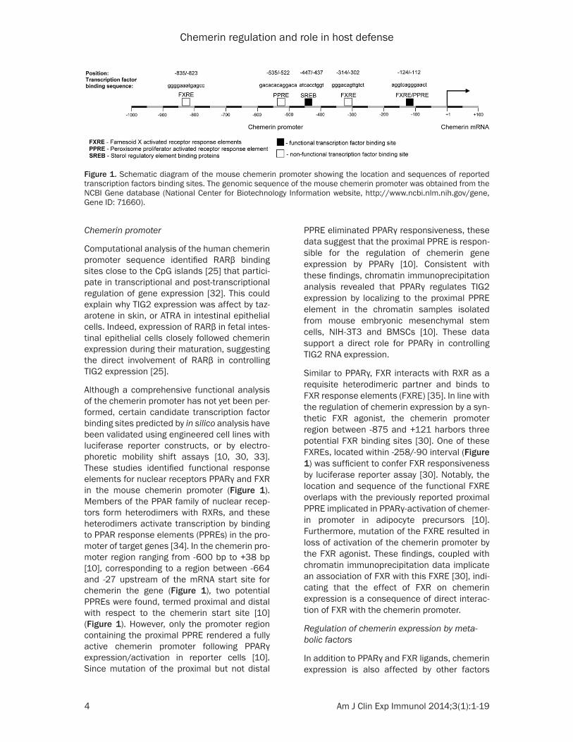
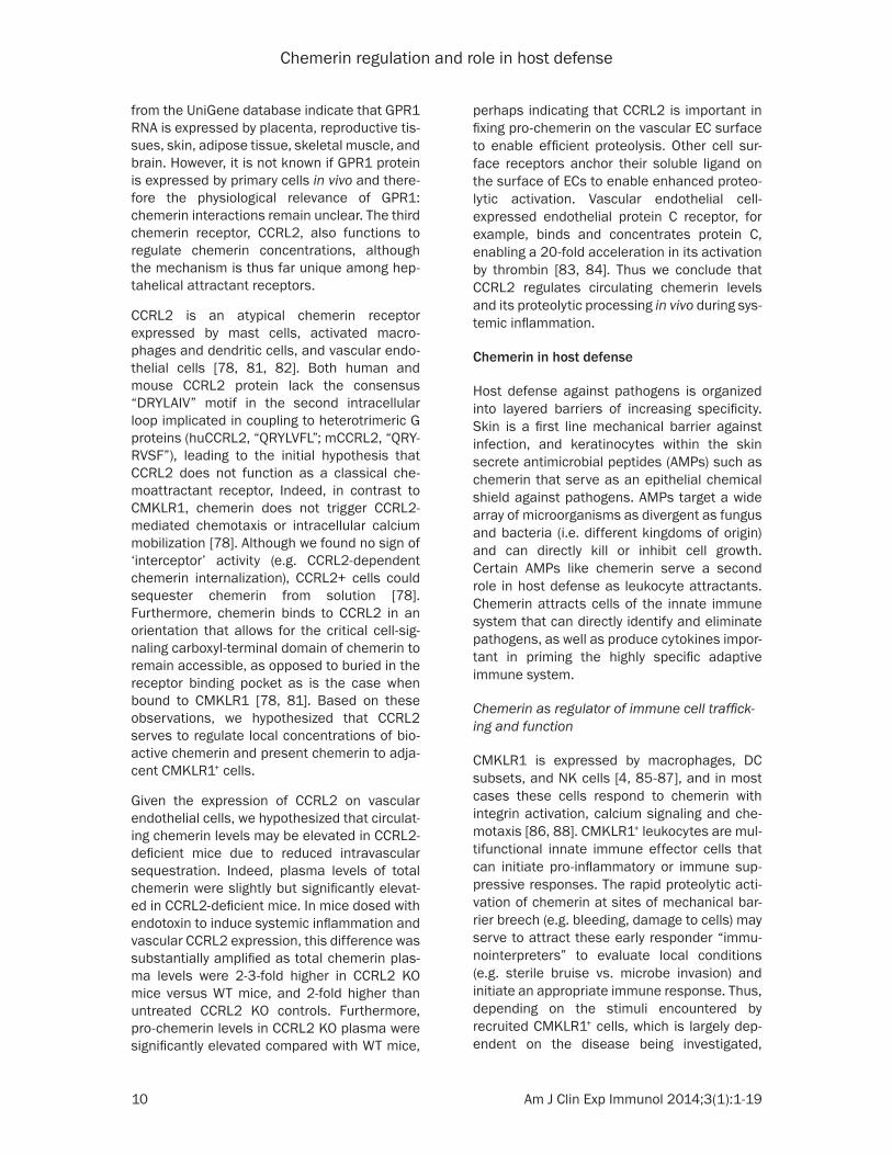
Although a comprehensive functional analysis of the chemerin promoter has not yet been per-formed, certain candidate transcription factor binding sites predicted by in silico analysis have been validated using engineered cell lines with luciferase reporter constructs, or by electro-phoretic mobility shift assays [10, 30, 33]. These studies identified functional response elements for nuclear receptors PPARγ and FXR in the mouse chemerin promoter (Figure 1). Members of the PPAR family of nuclear recep-tors form heterodimers with RXRs, and these heterodimers activate transcription by binding to PPAR response elements (PPREs) in the pro-moter of target genes [34]. In the chemerin pro-moter region ranging from -600 bp to +38 bp [10], corresponding to a region between -664 and -27 upstream of the mRNA start site for chemerin the gene (Figure 1), two potential PPREs were found, termed proximal and distal with respect to the chemerin start site [10] (Figure 1). However, only the promoter region containing the proximal PPRE rendered a fully active chemerin promoter following PPARγ expression/activation in reporter cells [10]. Since mutation of the proximal but not distal
PPRE eliminated PPARγ responsiveness, these data suggest that the proximal PPRE is respon-sible for the regulation of chemerin gene expression by PPARγ [10]. Consistent with these findings, chromatin immunoprecipitation analysis revealed that PPARγ regulates TIG2 expression by localizing to the proximal PPRE element in the chromatin samples isolated from mouse embryonic mesenchymal stem cells, NIH-3T3 and BMSCs [10]. These data support a direct role for PPARγ in controlling TIG2 RNA expression.
Similar to PPARγ, FXR interacts with RXR as a requisite heterodimeric partner and binds to FXR response elements (FXRE) [35]. In line with the regulation of chemerin expression by a syn-thetic FXR agonist, the chemerin promoter region between -875 and +121 harbors three potential FXR binding sites [30]. One of these FXREs, located within -258/-90 interval (Figure 1) was sufficient to confer FXR responsiveness by luciferase reporter assay [30]. Notably, the location and sequence of the functional FXRE overlaps with the previously reported proximal PPRE implicated in PPARγ-activation of chemer-in promoter in adipocyte precursors [10]. Furthermore, mutation of the FXRE resulted in loss of activation of the chemerin promoter by the FXR agonist. These findings, coupled with chromatin immunoprecipitation data implicate an association of FXR with this FXRE [30], indi-cating that the effect of FXR on chemerin expression is a consequence of direct interac-tion of FXR with the chemerin promoter.
Regulation of chemerin expression by meta‑bolic factors
In addition to PPARγ and FXR ligands, chemerin expression is also affected by other factors
Figure 1. Schematic diagram of the mouse chemerin promoter showing the location and sequences of reported transcription factors binding sites. The genomic sequence of the mouse chemerin promoter was obtained from the NCBI Gene database (National Center for Biotechnology Information website, http://www.ncbi.nlm.nih.gov/gene, Gene ID: 71660).
Chemerin regulation and role in host defense
5 Am J Clin Exp Immunol 2014;3(1):1-19
implicated in metabolic regulation such as free fatty acids (FFA). Plasma FFA levels are com-monly elevated in obese individuals, mostly due to increased FFA release from hypertrophic adipose tissue [33]. Chronically elevated plas-ma FFA levels likely contribute to the pathology of hypertension or dyslipidemia [36]. Chemerin synthesis is up-regulated by FFA in 3T3-L1 cells during their differentiation into adipocytes [33]. Elevation in chemerin levels in these cells coin-cided with activation of sterol regulatory ele-ment-binding protein 2 (SREBP2), a transcrip-tion factor belonging to the basic-helix-loop-helix leucine zipper class of transcriptional reg-ulators [37]. Following cell cholesterol short-age, SREBP2 is activated by intramembrane proteolysis. SREPB2 processing frees it from the endoplasmic reticulum to move to the nucleus where it binds to sterol regulatory ele-ments in promoter regions of genes required for cholesterol biosynthesis [38]. Excess load-ing with FFA in vitro stimulated SREBP2 activa-tion in 3T3-L1 cells, indicating cholesterol shortage and suggesting that chemerin expres-sion may be affected by cholesterol levels. Indeed, forced cholesterol depletion by phar-macological inhibition of cholesterol synthesis upregulated chemerin expression and SREBP2 activation [33]. Furthermore, siRNA knockdown of SREBP2 reduced basal and FFA-induced chemerin levels, demonstrating that SREBP2 directly regulates chemerin synthesis. An analy-sis of the chemerin promoter identified a con-sensus recognition element for basic-helix-loop-helix proteins [39] as a potential SREBP2 binding site, SREB [33] (Figure 1). The binding of nuclear proteins from 3T3-L1 adipocytes to this site was confirmed by electrophoretic mobility shift assay [33]. In summary, these data suggest the following chain of events: exogenous FFA treatment induces 3T3-L1 adi-pocyte hypertrophy which likely leads to intra-cellular cholesterol shortage; this event trig-gers the nuclear translocation of SREBP2, which then binds to SREB in the chemerin pro-moter to induce its expression.
Although the cellular and molecular signaling pathways have yet to be defined, there is also evidence of communication among insulin, glu-cose and chemerin. In obese diabetic patients circulating chemerin levels were elevated com-pared with controls [8, 40-43]. Likewise, in patients with endocrine disorders associated
with insulin resistance such as polycystic ovary syndrome (PCOS), plasma chemerin levels were elevated [44, 45]. Plasma chemerin levels were significantly increased following insulin infusion in healthy individuals [44]. In human adipose tissue explants, insulin significantly upregulat-ed chemerin production [44]. On the other hand, metformin, an antidiabetic drug that sup-presses hepatic glucose production and enhances peripheral glucose uptake by induc-ing glucose transporter 4 (GLUT4) translocation [46], suppressed chemerin synthesis and secretion in adipocyte tissue explants as well as reduced circulating chemerin levels in PCOS patients [44]. Overall these data suggest that insulin and/or glucose can impact chemerin expression, and that hyperinsulinemia may drive overexpression of chemerin in adipose tissue, leading to elevations in systemic che- merin protein via plasma circulation. Inter- estingly, data obtained from recently generated chemerin knockout (KO) and transgenic mice suggest a feedback signaling mechanism between plasma chemerin levels and insulin synthesis, as chemerin regulates insulin secre-tion in the pancreas [47].
Regulation of chemerin expression by cyto‑kines
In addition to its well-defined pro-inflammatory role in acute inflammation, TNFα also contrib-utes to the chronic low-grade inflammation that occurs in obesity [48]. Visceral white adipose tissue (vWAT) plays a key metabolic and endo-crine role in overweight individuals and is a tar-get for TNFα. TNFα enhances the sensitivity of vWAT to insulin, for example [42, 48, 49]. In vitro, TNFα induced TIG2 mRNA and chemerin protein expression in 3T3-L1 adipocytes, sug-gesting that TNFα may promote chemerin pro-duction in adipocytes in vivo [50]. Indeed, TNFα treatment significantly increased plasma che- merin levels in wild type but not TNF receptor (TNFRS1a and 1b) deficient mice. Inter- estingly, this increase in circulating chemerin protein was not accompanied by an upregula-tion of TIG2 mRNA in vWAT in vivo. Similarly, BMSC-derived primary adipocytes treated with TNFα failed to upregulate TIG2 transcription despite an increase in immunodetectable and bioactive chemerin protein in the conditioned media [50]. These data suggest a complex reg-ulation of chemerin by TNFα in adipocyte tissue
Chemerin regulation and role in host defense
6 Am J Clin Exp Immunol 2014;3(1):1-19
and indicates that tissues other than vWAT are likely responsive to TNFα-induced chemerin expression in vivo.
The ambiguity of TNFα in controlling chemerin levels was also reported for renal proximal tubular epithelial cells, where TNFα suppressed TIG2 transcription and secretion of immunode-tectable chemerin protein, but increased the level of bioactive chemerin as demonstrated by chemerin-dependent chemotaxis of plasmacy-toid dendritic cells (pDCs) [51]. Given that chemerin bioactivity is controlled by proteolysis (see below), these findings suggest that TNFα concomitantly orchestrates proteolytic events required for chemerin conversion to a chemo-tactically-active form.
The source of chemerin in plasma is not clear at present. In addition to individuals with obe-sity or type 2 diabetes (T2D), elevated levels of chemerin were reported in several disorders associated with chronic inflammation such as Crohn’s disease or chronic hepatitis C [52, 53]. In addition, systemic chemerin levels increased with the severity of liver fibrosis [45, 54]. Due to the strong expression of chemerin in liver and adipose tissue, we and others hypothesize that these organs have the biggest impact on blood chemerin levels. The portal vein delivers vWAT-derived metabolites like FFA as well as adipo-kines directly to the liver [55, 56], and thus may contribute to inducing hepatic chemerin expres-sion. The analysis of portal, hepatic and sys-temic venous blood chemerin levels in humans with liver cirrhosis revealed similar chemerin concentrations in portal and systemic circula-tion, suggesting that vWAT is not a major site of chemerin production. On the other hand, higher chemerin levels in hepatic venous compared to portal venous likely indicate that chemerin is produced and secreted by the liver [57]. However, TNFα had no influence on chemerin synthesis by mouse primary hepatocytes or the liver in vivo [50], suggesting that liver is not a substantial source of pro-inflammatory cyto-kine-induced chemerin production. The relative contribution of liver and WAT to circulating chemerin levels may vary depending on stimu-lus and/or disease state: whereas elevated chemerin levels found in the blood of obese individuals may to a large extent originate from fat tissue, in liver cirrhosis, the liver may be pri-marily responsible for elevated plasma chemer-in levels.
IL1β and IFNγ are additional proinflammatory cytokines that can modulate chemerin synthe-sis [58]. IL1β is one of a growing list of newly appreciated adipocyte-derived inflammatory mediators [59]. IL1β can also influence the metabolic function of adipocytes and may, for example, induce insulin resistance [60, 61]. Blocking IL1β improved glycemic control in patients with T2D [62] and delayed the devel-opment of T2D in experimental animals [63]. In vitro, IL1β increased chemerin protein concen-trations approximately 1.3 fold in conditioned media from 3T3-L1 adipocytes and immortal-ized brown adipocyte tissue (BAT)-derived cells. TIG2 mRNA expression was up-regulated in these cells in a time- and dose-dependent manner as well [58]. IFNγ induced robust chemerin expression by fibroblast-like synovio-cytes [64]. Elevated serum levels of IFNγ have been detected in psoriasis [65] or rheumatoid arthritis [66], diseases with postulated chemer-in involvement [7, 24, 67, 68]. The cellular and molecular signaling pathways involved in IL1β- or IFNγ-mediated regulation of chemerin expression/secretion have yet to be defined, and it is unclear if this regulation occurs in vivo.
Moreover, although the reports referenced above largely focused on the effects of individ-ual mediators on chemerin synthesis, it is likely that chemerin expression is controlled in con-cert by variety of factors operating in specific anatomic locations dependent on the particu-lar ongoing pathophysiological process. A com-prehensive list of mediators that regulate chemerin synthesis is shown in Table 1.
Posttranslational regulation of chemerin activ-ity
Chemerin secretion
Chemerin secretion may be constitutive or reg-ulated, and for most cell types the mechanism underlying this process is not well-character-ized. As previously mentioned, modulation of TIG2 mRNA is not strictly accompanied by par-allel changes in chemerin protein in condi-tioned media, suggesting that chemerin secre-tion might be subject to additional control. For example, the strong reduction in chemerin pro-tein levels present in conditioned media of human adipocytes treated with the PPARγ ago-nist troglitazone was not reflected in TIG2 mRNA levels, which remained unchanged [54].
Chemerin regulation and role in host defense
7 Am J Clin Exp Immunol 2014;3(1):1-19
Likewise, the increase in chemerin protein lev-els detected in conditioned media in mouse primary adipocytes treated with TNFα was not reflected in TIG2 mRNA levels, which also remained unchanged [50]. While these exam-ples suggest that adipocytes employ a mecha-nism to regulate the secretion of chemerin, it is possible that reduced chemerin immunoreac-tivity in cell conditioned media might result from extensive proteolytic processing of this protein (described in the next paragraph), or from sequestration of secreted chemerin by atypical chemerin receptor CCRL2, which con-centrates chemerin on the cell surface and effectively removing it from the fluid phase
being analyzed. Nevertheless, it is clear that in the case of anuclear platelets, chemerin is reg-ulated at the level of secretion: chemerin pro-tein is stored in preformed granules that are rapidly released upon platelet activation by thrombin, thrombin receptor-activating peptide, and collagen [16].
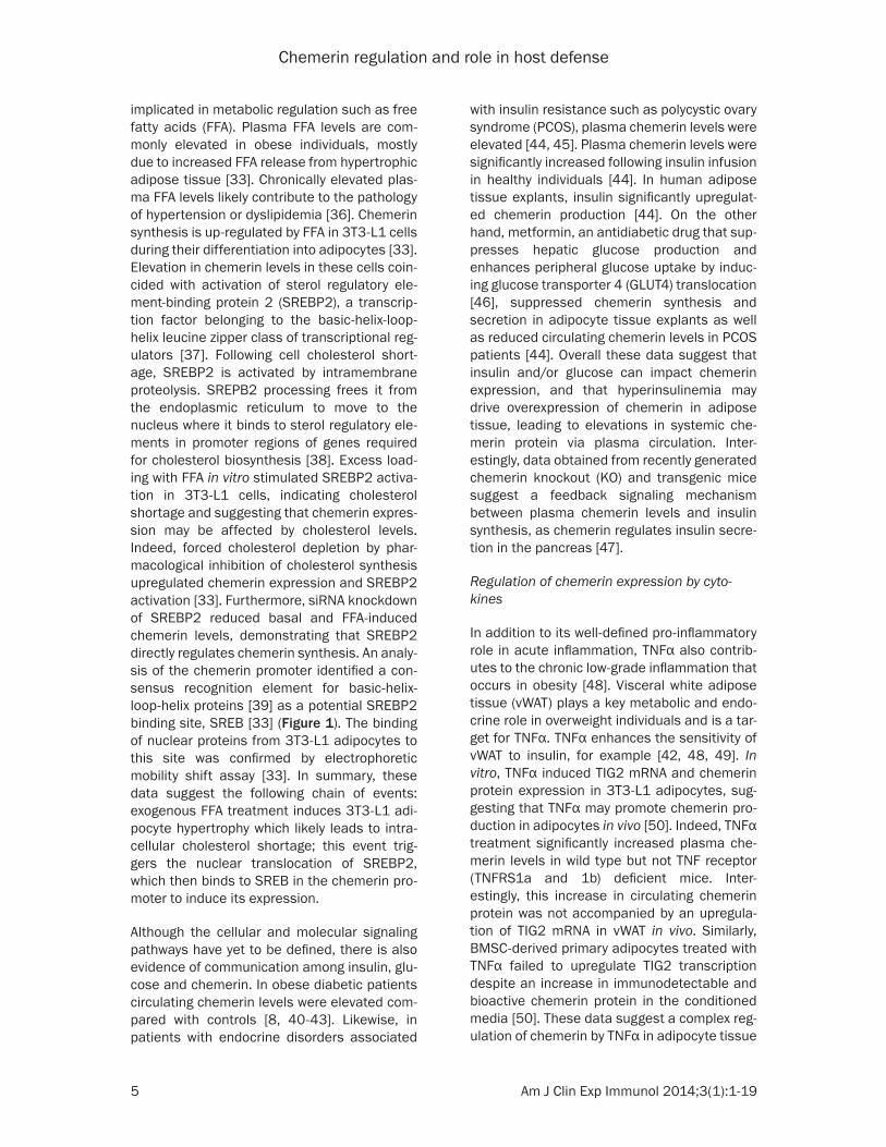
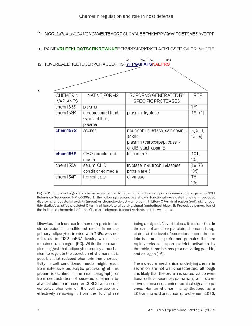
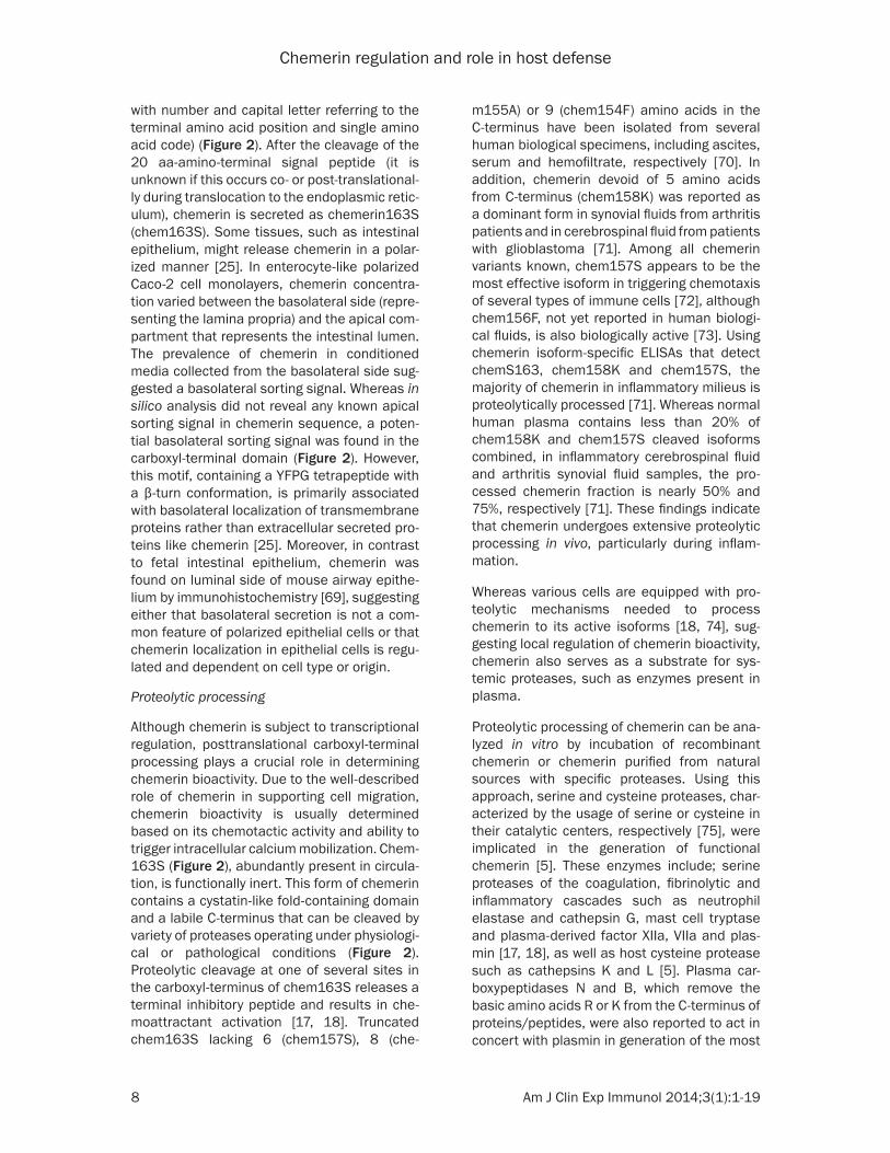
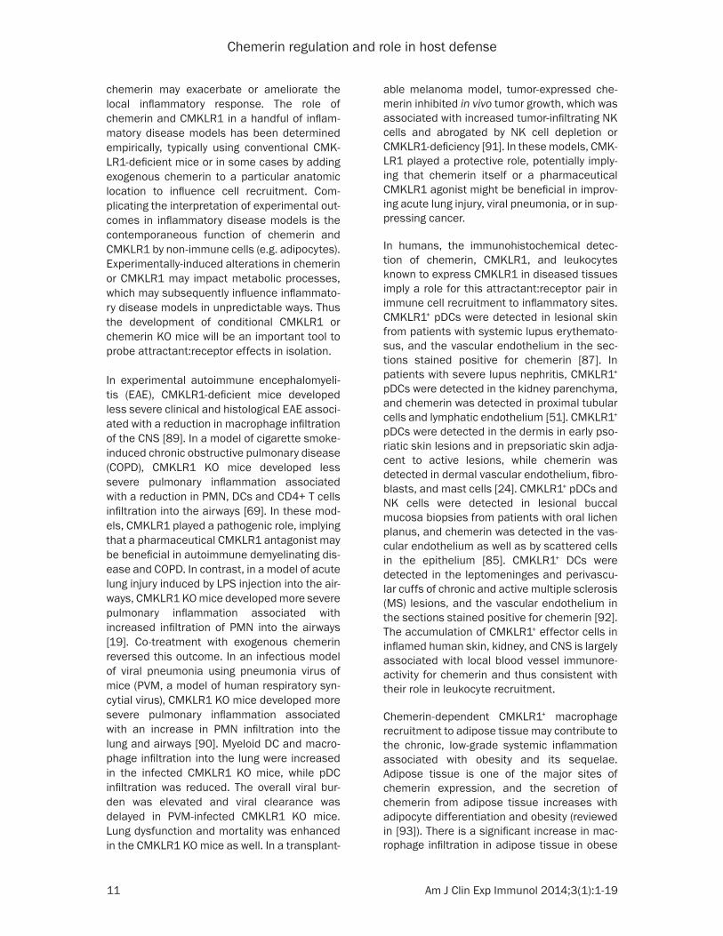
The molecular mechanism underlying chemerin secretion are not well-characterized, although it is likely that the protein is sorted via conven-tional cellular secretory pathways given its con-served consensus amino-terminal signal sequ- ence. Human chemerin is synthesized as a 163-amino acid precursor, (pro-chemerin163S,
Figure 2. Functional regions in chemerin sequence. A: In the human chemerin primary amino acid sequence (NCBI Reference Sequence: NP_002880.1) the following regions are shown: functionally-evaluated chemerin peptides displaying antibacterial activity (green) or chemotactic activity (blue), inhibitory C-terminal region (red), signal pep-tide (italics), in silico predicted C-terminal basolateral sorting signal (underlined blue). B: Proteolytic generation of the indicated chemerin isoforms. Chemerin chemoattractant variants are shown in blue.
Chemerin regulation and role in host defense
8 Am J Clin Exp Immunol 2014;3(1):1-19
with number and capital letter referring to the terminal amino acid position and single amino acid code) (Figure 2). After the cleavage of the 20 aa-amino-terminal signal peptide (it is unknown if this occurs co- or post-translational-ly during translocation to the endoplasmic retic-ulum), chemerin is secreted as chemerin163S (chem163S). Some tissues, such as intestinal epithelium, might release chemerin in a polar-ized manner [25]. In enterocyte-like polarized Caco-2 cell monolayers, chemerin concentra-tion varied between the basolateral side (repre-senting the lamina propria) and the apical com-partment that represents the intestinal lumen. The prevalence of chemerin in conditioned media collected from the basolateral side sug-gested a basolateral sorting signal. Whereas in silico analysis did not reveal any known apical sorting signal in chemerin sequence, a poten-tial basolateral sorting signal was found in the carboxyl-terminal domain (Figure 2). However, this motif, containing a YFPG tetrapeptide with a β-turn conformation, is primarily associated with basolateral localization of transmembrane proteins rather than extracellular secreted pro-teins like chemerin [25]. Moreover, in contrast to fetal intestinal epithelium, chemerin was found on luminal side of mouse airway epithe-lium by immunohistochemistry [69], suggesting either that basolateral secretion is not a com-mon feature of polarized epithelial cells or that chemerin localization in epithelial cells is regu-lated and dependent on cell type or origin.
Proteolytic processing
Although chemerin is subject to transcriptional regulation, posttranslational carboxyl-terminal processing plays a crucial role in determining chemerin bioactivity. Due to the well-described role of chemerin in supporting cell migration, chemerin bioactivity is usually determined based on its chemotactic activity and ability to trigger intracellular calcium mobilization. Chem- 163S (Figure 2), abundantly present in circula-tion, is functionally inert. This form of chemerin contains a cystatin-like fold-containing domain and a labile C-terminus that can be cleaved by variety of proteases operating under physiologi-cal or pathological conditions (Figure 2). Proteolytic cleavage at one of several sites in the carboxyl-terminus of chem163S releases a terminal inhibitory peptide and results in che-moattractant activation [17, 18]. Truncated chem163S lacking 6 (chem157S), 8 (che-
m155A) or 9 (chem154F) amino acids in the C-terminus have been isolated from several human biological specimens, including ascites, serum and hemofiltrate, respectively [70]. In addition, chemerin devoid of 5 amino acids from C-terminus (chem158K) was reported as a dominant form in synovial fluids from arthritis patients and in cerebrospinal fluid from patients with glioblastoma [71]. Among all chemerin variants known, chem157S appears to be the most effective isoform in triggering chemotaxis of several types of immune cells [72], although chem156F, not yet reported in human biologi-cal fluids, is also biologically active [73]. Using chemerin isoform-specific ELISAs that detect chemS163, chem158K and chem157S, the majority of chemerin in inflammatory milieus is proteolytically processed [71]. Whereas normal human plasma contains less than 20% of chem158K and chem157S cleaved isoforms combined, in inflammatory cerebrospinal fluid and arthritis synovial fluid samples, the pro-cessed chemerin fraction is nearly 50% and 75%, respectively [71]. These findings indicate that chemerin undergoes extensive proteolytic processing in vivo, particularly during inflam- mation.
Whereas various cells are equipped with pro-teolytic mechanisms needed to process chemerin to its active isoforms [18, 74], sug-gesting local regulation of chemerin bioactivity, chemerin also serves as a substrate for sys-temic proteases, such as enzymes present in plasma.
Proteolytic processing of chemerin can be ana-lyzed in vitro by incubation of recombinant chemerin or chemerin purified from natural sources with specific proteases. Using this approach, serine and cysteine proteases, char-acterized by the usage of serine or cysteine in their catalytic centers, respectively [75], were implicated in the generation of functional chemerin [5]. These enzymes include; serine proteases of the coagulation, fibrinolytic and inflammatory cascades such as neutrophil elastase and cathepsin G, mast cell tryptase and plasma-derived factor XIIa, VIIa and plas-min [17, 18], as well as host cysteine protease such as cathepsins K and L [5]. Plasma car-boxypeptidases N and B, which remove the basic amino acids R or K from the C-terminus of proteins/peptides, were also reported to act in concert with plasmin in generation of the most
Chemerin regulation and role in host defense
9 Am J Clin Exp Immunol 2014;3(1):1-19
active chemS157 isoform form plasmin-pro-duced chemK158 intermediate [16]. This example illustrates that chemerin can be pro-teolytically activated in direct or sequential fashion.
In addition to host proteases that regulate chemerin bioactivity under homeostatic or pathogenic conditions, chemerin activity is also controlled by proteases secreted by human pathogens. Staphopain B, a cysteine protease derived from Staphylococcus aureus, is a potent enzymatic chemerin activator that gen-erates the chemS157 variant [6].
As much as proteases are needed to produce active chemerin isoforms, they may also inacti-vate or degrade the attractant and thus limit the extent of chemerin activity. Tissue-recruited neutrophils secrete chemerin-activating elas-tase and cathepsin G, but also release protein-ase 3 that cleaves chem163S to chemotacti-cally inactive chem155A [76]. Of note, this chemerin isoform differs from the most active chem157S by two C-terminal amino acids. Interestingly, the net result of chemerin prote-olysis may depend not only on the interaction of proteases with unprocessed chem163S, but also with other chemerin variants, since chemerin processing intermediates are differ-entially targeted by proteases. For example, mast cell chymase does not use chem163S as a substrate, but abolishes the activity of func-tional variants chem157S and chem156F by conversion to the chem154F isoform [76].
The subtle amino acid sequence differences between biologically active and inactive che- merin variants raise the question of whether they compete for the same receptor binding site and can therefore influence each other’s activity. This was partially addressed by using chem157S as a cell stimulant in the presence of the chem155A variant that itself shows poor activity by calcium flux assay [72]. Chem155A inhibited 50% of chem157S activity at a molar ratio of 100:1, demonstrating a weak antago-nistic effect [72].
The chemerin analog nonapeptide 149YFPG- QFAFS157, corresponding to the C terminus of chem157S (Figure 2), has been reported to retain most of the activity of the chem157S [73], although in our hands this peptide was approximately 100-fold less-potent than
chem157S. Bioactive chemerin protein iso-forms are more potent than their C-terminal peptide counterparts, suggesting that the N-terminal portion of the intact protein contrib-utes to chemerin chemotactic activity. Given the isolation of various carboxyl-terminal chemerin variants from endogenous biofluids, most of the effort in defining the posttransla-tional regulation of chemerin has focused on C-terminal truncations. It is possible, however, that amino-terminal processing may also have a role in defining the bioactivity of chemerin. For example, pharmacological inhibition of ami-nopeptidases, enzymes that catalyze the cleav-age of amino acids from the N-terminus of pro-tein/peptide substrates, significantly increased the concentration of active chemerin in condi-tioned media from TNFα-treated 3T3-L1 adipo-cytes [74]. These data suggest that aminopep-tidases cleave and inactivate chemerin via amino-terminal truncation. Since aminopepti-dases are typically ubiquitous, their signifi-cance in regulating chemerin bioactivity requi- res further study.
Localization‑dependent (receptor‑based) regu‑lation of chemerin availability
There are three known heptahelical receptors for chemerin: CMKLR1, GPR1, and CC-motif chemokine receptor-like 2 (CCRL2). The three chemerin receptors bind to chemerin with simi-lar low nanomolar affinities [77, 78], but with substantially different functional outcomes. Chemerin binding to CMKLR1 triggers cell migration, intracellular calcium mobilization, and β-arrestin2 association and receptor inter-nalization, all features common to classical G-protein coupled receptors (GPCRs), including most leukocyte-expressed chemoattractant receptors [3, 4, 70, 77, 78]. Chemerin binding to GPR1 triggers β-arrestin2 association and receptor internalization; whether or not chemer-in induces GPR1-dependent intracellular calci-um signaling is controversial [3, 77]. Further- more, we found no evidence of chemerin-medi-ated GPR1+ cell migration (BAZ, unpublished). Thus GPR1 may function as a chemerin-binding ‘interceptor’, a class of attractant receptors including D6, DARC, CCX-CKR, and CXCR7 that binds, internalizes, and degrades chemoattrac-tants, thereby regulating local attractant levels [79, 80]. Publically available microarray data from the GEO (Gene Expression Omnibus) data-base and EST (expressed sequence tag) data
Chemerin regulation and role in host defense
10 Am J Clin Exp Immunol 2014;3(1):1-19
from the UniGene database indicate that GPR1 RNA is expressed by placenta, reproductive tis-sues, skin, adipose tissue, skeletal muscle, and brain. However, it is not known if GPR1 protein is expressed by primary cells in vivo and there-fore the physiological relevance of GPR1: chemerin interactions remain unclear. The third chemerin receptor, CCRL2, also functions to regulate chemerin concentrations, although the mechanism is thus far unique among hep-tahelical attractant receptors.
CCRL2 is an atypical chemerin receptor expressed by mast cells, activated macro-phages and dendritic cells, and vascular endo-thelial cells [78, 81, 82]. Both human and mouse CCRL2 protein lack the consensus “DRYLAIV” motif in the second intracellular loop implicated in coupling to heterotrimeric G proteins (huCCRL2, “QRYLVFL”; mCCRL2, “QRY- RVSF”), leading to the initial hypothesis that CCRL2 does not function as a classical che-moattractant receptor, Indeed, in contrast to CMKLR1, chemerin does not trigger CCRL2-mediated chemotaxis or intracellular calcium mobilization [78]. Although we found no sign of ‘interceptor’ activity (e.g. CCRL2-dependent chemerin internalization), CCRL2+ cells could sequester chemerin from solution [78]. Furthermore, chemerin binds to CCRL2 in an orientation that allows for the critical cell-sig-naling carboxyl-terminal domain of chemerin to remain accessible, as opposed to buried in the receptor binding pocket as is the case when bound to CMKLR1 [78, 81]. Based on these observations, we hypothesized that CCRL2 serves to regulate local concentrations of bio-active chemerin and present chemerin to adja-cent CMKLR1+ cells.
Given the expression of CCRL2 on vascular endothelial cells, we hypothesized that circulat-ing chemerin levels may be elevated in CCRL2-deficient mice due to reduced intravascular sequestration. Indeed, plasma levels of total chemerin were slightly but significantly elevat-ed in CCRL2-deficient mice. In mice dosed with endotoxin to induce systemic inflammation and vascular CCRL2 expression, this difference was substantially amplified as total chemerin plas-ma levels were 2-3-fold higher in CCRL2 KO mice versus WT mice, and 2-fold higher than untreated CCRL2 KO controls. Furthermore, pro-chemerin levels in CCRL2 KO plasma were significantly elevated compared with WT mice,
perhaps indicating that CCRL2 is important in fixing pro-chemerin on the vascular EC surface to enable efficient proteolysis. Other cell sur-face receptors anchor their soluble ligand on the surface of ECs to enable enhanced proteo-lytic activation. Vascular endothelial cell-expressed endothelial protein C receptor, for example, binds and concentrates protein C, enabling a 20-fold acceleration in its activation by thrombin [83, 84]. Thus we conclude that CCRL2 regulates circulating chemerin levels and its proteolytic processing in vivo during sys-temic inflammation.
Chemerin in host defense
Host defense against pathogens is organized into layered barriers of increasing specificity. Skin is a first line mechanical barrier against infection, and keratinocytes within the skin secrete antimicrobial peptides (AMPs) such as chemerin that serve as an epithelial chemical shield against pathogens. AMPs target a wide array of microorganisms as divergent as fungus and bacteria (i.e. different kingdoms of origin) and can directly kill or inhibit cell growth. Certain AMPs like chemerin serve a second role in host defense as leukocyte attractants. Chemerin attracts cells of the innate immune system that can directly identify and eliminate pathogens, as well as produce cytokines impor-tant in priming the highly specific adaptive immune system.
Chemerin as regulator of immune cell traffick‑ing and function
CMKLR1 is expressed by macrophages, DC subsets, and NK cells [4, 85-87], and in most cases these cells respond to chemerin with integrin activation, calcium signaling and che-motaxis [86, 88]. CMKLR1+ leukocytes are mul-tifunctional innate immune effector cells that can initiate pro-inflammatory or immune sup-pressive responses. The rapid proteolytic acti-vation of chemerin at sites of mechanical bar-rier breech (e.g. bleeding, damage to cells) may serve to attract these early responder “immu-nointerpreters” to evaluate local conditions (e.g. sterile bruise vs. microbe invasion) and initiate an appropriate immune response. Thus, depending on the stimuli encountered by recruited CMKLR1+ cells, which is largely dep- endent on the disease being investigated,
Chemerin regulation and role in host defense
11 Am J Clin Exp Immunol 2014;3(1):1-19
chemerin may exacerbate or ameliorate the local inflammatory response. The role of chemerin and CMKLR1 in a handful of inflam-matory disease models has been determined empirically, typically using conventional CMK- LR1-deficient mice or in some cases by adding exogenous chemerin to a particular anatomic location to influence cell recruitment. Com- plicating the interpretation of experimental out-comes in inflammatory disease models is the contemporaneous function of chemerin and CMKLR1 by non-immune cells (e.g. adipocytes). Experimentally-induced alterations in chemerin or CMKLR1 may impact metabolic processes, which may subsequently influence inflammato-ry disease models in unpredictable ways. Thus the development of conditional CMKLR1 or chemerin KO mice will be an important tool to probe attractant:receptor effects in isolation.
In experimental autoimmune encephalomyeli-tis (EAE), CMKLR1-deficient mice developed less severe clinical and histological EAE associ-ated with a reduction in macrophage infiltration of the CNS [89]. In a model of cigarette smoke-induced chronic obstructive pulmonary disease (COPD), CMKLR1 KO mice developed less severe pulmonary inflammation associated with a reduction in PMN, DCs and CD4+ T cells infiltration into the airways [69]. In these mod-els, CMKLR1 played a pathogenic role, implying that a pharmaceutical CMKLR1 antagonist may be beneficial in autoimmune demyelinating dis-ease and COPD. In contrast, in a model of acute lung injury induced by LPS injection into the air-ways, CMKLR1 KO mice developed more severe pulmonary inflammation associated with increased infiltration of PMN into the airways [19]. Co-treatment with exogenous chemerin reversed this outcome. In an infectious model of viral pneumonia using pneumonia virus of mice (PVM, a model of human respiratory syn-cytial virus), CMKLR1 KO mice developed more severe pulmonary inflammation associated with an increase in PMN infiltration into the lung and airways [90]. Myeloid DC and macro-phage infiltration into the lung were increased in the infected CMKLR1 KO mice, while pDC infiltration was reduced. The overall viral bur-den was elevated and viral clearance was delayed in PVM-infected CMKLR1 KO mice. Lung dysfunction and mortality was enhanced in the CMKLR1 KO mice as well. In a transplant-
able melanoma model, tumor-expressed che- merin inhibited in vivo tumor growth, which was associated with increased tumor-infiltrating NK cells and abrogated by NK cell depletion or CMKLR1-deficiency [91]. In these models, CMK- LR1 played a protective role, potentially imply-ing that chemerin itself or a pharmaceutical CMKLR1 agonist might be beneficial in improv-ing acute lung injury, viral pneumonia, or in sup-pressing cancer.
In humans, the immunohistochemical detec-tion of chemerin, CMKLR1, and leukocytes known to express CMKLR1 in diseased tissues imply a role for this attractant:receptor pair in immune cell recruitment to inflammatory sites. CMKLR1+ pDCs were detected in lesional skin from patients with systemic lupus erythemato-sus, and the vascular endothelium in the sec-tions stained positive for chemerin [87]. In patients with severe lupus nephritis, CMKLR1+ pDCs were detected in the kidney parenchyma, and chemerin was detected in proximal tubular cells and lymphatic endothelium [51]. CMKLR1+ pDCs were detected in the dermis in early pso-riatic skin lesions and in prepsoriatic skin adja-cent to active lesions, while chemerin was detected in dermal vascular endothelium, fibro-blasts, and mast cells [24]. CMKLR1+ pDCs and NK cells were detected in lesional buccal mucosa biopsies from patients with oral lichen planus, and chemerin was detected in the vas-cular endothelium as well as by scattered cells in the epithelium [85]. CMKLR1+ DCs were detected in the leptomeninges and perivascu-lar cuffs of chronic and active multiple sclerosis (MS) lesions, and the vascular endothelium in the sections stained positive for chemerin [92]. The accumulation of CMKLR1+ effector cells in inflamed human skin, kidney, and CNS is largely associated with local blood vessel immunore-activity for chemerin and thus consistent with their role in leukocyte recruitment.
Chemerin-dependent CMKLR1+ macrophage recruitment to adipose tissue may contribute to the chronic, low-grade systemic inflammation associated with obesity and its sequelae. Adipose tissue is one of the major sites of chemerin expression, and the secretion of chemerin from adipose tissue increases with adipocyte differentiation and obesity (reviewed in [93]). There is a significant increase in mac-rophage infiltration in adipose tissue in obese
Chemerin regulation and role in host defense
12 Am J Clin Exp Immunol 2014;3(1):1-19
patients [94]. In chemerin-deficient mice, there was a significant reduction in macrophage accumulation in epididymal adipose tissue compared to WT (reduction in percent Mac3-positive cells) [47]. However, this effect was not recapitulated in CMKLR1-deficient mice, as there was no difference in percent macro-phages in white adipose tissue versus WT [95]. It is possible that other chemerin receptors can compensate for the loss of CMKLR1 in coordi-nating macrophage infiltration into adipose tissue.
In addition to CMKLR1, there are a small num-ber of in vivo studies that implicate chemerin receptor CCRL2 in leukocyte trafficking and the pathophysiology of inflammatory disease. Using CCRL2 KO mice, we found that the recep-tor is required for maximal ear swelling and leu-kocyte infiltration in passive cutaneous ana-phylaxis (PCA), a mast cell-dependent in vivo allergy model [78]. Specifically, using mast cell-deficient animals engrafted with either WT or CCRL2 KO bone marrow-cultured mast cells, we showed that mast cell-expressed CCRL2 is required for optimal induction of PCA in mice sensitized with a low dose of antigen-specific IgE (high levels of sensitizing antigen abrogated this difference). In a model of ovalbumin (ova)-induced airway inflammation, CCRL2 KO mice were protected against pulmonary inflamma-tion, which was associated with a defect in the trafficking of antigen-laden lung DC to medias-tinal lymph nodes [35]. In the PCA model and the ova-induced airway inflammation models, it is not known whether the protection in CCRL2 KO mice is related to the role of CCRL2 as an atypical chemerin receptor.
As mentioned above, chemerin co-localized with vascular endothelial cells in MS, lupus, psoriasis, and in the endothelial venules of sec-ondary lymphoid tissues [87]. In vitro, we showed that chemerin binding to CCRL2 on endothelial cells triggered robust CMKLR1+ lymphoid cell adhesion dependent on α4β1 inte-grin/VCAM-1 [81]. Furthermore, CMKLR1+ NK cell recruitment to the airways was significantly impaired in CCRL2 KO mice following acute LPS-induced pulmonary inflammation in vivo [81]. CCRL2 expression by vascular endothelial cells thus provides a specific mechanism for the local enrichment of chemerin at inflamma-tory sites, regulating the recruitment of CMK- LR1+ cells.
Defensive function of chemerin in body barri‑ers
Chemerin protein is abundant in a variety of normal epithelial cell types, including human and mouse epidermis [15, 19, 24], human fetal and mouse adult enterocytes [19, 25], and mouse epithelium lining the airways [19, 69]. By immunohistochemistry, the bronchial epi-thelium is the predominant source of chemerin in the normal mouse lung, likely responsible for the secretion of chemerin into the airways fol-lowing cigarette smoke exposure [69]. Thus the strategic positioning of chemerin at body barri-ers alone suggests a role in antimicrobial defense.
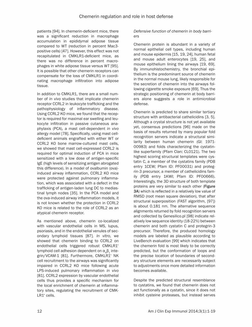
Chemerin is predicted to share similar tertiary structure with antibacterial cathelicidins [3, 5]. Although a crystal structure is not yet available yet, consensus predictions [96] made on the basis of results returned by many popular fold recognition servers indicate a structural simi-larity between human chemerin (GI: 1971- 00983) and folds characterizing the cystatin-like superfamily (Pfam Clan: CL0121). The two highest scoring structural templates were cys-tatin C, a member of the cystatins family (PDB entry 1CEW; Pfam ID: PF00031) and proteg-rin-3 precursor, a member of cathelicidins fam-ily (PDB entry 1KWI; Pfam ID: PF00666). Interestingly, the 3D structure of both template proteins are very similar to each other (Figure 3A) which is reflected in a relatively low value of RMSD (root mean square deviation), that after structural superposition (FAST algorithm, [97]) is about 0.181 nm. The alternative sequence alignments returned by fold recognition servers and collected by Genesilico.pl [98] indicate rel-atively low sequence identity (18-22%) between chemerin and both cystatin C and protegrin-3 precursor. Therefore, the produced homology models are labeled as plausible according to LiveBench evaluation [99] which indicates that the chemerin fold is most likely to be correctly predicted, but the conformation of loops and the precise location of boundaries of second-ary structure elements are necessarily subject to adjustments once more detailed information becomes available.
Despite the predicted structural resemblance to cystatins, we found that chemerin does not act functionally as a cystatin, since it does not inhibit cysteine proteases, but instead serves

Chemerin regulation and role in host defense
13 Am J Clin Exp Immunol 2014;3(1):1-19
as substrate for these enzymes [5]. However, in line with the predicted similarity to antibacterial cathelicidin, human recombinant chemerin as well as endogenous chemerin in exudates from organ cultures of primary human skin keratino-cytes significantly inhibited bacteria growth [5, 15]. As is the case for chemoattractant activity, the inhibitory C-terminal peptide present in the chemerin holoprotein chem163S must be removed for full antibacterial effects. Using a panel of overlapping chemerin-derived synthet-ic peptides, we also demonstrated that chemer-in antimicrobial activity can be largely narrowed down to an internal 20-amino acid long peptide V66-P85, which is comparable in potency to other AMPs [15]. Since this region is localized in the middle of the chemerin sequence (Figures 2
and 3), all chemerin isoforms truncated at the C-terminal end will likely be equipped with anti-microbial activity, despite their differing potentials to signal through CMKLR1. Indeed, che-motactically active chemS157 and chemotactically inert chem- R125 have similar antimicrobial activity against E. coli [5]. Alth- ough it remains to be deter-mined which chemerin isoform(s) are present in the epidermis, a pathogen-challenged epithelium will likely deliver specific pro- tease(s) required to activate the antibacterial activity of chemer-in [100]. Kallikrein 7, a serine protease constitutively expre- ssed in skin and recently report-ed to convert chem163S to chem156F, is a likely protease candidate to generate active chemerin in this tissue [101]. Alternatively, epithelium-coloniz-ing pathogens might themselves provide another source of pro-teinases capable of generating chemerin variants equipped with bactericidal potential. For exam-ple, chem157S generated by S. aureus would be expected to act as both chemoattractant and AMP. Although the growth of Gram-positive S. aureus is inhib-ited by chemerin peptide V66-P85, other microorganisms such as
Figure 3. Location of anti-bacterial activity in the predicated chemerin structure. A: The 3D structure of chicken egg white cystatin (PDB entry: 1CEW) and porcine protegrin-3 precursor (PDB entry: 1KWI). Helices, β-strands, and loops are shown, respectively, in blue, green, and orange. N- and C-termini are marked with letters. B: V66-P85 peptide (red) mapped on the secondary structure elements of porcine protegrin-3 precursor ac-cording to the alignment with human chemerin sequence constructed with hhsearch program [104]. The chemerin sequence VRLEFKLQQTSCRKRD-WKKP (positions 66-85) aligns with the KPVSFTVKETVCPRPTRQPP se-quence of protegrin-3 (75-94), which adopts a twisted β-strand that termi-nates at P88 and a four residue helical fragment starting with P92. The 3D structure of several discontinuous fragments of protegrin-3 remain unresolved.
Gram-negative E. coli or fungus Candida are more sensitive to this peptide [15]. Therefore, S. aureus, via secretion of specific proteolytic enzymes such as staphopain B, may activate chemerin to create a relatively hospitable niche for itself and prevent displacement by other skin-colonizing microorganisms that are more sensitive to chemerin-mediated killing.
Keratinocytes are the main source of chemerin in healthy skin by immunostaining, while in chronically inflamed skin (e.g. psoriasis lesion) chemerin co-localizes with dermal infiltrating pDCs [20]. Psoriatic skin-derived chemerin stimulates CMKLR+ cell migration, indicating that chemerin is activated locally in the skin likely through neutrophil-derived proteases [7,
Chemerin regulation and role in host defense
14 Am J Clin Exp Immunol 2014;3(1):1-19
100]. Given the presence of chemerin-mediat-ed attractant activity and chemerin-responsive pDCs in psoriatic dermis [7], this protein may contribute to the recruitment of pDCs to inflamed skin. Thus, the concerted action of chemerin and skin-associated proteases enables a dual role for chemerin in skin immu-nity as an AMP and chemoattractant: chemerin may directly contain microbes or act together with recruited immune cells to better control microbial infections.
Skin keratinocytes are not the only epithelial cell engaged in immune cell cross talk via chemerin. For example, chemerin is secreted by human fetal enterocytes [25]. Recruitment of macrophages to the intestinal mucosa is believed to play a role in clearance of luminal bacteria that breach the epithelium and gain access to the lamina propria. Given their phago-cytic and bactericidal potential, intestinal mac-rophages might be particularly important in the intestine of premature infants who are predis-posed to bacterial translocation due to a rela-tively permeable gut epithelium. Macrophage chemotaxis to conditioned media from fetal epithelial cells was chemerin-dependent, sug-gesting that chemerin may serve as a macro-phage chemoattractant in fetal gut [25]. This chemotactic function in the developing intes-tine is consistent with the basolateral secretion of chemerin by epithelial cells to the subepithe-lial compartments [25].
Interestingly, having dual antimicrobial and chemotactic activities is not unprecedented for a chemoattractant, as many chemokines (such as CCL20, CXCL10 or CXCL14) have similar activities [102]. Due to constant microbial chal-lenge the skin and gastrointestinal epithelium might be particularly suited to reveal the com-plexity of these multifunctional molecules in host defense.
Acknowledgements
This work was supported in part by the grant from the Foundation for Polish Science TEAM/ 2010-5/1, co-financed by the European Union within European Regional Development Fund and Polish National Science Center grant 0724/B/P01/2011/40 (to JC); and by DoD grant W81XWH-11-1-0512 and NIH grant R01 AI-079320 (to BAZ).
Disclosure of conflict of interest
None.
Address correspondence to: Dr. Joanna Cichy, Department of Immunology, Faculty of Biochemistry, Biophysics and Biotechnology, Jagiellonian Univer- sity, Gronostajowa 7 St., 30-387 Krakow, Poland. Tel: 48-12-664 6127; Fax: 48-12-664 6904; E-mail: [email protected]
References
[1] Nagpal S, Patel S, Jacobe H, DiSepio D, Ghosn C, Malhotra M, Teng M, Duvic M and Chandr-aratna RA. Tazarotene-induced gene 2 (TIG2), a novel retinoid-responsive gene in skin. J In-vest Dermatol 1997; 109: 91-95.
[2] Davenport AP, Alexander SP, Sharman JL, Paw-son AJ, Benson HE, Monaghan AE, Liew WC, Mpamhanga CP, Bonner TI, Neubig RR, Pin JP, Spedding M and Harmar AJ. International Union of Basic and Clinical Pharmacology. LXXXVIII. G protein-coupled receptor list: rec-ommendations for new pairings with cognate ligands. Pharmacol Rev 2013; 65: 967-986.
[3] Wittamer V, Franssen JD, Vulcano M, Mirjolet JF, Le Poul E, Migeotte I, Brezillon S, Tyldesley R, Blanpain C, Detheux M, Mantovani A, Soz-zani S, Vassart G, Parmentier M and Communi D. Specific Recruitment of Antigen-presenting Cells by Chemerin, a Novel Processed Ligand from Human Inflammatory Fluids. J Exp Med 2003; 198: 977-985.
[4] Zabel BA, Silverio AM and Butcher EC. Chemo-kine-like receptor 1 expression and chemerin-directed chemotaxis distinguish plasmacytoid from myeloid dendritic cells in human blood. J Immunol 2005; 174: 244-251.
[5] Kulig P, Kantyka T, Zabel BA, Banas M, Chyra A, Stefanska A, Tu H, Allen SJ, Handel TM, Kozik A, Potempa J, Butcher EC and Cichy J. Regula-tion of chemerin chemoattractant and antibac-terial activity by human cysteine cathepsins. J Immunol 2011; 187: 1403-1410.
[6] Kulig P, Zabel BA, Dubin G, Allen SJ, Ohyama T, Potempa J, Handel TM, Butcher EC and Cichy J. Staphylococcus aureus-derived staphopain B, a potent cysteine protease activator of plasma chemerin. J Immunol 2007; 178: 3713-3720.
[7] Skrzeczynska-Moncznik J, Wawro K, Stefanska A, Oleszycka E, Kulig P, Zabel BA, Sulkowski M, Kapinska-Mrowiecka M, Czubak-Macugowska M, Butcher EC and Cichy J. Potential role of chemerin in recruitment of plasmacytoid den-dritic cells to diseased skin. Biochem Biophys Res Commun 2009; 380: 323-327.
[8] Bozaoglu K, Bolton K, McMillan J, Zimmet P, Jowett J, Collier G, Walder K and Segal D.
Chemerin regulation and role in host defense
15 Am J Clin Exp Immunol 2014;3(1):1-19
Chemerin is a novel adipokine associated with obesity and metabolic syndrome. Endocrinolo-gy 2007; 148: 4687-4694.
[9] Goralski KB, McCarthy TC, Hanniman EA, Za-bel BA, Butcher EC, Parlee SD, Muruganandan S and Sinal CJ. Chemerin, a novel adipokine that regulates adipogenesis and adipocyte me-tabolism. J Biol Chem 2007; 282: 28175-28188.
[10] Muruganandan S, Parlee SD, Rourke JL, Ernst MC, Goralski KB and Sinal CJ. Chemerin, a novel peroxisome proliferator-activated recep-tor gamma (PPARgamma) target gene that pro-motes mesenchymal stem cell adipogenesis. J Biol Chem 2011; 286: 23982-23995.
[11] Kaur J, Adya R, Tan BK, Chen J and Randeva HS. Identification of chemerin receptor (ChemR23) in human endothelial cells: chemerin-induced endothelial angiogenesis. Biochem Biophys Res Commun 2010; 391: 1762-1768.
[12] Issa ME, Muruganandan S, Ernst MC, Parlee SD, Zabel BA, Butcher EC, Sinal CJ and Goral-ski KB. Chemokine-like receptor 1 regulates skeletal muscle cell myogenesis. Am J Physiol Cell Physiol 2012; 302: C1621-1631.
[13] Ernst MC, Issa M, Goralski KB and Sinal CJ. Chemerin exacerbates glucose intolerance in mouse models of obesity and diabetes. Endo-crinology 2010; 151: 1998-2007.
[14] Sell H, Laurencikiene J, Taube A, Eckardt K, Cramer A, Horrighs A, Arner P and Eckel J. Chemerin is a novel adipocyte-derived factor inducing insulin resistance in primary human skeletal muscle cells. Diabetes 2009; 58: 2731-2740.
[15] Banas M, Zabieglo K, Kasetty G, Kapinska-Mrowiecka M, Borowczyk J, Drukala J, Murzyn K, Zabel BA, Butcher EC, Schroeder JM, Schmidtchen A and Cichy J. Chemerin is an an-timicrobial agent in human epidermis. PLoS One 2013; 8: e58709.
[16] Du XY, Zabel BA, Myles T, Allen SJ, Handel TM, Lee PP, Butcher EC and Leung LL. Regulation of chemerin bioactivity by plasma carboxypep-tidase N, carboxypeptidase B (activated throm-bin-activable fibrinolysis inhibitor), and plate-lets. J Biol Chem 2009; 284: 751-758.
[17] Wittamer V, Bondue B, Guillabert A, Vassart G, Parmentier M and Communi D. Neutrophil-me-diated maturation of chemerin: a link between innate and adaptive immunity. J Immunol 2005; 175: 487-493.
[18] Zabel BA, Allen SJ, Kulig P, Allen JA, Cichy J, Handel TM and Butcher EC. Chemerin activa-tion by serine proteases of the coagulation, fi-brinolytic, and inflammatory cascades. J Biol Chem 2005; 280: 34661-34666.
[19] Luangsay S, Wittamer V, Bondue B, De Henau O, Rouger L, Brait M, Franssen JD, de Nadai P, Huaux F and Parmentier M. Mouse ChemR23 is expressed in dendritic cell subsets and mac-rophages, and mediates an anti-inflammatory activity of chemerin in a lung disease model. J Immunol 2009; 183: 6489-6499.
[20] Das S, Schapira M, Tomic-Canic M, Goyanka R, Cardozo T and Samuels HH. Farnesyl pyro-phosphate is a novel transcriptional activator for a subset of nuclear hormone receptors. Mol Endocrinol 2007; 21: 2672-2686.
[21] Mangelsdorf DJ, Thummel C, Beato M, Herrlich P, Schutz G, Umesono K, Blumberg B, Kastner P, Mark M, Chambon P and Evans RM. The nuclear receptor superfamily: the second de-cade. Cell 1995; 83: 835-839.
[22] Allenby G, Bocquel MT, Saunders M, Kazmer S, Speck J, Rosenberger M, Lovey A, Kastner P, Grippo JF, Chambon P, et al. Retinoic acid re-ceptors and retinoid X receptors: interactions with endogenous retinoic acids. Proc Natl Acad Sci U S A 1993; 90: 30-34.
[23] Germain P, Chambon P, Eichele G, Evans RM, Lazar MA, Leid M, De Lera AR, Lotan R, Man-gelsdorf DJ and Gronemeyer H. International Union of Pharmacology. LX. Retinoic acid re-ceptors. Pharmacol Rev 2006; 58: 712-725.
[24] Albanesi C, Scarponi C, Pallotta S, Daniele R, Bosisio D, Madonna S, Fortugno P, Gonzalvo-Feo S, Franssen JD, Parmentier M, De Pita O, Girolomoni G and Sozzani S. Chemerin expres-sion marks early psoriatic skin lesions and cor-relates with plasmacytoid dendritic cell recruit-ment. J Exp Med 2009; 206: 249-258.
[25] Maheshwari A, Kurundkar AR, Shaik SS, Kelly DR, Hartman Y, Zhang W, Dimmitt R, Saeed S, Randolph DA, Aprahamian C, Datta G and Ohls RK. Epithelial cells in fetal intestine produce chemerin to recruit macrophages. Am J Physiol Gastrointest Liver Physiol 2009; 297: G1-G10.
[26] Adams AE, Abu-Amer Y, Chappel J, Stueckle S, Ross FP, Teitelbaum SL and Suva LJ. 1,25 dihy-droxyvitamin D3 and dexamethasone induce the cyclooxygenase 1 gene in osteoclast-sup-porting stromal cells. J Cell Biochem 1999; 74: 587-595.
[27] Kliewer SA, Lenhard JM, Willson TM, Patel I, Morris DC and Lehmann JM. A prostaglandin J2 metabolite binds peroxisome proliferator-activated receptor gamma and promotes adi-pocyte differentiation. Cell 1995; 83: 813-819.
[28] Yu K, Bayona W, Kallen CB, Harding HP, Ravera CP, McMahon G, Brown M and Lazar MA. Dif-ferential activation of peroxisome proliferator-activated receptors by eicosanoids. J Biol Chem 1995; 270: 23975-23983.
[29] Roh SG, Song SH, Choi KC, Katoh K, Wittamer V, Parmentier M and Sasaki S. Chemerin--a
Chemerin regulation and role in host defense
16 Am J Clin Exp Immunol 2014;3(1):1-19
new adipokine that modulates adipogenesis via its own receptor. Biochem Biophys Res Commun 2007; 362: 1013-1018.
[30] Deng Y, Wang H, Lu Y, Liu S, Zhang Q, Huang J, Zhu R, Yang J, Zhang R, Zhang D, Shen W, Ning G and Yang Y. Identification of chemerin as a novel FXR target gene down-regulated in the progression of nonalcoholic steatohepatitis. Endocrinology 2013; 154: 1794-1801.
[31] Russell DW. The enzymes, regulation, and ge-netics of bile acid synthesis. Annu Rev Bio-chem 2003; 72: 137-174.
[32] Gardiner-Garden M and Frommer M. CpG is-lands in vertebrate genomes. J Mol Biol 1987; 196: 261-282.
[33] Bauer S, Wanninger J, Schmidhofer S, Weigert J, Neumeier M, Dorn C, Hellerbrand C, Zimara N, Schaffler A, Aslanidis C and Buechler C. Ste-rol regulatory element-binding protein 2 (SREBP2) activation after excess triglyceride storage induces chemerin in hypertrophic adi-pocytes. Endocrinology 2011; 152: 26-35.
[34] Keller H, Givel F, Perroud M and Wahli W. Sig-naling cross-talk between peroxisome prolifer-ator-activated receptor/retinoid X receptor and estrogen receptor through estrogen response elements. Mol Endocrinol 1995; 9: 794-804.
[35] Forman BM, Goode E, Chen J, Oro AE, Bradley DJ, Perlmann T, Noonan DJ, Burka LT, McMor-ris T, Lamph WW, Evans RM and Weinberger C. Identification of a nuclear receptor that is acti-vated by farnesol metabolites. Cell 1995; 81: 687-693.
[36] Boden G and Shulman GI. Free fatty acids in obesity and type 2 diabetes: defining their role in the development of insulin resistance and beta-cell dysfunction. Eur J Clin Invest 2002; 32 Suppl 3: 14-23.
[37] Yokoyama C, Wang X, Briggs MR, Admon A, Wu J, Hua X, Goldstein JL and Brown MS. SREBP-1, a basic-helix-loop-helix-leucine zipper protein that controls transcription of the low density li-poprotein receptor gene. Cell 1993; 75: 187-197.
[38] Horton JD, Shimomura I, Brown MS, Hammer RE, Goldstein JL and Shimano H. Activation of cholesterol synthesis in preference to fatty acid synthesis in liver and adipose tissue of transgenic mice overproducing sterol regula-tory element-binding protein-2. J Clin Invest 1998; 101: 2331-2339.
[39] Magana MM and Osborne TF. Two tandem binding sites for sterol regulatory element binding proteins are required for sterol regula-tion of fatty-acid synthase promoter. J Biol Chem 1996; 271: 32689-32694.
[40] Grundy SM, Brewer HB Jr, Cleeman JI, Smith SC Jr and Lenfant C. Definition of metabolic syndrome: Report of the National Heart, Lung, and Blood Institute/American Heart Associa-
tion conference on scientific issues related to definition. Circulation 2004; 109: 433-438.
[41] Bozaoglu K, Segal D, Shields KA, Cummings N, Curran JE, Comuzzie AG, Mahaney MC, Rain-water DL, Vandeberg JL, Maccluer JW, Collier G, Blangero J, Walder K and Jowett J. Chemerin is associated with metabolic syndrome pheno-types in a Mexican American Population. J Clin Endocrinol Metab 2009; 8: 3085-3088.
[42] Lehrke M, Becker A, Greif M, Stark R, Laubend-er R, von Ziegler F, Lebherz C, Tittus J, Reiser M, Becker C, Goke B, Leber A, Parhofer K and Broedl U. Chemerin is associated with markers of inflammation and components of the meta-bolic syndrome but does not predict coronary atherosclerosis. Eur J Endocrinol 2009 Aug; 161: 339-44.
[43] Stejskal D, Karpisek M, Hanulova Z and Svestak M. Chemerin is an independent mark-er of the metabolic syndrome in a Caucasian population--a pilot study. Biomed Pap Med Fac Univ Palacky Olomouc Czech Repub 2008; 152: 217-221.
[44] Tan BK, Chen J, Farhatullah S, Adya R, Kaur J, Heutling D, Lewandowski KC, O’Hare JP, Lehnert H and Randeva HS. Insulin and Met-formin Regulate Circulating and Adipose Tis-sue Chemerin. Diabetes 2009; 58: 1971-7.
[45] Weigert J, Neumeier M, Wanninger J, Filarsky M, Bauer S, Wiest R, Farkas S, Scherer MN, Schaffler A, Aslanidis C, Scholmerich J and Buechler C. Systemic chemerin is related to in-flammation rather than obesity in type 2 diabe-tes. Clin Endocrinol (Oxf) 2010; 72: 342-8.
[46] Lee JO, Lee SK, Jung JH, Kim JH, You GY, Kim SJ, Park SH, Uhm KO and Kim HS. Metformin induces Rab4 through AMPK and modulates GLUT4 translocation in skeletal muscle cells. J Cell Physiol 2011; 226: 974-981.
[47] Takahashi M, Okimura Y, Iguchi G, Nishizawa H, Yamamoto M, Suda K, Kitazawa R, Fujimoto W, Takahashi K, Zolotaryov FN, Hong KS, Kiyo-nari H, Abe T, Kaji H, Kitazawa S, Kasuga M, Chihara K and Takahashi Y. Chemerin regu-lates beta-cell function in mice. Sci Rep 2011; 1: 123.
[48] Hotamisligil GS, Arner P, Caro JF, Atkinson RL and Spiegelman BM. Increased adipose tissue expression of tumor necrosis factor-alpha in human obesity and insulin resistance. J Clin Invest 1995; 95: 2409-2415.
[49] Maachi M, Pieroni L, Bruckert E, Jardel C, Fel-lahi S, Hainque B, Capeau J and Bastard JP. Systemic low-grade inflammation is related to both circulating and adipose tissue TNFalpha, leptin and IL-6 levels in obese women. Int J Obes Relat Metab Disord 2004; 28: 993-997.
[50] Parlee SD, Ernst MC, Muruganandan S, Sinal CJ and Goralski KB. Serum chemerin levels vary with time of day and are modified by obe-
Chemerin regulation and role in host defense
17 Am J Clin Exp Immunol 2014;3(1):1-19
sity and tumor necrosis factor-{alpha}. Endocri-nology 2010; 151: 2590-2602.
[51] De Palma G, Castellano G, Del Prete A, Sozzani S, Fiore N, Loverre A, Parmentier M, Gesualdo L, Grandaliano G and Schena FP. The possible role of ChemR23/Chemerin axis in the recruit-ment of dendritic cells in lupus nephritis. Kid-ney Int 2011; 79: 1228-1235.
[52] Kukla M, Zwirska-Korczala K, Gabriel A, Walu-ga M, Warakomska I, Szczygiel B, Berdowska A, Mazur W, Wozniak-Grygiel E and Kryczka W. Chemerin, vaspin and insulin resistance in chronic hepatitis C. J Viral Hepat 2010; 17: 661-667.
[53] Weigert J, Obermeier F, Neumeier M, Wann-inger J, Filarsky M, Bauer S, Aslanidis C, Rogler G, Ott C, Schaffler A, Scholmerich J and Buechler C. Circulating levels of chemerin and adiponectin are higher in ulcerative colitis and chemerin is elevated in Crohn’s disease. In-flamm Bowel Dis 2010; 16: 630-637.
[54] Sell H, Divoux A, Poitou C, Basdevant A, Bouil-lot JL, Bedossa P, Tordjman J, Eckel J and Clement K. Chemerin correlates with markers for fatty liver in morbidly obese patients and strongly decreases after weight loss induced by bariatric surgery. J Clin Endocrinol Metab 2010; 95: 2892-2896.
[55] Fontana L, Eagon JC, Trujillo ME, Scherer PE and Klein S. Visceral fat adipokine secretion is associated with systemic inflammation in obese humans. Diabetes 2007; 56: 1010-1013.
[56] Schaffler A, Scholmerich J and Buchler C. Mechanisms of disease: adipocytokines and visceral adipose tissue--emerging role in intes-tinal and mesenteric diseases. Nat Clin Pract Gastroenterol Hepatol 2005; 2: 103-111.
[57] Weigert J, Neumeier M, Wanninger J, Filarsky M, Bauer S, Wiest R, Farkas S, Scherer MN, Schaffler A, Aslanidis C, Scholmerich J and Buechler C. Systemic chemerin is related to in-flammation rather than obesity in type 2 diabe-tes. Clin Endocrinol (Oxf) 2010; 72: 342-348.
[58] Kralisch S, Weise S, Sommer G, Lipfert J, Loss-ner U, Bluher M, Stumvoll M and Fasshauer M. Interleukin-1beta induces the novel adipokine chemerin in adipocytes in vitro. Regul Pept 2009; 154: 102-106.
[59] Trayhurn P and Wood IS. Adipokines: inflam-mation and the pleiotropic role of white adi-pose tissue. Br J Nutr 2004; 92: 347-355.
[60] Jager J, Gremeaux T, Cormont M, Le March-and-Brustel Y and Tanti JF. Interleukin-1beta-induced insulin resistance in adipocytes through down-regulation of insulin receptor substrate-1 expression. Endocrinology 2007; 148: 241-251.
[61] Lagathu C, Yvan-Charvet L, Bastard JP, Maachi M, Quignard-Boulange A, Capeau J and Caron M. Long-term treatment with interleukin-1beta induces insulin resistance in murine and hu-man adipocytes. Diabetologia 2006; 49: 2162-2173.
[62] Larsen CM, Faulenbach M, Vaag A, Volund A, Ehses JA, Seifert B, Mandrup-Poulsen T and Donath MY. Interleukin-1-receptor antagonist in type 2 diabetes mellitus. N Engl J Med 2007; 356: 1517-1526.
[63] Thomas HE, Irawaty W, Darwiche R, Brodnicki TC, Santamaria P, Allison J and Kay TW. IL-1 re-ceptor deficiency slows progression to diabe-tes in the NOD mouse. Diabetes 2004; 53: 113-121.
[64] Kaneko K, Miyabe Y, Takayasu A, Fukuda S, Mi-yabe C, Ebisawa M, Yokoyama W, Watanabe K, Imai T, Muramoto K, Terashima Y, Sugihara T, Matsushima K, Miyasaka N and Nanki T. Chemerin activates fibroblast-like synoviocytes in patients with rheumatoid arthritis. Arthritis Res Ther 2011; 13: R158.
[65] Arican O, Aral M, Sasmaz S and Ciragil P. Se-rum levels of TNF-alpha, IFN-gamma, IL-6, IL-8, IL-12, IL-17, and IL-18 in patients with active psoriasis and correlation with disease severity. Mediators Inflamm 2005; 2005: 273-279.
[66] al-Janadi M, al-Balla S, al-Dalaan A and Raziud-din S. Cytokine profile in systemic lupus erythe-matosus, rheumatoid arthritis, and other rheu-matic diseases. J Clin Immunol 1993; 13: 58-67.
[67] Nakajima H, Nakajima K, Nagano Y, Yamamoto M, Tarutani M, Takahashi M, Takahashi Y and Sano S. Circulating level of chemerin is upregu-lated in psoriasis. J Dermatol Sci 2010; 60: 45-47.
[68] Skrzeczynska-Moncznik J, Stefanska A, Zabel BA, Kapinska-Mrowiecka M, Butcher EC and Cichy J. Chemerin and the recruitment of NK cells to diseased skin. Acta Biochim Pol 2009; 56: 355-360.
[69] Demoor T, Bracke KR, Dupont LL, Plantinga M, Bondue B, Roy MO, Lannoy V, Lambrecht BN, Brusselle GG and Joos GF. The role of ChemR23 in the induction and resolution of cigarette smoke-induced inflammation. J Immunol 2011; 186: 5457-5467.
[70] Zabel BA, Zuniga L, Ohyama T, Allen SJ, Cichy J, Handel TM and Butcher EC. Chemoattractants, extracellular proteases, and the integrated host defense response. Exp Hematol 2006; 34: 1021-1032.
[71] Zhao L, Yamaguchi Y, Sharif S, Du XY, Song JJ, Lee DM, Recht LD, Robinson WH, Morser J and Leung LL. Chemerin158K protein is the domi-nant chemerin isoform in synovial and cerebro-
Chemerin regulation and role in host defense
18 Am J Clin Exp Immunol 2014;3(1):1-19
spinal fluids but not in plasma. J Biol Chem 2011; 286: 39520-39527.
[72] Yamaguchi Y, Du XY, Zhao L, Morser J and Leung LL. Proteolytic cleavage of chemerin protein is necessary for activation to the active form, Chem157S, which functions as a signal-ing molecule in glioblastoma. J Biol Chem 2011; 286: 39510-39519.
[73] Wittamer V, Gregoire F, Robberecht P, Vassart G, Communi D and Parmentier M. The C-termi-nal nonapeptide of mature chemerin activates the chemerin receptor with low nanomolar po-tency. J Biol Chem 2004; 279: 9956-9962.
[74] Parlee SD, McNeil JO, Muruganandan S, Sinal CJ and Goralski KB. Elastase and tryptase gov-ern TNFalpha-mediated production of active chemerin by adipocytes. PLoS One 2012; 7: e51072.
[75] Barrett Alan RN, Woessner J. Handbook of Pro-teolytic Enzymes. London: Academic Press 1998; pp: 543-799.
[76] Guillabert A, Wittamer V, Bondue B, Godot V, Imbault V, Parmentier M and Communi D. Role of neutrophil proteinase 3 and mast cell chy-mase in chemerin proteolytic regulation. J Leu-koc Biol 2008; 84: 1530-1538.
[77] Barnea G, Strapps W, Herrada G, Berman Y, Ong J, Kloss B, Axel R and Lee KJ. The genetic design of signaling cascades to record recep-tor activation. Proc Natl Acad Sci U S A 2008; 105: 64-69.
[78] Zabel BA, Nakae S, Zuniga L, Kim JY, Ohyama T, Alt C, Pan J, Suto H, Soler D, Allen SJ, Handel TM, Song CH, Galli SJ and Butcher EC. Mast cell-expressed orphan receptor CCRL2 binds chemerin and is required for optimal induction of IgE-mediated passive cutaneous anaphy-laxis. J Exp Med 2008; 205: 2207-2220.
[79] Haraldsen G and Rot A. Coy decoy with a new ploy: interceptor controls the levels of homeo-static chemokines. Eur J Immunol 2006; 36: 1659-1661.
[80] Graham GJ, Locati M, Mantovani A, Rot A and Thelen M. The biochemistry and biology of the atypical chemokine receptors. Immunol Lett 2012; 145: 30-38.
[81] Monnier J, Lewen S, O’Hara E, Huang K, Tu H, Butcher EC and Zabel BA. Expression, Regula-tion, and Function of Atypical Chemerin Recep-tor CCRL2 on Endothelial Cells. J Immunol 2012; 189: 956-967.
[82] Otero K, Vecchi A, Hirsch E, Kearley J, Vermi W, Del Prete A, Gonzalvo-Feo S, Garlanda C, Az-zolino O, Salogni L, Lloyd CM, Facchetti F, Man-tovani A and Sozzani S. Nonredundant role of CCRL2 in lung dendritic cell trafficking. Blood 2010; 116: 2942-2949.
[83] Esmon CT and Esmon NL. The link between vascular features and thrombosis. Annu Rev Physiol 2011; 73: 503-514.
[84] Van de Wouwer M, Collen D and Conway EM. Thrombomodulin-protein C-EPCR system: inte-grated to regulate coagulation and inflamma-tion. Arterioscler Thromb Vasc Biol 2004; 24: 1374-1383.
[85] Parolini S, Santoro A, Marcenaro E, Luini W, Massardi L, Facchetti F, Communi D, Parmen-tier M, Majorana A, Sironi M, Tabellini G, Moret-ta A and Sozzani S. The role of chemerin in the colocalization of NK and dendritic cell subsets into inflamed tissues. Blood 2007; 109: 3625-3632.
[86] Zabel BA, Ohyama T, Zuniga L, Kim JY, John-ston B, Allen SJ, Guido DG, Handel TM and Butcher EC. Chemokine-like receptor 1 expres-sion by macrophages in vivo: regulation by TGF-beta and TLR ligands. Exp Hematol 2006; 34: 1106-1114.
[87] Vermi W, Riboldi E, Wittamer V, Gentili F, Luini W, Marrelli S, Vecchi A, Franssen JD, Communi D, Massardi L, Sironi M, Mantovani A, Parmen-tier M, Facchetti F and Sozzani S. Role of ChemR23 in directing the migration of myeloid and plasmacytoid dendritic cells to lymphoid organs and inflamed skin. J Exp Med 2005; 201: 509-515.
[88] Hart R and Greaves DR. Chemerin contributes to inflammation by promoting macrophage ad-hesion to VCAM-1 and fibronectin through clus-tering of VLA-4 and VLA-5. J Immunol 2010; 185: 3728-3739.
[89] Graham KL, Zabel BA, Loghavi S, Zuniga LA, Ho PP, Sobel RA and Butcher EC. Chemokine-like receptor-1 expression by central nervous sys-tem-infiltrating leukocytes and involvement in a model of autoimmune demyelinating dis-ease. J Immunol 2009; 183: 6717-6723.
[90] Bondue B, Wittamer V and Parmentier M. Chemerin and its receptors in leukocyte traf-ficking, inflammation and metabolism. Cyto-kine Growth Factor Rev 2011; 22: 331-338.
[91] Pachynski RK, Zabel BA, Kohrt HE, Tejeda NM, Monnier J, Swanson CD, Holzer AK, Gentles AJ, Sperinde GV, Edalati A, Hadeiba HA, Alizadeh AA and Butcher EC. The chemoattractant chemerin suppresses melanoma by recruiting natural killer cell antitumor defenses. J Exp Med 2012; 209: 1427-1435.
[92] Lande R, Gafa V, Serafini B, Giacomini E, Vis-conti A, Remoli M, Severa M, Parmentier M, Ristori G, Salvetti M, Aloisi F and Coccia E. Plasmacytoid dendritic cells in multiple sclero-sis: Intracerebral recruitment and impaired maturation response to interferon-β. J Neuro-pathol Exp Neurol 2008; 67: 388-401.
[93] Ernst MC and Sinal CJ. Chemerin: at the cross-roads of inflammation and obesity. Trends En-docrinol Metab 2010; 21: 660-667.
[94] Weisberg SP, McCann D, Desai M, Rosenbaum M, Leibel RL and Ferrante AW Jr. Obesity is as-
Chemerin regulation and role in host defense
19 Am J Clin Exp Immunol 2014;3(1):1-19
sociated with macrophage accumulation in adipose tissue. J Clin Invest 2003; 112: 1796-1808.
[95] Ernst MC, Haidl ID, Zuniga LA, Dranse HJ, Rourke JL, Zabel BA, Butcher EC and Sinal CJ. Disruption of the chemokine-like receptor-1 (CMKLR1) gene is associated with reduced adiposity and glucose intolerance. Endocrinol-ogy 2012; 153: 672-682.
[96] Wallner B and Elofsson A. Pcons5: combining consensus, structural evaluation and fold rec-ognition scores. Bioinformatics 2005; 21: 4248-4254.
[97] Zhu J and Weng Z. FAST: a novel protein struc-ture alignment algorithm. Proteins 2005; 58: 618-627.
[98] Kurowski MA and Bujnicki JM. GeneSilico pro-tein structure prediction meta-server. Nucleic Acids Res 2003; 31: 3305-3307.
[99] Rychlewski L and Fischer D. LiveBench-8: the large-scale, continuous assessment of auto-mated protein structure prediction. Protein Sci 2005; 14: 240-245.
[100] Cichy J, Zabel BA. Proteinase Regulators of Plasmacytoid Dendritic Cell Trafficking and Function in Psoriasis. Psoriasis: Causes, Diag-nosis and Treatment 2011; 137-157.
[101] Schultz S, Saalbach A, Heiker JT, Meier R, Zell-mann T, Simon JC and Beck-Sickinger AG. Pro-teolytic activation of prochemerin by kallikrein 7 breaks an ionic linkage and results in C-ter-minal rearrangement. Biochem J 2013; 452: 271-280.
[102] Wolf M and Moser B. Antimicrobial activities of chemokines: not just a side-effect? Front Im-munol 2012; 3: 213.
[103] Conde J, Gomez R, Bianco G, Scotece M, Lear P, Dieguez C, Gomez-Reino J, Lago F and Gual-illo O. Expanding the adipokine network in car-tilage: identification and regulation of novel factors in human and murine chondrocytes. Ann Rheum Dis 2011; 70: 551-559.
[104] Soding J. Protein homology detection by HMM-HMM comparison. Bioinformatics 2005; 21: 951-960.
[105] Meder W, Wendland M, Busmann A, Kutzleb C, Spodsberg N, John H, Richter R, Schleuder D, Meyer M and Forssmann WG. Characterization of human circulating TIG2 as a ligand for the orphan receptor ChemR23. FEBS Lett 2003; 555: 495-499.