review dna methylation in genetic and sporadic forms of
TRANSCRIPT
Review
DNA methylation in genetic and sporadic forms of
neurodegeneration: Lessons from Alzheimer´s, related
tauopathies and genetic tauopathies
Geraldine Zimmer-Bensch 1,2* and Hans Zempel 3,4*
1 Functional Epigenetics in the Animal Model, Institute for Biology II, RWTH Aachen University, 52074
Aachen, Germany 2 Research Training Group 2416 MultiSenses-MultiScales, Institute for Biology II, RWTH Aachen University;
52074 Aachen, Germany 3 Institute of Human Genetics, Faculty of Medicine and University Hospital Cologne, University of Cologne;
50931 Cologne, Germany 4 Center for Molecular Medicine Cologne (CMMC), Faculty of Medicine and University Hospital Cologne,
University of Cologne; 50931 Cologne, Germany
* Correspondence: [email protected]; [email protected]
Abstract: Genetic and sporadic forms of tauopathies, the most prevalent of which is Alzheimer’s
Disease, are a scourge of the aging society, and in case of genetic forms, can also affect children and
young adults. All tauopathies share ectopic expression, mislocalization, or aggregation of the
microtubule associated protein TAU, encoded by the MAPT gene. As TAU is a neuronal protein
widely expressed in the CNS, the overwhelming majority of tauopathies are neurological disorders.
They are characterized by cognitive dysfunction often leading to dementia, and are frequently
accompanied by movement abnormalities such as parkinsonism. Tauopathies can lead to severe
neurological deficits and premature death. For some tauopathies there is a clear genetic cause and/
or an epigenetic contribution. However, for several others the disease etiology is unclear, with few
tauopathies being environmentally triggered. Here we review current knowledge of tauopathies
listing known genetic and important sporadic forms of this disease. Further, we discuss how DNA
methylation as a major epigenetic mechanism emerges to be involved in the disease
pathophysiology of Alzheimer’s, and related genetic and non-genetic tauopathies. Finally, we
debate the application of epigenetic signatures in peripheral blood samples as diagnostic tool and
usage of epigenetic therapy strategies for these diseases.
Keywords: Alzheimer; TAU; MAPT; epigenetics; neurodegeneration; neurogenetic disease; DNA
methylation
Introduction: Features of sporadic and genetic forms of Alzheimer’s and other
tauopathies
Background: Neurodegenerative Diseases (NDDs) are characterized by
irreversible loss of neurons or neuronal functions. They involve any neuronal
subtype or function. Peripheral nervous system diseases leading to loss of
neuronal functionality, e.g. due to loss or dysfunction of Schwann-cells in
Charcot-Marie-Tooth and loss of axonal tracts in the absence or presence of
neurodegeneration are also considered neurodegenerative disease. However, the
term NDD is often used to describe conditions of the central nervous system
(CNS) that are associated with neuronal loss and brain atrophy. The
overwhelming majority of NDD-patients suffers from Alzheimer Disease (AD).
Preprints (www.preprints.org) | NOT PEER-REVIEWED | Posted: 31 May 2021 doi:10.20944/preprints202105.0717.v1
© 2021 by the author(s). Distributed under a Creative Commons CC BY license.
Clinics of AD: Clinically, AD is characterized by cognitive dysfunction
leading to dementia. Dementia describes the loss of once acquired cognitive
functions, but should be discriminated from age-associated decline in cognitive
abilities and performance. Cognitive impairment (CI) in AD typically begins with
subtle failure of memory that may not be recognized clinically, but rather being
recognized by the affected patient. This very subtle CI eventually develops into
the so-called „mild cognitive impairment“ (MCI) stage, with poorly recognized,
but detectable failure of memory. Not all patients affected by MCI progress to AD:
Progression of MCI to full blown dementia occurs with a 60% chance over ten
years, and can be predicted by combining clinical, imaging-based, and laboratory
diagnosis [1]. In most cases, however, CI/MCI becomes more severe and,
eventually, incapacitating. Other common findings include confusion, poor
judgment, language disturbance, visual complaints, agitation, withdrawal, and
hallucinations. Occasionally, seizures, Parkinsonian features, increased muscle
tone, myoclonus, incontinence, and mutism occur. Death usually results from
accompanying inanition (meaning severe loss of body weight, including muscle
mass and subcutaneous fat), but also simply due to malnutrition, and often due
to pneumonia or other typical diseases of patients that are bed-ridden [1]. The
typical clinical duration of the disease is 9-11 years, but can last up to 25 years.
Roughly 95% of all AD is late onset (LOAD; age >60-65 years) and about 5% is
early onset (EOAD; age <60-65 years). The risk of Alzheimer’s is 60-80%
dependent on heritable factors (see also current reviews [2] [3].
Pathomechanisms of AD – a role of Aβ and TAU in disease progression
(Neuroanatomics, Clinics and Imaging Level): In AD, extracellular deposition of
plaques composed of the Amyloid-beta (Aβ) protein, and intracellular
aggregation of the protein TAU, are pathognomic of the disease. At autopsy, AD
presents with diffuse extracellular and neuritic amyloid-β (Aβ) plaques and
intracellular neurofibrillary tangles and neuropil threads of hyperphosphorylated
TAU, along with extensive neurodegeneration [4]. Leading hypotheses have
postulated that these two hallmark proteins, Aβ and TAU, either alone or in
combination, are causative agents in disease etiology and progression, while local
or global decrease in glucose consumption, observable at late stages, is rather
considered as a downstream consequence of disease [5]. Aberrant production of
Aβ, a 37-43 amino acid long peptide cleaved out of the Amyloid-Precursor-Protein
(APP) by a.o. a complex composed of PSEN1/PSEN2, and Aβ deposition in the
form of amyloid plaques occur 20-30 years before the onset of CI. This is
commonly placed as the upstream event in disease, but this amyloid hypothesis
is heavily debated for 30 years [6,7]. In genetic forms of AD, which usually
manifest as EOAD, all known disease-causing mutations (namely APP, PSEN1,
PSEN2, s. below and table 1) lead to an increase or aberration of Aβ production.
Yet, in mouse and cell culture models of AD, even dramatic exposure or
overproduction of Aβ is without significant effect, if the TAU protein is knocked-
out (or otherwise suppressed). This clearly hints towards TAU and proper cellular
TAU distribution as an essential executor in mediating neurodegeneration and
cognitive dysfunction [8–11]. Also, Aβ deposition may be upstream of disease in
AD, but does not correlate with CI. The histopathological hallmark of AD that
correlates best with clinical symptoms and synapse loss, and in particular with
CI, is the progressive accumulation and aggregation of the microtubule associated
Preprints (www.preprints.org) | NOT PEER-REVIEWED | Posted: 31 May 2021 doi:10.20944/preprints202105.0717.v1

protein (MAP) TAU, encoded by the MAPT-Gene. Disease progression from MCI
to full blown AD as well as functional and structural brain deterioration is more
closely related to, and can be predicted by (PET-) imaging based on TAU [12–14].
The appearance of TAU in specific cognitive networks leads to domain-specific
cognitive impairments [15]. On a population basis, a stereotypical progression
derived from histopathological stainings at autopsy, formalized via the Braak
stageing system, exhibits a cortical spreading of TAU aggregates. The first
appearance of TAU tangles is in the transentorhinal cortex. Tangles subsequently
spread throughout the medial and basal temporal lobes, then into neocortical
associative regions and finally into the unimodal sensory and motor cortex
[16,17]. While this certainly holds true on a population basis, advances in PET-
imaging improved longitudinal clinical studies, and artificial intelligence (AI)-
based data interpretation have very recently allowed to better correlate the clinical
symptoms in AD with structural and TAU-based brain abnormalities. These new
data have revealed four different subtypes of AD, that can now be described not
only clinically, but in close correlation also using TAU-based PET-imaging [18].
While Aβ deposition may be a far-reaching upstream trigger or indicator of
disease onset, it is now fairly clear from a clinical, anatomical and imaging-based
perspective that aberrant deposition and other changes in TAU are the main
driver of AD pathology and consequent CI.
Pathomechanisms of AD and related tauopathies – a role of TAU in
neuronal dysfunction on a cellular & molecular level: What are the molecular
and cellular functions of TAU, and how are they impaired in disease? The protein
TAU (UniProt: P10636, with isoform F being the largest isoform in the human
CNS with 441 residues) is a microtubule-associated protein (MAP). MAPs are
proteins that bind to assembled microtubules, and can promote the assembly of
tubulin dimers into microtubules. TAU expression is upregulated during
neuronal differentiation together with tubulin [19]. In normally matured healthy
neurons in the Central Nervous System (CNS), TAU is predominantly present in
neuronal axons and only very little evident in dendrites. There are several
mechanisms discussed that enable the strong axonal targeting of TAU (for review
see [11]). Human TAU is encoded on chromosome 17q21 [20], and the human CNS
comprises six major alternatively spliced isoforms, with different isoform ratios
depending on the developmental stage, cell type, and brain region [21,22]. A
balanced isoform expression and epigenetic regulation of the isoforms may be
crucial for both sporadic and genetic TAU-related diseases. Mutations in intronic
MAPT regions that lead to altered isoform expression of TAU, but not overall
protein amount or the amino acid sequence, are sufficient to cause FTD-TAU
reminiscent of AD and PSP [23,24]. Not only maintenance of the isoform
ratio/distribution, but also the intracellular axodendritic distribution of TAU, i.e.
successful axonal targeting, is essential to avoid neuronal dysfunction. In virtually
all tauopathies, TAU protein is mislocalized and/or ectopically expressed, with
mislocalization of TAU protein („TAU missorting“) being the earliest sign of
disease progression (for review see [25]). The individual TAU isoforms display
differential sorting and expression patterns [26,27]. TAU missorting to dendrites
and spines is associated with synaptic dysfunction, loss of microtubules and
mitochondria, all of which are the first signs of neurodegeneration in AD. Also,
missorting of TAU into the somatodendritic compartment occurs prior to the
appearance of TAU aggregation and hyperphosphorylation in AD. It is thus clear
Preprints (www.preprints.org) | NOT PEER-REVIEWED | Posted: 31 May 2021 doi:10.20944/preprints202105.0717.v1
that elucidating the molecular and cellular regulation of TAU is crucial to unravel
the pathomechanism of AD. The epigenetic mechanisms regulating intracellular
and splice-isoform distribution are unresolved, but may be essential to
elucidating key components of TAU driven toxicity in AD and related
tauopathies: Sorting mechanisms are in part RNA-based, and epigenetic
regulation of splicing is likely [11] (Zempel 2019).
The role of TAU as a driver of neuronal dysfunction becomes even clearer
when we look beyond AD: Aberrant deposition of TAU in the somatodendritic
compartment is a common feature of numerous neurodegenerative disorders
known as tauopathies [25]. The formation of neurofibrillary tangles (NFTs),
neuropil threads including hyperphosphorylated TAU protein, is the
histopathological hallmark of tauopathies. This holds true not only for AD, but
also for Frontotemporal Dementia (FTD) and variants thereof, such as Pick’s
Disease (PiD), Corticobasal Degeneration (CBD), Progressive Supranuclear Palsy
(PSP), as well as other common (Parkinson Disease (PD), Traumatic Brain Injury
(TBI)) and rare genetic diseases [28–31] (see also table 1 for a list of genetic,
epigenetic, primary and putative secondary tauopathies).
Clinics and classification of taupathies: Besides AD, what are tauopathies?
The spectrum of tauopathy encompasses a rather heterogenous group of NDDs
characterized by neural or glial deposition of the protein TAU. Clinically they can
present as cognitive syndromes, movement disorders, motor neuron disease,
either with isolated disease manifestation or in various combinations [32], based
on the vulnerable anatomical structures being affected by the pathological TAU
protein accumulations. The heterogeneity in clinical presentation, genetic
background, and underlying pathology make it difficult to classify and clinically
approach tauopathy. Traditionally, tauopathies are classified as i) primary
tauopathies, with TAU being either the predominant or the causative pathology,
and ii) secondary tauopathies, where TAU pathology is either secondary to or
appears in combination with other brain pathologies or insults (see below). Other
classifications are e.g. the syndromic classification by Höglinger et al., that
separates Cognitive Syndromes (e.g. behavioral variant of
frontotemporaldementia (bvFTD), non-fluent agrammatic variant of primary
progressive aphasia (nfavPPA), semantic variant of primary-progressive aphasia
(svPPA), and amnestic syndrome of hippocampal type (AS)) from Motor
Syndromes (e.g. Richardson syndrome (RS), Parkinson syndrome (PS),
Corticobasal Syndrome (CBS), primary gait freezing (PGF), Cerebellar Syndrome
(CS), and primary lateral sclerosis (PLS)). However, there is often overlap within
the same disease etiology. Many primary tauopathies show aggregation or
pathology only of 3-repeat (3R) or 4-repeat (4R) TAU isoforms, while most
secondary tauopathies present with aggregation of all isoforms (3R+4R / mixed).
Thus, tauopathies can also be classified into 3R, 4R or mixed tauopathies. As the
same genetic mutation in case of genetic forms of tauopathies, or different
secondary tauopathies can disply similar disease manifestations, we here roughly
reclassify tauopathies as either clearly genetic or epigenetic, idiopathic/sporadic,
secondary/existing copathology, and (likely) environmental (for a list of
tauopathies see table 1).
Preprints (www.preprints.org) | NOT PEER-REVIEWED | Posted: 31 May 2021 doi:10.20944/preprints202105.0717.v1

Established genetic mechanisms of AD and tauopathies: For autosomal-
dominant inheritable forms of early onset AD, mutations in the genes APP, PSEN1
and PSEN2 coding for the amyloid-precursor-protein, presenelin-1 and
presenelin-2, respectively, are causative for the disease. Homozygote carriers of
the ε4 allele of the apolipoprotein E, encoded by APOE(4), have a ~15-fold
increased risk to be affected by early or late onset AD. Heterozygote carriers still
have a 3-fold higher risk, but the overall risk is below 35% and 20%, respectively,
while the Eta2 allele is clearly protective. In the last 10 years, mainly GWAS but
also genome studies have revealed more than 20 (and possibly up to 75 according
to recent preprints) risk loci/genes, that modulate disease risk. While the odds
ratio for APOE(2) and APOE(4) for disease risk ranges from 0.56 to 14.49, the odds
ratio for other genes lies between 0.68 (PLCG2) and 2.08 (CD2AP). Of note, loci in
the vicinity of MAPT is associated with a reduced odds ratio for AD (0.73 or 0.94),
indicating that MAPT can also modify the risk for AD (for a full list of current risk
genes and odds ratios we refer to other review articles [33–35].
Preprints (www.preprints.org) | NOT PEER-REVIEWED | Posted: 31 May 2021 doi:10.20944/preprints202105.0717.v1
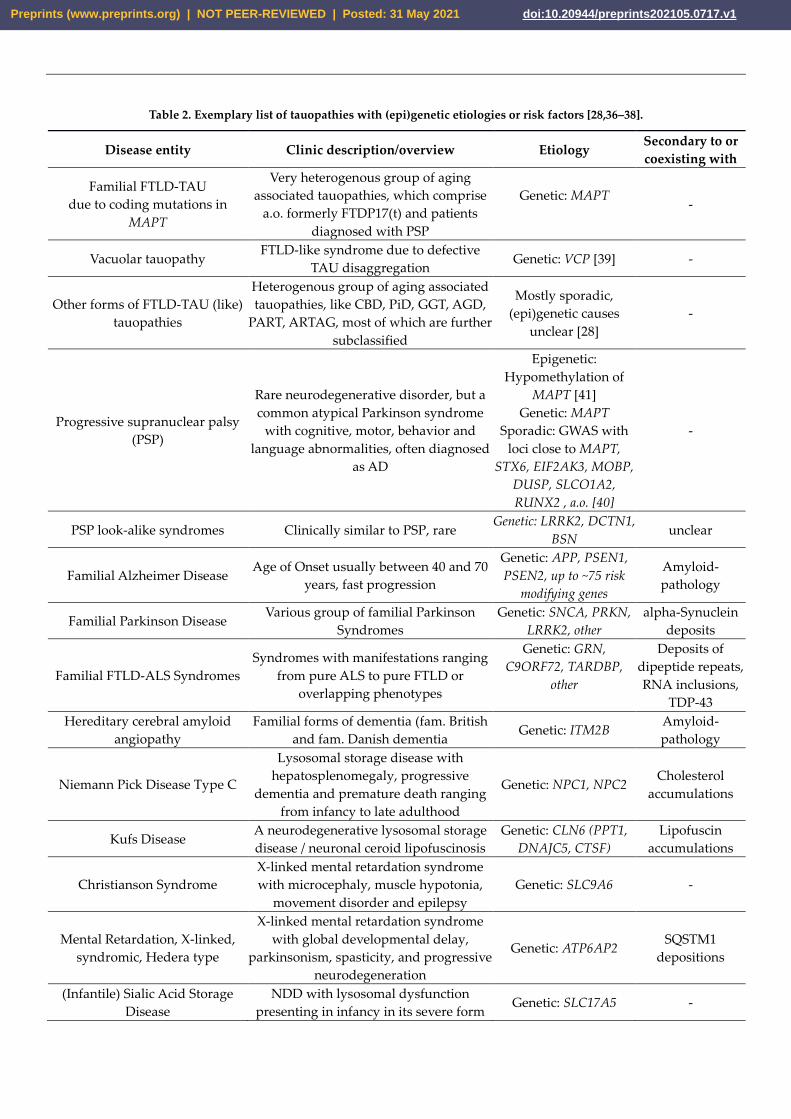
Table 2. Exemplary list of tauopathies with (epi)genetic etiologies or risk factors [28,36–38].
Disease entity Clinic description/overview Etiology Secondary to or
coexisting with
Familial FTLD-TAU
due to coding mutations in
MAPT
Very heterogenous group of aging
associated tauopathies, which comprise
a.o. formerly FTDP17(t) and patients
diagnosed with PSP
Genetic: MAPT
-
Vacuolar tauopathy FTLD-like syndrome due to defective
TAU disaggregation Genetic: VCP [39] -
Other forms of FTLD-TAU (like)
tauopathies
Heterogenous group of aging associated
tauopathies, like CBD, PiD, GGT, AGD,
PART, ARTAG, most of which are further
subclassified
Mostly sporadic,
(epi)genetic causes
unclear [28]
-
Progressive supranuclear palsy
(PSP)
Rare neurodegenerative disorder, but a
common atypical Parkinson syndrome
with cognitive, motor, behavior and
language abnormalities, often diagnosed
as AD
Epigenetic:
Hypomethylation of
MAPT [41]
Genetic: MAPT
Sporadic: GWAS with
loci close to MAPT,
STX6, EIF2AK3, MOBP,
DUSP, SLCO1A2,
RUNX2 , a.o. [40]
-
PSP look-alike syndromes Clinically similar to PSP, rare Genetic: LRRK2, DCTN1,
BSN unclear
Familial Alzheimer Disease Age of Onset usually between 40 and 70
years, fast progression
Genetic: APP, PSEN1,
PSEN2, up to ~75 risk
modifying genes
Amyloid-
pathology
Familial Parkinson Disease Various group of familial Parkinson
Syndromes
Genetic: SNCA, PRKN,
LRRK2, other
alpha-Synuclein
deposits
Familial FTLD-ALS Syndromes
Syndromes with manifestations ranging
from pure ALS to pure FTLD or
overlapping phenotypes
Genetic: GRN,
C9ORF72, TARDBP,
other
Deposits of
dipeptide repeats,
RNA inclusions,
TDP-43
Hereditary cerebral amyloid
angiopathy
Familial forms of dementia (fam. British
and fam. Danish dementia Genetic: ITM2B
Amyloid-
pathology
Niemann Pick Disease Type C
Lysosomal storage disease with
hepatosplenomegaly, progressive
dementia and premature death ranging
from infancy to late adulthood
Genetic: NPC1, NPC2 Cholesterol
accumulations
Kufs Disease A neurodegenerative lysosomal storage
disease / neuronal ceroid lipofuscinosis
Genetic: CLN6 (PPT1,
DNAJC5, CTSF)
Lipofuscin
accumulations
Christianson Syndrome
X-linked mental retardation syndrome
with microcephaly, muscle hypotonia,
movement disorder and epilepsy
Genetic: SLC9A6 -
Mental Retardation, X-linked,
syndromic, Hedera type
X-linked mental retardation syndrome
with global developmental delay,
parkinsonism, spasticity, and progressive
neurodegeneration
Genetic: ATP6AP2 SQSTM1
depositions
(Infantile) Sialic Acid Storage
Disease
NDD with lysosomal dysfunction
presenting in infancy in its severe form Genetic: SLC17A5 -
Preprints (www.preprints.org) | NOT PEER-REVIEWED | Posted: 31 May 2021 doi:10.20944/preprints202105.0717.v1

or in adulthood with progressive brain
atrophy.
PKAN NDD with brain iron accumulation Genetic: PANK2 Iron depositions
Genetic forms of AD and related tauopathies, as well as the histopathology
and imaging-based findings have elucidated possible connections and allowed
initial mapping of cellular components and pathways involved. Still, genetic and
signaling-based mechanisms alone can by far not exhaustively describe the
pathomechanisms both of genetic and sporadic forms of neurodegeneration, in
particular of AD and related pathology. Epigenetic mechanisms likely bridge the
gaps in current knowledge of tauopathy disease mechanisms, and might provide
mechanistic basis for environmental triggers.
The epigenetic toolkit
The epigenetic toolkit involves histone variants and modifications, alterations
in nucleosome positioning, DNA methylation, and non-coding RNAs. As
enormous progress was made in investigating the functional implications and
disease-relevance of DNA methylation and non-coding RNAs in the context of
AD and tauopathies, we will mainly focus on the discussion of these epigenetic
mechanisms. However, we will shortly introduce all aforementioned epigenetic
signatures.
Histone Variants and modifications: The repeated basic unit of chromatin –
the nucleosome – is composed of DNA wrapped around histone octamers, which
are formed of the highly conserved H2A, H2B, H3, and H4 histone proteins. In
addition to replication-dependent canonical histones, particular histone variants
exist, which either display significant structural dissimilarities (e.g., macroH2A,
the centromere-specific protein CENP-A), or minor sequence differences (e.g., the
canonical H3.1 and H3.2, and the variant H3.3). Histone variants display different
cell-type specific expression levels [42], and undergo particular posttranslational
histone modifications [43]. Dependent on the incorporated variant, the site of
insertion in the genome as well as the variant-specific posttranslational
modifications, histone variants can differentially affect gene transcription.
Posttranslational histone modifications come in different flavors, such as
acetylation, methylation, phosphorylation, SUMOylation and ADP-ribosylation
[44], and occur mostly at the N-terminal tails of the histone proteins. They
represent dynamic and reversible modifications differently influencing gene
transcription. The regulation of these post-translational histone modifications
depends on antagonistic sets of enzyme complexes that either attach (writers) or
remove (erasers) the respective chemical groups. One of the most intensively
studied histone modifications is the acetylation of lysine residues. While adding
of acetyl groups, catalyzed by acetyl transferases (HATs), is associated with
transcriptional activation, the erasure of acetyl groups by histone deacetylases
(HDACs) leads to condensed chromatin [45]. Histone methylation likewise
influences the accessibility of the DNA, which is associated either with
transcriptional repression or activation. This depends on the site and the degree
of methylation, being bound by particular readers, which mediate the
transcriptional repression or activation by interacting with respective regulatory
proteins or complexes. While H3K4-trimethylation (me3) leads to open
Preprints (www.preprints.org) | NOT PEER-REVIEWED | Posted: 31 May 2021 doi:10.20944/preprints202105.0717.v1
chromatin, H3K27me3 causes chromatin condensation associated with gene
repression [46].
DNA methylation: In addition to modification of histone proteins, the DNA
itself can be chemically modified by DNA methylation, occurring mostly on
cytosines, but also on adenines [47]. DNA methylation is catalyzed by DNA
methyltransferases (DNMTs), with DNMT1 and DNMT3A being predominantly
expressed in the adult brain [48]. DNA methylation at enhancer and promoter
sites is associated with transcriptional regulation. Further, DNA methylation can
occur in gene bodies and intergenic regions, being involved in repression of
repetitive elements, alternative splicing and alternative promoter choice [49,50].
DNA methylation affects transcription by diverse mechanisms. For example,
methylation of transcription factor binding motifs can physically impede
transcription factor binding, resulting in suppression of transcription. In addition,
methyl-CpG-binding domain proteins (MBDs) interact with methylated DNA,
which then recruit other chromatin and nucleosome remodeling factors,
promoting inactive heterochromatin formation [51]. This is in line with the
textbook model of repressive DNA methylation in gene promoter regions.
Contrasting the accepted function of DNA methylation preventing the binding of
proteins without a methyl-CpG-binding domain (MBD), different studies
reported that DNA methylation signatures can also serve as binding motifs for
specific transcription factors without a methyl binding domain [52]. In silico
studies revealed an increasing pool of transcription factors predicted to bind
methylated DNA sequences, and emerging scenarios propose that DNA
methylation signatures could create new binding motifs for particular
transcription factors. Likewise, certain transcription factors might even recognize
different sequences, dependent on whether being methylated or not [53].
Alike the modification of histone proteins, DNA methylation is a dynamic
process. In addition to passive DNA demethylation in dividing progenitors, Ten-
eleven translocation (TET) family enzyme-dependent mechanisms initiate active
DNA demethylation, also in non-dividing cells such as neurons [54]. Through
TET-mediated oxidation of 5-methylcytosine (5mc) to 5-hydroxymethylcytosine
(5hmc) and iterative oxidation forms, active reversion to cytosine can be achieved
by thymine DNA glycosylase (TDG)-mediated base excision repair [55,56], which
is also observed in neurons [57,58].
Non-coding RNAs: Non-coding RNAs are distinguished in small and long
non-coding RNAs (scnRNAs and lncRNAs, respectively), which differ in size,
biogenesis, and function. The sncRNAs, including miRNAs, siRNAs and piRNAs,
mainly act on posttranscriptional regulation in the cytoplasm [59]. In contrast, the
functional spectrum of lncRNAs is tremendously diverse, modulating
transcription and post-transcriptional events in the nucleus, and further acting on
translation, e.g. by acting as a sponge for, or precursors of miRNAs [54].
Crosstalk between epigenetic mechanisms: The different mechanisms
concertedly modulate chromatin structure and gene expression involving an
intensive crosstalk. For example, particular histone modifications predispose for
the establishment of DNA methylation marks and vice versa [51,60]. Moreover,
DNMTs influence histone modifications by regulating the transcription of genes
encoding for enzymes of histone modifying complexes as well as by interacting
Preprints (www.preprints.org) | NOT PEER-REVIEWED | Posted: 31 May 2021 doi:10.20944/preprints202105.0717.v1
e.g. with the PRC2 at protein level [61,62]. Moreover, lncRNAs were reported to
recruit or evict the binding of DNMTs and histone modifying complexes [59,63],
mediating target specificity. Hence, a complex interplay between different
epigenomic remodelers exists, which concertedly orchestrate transcriptional and
posttranscriptional regulation, coordinating diverse physiological processes in
health and disease.
Age-dependent changes of DNA methylation signatures and the relevance
for AD and tauopathies
Aberrant gene expression, genomic instability, and the loss in chromatin structure
are features of both aging and multifactorial or complex diseases such as AD
[64,65]. These alterations are intimately associated to changes in the epigenome
[66], and can be responsive to environmental influence [67]. As aging represents
the main risk factor for AD and most tauopathies, it is conceivable that the
epigenetic changes coming along with the aging process that affect brain function
predispose for or are associated with AD and tauopathies. A common hallmark
of both healthy aging and AD/tauopathies, is the decline in memory function.
Changes in the gene expression of chromatin remodeling enzymes, such as
DNMTs and histone modifying proteins, have been associated with alterations in
synaptic plasticity, learning and memory [68–73]. Moreover, the expression or
activity of epigenetic modifiers is altered in the aging brain [74]. Together, this
underlines the relevance of epigenetic modifications in the context of aging and
AD, which will be discussed as follows.
The age-related decline in Dnmt3a2 expression seems to be associated with
diminished cognitive abilities, as these were restored upon the rescue of
decreased Dnmt3a2 levels in mice [75]. In line with the decline in DNMT
expression upon aging, global hypomethylation with local sites of
hypermethylation were observed in aging brains across species, affecting the
expression of genes related to synapse function, cellular homeostasis but also
neuronal development [76,77]. Such age-associated DNA methylation changes are
proposed to contribute to transcriptional alterations of AD-related genes, possibly
predisposing for the disease [78,79]. Indeed, the expression levels of key genes
associated with AD and taupathies´ pathophysiology were suggested to be
regulated by DNA methylation in an age-dependent fashion. This is true e.g. for
the membrane protein APP (Amyloid-Precursor Protein), concentrated in the
synapses. As indicated above, mutations in APP lead to a rare genetic and early
onset form of AD, due to increased production of the amyloidogenic Amyloid-
beta (Aβ) protein, the main component of the plaques deposited in the brain of
AD patients. The APP coding gene, which is frequently methylated, displays an
age-related demethylation of cytosines in the promoter region (those at −207 to
approximately −182), suggested to be linked to the Aβ deposition in the aged brain
[80,81]. In contrast, the promoter regions of the neprilysin (NEP) gene, known to
inhibit AD occurrence, turned out to be highly methylated in AD brains [82]. NEP,
which is a major Aβ-degrading enzyme in the brain fulfilling critical functions in
the clearance of Aβ, was found down-regulated in both AD and aged healthy
brains [83]. The increased methylation of the NEP gene results in decreased
expression, which then negatively impacts Aβ clearance, potentially being
causative for the elevated Aβ plaque burden of the aging brain [82].
Preprints (www.preprints.org) | NOT PEER-REVIEWED | Posted: 31 May 2021 doi:10.20944/preprints202105.0717.v1
Moreover, age-related changes in the cytosine methylation status in a
promoter region of the microtubule-related protein TAU (MAPT) gene were
identified. While in the binding sites of the transcriptional activator SP1 a
significant age-related increase in 5mC was observed in autopsy human cerebral
cortex, a decrease with age of 5mC in the binding sites for GCF, a repressor of GC-
rich promoters, was revealed [81]. The authors propose that the methylation
status of cytosines in the promoter region of the MAPT gene changes with age to
reduce its transcriptional activity in the human cerebral cortex.
Evidence for the implication of altered DNA methylation signatures in AD
and tauopathies
Similar to the aging brain, global DNA hypomethylation was reported for
AD, supported by decreased immunoreactivity for 5-methylcytosine (5-mC) in
cortical neurons of postmortem AD brains compared to controls [84]. Reduced
levels of 5mC were observed in the hippocampus, entorhinal and prefrontal
cortex, as well as in the cerebellum of patients with AD [84–86]). In line with this,
Mastroeni et al. (2010) found weak staining with antibodies directed against DNA
methylation maintenance factors in hippocampal tissue of AD patients compared
to normal brains. These studies are in line with findings obtained from a rare set
of monozygotic twins discordant for AD, revealing reduced levels of DNA
methylation in neuronal nuclei of the AD twin in the temporal neocortex [87].
For genes such as MCF2L, ANK1, MAP2, LRRC8B, STK32C and S100B,
neuronal and glia cell-type specific differential methylation dynamics associated
with AD Braak stage progression were observed, and methylation changes at the
key AD risk genes APP and ADAM17 were identified in a meta-analysis [88].
Elevated DNA methylation levels in the promoter region of apolipoprotein E
(APOE) gene were demonstrated to increase the odds of dementia and AD [89].
Genetic variation in the APOE gene is related with AD risk and the severity of
amyloid-β (Aβ) burden, with the APOE ε4 variant described to be by far the most
consistent genetic risk factor [90,91]. The DNA methylation-dependent effect was
independent of APOE genotype, suggesting that allelic variation and methylation
variation in APOE may independently affect the risk of dementia [89].
DNA methylation changes leading to pathological phosphorylation of
TAU: Disturbed methylation levels in promoter regions of genes related to TAU
phosphorylation, which plays a critical role in tauopathies, were revealed by
diverse clinical and basic research studies in the context of AD [92]. During early
AD development, low DNA methylation levels were found in the promoter
region of the GSK3β gene (GSK3B) from the prefrontal cortex tissue of AD
patients. GSK3β was further found overexpressed in patients with initial AD [93].
GSK3β is the kinase most commonly implicated in hyperphosphorylation of the
TAU protein, which is in turn believed to be a prerequisite for aggregation and
formation of NFTs [94]. While at Braak stages I-II a decrease of the inactive GSK3β
was found in the cortex from AD patients, considerable increase was observed in
AD patients at stages V-VI, when being compared to the control subjects. It is
proposed by the authors that GSK3β hyperactivity, and then NFTs formation,
could be initiated at an early stage of the disease and turned off at final stages [93].
Preprints (www.preprints.org) | NOT PEER-REVIEWED | Posted: 31 May 2021 doi:10.20944/preprints202105.0717.v1
TAU hyperphosphorylation is further driven by upregulated Cdk5
expression, causing diminished long-term synaptic potentiation and culminating
in impairments of spatial learning and memory. Low levels of cytosine
methylation were detected in the promoter region of Cdk5 in the hippocampal
CA1 region in a rat model with Aβ1-42-induced memory deficiency [95].
Increased DNA methylation, linearly correlating with the Braak stage, was
observed in the promoter region of the dual specificity phosphatase 22 gene
(DUSP22) in hippocampi of AD patients. In line with the well-accepted
transcriptionally repressive function of promoter methylation, reduced DUSP22
expression was detected at mRNA and protein level. As DUSP22 was found to
inhibit the PKA-mediated TAU phosphorylation [96], its reduced expression
could have direct consequence for disease progression. In addition to TAU, PKA
activates the Ser133phosphorylation of the cAMP response element-binding
protein (CREB). CREB activity is known to be relevant for neuronal function and
synaptic plasticity, long-term memory formation and neuronal survival
regulation [97]; all being compromised in AD [98]. Hence, the reduced DUSP22
expression could promote neuronal survival through elevated PKA/CREB
activation. The authors propose the increase in DUSP22 promoter methylation to
be a consequence of Aβ-induced toxicity, in the sense that cells respond with
active methylation to improve their survival [96].
TAU phosphorylation can further be influenced by Tet-dependent DNA
demethylation. BDNF, as a key component in the maintenance of synaptic
plasticity and synaptogenesis in the hippocampus [99], is closely related to TAU
hyperphosphorylation [100,101]. It was reported that the BDNF chromatin status
and promoter accessibility is regulated by TET1 and ERK1/2 [102]. Due to this, it
is assumed that TET1-dependent BDNF DNA demethylation influences TAU
phosphorylation levels. Overall, these studies provide evidence for an implication
of DNA methylation dependent transcriptional control of TAU phosphorylation-
related genes in AD. Most of these genes are effectors downstream of Aβ
pathology, but upstream of TAU pathology closely related to cognitive
dysfunction in patients. Thus, targeting these genes might disrupt the amyloid
cascade upstream of TAU and are thus potential targets for AD treatment
strategies.
Altered DNA methylation signatures as a consequence of disease
pathophysiology, such as amyloid β (Aβ) burden and TAU-phosphorylation
As mentioned above for DUSP22, changes in DNA methylation could be a
consequence of the altered neuronal physiology in AD and tauopathies, such as
the accumulation of Aβ peptides. Hence, altered epigenetic signatures could be a
bystander of disease progression, leading to the devastating dysregulation of
genes and driving the further progression of neurodegeneration in AD and other
tauopathies. Furthermore, distinct mutations associated with these diseases could
elicit “secondary” changes in DNA methylation pattern.
Aβ peptide and TAU-phosphorylation-driven changes in the expression
and localization of DNA repair related proteins: Disruption of maintenance of
genomic integrity emerges to play a central role in AD and related pathology
[103]. Early intraneuronal accumulation of Aβ peptides promotes global DNA
hypomethylation and through this, increased expression of genes being involved
Preprints (www.preprints.org) | NOT PEER-REVIEWED | Posted: 31 May 2021 doi:10.20944/preprints202105.0717.v1
in DNA repair such as BRCA1, in the mouse model [104]. BRCA1 has been
identified to be upregulated in response to Aβ stimulation, in both cellular in vitro
and in vivo mouse models, acting neuroprotectively against Aβ-induced DNA
double-strand breaks. Hypomethylation and up-regulated expression of BRCA1
was further observed in postmortem brain samples from AD patients [105].
However, in the hippocampal CA1 region and entorhinal cortex of the AD brain,
BRCA1 protein has been found mislocalized to the cytoplasm and insoluble [105].
In line with the cytosolic mislocalization, nuclear BRCA1 protein, but not of other
members of Defective DNA Repair (DDR) mechanisms, were found to be reduced
in AD brains [106]. The cytoplasmic BRCA1 mislocalization is proposed to
represent a consequence of pTAU deposition, which is in line with the observation
that brain regions without TAU pathology, namely the occipital lobe and the
cerebellum, are free of cytoplasmic accumulation of BRCA1 despite decreased
methylation of the respective gene. The insolubility of BRCA1 under the presence
of aggregated TAU is proposed to be the reason for its dysfunction despite
enhanced expression, contributing to compromised genomic integrity of neurons
and hence, disease pathophysiology [105]. BRCA1 was sequestered to TAU
inclusions not only in AD brains, but also in brains of patients suffering from other
tauopathies (namely PiD, PSP, CBD, FTDP17/FTLD-TAU) [107,108],
strengthening the role for TAU in disruption of DDR.
Aβ-associated changes in DNA methylation of cell cycle-related genes: In
addition to compromised genomic integrity, dysregulated cell cycle control is an
integral part of AD. While in a healthy neuron, abnormal cell cycle reentry leads
to apoptosis, abnormal reentry in neurons of aged subjects with AD triggers a
cycle of oxidative damage and mitogen production facilitating TAU
hyperphosphorylation and Aβ deposition, leading to full blown pathology and
cognitive decline [109].
For genes promoting the activation of cell cycle reentry (i.e., via CDK5),
hypomethylation was observed in AD or in AD disease paradigms [110]. In
support of the hypothesis that DNA methylation changes represent a downstream
effect triggered by Aß plaques, the exposure of differentiated human neurons to
Aβ results in DNA methylation abnormalities of cell-fate genes controlling
neuronal differentiation and apoptosis [110].
In this context, a recent study described a potential mechanism for DNA
methylation-mediated Aβ peptide overproduction, which then drives Aβ
peptide-triggered hypomethylation of cell cycle-associated genes [111]. Li et al.
(2019) [111] found that AD neurons display significant hypomethylation in the
enhancer of the DSCAML1 gene that targets BACE1. BACE1 encodes the β-
secretase, which cleaves amyloid precursor protein (APP) thereby acting on Aβ
production. Hence, the DSCAML1 enhancer hypomethylation is suggested to
activate BACE1 transcription, putatively leading to the overproduction of Aβ
peptides, which eventually form plaques. As mentioned above, plaques are
reported to occur early in AD typically preceding the spread of neurofibrillary
tangles and neurodegeneration [112,113]. In addition to plaque formation, the Aβ
peptides are proposed to engage the pathogenic hypomethylation of enhancers
affecting neurogenesis and cell cycle genes, which are suggested to be already
primed for activation due to the age-related global hypomethylation [111].
Changes of DNA methylation signatures in enhancer regulatory elements are
Preprints (www.preprints.org) | NOT PEER-REVIEWED | Posted: 31 May 2021 doi:10.20944/preprints202105.0717.v1
frequently observed in AD brains [114,115]. Together, this indicates that
epigenetic impairment of enhancer function is implicated in AD.
The studies described so far illustrate that changes in DNA methylation
signatures can be elicited in response to pathophysiological processes induced in
AD and/or tauopathies, making the role of DNA methylation in these diseases
difficult to judge. Moreover, it should be noted that changes in neuronal activity
can also modify the DNA methylation landscape [116], and that altered synaptic
and neuronal function is a hallmark of AD and tauopathies.
The resulting changes in DNA methylation seem to contribute to the
progressive neurodegeneration by transcriptional dysregulation, whereas the
detailed implication requires more intensive investigations. Dissecting the
relevance of DNA methylation for AD and tauopathies is further complicated by
newly arising scenarios of the biological relevance of DNA methylation. In
addition, to repressive promoter methylation, intragenic DNA methylation
mediates alternative splicing and promoter choice. Moreover, apart from
impeding transcription factor binding, certain DNA methylation patterns seem to
create new motifs for transcription factors, for which increased methylation can
also result in elevated expression [117]. Hence, the transcriptional consequences
of certain changes in DNA methylation need to be dissected in much more detail.
Epigenetic Treatment? – The potential and limitations of DNA
methylation-based therapy approaches
As described above, hypomethylation of AD risk genes (like APP, PSEN1,
and PSEN2) was described to be associated with defects in learning and memory.
Increases in methyl donor S-adenosyl-L-methionine (SAM) was reported to
reduce APP and PSEN1 expression by promoter hypermethylation [118,119]. In
line with this, elevated levels of B12, folate and other methionine sources in the
diet improve methionine bioavailability and were shown to reverse elevated
expression of APP and PSEN1 [120–122].
In addition to driving hypermethylation, there is ongoing screening for
DNMT inhibitors capable of modulating the methylation of AD or tauopathy risk
genes. DNMT inhibitors such as azacitidine and decitabine have already been
approved by the FDA for cancer treatment such as leukemia [123–125]. The use of
DNA demethylating agents has also been used in some other neurodegenerative
diseases, such as Friedreich’s ataxia [126], which however did not provide
promising results in human cells.
Finally, due to gene locus-specific changes in DNA methylation signatures,
sequence-specific DNA demethylating agents, such as the oligonucleotide
antisense inhibitor MG98 [127–129], seem rather be useful for future therapeutic
approaches to reduce DNA methylation site-specifically than the usage of general
DNMT inhibitors. Moreover, apart from site-specific hypermethylation,
hypomethylation of particular genes was described to be implicated in AD´ and
tauopathy´s pathomechanisms. Hence, locus-specific editing technologies are
required for altering DNA methylation, which can be achieved by clustered
regulatory interspaced short palindromic repeats (CRISPR)-deactivated Cas9
(dCas9)-based editing systems that have been described as a specific and efficient
method capable of manipulating site-specific DNA methylation [130]. This, in
combination with improvements in cell type specific application strategies, would
open the way for targeted epigenetic therapies. A major challenge for such
Preprints (www.preprints.org) | NOT PEER-REVIEWED | Posted: 31 May 2021 doi:10.20944/preprints202105.0717.v1
epigenetic therapy is to find ways for applied molecules to pass through the
blood-brain barrier, and enter specific subsets of affected cells. Moreover, as
disease progression-specific alterations in DNA methylation have also been
described, with some changes rather being causative or a consequence of disease
pathophysiology, determining the exact stage of disease would be very beneficial.
Herein, DNA methylation signatures might also help in the future, as discussed
as follows.
Altered DNA methylation signatures as potential biomarkers for
AD/Tauopathies disease and disease progression?
The use of reliable biomarkers for AD and tauopathies would be extremely
beneficial for early diagnosis and monitoring disease progression, and eventually
the response towards potential therapies. So, may DNA methylation signatures
be useful to serve as biomarkers for AD and tauopathies in blood cells?
It was found that in leukocytes the intron 1 of the TREM2 gene (triggering
receptor expression on myeloid cells 2) displays reduced methylation coming
along with elevated expression at mRNA level in AD subjects compared to
controls [131,132]. Moreover, increased levels of peripheral BDNF promoter
methylation was proposed to be an epigenetic biomarker indicating the
transformation of MCI to AD [133]. Similarly, increased DNA methylation levels
were detected in promoter regions of the COASY and SPINT genes in plasma
samples of AD and aMCI subjects compared to controls [134]. Methylation of the
PICALM gene in blood cells was found to be related to cognitive decline of AD
patients [135].
In general, global DNA methylation levels were found higher in peripheral
blood mononuclear cells of LOAD patients compared to healthy controls,
suggested to be associated with the presence of the APOE(4) allele (p=0.0043) and
APOE(3) carriers (p=0.05) in the global population [136]). In sum, current studies
indicate that monitoring global and site-specific DNA methylation in peripheral
samples might be applied for individual risk screening to develop AD. However,
more detailed studies and correlations are required that strengthen the use of
DNA methylation as biomarkers for AD risk, diagnosis and progression, which
might be expected in near future.
Conclusions
Changes in DNA methylation seem to be critically implicated in causing
and/or driving the progression of AD and tauopathies, for which epigenetic
therapy strategies hitting DNMTs and DNA methylation are discussed to be
promising. However, there is still a long way to go. Firstly, locus, cell-type and
disease progression-specific changes have to be clearly dissected and correlated
to the transcriptional output as well as the physiological consequences. Then,
targeted strategies, such as being offered by (CRISPR)-deactivated Cas9 (dCas9)-
based editing systems, have to be exploited and developed for site-specific
manipulation of DNA methylation signatures. And finally, these manipulation
systems have to be safely applicable to the brain and to specific neuronal subtypes
at certain disease-progression stages. Hence, a new generation of selective probes
has to be developed. In addition to potential therapeutic targets, epigenetic
signatures may also help to improve diagnosis of AD and tauopathies, for which
Preprints (www.preprints.org) | NOT PEER-REVIEWED | Posted: 31 May 2021 doi:10.20944/preprints202105.0717.v1

epigenetic regulation likely will become an important tool in the treatment of
these diseases.
Funding: This research was funded by the Deutsche Forschungsgemeinschaft (DFG, German Research Foundation) -
368482240/GRK2416DFG; DFG ZI1224/13-1 (both assigned to GZB); and the Else-Kröner-Fresenius Stiftung (2019_A41) and Köln
Fortune (both assigned to HZ)
References
1. Korolev, I.O.; Symonds, L.L.; Bozoki, A.C. Predicting progression from mild cognitive impairment to Alzheimer’s dementia using clinical, MRI, and plasma
biomarkers via probabilistic pattern classification. PLoS One 2016, 11, doi:10.1371/journal.pone.0138866.
2. Scheltens, P.; De Strooper, B.; Kivipelto, M.; Holstege, H.; Chételat, G.; Teunissen, C.E.; Cummings, J.; van der Flier, W.M. Alzheimer’s disease. Lancet 2021,
397, 1577–1590.
3. Bird, T.D. Alzheimer Disease Overview; 1993;
4. Hyman, B.T.; Phelps, C.H.; Beach, T.G.; Bigio, E.H.; Cairns, N.J.; Carrillo, M.C.; Dickson, D.W.; Duyckaerts, C.; Frosch, M.P.; Masliah, E.; et al. National
Institute on Aging-Alzheimer’s Association guidelines for the neuropathologic assessment of Alzheimer’s disease. Alzheimer’s Dement. 2012, 8,
doi:10.1016/j.jalz.2011.10.007.
5. Hammond, T.C.; Xing, X.; Wang, C.; Ma, D.; Nho, K.; Crane, P.K.; Elahi, F.; Ziegler, D.A.; Liang, G.; Cheng, Q.; et al. β-amyloid and tau drive early
Alzheimer’s disease decline while glucose hypometabolism drives late decline. Commun. Biol. 2020, 3, doi:10.1038/s42003-020-1079-x.
6. Gulisano, W.; Maugeri, D.; Baltrons, M.A.; Fà, M.; Amato, A.; Palmeri, A.; D’Adamio, L.; Grassi, C.; Devanand, D.P.; Honig, L.S.; et al. Role of Amyloid-β
and Tau Proteins in Alzheimer’s Disease: Confuting the Amyloid Cascade. J. Alzheimer’s Dis. 2018, 64.
7. D.J., S.; J., H. The amyloid hypothesis of Alzheimer’s disease at 25 years. EMBO Mol. Med. 2016, 8.
8. Roberson, E.D.; Scearce-Levie, K.; Palop, J.J.; Yan, F.; Cheng, I.H.; Wu, T.; Gerstein, H.; Yu, G.Q.; Mucke, L. Reducing endogenous tau ameliorates amyloid
beta-induced deficits in an Alzheimer’s disease mouse model. Science (80-. ). 2007, 316, 750–754.
9. Ittner, L.M.; Ke, Y.D.; Delerue, F.; Bi, M.; Gladbach, A.; van Eersel, J.; Wolfing, H.; Chieng, B.C.; Christie, M.J.; Napier, I.A.; et al. Dendritic function of tau
mediates amyloid-beta toxicity in Alzheimer’s disease mouse models. Cell 2010, 142, 387–397, doi:10.1016/j.cell.2010.06.036.
10. Zempel, H.; Luedtke, J.; Kumar, Y.; Biernat, J.; Dawson, H.; Mandelkow, E.; Mandelkow, E.M. Amyloid-β oligomers induce synaptic damage via Tau-
dependent microtubule severing by TTLL6 and spastin. EMBO J. 2013, 32, 2920–2937, doi:10.1038/emboj.2013.207.
11. Zempel, H.; Mandelkow, E. Mechanisms of Axonal Sorting of Tau and Influence of the Axon Initial Segment on Tau Cell Polarity. In Advances in
Experimental Medicine and Biology; 2019; Vol. 1184, pp. 69–77.
12. Cho, H.; Choi, J.Y.; Hwang, M.S.; Lee, J.H.; Kim, Y.J.; Lee, H.M.; Lyoo, C.H.; Ryu, Y.H.; Lee, M.S. Tau PET in Alzheimer disease and mild cognitive
impairment. Neurology 2016, 87, doi:10.1212/WNL.0000000000002892.
13. Joie, R. La; Visani, A. V.; Baker, S.L.; Brown, J.A.; Bourakova, V.; Cha, J.; Chaudhary, K.; Edwards, L.; Iaccarino, L.; Janabi, M.; et al. Prospective longitudinal
atrophy in Alzheimer’s disease correlates with the intensity and topography of baseline tau-PET. Sci. Transl. Med. 2020, 12, doi:10.1126/scitranslmed.aau5732.
14. Biel, D.; Brendel, M.; Rubinski, A.; Buerger, K.; Janowitz, D.; Dichgans, M.; Franzmeier, N. Tau-PET and in vivo Braak-staging as a prognostic marker in
Alzheimer’s disease. medRxiv 2021.
15. Bejanin, A.; Schonhaut, D.R.; La Joie, R.; Kramer, J.H.; Baker, S.L.; Sosa, N.; Ayakta, N.; Cantwell, A.; Janabi, M.; Lauriola, M.; et al. Tau pathology and
neurodegeneration contribute to cognitive impairment in Alzheimer’s disease. Brain 2017, 140, doi:10.1093/brain/awx243.
16. Braak, H.; Braak, E. Neuropathological stageing of Alzheimer-related changes. Acta Neuropathol 1991, 82, 239–259.
17. Braak, H.; Alafuzoff, I.; Arzberger, T.; Kretzschmar, H.; Tredici, K. Staging of Alzheimer disease-associated neurofibrillary pathology using paraffin sections
and immunocytochemistry. Acta Neuropathol. 2006, 112, 389–404, doi:10.1007/s00401-006-0127-z.
18. Vogel, J.W.; Young, A.L.; Oxtoby, N.P.; Smith, R.; Ossenkoppele, R.; Strandberg, O.T.; Joie, R. La; Aksman, L.M.; Grothe, M.J.; Iturria-Medina, Y.; et al.
Characterizing the spatiotemporal variability of Alzheimer’s disease pathology. medRxiv 2020.
19. Drubin, D.G. Tau protein function in living cells. J. Cell Biol. 2004, 103, 2739–2746, doi:10.1083/jcb.103.6.2739.
20. Neve, R.L.; Donlon, T.A.; Kurnit, D.M.; Harris, P.; Kosik, K.S. Identification of cDNA clones for the human microtubule-associated protein tau and
chromosomal localization of the genes for tau and microtubule-associated protein 2. Mol. Brain Res. 2003, 1, 271–280, doi:10.1016/0169-328x(86)90033-1.
21. Andreadis, A. Tau splicing and the intricacies of dementia. J. Cell. Physiol. 2012, 227, 1220–1225, doi:10.1002/jcp.22842.
22. Trabzuni, D.; Wray, S.; Vandrovcova, J.; Ramasamy, A.; Walker, R.; Smith, C.; Luk, C.; Gibbs, J.R.; Dillman, A.; Hernandez, D.G.; et al. MAPT expression
and splicing is differentially regulated by brain region: relation to genotype and implication for tauopathies. Hum Mol Genet 2012, 21, 4094–4103,
doi:10.1093/hmg/dds238.
23. Doran, M.; Du Plessis, D.G.; Ghadiali, E.J.; Mann, D.M.A.; Pickering-Brown, S.; Larner, A.J. Familial early-onset dementia with tau intron 10 + 16 mutation
with clinical features similar to those of Alzheimer disease. Arch. Neurol. 2007, 64, doi:10.1001/archneur.64.10.1535.
24. Morris, H.R.; Osaki, Y.; Holton, J.; Lees, A.J.; Wood, N.W.; Revesz, T.; Quinn, N. Tau exon 10 + 16 mutation FTDP-17 presenting clinically as sporadic young
onset PSP. Neurology 2003, 61, doi:10.1212/01.WNL.0000072325.27824.A5.
25. Zempel, H.; Mandelkow, E. Lost after translation: Missorting of Tau protein and consequences for Alzheimer disease. Trends Neurosci. 2014, 37, 721–732,
doi:10.1016/j.tins.2014.08.004.
26. Zempel, H.; Dennissen, F.J.A.; Kumar, Y.; Luedtke, J.; Biernat, J.; Mandelkow, E.M.; Mandelkow, E. Axodendritic sorting and pathological missorting of Tau
are isoform-specific and determined by axon initial segment architecture. J. Biol. Chem. 2017, 292, 12192–12207, doi:10.1074/jbc.M117.784702.
Preprints (www.preprints.org) | NOT PEER-REVIEWED | Posted: 31 May 2021 doi:10.20944/preprints202105.0717.v1
27. Bell, M.; Bachmann, S.; Klimek, J.; Langerscheidt, F.; Zempel, H. Axonal TAU sorting in SH-SY5Y-derived neurons requires the C-terminus of TAU but is
independent of Ankyrin G. bioRxiv 2020, 2020.06.26.173526, doi:10.1101/2020.06.26.173526.
28. Ali, F.; Josephs, K. Rare Tauopathies. Semin. Neurol. 2019, 39, doi:10.1055/s-0039-1678584.
29. Spillantini, M.G.; Goedert, M. Tau pathology and neurodegeneration. Lancet Neurol. 2013, 12, 609–622, doi:10.1016/S1474-4422(13)70090-5.
30. Wang, Y.; Mandelkow, E. Tau in physiology and pathology. Nat. Rev. Neurosci. 2016, 17, 5–21.
31. Sultan, A.; Nesslany, F.; Violet, M.; Begard, S.; Loyens, A.; Talahari, S.; Mansuroglu, Z.; Marzin, D.; Sergeant, N.; Humez, S.; et al. Nuclear tau, a key player
in neuronal DNA protection. J Biol Chem 2011, 286, 4566–4575, doi:10.1074/jbc.M110.199976.
32. Murley, A.G.; Coyle-Gilchrist, I.; Rouse, M.A.; Simon Jones, P.; Li, W.; Wiggins, J.; Lansdall, C.; Rodríguez, P.V.; Wilcox, A.; Tsvetanov, K.A.; et al.
Redefining the multidimensional clinical phenotypes of frontotemporal lobar degeneration syndromes. Brain 2020, 143, doi:10.1093/brain/awaa097.
33. Bellenguez, C.; Grenier-Boley, B.; Lambert, J.C. Genetics of Alzheimer’s disease: where we are, and where we are going. Curr. Opin. Neurobiol. 2020, 61.
34. Bellenguez, C.; Küçükali, F.; Jansen, I.; Andrade, V.; Morenau-Grau, S.; Amin, N.; Grenier-Boley, B.; Boland, A.; Kleineidam, L.; Holmans, P.; et al. Large
meta-analysis of genome-wide association studies expands knowledge of the genetic etiology of Alzheimer’s disease and highlights potential translational
opportunities. medRxiv 2020.
35. Schwartzentruber, J.; Cooper, S.; Liu, J.Z.; Barrio-Hernandez, I.; Bello, E.; Kumasaka, N.; Young, A.M.H.; Franklin, R.J.M.; Johnson, T.; Estrada, K.; et al.
Genome-wide meta-analysis, fine-mapping and integrative prioritization implicate new Alzheimer’s disease risk genes. Nat. Genet. 2021, 53, doi:10.1038/s41588-020-
00776-w.
36. Tacik, P.; Sanchez-Contreras, M.; Rademakers, R.; Dickson, D.W.; Wszolek, Z.K. Genetic disorders with tau pathology: A review of the literature and report
of two patients with tauopathy and positive family histories. In Proceedings of the Neurodegenerative Diseases; 2016; Vol. 16, pp. 12–21.
37. Tacik, P.; Wszolek, Z.; SanchezContreras, M.; Rademakers, R.; Dickson, D. Literature review of genetic disorders with tau pathology. Neurology 2016.
38. Mulroy, E.; Jaunmuktane, Z.; Balint, B.; Erro, R.; Latorre, A.; Bhatia, K.P. Some New and Unexpected Tauopathies in Movement Disorders. Mov. Disord.
Clin. Pract. 2020, 7, 616–626, doi:10.1002/mdc3.12995.
39. Darwich, N.F.; Phan, J.M.; Kim, B.; Suh, E.; Papatriantafyllou, J.D.; Changolkar, L.; Nguyen, A.T.; O’Rourke, C.M.; He, Z.; Porta, S.; et al. Autosomal
dominant VCP hypomorph mutation impairs disaggregation of PHF-tau. Science (80-. ). 2020, 370, doi:10.1126/science.aay8826.
40. Wen, Y.; Zhou, Y.; Jiao, B.; Shen, L. Genetics of progressive supranuclear palsy: A review. J. Parkinsons. Dis. 2021, 11.
41. Huin, V.; Deramecourt, V.; Caparros-Lefebvre, D.; Maurage, C.A.; Duyckaerts, C.; Kovari, E.; Pasquier, F.; Buée-Scherrer, V.; Labreuche, J.; Behal, H.; et al.
The MAPT gene is differentially methylated in the progressive supranuclear palsy brain. Mov. Disord. 2016, 31, doi:10.1002/mds.26820.
42. Rogakou, E.P.; Sekeri-Pataryas, K.E. Histone variants of H2A and H3 families are regulated during in vitro aging in the same manner as during
differentiation. Exp. Gerontol. 1999, 34, doi:10.1016/S0531-5565(99)00046-7.
43. Yadav, T.; Quivy, J.P.; Almouzni, G. Chromatin plasticity: A versatile landscape that underlies cell fate and identity. Science (80-. ). 2018, 361.
44. Taylor, B.C.; Young, N.L. Combinations of histone post-Translational modifications. Biochem. J. 2021, 478.
45. Brownell, J.E.; Zhou, J.; Ranalli, T.; Kobayashi, R.; Edmondson, D.G.; Roth, S.Y.; Allis, C.D. Tetrahymena histone acetyltransferase A: A homolog to yeast
Gcn5p linking histone acetylation to gene activation. Cell 1996, 84, doi:10.1016/S0092-8674(00)81063-6.
46. Lachner, M.; O’Sullivan, R.J.; Jenuwein, T. An epigenetic road map for histone lysine methylation. J. Cell Sci. 2003, 116.
47. Greenberg, M.V.C. Get Out and Stay Out: New Insights Into DNA Methylation Reprogramming in Mammals. Front. Cell Dev. Biol. 2021, 8.
48. Bayraktar, G.; Kreutz, M.R. Neuronal DNA Methyltransferases: Epigenetic Mediators between Synaptic Activity and Gene Expression? Neuroscientist 2018,
24.
49. Smith, Z.D.; Meissner, A. DNA methylation: Roles in mammalian development. Nat. Rev. Genet. 2013, 14.
50. Maunakea, A.K.; Nagarajan, R.P.; Bilenky, M.; Ballinger, T.J.; Dsouza, C.; Fouse, S.D.; Johnson, B.E.; Hong, C.; Nielsen, C.; Zhao, Y.; et al. Conserved role of
intragenic DNA methylation in regulating alternative promoters. Nature 2010, 466, doi:10.1038/nature09165.
51. Du, J.; Johnson, L.M.; Jacobsen, S.E.; Patel, D.J. DNA methylation pathways and their crosstalk with histone methylation. Nat. Rev. Mol. Cell Biol. 2015, 16,
doi:10.1038/nrm4043.
52. Hudson, N.O.; Whitby, F.G.; Buck-Koehntop, B.A. Structural insights into methylated DNA recognition by the C-terminal zinc fingers of the DNA reader
protein ZBTB38. J. Biol. Chem. 2018, 293, doi:10.1074/jbc.RA118.005147.
53. Zhu, H.; Wang, G.; Qian, J. Transcription factors as readers and effectors of DNA methylation. Nat. Rev. Genet. 2016, 17, doi:10.1038/nrg.2016.83.
54. Wu, X.; Zhang, Y. TET-mediated active DNA demethylation: Mechanism, function and beyond. Nat. Rev. Genet. 2017, 18.
55. Ito, S.; Shen, L.; Dai, Q.; Wu, S.C.; Collins, L.B.; Swenberg, J.A.; He, C.; Zhang, Y. Tet proteins can convert 5-methylcytosine to 5-formylcytosine and 5-
carboxylcytosine. Science (80-. ). 2011, 333, doi:10.1126/science.1210597.
56. Kohli, R.M.; Zhang, Y. TET enzymes, TDG and the dynamics of DNA demethylation. Nature 2013, 502.
57. Kaas, G.A.; Zhong, C.; Eason, D.E.; Ross, D.L.; Vachhani, R. V.; Ming, G. li; King, J.R.; Song, H.; Sweatt, J.D. TET1 controls CNS 5-Methylcytosine
Hydroxylation, active DNA demethylation, gene transcription, and memory formation. Neuron 2013, 79, doi:10.1016/j.neuron.2013.08.032.
58. Li, M.; Guo, S.; Xu, Y.; Meng, Q.; Li, G.; Yang, X. Glycine betaine-mediated potentiation of HSP gene expression involves calcium signaling pathways in
tobacco exposed to NaCl stress. Physiol Plant 2014, 150, 63–75, doi:10.1111/ppl.12067.
59. Zimmer-Bensch, G. Emerging Roles of Long Non-Coding RNAs as Drivers of Brain Evolution. Cells 2019, 8.
60. Hashimshony, T.; Zhang, J.; Keshet, I.; Bustin, M.; Cedar, H. The role of DNA methylation in setting up chromatin structure during development. Nat.
Genet. 2003, 34, doi:10.1038/ng1158.
61. Symmank, J.; Zimmer, G. Regulation of neuronal survival by DNA methyltransferases. Neural Regen. Res. 2017, 12.
62. Symmank, J.; Bayer, C.; Schmidt, C.; Hahn, A.; Pensold, D.; Zimmer-Bensch, G. DNMT1 modulates interneuron morphology by regulating Pak6 expression
through crosstalk with histone modifications. Epigenetics 2018, 13, doi:10.1080/15592294.2018.1475980.
Preprints (www.preprints.org) | NOT PEER-REVIEWED | Posted: 31 May 2021 doi:10.20944/preprints202105.0717.v1
63. Marchese, F.P.; Raimondi, I.; Huarte, M. The multidimensional mechanisms of long noncoding RNA function. Genome Biol. 2017, 18.
64. Spiegel, A.M.; Sewal, A.S.; Rapp, P.R. Epigenetic contributions to cognitive aging: Disentangling mindspan and lifespan. Learn. Mem. 2014, 21.
65. López-otín, C.; Blasco, M.A.; Partridge, L.; Serrano, M.; Kroemer, G. The Hallmarks of Aging Longevity. Cell 2013, 153.
66. Rowbotham, D.A. Epigenetic Changes in Aging and Age-related Disease. J. Aging Sci. 2015, 03, doi:10.4172/2329-8847.1000130.
67. Grant, W.B.; Campbell, A.; Itzhaki, R.F.; Savory, J. The significance of environmental factors in the etiology of Alzheimer’s disease. J. Alzheimer’s Dis. 2002, 4.
68. Levenson, J.M.; Roth, T.L.; Lubin, F.D.; Miller, C.A.; Huang, I.C.; Desai, P.; Malone, L.M.; Sweatt, J.D. Evidence that DNA (cytosine-5) methyltransferase
regulates synaptic plasticity in the hippocampus. J. Biol. Chem. 2006, 281, doi:10.1074/jbc.M511767200.
69. Morris, M.J.; Monteggia, L.M. Role of DNA methylation and the DNA methyltransferases in learning and memory. Dialogues Clin. Neurosci. 2014, 16,
doi:10.31887/dcns.2014.16.3/mmorris.
70. Laplant, Q.; Vialou, V.; Covington, H.E.; Dumitriu, D.; Feng, J.; Warren, B.L.; Maze, I.; Dietz, D.M.; Watts, E.L.; Iñiguez, S.D.; et al. Dnmt3a regulates
emotional behavior and spine plasticity in the nucleus accumbens. Nat. Neurosci. 2010, 13, doi:10.1038/nn.2619.
71. Xu, S.; Wilf, R.; Menon, T.; Panikker, P.; Sarthi, J.; Elefant, F. Epigenetic control of learning and memory in drosophila by Tip60 HAT action. Genetics 2014,
198, doi:10.1534/genetics.114.171660.
72. Guan, J.S.; Haggarty, S.J.; Giacometti, E.; Dannenberg, J.H.; Joseph, N.; Gao, J.; Nieland, T.J.F.; Zhou, Y.; Wang, X.; Mazitschek, R.; et al. HDAC2 negatively
regulates memory formation and synaptic plasticity. Nature 2009, 459, doi:10.1038/nature07925.
73. McQuown, S.C.; Barrett, R.M.; Matheos, D.P.; Post, R.J.; Rogge, G.A.; Alenghat, T.; Mullican, S.E.; Jones, S.; Rusche, J.R.; Lazar, M.A.; et al. HDAC3 is a
critical negative regulator of long-term memory formation. J. Neurosci. 2011, 31, doi:10.1523/JNEUROSCI.5052-10.2011.
74. Cui, D.; Xu, X. Dna methyltransferases, dna methylation, and age-associated cognitive function. Int. J. Mol. Sci. 2018, 19.
75. Oliveira, A.M.M.; Hemstedt, T.J.; Bading, H. Rescue of aging-associated decline in Dnmt3a2 expression restores cognitive abilities. Nat. Neurosci. 2012, 15,
doi:10.1038/nn.3151.
76. Johnson, A.A.; Akman, K.; Calimport, S.R.G.; Wuttke, D.; Stolzing, A.; De Magalhães, J.P. The role of DNA methylation in aging, rejuvenation, and age-
related disease. Rejuvenation Res. 2012, 15, doi:10.1089/rej.2012.1324.
77. Hahn, A.; Pensold, D.; Bayer, C.; Tittelmeier, J.; González-Bermúdez, L.; Marx-Blümel, L.; Linde, J.; Groß, J.; Salinas-Riester, G.; Lingner, T.; et al. DNA
Methyltransferase 1 (DNMT1) Function Is Implicated in the Age-Related Loss of Cortical Interneurons. Front. Cell Dev. Biol. 2020, 8, doi:10.3389/fcell.2020.00639.
78. McKinney, B.C.; Lin, C.W.; Rahman, T.; Oh, H.; Lewis, D.A.; Tseng, G.; Sibille, E. DNA methylation in the human frontal cortex reveals a putative
mechanism for age-by-disease interactions. Transl. Psychiatry 2019, 9, doi:10.1038/s41398-019-0372-2.
79. Salameh, Y.; Bejaoui, Y.; El Hajj, N. DNA Methylation Biomarkers in Aging and Age-Related Diseases. Front. Genet. 2020, 11.
80. Tohgi, H.; Utsugisawa, K.; Nagane, Y.; Yoshimura, M.; Genda, Y.; Ukitsu, M. Reduction with age in methylcytosine in the promoter region -224~-101 of the
amyloid precursor protein gene in autopsy human cortex. Mol. Brain Res. 1999, 70, doi:10.1016/S0169-328X(99)00163-1.
81. Tohgi, H.; Utsugisawa, K.; Nagane, Y.; Yoshimura, M.; Ukitsu, M.; Genda, Y. The methylation status of cytosines in a τ gene promoter region alters with age
to downregulate transcriptional activity in human cerebral cortex. Neurosci. Lett. 1999, 275, doi:10.1016/S0304-3940(99)00731-4.
82. Nagata, K.; Mano, T.; Murayama, S.; Saido, T.C.; Iwata, A. DNA methylation level of the neprilysin promoter in Alzheimer’s disease brains. Neurosci. Lett.
2018, 670, doi:10.1016/j.neulet.2018.01.003.
83. Hellström-Lindahl, E.; Ravid, R.; Nordberg, A. Age-dependent decline of neprilysin in Alzheimer’s disease and normal brain: Inverse correlation with Aβ
levels. Neurobiol. Aging 2008, 29, doi:10.1016/j.neurobiolaging.2006.10.010.
84. Mastroeni, D.; Grover, A.; Delvaux, E.; Whiteside, C.; Coleman, P.D.; Rogers, J. Epigenetic changes in Alzheimer’s disease: Decrements in DNA
methylation. Neurobiol. Aging 2010, 31, doi:10.1016/j.neurobiolaging.2008.12.005.
85. Chouliaras, L.; Mastroeni, D.; Delvaux, E.; Grover, A.; Kenis, G.; Hof, P.R.; Steinbusch, H.W.M.; Coleman, P.D.; Rutten, B.P.F.; van den Hove, D.L.A.
Consistent decrease in global DNA methylation and hydroxymethylation in the hippocampus of Alzheimer’s disease patients. Neurobiol. Aging 2013, 34,
doi:10.1016/j.neurobiolaging.2013.02.021.
86. Pellegrini, C.; Pirazzini, C.; Sala, C.; Sambati, L.; Yusipov, I.; Kalyakulina, A.; Ravaioli, F.; Kwiatkowska, K.M.; Durso, D.F.; Ivanchenko, M.; et al. A Meta-
Analysis of Brain DNA Methylation Across Sex, Age, and Alzheimer’s Disease Points for Accelerated Epigenetic Aging in Neurodegeneration. Front. Aging Neurosci.
2021, 13.
87. Mastroeni, D.; McKee, A.; Grover, A.; Rogers, J.; Coleman, P.D. Epigenetic differences in cortical neurons from a pair of monozygotic twins discordant for
Alzheimer’s disease. PLoS One 2009, 4, doi:10.1371/journal.pone.0006617.
88. Gasparoni, G.; Bultmann, S.; Lutsik, P.; Kraus, T.F.J.; Sordon, S.; Vlcek, J.; Dietinger, V.; Steinmaurer, M.; Haider, M.; Mulholland, C.B.; et al. DNA
methylation analysis on purified neurons and glia dissects age and Alzheimer’s disease-specific changes in the human cortex. Epigenetics and Chromatin 2018, 11,
doi:10.1186/s13072-018-0211-3.
89. Foraker, J.; Millard, S.P.; Leong, L.; Thomson, Z.; Chen, S.; Keene, C.D.; Bekris, L.M.; Yu, C.E.; Fischer, A. The APOE Gene is Differentially Methylated in
Alzheimer’s Disease. J. Alzheimer’s Dis. 2015, 48, doi:10.3233/JAD-143060.
90. Bertram, L.; Lill, C.M.; Tanzi, R.E. The genetics of alzheimer disease: Back to the future. Neuron 2010, 68.
91. Lambert, J.C.; Ibrahim-Verbaas, C.A.; Harold, D.; Naj, A.C.; Sims, R.; Bellenguez, C.; Jun, G.; DeStefano, A.L.; Bis, J.C.; Beecham, G.W.; et al. Meta-analysis
of 74,046 individuals identifies 11 new susceptibility loci for Alzheimer’s disease. Nat. Genet. 2013, 45, doi:10.1038/ng.2802.
92. Yu, C.C.; Jiang, T.; Yang, A.F.; Du, Y.J.; Wu, M.; Kong, L.H. Epigenetic modulation on tau phosphorylation in Alzheimer’s disease. Neural Plast. 2019, 2019.
93. Nicolia, V.; Ciraci, V.; Cavallaro, R.A.; Ferrer, I.; Scarpa, S.; Fuso, A. GSK3β 5’-flanking DNA Methylation and Expression in Alzheimer’s Disease Patients.
Curr. Alzheimer Res. 2017, 14, doi:10.2174/1567205014666170203153325.
94. Sayas, C.L.; Ávila, J. GSK-3 and Tau: A Key Duet in Alzheimer’s Disease. Cells 2021, 10.
Preprints (www.preprints.org) | NOT PEER-REVIEWED | Posted: 31 May 2021 doi:10.20944/preprints202105.0717.v1

95. Li, L.; Zhang, C.; Zi, X.; Tu, Q.; Guo, K. Epigenetic modulation of Cdk5 contributes to memory deficiency induced by amyloid fibrils. Exp. Brain Res. 2015,
233, doi:10.1007/s00221-014-4100-0.
96. Sanchez-Mut, J. V; Aso, E.; Heyn, H.; Matsuda, T.; Bock, C.; Ferrer, I.; Esteller, M. Promoter hypermethylation of the phosphatase DUSP22 mediates PKA-
dependent TAU phosphorylation and CREB activation in Alzheimer’s disease. Hippocampus 2014, doi:10.1002/hipo.22245.
97. Lonze, B.E.; Riccio, A.; Cohen, S.; Ginty, D.D. Apoptosis, axonal growth defects, and degeneration of peripheral neurons in mice lacking CREB. Neuron
2002, 34, doi:10.1016/S0896-6273(02)00686-4.
98. Querfurth, H.W.; LaFerla, F.M. Alzheimer’s disease: mechanism of disease. N. Engl. J. Med. 2010, doi:10.1016/B978-0-12-803699-0.00045-1.
99. Song, J.H.; Yu, J.T.; Tan, L. Brain-Derived Neurotrophic Factor in Alzheimer’s Disease: Risk, Mechanisms, and Therapy. Mol. Neurobiol. 2015, 52.
100. Tanila, H. The role of BDNF in Alzheimer’s disease. Neurobiol. Dis. 2017, 97.
101. Xu, A.H.; Yang, Y.; Sun, Y.X.; Zhang, C.D. Exogenous brain-derived neurotrophic factor attenuates cognitive impairment induced by okadaic acid in a rat
model of Alzheimer’s disease. Neural Regen. Res. 2018, 13, doi:10.4103/1673-5374.241471.
102. Ambigapathyy, G.; Zhengy, Z.; Keifer, J. Regulation of BDNF chromatin status and promoter accessibility in a neural correlate of associative learning.
Epigenetics 2015, 10, doi:10.1080/15592294.2015.1090072.
103. Hou, Y.; Song, H.; Croteau, D.L.; Akbari, M.; Bohr, V.A. Genome instability in Alzheimer disease. Mech. Ageing Dev. 2017, 161,
doi:10.1016/j.mad.2016.04.005.
104. Do Carmo, S.; Hanzel, C.E.; Jacobs, M.L.; MacHnes, Z.; Iulita, M.F.; Yang, J.; Yu, L.; Ducatenzeiler, A.; Danik, M.; Breuillaud, L.S.; et al. Rescue of Early bace-
1 and Global DNA Demethylation by S-Adenosylmethionine Reduces Amyloid Pathology and Improves Cognition in an Alzheimer’s Model. Sci. Rep. 2016, 6,
doi:10.1038/srep34051.
105. Mano, T.; Nagata, K.; Nonaka, T.; Tarutani, A.; Imamura, T.; Hashimoto, T.; Bannai, T.; Koshi-Mano, K.; Tsuchida, T.; Ohtomo, R.; et al. Neuron-specific
methylome analysis reveals epigenetic regulation and tau-related dysfunction of BRCA1 in Alzheimer’s disease. Proc. Natl. Acad. Sci. U. S. A. 2017, 114,
doi:10.1073/pnas.1707151114.
106. Suberbielle, E.; Djukic, B.; Evans, M.; Kim, D.H.; Taneja, P.; Wang, X.; Finucane, M.; Knox, J.; Ho, K.; Devidze, N.; et al. DNA repair factor BRCA1 depletion
occurs in Alzheimer brains and impairs cognitive function in mice. Nat. Commun. 2015, 6, doi:10.1038/ncomms9897.
107. Nakamura, M.; Kaneko, S.; Dickson, D.W.; Kusaka, H. Aberrant accumulation of BRCA1 in Alzheimer disease and other tauopathies. J. Neuropathol. Exp.
Neurol. 2020, 79, doi:10.1093/jnen/nlz107.
108. Kurihara, M.; Mano, T.; Saito, Y.; Murayama, S.; Toda, T.; Iwata, A. Colocalization of BRCA1 with Tau aggregates in human tauopathies. Brain Sci. 2020, 10,
doi:10.3390/brainsci10010007.
109. Moh, C.; Kubiak, J.Z.; Bajic, V.P.; Zhu, X.; Smith, M.A.; Lee, H.G. Cell cycle deregulation in the neurons of Alzheimer’s disease. Results Probl. Cell Differ.
2011, 53, doi:10.1007/978-3-642-19065-0_23.
110. Taher, N.; McKenzie, C.; Garrett, R.; Baker, M.; Fox, N.; Isaacs, G.D. Amyloid-β alters the DNA methylation status of cell-fate genes in an Alzheimer’s
disease model. J. Alzheimer’s Dis. 2014, 38, doi:10.3233/JAD-131061.
111. Li, P.; Marshall, L.; Oh, G.; Jakubowski, J.L.; Groot, D.; He, Y.; Wang, T.; Petronis, A.; Labrie, V. Epigenetic dysregulation of enhancers in neurons is
associated with Alzheimer’s disease pathology and cognitive symptoms. Nat. Commun. 2019, 10, doi:10.1038/s41467-019-10101-7.
112. Khan, U.A.; Liu, L.; Provenzano, F.A.; Berman, D.E.; Profaci, C.P.; Sloan, R.; Mayeux, R.; Duff, K.E.; Small, S.A. Molecular drivers and cortical spread of
lateral entorhinal cortex dysfunction in preclinical Alzheimer’s disease. Nat. Neurosci. 2014, 17, doi:10.1038/nn.3606.
113. Jack, C.R.; Knopman, D.S.; Jagust, W.J.; Shaw, L.M.; Aisen, P.S.; Weiner, M.W.; Petersen, R.C.; Trojanowski, J.Q. Hypothetical model of dynamic biomarkers
of the Alzheimer’s pathological cascade. Lancet Neurol. 2010, 9.
114. De Jager, P.L.; Srivastava, G.; Lunnon, K.; Burgess, J.; Schalkwyk, L.C.; Yu, L.; Eaton, M.L.; Keenan, B.T.; Ernst, J.; McCabe, C.; et al. Alzheimer’s disease:
Early alterations in brain DNA methylation at ANK1, BIN1, RHBDF2 and other loci. Nat. Neurosci. 2014, 17, doi:10.1038/nn.3786.
115. Watson, C.T.; Roussos, P.; Garg, P.; Ho, D.J.; Azam, N.; Katsel, P.L.; Haroutunian, V.; Sharp, A.J. Genome-wide12 DNA methylation profiling in the superior
temporal gyrus reveals epigenetic signatures associated with Alzheimer’s disease. Genome Med. 2016, 8, doi:10.1186/s13073-015-0258-8.
116. Guo, J.U.; Ma, D.K.; Mo, H.; Ball, M.P.; Jang, M.H.; Bonaguidi, M.A.; Balazer, J.A.; Eaves, H.L.; Xie, B.; Ford, E.; et al. Neuronal activity modifies the DNA
methylation landscape in the adult brain. Nat. Neurosci. 2011, 14, doi:10.1038/nn.2900.
117. Héberlé, É.; Bardet, A.F. Sensitivity of transcription factors to DNA methylation. Essays Biochem. 2019, 63.
118. Scarpa, S.; Cavallaro, R.A.; D’Anselmi, F.; Fuso, A. Gene silencing through methylation: An epigenetic intervention on Alzheimer disease. J. Alzheimer’s Dis.
2006, 9.
119. Fuso, A.; Seminara, L.; Cavallaro, R.A.; D’Anselmi, F.; Scarpa, S. S-adenosylmethionine/homocysteine cycle alterations modify DNA methylation status
with consequent deregulation of PS1 and BACE and beta-amyloid production. Mol. Cell. Neurosci. 2005, 28, doi:10.1016/j.mcn.2004.09.007.
120. Chan, A.; Shea, T.B. Effects of dietary supplementation with N-acetyl cysteine, acetyl-L-carnitine and S-adenosyl methionine on cognitive performance and
aggression in normal mice and mice expressing human ApoE4 (Neuromolecular Medicine DOI: 10.1007/s12017-007-8005-y). NeuroMolecular Med. 2008, 10.
121. Fuso, A. The “golden age” of DNA methylation in neurodegenerative diseases. In Proceedings of the Clinical Chemistry and Laboratory Medicine; 2013;
Vol. 51.
122. Fuso, A.; Scarpa, S. One-carbon metabolism and Alzheimer’s disease: Is it all a methylation matter? Neurobiol. Aging 2011, 32.
123. Momparler, R.L.; Momparler, L.F.; Samson, J. Comparison of the antileukemic activity of 5-aza-2′-deoxycytidine, 1-β-d-arabinofuranosylcytosine and 5-
azacytidine against L1210 leukemia. Leuk. Res. 1984, 8.
124. Issa, J.P.J.; Garcia-Manero, G.; Giles, F.J.; Mannari, R.; Thomas, D.; Faderl, S.; Bayar, E.; Lyons, J.; Rosenfeld, C.S.; Cortes, J.; et al. Phase 1 study of low-dose
prolonged exposure schedules of the hypomethylating agent 5-aza-2′-deoxycytidine (decitabine) in hematopoietic malignancies. Blood 2004, 103, doi:10.1182/blood-
2003-03-0687.
Preprints (www.preprints.org) | NOT PEER-REVIEWED | Posted: 31 May 2021 doi:10.20944/preprints202105.0717.v1
125. Lübbert, M.; Daskalakis, M.; Sander, P.N.; Kündgen, A. Azanucleoside DNA Methyltransferase inhibitor drugs: Update on clinical applications in
Myelodysplastic syndromes and acute myeloid leukemia. In Proceedings of the Epigenetics and Human Health; 2016.
126. Sandi, C.; Sandi, M.; Virmouni, S.A.; Al-Mahdawi, S.; Pook, M.A. Epigenetic-based therapies for Friedreich ataxia. Front. Genet. 2014, 5.
127. Plummer, R.; Vidal, L.; Griffin, M.; Lesley, M.; De Bono, J.; Coulthard, S.; Sludden, J.; Siu, L.L.; Chen, E.X.; Oza, A.M.; et al. Phase I study of MG98, an
oligonucleotide antisense inhibitor of human DNA methyltransferase 1, given as a 7-day infusion in patients with advanced solid tumors. Clin. Cancer Res. 2009, 15,
doi:10.1158/1078-0432.CCR-08-2859.
128. Winquist, E.; Knox, J.; Ayoub, J.P.; Wood, L.; Wainman, N.; Reid, G.K.; Pearce, L.; Shah, A.; Eisenhauer, E. Phase II trial of DNA methyltransferase 1
inhibition with the antisense oligonucleotide MG98 in patients with metastatic renal carcinoma: A National Cancer Institute of Canada Clinical Trials Group
investigational new drug study. Invest. New Drugs 2006, 24, doi:10.1007/s10637-006-5938-1.
129. Stewart, D.J.; Donehower, R.C.; Eisenhauer, E.A.; Wainman, N.; Shah, A.K.; Bonfils, C.; MacLeod, A.R.; Besterman, J.M.; Reid, G.K. A phase I
pharmacokinetic and pharmacodynamic study of the DNA methyltransferase 1 inhibitor MG98 administered twice weekly. Ann. Oncol. 2003, 14,
doi:10.1093/annonc/mdg216.
130. Urbano, A.; Smith, J.; Weeks, R.J.; Chatterjee, A. Gene-specific targeting of DNA methylation in the mammalian genome. Cancers (Basel). 2019, 11.
131. Ozaki, Y.; Yoshino, Y.; Yamazaki, K.; Sao, T.; Mori, Y.; Ochi, S.; Yoshida, T.; Mori, T.; Iga, J. ichi; Ueno, S. ichi DNA methylation changes at TREM2 intron 1
and TREM2 mRNA expression in patients with Alzheimer’s disease. J. Psychiatr. Res. 2017, 92, doi:10.1016/j.jpsychires.2017.04.003.
132. Mori, Y.; Yoshino, Y.; Ochi, S.; Yamazaki, K.; Kawabe, K.; Abe, M.; Kitano, T.; Ozaki, Y.; Yoshida, T.; Numata, S.; et al. TREM2 mRNA expression in
leukocytes is increased in Alzheimer’s disease and schizophrenia. PLoS One 2015, 10, doi:10.1371/journal.pone.0136835.
133. Xie, B.; Xu, Y.; Liu, Z.; Liu, W.; Jiang, L.; Zhang, R.; Cui, D.; Zhang, Q.; Xu, S. Elevation of peripheral BDNF promoter methylation predicts conversion from
amnestic mild cognitive impairment to Alzheimer’s disease: a 5-year longitudinal study. J. Alzheimer’s Dis. 2017, 56, doi:10.3233/JAD-160954.
134. Kobayashi, N.; Shinagawa, S.; Nagata, T.; Shimada, K.; Shibata, N.; Ohnuma, T.; Kasanuki, K.; Arai, H.; Yamada, H.; Nakayama, K.; et al. Usefulness of
DNA methylation levels in COASY and SPINT1 gene promoter regions as biomarkers in diagnosis of Alzheimer’s disease and Amnestic mild cognitive impairment.
PLoS One 2016, 11, doi:10.1371/journal.pone.0168816.
135. Mercorio, R.; Pergoli, L.; Galimberti, D.; Favero, C.; Carugno, M.; Dalla Valle, E.; Barretta, F.; Cortini, F.; Scarpini, E.; Valentina, V.B.; et al. PICALM gene
methylation in blood of Alzheimer’s disease patients is associated with cognitive decline. J. Alzheimer’s Dis. 2018, 65, doi:10.3233/JAD-180242.
136. Di Francesco, A.; Arosio, B.; Falconi, A.; Micioni Di Bonaventura, M.V.; Karimi, M.; Mari, D.; Casati, M.; Maccarrone, M.; D’Addario, C. Global changes in
DNA methylation in Alzheimer’s disease peripheral blood mononuclear cells. Brain. Behav. Immun. 2015, 45, doi:10.1016/j.bbi.2014.11.002.
1
2
Preprints (www.preprints.org) | NOT PEER-REVIEWED | Posted: 31 May 2021 doi:10.20944/preprints202105.0717.v1