rhs 221 manual muscle testing theory department of...
TRANSCRIPT

RHS 221 Manual Muscle Testing
Theory – 1 hour practical – 2 hours Ali Aldali, MS, PT
Tel# 4693601 Department of Physical Therapy
King Saud University
Brief Review of Anatomy
Evaluation of the knee and foot Joints
Muscle Testing and rang of motion measurement of the knee and ankle Joints.
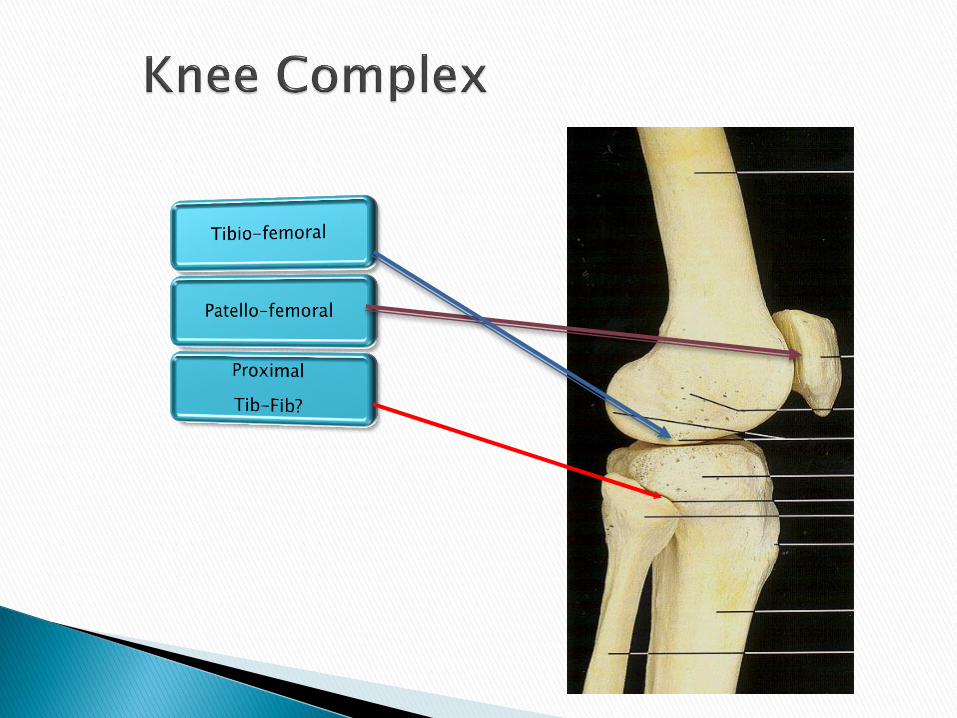
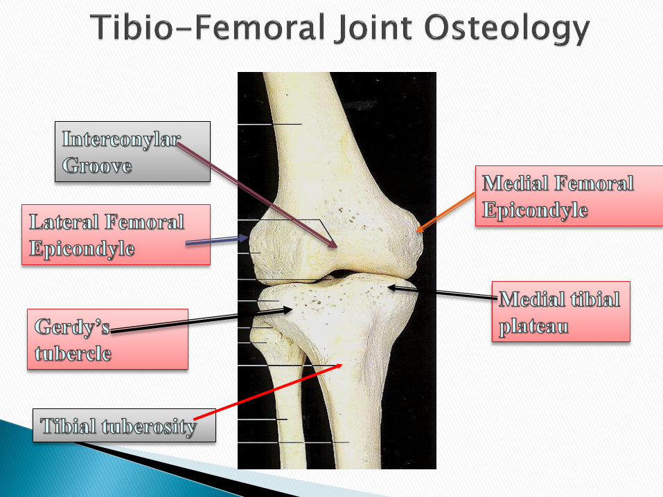
The knee is a mechanism of three joints and four bones- the femur, tibia, patella and fibula.
The function of these joints is to allow certain movements, restrict others, and to provide load transfer through the lower limb.


Meniscii
• Glide anteriorly with extension
• Glide posteriorly with flexion
Motion
Absorb shock Distribute weight
bearing(WB) Provide stability Aid in lubrication

Medial Collateral Ligament
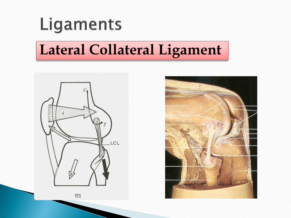
Lateral Collateral Ligament


Direction of Force ◦ Varus/LCL
◦ Valgus/MCL
◦ Anterior/ACL
◦ Posterior/PCL


Semitendinosis
Semimembranosis
Biceps Femoris
Gastrocnemius

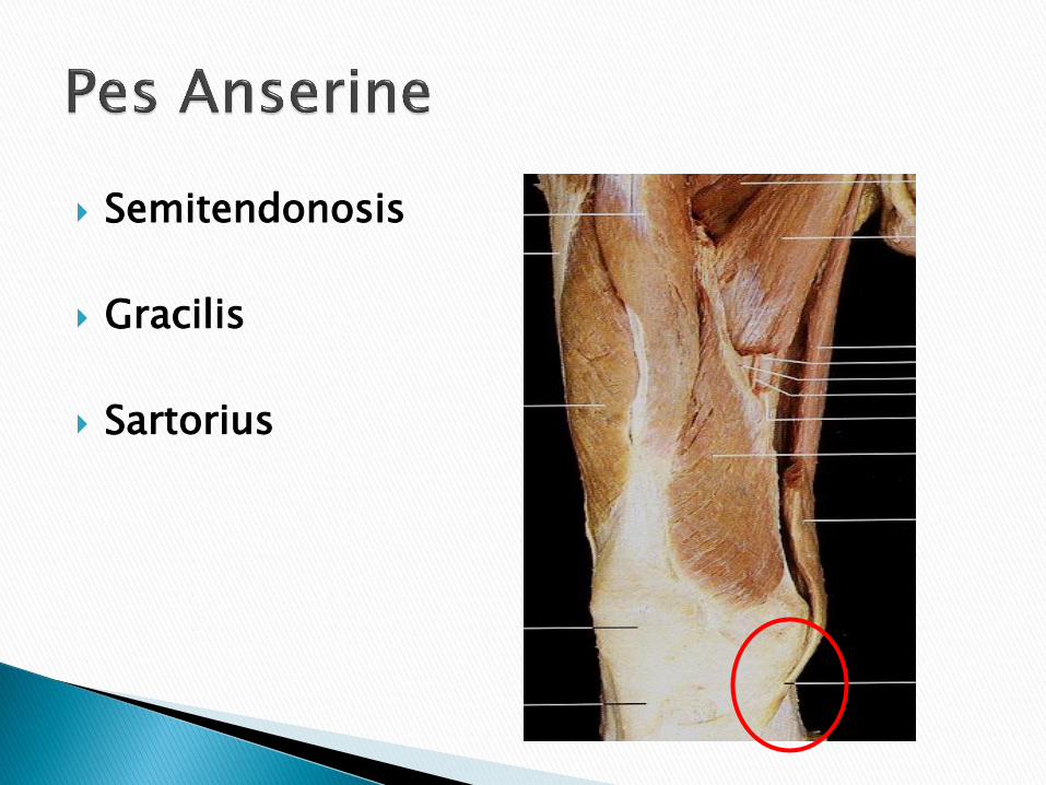
Semitendonosis
Gracilis
Sartorius

Plantaris
Popliteus

Tibia Fibula
Distal Tib-fib joint
Talus Ankle
Mortise
Calcaneus Subtalar Joint

Talus Calcaneus
Navicular Cuboid
Cuneiforms
1, 2, 3
Metatarsals
Proximal Phalanges
Middle Phalanges
Distal Phalanges

Rearfoot
Forefoot
Talonavicular
Joint
Calcaneocuboid
Joint

Tibonavicular
(Deltoid)
Tibocalcaneal
(Deltoid) Posterior
(Deltoid)
Long Plantar Plantar calcaneonavicular
(Spring Ligament)

Ant Tibiofibular Ant Talofibular
Calcaneofibular Long Plantar
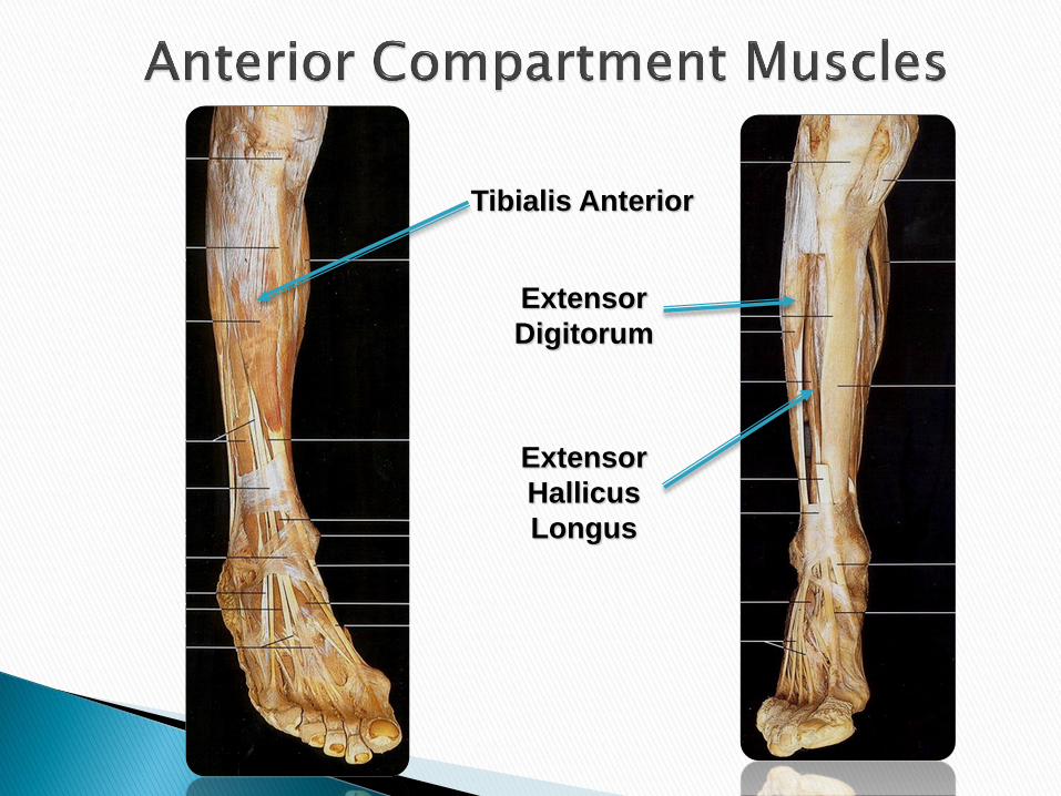
Tibialis Anterior
Extensor
Digitorum
Extensor
Hallicus
Longus

Gastrocnemius
: Medial and
Lateral heads
Achilles
Tendon
Plantaris
Soleus

Flexor
Digitorum Tibialis
Posterior
Flexor
Digitorum
Tendon
Tibialis
Posterior
Tendon
Flexor
Hallicus
Longus

Peroneus L
Peroneus B
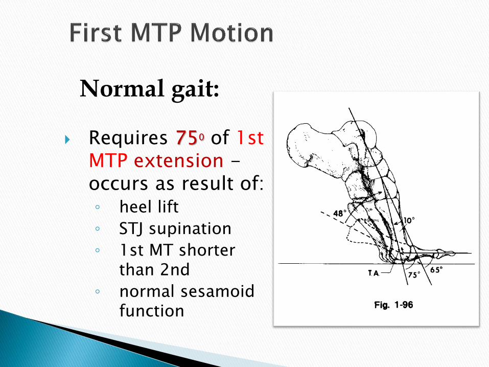
Requires 750 of 1st MTP extension - occurs as result of: ◦ heel lift
◦ STJ supination
◦ 1st MT shorter than 2nd
◦ normal sesamoid function
Normal gait:


Rectus Femoris, Hamstrings.
Gastrocnemius: Knee and Ankle.

Sciatic N.
Tibial N.
Common Peroneal N.

1. Knee Flexion and Extension
2. Ankle Planter Flexion
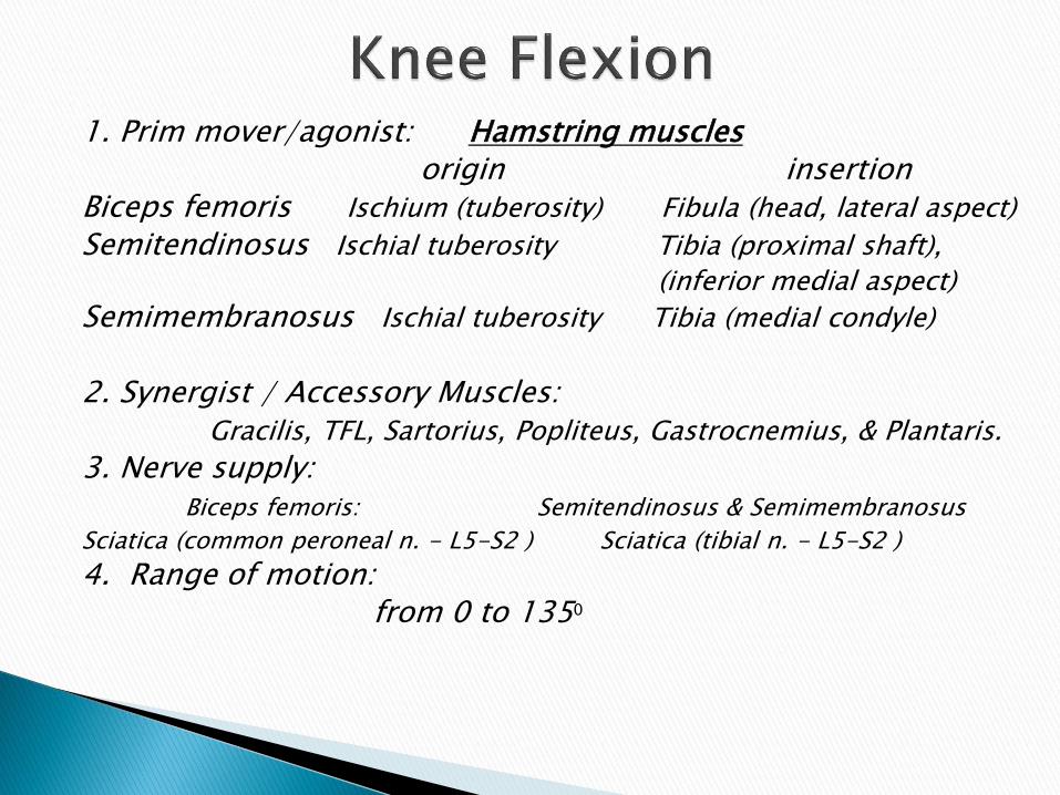
1. Prim mover/agonist: Hamstring muscles origin insertion Biceps femoris Ischium (tuberosity) Fibula (head, lateral aspect)
Semitendinosus Ischial tuberosity Tibia (proximal shaft),
(inferior medial aspect)
Semimembranosus Ischial tuberosity Tibia (medial condyle)
2. Synergist / Accessory Muscles: Gracilis, TFL, Sartorius, Popliteus, Gastrocnemius, & Plantaris. 3. Nerve supply: Biceps femoris: Semitendinosus & Semimembranosus
Sciatica (common peroneal n. - L5-S2 ) Sciatica (tibial n. - L5-S2 )
4. Range of motion: from 0 to 1350


Semitendinosis
Semimembranosis
Biceps Femoris
Gastrocnemius
5. Fixation: a. Contraction of iliocostalis lumborum and quadratus lumborum
muscles. b. Weight of thigh and pelvis. 6. Effect of weakness and contracture: Effect of weakness and contracture:-Video? weakness of both med & lat hamstring causes knee hyperextension. weakness of lat. Hamstring causes loss of lateral knee stability. weakness of med. Hamstring decrease medial knee stability. effect of contracture: knee flexion deformity accompanied by
posterior tilting of the pelvis and flattening of the lumbar curve? Why? - restriction of knee extension when the hip is flexed or restriction
of the hip flexion when the knee is extended. Why? 7. Factor Limiting of motion:
a. Contact or approximation between calf muscles with posterior thigh muscles.
b. Tension of quadriceps muscles. 8. Substitution: 1) hip flexion. 2) sartorius 3) grcilis
9. Procedures: a- patient position (pt):
b- Therapist Position:
inner hand:
Outer hand: Direction of Resistance :
Instruction to patient:
c- grading system:
Normal(5), Good(4), Fair(3), Poor(2), Trace(1), Zero(0)
make sure patient tolerates maximal resistance pluse hold 3 sec.
e. Palpation site:
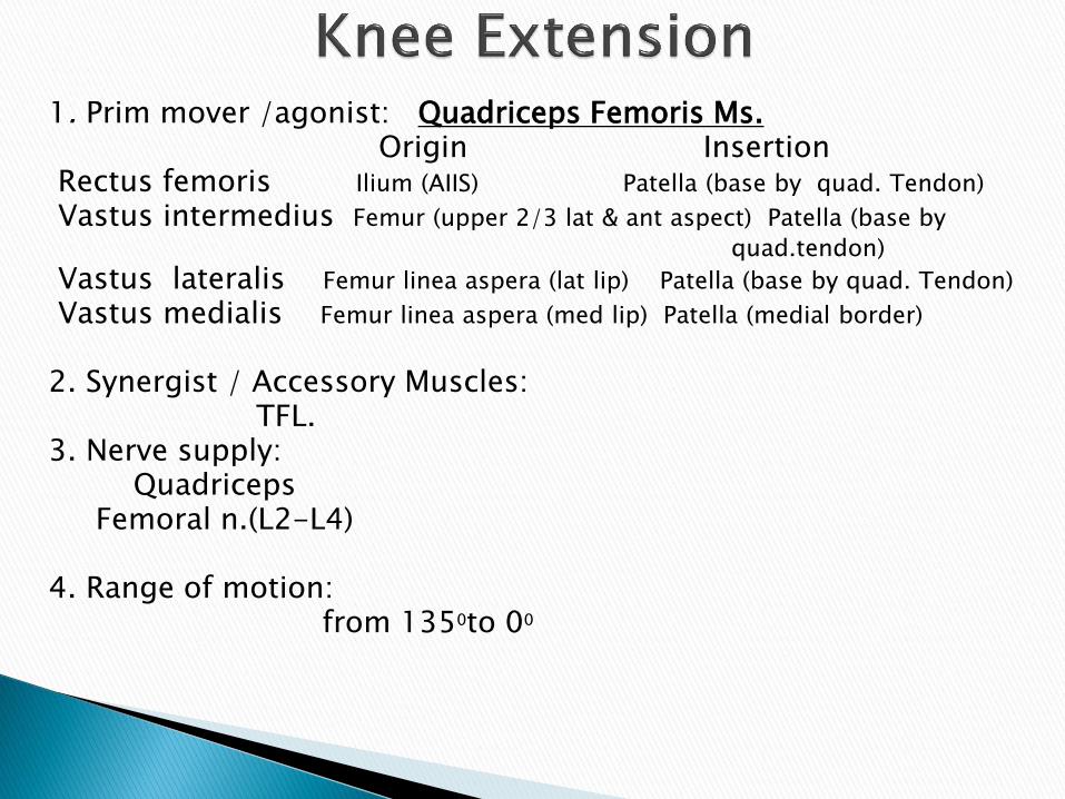
1. Prim mover /agonist: Quadriceps Femoris Ms. Origin Insertion Rectus femoris Ilium (AIIS) Patella (base by quad. Tendon)
Vastus intermedius Femur (upper 2/3 lat & ant aspect) Patella (base by
quad.tendon)
Vastus lateralis Femur linea aspera (lat lip) Patella (base by quad. Tendon)
Vastus medialis Femur linea aspera (med lip) Patella (medial border)
2. Synergist / Accessory Muscles: TFL. 3. Nerve supply: Quadriceps Femoral n.(L2-L4) 4. Range of motion: from 1350to 00


5. Fixation: a. Contraction of antero-lateral abdominal muscles to fix origin
of rectus femoris. b. Weight of thigh and pelvis.
6. Effect of weakness and contracture: Effect of weakness and contracture:-Video? Difficulty in getting up & down from sitting position as well as
on going up & down stairs. - knee hyperextension. effect of shortness: shortness of the Rectus femoris will results
in a restriction of the knee flexion when the hip is extended or a restriction of the hip extension when the knee is flexed.
effect of contracture: restriction of knee flexion. 7. Factor Limiting of motion: a. Tension of oblique popliteal, cruciate and collateral ligaments
of knee joint. b. Tension of knee flexor muscles. 8. Substitution: when pt in side-lying (grade 2) pt may use hip internal rotators.

9. Procedures: a- patient position (pt):
b- Therapist Position:
inner hand:
Outer hand: Direction of Resistance :
Instruction to patient:
c- grading system:
Normal(5), Good(4), Fair(3), Poor(2), Trace(1), Zero(0)
make sure patient tolerates maximal resistance plus hold 3 sec.
e. Palpation site:

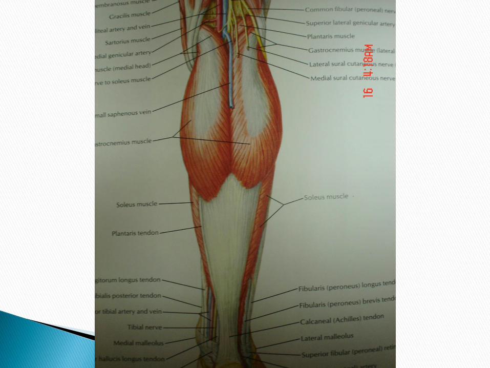
1. Prim mover /agonist:
Origin Insertion
Gastrocnemius
Medial head Femur (medial condyle) Tendo Calcaneus-calcaneu (posterior)
Lateral head Femur (lateral condyle) Tendo Calcaneus …..
Soleus Fibula (posterior) Tendo Calcaneus-calcaneus (posterior)
2. Synergist / Accessory Muscles:
Tibialis posterior, plantaris, peroneus longus & brevis, Flexor digitorum &
hallucis longus.
3. Nerve supply:
Gastrocnemius & Soleus: Tibia n.
4. Range of motion:
from 00 to 450


5. Fixation: By Weight of thigh.
6. Effect of weakness and contracture: Effect of weakness and contracture:-Video? Result in an hyperextension of the knee(why?) as well as in a non-weight
bearing position as in standing. During walking the inability to rise on toes.
effect of contracture: result in an equinus position of the foot and flexion of the knee. - also a restriction of the ankle dorsiflexion when the knee is extended and a restriction of the knee extension when the ankle is
dorsiflexed.
7. Factor Limiting of motion: a. Tension of anterior talofibular ligament and anterior fibers of deltoid
ligaments. b. Tension of dorsiflexor muscles. c. Contact of posterior portion of talus with tibia.
8. Substitution: by 1) Flexor hallucis longus and flexor digitorum longus 2) Peroneus longus and brevis. 3) Tibialis posterior.

9. Procedures: WB test and Non WB test
a- patient position (pt):
b- Therapist Position:
inner hand:
Outer hand: Direction of Resistance :
Instruction to patient:
c- grading system:
Normal(5), Good(4), Fair(3), Poor(2), Trace(1), Zero(0)
make sure patient tolerates maximal resistance plus hold 3 sec.
e. Palpation site:

Thank You