rida gene bacterial stool panel
TRANSCRIPT

RIDA®GENE Bacterial Stool Panel
real-time PCR
Art. No.: PG2405
100 reactions
For in vitro diagnostic use.
-20 °C
R-Biopharm AG, An der neuen Bergstraße 17, D-64297 Darmstadt, Germany
Tel.: +49 (0) 61 51 81 02-0 / Telefax: +49 (0) 61 51 81 02-20

2 RIDAGENE Bacterial Stool Panel 12-11-28
1. Intended use
For in vitro diagnostic use. RIDA®GENE Bacterial Stool Panel is a multiplex real-time
PCR for the direct, qualitative detection and differentiation of Salmonella spp.,
Campylobacter spp. and Yersinia enterocolitica in human stool samples.
RIDA®GENE Bacterial Stool Panel multiplex real-time PCR is intended for use as an
aid in diagnosis of gastrointestinal infections caused by bacteria.
2. Explanation of the test
Diarrheal disease is a major health care problem and causes about 2 billion cases
worldwide. The World Health Organization (WHO) ranks diarrheal disease as 2nd
most common cause of child deaths among children under 5 years globally,
particularly in developing countries. About 1.9 million children younger than 5 years
of age perish from diarrhea each year, more than AIDS, malaria and measles
combined.1,2 Common causes of bacterial diarrheal disease are Campylobacter spp.,
Salmonella spp. and Y. enterocolitica.
Campylobacter species are one of the most common causes of bacterial diarrhea
worldwide, responsible for 400 million – 500 million cases annually. The disease
caused by the genus Campylobacter is called campylobacteriosis. More than 80% of
Campylobacter infections are caused by C. jejuni. The Centers for Disease Control
and Prevention (CDC) estimates more than 2 million cases of campylobacteriosis
each year in the US. The Foodborne Diseases Active Surveillance Network
(FoodNet) reported an incidence rate of 13 cases per 100,000 population in 2008.
C. jejuni was detected in 5 - 16% of children with diarrhea in developed countries and
in 8 - 45% of children with diarrhea in developing countries.4 Approximately 100
persons with Campylobacter infections die each year in the US.3,4 Infection with
Campylobacter occurs through contaminated food, especially poultry, water, contact
with infected animals or by fecal-oral route, particularly in children. The infectious
dose is with 500 bacteria relatively low. After an incubation period of 2 to 5 days
people with campylobacteriosis get fever, diarrhea, abdominal cramps, vomiting,
abdominal pain and nausea. Potential long-term complications are autoimmune
disorders, for example the Guillain-Barré syndrome (GBS).4
Salmonella species are also a leading cause of bacterial gastroenteritis worldwide.
The genus Salmonella is divided into two species, S. enterica and S. bongori. So far,
more than 2,500 Salmonella serotpyes are described which are pathogenic for
humans. Salmonella species are causing nontyphoidal salmonellosis or typhoid
fever. It is estimated that 93.8 million cases of nontyphoidal salmonellosis infections
with 155,000 deaths occuring globally each year.6 The CDC estimates more than
1.2 million cased of nontyphoidal salmonellosis infections each year in the United

RIDAGENE Bacterial Stool Panel 12-11-28 3
States, with more than 23,000 hospitalizations and 450 deaths.5 Most of the
nontyphoidal salmonellosis infections are caused by the S. typhimurium and
S. enteritidis, while typhoid fever is caused by S. typhi and S. paratyphi A, B or C.
The CDC estimates more than 1,800 cases of typhoid fever annually in the U.S.
Transmission of Salmonella occurs through contaminated food, water or contact with
infected animals. The infectious dose of Salmonella species is varying from 1 to 1000
bacteria. Nontyphoidal salmonellosis infection occurs after an incubation period of 6
- 72h with clinical symptoms of nausea, vomiting, abdominal cramps, diarrhea, fever
and headache. People with typhoid fever get headache, achiness, high fever (from
39 °C to 41 °C), gastrointestinal symptoms, including abdominal pains and diarrhea
within 1 to 3 weeks after exposure to the organism.3,7
Yersinia enteroclitica is one of three Yersinia species (Y. pestis,
Y. pseudotuberculosis) of the genus Yersinia that are pathogenic for humans and
cause of the gastrointestinal disease called Yersiniosis. According to FoodNet an
incidence rate of 1 Y. enterocolitica infection per 100,000 persons occurs each year
in the U.S. The European Centre for Disease Prevention and Control reported 8,874
cases in 2007, of which about 5000 cases were from Germany. Infection with
Yersiniosis occurs after ingestion of contaminated food or water. The estimated
infectious dose is between 104 to 106 bacteria. After an incubation period of 1 to 11
days people with yersiniosis get diarrhea, vomiting and abdominal pain.
Y. enterocolitica has also been associated with reactive arthritis 3,8
Culture is the classical method and for establishing the laboratory diagnosis of
bacterial diarrhea, but requires several days.
RIDA®GENE Bacterial Stool Panel multiplex real-time PCR is a new and attractive
alternative method for testing stool samples and has proven to be highly sensitive
and specific for the simultaneous detection of three of the most important diarrhea
causing bacteria (Campylobacter spp., Salmonella spp. and Y. enterocolitica).
3. Test principle
The RIDA®GENE Bacterial Stool Panel assay is a multiplex real-time PCR for the
direct, qualitative detection and differentiation of Salmonella spp., Campylobacter
spp. and Yersinia enterocolitica. After DNA-isolation, amplification of the gene
fragments specific for Salmonella spp. (ttr), Campylobacter spp. (16s-rDNA) and
Y. enterocolitica (ail) occurs, if present. The amplified targets are detected with
hydrolysis probes, which are labeled at one end with a quencher and at the other end
with a fluorescent reporter dye (fluorophore). In the presence of a target the probes
hybridize to the amplicons. During the extension step the Taq-polymerase breaks the
reporter-quencher proximity.

4 RIDAGENE Bacterial Stool Panel 12-11-28
The reporter emits a fluorescent signal which is detected by the optical unit of a real-
time PCR instrument. The fluorescence signal increases with the amount of formed
amplicons. The RIDA®GENE Bacterial Stool Panel assay contains an Internal Control
DNA (ICD) as an internal control of sample preparation procedure and to determine
possible PCR-inhibition.
4. Reagents provided
Tab.1: Reagents provided (Reagents provided in the kit are sufficient for 100 determinations)
Kit Code Reagent Amount Lid Color
1 Reaction Mix 2x 1100 µl yellow
2 Taq-Polymerase 1x 11 µl red
D Internal Control DNA 2x 1800 µl orange
N PCR Water 1x 500 µl white
P Positive Control 1x 200 µl blue
5. Storage instructions
- Protect all reagents from light and store at -20 °C. All reagents can be used until
the expiration date. After expiry the quality guarantee is no longer valid.
- Carefully thaw reagents before using (e.g. in a refrigerator at 2 - 8 °C).
- Reagents can sustain up to 5 freeze/thaw cycles without influencing the assay
performance (e.g. after the first thawing separate it in aliquots and freeze
immediately).
- During PCR preparation all the reagents should be stored cold in an appropriate
way (2 - 8 °C).

RIDAGENE Bacterial Stool Panel 12-11-28 5
6. Additional equipment and materials required
- DNA-Extraction kit (e.g. RTP® Pathogen Kit, STRATEC Molecular)
or
DNA-Extraction system (e.g. Maxwell® 16 (Promega), NucliSENS easy®MAG™
(bioMérieux))
- Real-time PCR instrument:
Roche: LightCycler 480II
- Cepheid: SmartCycler
Applied Biosystems: ABI 7500
Abbott: m2000rt
Stratagene: Mx3000P, Mx3005P
QIAGEN: Rotor-Gene Q
- RIDA®GENE Color Compensation I (PG0001) for run the LightCycler 480II
- Real-time PCR consumables (plates, tubes, foil)
- Centrifuge with a rotor for the reaction vials
- Vortexer
- Pipettes (0.5 – 20 µl, 20 – 200 µl, 100 – 1000 µl)
- Filter tips
- Powder-free disposal gloves
7. Precautions for users
- For in vitro diagnostic use only.
- DNA isolation, PCR preparation and the PCR run should be separated in different
rooms to avoid cross-contaminations.
- This test must only be performed by laboratory personnel trained in molecular
biology methods.
- Strictly follow the working instructions.
- When handling samples, wear disposable gloves. After finishing the test, wash
your hands.
- Do not smoke, eat or drink in areas where samples or test reagents are being
used.
- Samples must be treated as potentially infectious as well as all reagents and
materials being exposed to the samples and have to be handled according to the
national safety regulations.
- Do not use the kit after the expiration date.

6 RIDAGENE Bacterial Stool Panel 12-11-28
8. Test procedure
8.1 DNA-Extraction
Use for DNA-extraction of human stool samples a commercially available DNA
isolation kit (e.g. RTP® Pathogen Kit (STRATEC Molecular)) or DNA extraction
system (e.g. Maxwell® 16 (Promega)). Extract DNA according to the manufacturer’s
instructions.
We recommend to dilute the stool samples before extraction 1:3 with water. Vortex
the diluted stool sample intensely and centrifuge at 3,000 rpm for 30 sec. Use from
the supernatant the appropriate volume according to the manufacturer’s instruction.
The RIDA®GENE Bacterial Stool Panel assay contains an Internal Control DNA
(ICD), which can either be used as PCR inhibition control or as extraction control for
the sample preparation procedure and as a PCR inhibition control.
If the ICD is used only as a PCR inhibition control, 1µl of the ICD should be added to
the Master-Mix (see Tab.3).
If the ICD is used as a extraction control for the sample preparation procedure and
as PCR inhibition control, 20 µl of the ICD has to be added during extraction
procedure. The ICD should always be added to the specimen-lysis buffer mixture and
must not be added directly to the specimen. We also recommend to add 1 µl of the
ICD to the negative and positive control PCR-Mix.
8.2 Master-Mix preparation
Calculate the total number of PCR reactions (sample and control reactions) needed.
One positive control and negative control must be included in each assay run.
We recommend to calculate an additional volume of 10 % to compensate imprecise
pipetting (see Tab.2, Tab.3). Thaw, mix gently and centrifuge briefly the Reaction
Mix, the Taq-Polymerase, the Positive Control, the PCR Water and the ICD before
using. Keep reagents appropriately cold during working step (2 - 8 °C).
Tab.2: Calculation and pipetting example for 10 reactions of the Master-Mix
(ICD as extraction and PCR inhibition control)
Kit code Master-Mix components Volume per reaction 10 reactions (10 % extra)
1 Reaction Mix 19.9 µl 218.9 µl
2 Taq-Polymerase 0.1 µl 1.1 µl
Total 20.0 µl 220 µl
Mix the components of the Master-Mix gently and briefly spin down.

RIDAGENE Bacterial Stool Panel 12-11-28 7
Tab.3: Calculation and pipetting example for 10 reactions of the Master-Mix
(ICD only as PCR inhibition control)
Kit Code Master-Mix components Volume per reaction 10 reactions
(10 % extra)
1 Reaction Mix 19.9 µl 218.9 µl
2 Taq-Polymerase 0.1 µl 1.1 µl
D Internal Control DNA 1.0 µl 11 µl
Total 21.0 µl 231.0 µl
Mix the components of the Master-Mix gently and briefly spin down.
8.3 Preparation of the PCR-Mix
Pipette 20 µl of the Master-Mix in each reaction vial (tube or plate).
Negative control: Add 5 µl PCR Water as negative control to the pre-pipetted
Master-Mix.
Note: If the ICD is used as extraction control for the sample preparation procedure and as
PCR inhibition control, we recommend to add 1 µl of the ICD to the negative control PCR
Mix.
Sample: Add 5 µl DNA-Extract to the pre-pipetted Master-Mix.
Positive control: Add 5 µl Positive Control to the pre-pipetted Master-Mix.
Note: If the ICD is used as extraction control for the sample preparation procedure and as
PCR inhibition control, we recommend to add 1 µl of the ICD to the positive control PCR Mix.
Cover tubes or plate. Spin down and place in the real-time PCR instrument. The
PCR reaction should be started according to the PCR instrument Set-up (see Tab.4,
Tab. 5).

8 RIDAGENE Bacterial Stool Panel 12-11-28
8.4 PCR Instrument Set-up
Tab.4: Real-time PCR profile for LightCyler 480II, SmartCycler and Rotor-Gene Q
Initial Denaturation
Cycles
PCR Denaturation
Annealing/Extension
1 min, 95 °C
45 Cycles
10 sec, 95 °C
15 sec, 60 °C
Temperature Transition Rate / Ramp Rate
Maximum
Note: Annealing and Extension occur in the same step.
Tab.5: Real-time PCR profile for Mx3000P, Mx3005P, ABI 7500 and m2000rt
Initial Denaturation
Cycles
PCR Denaturation
Annealing/Extension
1 min, 95 °C
45 Cycles
10 sec, 95 °C
30 sec, 60 °C
Temperature Transition Rate / Ramp Rate
Maximum
Note: Annealing and Extension occur in the same step.

RIDAGENE Bacterial Stool Panel 12-11-28 9
8.5. Detection Channel Set-up
Tab.6: Selection of appropriate detection channels
Real-time PCR instrument
Detection Detection Channel
Dark-Quencher
Note
Roche
LightCycler 480II
Salmonella spp. 465/510 + RIDAGENE Color
Compensation Kit I (PG0001)
is required
ICD 533/580 +
Yersinia enterocolitica 533/610 +
Campylobacter spp. 618/660 +
Cepheid
SmartCycler
Salmonella spp. Channel 1 +
- ICD Channel 2 +
Yersinia enterocolitica Channel 3 +
Campylobacter spp. Channel 4 +
ABI 7500
Salmonella spp. FAM none Check that passive
reference option ROX is none
ICD VIC none
Yersinia enterocolitica ROX none
Campylobacter spp. Cy5 none
Abbott m2000rt
Salmonella spp. FAM none
- ICD VIC none
Yersinia enterocolitica ROX none
Campylobacter spp. Cy5 none
Stratagene Mx3000P/ Mx3005P
Salmonella spp. FAM +
Check that reference dye is none
ICD HEX +
Yersinia enterocolitica ROX +
Campylobacter spp. Cy5 +
Qiagen Rotor-Gene Q
Salmonella spp. Green +
- ICD Yellow +
Yersinia enterocolitica Orange +
Campylobacter spp. Red +

10 RIDAGENE Bacterial Stool Panel 12-11-28
9. Result interpretation
The analysis of the samples is done by the software of the used real-time PCR
instrument according to the manufacturer’s instructions. Positive and negative
controls have to show correct results (see Fig.1, Fig.2, Fig.3).
The positive control has a concentration of 103 copies / µl. In each PCR run it is used
in a total amount of 5 x 10³ copies.
Fig.1: Correct run of the positive and negative control (Salmonella spp.) on the
LightCycler 480II

RIDAGENE Bacterial Stool Panel 12-11-28 11
Fig.2: Correct run of the positive and negative control (Yersinia enterocolitica) on the
LightCycler 480II
Fig.3: Correct run of the positive and negative control (Campylobacter spp.) on the
LightCycler 480II

12 RIDAGENE Bacterial Stool Panel 12-11-28
The result interpretation is done according to Table 7.
Tab.7: Sample interpretation
Target genes
Salmonella spp.
Yersinia enterocolitica
Campylobacter spp.
ICD Result
positive negative negative positive/negative Salmonella spp.
negative positive negative positive/negative Yersinia enterocolitica
negative negative positive positive/negative Campylobacter spp.
negative negative negative positive Negative
(Target genes are not detectable)
negative negative negative negative Not evaluable
A sample is evaluated negative, if the sample shows no amplification signal in the
detection system, but the Internal Control DNA (ICD) is positive. An inhibition of the
PCR reaction or a failure in the extraction procedure can be excluded by the
detection of the Internal Control DNA (ICD).
A sample is evaluated positive, if both, the sample and the Internal Control DNA,
(ICD) show an amplification signal in the detection system.
A sample is evaluated positive, if the sample shows an amplification signal in the
detection system, but the Internal Control DNA (ICD) is negative. The detection of the
internal amplification control is not necessary, because high concentrations of the
amplicon can cause a weak or absent signal of the internal amplification control.
A sample is evaluated invalid, if both, the sample and the Internal Control DNA (ICD)
show no amplification signal in the detection system. The sample contained a
PCR inhibitor or a failure occurred in the extraction procedure. The extracted sample
needs to be further diluted with PCR water (1:10) and re-amplified, or the isolation
and purification of the sample has to be improved.

RIDAGENE Bacterial Stool Panel 12-11-28 13
10. Performance characteristics
10.1 Clinical Efficiency
In a prospective study at the Medical Laboratory of Professor
Schenk / Ansorge & Colleagues in Magdeburg (Germany), stool samples from
symptomatic patients were tested using the RIDA®GENE Bacterial Stool Panel
multiplex real-time PCR and the results were compared with those obtained by
culture followed by MALDI-TOF analysis. The results are summarized in the following
tables:
Tab.8: Correlation of the Salmonella spp. results with the RIDA®GENE Bacterial Stool Panel
multiplex real-time PCR and culture
Culture
positive negative
RIDA®GENE
Bacterial Stool Panel
positive 44 0
negative 0 250
Tab.9: Correlation of the Y. enterocolitica results with the RIDA®GENE Bacterial Stool Panel
multiplex real-time PCR and culture
Culture
positive negative
RIDA®GENE
Bacterial Stool Panel
positive 11 0
negative 0 283
Tab.10: Correlation of the Campylobacter spp. results with the RIDA®GENE Bacterial
Stool Panel multiplex real-time PCR and culture
Culture
positive negative
RIDA®GENE
Bacterial Stool Panel
positive 51 3
negative 0 240
Sensitivity 100.0 %
Specificity 100.0 %
PPV 100.0 %
NPV 100.0 %
Sensitivity 100.0 %
Specificity 100.0 %
PPV 100.0 %
NPV 100.0 %
Sensitivity 100.0 %
Specificity 98.4 %
PPV 94.4 %
NPV 100.0 %

14 RIDAGENE Bacterial Stool Panel 12-11-28
10.2 Analytical sensitivity
The RIDAGENE Bacterial Stool Panel multiplex real-time PCR has a detection limit
of 5 DNA copies per reaction for Salmonella spp., Yersinia enterocolitica and
Campylobacter spp. (see Fig.4, Fig.5, Fig.6).
Fig.4: Dilution series Salmonella spp. (105 – 101 DNA copies per µl) on the
LightCycler 480II
Fig.5: Dilution series Yersinia enterocolitica (105 – 101 DNA copies per µl) on the
LightCycler 480II

RIDAGENE Bacterial Stool Panel 12-11-28 15
Fig.6: Dilution series Campylobacter spp. (105 – 101 DNA copies per µl) on the
LightCycler 480II
The detection limit of the whole procedure depends on the sample matrix,
DNA-extraction and DNA-concentration.
16 RIDAGENE Bacterial Stool Panel 12-11-28
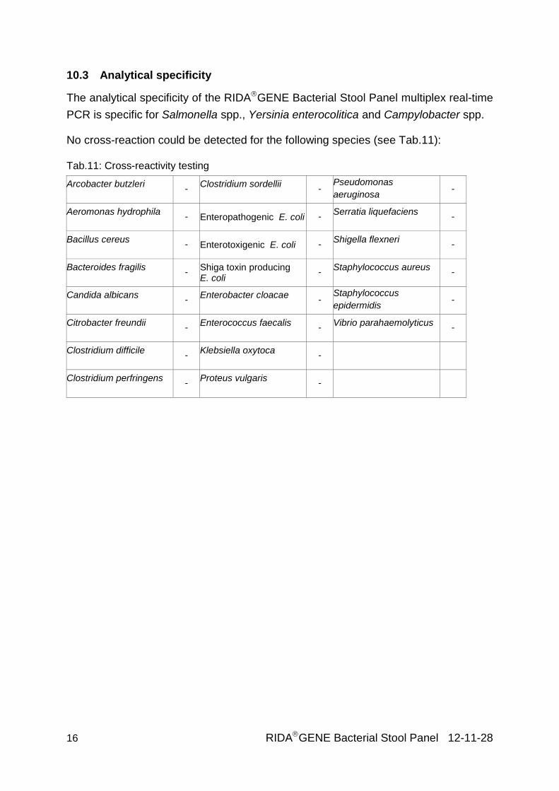
10.3 Analytical specificity
The analytical specificity of the RIDAGENE Bacterial Stool Panel multiplex real-time
PCR is specific for Salmonella spp., Yersinia enterocolitica and Campylobacter spp.
No cross-reaction could be detected for the following species (see Tab.11):
Tab.11: Cross-reactivity testing
Arcobacter butzleri -
Clostridium sordellii -
Pseudomonas
aeruginosa -
Aeromonas hydrophila - Enteropathogenic E. coli -
Serratia liquefaciens -
Bacillus cereus - Enterotoxigenic E. coli -
Shigella flexneri -
Bacteroides fragilis -
Shiga toxin producing E. coli
- Staphylococcus aureus
-
Candida albicans -
Enterobacter cloacae -
Staphylococcus
epidermidis -
Citrobacter freundii -
Enterococcus faecalis -
Vibrio parahaemolyticus -
Clostridium difficile -
Klebsiella oxytoca -
Clostridium perfringens -
Proteus vulgaris -

RIDAGENE Bacterial Stool Panel 12-11-28 17
10.4 Analytical reactivity
The reactivity of the RIDAGENE Bacterial Stool Panel multiplex real-time PCR was
evaluated against multiple Campylobacter species, Salmonella serotypes and
Yersinia enterocolitica (see Tab. 12). All Campylobacter species, Salmonella
serotypes and Yersinia enterocolitica of the panel were detected by the RIDAGENE
Bacterial Stool Panel multiplex real-time PCR.
Tab.12: Analytical reactivity testing
Campylobacter species
C. coli + C. jejuni + C. fetus +
C. lari + C. upsaliensis +
Salmonella serotypes
S. enteritidis + S. typhimurium + S. heidelberg +
S. brandenburg + S. agona + S. infantis +
S. kiel + S. paratyphi A + S. gloucester +
S. haifa + S. wilhelmsburg + S. essen +
S. virchow + S. montevideo + S. blegdam +
S. rostock + S. moscow + S. pullorum +
S. wernigerode + S. hadar + S. duesseldorf +
S. glostrup Serovar + S. poona + S. oranienburg +
S. senftenberg + S. bongori + S. bovismorbificans +
S. derby + S. anatum + S. newport +
S. goldcoast + S. dublin + S. livingston +
S. muenchen + S. kentucky + S. amsterdam +
S. berta + S. caracas + S. ealing +
S. augustenbourg +
Yersinia species
Y. enterocolitica
+

18 RIDAGENE Bacterial Stool Panel 12-11-28
11. Limitations of the method
1. The result of molecular analysis should not lead to the diagnosis, but always be
considered in the context of medical history and symptoms of the patient.
2. This assay is only validated for stool samples.
3. Inappropriate specimen collection, transport, storage and processing or a
pathogen load in the specimen below the analytical sensitivity can result in false
negative results.
4. The presence of PCR inhibitors may cause invalid results.
5. Mutations or polymorphisms in primer or probe binding regions may affect
detection of new variants resulting in a false negative result with the RIDAGENE
Bacterial Stool Panel assay.
6. As with all PCR based in vitro diagnostic tests, extremely low levels of target
below the limit of detection (LoD) may be detected, but results may not be
reproducible.
12. Literature
1. World Gastroenterology Organisation Global Guidelines: Acute diarrhea in adults
and children : a global perspective.
2. UNICEF/WHO, Diarrhoea: Why children are still dying and what can be done,
2009.
3. FDA 2012. Bad Bug Book 2nd Edition. Foodborne Pathogenic Microorganisms
and Natural Toxins Handbook.
4. Ruiz-Palacios GM. Clinical Infectious Diseases 2007; 44:701–703.
5. CDC. National Salmonella Surveillance Overview. Atlanta, Georgia: US
Department of Health and Human Services, CDC, 2011.
6. Majowicz SE et al. Clinical Infectious Diseases 2010; 50:882–889.
7. Pui CF et al. International Food Research Journal 2011; 18: 465-473.
8. Rosner BM et al. BMC Public Health 2010; 10:337.