robotics for surgery - annual reviews
TRANSCRIPT
Annu. Rev. Biomed. Eng. 1999. 01:211–240Copyright q 1999 by Annual Reviews. All rights reserved
1523–9829/99/0820–0211$08.00 211
Robotics for Surgery
Robert D. Howe and Yoky MatsuokaDivision of Engineering and Applied Sciences, Harvard University, Cambridge,Massachusetts 02138; e-mail: [email protected], [email protected]
Key Words robot, manipulator, minimally invasive surgery, image-guidedsurgery, registration
Abstract Robotic technology is enhancing surgery through improved precision,stability, and dexterity. In image-guided procedures, robots use magnetic resonanceand computed tomography image data to guide instruments to the treatment site. Thisrequires new algorithms and user interfaces for planning procedures; it also requiressensors for registering the patient’s anatomy with the preoperative image data. Min-imally invasive procedures use remotely controlled robots that allow the surgeon towork inside the patient’s body without making large incisions. Specialized mechanicaldesigns and sensing technologies are needed to maximize dexterity under these accessconstraints. Robots have applications in many surgical specialties. In neurosurgery,image-guided robots can biopsy brain lesions with minimal damage to adjacent tissue.In orthopedic surgery, robots are routinely used to shape the femur to precisely fitprosthetic hip joint replacements. Robotic systems are also under development forclosed-chest heart bypass, for microsurgical procedures in ophthalmology, and forsurgical training and simulation. Although results from initial clinical experience ispositive, issues of clinician acceptance, high capital costs, performance validation,and safety remain to be addressed.
CONTENTS
Introduction ..................................................................................... 212Robotic Techniques for Surgery .......................................................... 213
Minimally Invasive Procedures. . . . . . . . . . . . . . . . . . . . . . . . . . . . . . . . . . . . . . . . . . . . . . . . . . . . . . . . . . . . . 214Image-Based Procedures . . . . . . . . . . . . . . . . . . . . . . . . . . . . . . . . . . . . . . . . . . . . . . . . . . . . . . . . . . . . . . . . . . . . 214Interaction Modes . . . . . . . . . . . . . . . . . . . . . . . . . . . . . . . . . . . . . . . . . . . . . . . . . . . . . . . . . . . . . . . . . . . . . . . . . . . . 218Limitations of Robotic Surgery . . . . . . . . . . . . . . . . . . . . . . . . . . . . . . . . . . . . . . . . . . . . . . . . . . . . . . . . . . . . . 219
Surgical Applications......................................................................... 220Orthopedic Surgery . . . . . . . . . . . . . . . . . . . . . . . . . . . . . . . . . . . . . . . . . . . . . . . . . . . . . . . . . . . . . . . . . . . . . . . . . . 220Neurosurgery . . . . . . . . . . . . . . . . . . . . . . . . . . . . . . . . . . . . . . . . . . . . . . . . . . . . . . . . . . . . . . . . . . . . . . . . . . . . . . . . . . 226General and Thoracic Surgery . . . . . . . . . . . . . . . . . . . . . . . . . . . . . . . . . . . . . . . . . . . . . . . . . . . . . . . . . . . . . 227
Training and Simulation ..................................................................... 231Technical and Implementation Challenges ............................................. 232
Technical Issues . . . . . . . . . . . . . . . . . . . . . . . . . . . . . . . . . . . . . . . . . . . . . . . . . . . . . . . . . . . . . . . . . . . . . . . . . . . . . . 232Clinical Implementation and Acceptance Issues . . . . . . . . . . . . . . . . . . . . . . . . . . . . . . . . . . . . . . . . . 234
212 HOWE n MATSUOKA
INTRODUCTION
Over the past decade, robots have been appearing in the operating room. Robotictechnology is now regularly used to aim endoscopes in minimally invasive sur-gery and to guide instruments to tumors in brain surgery. The use of a robot toshape bones in hip replacement surgery was one of the groundbreaking applica-tions (2, 3, 36). Based on three-dimensional (3-D) computed tomography images,the surgeon plans the location of the prosthetic replacement joint within the femur.In surgery, the robot moves a high-speed cutting tool to form the precise shapespecified in the presurgical plan. The result is a far better fit between the boneand replacement joint than has been possible with conventional hand-held cuttinginstruments.
One reason surgical applications are progressing quickly is the large technol-ogy base that has been developed in robotics research in the past three decades(11, 37). Results in mechanical design, kinematics, control algorithms, and pro-gramming that were developed for industrial robots are directly applicable tomany surgical applications. Robotics researchers have also worked to enhancerobotic capabilities through adaptability (the use of sensory information torespond to changing conditions) and autonomy (the ability to carry out taskswithout human supervision). The resulting sensing and interpretation techniquesthat are proving useful in surgery include methods for image processing, spatialreasoning and planning, and real-time sensing and control.
To understand the advantages of using robots in surgery, it is helpful to con-sider the differences in human and machine characteristics (summarized inTable 1); many promising applications are based on unique robotic capabilities.One key difference is precision and accuracy, or more generally, the ability touse copious, detailed, quantitative information. The combination of 3-D imagingdata, computers, and intrasurgical sensors, for example, allows robots to accu-rately guide instruments to pathological structures deep within the body. Anotherimportant difference is that specialized manipulator designs allow robots to workthrough incisions that are much smaller than would be required for human handsor to work at small scales, where hand tremor poses fundamental limitations.
Humans are superior, however, at integrating diverse sources of information,using qualitative information, and exercising judgment. Humans have unexcelleddexterity and hand-eye coordination, as well as a finely developed sense of touch.Unlike interaction with robots, interaction with human members of a surgicalteam for instruction and explanation is straightforward. These differences in capa-bilities mean that current robots are restricted to simple procedures, and humansmust provide detailed commands, using preoperative planning systems or by pro-viding explicit move-by-move instructions. Even in the most sophisticated sys-tems, robots are specialized to specific tasks within procedures; humans mustprepare the patient, make many of the incisions and sutures, and perform manyother functions. Robotic systems are best described as ‘‘extending human capa-bilities’’ rather than ‘‘replacing human surgeons.’’
ROBOTICS FOR SURGERY 213
Humans Robots
Strengths Strengths
Strong hand-eye coordination Good geometric accuracy
Dexterous (at human scale) Stable and untiring
Flexible and adaptable Can be designed for a wide range of scales
Can integrate extensive and diverseinformation
May be sterilized
Able to use qualitative informationResistant to radiation and infection
Good judgmentCan use diverse sensors (chemical, force,
acoustic, etc.) in control
Easy to instruct and debrief
Limitations Limitations
Limited dexterity outside natural scale Poor judgment
Prone to tremor and fatigue Limited dexterity and hand-eyecoordinationLimited geometric accuracy
Limited to relatively simple proceduresLimited ability to use quantitativeinformation Expensive
Large operating room space requirement Technology in flux
Limited sterility Difficult to construct and debug
Susceptible to radiation and infection
aAdapted from Taylor & Stulberg (75).
In this article, we review enhancements and extensions of surgical practice byrobotic technology. The article is divided into two main parts. In the first part,we characterize the main technical approaches under development for roboticsurgery. In the second part, we describe specific surgical procedures where robotsare used, including orthopedic, general, thoracic, and neurosurgery. We concludewith a discussion of current research issues and promising areas for futureresearch.
ROBOTIC TECHNIQUES FOR SURGERY
Several trends in surgery are contributing to the growing acceptance of robots.Primary factors include the increasing emphasis on minimally invasive surgicaltechniques and the widespread availability of 3-D image data. Other robotic char-acteristics, particularly stability and the ability to work at small scales, providethe incentive for additional robotic applications.
214 HOWE n MATSUOKA
Minimally Invasive Procedures
Over the past decade, several surgical specialties have been rapidly transformedby minimally invasive surgery (also called minimal access surgery) (12). A cen-tral example is laparoscopic cholecystectomy, or gallbladder excision, a commonprocedure that is executed almost exclusively using minimally invasive surgerytechniques. Surgeons work through a set of three to five incisions approximately1 cm in size. Long-handled instruments are used to grip and cut tissue within thebody, and a video laparoscope provides a view of the internal operating field.Because this procedure avoids the long incision through the abdominal wall usedin the conventional open procedure, patients recover more quickly. Benefitsinclude greatly reduced discomfort, improved cosmesis, reduced convalescenceand hospitalization costs, and less time away from productive work. Minimallyinvasive approaches have produced the same benefits in a number of other pro-cedures, such as arthroscopic knee reconstruction and thoracoscopic lungresection.
The necessity of working through a few fixed incisions places severe limita-tions on dexterity in manipulation, and only a few procedures are possible withthe current hand-held instruments. Lateral movement of the instrument shaft isnot possible at the incision, which thus acts as a fulcrum, reversing the directionsof the surgeon’s hand motions at the instrument tip and varying the mechanicaladvantage as the instruments move in and out. The video monitor is often locatedon the far side of the patient, and the difference in orientation between the endo-scope and the monitor requires the surgeon to perform a difficult mental trans-formation between visual and motor coordinate frames (76). Contact forceperception is impaired by friction and varying mechanical advantage at the inci-sion, and distributed tactile information is absent (34).
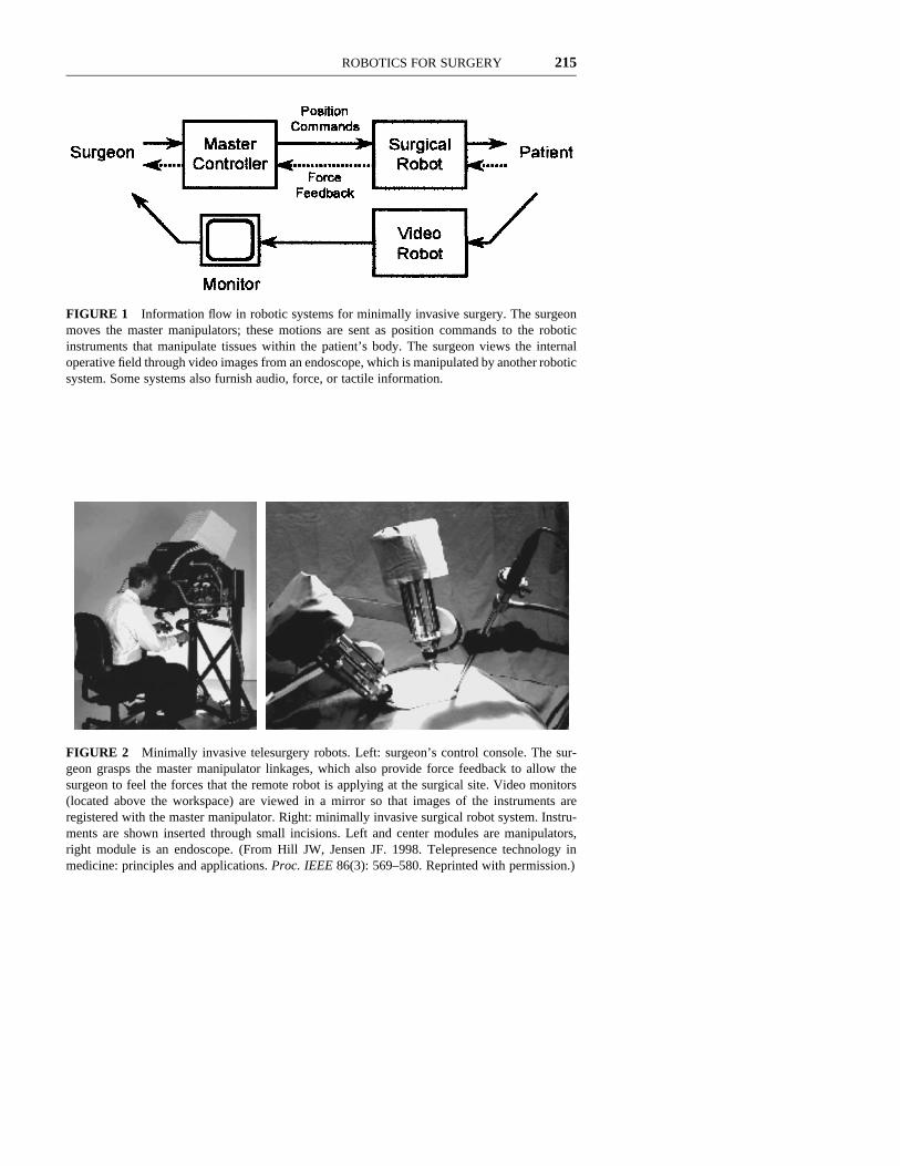
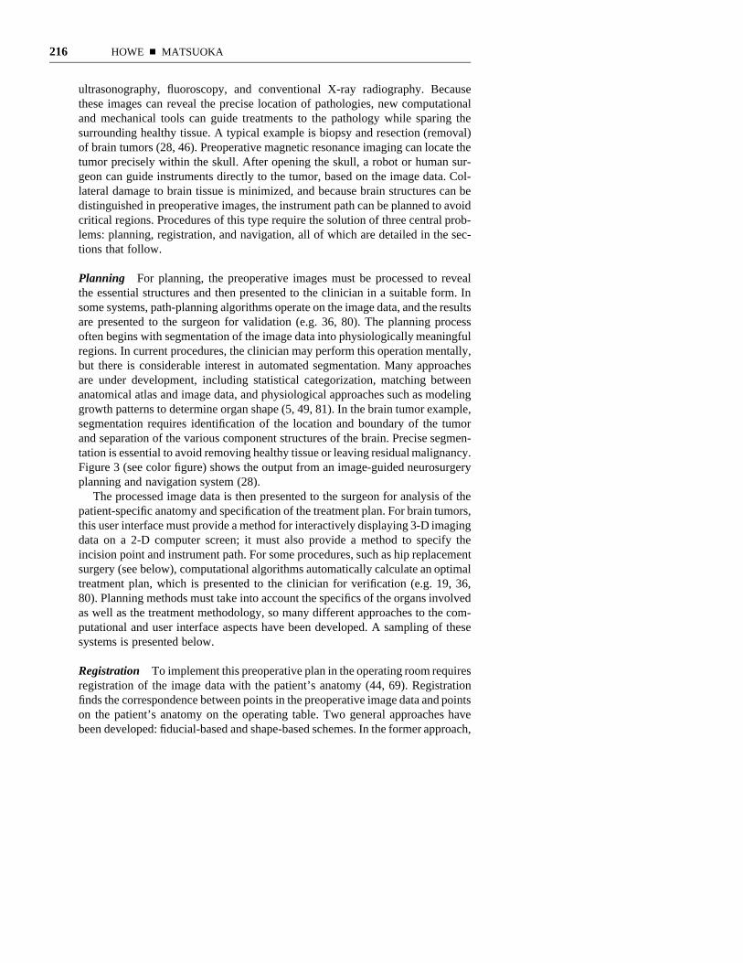
Robotic manipulators promise to solve many of these problems. The challengeis to design devices with good dexterity and intuitive control that can be insertedthrough small incisions. One focus is the development of general purpose systemsthat can execute a range of procedures in general, thoracic, and gynecologicalsurgery (9, 31, 47). These systems are often configured so that the surgeon sitsat a console in the operating room and uses a master control manipulator thatsends commands to the robots performing the surgical procedure (Figures 1 and2). Video images, and sometimes force sensations, are reproduced at the surgeon’sconsole. Other systems under development are aimed at specific access modali-ties, such as percutaneous needle puncture and transurethral prostate resection.There are also systems that take advantage of robotic ability to perform stableand untiring holding tasks, such as endoscope pointing and organ retraction, andto work at microscopic scales.
Image-Based Procedures
Another catalyst for robotic surgery applications has been the development ofnoninvasive imaging techniques, including 3-D modalities such as computedtomography (CT) and magnetic resonance imaging, and 2-D techniques such as
ROBOTICS FOR SURGERY 215
FIGURE 1 Information flow in robotic systems for minimally invasive surgery. The surgeonmoves the master manipulators; these motions are sent as position commands to the roboticinstruments that manipulate tissues within the patient’s body. The surgeon views the internaloperative field through video images from an endoscope, which is manipulated by another roboticsystem. Some systems also furnish audio, force, or tactile information.
FIGURE 2 Minimally invasive telesurgery robots. Left: surgeon’s control console. The sur-geon grasps the master manipulator linkages, which also provide force feedback to allow thesurgeon to feel the forces that the remote robot is applying at the surgical site. Video monitors(located above the workspace) are viewed in a mirror so that images of the instruments areregistered with the master manipulator. Right: minimally invasive surgical robot system. Instru-ments are shown inserted through small incisions. Left and center modules are manipulators,right module is an endoscope. (From Hill JW, Jensen JF. 1998. Telepresence technology inmedicine: principles and applications. Proc. IEEE 86(3): 569–580. Reprinted with permission.)
216 HOWE n MATSUOKA
ultrasonography, fluoroscopy, and conventional X-ray radiography. Becausethese images can reveal the precise location of pathologies, new computationaland mechanical tools can guide treatments to the pathology while sparing thesurrounding healthy tissue. A typical example is biopsy and resection (removal)of brain tumors (28, 46). Preoperative magnetic resonance imaging can locate thetumor precisely within the skull. After opening the skull, a robot or human sur-geon can guide instruments directly to the tumor, based on the image data. Col-lateral damage to brain tissue is minimized, and because brain structures can bedistinguished in preoperative images, the instrument path can be planned to avoidcritical regions. Procedures of this type require the solution of three central prob-lems: planning, registration, and navigation, all of which are detailed in the sec-tions that follow.
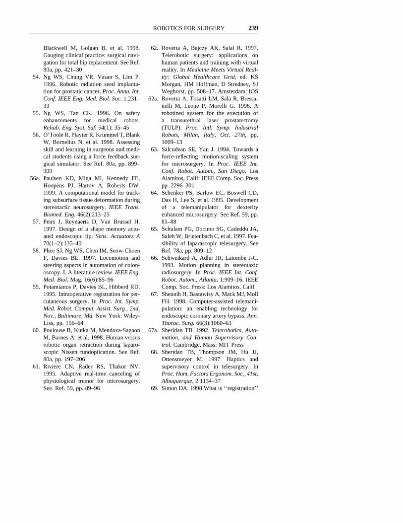
Planning For planning, the preoperative images must be processed to revealthe essential structures and then presented to the clinician in a suitable form. Insome systems, path-planning algorithms operate on the image data, and the resultsare presented to the surgeon for validation (e.g. 36, 80). The planning processoften begins with segmentation of the image data into physiologically meaningfulregions. In current procedures, the clinician may perform this operation mentally,but there is considerable interest in automated segmentation. Many approachesare under development, including statistical categorization, matching betweenanatomical atlas and image data, and physiological approaches such as modelinggrowth patterns to determine organ shape (5, 49, 81). In the brain tumor example,segmentation requires identification of the location and boundary of the tumorand separation of the various component structures of the brain. Precise segmen-tation is essential to avoid removing healthy tissue or leaving residual malignancy.Figure 3 (see color figure) shows the output from an image-guided neurosurgeryplanning and navigation system (28).
The processed image data is then presented to the surgeon for analysis of thepatient-specific anatomy and specification of the treatment plan. For brain tumors,this user interface must provide a method for interactively displaying 3-D imagingdata on a 2-D computer screen; it must also provide a method to specify theincision point and instrument path. For some procedures, such as hip replacementsurgery (see below), computational algorithms automatically calculate an optimaltreatment plan, which is presented to the clinician for verification (e.g. 19, 36,80). Planning methods must take into account the specifics of the organs involvedas well as the treatment methodology, so many different approaches to the com-putational and user interface aspects have been developed. A sampling of thesesystems is presented below.
Registration To implement this preoperative plan in the operating room requiresregistration of the image data with the patient’s anatomy (44, 69). Registrationfinds the correspondence between points in the preoperative image data and pointson the patient’s anatomy on the operating table. Two general approaches havebeen developed: fiducial-based and shape-based schemes. In the former approach,
ROBOTICS FOR SURGERY 217
fiducials, or markers, are attached to the pertinent anatomical structure prior toimaging. From the image data, the robot control computer knows the location ofthe pathology with respect to the fiducials. During surgery, the markers areexposed and a sensor system conveys their location to the computer. Many sens-ing systems can be used for determining fiducial location. The most direct is aprobe attached to the robot manipulator itself, so that when the robot contacts afiducial, its location in the robot’s coordinate space is immediately determined.From contact with several fiducials, the complete spatial transformation betweenthe preoperative image and the patient can be found.
A number of sensing systems are used in surgery (51, 70). One of the mostcommon is the optical tracker. Light-emitting diodes or reflective targets areattached to a probe, and a set of cameras or optical sensors view the probe fromknown locations. Triangulation can then determine the location of each target inthe robot coordinate frame; submillimeter resolutions are readily achieved. Othersensing techniques include electromagnetic transceivers, articulated probe arms,and ultrasonic and laser range finders. Many of these tracking modalities areavailable as an integrated part of commercial image-guided treatment systems.
One problem with fiducial-based registration is that the attachment of themarkers, which must be carried out prior to imaging, can be a significant surgicalprocedure in itself. For example, the ROBODOC system for hip replacementsurgery uses fiducials that are pins implanted in the femur at both the proximaland the distal ends (see below). This adds time and cost to the robotic procedureand can cause significant discomfort for the patient.
The alternative approach, shape-based registration, avoids these problems byfitting the shape of anatomical structures from intraoperative measurements to thepreoperative image data. The patient measurements can be obtained from a varietyof sensing techniques, including tracing curves on the pertinent anatomical struc-ture with an optical tracker probe, scanning the surface with a laser range finder,or processing video images of the patient. The result is a description of the shapeof the anatomical structure in patient coordinates. A computational algorithm thenfinds the spatial transformation that minimizes the error between the intraopera-tively sensed shape and the shape that has been segmented from the preoperativeimage data.
There are many other variants on the registration problem. One potentiallyadvantageous approach uses readily obtained 2-D ultrasound or X-ray images asthe intraoperative sensing technique. The resulting ‘‘slices’’ or projections of theanatomy are then fitted to the 3-D preoperative image data (20, 77). A significantproblem in registration is correcting for motion of the patient or deformation oftissue during surgery. This is particularly important in neurosurgery, where swell-ing of the brain follows a craniotomy. Deformable template matching and bio-mechanical models that incorporate response to mechanical loading or the edemaprocess have been proposed as a way to deal with this problem (41, 56a). Othertracking approaches use video images to follow patient motion in real time (27).Verification of the accuracy of registration techniques has also become an impor-tant research question (17, 25). Because it sets fundamental accuracy limits, reg-
218 HOWE n MATSUOKA
istration is important for all areas of image-guided therapy and has attracted agreat deal of research interest in recent years.
Navigation Following registration, the preoperative plan and image data can beused for navigation or guidance, by either a robot or a human surgeon. In thecase of a robotic manipulator handling an instrument, the sensors in the robot’sjoints are used with the kinematic model of the manipulator to control the motionof the instrument in a fixed coordinate frame (11). Because the patient and theimage data have been registered with this frame, the control computer can relateinstrument motion to the patient’s anatomy and the presurgical plan. For a humansurgeon, guidance is provided for maneuvering hand-held instruments. Sensorstrack the motion of the instruments, and a computer displays motion instructionsto enable the surgeon to navigate to the pathological tissue (28). The choice ofrobotic versus manual navigation is based on a number of factors, including cost,implementation difficulty, clinical acceptance, and safety concerns. In bothapproaches, the treatments are enabled by the use of computers and sensors tomanipulate quantitative image data in ways that are impossible for humans alone.Further development of robotic technology can be expected to lower developmentand system costs, and to increase precision so that in the future more of the manualprocedures may be executed robotically.
Interaction Modes
Surgeons can interact with robots in many ways. One fundamental categorizationis in terms of the level of autonomy exercised by the robot. Currently, a fewprocedures are executed autonomously, i.e. the robot carries out a preoperativeplan without immediate human intervention. Examples are found in hip jointreplacement (36) and radiosurgery (66), where the complex or repetitive optimalpaths that are calculated would be impossible for a human surgeon to follow withsufficient precision. In this situation, the surgeon plans and sets up the procedure,then monitors its execution to ensure compliance and safety.
Other procedures are performed interactively or assistively, meaning the sur-geon and robot share control (79). One example is a robotic system for bonecutting in knee joint–replacement procedures (32). The surgeon grasps the cuttingtool at the end of a low-impedance robot manipulator and moves the tool toreshape the bone to fit the prosthetic joint. The robot monitors the surgeon’sactions and permits free motion in the appropriate cutting region but applies forcesto prevent motion into regions where bone should not be removed. This allowsthe surgeon to supervise and control the robot, using innate human sensing andjudgment, while it also provides ‘‘active constraints’’ that increase safety andaccuracy of the cutting process. This approach may also improve acceptance ofrobotic systems by surgeons and patients, as the surgeon remains in control ofthe procedure. Robots for assistive control applications may require new manip-ulator designs; most robots are designed for high stiffness to ensure geometric
ROBOTICS FOR SURGERY 219
accuracy at the tip in the presence of variable task loads. This makes it difficultto design a sensing and control scheme that allows the robot to follow the sur-geon’s hand without the application of large forces or significant time delays.
At the other extreme of the autonomy scale, the minimally invasive surgicalrobot systems described in the previous section are often controlled explicitly bythe surgeon. Each motion the surgeon makes with the master manipulator at thecontrol console is transmitted to the robot working inside the patient’s body (Fig-ures 1 and 2). The surgeon formulates all motion commands on the basis ofsensory information returned from the surgical site, which usually consists ofvideo images. Because the master manipulator is physically separate from thesurgical robot, this control mode falls under the category of teleoperation, eventhough the surgeon is usually located in the operating room with the surgicalrobot (67a).
Researchers have proposed that this technology will allow surgeons to treatpatients from a considerable distance (31, 62). This could reduce the need totransport patients to highly specialized surgeons and avoid exposing surgical per-sonnel to hazardous conditions in wartime or following natural disasters. A centralproblem is communication delays: Satellite links, for example, often have round-trip delays that last from a fraction of a second to several seconds. This can greatlyslow task execution, as the surgeon must pace the procedure to wait to see effectsof commanded motions. In the case of force feedback, it has been known fordecades that delays of this magnitude can cause instability of the robot controlsystem, although various techniques can help to minimize this problem (67a, 68).A less ambitious application is telementoring, where an experienced surgical spe-cialist can observe and advise surgeons performing a procedure in a distant loca-tion. Robotics permits new forms of interaction in telementoring, such as givingthe mentor control of the endoscopic camera (65). It remains to be seen whetherthe benefits of long-distance telerobotic surgical applications will outweigh thetechnical hurdles, acceptance barriers, and attendant costs (39).
Limitations of Robotic Surgery
There are, of course, many limitations to the application of robotics to surgery.Currently, the mechanical design of manipulators limits dexterity, particularly forminimally invasive procedures with severe size constraints. There is considerableroom for improved kinematic configurations, as well as more compact and effi-cient actuator and transmission technologies. In terms of sensing and control,robots are controlled by computers and thus share many of their all-too-familiarshortcomings, especially for autonomous operation. Robots follow instructionsliterally, are unable to integrate diverse sources of information, and cannot usequalitative reasoning or exercise meaningful judgment. Although complex 3-Dimaging information can be preprocessed to allow execution of very precise tasks,robots have a limited ability to use information from disparate sensors to controlbehavior during the course of a procedure. Increasing computational power may
220 HOWE n MATSUOKA
improve robot control capabilities, but the resulting complexity makes it increas-ingly difficult to program and debug these systems.
SURGICAL APPLICATIONS
Robotic technology is finding its way into diverse surgical procedures, both revis-ing the way current procedures are executed and enabling new procedures. Wereview the current state of research for the main surgical specialties that havebeen the focus of robot applications, emphasizing orthopedics, general and tho-racic surgery, and neurosurgery.
Orthopedic Surgery
Orthopedics was one of the first areas of surgery in which robot applications weredeveloped. Compared with soft tissues, bones are relatively easy to manipulateand deform little during cutting, so image-guided techniques are relativelystraightforward to implement. The result is that robotic procedures can result infar better agreement with a preoperative plan than with the analogous manualprocedure. Orthopedic applications that have received the greatest attention arehip and knee replacement and spinal fusion; additional work is under way in avariety of other areas, including craniofacial reconstruction and fracture treatment.
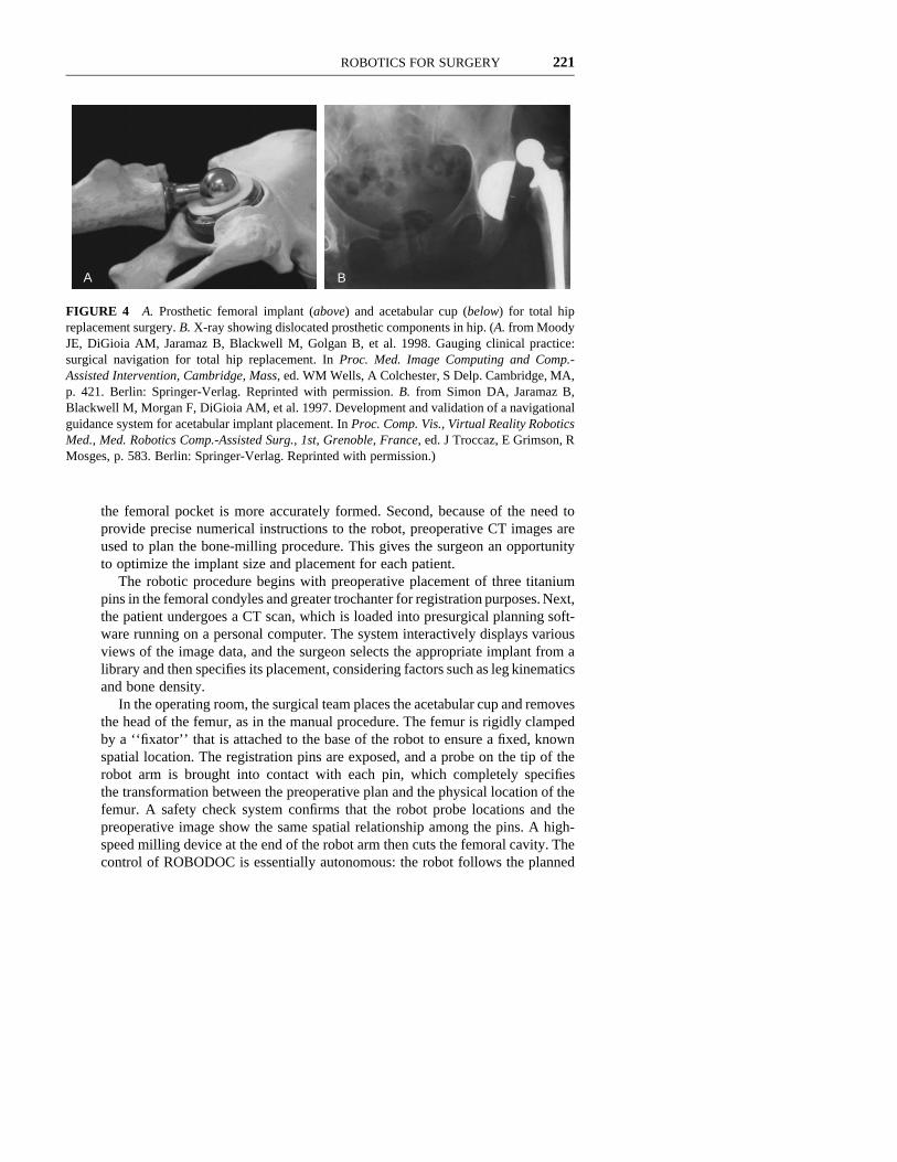
Total Hip Arthroplasty: Femur Preparation The replacement of hip joints thathave failed as a result of disease or trauma has become commonplace. The pro-cedure begins with disarticulation of the joint and removal of the proximal headof the femur. A metal and polymer prosthetic cup is then placed in the acetabulum.The femoral implant consists of a long metal shaft (up to 220 mm) that is insertedinto a deep cavity that must be formed along the proximal axis of the femur (52).The prosthetic components are shown in Figure 4.
In the current manual procedure, the surgeon cuts the cavity by forcing hand-held broaches and reamers into the femur, which leaves a rough and unevensurface. Until recently, the implant was cemented in place in this pocket, butlong-term postoperative data indicated that the cement could crack, loosen, orcause osteolysis, leading to failure of the implant. Newer cementless implantshave a porous metal surface and rely on natural bone growth into the metal forfixation. This ingrowth requires close proximity (0.25 mm or less) between thebone surface and the implant, so long-term success is highly dependent on a tightfit between the implant and the femur (52).
The need for improved precision led to the creation of a robotic approach toforming the femoral cavity. Development of the ROBODOC system began in themid-1980s, and it is now commercially available in Europe and is undergoingFDA approval trials in the United States (36). The system provides two mainadvantages over the manual procedure. First, clinical trials have confirmed that
ROBOTICS FOR SURGERY 221
A B
FIGURE 4 A. Prosthetic femoral implant (above) and acetabular cup (below) for total hipreplacement surgery. B. X-ray showing dislocated prosthetic components in hip. (A. from MoodyJE, DiGioia AM, Jaramaz B, Blackwell M, Golgan B, et al. 1998. Gauging clinical practice:surgical navigation for total hip replacement. In Proc. Med. Image Computing and Comp.-Assisted Intervention, Cambridge, Mass, ed. WM Wells, A Colchester, S Delp. Cambridge, MA,p. 421. Berlin: Springer-Verlag. Reprinted with permission. B. from Simon DA, Jaramaz B,Blackwell M, Morgan F, DiGioia AM, et al. 1997. Development and validation of a navigationalguidance system for acetabular implant placement. In Proc. Comp. Vis., Virtual Reality RoboticsMed., Med. Robotics Comp.-Assisted Surg., 1st, Grenoble, France, ed. J Troccaz, E Grimson, RMosges, p. 583. Berlin: Springer-Verlag. Reprinted with permission.)
the femoral pocket is more accurately formed. Second, because of the need toprovide precise numerical instructions to the robot, preoperative CT images areused to plan the bone-milling procedure. This gives the surgeon an opportunityto optimize the implant size and placement for each patient.
The robotic procedure begins with preoperative placement of three titaniumpins in the femoral condyles and greater trochanter for registration purposes. Next,the patient undergoes a CT scan, which is loaded into presurgical planning soft-ware running on a personal computer. The system interactively displays variousviews of the image data, and the surgeon selects the appropriate implant from alibrary and then specifies its placement, considering factors such as leg kinematicsand bone density.
In the operating room, the surgical team places the acetabular cup and removesthe head of the femur, as in the manual procedure. The femur is rigidly clampedby a ‘‘fixator’’ that is attached to the base of the robot to ensure a fixed, knownspatial location. The registration pins are exposed, and a probe on the tip of therobot arm is brought into contact with each pin, which completely specifiesthe transformation between the preoperative plan and the physical location of thefemur. A safety check system confirms that the robot probe locations and thepreoperative image show the same spatial relationship among the pins. A high-speed milling device at the end of the robot arm then cuts the femoral cavity. Thecontrol of ROBODOC is essentially autonomous: the robot follows the planned
222 HOWE n MATSUOKA
cutting paths without the surgeon’s guidance. After the pocket is milled, thesurgeon continues as in the manual procedure.
The first human trial of the system took place in 1992. Recent reports onapproximately 130 hip replacements from an ongoing clinical study in the UnitedStates used radiographs to compare ROBODOC-treated patients with a controlgroup (2). The ROBODOC cases showed significantly less space between theprosthetic and the bone. Placement of the implant was also improved. Further-more, no intraoperative femoral fractures occurred for the ROBODOC group,whereas three were observed in the control group. In Europe, the regulatory envi-ronment has permitted wider deployment of the system. Between November 1994and February 1998, more than 1000 patients were successfully treated at 17 sitesin Germany and Austria (3). The results also showed improved prosthetic fit, andthe overall complication rate was reduced to 11.6% from the reported manualprocedure rates of 16.6% to 33.7%. In addition, the surgical time decreased dra-matically as surgeons gained experience with the system and modified the pro-cedure: the first 10 cases averaged 220 min, whereas the current level is 90–100min (4).
Although results of these studies show that the system successfully achievesthe goal of improved fit, there are a number of difficulties that are common tomany image-guided surgical procedures. One area for improvement is the trau-matic pin placement procedure and slow pin-finding registration process. Workis under way to reduce the number of pins and then to eliminate them altogether,using other registration techniques (71). Another issue is the complex method forfixing the femur to the base of the robot, which is time consuming to set up andcan cause postoperative pain in the knee. A related problem is motion of the bonewithin the fixator during cutting. Currently, a separate sensing system is requiredto check for motion; if bone shift is detected, cutting is interrupted and the reg-istration process must be repeated. Several incidents of femur motion can pushthe surgical time over the limit of acceptability. An improved fixation techniqueor continuous registration method could eliminate these problems. Finally,although prosthetic fit and positioning appear to be improved, it is crucial toaddress the question of whether this improves treatment in the long term, as thecurrent orthopedic literature does not show a significant correlation betweenimplant fit and long-term outcome (2).
Total Hip Arthroplasty: Acetabular Cup Placement Hip dislocation occurswhen the head of the femur disengages from the acetabular cup, as shown inFigure 4. Dislocation is one of the most common postoperative complicationsfollowing total hip replacement surgery, with a rate of 1%–5% (53). The causeof dislocation is related to a number of factors, particularly malposition of theacetabular implant component. Incorrect positioning can allow the neck of thefemoral implant component to impinge on the edge of the cup or a bony promi-nence on the pelvis, forcing out the femoral head. Unfortunately, current manualalignment devices configure the implant with respect to the gross body axes of
ROBOTICS FOR SURGERY 223
the patient and do not take account of the pelvic orientation on the operating tableand individual variations in pelvis geometry.
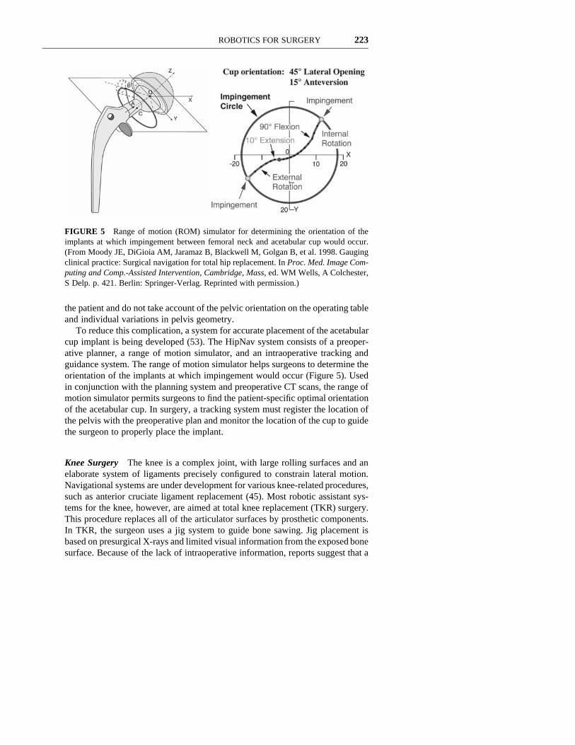
To reduce this complication, a system for accurate placement of the acetabularcup implant is being developed (53). The HipNav system consists of a preoper-ative planner, a range of motion simulator, and an intraoperative tracking andguidance system. The range of motion simulator helps surgeons to determine theorientation of the implants at which impingement would occur (Figure 5). Usedin conjunction with the planning system and preoperative CT scans, the range ofmotion simulator permits surgeons to find the patient-specific optimal orientationof the acetabular cup. In surgery, a tracking system must register the location ofthe pelvis with the preoperative plan and monitor the location of the cup to guidethe surgeon to properly place the implant.
Knee Surgery The knee is a complex joint, with large rolling surfaces and anelaborate system of ligaments precisely configured to constrain lateral motion.Navigational systems are under development for various knee-related procedures,such as anterior cruciate ligament replacement (45). Most robotic assistant sys-tems for the knee, however, are aimed at total knee replacement (TKR) surgery.This procedure replaces all of the articulator surfaces by prosthetic components.In TKR, the surgeon uses a jig system to guide bone sawing. Jig placement isbased on presurgical X-rays and limited visual information from the exposed bonesurface. Because of the lack of intraoperative information, reports suggest that a
FIGURE 5 Range of motion (ROM) simulator for determining the orientation of theimplants at which impingement between femoral neck and acetabular cup would occur.(From Moody JE, DiGioia AM, Jaramaz B, Blackwell M, Golgan B, et al. 1998. Gaugingclinical practice: Surgical navigation for total hip replacement. In Proc. Med. Image Com-puting and Comp.-Assisted Intervention, Cambridge, Mass, ed. WM Wells, A Colchester,S Delp. p. 421. Berlin: Springer-Verlag. Reprinted with permission.)
224 HOWE n MATSUOKA
sizable fraction of current manual procedures result in clinically significant inac-curacies, and up to 40% of the patients are left with patellofemoral pain or limitedflexion after conventional TKR surgery (1). The alignment of femur and tibia andthe location of ligament attachments are crucial; small displacements (2.5 mm)of the femoral component have been shown to alter the range of motion by asmuch as 208 (22).
Several robotic TKR assistant systems have been developed to increase theaccuracy of the prosthetic alignment. Many of these systems include an image-based preoperative planner and a robot to perform the bone cutting (19). Kienzleet al (38) have developed a system that uses the robot to guide jigs to the correctlocation, which then allows the surgeon to make accurate bone resections. First,the PUMA 560 robot tracks the motion and locates the center of the femoral headwhile the surgeon manually flexes and abducts the thigh. The robot uses thislandmark as a fiducial in addition to the preoperatively implanted pins to guidethe cutting tools to the position where the femur is to be resected. After thesurgeon makes the cut for the femur, the robot guides the cutting location for thetibia using the implanted pins. To maintain registration, the pelvis and the ankleare fixed to the surgical table, and the distal femur and proximal tibia are lockedwith respect to the base of the robot using a six degree-of-freedom arm. Thismechanical arm must be attached to the bones without interfering with the activ-ities of the surgeon. Accurate calibration of the robot proved to be one of thelargest obstacles for this project. Most industrial robots are built with high repeat-ability but insufficient positional accuracy. A specialized calibration techniquehas been implemented, and preliminary results indicate that an accuracy of lessthan 1 mm and 18 is plausible.
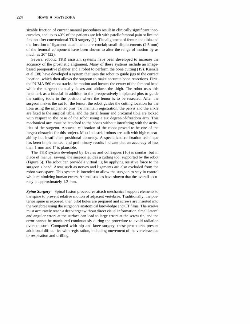
The TKR system developed by Davies and colleagues (16) is similar, but inplace of manual sawing, the surgeon guides a cutting tool supported by the robot(Figure 6). The robot can provide a virtual jig by applying resistive force to thesurgeon’s hand. Areas such as nerves and ligaments are also excluded from therobot workspace. This system is intended to allow the surgeon to stay in controlwhile minimizing human errors. Animal studies have shown that the overall accu-racy is approximately 1.3 mm.
Spine Surgery Spinal fusion procedures attach mechanical support elements tothe spine to prevent relative motion of adjacent vertebrae. Traditionally, the pos-terior spine is exposed, then pilot holes are prepared and screws are inserted intothe vertebrae using the surgeon’s anatomical knowledge and CT films. The screwsmust accurately reach a deep target without direct visual information. Small lateraland angular errors at the surface can lead to large errors at the screw tip, and theerror cannot be monitored continuously during the procedure to avoid radiationoverexposure. Compared with hip and knee surgery, these procedures presentadditional difficulties with registration, including movement of the vertebrae dueto respiration and drilling.
ROBOTICS FOR SURGERY 225
Current research in spinal surgery focuses on image-guided passive assistancein aligning the hand-held surgical drill. Preoperative CT images are integratedwith tracking devices during the procedure. Targets may be attached to eachvertebra to permit constant optical motion tracking during the procedure. Usingthese techniques, Merloz et al (50) report a far lower rate of cortical penetrationfor computer-assisted techniques compared with the manual procedure. Work isunder way on the use of intraoperative ultrasound or radiograph images to registerthe CT data with the patient (43). The screws may then be inserted percutaneously,eliminating the need for exposing the spine.
FIGURE 6 Total knee replacement robot. The surgeon guides the cutting tool while therobot generates constraint forces to ensure accuracy and protect key structures. (FromHarris SJ, Jakopec M, Hibberd RD, Cobb J, Davies BL, 1998. Interactive pre-operativeselection of cutting constraints, and interactive force controlled knee surgery by a surgicalrobot. In Proc. Med. Image Computing and Comp.-Assisted Intervention, Cambridge,Mass, ed. WM Wells, A Colchester, S Delp. pp. 996–1006. Berlin: Springer-Verlag.Reprinted with permission.)
226 HOWE n MATSUOKA
Neurosurgery
Neurosurgery was the first surgical specialty to use image-guided techniques,beginning with stereotactic frames that were attached to the patient’s craniumbefore the imaging process and remained in place during surgery. The relationshipbetween the frame and lesion observed in the image was used to guide the instru-ments within the brain. Newer image-guided techniques, sometimes called frame-less stereotaxy, use less invasive fiducial markers or video images for registrationand optical trackers for navigation of hand-held instruments (27, 28, 44). Toenhance stability, accuracy, and ease of use, a number of robotic systems havebeen developed for these procedures over the past 15 years (e.g. 24a, 26, 40, 46,48).
One issue in image-guided neurosurgery is shifting of the brain during theprocedure, which alters the spatial relationship between the preoperative imagedata and the anatomy of the patient. Various solutions have been proposed to dealwith this problem, including deformable templates for nonrigid registration,sometimes based on biomechanical models of soft tissue (41, 56a). Anothersolution is to perform the procedure inside an imaging system, which permitscontinuous monitoring of the anatomy and instrumentation. This requires roboticmanipulators that are compatible with the imaging modality and space constraints(48).
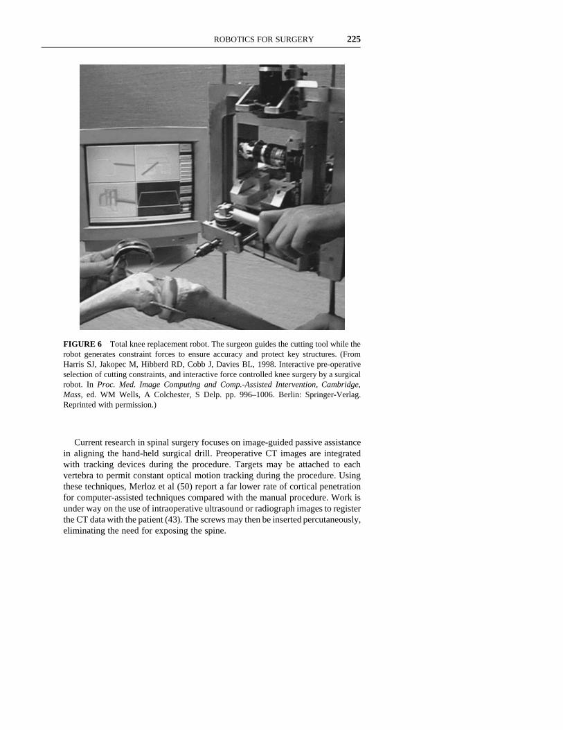
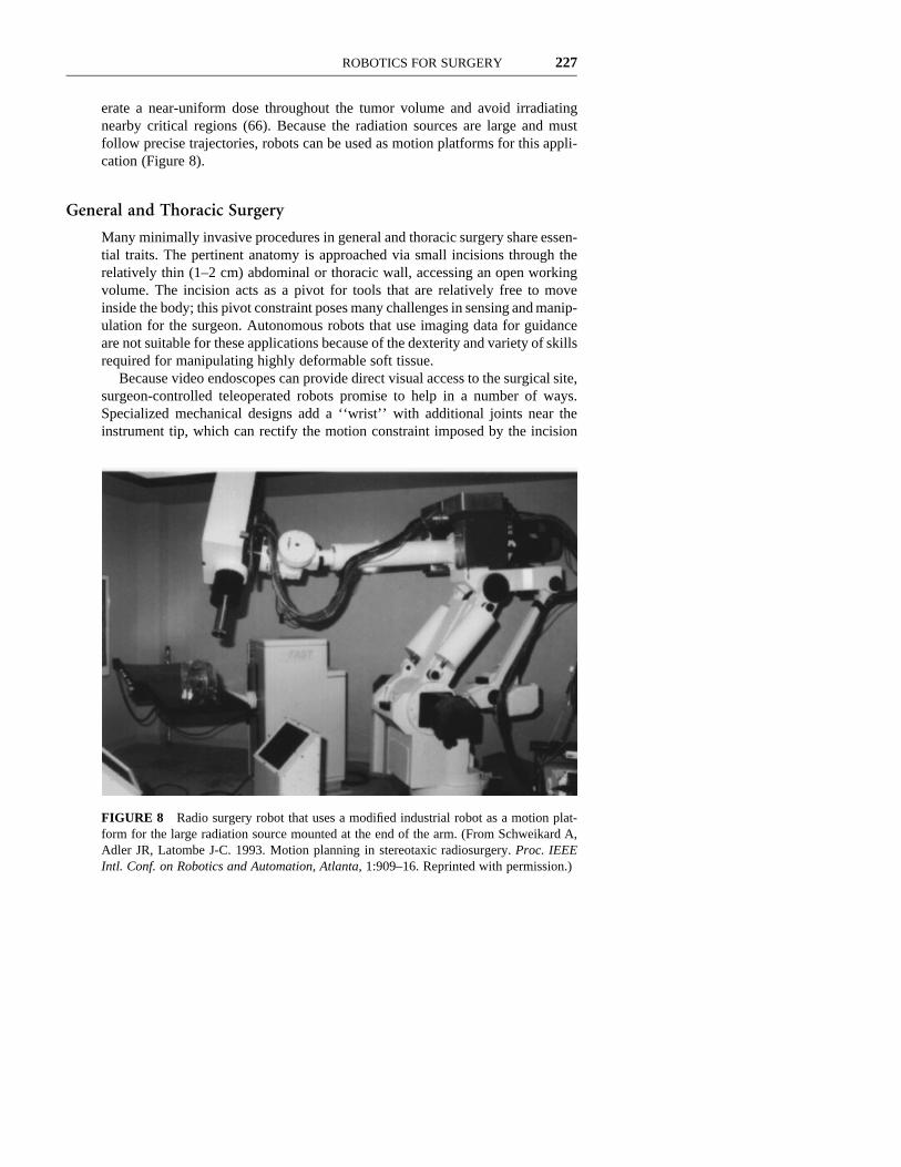
Radiosurgery uses a beam of radiation as a surgical instrument to destroy braintumors. If the angle of incidence of the beam is pivoted through a large range,the beam passes through the tumor at all times but intersects each point of adjacenttissues only briefly (Figure 7). Planning algorithms can optimize the path to gen-
FIGURE 7 Radiation beam for radiosurgery passes through the tumor at all times butintersects each point of adjacent tissue only briefly. (From Schweikard A, Adler JR,Latombe J-C. 1993. Motion planning in stereotaxic radiosurgery. Proc. IEEE Intl. Conf.on Robotics and Automation, Atlanta, 1:909–16. Reprinted with permission.)
ROBOTICS FOR SURGERY 227
erate a near-uniform dose throughout the tumor volume and avoid irradiatingnearby critical regions (66). Because the radiation sources are large and mustfollow precise trajectories, robots can be used as motion platforms for this appli-cation (Figure 8).
General and Thoracic Surgery
Many minimally invasive procedures in general and thoracic surgery share essen-tial traits. The pertinent anatomy is approached via small incisions through therelatively thin (1–2 cm) abdominal or thoracic wall, accessing an open workingvolume. The incision acts as a pivot for tools that are relatively free to moveinside the body; this pivot constraint poses many challenges in sensing and manip-ulation for the surgeon. Autonomous robots that use imaging data for guidanceare not suitable for these applications because of the dexterity and variety of skillsrequired for manipulating highly deformable soft tissue.
Because video endoscopes can provide direct visual access to the surgical site,surgeon-controlled teleoperated robots promise to help in a number of ways.Specialized mechanical designs add a ‘‘wrist’’ with additional joints near theinstrument tip, which can rectify the motion constraint imposed by the incision
FIGURE 8 Radio surgery robot that uses a modified industrial robot as a motion plat-form for the large radiation source mounted at the end of the arm. (From Schweikard A,Adler JR, Latombe J-C. 1993. Motion planning in stereotaxic radiosurgery. Proc. IEEEIntl. Conf. on Robotics and Automation, Atlanta, 1:909–16. Reprinted with permission.)
228 HOWE n MATSUOKA
(9, 31, 47). With these manipulators, surgeons can orient the instrument to arbi-trary angles and reach around anatomical structures. Second, the controller canscale the surgeon’s motions so that the robot works at smaller scale than is pos-sible with hand-held instruments. This enables microsurgical procedures usingminimally invasive techniques, as has been demonstrated for tubal anastomosisin heart bypass procedures (67, 73). A third advantage is that the control computercan interpose rotational transformations between the surgeon’s master controlinterface and the surgical robot, so that, for example, orientations in the videoimage match motion direction at the surgeon’s hands. Studies indicate that appro-priate mappings can improve manipulation performance (42).
Teleoperation is also a promising approach for microsurgery in a number ofspecialties, including vascular, gynecological, neuro-, and ophthalmological sur-gery. A number of specialized systems have been developed (31, 64), in additionto the general-purpose telesurgical systems described above with motion scalingcapabilities. These systems pose unique research problems, including develop-ment of specialized manipulators and grippers, control methods for optimal map-ping between the human scale and microscales, and elimination of hand tremor(61, 63).
Minimally Invasive Surgery—Specialized Designs Specialized robotic systemscan enable new procedures where access is limited to long lumens, as in gastro-intestinal or urinary surgery. One example is transurethral resection of the prostate(30, 54). This procedure, to ameliorate benign enlargement of the prostate, is nowa skilled manual process of inserting instruments through the urethra and remov-ing tissue with repetitive cutting motions. In the system developed by Harris etal (30), the robotic system incorporates real-time ultrasonic imaging as well ascutting instruments. The surgeon uses the images to select the volume of theprostate to be excised. As with the ROBODOC system for hip surgery, the robotexecutes the planned resection autonomously, and the user interface provides thesurgeon with continuous information about the progress of the procedure; thesurgeon may halt or modify the procedure at any time. By developing a special-purpose mechanism, a number of safety features may be incorporated, includinglimiting the workspace accessible to the robot to the volume of the prostate, thuseliminating the possibility of more extensive tissue injury in the event ofmalfunction.
Another example of procedure-specialized mechanism design is robotic endos-copy. The most common application is colonoscopy, where the goal is inspection,biopsy, or treatment of the colon. Conventional endoscopes are rigid tubes, some-times with a few manually operated joints. The limited articulation capabilitiespermit access only to the lower portion of the intestinal tract, and the limitedconformation may produce large forces that cause considerable discomfort to thepatient. There are two robotic approaches to endoscopes for these applications(58). One is a highly articulated mechanism with many joints that can conformto the sinuous passages of the bowel (35, 57). This approach requires incorpo-
ROBOTICS FOR SURGERY 229
rating a large number of actuators and sensors into the endoscope structure; sizeconstraints suggest that novel technologies such as shape-memory alloy actuatorsmay be useful here. Robotics research has only partially solved the problem ofpath planning and control algorithms for mechanisms with many redundantdegrees of freedom.
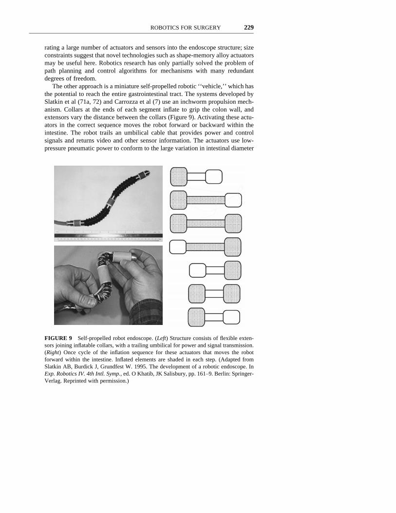
The other approach is a miniature self-propelled robotic ‘‘vehicle,’’ which hasthe potential to reach the entire gastrointestinal tract. The systems developed bySlatkin et al (71a, 72) and Carrozza et al (7) use an inchworm propulsion mech-anism. Collars at the ends of each segment inflate to grip the colon wall, andextensors vary the distance between the collars (Figure 9). Activating these actu-ators in the correct sequence moves the robot forward or backward within theintestine. The robot trails an umbilical cable that provides power and controlsignals and returns video and other sensor information. The actuators use low-pressure pneumatic power to conform to the large variation in intestinal diameter
FIGURE 9 Self-propelled robot endoscope. (Left) Structure consists of flexible exten-sors joining inflatable collars, with a trailing umbilical for power and signal transmission.(Right) Once cycle of the inflation sequence for these actuators that moves the robotforward within the intestine. Inflated elements are shaded in each step. (Adapted fromSlatkin AB, Burdick J, Grundfest W. 1995. The development of a robotic endoscope. InExp. Robotics IV. 4th Intl. Symp., ed. O Khatib, JK Salisbury, pp. 161–9. Berlin: Springer-Verlag. Reprinted with permission.)
230 HOWE n MATSUOKA
and avoid concentrated local pressure on the intestinal wall. This also minimizessafety concerns associated with electrical actuation.
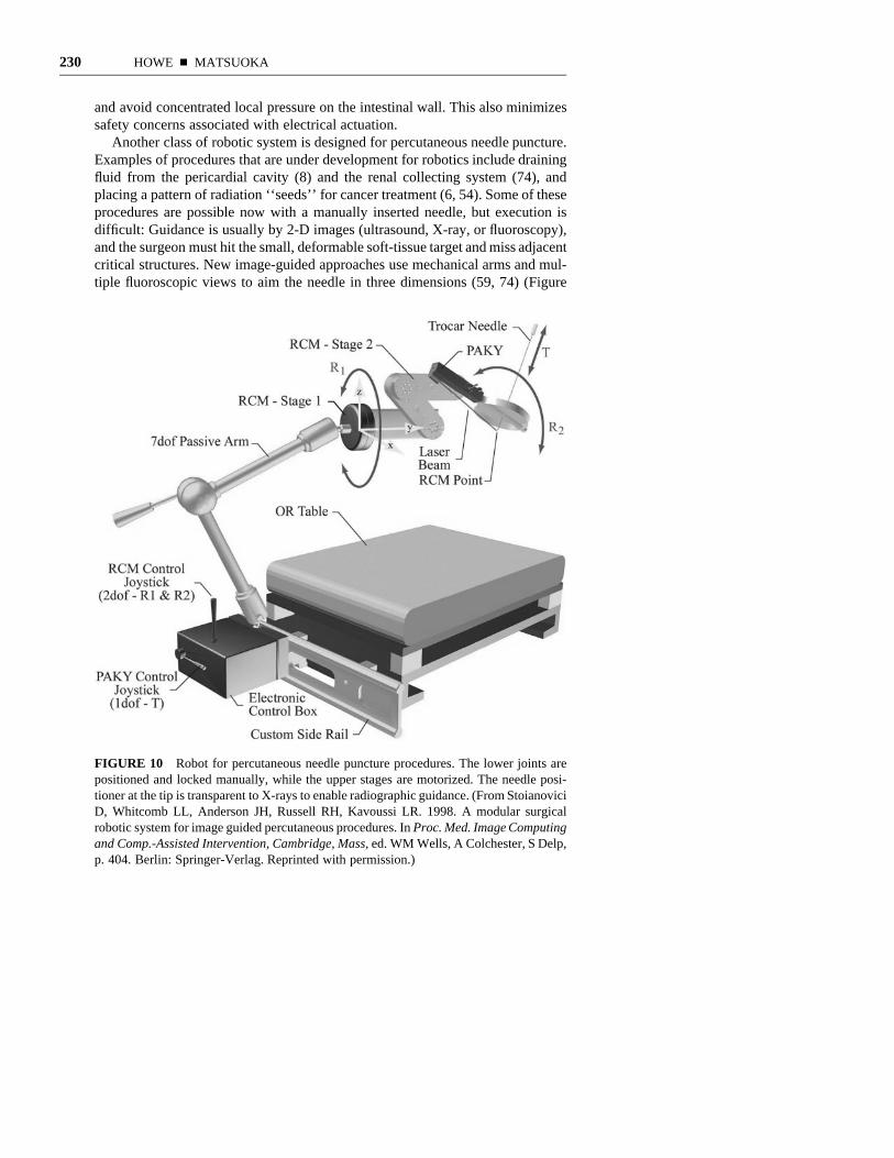
Another class of robotic system is designed for percutaneous needle puncture.Examples of procedures that are under development for robotics include drainingfluid from the pericardial cavity (8) and the renal collecting system (74), andplacing a pattern of radiation ‘‘seeds’’ for cancer treatment (6, 54). Some of theseprocedures are possible now with a manually inserted needle, but execution isdifficult: Guidance is usually by 2-D images (ultrasound, X-ray, or fluoroscopy),and the surgeon must hit the small, deformable soft-tissue target and miss adjacentcritical structures. New image-guided approaches use mechanical arms and mul-tiple fluoroscopic views to aim the needle in three dimensions (59, 74) (Figure
FIGURE 10 Robot for percutaneous needle puncture procedures. The lower joints arepositioned and locked manually, while the upper stages are motorized. The needle posi-tioner at the tip is transparent to X-rays to enable radiographic guidance. (From StoianoviciD, Whitcomb LL, Anderson JH, Russell RH, Kavoussi LR. 1998. A modular surgicalrobotic system for image guided percutaneous procedures. In Proc. Med. Image Computingand Comp.-Assisted Intervention, Cambridge, Mass, ed. WM Wells, A Colchester, S Delp,p. 404. Berlin: Springer-Verlag. Reprinted with permission.)
ROBOTICS FOR SURGERY 231
10). Another approach uses optical trackers on an ultrasound head, so a computercan reconstruct the 3-D anatomical structure (8). An optical tracker on the needleholder guides the surgeon to insert the needle into the target. Challenges in theseprocedures include the 2-D to 3-D registration problem, misregistration frommotion of the patient as a result of respiration, heartbeat, or discomfort, deflectionof the needle, and the development of intuitive computer interfaces for 3-Dguidance.
Stability Enhancement Because robots are stable and untiring, they are effec-tive assistants for a number of surgical procedures. One task that has receivedmuch attention is holding endoscopes for minimally invasive surgery. Severalrobotic systems for laparoscopic general surgery are now commercial products(1a, 21, 23). This function is particularly appealing for robotic implementationbecause contact with tissue is limited to the sides of the incision, so safety con-cerns and control complexity are minimized. Various methods for controllingscope pointing have been implemented, from simple instrument-mounted joy-sticks and foot pedals to voice commands (1a, 23) and head tracking (21). Forneurosurgery, Gorodia et al (26) have demonstrated an assistive control systemwhere the surgeon manually guides a robot-mounted endoscope. For proceduressuch as evacuation of hematomas, this approach may overcome problems withsteadiness and precision when the endoscope is supported only by hand. Otherapplications where stability and lack of fatigue are important include limb holding(18) and organ retraction (60).
TRAINING AND SIMULATION
Robots are also finding applications in surgical training and simulation, wherethey provide force feedback from computer models of instrument–tissue inter-action. In these systems, users manipulate surgical instrument handles that areattached to specialized robot manipulators (Figure 11; see color figure). A com-puter senses the user-generated motions and commands the robot to apply theforces that would have resulted from the instruments’ interaction with real tissue.The computer also generates images of the simulated surgical site. These systemsare similar to telesurgical systems where the user interacts with the master manip-ulator, but here a computer model replaces the actual surgical robot and patient.Systems have been developed for many procedures, including arthroscopic kneesurgery (24), tubal anastomosis (56), and laparoscopic surgery (10).
These virtual environment systems offer a number of potential advantages.Compared with cadaver and animal training, costs may be reduced, and comparedwith conventional patient-based surgical training, there are fewer time and per-formance constraints. Because these systems measure all of the actions duringeach procedure, trainees can review their data to analyze technique, and trainerscan evaluate progress and skill level. Finally, surgeons can explore new and
232 HOWE n MATSUOKA
enhanced surgical techniques, and by incorporating preoperative image data,patient-specific procedures may be rehearsed.
The field of surgical simulation is still in an early state of development. Spe-cialized ‘‘haptic interface’’ robots with sufficient fidelity to produce realistic sen-sations have been available for fewer than 10 years (13, 30a). One unsolvedproblem is tissue modeling. To determine the correct force to feed back inresponse to user motions, the system must calculate the deformation of modeltissues in real time. Current mechanical modeling techniques, based on the finite-element method, are too slow for real-time use (1b, 10, 15a). In addition, themechanical properties of many of the tissues of interest have not been measured.Another problem is generation of patient-specific models from 3-D image data.Gibson et al (24) have developed a voxel-based object representation scheme thatoperates directly on the medical image data structure. This approach can representvolumetric information that is hidden from the surface to allow realistic modelingof deformation, cutting, and tearing of tissues. Limitations of this approachinclude slow visual rendering and the need for more resolution for hapticfeedback.
A central question for these systems is the relevance to actual surgery: Can asimulator effectively train medical students in good surgical skills? A report byO’Toole et al (56) suggests that the answer is yes. This study used a simulator toassess suturing technique. The user interface was a needle holder and forcepsattached to force feedback devices (Figure 11). The user could see and feel sim-ulated organs and interact with them in various ways (grasp, poke, pluck, andsuture). Physics-based models made the vessels statically and dynamically real-istic. Twelve medical students and eight practicing vascular surgeons performedlarge flexible-vessel anastomosis with the simulator. Their performance was eval-uated in terms of errors, accuracy, and tissue damage. The average medical stu-dent’s score was significantly worse than the average of practicing surgeons formost measures. In addition, performance improved more for the students duringthe study. Although these results demonstrate that untrained subjects learned thesimulated surgical technique, the transference of skills to real surgery was notevaluated.
TECHNICAL AND IMPLEMENTATION CHALLENGES
The results reviewed above demonstrate that robotic technology can enhancesurgery in many ways. Surgical robotics has been an active area of research foronly a decade, and innovation continues at a rapid pace. In this concluding section,we review some of the leading research problems and consider issues that mayconstrain widespread clinical acceptance of robotic surgery.
Technical Issues
As discussed above, the great majority of surgical applications take advantage ofthe unique characteristics of robots. The main benefits may be summarized asimproved precision, stability, and dexterity. To extend these benefits to additionalprocedures will require advances in mechanical design, sensing, and control.
ROBOTICS FOR SURGERY 233
Mechanical Design Currently, many image-guided surgical applications useoff-the-shelf industrial robot manipulators. This speeds development and reducescosts, but these devices have not been optimized for the characteristics of specificsurgical tasks. For example, most industrial robots are designed for good repeat-ability but may lack sufficient positional accuracy (38, 40). Similarly, assistivesystems that share control between the robot and human surgeon would benefitfrom the development of low impedance manipulators in place of highly geared,stiff industrial arms (32, 79). Other advantages that can accrue to specializeddesigns include improved sterility and compatibility with imaging systems (e.g.transparency to X-rays). In teleoperated systems, access constraints have alwaysnecessitated the development of new manipulator configurations, but kinematicstructures and actuator technologies are far from perfected. These technologiesalso limit the development of microrobots for medical applications (14).
Sensing and Control In teleoperated systems for minimally invasive or micro-surgical procedures, there is substantial room for improvement of control andsensory feedback interfaces. In general, the human factors aspects of these sys-tems have been little studied. Research questions include master manipulatorconfiguration, mapping between master and remote robot coordinate systems,scaling laws for micromanipulation systems, and video, force, and tactile feed-back fidelity and bandwidth requirements (31, 34, 42, 63, 67a, 68).
Image-guided procedures have been an area of great success for robotic sur-gery, but there are many unresolved issues. Improved automatic segmentationand planning systems promise to improve efficiency and accuracy. Areas forimprovement in registration include elimination of invasively placed fiducials andmethods for nonrigid registration and tracking of tissue deformation in real time.The use of 2-D imaging modalities such as ultrasound in combination with 3-Dtracking may lower costs and enable wider application of image-guided tech-niques (8, 77).
For autonomous robotics in general, almost all successful applications overthe past three decades have come in areas where tasks are narrowly specified andthe environment is predictable, as in manufacturing. The early success of roboticsin orthopedic surgery is due at least in part to the fact that bones are essentiallyrigid and relatively straightforward to manipulate, immobilize, and cut. The useof robots for autonomously manipulating soft tissue raises a host of new chal-lenges, many without precedent in robotics research.
Currently, large deformation manipulation of soft tissue requires teleoperation,where the surgeon provides the required sensory integration and dexterous con-trol. For autonomous robots to undertake these tasks will require good hand-eyecoordination, tactile sensing of the instrument–tissue contact state, and an abilityto predict the outcome of manipulative actions. Increased computational powerhas enabled new capabilities in ‘‘visual servoing’’ of manipulator motion, whichbegins to address the visual coordination problem (29, 34a). In contrast, inte-grating tactile information into control is still a largely unsolved problem in robot-ics, even for rigid objects (33). Alternative sensing schemes, such as real-time
234 HOWE n MATSUOKA
continuous magnetic resonance imaging, may prove superior to visual and tactileapproaches, but cost and manipulator compatibility issues are severe obstacles.Predicting the results of manipulative actions may require mechanical modelingof the tissue–instrument interaction. Initial research in this area for surgical simu-lation has showed that conventional techniques are far too slow for use in real-time control (10, 15a, 24).
In addition to these quantitative abilities, the actions of a skilled surgeon arebased on broad and deep knowledge of anatomy and surgical technique. Forcomplete autonomy, robots must be able to use such qualitative reasoning andbroad sensory integration in control. This will require fundamental advances inseveral areas of computer science as well as robotics. As a more immediate goal,it may be possible to add semiautonomous capabilities that exploit the quantitativeadvantages of robots to decrease the demands on the surgeon, enable new pro-cedures, and improve safety.
Clinical Implementation and Acceptance Issues
Safety Safety is an obvious concern for robotic surgery, and regulatory agenciesrequire that it be addressed for every clinical implementation. As with most com-plex computer-controlled systems, there is no accepted technique that can guar-antee safety for all systems in every circumstance (15, 55, 78). Various roboticsystems approach the problem in different ways. One common technique is toinclude passive and active safety mechanisms in the mechanical design of themanipulator. A good example is the AESOP endoscopic pointing robot, used forminimally invasive general surgery (23). The end of the robot arm is attached tothe endoscope through a gimbal and a magnetic coupling. Because the incisionprevents lateral motion of the endoscope tube, as the robot moves the endoscopein space above the patient, the gimbal allows the endoscope tube to pivot aboutthe incision. This makes it impossible for the robot to apply lateral forces on theincision. The magnetic coupling acts as an emergency release: If forces on theendoscope exceed the magnetic holding force, the endoscope disconnects andfalls onto the patient’s abdomen, which is unlikely to cause injury.
Examples of designed-in hardware safety features in other robot systemsinclude the use of low-pressure pneumatic power to minimize dangers from elec-trical actuation (71a, 72), and limiting the size of the robot workspace to eliminatethe possibility of damage to tissue away from the intended surgical site (30).
Safety features of the software portion of the system are also essential. In thecontext of a urology robot, Ng & Tan (55) used mathematical logic to analyzeprogram flow and determine if it is possible for control to evade the safety featuresincorporated into the code. In addition, they implemented a completely indepen-dent safety monitor that can arrest a servo runaway and detect out-of-safe-bound-ary conditions, using joint encoder signals as input.
Some robotics developers have asserted that it is important to keep control ofthe procedure in the hands of the surgeon, even in image-guided surgery (32, 78).
ROBOTICS FOR SURGERY 235
For example, the knee surgery system developed by Ho et al (32) (see above) hasthe surgeon moving the cutting tool while the robot prevents motion outside ofthe planned workspace. In contrast, the ROBODOC hip replacement system hasthe robot moving the cutting tool under autonomous control while the surgeonmonitors progress (36). Early results with ROBODOC from Europe suggest fewproblems with clinician acceptance of the autonomous control mode (2–4). Asexperience with robotic systems increases, the level of comfort with autonomouscontrol may rise. It is, however, undeniably important to design user interfacesso that the surgeon is fully informed of the system’s plans and status.
Other Acceptance Issues Robots will be successful in surgery only if theyimprove patient outcome, lower cost, or both. Unfortunately, in many cases out-come cannot be assessed until many years after the procedure. For example, itmay take 15 years to accurately measure the difference in durability of roboticversus manual hip replacements. This is a prohibitive delay, both for the devel-opers of the systems and for the patients who are denied this potentially improvedcare in the intervening years. As a result, an alternative measure of outcome maybe necessary. In the hip replacement case, one measure is comparison of thecloseness of the fit between the femur and the implant. As previous bone growthstudies have shown that close fit is essential for good fixation, this is a plausiblecorrelate with long-term success of the procedure. The space between the implantshaft and the bone can be measured radiographically soon after surgery, so thisprovides a means to measure outcome promptly, if indirectly, and facilitate morerapid acceptance. One benefit from early acceptance of robotic technology is thatas the number of cases increases, clinicians often improve the procedure, whichmay result in better outcomes and lower costs (4).
Expense is also an issue with some robots. Although there is a large range incost, some systems exceed one million dollars. This may reduce the rate of imple-mentation, especially in the early years, when benefits have not been fully realizedor documented. As the field matures and engineering expertise with these systemsincreases, costs will likely decrease. In addition, many robotic systems are nowdedicated to specific procedures, so that systems for knee replacement are unableto perform hip replacements, even though the procedures are similar in manyrespects. With growing maturity of the field, systems may gain flexibility, so thatthe same robot can be used for a variety of procedures in a surgical specialty,serving to reduce costs.
Finally, we note that the progress reviewed here demonstrates that robotictechnology will transform surgery in the coming years. Robots promise to becomethe standard modality for many common procedures, including hip replacement,heart bypass, and abdominal surgery. This suggests that surgeons, particularlyresearchers working to enhance and extend the field, will need to become familiarwith robotic technology. The same is true for robotics researchers: Creating effec-tive systems requires understanding the demands of surgical procedures and theculture of surgical practice. The research teams that have created groundbreaking
236 HOWE n MATSUOKA
systems demanded close collaborations among robotics researchers, computerscientists, and surgeons. Future progress will require similar interdisciplinaryteamwork.
Visit the Annual Reviews home page at http://www.AnnualReviews.org.
LITERATURE CITED
1. Aglietti P, Buzzi R, Gaudenzi A. 1988.Patellofemoral functional results andcomplications with the posterior stabi-lized total condylar knee prosthesis. J.Arthroplasty 3:17–25
1a. Allaf ME, Jackman SV, Schulam PG,Cadeddu JA, Lee BR, et al. 1998. Lapa-roscopic visual field. Voice vs foot pedalinterfaces for control of the AESOProbot. Surg. Endoscopy 12(12):1415–8
1b. Astley OR, Hayward V. 1998. Multiratehaptic simulation achieved by couplingfinite element meshes through Nortonequivalents. Proc. IEEE Intl. Conf.Robotics Autom. Leuven, Belgium, May1998. pp. 989–94
2. Bargar WL, Bauer A, Borner M. 1998.Primary and revision total hip replace-ment using the ROBODOC system. J.Clin. Orthop. Relat. Res. 354:82–91
3. Bauer A, Borner M, Lahmer A. 1998.Primary and revision THR using theROBODOC system. In Proc. Comp.Aided Orthop. Surg. Conf., p. 149. Pitts-burgh, Pa: Cent. Orthop. Res., ShadysideHosp.
4. Bauer A, Lahmer A, Borner M. 1998.Men-machine interaction—pitfalls inrobotic orthopedic surgery. See Ref. 3, p.68. (Abstr.)
5. Bro-Nielsen M, Gramkow C, Kreiborg S.1997. Non-rigid image registration usingbone growth model. See Ref. 78a, pp.3–12
6. Bzostek A, Schreiner S, Barnes AC,Cadeddu JA, Roberts W, et al. 1997. Anautomated system for precise percutane-ous access of the renal collecting system.See Ref. 78a, pp. 299–308
7. Carrozza MC, Lencioni L, Magnani B,D’Attanasio S, Dario P. 1997. The devel-opment of a microrobot system forcolonoscopy. See Ref. 78a, pp. 779–88
8. Chavanon O, Barbe C, Troccaz J, CarratL, Ribuot C, Blin D. 1997. Computerassisted pericardial puncture: work inprogress. Comput. Aided Surg. 2(6):356–64
9. Cohn MB, Crawford LS, Wendlandt JM,Sastry SS. 1996. Surgical applications ofmilli-robots. J. Robot. Syst. 12(6):401–16
10. Cotin S, Delingette H, Clement JM, Tas-setti V, Marescaux J, Ayache N. 1996.Volumetric deformable models for simu-lation of laparoscopic surgery. In Com-put. Assist. Radiol.: Proc. Int. Symp.Comp. Commun. Syst. Image GuidedDiagn. Ther., ed. HU Lemke, MW Van-nier, K Inamura, AG Farman, pp. 793–98. Amsterdam: Elsevier
11. Craig JJ. 1989. Introduction to Robotics:Mechanics and Control. Reading, Mass:Addison-Wesley. 2nd ed.
12. Cuschieri A, Buess G, Perissat J. 1992.Operative Manual of Endoscopic Sur-gery. Berlin: Springer-Verlag
13. Cutkosky MR, Srinivasan M, SalisburyJK, Howe RD, eds. 1999. Human andMachine Haptics. Cambridge, Mass:MIT Press. In press
14. Dario P, Guglielmelli E, Allotta B, Car-rozza MC. 1996. Robotics for medicalapplications. IEEE Robot. Autom. Mag.3(3):44–56
15. Davies B. 1998. The safety of medicalrobots. In Proc. Int. Symp. Robot., 29th,Birmingham, ed. F Redmill, T Anderson,
ROBOTICS FOR SURGERY 237
pp. 70–74. Birmingham, United King-dom: Br. Robot. Assoc.
15a. Delingette, H. Toward realistic soft-tis-sue modeling in medical simulation.1998. Proc. IEEE 86(3):512–23
16. Delp SL, Stulberg D, Davies B, Picard F,Leitner F. 1998. Computer assisted kneereplacement. J. Clin. Orthop. Relat. Res.354:49–56
17. Ellis RE, Fleet DJ, Bryant JT, Rudan J,Fentan P. 1997. A method for evaluatingCT-based surgical registration. See Ref.78a, pp. 141–50
18. Erbse S, Radermacher K, Anton M, RauG, Boeckmann W, et al. 1997. Devel-opment of an automatic surgical holdingsystem based on ergonomic analysis. SeeRef. 78a, pp. 737–46
19. Fadda M, Bertelli D, Martelli S, Mar-cacci M, Dario P, et al. 1997. Computerassisted planning for total knee arthro-plasty. See Ref. 78a, pp. 619–28
20. Feldmar J, Ayache N, Betting F. 1997.3D-2D projective registration of free-form curves and surfaces. Comput. Vis.Image Underst. 65(3):403–24
21. Finlay PA, Ornstein MH. 1995. Control-ling the movement of a surgical laparo-scope. IEEE Eng. Med. Biol. 14(3):289–91
22. Garg A, Walker PS. 1990. Prediction oftotal knee motion using a three-dimen-sional computer graphics model. J. Bio-mech. 23:45–58
23. Geis WP, Kim HC, Brennan EJ Jr,McAfee PC, Wang Y. 1996. Robotic armenhancement to accommodate improvedefficiency and decreased resource utili-zation in complex minimally invasivesurgical procedures. In Proc. Med. MeetsVirtual Real.: Health Care Inf. Age, SanDiego, ed. SJ Weghorst, HB Sieburg, KSMorgan, pp. 471–81. Amsterdam: IOS
24. Gibson S, Samosky J, Mor A, Fyock C,Grimson E, et al. 1997. Simulatingarthroscopic knee surgery using volu-metric object representations, real-timevolume rendering and haptic feedback.See Ref. 78a, pp. 369–78
24a. Glauser D, Fankhauser H, Epitaux M,Hefti J-L, Jaccottet A. 1995. Neurosur-gical Robot Minerva: first results andcurrent developments. J. Image Guid.Surg. 1:266–72
25. Glossop ND, Hu RW. 1998. Accuracylimitations and tradeoffs in CAOS. SeeRef. 3, p. 200 (Abstr.)
26. Gorodia TM, Taylor RH, Auer LM.1997. Robot-assisted minimally invasiveneurosurgical procedures: first experi-mental experience. See Ref. 78a, pp.319–22
27. Grimson WEL, Ettinger GJ, White SJ,Lozano-Perez T, Wells WM, et al. 1996.An automatic registration method forframeless stereotaxy, image guided sur-gery, and enhanced reality visualization.IEEE Trans. Med. Imag. 15(2):129–40
28. Grimson WEL, Leventon ME, EttingerG, Chabrerie A, Ozlen F, et al. 1998.Clinical experience with a high precisionimage-guided neurosurgery system. Seeref. 80a, pp. 63–73
29. Guo-Qing W, Arbter K, Hirzinger G.1997. Real-time visual servoing for lap-aroscopic surgery: controlling robotmotion with color image segmentation.IEEE Eng. Med. Biol. 16(1):40–45
30. Harris SJ, Arambula-Cosio F, Mei Q,Hibberd RD, Davies BL, et al. 1997. TheProbot—an active robot for prostateresection. Proc. Inst. Mech. Eng. J. Eng.Med. 211(H4):317–25
30a. Hayward V, Gregorio P, Astley O,Greenish S, Doyon M, Lessard L, et al.1997. Freedom-7: a high fidelity sevenaxis haptic device with application tosurgical training. In Experimental Robot-ics V: Fifth International Symposium.Barcelona, Spain, 15–18 June 1997, ed.A Casals, AT de Almeida, pp. 445–56.Berlin: Springer-Verlag
31. Hill JW, Jensen JF. 1998. Telepresencetechnology in medicine: principles andapplications. Proc. IEEE 86(3):569–80
32. Ho SC, Hibberd RD, Davies BL. 1995.Robot assisted knee surgery. IEEE Eng.Med. Biol. 14(3):292–300
238 HOWE n MATSUOKA
33. Howe RD. 1994. Tactile sensing andcontrol of robotic manipulation. J. Adv.Robot. 8(3):245–61
34. Howe RD, Peine WJ, Kontarinis DA,Son JS. 1995. Remote palpation technol-ogy. IEEE Eng. Med. Biol. 14(3):318–23
34a. Hutchinson S, Hager GD, Corke PI.1996. A tutorial on visual servo control.IEEE Trans. Robotics Automation12(5):651–70
35. Ikuta K, Nokata M, Aritomi S. 1997.Development of hyper active endoscopefor remote minimal invasive surgery. SeeRef. 46a, pp. 1044–50
36. Kazanzides P, Mittelstadt BD, MusitsBL, Bargar WL, Zuhars JF, et al. 1995.An integrated system for cementless hipreplacement. IEEE Eng. Med. Biol.14(3):307–13
37. Khatib O, ed. 1992. Robotics Review 2.Cambridge, Mass: MIT Press
38. Kienzle TC, Stulberg SD, Peshkin M,Quaid A, Lea J, et al. 1995. A computer-assisted total knee replacement surgicalsystem using a calibrated robot. See Ref.74a, pp. 409–16
39. Kliegis UG, Zeilhofer HF, Sader R,Horch HH. 1997. Intraoperative telena-vigation—some critical remarks aboutthe concept. See Ref. 46a, pp. 843–48
40. Kwoh YS, Hou J, Jonckheere EA, HayatiS. 1988. A robot with improved absolutepositioning accuracy for CT guided ste-reotactic brain surgery. IEEE Trans. Bio-med. Eng. 35(2):153–60
41. Kyriacou SK, Davatzikos C. 1998. Abiomechanical model of soft tissue defor-mation, with applications to non-rigidregistration of brain images with tumorpathology. See Ref. 80a, p. 531
42. Lai F, Howe RD, Millman PA, Sur S.1999. Frame mapping and dexterity forsurgical task performance in roboticendoscopic surgery. Proc. ASME Dym.Sys. Contr. Div., Nashville, Nov., ed. N.Olgac
43. Lavallee S, Troccaz J, Sautot P, MazierB, Cinquin P, et al. 1995. Computer-
assisted spine surgery using anatomy-based registration. See Ref. 74a, pp. 425–50
44. Lavallee S. 1995. Registration for com-puter-integrated surgery: methodology,state of the art. See Ref. 74a, pp. 77–97
45. Lavallee S. 1998. Example: computerassisted ACL replacement. See Ref. 3,p. 46 (Abstr.)
46. Lavallee S, Troccaz J, Gaborit L, Cin-quin P, Benabid AL, et al. 1992. Imageguided operating robot: a clinical appli-cation in stereotactic neurosurgery. Proc.IEEE Int. Conf. Robot. Autom. 1:618–24
46a. Lemke HU, Inamura K, Vannier MW,eds. 1997. Proc. Int. Symp. Exhib. Com-put. Assist. Radiol. Surg., Berlin, 11th.Amsterdam: Elsevier
47. Madhani AJ. 1998. Design of teleoper-ated surgical instruments for minimallyinvasive surgery. PhD thesis. MIT, Dept.Mech. Eng., Cambridge, Mass
48. Masamune K, Kobayashi E, Masutani Y,Suzuki M, Dohi T, et al. 1995. Devel-opment of an MRI-compatible needleinsertion manipulator for stereotacticneurosurgery. J. Image Guid. Surg.1:242–48
49. McInerney T, Terzopoulos D. 1996.Deformable models in medical imageanalysis. In Proc. IEEE Workshop Math.Methods Biomed. Image Anal., SanFrancisco, pp. 171–80. IEEE Comp.Soc.: Los Alamitos, Calif
50. Merloz P, Tonetti J, Eid A, Faure C, Pit-tet L, et al. 1997. Computer-assisted ver-sus manual spine surgery: clinical report.See Ref. 78a, pp. 541–44
51. Meyer K, Applewhite HL, Biocca FA.1992. A survey of position trackers.Presence 1(2):173–200
52. Mittelstadt BD, Kazanzides P, Zuhars J,Williamson B, Cain P, et al. 1994. Theevolution of a surgical robot from pro-totype to human clinical use. In Proc.Med. Robot. Comp.-Assisted Surg., Pitts-burgh, Pa
53. Moody JE, DiGioia AM, Jaramaz B,
ROBOTICS FOR SURGERY 239
Blackwell M, Golgan B, et al. 1998.Gauging clinical practice: surgical navi-gation for total hip replacement. See Ref.80a, pp. 421–30
54. Ng WS, Chung VR, Vasan S, Lim P.1996. Robotic radiation seed implanta-tion for prostatic cancer. Proc. Annu. Int.Conf. IEEE Eng. Med. Biol. Soc. 1:231–33
55. Ng WS, Tan CK. 1996. On safetyenhancements for medical robots.Reliab. Eng. Syst. Saf. 54(1): 35–45
56. O’Toole R, Playter R, Krummel T, BlankW, Bornelius N, et al. 1998. Assessingskill and learning in surgeons and medi-cal students using a force feedback sur-gical simulator. See Ref. 80a, pp. 899–909
56a. Paulsen KD, Miga MI, Kennedy FE,Hoopens PJ, Hartov A, Roberts DW.1999. A computational model for track-ing subsurface tissue deformation duringstereotactic neurosurgery. IEEE Trans.Biomed. Eng. 46(2):213–25
57. Peirs J, Reynaerts D, Van Brussel H.1997. Design of a shape memory actu-ated endoscopic tip. Sens. Actuators A70(1–2):135–40
58. Phee SJ, Ng WS, Chen IM, Seow-ChoenF, Davies BL. 1997. Locomotion andsteering aspects in automation of colon-oscopy. I. A literature review. IEEE Eng.Med. Biol. Mag. 16(6):85–96
59. Potamianos P, Davies BL, Hibberd RD.1995. Intraoperative registration for per-cutaneous surgery. In Proc. Int. Symp.Med. Robot. Comput. Assist. Surg., 2nd,Nov., Baltimore, Md. New York: Wiley-Liss, pp. 156–64
60. Poulouse B, Kutka M, Mendoza-SagaonM, Barnes A, et al. 1998. Human versusrobotic organ retraction during laparo-scopic Nissen fundoplication. See Ref.80a, pp. 197–206
61. Riviere CN, Rader RS, Thakor NV.1995. Adaptive real-time canceling ofphysiological tremor for microsurgery.See. Ref. 59, pp. 89–96
62. Rovetta A, Bejczy AK, Salal R. 1997.Telerobotic surgery: applications onhuman patients and training with virtualreality. In Medicine Meets Virtual Real-ity: Global Healthcare Grid, ed. KSMorgan, HM Hoffman, D Stredney, SJWeghorst, pp. 508–17. Amsterdam: IOS
62a. Rovetta A, Tosatti LM, Sala R, Bressa-nelli M, Leone P, Morelli G. 1996. Arobotized system for the execution ofa transurethral laser prostatectomy(TULP). Proc. Intl. Symp. IndustrialRobots, Milan, Italy, Oct. 27th, pp.1009–13
63. Salcudean SE, Yan J. 1994. Towards aforce-reflecting motion-scaling systemfor microsurgery. In Proc. IEEE Int.Conf. Robot. Autom., San Diego, LosAlamitos, Calif: IEEE Comp. Soc. Presspp. 2296–301
64. Schenker PS, Barlow EC, Boswell CD,Das H, Lee S, et al. 1995. Developmentof a telemanipulator for dexterityenhanced microsurgery. See Ref. 59, pp.81–88
65. Schulam PG, Docimo SG, Cadeddu JA,Saleh W, Brietenbach C, et al. 1997. Fea-sibility of laparascopic telesurgery. SeeRef. 78a, pp. 809–12
66. Schweikard A, Adler JR, Latombe J-C.1993. Motion planning in stereotaxicradiosurgery. In Proc. IEEE Int. Conf.Robot. Autom., Atlanta, 1:909–16. IEEEComp. Soc. Press: Los Alamitos, Calif
67. Shennib H, Bastawisy A, Mack MJ, MollFH. 1998. Computer-assisted telemani-pulation: an enabling technology forendoscopic coronary artery bypass. Ann.Thorac. Surg. 66(3):1060–63
67a. Sheridan TB. 1992. Telerobotics, Auto-mation, and Human Supervisory Con-trol. Cambridge, Mass: MIT Press
68. Sheridan TB, Thompson JM, Hu JJ,Ottensmeyer M. 1997. Haptics andsupervisory control in telesurgery. InProc. Hum. Factors Ergonom. Soc., 41st,Albuquerque, 2:1134–37
69. Simon DA. 1998 What is ‘‘registration’’
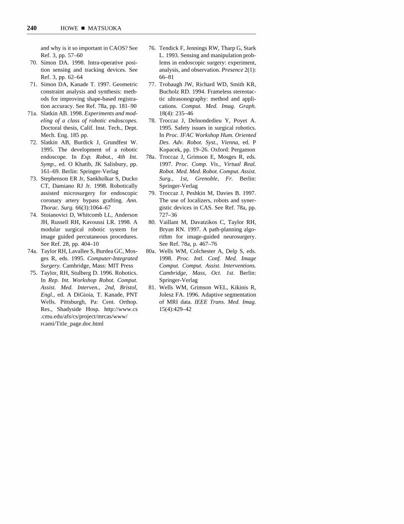
240 HOWE n MATSUOKA
and why is it so important in CAOS? SeeRef. 3, pp. 57–60
70. Simon DA. 1998. Intra-operative posi-tion sensing and tracking devices. SeeRef. 3, pp. 62–64
71. Simon DA, Kanade T. 1997. Geometricconstraint analysis and synthesis: meth-ods for improving shape-based registra-tion accuracy. See Ref. 78a, pp. 181–90
71a. Slatkin AB. 1998. Experiments and mod-eling of a class of robotic endoscopes.Doctoral thesis, Calif. Inst. Tech., Dept.Mech. Eng. 185 pp.
72. Slatkin AB, Burdick J, Grundfest W.1995. The development of a roboticendoscope. In Exp. Robot., 4th Int.Symp., ed. O Khatib, JK Salisbury, pp.161–69. Berlin: Springer-Verlag
73. Stephenson ER Jr, Sankholkar S, DuckoCT, Damiano RJ Jr. 1998. Roboticallyassisted microsurgery for endoscopiccoronary artery bypass grafting. Ann.Thorac. Surg. 66(3):1064–67
74. Stoianovici D, Whitcomb LL, AndersonJH, Russell RH, Kavoussi LR. 1998. Amodular surgical robotic system forimage guided percutaneous procedures.See Ref. 28, pp. 404–10
74a. Taylor RH, Lavallee S, Burdea GC, Mos-ges R, eds. 1995. Computer-IntegratedSurgery. Cambridge, Mass: MIT Press
75. Taylor, RH, Stulberg D. 1996. Robotics.In Rep. Int. Workshop Robot. Comput.Assist. Med. Interven., 2nd, Bristol,Engl., ed. A DiGioia, T. Kanade, PNTWells. Pittsburgh, Pa: Cent. Orthop.Res., Shadyside Hosp. http://www.cs.cmu.edu/afs/cs/project/mrcas/www/rcami/Title_page.doc.html
76. Tendick F, Jennings RW, Tharp G, StarkL. 1993. Sensing and manipulation prob-lems in endoscopic surgery: experiment,analysis, and observation. Presence 2(1):66–81
77. Trobaugh JW, Richard WD, Smith KR,Bucholz RD. 1994. Frameless stereotac-tic ultrasonography: method and appli-cations. Comput. Med. Imag. Graph.18(4): 235–46
78. Troccaz J, Delnondedieu Y, Poyet A.1995. Safety issues in surgical robotics.In Proc. IFAC Workshop Hum. OrientedDes. Adv. Robot. Syst., Vienna, ed. PKopacek, pp. 19–26. Oxford: Pergamon
78a. Troccaz J, Grimson E, Mosges R, eds.1997. Proc. Comp. Vis., Virtual Real.Robot. Med. Med. Robot. Comput. Assist.Surg., 1st, Grenoble, Fr. Berlin:Springer-Verlag
79. Troccaz J, Peshkin M, Davies B. 1997.The use of localizers, robots and syner-gistic devices in CAS. See Ref. 78a, pp.727–36
80. Vaillant M, Davatzikos C, Taylor RH,Bryan RN. 1997. A path-planning algo-rithm for image-guided neurosurgery.See Ref. 78a, p. 467–76
80a. Wells WM, Colchester A, Delp S, eds.1998. Proc. Intl. Conf. Med. ImageComput. Comput. Assist. Interventions.Cambridge, Mass, Oct. 1st. Berlin:Springer-Verlag
81. Wells WM, Grimson WEL, Kikinis R,Jolesz FA. 1996. Adaptive segmentationof MRI data. IEEE Trans. Med. Imag.15(4):429–42
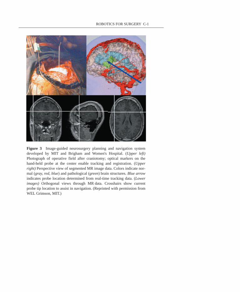
ROBOTICS FOR SURGERY C-1
Figure 3 Image-guided neurosurgery planning and navigation systemdeveloped by MIT and Brigham and Women's Hospital. (Upper left)Photograph of operative field after craniotomy; optical markers on thehand-held probe at the center enable tracking and registration. (Upperright) Perspective view of segmented MR image data. Colors indicate nor-mal (gray, red, blue) and pathological (green) brain structures. Blue arrowindicates probe location determined from real-time tracking data. (Lowerimages) Orthogonal views through MR data. Crosshairs show currentprobe tip location to assist in navigation. (Reprinted with permission fromWEL Grimson, MIT.)
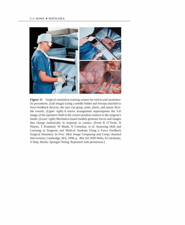
Figure 11 Surgical simulation training system for end-to-end anastomo-sis procedures. (Left image) Using a needle holder and forceps attached toforce-feedback devices, the user can grasp, poke, pluck, and suture flexi-ble vessels. (Upper right) A mirror arrangement superimposes the 3-Dimage of the operative field in the correct position relative to the surgeon’shands. (Lower right) Mechanics-based models generate forces and imagesthat change realistically in response to contact. (From R O’Toole, RPlayter, T Krummel, W Blank, N Cornelius, et al. Assessing Skill andLearning in Surgeons and Medical Students Using a Force FeedbackSurgical Simulator. In Proc. Med. Image Computing and Comp.-AssistedIntervention, Cambridge, MA, 1998, p. 404. Ed. WM Wells, A Colchester,S Delp. Berlin: Springer-Verlag. Reprinted with permission.)
C-2 HOWE ■ MATSUOKA