robust tlr4-induced gene expression patterns are not an
TRANSCRIPT

RESEARCH Open Access
Robust TLR4-induced gene expression patternsare not an accurate indicator of human immunityKelly L Brown1,2,4*, Reza Falsafi1, Winnie Kum1, Pamela Hamill1, Jennifer L Gardy1, Donald J Davidson2,3,Stuart Turvey2, Brett B Finlay1, David P Speert2, Robert EW Hancock1
Abstract
Background: Activation of Toll-like receptors (TLRs) is widely accepted as an essential event for defence againstinfection. Many TLRs utilize a common signalling pathway that relies on activation of the kinase IRAK4 and thetranscription factor NF�B for the rapid expression of immunity genes.
Methods: 21 K DNA microarray technology was used to evaluate LPS-induced (TLR4) gene responses in bloodmonocytes from a child with an IRAK4-deficiency. In vitro responsiveness to LPS was confirmed by real-time PCRand ELISA and compared to the clinical predisposition of the child and IRAK4-deficient mice to Gram negativeinfection.
Results: We demonstrated that the vast majority of LPS-responsive genes in IRAK4-deficient monocytes weregreatly suppressed, an observation that is consistent with the described role for IRAK4 as an essential componentof TLR4 signalling. The severely impaired response to LPS, however, is inconsistent with a remarkably low incidenceof Gram negative infections observed in this child and other children with IRAK4-deficiency. This unpredictedclinical phenotype was validated by demonstrating that IRAK4-deficient mice had a similar resistance to infectionwith Gram negative S. typhimurium as wildtype mice. A number of immunity genes, such as chemokines, wereexpressed at normal levels in human IRAK4-deficient monocytes, indicating that particular IRAK4-independentelements within the repertoire of TLR4-induced responses are expressed.
Conclusions: Sufficient defence to Gram negative immunity does not require IRAK4 or a robust, ‘classic’inflammatory and immune response.
BackgroundToll-like receptor-4 (TLR4) is a prominent member ofthe TLR family of host receptors that recognize micro-bial components in the intra- and extra-cellular environ-ment [1,2]. Lipopolysaccharide (LPS, endotoxin) is amajor component of the cell wall of Gram negative bac-teria, a potent TLR4 agonist and the driving forcebehind sepsis. TLR4 engagement by LPS results in theactivation of the transcription factor NF�B via signaltransduction cascades that are propagated eitherthrough, or independent of, the adaptor moleculeMyD88 [2-4]. Organisms amenable to genetic manipula-tion have been used to evaluate the importance of TLR4and various downstream signalling molecules for the
(LPS-)induced expression of immunity genes (mostcommonly cytokines including chemokines) and defenceagainst various pathogens [5,6], e.g., MyD88-knockoutmice fail to elevate serum cytokines when administereda high dose of LPS and are susceptible to infection by S.aureus [7], P. aeruginosa [8], M. tuberculosis [9], M.avium [10] and L. monocytogenes [11]. Such studieshave established that the MyD88-dependent pathway,via sequential activation of IRAK4, IRAK1, TRAF6, IKKand NF�B, drives a cellular response to TLR agoniststhat is responsible for the robust expression of early-response, NF�B-regulated immunity genes. It has beenoften assumed that this robust transcriptional response,involving the substantial induction of a large number ofgenes (>1000) is essential for normal immunologicalfunction(s) and, in turn, host defences. Moreover, thereduced expression, either in vitro or in vivo, of just asubset of TLR-responsive genes, such as the classic pro-
* Correspondence: [email protected] for Microbial Diseases and Immunity Research, Department ofMicrobiology and Immunology, University of British Columbia, Vancouver,British Columbia, V6T 1Z3, Canada
Brown et al. Journal of Translational Medicine 2010, 8:6http://www.translational-medicine.com/content/8/1/6
© 2010 Brown et al; licensee BioMed Central Ltd. This is an Open Access article distributed under the terms of the Creative CommonsAttribution License (http://creativecommons.org/licenses/by/2.0), which permits unrestricted use, distribution, and reproduction inany medium, provided the original work is properly cited.
inflammatory cytokines (TNF-a, IL-6), chemokines (IL-8, Gro-a/CXCL1) and antibacterials (antimicrobial pep-tides, reactive oxygen/nitrogen species) is often acceptedas sufficient evidence to predict a defect in host defenceagainst infectious agents.Since 2003, eighteen individuals, primarily children,
have been identified with a mutation in the gene thatencodes IL-1R-associated kinase 4 (IRAK4) that liesimmediately downstream of MyD88 in the TLR signal-ling cascade (reviewed in [12]). We previously reportedan IRAK4-deficiency in a 4-year old child due to ahomozygous single base substitution (C887T) in exon 8of the IRAK4 gene that introduced a premature stopcodon (Q293X) [13,14]; an identical mutation occurs inthe majority of cases of IRAK4 immunodeficiency.PBMC from children with IRAK4-deficiency consistentlyshow impaired in vitro responsiveness, as measured bythe production of pro-inflammatory cytokines, toselected TLR agonists [12-14]. Given the pivotal positionof IRAK4 in the TLR pathway and the failed responsive-ness of IRAK4-deficient (human and mouse) cells toboth Gram positive (LTA, PGN) and Gram negative(LPS) TLR agonists, it seemed logical that a defect inIRAK4 would render patients susceptible to a broadrange of both Gram positive and Gram negative bacter-ial pathogens. In contrast, children with an IRAK4-defi-ciency have a remarkably mild immunodeficiency withenhanced susceptibility to infection by only a narrowrange of Gram positive bacteria, particularly S. aureusand S. pneumoniae [12-18]. IRAK4 knockout mice arealso more susceptible to infections by (Gram positive) S.aureus than wild-type mice [19]. We were intrigued bythe relatively small number of documented cases ofGram negative infections in IRAK4-deficient children[20,21] in light of a consistently poor cellular responsesto LPS. It should be noted that these children are pre-scribed a regimen of antibiotics that are effective onlyagainst Gram positive bacteria, thus prophylaxis can notexplain their apparent immunity to Gram negative infec-tions. IRAK4 knockout mice and IRAK4-kinase-deadknock-in mice also fail, in vitro and in vivo, to produceinflammatory cytokines in response to LPS and are pro-tected against LPS-induced sepsis in vivo [19,22-24].There is, however, a resounding lack of experimentalevidence that IRAK4-deficiency (in humans or mice)actually compromises host defences to Gram negativeinfection.Herein we provide the first evidence that IRAK4
knockout mice are not overwhelmed by challenge withthe Gram negative bacterium S. typhimurium. Theseresults substantiated our hypothesis that the under-whelming number of documented Gram negative bac-terial infections in IRAK-deficient children is likelyattributable to sufficient host defence. The widely
accepted notion that the absence of IRAK4 should com-promise immunity to Gram negative pathogens wasbased primarily upon results demonstrating the compro-mised expression of a small number of classic LPS-responsive, pro-inflammatory markers (primarily cyto-kines) as well as the assumption that high expression ofthese genes in particular was required for immunity.Here we evaluated the impact of IRAK4-deficiency on aglobal scale using 21 K microarray technology to analyzetotal gene expression in LPS-activated monocytes froman IRAK4-deficient patient. In accordance with previousreports, and counter-intuitive to a resistance to Gramnegative infection, there was a profound failure ofIRAK4-deficient monocytes to produce, in response toLPS, substantial levels of classical immunity genes (cyto-kines, chemokines, NF�B subunits). Upon closer exami-nation, however, it was discovered that the expression ofthese genes, while low, was seldom abolished. Moreover,approximately 20% of LPS-responsive genes, includingcertain chemokines, transcription factors and regulatorsof translation, were expressed at similar levels in controland IRAK4-deficient cells. We conclude that IRAK4 isindeed essential for LPS-induced signal transduction viathe MyD88-dependent pathway that is responsible forrapid and vigorous transcription of classical NF�B-regu-lated immunity genes, but that neither IRAK4 nor arobust transcriptional response (typical of cells exposedto LPS) are required for immunity to Gram negativeinfections.
MethodsPBMC isolation and stimulationPBMC from healthy volunteers and the patient wereprepared as previously described [25], in accordancewith UBC Clinical Research Ethics Board protocol C04-0193. Due to the rarity of this syndrome only a singlepatient was available to us; however previous studieshave demonstrated similar phenotypic responses formost IRAK4 deficient patients including this one[13-18]. PBMC (2-3 × 10⁷ cells at 5 × 10⁶ cells/ml) werestimulated for 4 hr at 37°C 5% CO2 with 100 ng/mlEscherichia coli 0111:B4 LPS (Invivogen). Based on pre-vious studies, LPS-induced gene transcription in mono-cytes peaked (with respect to the number of genes andthe magnitude of gene expression) after 4 hr of stimula-tion, at which point a substantial level of inflammatorycytokines could also be detected in the tissue culturesupernatant [25]. We previously reported comparablecytokine responses in PBMC isolated from adults andchildren following in vitro stimulation with TLR ago-nists [26]; thus for ethical reasons we used adult PBMCas controls in these experiments. All reagents weretested for the absence of endotoxin and reconstituted inendotoxin-free water.
Brown et al. Journal of Translational Medicine 2010, 8:6http://www.translational-medicine.com/content/8/1/6
Page 2 of 12
Detection of cytokines and chemokinesFollowing culture of PBMC, the tissue culture superna-tants were centrifuged and stored at -80°C and/or mea-sured for cytokines using a cytokine 5-Plex kit (BiosourceInternational Inc) and Luminex 100™ StarStation software(Applied Cytometry Systems) as described [25]. CCL22(MDC) secretion in tissue culture supernatants wasdetected with a capture ELISA (R&D Systems).
Positive selection of CD14+ monocytes and DNAmicroarraysFollowing culture of PBMC, monocytes were positivelyselected using anti-CD14 conjugated magnetic beads(M450; Dynal; Invitrogen) as described [27,28]. RNAwas isolated from monocytes with RNeasy Mini kit andanalyzed using an Agilent 2100 Bioanalyzer (AgilentTechnologies) as described [25]. Equal quantities ofRNA from each of five healthy individuals were pooledper experimental condition to generate a reference sam-ple of the average (n = 5) expression of genes in controlmonocytes. The profile of LPS-responsive gene expres-sion in the control pool was consistent with what hasbeen observed by us in monocytes from individual sub-jects [29]. Microarray analyses were performed on21,000 gene arrays as previously described [29], analyzedby ArrayPipe software, version 1.6 [30], and the datadeposited into ArrayExpress under accession number E-FPMI-7. Differentially expressed genes were overlayedon known signal transduction pathways using Cytos-cape, an open-source bioinformatics visualization soft-ware [31]. The Gene Ontology Tree Machine software[32] was used to identify gene ontology (GO) categoriesand biological processes with a significantly enriched(ratio >1.0 and p < 0.01) number of differentiallyexpressed genes (indicative of dysregulation) in thepatient’s monocytes.
Quantitative real-time PCR (qPCR)Differential gene expression was validated using theSuperScript™ III Platinum® Two-Step qRT-PCR Kit withSYBR® Green (Invitrogen) [25], using 10 ng of totalRNA as the starting material. Reported fold changes ingene expression were normalized to GAPDH in eachsample and were relative to the expression of the genein resting control monocytes. The result of qPCR ana-lyses of more than twenty, TLR-inducible, NF�B-regu-lated genes was in agreement with the microarray geneexpression data 84% of the time; a correlation indicativeof a reliable microarray data set with a relatively lowincidence of false results.
Bacterial culture and infection of miceIRAK4-/- mice on the C57BL/6 background [19] wereobtained from the Canadian Network for Vaccines and
Immunotherapeutics of Cancer and Chronic Viral Dis-eases (CanVac). Sex- and age-matched control C57BL/6mice were purchased from Jackson Laboratories (BarHarbor, Maine). Animals were used at 8-10 weeks ofage. Studies were performed under pathogen-free condi-tions according to the standard animal care guidelinesand protocols of the UBC Animal Care Committee andCanadian Council on Use of Laboratory Animals. Sal-monella serovar Typhimurium wild-type strain SL1344[33] was grown with overnight shaking (220 rpm) in 3ml Luria-Bertani (LB) broth with 50 μg/ml streptomycinat 37°C for 18 hr. Groups of eight mice were infected byoral gavage with approximately 1 × 10⁷ CFU in 100 μlof sterile PBS. Infected mice were monitored twice daily.Mice that showed extreme distress or became moribundwere euthanized and survival of the animals wasrecorded.
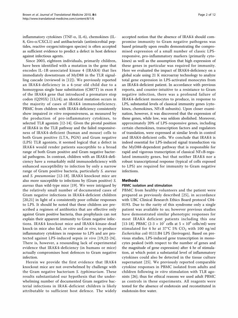
ResultsIRAK4-deficient mice survive infection by the Gramnegative pathogen Salmonella typhimuriumTo validate the unusually low occurrence of Gram-nega-tive infections in IRAK4-deficient children, IRAK4-defi-cient mice were challenged with the Gram negativebacterium, S. typhimurium. Results shown in Figure 1demonstrated no significant difference in survival ratesbetween IRAK4-deficient mice and wild type mice fol-lowing challenge with S. typhimurium. Thus, theabsence of IRAK4 does not render animals defencelessagainst a Gram negative bacterial challenge. These datatogether with that of Suzuki et al demonstrated thatIRAK4 knockout mice are highly susceptible to Grampositive [19] but not necessarily Gram negative bacterialinfections (Figure 1), an infection profile that emulatesthat of children with an IRAK4 deficiency. Thus thesedata strengthen our hypothesis that immunity, asopposed to prophylaxis or a lack of exposure to Gramnegative bacteria, is responsible for the underwhelmingnumber of documented Gram negative infections inchildren with IRAK4-deficiency.
Gene expression was generally severely compromised inIRAK4-deficient monocytes stimulated with LPSThe long-standing assumption that any deficiency in themajor TLR to NF�B pathway (such as an IRAK4-defi-ciency) would compromise immunity to Gram negativebacteria [34-36] was based on a substantial amount ofpublished data demonstrating compromised expressionin vitro of a limited set of LPS-induced, early-response,NF�B-regulated, pro-inflammatory markers in cells defi-cient for various components of the TLR to NF�B path-way (including IRAK4). A functional genomics approachwas utilized here to view the broader impact of anIRAK4 deficiency on LPS-induced gene expression.
Brown et al. Journal of Translational Medicine 2010, 8:6http://www.translational-medicine.com/content/8/1/6
Page 3 of 12
Blood monocytes were obtained from an IRAK4-defi-cient child and subsequently stimulated in vitro in twoindependent experiments, two months apart. Withhuman 21 K oligo-based DNA microarray technology,the expression of genes in IRAK4-deficient monocytesfrom each sampling was evaluated, in triplicate, relativeto gene expression in monocytes from five healthy indi-viduals. Previous studies that employed a similar metho-dology [28,29] served as a reference to confirm normal
LPS-responsive gene expression in the healthy, controlpool of monocytes.The expression of more than 500 genes in IRAK4-defi-
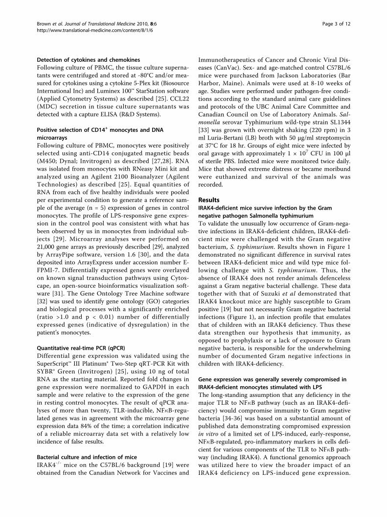
cient cells stimulated with LPS was significantly differentcompared to that in control cells (567 genes, differentialfold change ± 1.5, Student’s t-test p-value ≤ 0.05, resultsdeposited in ArrayExpress, accession number E-FPMI-7).An overlay of the gene expression data on a protein mapof the TLR-to-NF�B signal transduction pathway illu-strated that the majority of differentially expressed genesassociated with this pathway were i) suppressed relativeto gene expression in control monocytes and ii) tendedto congregate downstream of the I�B-NF�B complex(Figure 2). Gene ontology (GO) analysis [32] of the differ-entially expressed genes predicted that 56 GO categorieswere perturbed in IRAK4-deficient patients; these consti-tuted 5 major biological processes (p < 0.01) namely‘immune response’, ‘response to biotic stimulus’,‘response to stress’, ‘cell adhesion’ and ‘negative regula-tion of cellular processes’ (data not shown).Thus, the microarray analysis of monocytes from an
IRAK4-deficient child revealed a substantial disturbancein LPS-induced gene expression, in particular in genesregulated by the TLR-to-NF�B pathway and associatedwith biological processes related to immunity and cellu-lar responsiveness to stimuli. This global analysis ofgene expression in IRAK4-deficient monocytes was inaccordance with previously published results that ana-lyzed small subsets of inflammatory genes/proteins andconfirmed the previously ascribed role of IRAK4 inmediating signalling from TLR4 to genes downstream ofNF�B.
Figure 1 IRAK4-deficient mice maintain defence against Gramnegative bacteria. IRAK4 -/- (dotted line) and age- and sex-matched C57BL wild type (solid line) mice were infected with S.typhimurium SL1344 and monitored for survival (y-axis) over time (x-axis). The result is representative of two independent experimentswhere lack of statistical significance (p = 0.69 and p = 0.13) fordifferential survival between two groups of eight infected mice wasdetermined by the chi-square test.
Figure 2 Differentially expressed genes in LPS-stimulated IRAK4-deficient monocytes downstream of NF�B. Using the open-sourcebioinformatics visualization software Cytoscape [31] gene expression in LPS-stimulated, IRAK4-deficient monocytes was overlayed on the TLR4signal transduction pathway downstream of NF�B. Gene names are given in the left panel and gene expression in the right panel. The relativeexpression of the gene in IRAK4-deficient monocytes is depicted by the colour of the nodes on the protein map; green nodes indicate lowerexpression and red nodes indicate higher expression in IRAK4-deficient monocytes relative to that in a control pool of monocytes.
Brown et al. Journal of Translational Medicine 2010, 8:6http://www.translational-medicine.com/content/8/1/6
Page 4 of 12
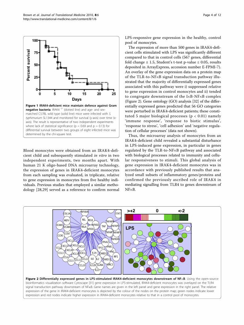
Expression of NF�B-regulated, pro-inflammatory geneswas compromised to varying degrees in IRAK4-deficientmonocytesCytokines, chemokines and certain antimicrobial pep-tides are TLR-sensitive, early response genes of theinnate immune system that are commonly used to pre-dict host defence and infectious disease risk. A subset ofthese TLR-to-NF�B-regulated immunity genes revealedin the microarray analyses were selected for furtherinvestigation. Genes were selected if they fit one ormore of the following criteria (i) the gene was regulatedby cellular activation with LPS (ii) the gene was a classicpro-inflammatory mediator, for e.g., a cytokine (TNF-a,IL-6), chemokine (IL-8, Gro-a/CXCL1) or antibacterial(DEFB1) (iii) in previously published work, alteration ofthe expression of the gene or protein product, either invitro or in vivo, was predicted to influence host defence,or (iv) the gene served as an internal control (for e.g.,induction of NF�B subunits). Real-time quantitativePCR (qPCR) and ELISA were used to confirm themicroarray results for relative gene expression betweencontrol and IRAK4-deficient monocytes, and to establishthe absolute expression of mRNA and protein prior toand following stimulation with LPS.The expression of classic pro-inflammatory cytokines
and chemokines including TNF-a, IL-6, IL-12b, IL-8,Gro-a/CXCL1, MCP-2/CCL8 and MIP-3a/CCL20 wascompromised in LPS-stimulated IRAK4-deficient mono-cytes compared to LPS-stimulated control monocytes(Figure 3 and Table 1). While the expression of thesegenes was unanimously compromised, the degree ofsuppression varied considerably, from 5- to 100- foldreduction in expression (Figure 3A, Table 1). The rela-tive level of protein expressed by PBMC was also heavilycompromised (ELISA shown for TNF-a, IL-8, IL-6 inFigure 3B). These data were consistent with previousreports of low levels of cytokines detected in the TCS ofLPS-stimulated PBMC from this patient and other indi-viduals with mutations in the IRAK4 gene [12-18] aswell as diminished, but not absent levels of TNF-a andIL-6 in the serum of IRAK4-kinase dead (KD) knockinmice following in vivo administration of LPS [37]. Thesedata (Figure 2, Figure 3, Table 1) illustrated that genetranscription and protein secretion of key pro-inflamma-tory mediators, while highly variable, was not completelyabolished in IRAK4-deficient monocytes exposed to LPS.
Identification of LPS-responsive, IRAK4-independentimmunity genesWhile LPS-induced expression of the majority of NF�B-regulated genes was lowered in IRAK4-deficient mono-cytes, the TLR-to-NF�B pathway map (shown in Figure2) revealed a subset of genes that were expressed atsimilar (white nodes) or higher (red nodes) levels in
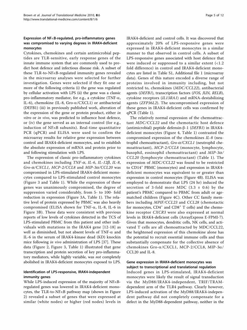
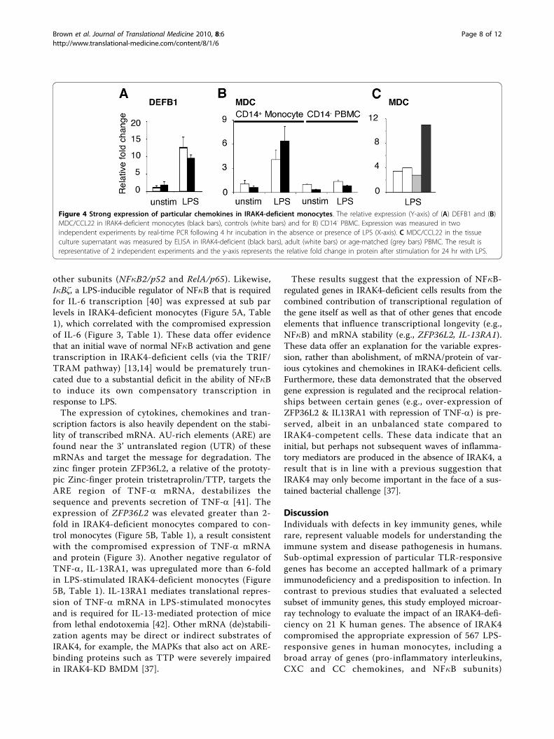
IRAK4-deficient and control cells. It was discovered thatapproximately 20% of LPS-responsive genes wereexpressed in IRAK4-deficient monocytes in a similarmanner to that observed in control cells. A subset ofLPS-responsive genes associated with host defence thatwere induced or suppressed to a similar extent (<1.2fold difference) in control and IRAK4-deficient mono-cytes are listed in Table S1, Additional file 1 (microarraydata). Genes of this nature encoded a diverse range ofproteins involved in immunity including, but notrestricted to, chemokines (MDC/CCL22), antibacterialagents (DEFB1), transcription factors (FOS, JUN, RELB),cytokine receptors (IL13RA1) and mRNA-destabilizingagents (ZFP36L2). The uncompromised expression ofthese genes in IRAK4-deficient cells was confirmed byqPCR (Table 1).The relatively normal expression of the chemoattrac-
tant MDC/CCL22 and the chemotactic host defence(antimicrobial) peptide defensin-b-1 (DEFB1) in IRAK4-deficient monocytes (Figure 4, Table 1) contrasted thecompromised expression of the chemokines IL-8 (neu-trophil chemoattractant), Gro-a/CXCL1 (neutrophil che-moattractant), MCP-2/CCL8 (monocyte, lymphocyte,basophil, eosinophil chemoattractant) and MIP-3a/CCL20 (lymphocyte chemoattractant) (Table 1). Theexpression of MDC/CCL22 was found to be restrictedto CD14+ PBMC (monocytes) and expression in IRAK4-deficient monocytes was equivalent to or greater thanexpression in control monocytes (Figure 4B). ELISA wasemployed to demonstrate that LPS (24 hr) induced thesecretion of 3-fold more MDC (3.3 ± 0.6) by thepatient’s PBMC compared to PBMC from adult or age-matched children (Figure 4C). Other CC family mem-bers including MPIF/CCL23 and CCL28 (chemotacticfor monocytes, CD4+ and CD8+ T cells) and the chemo-kine receptor CXCR3 were also expressed at normallevels in IRAK4-deficient cells (ArrayExpress E-FPMI-7).Given that monocytes, dendritic cells, NK cells, and acti-vated T cells are all chemoattracted by MDC/CCL22,the heightened expression of this chemokine alone hasthe potential to recruit essential immune cells and thussubstantially compensate for the collective absence ofchemokines Gro-a/CXCL1, MCP-2/CCL8, MIP-3a/CCL20 and IL-8.
Gene expression in IRAK4-deficient monocytes wassubject to transcriptional and translational regulationInduced genes in LPS-stimulated, IRAK4-deficientmonocytes were likely the result of signal transductionvia the MyD88/IRAK4-independent, TRIF/TRAM-dependent arm of the TLR4 pathway. Clearly however,LPS-induced activation of the MyD88/IRAK4-indepen-dent pathway did not completely compensate for adefect in the MyD88-dependent pathway, neither in the
Brown et al. Journal of Translational Medicine 2010, 8:6http://www.translational-medicine.com/content/8/1/6
Page 5 of 12

number of expressed genes nor magnitude of theirexpression.Kinetic models have estimated that MyD88/IRAK4-
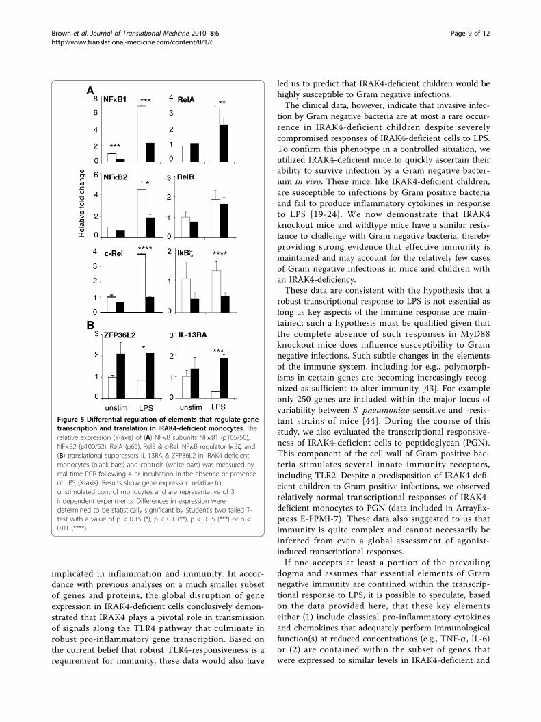
independent activation of NF�B via TRIF/TRAM lagsthe MyD88-dependent pathway by approximately 30min [38]. We and others previously demonstrated thatcells from the IRAK4-deficient patient [13,14] andMyD88-deficient mice [39] do indeed activate NF�Bshortly after exposure to LPS. The depletion of cytoplas-mic pools of NF�B as a consequence of nuclear
translocation stimulates new transcription of NF�B sub-units in order to replenish cytoplasmic supplies. Thisphenomenon was observed here (Figure 5A, Table 1) forLPS-activated control monocytes that induced the com-pensatory expression of all five NF�B subunits: NF�B1/p50, NF�B2/p52, RelA/p65, RelB and c-Rel. The com-pensatory expression of all subunits except RelB washowever diminished in IRAK4-deficient cells. Theexpression of NF�B1/p50 and c-Rel was significantly (p< 0.05) more impaired after LPS activation than two
Figure 3 Expression of inflammatory cytokines and chemokines in IRAK4-deficient monocytes. A Expression of IL-6, IL-8, IL-12b, TNF-a,Gro-a/CXCL1 and MIP-3a/CCL20 in the healthy control pooled monocytes (white bars) and in both biological replicates of the patient’smonocytes (black bars) were evaluated, in duplicate, by qPCR following a 4 hr incubation in the absence or presence of LPS (X-axis). B IL-6, IL-8and TNF-a were also measured in the tissue culture supernatant of stimulated PBMC by multiplex cytokine bead immunoassays. The Y-axisrepresents fold change (log scale) in gene expression relative to unstimulated control monocytes (qPCR) or pg/ml of protein determined by thecytokine assay. Differences in expression or protein secretion were determined to be statistically significant by Student’s two tailed T-test with avalue of p < 0.15 (*), p < 0.1 (**), p < 0.05 (***) and p < 0.003 (****).
Brown et al. Journal of Translational Medicine 2010, 8:6http://www.translational-medicine.com/content/8/1/6
Page 6 of 12
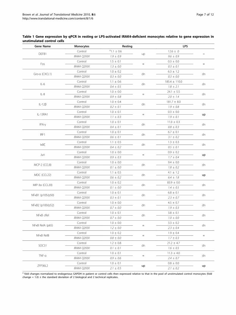
Table 1 Gene expression by qPCR in resting or LPS-activated IRAK4-deficient monocytes relative to gene expression inunstimulated control cells
Gene Name Monocytes Resting LPS
DEFB1Control a1.1 ± 0.6
up12.6 ± .0
=IRAK4 Q293X 1.9 ± 0.9 9.6 ± 0.9
FosControl 1.5 ± 0.1
=0.3 ± 0.0
=IRAK4 Q293X 1.3 ± 0.0 0.3 ± 0.1
Gro-α (CXCL1)Control 1.0 ± 0.2
dn6.3 ± 1.2
dnIRAK4 Q293X 0.3 ± 0.0 0.3 ± 0.0
IL-6Control 1.1 ± 0.6
dn185.4 ± 110.0
dnIRAK4 Q293X 0.4 ± 0.5 1.8 ± 2.1
IL-8Control 1.0 ± 0.0
=24.1 ± 5.5
dnIRAK4 Q293X 0.9 ± 0.8 2.0 ± 1.4
IL-12βControl 1.0 ± 0.4
dn181.7 ± 8.0
dnIRAK4 Q293X 0.2 ± 0.1 1.9 ± 0.8
IL-13RA1Control 1.0 ± 0.1
=0.3 ± 0.0
upIRAK4 Q293X 1.1 ± 0.3 1.9 ± 0.1
IFN-γControl 1.0 ± 0.1
dn11.0 ± 0.3
dnIRAK4 Q293X 0.4 ± 0.1 0.8 ± 0.3
IRF1Control 1.0 ± 0.1
dn6.7 ± 0.1
dnIRAK4 Q293X 0.6 ± 0.1 3.1 ± 0.2
IκBζControl 1.1 ± 0.5
dn1.3 ± 0.3
dnIRAK4 Q293X 0.4 ± 0.2 0.5 ± 0.1
JunControl 1.0 ± 0.0
=0.9 ± 0.2
upIRAK4 Q293X 0.9 ± 0.3 1.7 ± 0.4
MCP-2 (CCL8)Control 1.0 ± 0.0
dn9.4 ± 0.0
dnIRAK4 Q293X 0.1 ± 0.0 1.8 ± 0.2
MDC (CCL22)Control 1.1 ± 0.5
dn4.1 ± 1.2
upIRAK4 Q293X 0.6 ± 0.2 6.4 ± 1.8
MIP-3α (CCL20)Control 1.0 ± 0.2
dn83.9 ± 0.0
dnIRAK4 Q293X 0.1 ± 0.0 1.4 ± 0.5
NFκB1 (p105/p50)Control 1.0 ± 0.1
dn6.8 ± 0.1
dnIRAK4 Q293X 0.3 ± 0.1 2.3 ± 0.7
NFκB2 (p100/p52)Control 1.0 ± 0.0
dn4.5 ± 0.7
dnIRAK4 Q293X 0.7 ± 0.0 1.9 ± 0.3
NFκB cRelControl 1.0 ± 0.1
dn3.8 ± 0.1
dnIRAK4 Q293X 0.7 ± 0.0 1.0 ± 0.0
NFκB RelA (p65)Control 1.0 ± 0.0
=3.3 ± 0.2
dnIRAK4 Q293X 1.2 ± 0.0 2.3 ± 0.4
NFκB RelBControl 1.0 ± 0.2
=1.9 ± 0.4
=IRAK4 Q293X 0.8 ± 0.0 1.7 ± 0.3
SOCS1Control 1.2 ± 0.8
dn21.2 ± 4.7
dnIRAK4 Q293X 0.1 ± 0.1 1.6 ± 0.5
TNF-αControl 1.0 ± 0.1
=11.3 ± 4.0
dnIRAK4 Q293X 0.9 ± 0.6 2.4 ± 0.7
ZFP36L2Control 1.0 ± 0.1
up0.8 ± 0.0
upIRAK4 Q293X 2.1 ± 0.5 2.1 ± 0.2
a fold changes normalized to endogenous GAPDH in patient or control cells then expressed relative to that in the pool of unstimulated control monocytes (foldchange = 1.0) ± the standard deviation of 2 biological and 2 technical replicates.
Brown et al. Journal of Translational Medicine 2010, 8:6http://www.translational-medicine.com/content/8/1/6
Page 7 of 12
other subunits (NF�B2/p52 and RelA/p65). Likewise,I�Bζ, a LPS-inducible regulator of NF�B that is requiredfor IL-6 transcription [40] was expressed at sub parlevels in IRAK4-deficient monocytes (Figure 5A, Table1), which correlated with the compromised expressionof IL-6 (Figure 3, Table 1). These data offer evidencethat an initial wave of normal NF�B activation and genetranscription in IRAK4-deficient cells (via the TRIF/TRAM pathway) [13,14] would be prematurely trun-cated due to a substantial deficit in the ability of NF�Bto induce its own compensatory transcription inresponse to LPS.The expression of cytokines, chemokines and tran-
scription factors is also heavily dependent on the stabi-lity of transcribed mRNA. AU-rich elements (ARE) arefound near the 3’ untranslated region (UTR) of thesemRNAs and target the message for degradation. Thezinc finger protein ZFP36L2, a relative of the prototy-pic Zinc-finger protein tristetraprolin/TTP, targets theARE region of TNF-a mRNA, destabilizes thesequence and prevents secretion of TNF-a [41]. Theexpression of ZFP36L2 was elevated greater than 2-fold in IRAK4-deficient monocytes compared to con-trol monocytes (Figure 5B, Table 1), a result consistentwith the compromised expression of TNF-a mRNAand protein (Figure 3). Another negative regulator ofTNF-a, IL-13RA1, was upregulated more than 6-foldin LPS-stimulated IRAK4-deficient monocytes (Figure5B, Table 1). IL-13RA1 mediates translational repres-sion of TNF-a mRNA in LPS-stimulated monocytesand is required for IL-13-mediated protection of micefrom lethal endotoxemia [42]. Other mRNA (de)stabili-zation agents may be direct or indirect substrates ofIRAK4, for example, the MAPKs that also act on ARE-binding proteins such as TTP were severely impairedin IRAK4-KD BMDM [37].
These results suggest that the expression of NF�B-regulated genes in IRAK4-deficient cells results from thecombined contribution of transcriptional regulation ofthe gene itself as well as that of other genes that encodeelements that influence transcriptional longevity (e.g.,NF�B) and mRNA stability (e.g., ZFP36L2, IL-13RA1).These data offer an explanation for the variable expres-sion, rather than abolishment, of mRNA/protein of var-ious cytokines and chemokines in IRAK4-deficient cells.Furthermore, these data demonstrated that the observedgene expression is regulated and the reciprocal relation-ships between certain genes (e.g., over-expression ofZFP36L2 & IL13RA1 with repression of TNF-a) is pre-served, albeit in an unbalanced state compared toIRAK4-competent cells. These data indicate that aninitial, but perhaps not subsequent waves of inflamma-tory mediators are produced in the absence of IRAK4, aresult that is in line with a previous suggestion thatIRAK4 may only become important in the face of a sus-tained bacterial challenge [37].
DiscussionIndividuals with defects in key immunity genes, whilerare, represent valuable models for understanding theimmune system and disease pathogenesis in humans.Sub-optimal expression of particular TLR-responsivegenes has become an accepted hallmark of a primaryimmunodeficiency and a predisposition to infection. Incontrast to previous studies that evaluated a selectedsubset of immunity genes, this study employed microar-ray technology to evaluate the impact of an IRAK4-defi-ciency on 21 K human genes. The absence of IRAK4compromised the appropriate expression of 567 LPS-responsive genes in human monocytes, including abroad array of genes (pro-inflammatory interleukins,CXC and CC chemokines, and NF�B subunits)
Figure 4 Strong expression of particular chemokines in IRAK4-deficient monocytes. The relative expression (Y-axis) of (A) DEFB1 and (B)MDC/CCL22 in IRAK4-deficient monocytes (black bars), controls (white bars) and for B) CD14- PBMC. Expression was measured in twoindependent experiments by real-time PCR following 4 hr incubation in the absence or presence of LPS (X-axis). C MDC/CCL22 in the tissueculture supernatant was measured by ELISA in IRAK4-deficient (black bars), adult (white bars) or age-matched (grey bars) PBMC. The result isrepresentative of 2 independent experiments and the y-axis represents the relative fold change in protein after stimulation for 24 hr with LPS.
Brown et al. Journal of Translational Medicine 2010, 8:6http://www.translational-medicine.com/content/8/1/6
Page 8 of 12
implicated in inflammation and immunity. In accor-dance with previous analyses on a much smaller subsetof genes and proteins, the global disruption of geneexpression in IRAK4-deficient cells conclusively demon-strated that IRAK4 plays a pivotal role in transmissionof signals along the TLR4 pathway that culminate inrobust pro-inflammatory gene transcription. Based onthe current belief that robust TLR4-responsiveness is arequirement for immunity, these data would also have
led us to predict that IRAK4-deficient children would behighly susceptible to Gram negative infections.The clinical data, however, indicate that invasive infec-
tion by Gram negative bacteria are at most a rare occur-rence in IRAK4-deficient children despite severelycompromised responses of IRAK4-deficient cells to LPS.To confirm this phenotype in a controlled situation, weutilized IRAK4-deficient mice to quickly ascertain theirability to survive infection by a Gram negative bacter-ium in vivo. These mice, like IRAK4-deficient children,are susceptible to infections by Gram positive bacteriaand fail to produce inflammatory cytokines in responseto LPS [19-24]. We now demonstrate that IRAK4knockout mice and wildtype mice have a similar resis-tance to challenge with Gram negative bacteria, therebyproviding strong evidence that effective immunity ismaintained and may account for the relatively few casesof Gram negative infections in mice and children withan IRAK4-deficiency.These data are consistent with the hypothesis that a
robust transcriptional response to LPS is not essential aslong as key aspects of the immune response are main-tained; such a hypothesis must be qualified given thatthe complete absence of such responses in MyD88knockout mice does influence susceptibility to Gramnegative infections. Such subtle changes in the elementsof the immune system, including for e.g., polymorph-isms in certain genes are becoming increasingly recog-nized as sufficient to alter immunity [43]. For exampleonly 250 genes are included within the major locus ofvariability between S. pneumoniae-sensitive and -resis-tant strains of mice [44]. During the course of thisstudy, we also evaluated the transcriptional responsive-ness of IRAK4-deficient cells to peptidoglycan (PGN).This component of the cell wall of Gram positive bac-teria stimulates several innate immunity receptors,including TLR2. Despite a predisposition of IRAK4-defi-cient children to Gram positive infections, we observedrelatively normal transcriptional responses of IRAK4-deficient monocytes to PGN (data included in ArrayEx-press E-FPMI-7). These data also suggested to us thatimmunity is quite complex and cannot necessarily beinferred from even a global assessment of agonist-induced transcriptional responses.If one accepts at least a portion of the prevailing
dogma and assumes that essential elements of Gramnegative immunity are contained within the transcrip-tional response to LPS, it is possible to speculate, basedon the data provided here, that these key elementseither (1) include classical pro-inflammatory cytokinesand chemokines that adequately perform immunologicalfunction(s) at reduced concentrations (e.g., TNF-a, IL-6)or (2) are contained within the subset of genes thatwere expressed to similar levels in IRAK4-deficient and
Figure 5 Differential regulation of elements that regulate genetranscription and translation in IRAK4-deficient monocytes. Therelative expression (Y-axis) of (A) NF�B subunits NF�B1 (p105/50),NF�B2 (p100/52), RelA (p65), RelB & c-Rel, NF�B regulator I�Bζ, and(B) translational suppressors IL-13RA & ZFP36L2 in IRAK4-deficientmonocytes (black bars) and controls (white bars) was measured byreal-time PCR following 4 hr incubation in the absence or presenceof LPS (X-axis). Results show gene expression relative tounstimulated control monocytes and are representative of 3independent experiments. Differences in expression weredetermined to be statistically significant by Student’s two tailed T-test with a value of p < 0.15 (*), p < 0.1 (**), p < 0.05 (***) or p <0.01 (****).
Brown et al. Journal of Translational Medicine 2010, 8:6http://www.translational-medicine.com/content/8/1/6
Page 9 of 12
control monocytes (e.g., MDC/CCL22, DEFB1). Consis-tent with these concepts, it was recently demonstratedthat innate defence regulator peptides, which suppresspro-inflammatory cytokines but substantially maintainchemokine responses, are able to protect against Salmo-nella infections [30]. The functional redundancy of che-mokines is an example of how the expression of just afew genes might be sufficient to support critical immu-nological functions such as cellular recruitment, despitethe severe impairment of the expression of other familymembers. As demonstrated here, the expression of IL-8/CXCL8, Gro-a/CXCL1, MCP-2/CCL8 and MIP-3a/CCL20 was compromised in LPS-stimulated IRAK4-deficient monocytes (Figure 3, Table 1), suggesting adiminished recruitment of neutrophils, monocytes, lym-phocytes, basophils and eosinophils to sites of infection.However, the chemokines MDC/CCL22, MPIF/CCL23,CCL28 and DEFB1 were robustly expressed in theabsence of IRAK4 (Figure 5 and ArrayExpress), implyingthat monocytes, dendritic cells, natural killer cells, mem-ory T cells, and activated CD4+ and CD8+ T cells, parti-cularly CD4+ T-helper-2 (Th2) cells could still bemobilized in response to LPS. The enhanced recruit-ment of Th2 cells that express IL-4 and IL-13 would beconsistent with the elevated expression of IL-13RA1(Figure 5), as well as reports of relatively normal anti-body responses to vaccination in some children withIRAK4-deficiency [15,16]. Similar to our findings inhuman IRAK4-deficient monocytes, a number of che-mokines were expressed at similar levels in murineBMDM from wildtype and IRAK4-kinase-defectiveknock-in mice, such as CXCL2, CXCL10, CXCL11,CCL2 and CCL4 [Clusters I and III in [37]]. We proposethat the observed differential expression of chemokinesis sufficient to appropriately change the cellular milieuat the site of infection and favourably impact on theoutcome to infection [45]. Without supporting in vivodata however, these results are at best speculative. Itshould however be noted that infections in patients withIRAK4-deficiency are pyogenic (pus-forming) and canlead to mild fever and inflammation at late stages ofinfection [12]. This in vivo evidence implies that certaininflammatory mediators must have been produced andthat cells are actively recruited to the site of infection inIRAK4-deficient individuals. Furthermore, TNFa andIL-6 were detected, albeit at low levels in the serum ofIRAK4-KD mice following administration of LPS [37].Other potential candidate elements that might mediate
defence include the >60 upregulated genes and 20 downregulated genes listed in Table S1, Additional file 1(genes with similar or exaggerated expression in IRAK4-deficient cells compared to controls). Some of thesegenes in the ADAM, ICAM, integrin and NF�B familieswere also regulated in a similar fashion in IRAK4-KD
and control BMDM stimulated with LPS [37]. AlthoughGO analysis did not predict a disturbance in any majorbiological process in resting (unstimulated) IRAK4-defi-cient monocytes, we identified more than 50 LPS-responsive genes that were expressed in resting IRAK4-deficient cells not exposed to LPS (Table S2, Additionalfile 2). Notable genes include cytokines & chemokines(IL1F9, MIP-2a/CXCL2), transcription factors & tran-scriptional regulators (c-REL, NFKB1A, IBRDC2,UBE2N, USP9Y, BACH1), cell adhesion molecules(CD44), signalling molecules (MAPK6, PRKAG2,CALM3) and other pro-inflammatory mediators(ALOX5, HIF3a, HLA-DMA, LILRA3). It is temptingspeculate that these differentially expressed genes inunstimulated, IRAK4-deficient cells may also contributeto successful host defences.LPS-induced gene expression in IRAK4-deficient cells
is subject to regulation by both transcriptional andtranslational mechanisms and is the product of signaltransduction via either (1) the MyD88-independent,TRIF/TRAM pathway or (2) MyD88-dependent, IRAK4-independent pathways. Björkbacka and colleagues havedemonstrated that only 20% of more than 1000 LPS-responsive genes in macrophages are in fact dependenton MyD88 [46]. We however favour the hypothesis thata subset of MyD88-dependent, IRAK4-independentgenes are imperative for immunity since MyD88-defi-cient mice are susceptible to a broader range of patho-gens, including Gram negative bacteria, than are causedby IRAK4-deficiency in mice and humans. MyD88-dependent pathways that transduce signals independentof IRAK4 utilize signalling molecules such as PI3K, Btk,Tlp-2, and NIK and activate MAPKs and NF�B. Regard-less, it can be concluded that pathways other than theclassic MyD88-dependent pathway (via sequential acti-vation of IRAK4, IRAK1, TRAF6, IKK and NF�B) haveessential and under-appreciated roles in defence againstGram negative bacteria.
ConclusionsIn this study, we demonstrated that the expression ofthe vast majority of LPS-induced inflammatory andimmunity genes were compromised (compared to theexpression levels in control cells) in monocytes from achild with IRAK4 deficiency. We conclude that whileIRAK4 is imperative for a comprehensive transcriptionalresponse to LPS, neither IRAK4, nor this classical,robust transcriptional response is required for effectivehost defences against Gram negative infection. Instead,the data implies that sufficient defence could lie withina small repertoire of LPS-responsive, IRAK4-indepen-dent genes. A subset of transcribed genes amidst aseverely impaired response to LPS was also observed byKoziczak-Holbro et al in murine BMDM from IRAK4-
Brown et al. Journal of Translational Medicine 2010, 8:6http://www.translational-medicine.com/content/8/1/6
Page 10 of 12
kinase-defective knockin mice [37]. These observationsare consistent with the concept that it may be possibleto generate an effective immune response without arobust inflammatory response [47] and such propertiesmay even make IRAK4 an attractive drug target fortreating inflammation without compromising effectiveimmune defences [48].
Additional file 1: Table S1. Selection of LPS-responsive immunity genesthat were expressed to similar or exaggerated (italics) levels in IRAK4-deficient monocytes compared to controls as assayed by microarrayanalysisClick here for file[ http://www.biomedcentral.com/content/supplementary/1479-5876-8-6-S1.PDF ]
Additional file 2: Table S2. Selection of LPS-responsive genes expressedin unstimulated IRAK4-deficient monocytesClick here for file[ http://www.biomedcentral.com/content/supplementary/1479-5876-8-6-S2.PDF ]
AcknowledgementsWe gratefully acknowledge the support of Genome Prairie and Genome BCfor the Pathogenomics of Innate Immunity research program and theCanadian Institutes for Health Research (CIHR). KLB received support from apostdoctoral fellowship from the CIHR-UBC Strategic Training Program forTranslational Research in Infectious Diseases (TRID) and the EuropeanCommunity’s Seventh Framework Programme (FP7/2007-2013) under grantagreement number 221094. DJD is supported by the Wellcome Trust. JLGreceived a postdoctoral fellowship from the Michael Smith Foundation forHealth Research. DPS is supported by operating grants from the CIHR andthe Canadian Bacterial Diseases Network. B.B.F. is a CIHR DistinguishedInvestigator, a Howard Hughes Medical Institute International ResearchScholar, and the University of British Columbia Peter Wall DistinguishedProfessor. REWH is the recipient of a Canada Research Chair Award. Theauthors would like to thank Rebecca Ma, Yuexin Li, Dawn Bowdish, ShuYuFan, Jessica Li, George Song-Zhao, and Aaron Hirschfeld for technicalsupport, and the patient and his family for their cooperation.
Author details1Centre for Microbial Diseases and Immunity Research, Department ofMicrobiology and Immunology, University of British Columbia, Vancouver,British Columbia, V6T 1Z3, Canada. 2Child and Family Research Institute,University of British Columbia, 950 West 28th Avenue, Vancouver, BritishColumbia, V5Z 4H4, Canada. 3Current address: MRC/University of EdinburghCentre for Inflammation Research, Queen’s Medical Research Institute W2 03,47 Little France Crescent, Edinburgh EH16 4T, UK. 4Current address:Department of Rheumatology and Inflammation Research, University ofGothenburg, Guldhedsgatan 10, S-413 46 Gothenburg, Sweden.
Authors’ contributionsKLB carried out the in vitro cell culture, RNA isolation, qPCR, ELISA andmanuscript preparation. RF carried out the microarray analysis. WK carriedout the animal studies. JLG performed bioinformatics analyses. PH addedvaluable insight into data analysis, manuscript content and presentation.DJD, ST and BBF provided advice on the conception and design of theclinical and animal experiments. DPS and REWH were imperative to theconception, design and implementation of the experiments and preparationof the manuscript. All authors read and approved the final manuscript.
Competing interestsThe authors declare that they have no competing interests.
Received: 27 July 2009Accepted: 27 January 2010 Published: 27 January 2010
References1. Akira S, Uematsu S, Takeuchi O: Pathogen Recognition and Innate
Immunity. Cell 2005, 124:783-801.2. Martino F, Tschoop J: NLRs join TLRs as innate sensors of pathogens.
Trends Immunol 2005, 26:449-454.3. Akira S, Takeda K: Toll-like receptor signalling. Nat Rev Immunol 2004,
4:499-511.4. Dunne A, O’Neill LA: Adaptor usage and Toll-like receptor signalling
specificity. FEBS Lett 2005, 579:3330-3335.5. Kim T, Kim YJ: Overview of innate immunity in Drosophila. J Biochem Mol
Biol 2005, 38:121-127.6. Akira S: Toll-like receptors: lessons from knockout mice. Biochem Soc
Trans 2000, 28:551-556.7. Takeuchi O, Hoshino K, Akira S: Cutting edge: TLR2-deficient and MyD88-
deficient mice are highly susceptible to Staphylococcus aureus infection.J Immunol 2000, 165:5392-5396.
8. Skerrett SJ, Liggitt HD, Hajjar AM, Wilson CB: Cutting edge: myeloiddifferentiation factor 88 is essential for pulmonary host defence againstPseudomonas aeruginosa but not Staphylococcus aureus. J Immunol 2004,172:3377-3381.
9. Fremond CM, Yeremeev V, Nicolle DM, Jacobs M, Quesniaux VF, Ryffel B:Fatal Mycobacterium tuberculosi s infection despite adaptive immuneresponse in the absence of MyD88. J Clin Invest 2004, 114:1790-1799.
10. Feng CG, Scanga CA, Collazo-Custodio CM, Cheever AW, Hieny S, Caspar P,Sher A: Mice lacking myeloid differentiation factor 88 display profounddefects in host resistance and immune responses to Mycobacteriumavium infection not exhibited by Toll-like receptor 2 (TLR2)- and TLR4-deficient animals. J Immunol 2003, 171:4758-4764.
11. Seki E, Tsutsui H, Tsuji NM, Hayashi N, Adachi K, Nakano H, Futatsugi-Yumikura S, Takeuchi O, Hoshino K, Akira S, Fujimoto J, Nakanishi K: Criticalroles of myeloid differentiation factor 88-dependent proinflammatorycytokine release in early phase clearance of Listeria monocytogenes inmice. J Immunol 2002, 169:3863-3868.
12. Ku CL, Yang K, Bustamante Puel A, von Bernuth A, Santos OF, Lawrence T,Chang HH, Al-Mousa H, Picard C: Inherited disorders of human Toll-likereceptor signalling: immunological implications. Immunol Rev 2005,203:10-20.
13. Currie AJ, Davidson DJ, Reid GS, Bharya S, MacDonald KL, Devon RS,Speert DP: Primary immunodeficiency to pneumococcal infection due toa defect in Toll-like receptor signalling. J Pediatr 2004, 144:512-518.
14. Davidson DJ, Currie AJ, Bowdish DME, Brown KL, Rosenberger CM, Ma RC,Bylund J, Campsall PA, Puel A, Picard C, Casanova JL, Turvey SE, Devon RE,Hancock REW, Speert DP: IRAK-4 mutation (Q293X): rapid detection andcharacterization of defective post-transcriptional TLR/IL1R responses inhuman myeloid and non-myeloid cells. J Immunol 2006, 177:8202-8211.
15. Picard C, Puel A, Bonnet M, Ku CL, Bustamante J, Yang K, Soudais C,Dupuis S, Feinberg J, Fieschi C, Elbim C, Hitchcock R, Lammas D, Davies G,Al-Ghonaium A, Al-Rayes H, Al-Jumaah A, Al-Hajjar S, Al-Mohsen IZ,Frayha HH, Rucker R, Hawn TR, Aderem A, Tufenkji H, Haraguchi S, Day NK,Good RA, Gougerot-Pocidalo MA, Ozinsky A, Casanova JL: Pyogenicbacterial infections in humans with IRAK-4 deficiency. Science 2003,299:2076-2079.
16. Medvedev AE, Lentschat A, Kuhns DB, Blanco JC, Salkowski C, Zhang S,Arditi M, Gallin JI, Vogel SN: Distinct mutations in IRAK-4 Conferhyporesponsiveness to lipopolysaccharide and interleukin-1 in a patientwith recurrent bacterial infections. J Exp Med 2003, 198:521-531.
17. Chapel H, Puel A, von Bernuth H, Picard C, Casanova JL: Shigella sonneimeningitis due to interleukin-1 receptor-associated kinase-4 deficiency:first association with a primary immune deficiency. Clin Infect Dis 2005,40:1227-1231.
18. Enders A, Pannicke U, Berner R, Henneke P, Radlinger K, Schwarz K, Ehl S:Two siblings with lethal pneumococcal meningitis in a family with amutation in Interleukin-1 receptor-associated kinase 4. J Pediatr 2004,145:698-700.
19. Suzuki N, Suzuki S, Duncan GS, Millar DG, Wada T, Mirtsos C, Takada H,Wakeham A, Itie A, Li S, Penninger JM, Wesche H, Ohashi PS, Mak TW,Yeh WC: Severe impairment of interleukin-1 and Toll-like receptorsignalling in mice lacking IRAK-4. Nature 2002, 416:750-756.
20. Vosse van de E, van Dissel JT, Ottenhoff TH: Genetic deficiencies of innateimmune signalling in human infectious disease. Lancet Infect Dis 2009,9:688-698.
Brown et al. Journal of Translational Medicine 2010, 8:6http://www.translational-medicine.com/content/8/1/6
Page 11 of 12

21. Ku CL, von Bernuth H, Picard C, Zhang SY, Chang HH, Yang K, Chrabieh M,Issekutz AC, Cunningham CK, Gallin J, Holland SM, Roifman C, Ehl S,Smart J, Tang M, Barrat FJ, Levy O, McDonald D, Day-Good NK, Miller R,Takada H, Hara T, Al-Hajjar S, Al-Ghonaium A, Speert D, Sanlaville D, Li X,Geissmann F, Vivier E, Maródi L, Garty BZ, Chapel H, Rodriguez-Gallego C,Bossuyt X, Abel L, Puel A, Casanova JL: Selective predisposition tobacterial infections in IRAK-4-deficient children: IRAK-4-dependent TLRsare otherwise redundant in protective immunity. J Exp Med 2007,204:2407-2422.
22. Suzuki N, Suzuki S, Eriksson U, Hara H, Mirtosis C, Chen NJ, Wada T,Bouchard D, Hwang I, Takeda K, Fujita T, Der S, Penninger JM, Akira S,Saito T, Yeh WC: IL-1R-associated kinase 4 is required forlipopolysaccharide-induced activation of APC. J Immunol 2003,171:6065-6071.
23. Kawagoe T, Sato S, Jung A, Yamamoto M, Matsui K, Kato H, Uematsu S,Takeuchi O, Akira S: Essential role of IRAK-4 protein and its kinase activityin Toll-like receptor-mediated immune responses but not in TCRsignalling. J Exp Med 2007, 204:1013-1024.
24. Kim TW, Staschke K, Bulek K, Yao J, Peters K, Oh KH, Vandenburg Y, Xiao H,Qian W, Hamilton T, Min B, Sen G, Gilmour R, Li X: A critical role for IRAK4kinase activity in Toll-like receptor-mediated innate immunity. J Exp Med2007, 204:1025-1036.
25. Mookherjee N, Brown KL, Bowdish DME, Doria S, Falsafi R, Hokamp K,Roche RM, Mu R, Doho GH, Pistolic J, Powers JP, Bryan J, Brinkman FS,Hancock RE: Modulation of the Toll-like receptor-mediated inflammatoryresponse by the endogenous human host defence peptide LL-37. JImmunol 2006, 176:2455-2464.
26. Hirschfeld AF, Bettinger JA, Victor RE, Davidson DJ, Currie AJ, Ansermino JM,Scheifele DW, Orange JS, Turvey SE: Prevalence of Toll-like receptorsignalling defects in apparently healthy children who developedinvasive pneumococcal infection. Clin Immunol 2007, 122:271-8.
27. Brown KL, Maiti A, Johnson P: Role of sulfation in CD44-mediatedhyaluronan binding induced by inflammatory mediators in human CD14+ peripheral blood monocytes. J Immunol 2001, 167:5367-5374.
28. Brown KL, Bylund J, MacDonald KL, Song-Zhao GX, Elliott M, Falsafi R,Hancock R, Speert DP: ROS-deficient monocytes have aberrant geneexpression that correlates with inflammatory disorders of chronicgranulomatous disease. Clin Immunol 2008, 129:90-102.
29. Scott MG, Dullaghan E, Mookherjee N, Glavas N, Waldbrook M,Thompson A, Wang A, Lee K, Doria S, Hamill P, Yu JJ, Li Y, Donini O,Guarna MM, Finlay BB, North JR, Hancock REW: An anti-infective peptidethat selectively modulates the innate immune response. Nat Biotechnol2007, 25:465-472.
30. Hokamp K, Roche FM, Acab M, Rousseau ME, Kuo B, Goode D,Aeschliman D, Bryan J, Babiuk LA, Hancock REW, Brinkman FS: ArrayPipe: aflexible processing pipeline for microarray data. Nucl Acids Res 2004, 32:W457-459.
31. Shannon P, Markiel A, Ozier Q, Baliga NS, Wang JT, Ramage D, Amin N,Schwikowski B, Ideker T: Cytoscape: a software environment forintegrated models of biomolecular interaction networks. Genome Res2003, 13:2498-2504.
32. Zhang B, Schmoyer D, Kirov K, Snoddy J: GOTree Machine (GOTM): a web-based platform for interpreting sets of interesting genes using GeneOntology hierarchies. BMC Bioinformatics 2004, 5:16-23.
33. Hoiseth SK, Stocker BA: Aromatic-dependent Salmonella typhimurium arenon-virulent and effective as live vaccines. Nature 1981, 291:238-239.
34. Faure K, Sawa T, Ajayi T, Fujimoto J, Moriyama K, Shime N, Wiener-Kronish JP: TLR signalling is essential for survival in acute lung injuryinduced by virulent Pseudomonas aeruginosa secreting type III secretorytoxins. Respir Res 2004, 5:1-10.
35. Mahieu T, Park JM, Revets H, Paschel B, Lengeling A, Staelens J, Wullaert A,Vanlaere I, Hochepied T, van Roy F, Karin M, Libert C: The wild-derivedinbred mouse strain SPRET_Ei is resistant to LPS and defective in IFN-bproduction. PNAS 2006, 103:2292-2297.
36. Modlin RL, Brightbill HD, Godowski PJ: The Toll of Innate Immunity onMicrobial Pathogens. NEJM 1999, 340:1834-1835.
37. Koziczak-Holbro M, Glück A, Tschopp C, Mathison JC, Gram H: IRAK-4kinase activity-dependent and -independent regulation oflipopolysaccharide-inducible genes. Eur J Immunol 2008, 38:788-96.
38. Covert MW, Leung TH, Gaston JE, Baltimore D: Achieving stability oflipopolysaccharide-induced NF-kappaB activation. Science 2005,309:1854-1857.
39. Kawai T, Adachi O, Ogawa T, Takeda K, Akira S: Unresponsiveness ofMyD88-deficient mice to endotoxin. Immunity 1999, 11:115-122.
40. Yamamoto M, Yamazaki S, Uematsu S, Sato S, Hemmi H, Hoshino K,Kaisho T, Kuwata H, Takeuchi O, Takeshige K, Saitoh T, Yamaoka S,Yamamoto N, Yamamoto S, Muta Tm, Takeda K, Akira S: Regulation of Toll/IL-1-receptor-mediated gene expression by the inducible nuclear proteinIkappaBzeta. Nature 2004, 430:218-222.
41. Carballo E, Lai WS, Blackshear PJ: Feedback inhibition of macrophagetumor necrosis factor-alpha production by tristetraprolin. Science 1998,281:1001-1005.
42. Muchamuel T, Menon S, Pisacane P, Howard MC, Cockayne DA: IL-13protects mice from lipopolysaccharide-induced lethal endotoxemia:correlation with down-modulation of TNF-alpha, IFN-gamma, and IL-12production. J Immunol 1997, 158:2898-2903.
43. Pasare C, Medzhitov R: Control of B-cell responses by Toll-like receptors.Nature 2005, 438:364-368.
44. Denny P, Hopes E, Gingles N, Broman KW, McPheat W, Morten J,Alexander J, Andrew PW, Brown SD: A major locus conferringsusceptibility to infection by Streptococcus pneumoniae in mice. MammGenome 2003, 14:448-453.
45. Esche C, Stellato C, Beck LA: Chemokines: key players in innate andadaptive immunity. J Invest Dermatol 2005, 125:615-628.
46. Björkbacka H, Fitzgerald KA, Huet F, LI X, Gregory JA, Lee MA, Ordija CM,Dowley NE, Golenbock DT, Freeman MW: The induction of macrophagegene expression by LPS predominantly utilizes Myd88-independentsignaling cascades. Physiological Genomics 2004, 19:319-330.
47. Brown KL, Cosseau C, Gardy JL, Hancock REW: Complexities of targetinginnate immunity to treat infection. Trends Immunol 2007, 28:260-66.
48. Li X: IRAK4 in TLR/IL-1R signalling: possible clinical applications. Eur JImmunol 2008, 38:614-618.
doi:10.1186/1479-5876-8-6Cite this article as: Brown et al.: Robust TLR4-induced gene expressionpatterns are not an accurate indicator of human immunity. Journal ofTranslational Medicine 2010 8:6.
Submit your next manuscript to BioMed Centraland take full advantage of:
• Convenient online submission
• Thorough peer review
• No space constraints or color figure charges
• Immediate publication on acceptance
• Inclusion in PubMed, CAS, Scopus and Google Scholar
• Research which is freely available for redistribution
Submit your manuscript at www.biomedcentral.com/submit
Brown et al. Journal of Translational Medicine 2010, 8:6http://www.translational-medicine.com/content/8/1/6
Page 12 of 12