role of yeast beta glucan on blood coagulation in ...3) 60-2015.pdf · the role of yeast beta...
TRANSCRIPT

Arab Journal of Nuclear Science and Applications, 94 (2 ), (164-187) 2016
164
The Role of Yeast Beta Glucan on Blood Coagulation in Streptozotocin-
Induced Diabetes and Irradiated Rats
M.M.A, El-Kashoury1, S.M. Abdel Fattah1, L.A. Ramadan1 and E.S. El-Denshary2 1Department of Drug Radiation Research, National Center for Radiation Research and Technology,
Atomic Energy Authority,, Cairo, Egypt
2Department of Pharmacology and Toxicology, Faculty of Pharmacy, Cairo University, Egypt
Received: 20/2/2015 Accepted: 2/5/2015
ABSTRACT1
Clotting abnormalities are observed after exposure to ionizing radiation as
well as in diabetes melittus. The objective of this study is to elucidate the role of
yeast beta glucan (YBG) in the modulation of some biochemical variations observed
in -irradiated, diabetic and diabetic-irradiated rats.
Gamma-irradiation was performed through the whole body exposure of rats
to 6 Gy administered in four fractions of 1.5 Gy two times per week for two weeks.
Diabetes was induced by a single intraperitoneal injection of streptozotocin (55
mg/kg body weight). YBG was given orally to male albino rats (1 g/kg body weight)
for two weeks post irradiation and/or induction of diabetes.
Animals were divided into 4 main groups: 1- control, 2- -irradiated, 3-
diabetic and 4- diabetic--irradiated rats. Each group was subdiveded into 2
subgroups (a) untreated and (b) treated. The 3rd and 14th day, after the last dose of
radiation in the irradiated groups and after the induction of diabetes in diabetic
groups, were chosen to evaluate the effect of oral YBG in irradiated and/or diabetic
rats.
The results revealed that the body weight decreased significantly in irradiated,
diabetic and diabetic–irradiated rats. The loss of weight was accompanied by a
reduction in the pancreas weight. Glucose concentration was significantly increased
in diabetic group at the two time intervals. It is worth noting that, radiation
ameliorated blood glucose level in diabetic--irradiated group. Radiation exposure
and/or diabetes caused an oxidative stress manifested by a significant increase of malondialdhyde (MDA) accompanied by a significant decrease in glutathione (GSH)
level. This oxidative stress caused disturbances in the measured clotting parameters
by enhancing platelet aggregation (PA) induced by arachidonic acid and increased
thrombin level as concluded from the significant shortening of prothrombin time
(PT) and activated partial thromboplastin time (APTT). Also, exposure to radiation
and/or diabetes disturbed blood counts by decreasing red blood cells (RBCs); white
blood celld (WBCs) and platelets and increasing platelet indices platelet mean volume (MPV) and platelet distribution width (PDW) as well as disturbances in lipid
profile . The levels of these estimated parameters approached to normal levels by
YBG treatment.
The obtained results show that YBG ameliorate hyperglycemia, exhibit antiplatelet activity, acts as immune enhancer by improving blood counts ,
ameliorate dyslipidemia and atherosclerotic index. It is concluded that YBG
minimizes the harmful effects induced by exposure to ionizing radiation and reduces
various complications of diabetes
Key Words: Yeast Beta Glucan/ -irradiation/ Coagulation/ Diabetes Mellitus.
1 Corresponding author E-mail: [email protected]

Arab Journal of Nuclear Science and Applications, 94 (2 ), (164-187) 2016
165
INTRODUCTION
In recent years, a great deal of research has taken place in the area of free radicals and reactive oxygen species (ROS). It is now believed that they are either initiating or promoting agents in nearly every known disease (1). Aerobic cells have multiple defense mechanisms against free radical attack. There are both enzymatic defense systems and non-enzymatic antioxidants. Free radicals are generated in both the aqueous and lipid portions of the intracellular and extracellular environments. Therefore, it is crucial for the body to have a combination of water-soluble and lipid- soluble antioxidants to provide the full range of protection. Some antioxidants are synthesized by the body, whereas others must be obtained from food or food supplements (2). The oxidative stress that results from excessive free radical formation and limited antioxidant defense leads to changes in proteins, lipids, polysaccharides, and DNA in biological systems (3). Eventually, it results in pathophysiological conditions such as aging, cardiovascular disease, Alzheimer’s disease, and diabetes (4,5). In recent years, much attention has been focused on the role of oxidative stress, and it has been reported that oxidative stress may constitute the key and common event in the pathogenesis of secondary diabetic complications (6,7). So, one of the important goals of DM treatment is to prevent its complications (8).
Radiation injury of blood vessels was originally described more than a century ago and remains a contemporary clinical problem, despite dramatic advances in the field of radiation oncology (9). Ionizing radiation (IR) is associated with an increased risk of thrombotic occlusion of vessels and organ fibrosis (10,11). Also, Secondary vascular complications are frequently observed in patients with diabetes mellitus. The role of diabetes as an independent risk factor for cardiovascular disease has been well established in many studies (12).
Although almost all organisms possess antioxidant defense and repair systems that have evolved to protect them against free radicals, these systems are insufficient to protect them completely against oxidative damage (13). Many of the types of damage observed after irradiation can be ameliorated by antioxidants (14). Restriction on the use of synthetic antioxidants due to their carcinogenic nature has led to a growing interest in recent years in natural antioxidants of plant origin. The potential value of antioxidants has prompted researchers to look for natural antioxidants with low cytotoxicity (15).
Hofer and Pospisil(16) reported that glucan is a stimulator of the function of the reticuloendothelial system and a modulator of cellular and humoral immunity. Glucan was successfully tested from the point of view of stimulation of nonspecific immunity against bacterial, viral, mycotic and microparazitic infections, as well as against malignant cell growth. YBG has beneficial effects on the immune system and are claimed to have no toxic or adverse effects (8).
The aim of the present study is to evaluate the beneficial role of yeast beta glucan in minimizing radiation hazards and diabetic complications.
MATERIAL AND METHODS
Animals
Male Wistar rats purchased from the Research Institute of Ophthalmology (Giza, Egypt) weighing 150-200 g were used in this study. Animals were maintained under standard conditions of ventilation, temperature, humidity, lighting (light/dark: 13h/11h) and fed on standard pellets diet containing all nutritive elements (proteins, fats, carbohydrates, vitamins, salts and minerals). Food and water were available ad libitum.. All animal procedures were carried out in accordance to the Ethics Committee of the Faculty of Pharmacy, Cairo University conformed to the “Guide for the care and use of Laboratory Animals” published by the National Institutes of Health (NIH publication No. 85–23,
revised 1996).
Irradiation Procedure
Rats were whole body -irradiated at the National Center for Radiation Research and Technology (NCRRT), Cairo, Egypt, using an AECL Gamma cell-40, which is a Cesium-137 irradiation unit manufactured by Atomic Energy of Canada Limited. Animals received 6 Gy administered in four doses of 1.5 Gy two times per week for two weeks at a dose rate of 0.43 Gy/min.

Arab Journal of Nuclear Science and Applications, 94 (2 ), (164-187) 2016
166
Induction of Diabetes Diabetes was induced by a single intraperitoneal injection of streptozotocin (STZ) (Sigma, St.
Louis, MO, USA) (55 mg/kg body weight) to animals fasted overnight (17) in 0.1 citrate buffer PH 4.5 freshly prepared before injection. Diabetes was verified 48 h later by measuring tail vein blood glucose, and rats with blood glucose 300 mg/dl or more were considered diabetic (17).
Beta Glucan Treatment Yeast beta glucan (Shaanxi Sciphar Hi-Tech Industry Co., Ltd, China), was dissolved in
distilled water and administered to the experimental animals (1g/kg body weight) by oral intragastric tube daily for two weeks (18).
Experimental design and animal groups: Animals were categorized into four groups each one subdivided in two subgroups (a and b) as follows:
Group 1: Control groups a- Control group not treated with yeast beta glucan (30 rats): was given a single
intraperitoneal (i.p) injection of citrate buffer pH 4.5. b- Control group treated with yeast beta glucan (30 rats): given a single intraperitoneal (i.p)
injection of citrate buffer pH 4.5 and 48 h later received YBG treatment daily for two weeks. Group 2: Gamma irradiated groups a- Gamma-irradiated control group (45 rats): given a single intraperitoneal (i.p) injection of
citrate buffer pH 4.5 and 48 h later exposed to -irradiation regimen. b- Gamma-irradiated group treated with yeast beta glucan (45 rats): given a single
intraperitoneal (i.p) injection of citrate buffer pH 4.5 and 48 h later exposed to the gamma-
irradiation regimen then followed by YBG daily for two weeks starting post the last -radiation dose.
Group 3: Diabetic groups a- Diabetic control group (45 rats): rats with blood glucose 300 mg/dl or more. b- Diabetic group treated with yeast beta glucan (45 rats): Diabetic rats receiving YBG
treatment daily for two weeks starting 48 h after STZ injection. Group 4: Diabetic gamma irradiated groups
a- Diabetic--irradiated control group (n=45): Diabetic rats exposed to -radiation regimen 48 hr after STZ injection.
b- Diabetic--irradiated group treated with yeast beta glucan (n=45): Diabetic rats exposed to
-radiation regimen followed by YBG treatment daily during 2 weeks starting post the last -radiation dose.
Normal control rats and diabetic rats, not subjected to YBG treatment, were sacrified 3 and 14 days post citrate buffer or STZ injection, respectively.
-irradiated and diabetic--irradiated rats not subjected to YBG treatment were sacrified 3 and
14 days post the last -irradiation dose.
Normal control, -irradiated, diabetic and diabetic--irradiated rats given YBG were sacrified 3 and 14 days post YBG treatment.
Blood Sampling and Analysis }}}}
Rats were anaesthetized with diethyl ether; then weighed. Samples were withdrawn from the retro-orbital venous plexus. Plasma were separated using non-heparinized capillaries in sterile sodium fluoride tubes for measuring glucose level (Glu), in ethylene diamine tetra acetic acid (EDTA) tubes for measuring the haematological parameters complete blood count (CBC) and in sterile 3.2% sodium citrate tubes for measuring platelet aggregation test (PA), prothrombin time (PT) and activated partial thrombinoplastin time (APTT) and heparinized capillaries for measuring malondialdhyde (MDA) and glutathione (GSH). Serum was separated using non heparinized capillaries in sterile plain tubes for measuring lipid profile

Arab Journal of Nuclear Science and Applications, 94 (2 ), (164-187) 2016
167
For the determination of body and pancreas weight: Rats were weighed then killed by cervical dislocation, the pancreas was immediately excised washed with cold saline, blotted and weighed.
Glucose was determined using reagent kit (Stanbio, USA), according to Young (19). Malondialdehyde (MDA) and reduced glutathione (GSH) levels were determined according to Yoshioka et al. (20) and Beutler et al. (21), respectively
Platelet aggregation test was determined using reagent kit (Bio/Data Corporation, USA) using Apact 4004, platelet aggregometer, Germany according to McCabe-White & Jennings (22) and Bain et al. (23).
Prothrombin time (PT) and activated partial thromboplastin time (APTT) were determined using reagent kit (Biomed, Egypt), as indicated by Hirsh et al. (24) and CRC (25), respectively.
The haematological parameters including haemoglobin (Hb), haematocrit (Hct), red blood cells count (RBCs), packed cell volume (PCV), mean corpuscular haemoglobin (MCH), mean corpuscular haemoglobin concentration (MCHC), platelet count, mean platelet volume (MPV), platelet distribution width (PDW), total leucucytic count (TLC), neutrophils, lymphocytes, monocytes, eosinophils and basophils were determined using Sysmex XE 2100 and XT 2000i operator manual, USA as indicated by Bain et al. (23).
For the determination of lipid profile: Total lipids were determined using reagent kit (Constant Medical Scientific, Egypt), according to the method of Frings et al.(26). Total cholesterol (TC), triglycerides (TG) and high density lipoprotein-cholesterol (HDL-C) were determined using the corresponding reagent kit (Stanbio, USA) as described by Stein (27), Buccolo & David (28), and NIHP(29), respectively. Low density lipoprotein-cholesterol (LDL-C), very low density lipoprotein-cholesterol (VLDL-C) and atherogenic index (AI) were determined according to Friedewald et al. (30) equations: LDL-C = TC- HDL-C- (TG/5); VLDL-C = TG/5 and AI = (TC- HDL-C)/ HDL-C. Antiatherogenic index (AAI) was determined according to Guido & Joseph (31) AAI = (HDL-C×100)/(TC-HDL-C). In addition, risk factor TC/HDL-C ratio, TC/LDL-C ratio and HDL-C/LDL-C ratio were determined according to the previous parameters.
All biochemical determination were done using spectro-photometric technique using UNICAM 8625,UV/VIS, England, Spectrophotometer.
Methods of disposal of remaining animals:Animals were disposed though the holocaust of Atomic Energy Authority.
Statistical analysis: Results are given as means ± SD. Comparisons between means were carried out using one-way ANOVA followed by the Tukey-kramer multiple comparisons test using Instat software, version 2 (Graphpad Software, Inc., Diego, USA).
RESULTS
Effect of YBG on Blood Glucose Level, Body Weight and Pancreas Weight in Different Groups
of Rats (Fig. 1 A, B and C)
The diabetic control group displayed a highly significant increase in blood glucose level when
compared to control, -irradiated and diabetic -irradiated groups (p <0.01). While, with all other
groups including -irradiated, diabetic -irradiated control and treated groups, a highly significant decrease in blood glucose level occurred gradually during both intervals approaching the normal control level (p <0.01).
There was no significant difference in body weight observed between the different groups on the 3rd day (p >0.05), but on the 14th day, diabetic control group displayed a high significant decrease
of body weight when compared with control, -irradiated and diabetic -irradiated groups (p <0.01).
Meanwhile, all other groups including -irradiated, both diabetic -irradiated control and treated and

Arab Journal of Nuclear Science and Applications, 94 (2 ), (164-187) 2016
168
diabetic treated groups exhibited a high significant increase in the body weight approximating the normal control level (p >0.05).
There was a non-significant difference between all groups of rats in the pancreas weight recorded the 3rd day (p >0.05) but on the 14th day, diabetic control group showed a high significant decrease in pancreas weight (p <0.01). The reduction of pancreas weight was less significant in
diabetic -irradiated group (p <0.05). In addition, both treated -irradiated and diabetic -irradiated groups showed a significant increase in the pancreas weight with some significant differences between groups approaching the normal control level.
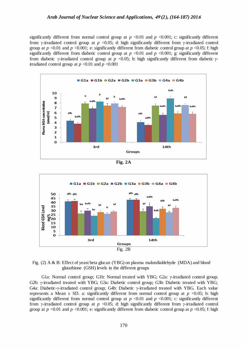
Effect of YBG on Plasma MDA and Blood GSH in Different Groups (Fig. 2 A& B)
-irradiated, diabetic, and diabetic--irradiated control groups displayed a highly significant
increase in plasma MDA level (p <0.01). However both treated -irradiated and diabetic -irradiated groups showed a significant decrease in the MDA level occurred especially in 14th day with some significant differences between groups but didn't approach the normal control level.
-irradiated, diabetic, and diabetic -irradiated control groups displayed a highly significant
decrease in blood GSH level (p <0.01). Meanwhile with both treated -irradiated and diabetic -irradiated groups a significant increase in the GSH level occurred especially at the 14th day with some significant differences between groups but didn't approach the normal control level.
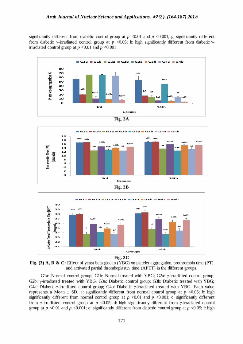
Effect of YBG on Platelet Aggregation (Induced by Arachidonic Acid), Prothrombin Time (PT) and Activated Partial Thromboplastin Time (APTT) in Different Groups (Fig. 3 A, B & C)
-irradiated, diabetic and diabetic -irradiated control groups displayed a highly significant increase in platelet aggregation at the 3rd day which gradually decrease till the 14th day except diabetic control group which exhibited an increased platelet aggregation when compared with other groups (p
<0.01). However, all treated groups including -irradiated, diabetic and diabetic -irradiated groups exhibited a gradual highly significant decrease in platelet aggregation with some significant differences between groups at both intervals reaching approximately the normal control level.
-irradiated, diabetic and diabetic -irradiated control groups displayed a highly significant decrease of PT and APTT during both time intervals (p <0.01). On the contrary, all other treated
groups including -irradiated, diabetic and diabetic -irradiated groups showed a highly significant gradual increase of PT and APTT with some significant differences between groups during both time intervals but didn’t approach the normal control level.
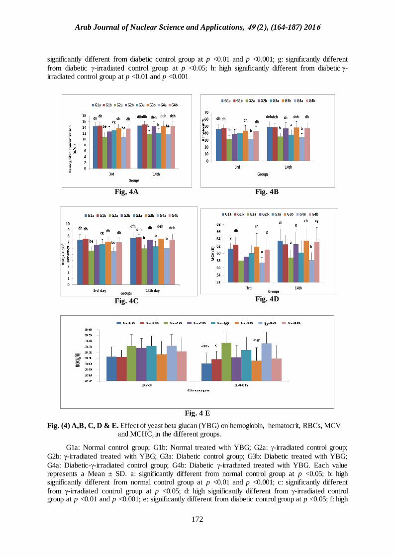
Effect of YBG on on Complete Blood Count In Different Groups (Fig. 4A, B, C, D and E; Fig.
5A, B and C and Fig. 6A, B, C, D and E)
In -irradiated, diabetic and diabetic -irradiated control groups a highly significant decrease in Hb, Hct, RBCs, MCV, MCHC, platelet count, WBCs, neutrophils, lymphocytes, monocytes and eosinophils as well as a high significant increased in MPV and PDW were recorded at both time
intervals (p <0.01). On the contrary, all other treated groups including the -irradiated, diabetic and
diabetic -irradiated groups showed a highly significant increase in Hb, Hct, RBCs, MCH, platelet count, TLC, WBCs, neutrophils, lymphocytes, monocytes and eosinophils and high significantly decreased MPV and PDW were recorded at both time intervals with some significant differences between groups at both intervals reaching nearly normal control level.
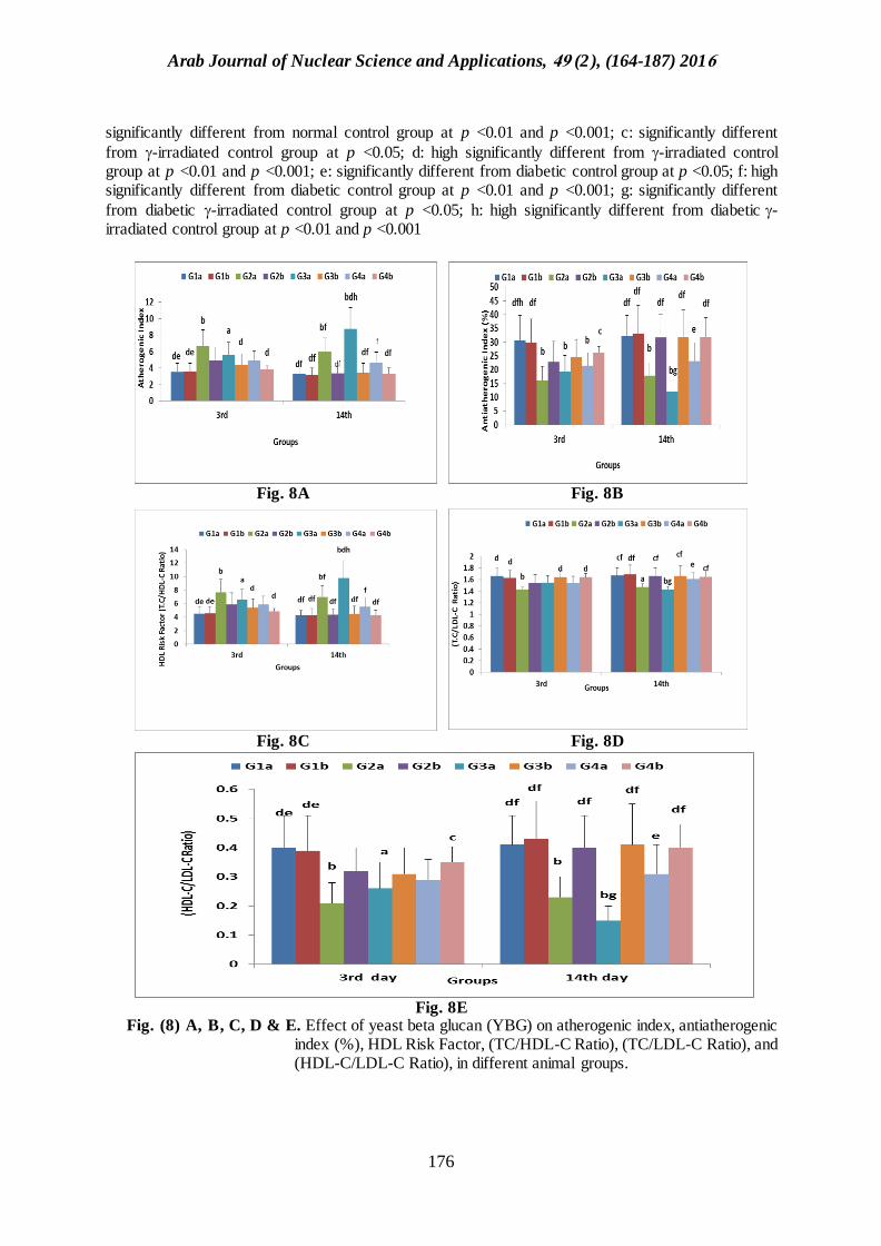
Effect of YBG on on Lipid Profile in Different Groups (Fig. 7A,B, C, D,E and F and Fig. 8A,B,
C, D)
-irradiated, diabetic and diabetic--irradiated control groups showed a highly significant increase of total lipids (TL), TC ,TG, LDL-C, VLDL-C, AI, HDL risk factor and TC/LDL-C ratio and a high significant decrease in HDL-C, AAI and HDL-C/LDL-C ratio at both time intervals (p <0.01).
On the other hand, all other treated groups including -irradiated, diabetic and diabetic -irradiated groups exhibited a highly significant decrease in the levels of TC, TG, LDL-C, VLDL-C, AI, HDL

Arab Journal of Nuclear Science and Applications, 94 (2 ), (164-187) 2016
169
risk factor, TC/LDL-C ratio and TL and a highly significant increased HDL-C, AAI and HDL-C/LDL-C ratio at both time intervals with some significant differences between groups in both intervals approaching the normal control level.
Fig. 1A
Fig. 1B
Fig. 1C
Fig. (1) A, B & C: Effect of yeast beta glucan (YBG) on body weight, pancreas weight and blood glucose level in the different groups
G1a: Normal control group; G1b: Normal treated with YBG; G2a: -irradiated control group;
G2b: -irradiated treated with YBG; G3a: Diabetic control group; G3b: Diabetic treated with YBG;
G4a: Diabetic--irradiated control group; G4b: Diabetic -irradiated treated with YBG. Each value represents a Mean ± SD. a: significantly different from normal control group at p <0.05; b: high
Arab Journal of Nuclear Science and Applications, 94 (2 ), (164-187) 2016
170
significantly different from normal control group at p <0.01 and p <0.001; c: significantly different
from -irradiated control group at p <0.05; d: high significantly different from -irradiated control group at p <0.01 and p <0.001; e: significantly different from diabetic control group at p <0.05; f: high significantly different from diabetic control group at p <0.01 and p <0.001; g: significantly different
from diabetic -irradiated control group at p <0.05; h: high significantly different from diabetic-irradiated control group at p <0.01 and p <0.001
Fig. 2A
Fig. 2B
Fig. (2) A & B: Effect of yeast beta glucan (YBG) on plasma malondialdehyde (MDA) and blood glutathione (GSH) levels in the different groups
G1a: Normal control group; G1b: Normal treated with YBG; G2a: -irradiated control group;
G2b: -irradiated treated with YBG; G3a: Diabetic control group; G3b: Diabetic treated with YBG;
G4a: Diabetic--irradiated control group; G4b: Diabetic -irradiated treated with YBG. Each value represents a Mean ± SD. a: significantly different from normal control group at p <0.05; b: high significantly different from normal control group at p <0.01 and p <0.001; c: significantly different from -irradiated control group at p <0.05; d: high significantly different from -irradiated control group at p <0.01 and p <0.001; e: significantly different from diabetic control group at p <0.05; f: high
Arab Journal of Nuclear Science and Applications, 94 (2 ), (164-187) 2016
171
significantly different from diabetic control group at p <0.01 and p <0.001; g: significantly different
from diabetic -irradiated control group at p <0.05; h: high significantly different from diabetic-irradiated control group at p <0.01 and p <0.001
Fig. 3A
Fig. 3B
Fig. 3C
Fig. (3) A, B & C: Effect of yeast beta glucan (YBG) on platelet aggregation, prothrombin time (PT) and activated partial thromboplastin time (APTT) in the different groups.
G1a: Normal control group; G1b: Normal treated with YBG; G2a: -irradiated control group;
G2b: -irradiated treated with YBG; G3a: Diabetic control group; G3b: Diabetic treated with YBG;
G4a: Diabetic--irradiated control group; G4b: Diabetic -irradiated treated with YBG. Each value represents a Mean ± SD. a: significantly different from normal control group at p <0.05; b: high significantly different from normal control group at p <0.01 and p <0.001; c: significantly different
from -irradiated control group at p <0.05; d: high significantly different from -irradiated control group at p <0.01 and p <0.001; e: significantly different from diabetic control group at p <0.05; f: high
Arab Journal of Nuclear Science and Applications, 94 (2 ), (164-187) 2016
172
significantly different from diabetic control group at p <0.01 and p <0.001; g: significantly different
from diabetic -irradiated control group at p <0.05; h: high significantly different from diabetic-irradiated control group at p <0.01 and p <0.001
Fig, 4A
Fig. 4B
Fig. 4C
Fig. 4D
Fig. 4 E
Fig. (4) A,B, C, D & E. Effect of yeast beta glucan (YBG) on hemoglobin, hematocrit, RBCs, MCV and MCHC, in the different groups.
G1a: Normal control group; G1b: Normal treated with YBG; G2a: -irradiated control group;
G2b: -irradiated treated with YBG; G3a: Diabetic control group; G3b: Diabetic treated with YBG;
G4a: Diabetic--irradiated control group; G4b: Diabetic -irradiated treated with YBG. Each value represents a Mean ± SD. a: significantly different from normal control group at p <0.05; b: high significantly different from normal control group at p <0.01 and p <0.001; c: significantly different
from -irradiated control group at p <0.05; d: high significantly different from -irradiated control group at p <0.01 and p <0.001; e: significantly different from diabetic control group at p <0.05; f: high

Arab Journal of Nuclear Science and Applications, 94 (2 ), (164-187) 2016
173
significantly different from diabetic control group at p <0.01 and p <0.001; g: significantly different
from diabetic -irradiated control group at p <0.05; h: high significantly different from diabetic-irradiated control group at p <0.01 and p <0.001.
Fig. 5A
Fig. 5B
Fig. 5C
Fig. (5) A, B and C. Effect of yeast beta glucan (YBG) on Platelet count, MPV and PDW
respectively, respectively in diabetic gamma/irradiated rats.
G1a: Normal control group; G1b: Normal treated with YBG; G2a: -irradiated control group; G2b: -irradiated treated with YBG; G3a: Diabetic control group; G3b: Diabetic treated with YBG;
G4a: Diabetic--irradiated control group; G4b: Diabetic -irradiated treated with YBG. Each value represents a Mean ± SD. a: significantly different from normal control group at p <0.05; b: high significantly different from normal control group at p <0.01 and p <0.001; c: significantly different

Arab Journal of Nuclear Science and Applications, 94 (2 ), (164-187) 2016
174
from -irradiated control group at p <0.05; d: high significantly different from -irradiated control group at p <0.01 and p <0.001; e: significantly different from diabetic control group at p <0.05; f: high significantly different from diabetic control group at p <0.01 and p <0.001; g: significantly different
from diabetic -irradiated control group at p <0.05; h: high significantly different from diabetic-irradiated control group at p <0.01 and p <0.001
Fig. 6A
Fig. 6B
Fig. 6 C
Fig. 6 D
Fig. 6 E
Fig. (6) A,B, C, D & E . Effect of yeast beta (YBG) on total leucocytic count, neutrophil, lymphocytes, monocytes and eosinophils, in different animal groups.
G1a: Normal control group; G1b: Normal treated with YBG; G2a: -irradiated control group;
G2b: -irradiated treated with YBG; G3a: Diabetic control group; G3b: Diabetic treated with YBG;
G4a: Diabetic--irradiated control group; G4b: Diabetic -irradiated treated with YBG. Each value represents a Mean ± SD. a: significantly different from normal control group at p <0.05; b: high significantly different from normal control group at p <0.01 and p <0.001; c: significantly different

Arab Journal of Nuclear Science and Applications, 94 (2 ), (164-187) 2016
175
from -irradiated control group at p <0.05; d: high significantly different from -irradiated control group at p <0.01 and p <0.001; e: significantly different from diabetic control group at p <0.05; f: high significantly different from diabetic control group at p <0.01 and p <0.001; g: significantly different
from diabetic -irradiated control group at p <0.05; h: high significantly different from diabetic-irradiated control group at p <0.01 and p <0.001
Fig. 7A
Fig. 7B
Fig. 7C
Fig. 7D
Fig. 7E
Fig. 7F
Fig. (7) A, B, C, D, E & F. Effect of yeast beta glucan (YBG) on total lipids, total cholesterol, triglyceride, HDL-cholesterol, LDL-cholesterol VLDL-cholesterol, in different animal groups
G1a: Normal control group; G1b: Normal treated with YBG; G2a: -irradiated control group;
G2b: -irradiated treated with YBG; G3a: Diabetic control group; G3b: Diabetic treated with YBG;
G4a: Diabetic--irradiated control group; G4b: Diabetic -irradiated treated with YBG. Each value represents a Mean ± SD; a: significantly different from normal control group at p <0.05; b: high
Arab Journal of Nuclear Science and Applications, 94 (2 ), (164-187) 2016
176
significantly different from normal control group at p <0.01 and p <0.001; c: significantly different
from -irradiated control group at p <0.05; d: high significantly different from -irradiated control group at p <0.01 and p <0.001; e: significantly different from diabetic control group at p <0.05; f: high significantly different from diabetic control group at p <0.01 and p <0.001; g: significantly different
from diabetic -irradiated control group at p <0.05; h: high significantly different from diabetic-irradiated control group at p <0.01 and p <0.001
Fig. 8A
Fig. 8B
Fig. 8C
Fig. 8D
Fig. 8E
Fig. (8) A, B, C, D & E. Effect of yeast beta glucan (YBG) on atherogenic index, antiatherogenic index (%), HDL Risk Factor, (TC/HDL-C Ratio), (TC/LDL-C Ratio), and (HDL-C/LDL-C Ratio), in different animal groups.

Arab Journal of Nuclear Science and Applications, 94 (2 ), (164-187) 2016
177
G1a: Normal control group; G1b: Normal treated with YBG; G2a: -irradiated control group;
G2b: -irradiated treated with YBG; G3a: Diabetic control group; G3b: Diabetic treated with YBG;
G4a: Diabetic--irradiated control group; G4b: Diabetic -irradiated treated with YBG. Each value represents a Mean ± SD. a: significantly different from normal control group at p <0.05; b: high significantly different from normal control group at p <0.01 and p <0.001; c: significantly different
from -irradiated control group at p <0.05; d: high significantly different from -irradiated control group at p <0.01 and p <0.001; e: significantly different from diabetic control group at p <0.05; f: high significantly different from diabetic control group at p <0.01 and p <0.001; g: significantly different
from diabetic -irradiated control group at p <0.05; h: high significantly different from diabetic-irradiated control group at p <0.01 and p <0.001.
DISCUSSION
Free radicals are important mediators in the complex pathogenesis of acute and chronic inflammatory reactions (2). In biological systems, inactivation and removal of free radicals depend on reactions involving the antioxidant defense system. Natural antioxidants from vegetables and fruits that can reduce the risk of chronic diseases such as diabetes have been the focus of much investigation(32). Beta glucans are polysaccharides of glucose that can be produced by many prokaryotic and eukaryotic organisms. This group of compounds has several beneficial properties and because of that they have found a wide variety of uses in human and in veterinary medicine, pharmaceutical, cosmetic and chemical industries as well as food and feed production (33). Beta glucan activates the immune response through the immune cells, called macrophages, showing various therapeutic effects (34).
In the present study, the body weight and pancreas weight of untreated irradiated, diabetic and
diabetic -irradiated rat groups were reduced significantly. The results also revealed that exposure of rats to radiation didn't change glucose concentration in irradiated animal groups, while it caused a
hypoglycemic effect in diabetic -irradiated groups.
The body weight reduction observed post-irradiation of rats could be due to anorexia and intestinal damage provoking a reduction in gastric secretion associated with a great decrease in acidity. However, absorption would also be impaired as an extensive injury to the gastrointestinal tract
following -irradiation. The results confirmed that there was a significant decrease in pancreas weight due to irradiation of rats (35). The loss in body weight of diabetic animals agrees with that found by Oyedemi et al. (36) who observed a similar effect. The explanation of this reduction has been possibly linked to degradation of structural proteins and muscle wasting.
The results of the present study showed that the gain of body weight was achieved by oral administration of yeast beta glucan. Urao et al. (37) suggested that the glucan administration allowed the beneficial microorganisms e.g. bifidobacterium to quickly reproduce in the animal intestine. These beneficial microorganisms can synthesize vitamins and amino acids, stimulate immunoglobulin activity and improve immune function. The anti-diabetic effect of beta glucans was attributed to its activation of macrophages, the main source of IL-1 in the body, which increases insulin production resulting in lowering of blood glucose level (38). Increasing in the insulin level by yeast beta glucan administration may prevent catabolism of structural protein and increases the peripheral utilization of glucose leading to improvement in body weight and reduction in the blood glucose levels in the YBG treated diabetic rats. Moreover, the effect of yeast beta glucan to reduce blood glucose could be mediated possibly by delaying stomach emptying so that dietary glucose is absorbed more gradually(39). Yeast beta glucan passes the stomach virtually un-changed. In the intestine, there are macrophages that inhabit the intestinal wall and are able to pick yeast beta glucan particles through beta glucan receptors via phagocytic transport mechanism (40). Another possible mechanism for beta-glucans to reduce blood glucose level is mediated by signal pathway through PI3K/Akt activation. Decreased PI3K/Akt activity has been shown to play a key role in the pathogenesis of diabetes. Beta-glucans have been demonstrated to increase PI3K/Akt through several receptors (41).

Arab Journal of Nuclear Science and Applications, 94 (2 ), (164-187) 2016
178
Radiation exposure induced hypoglycemia in experimental rats. This conclusion could be attributed to inhibition of glucose absorption by damaged intestinal epithelium of irradiated intestine. Also, direct effect of radiation on the pancreatic β-cells of islets of Langerhans, thus stimulating rise in insulin secretion (42). The results of Ashry et al. (42) showed an increase in glucose oxidation subsequent to the increase in oxygen consumption as a result of stimulation of thyroid gland activity by gamma irradiation.
Data of the current study revealed that irradiation of rats and STZ administration caused a significant increase in plasma MDA level accompanied by a significant reduction in blood GSH level.
The interaction of ionizing radiation with biological system results in generations of free radicals, H and OH radicals, H2 and H2O2. Radiations-induced free radicals in turn impair the antioxidant defense mechanism leading to increased membrane lipid peroxidation, which results in the damage of membrane bound enzymes (43,44). Also, it is well known that oxidative stress is a contributor to the development of complications in DM. Previous studies have demonstrated that diabetes exhibits enhanced oxidative stress and high reactive oxygen species in pancreatic islets due to persistent and chronic hyperglycemia, thereby depletes the activity of antioxidative defense system, and thus promotes free radical generation (45).
The oxidative stress due to diabetes which induced complications in body hemostasis agrees with that found by Shukla et al. (46) and Kakadiya et al. (47).
Treated animals with yeast beta glucan showed an improvement of oxidative stress parameters due to the antioxidant properties of yeast beta glucan. Cheng et al. (48) reported that the control of hyperglycemia leads to improvement in oxidative stress profile, and enhancing antioxidant defense mechanisms in pancreatic islets helps them to cope better with oxidative stress.
The present results showed increased platelet aggregation on the 3rd day in irradiated and diabetic irradiated groups which then decreased on the 14th day. Diabetic group exhibited an increased platelet aggregation throughout the experimental periods and shortened PT and APTT in untreated groups at both days.
Thrombosis induced by radiation has been explained in many studies. The vascular endothelium is believed to be a target for radiation-induced injury. Overexpression and increased activity of tissue factor (TF) have been shown to be involved in radiation-induced changes (49). In response to IR, monocytes have been reported to produce inflammatory cytokines such as TNF-α. NFĸB, one of the main mediators of cellular responses involved in inflammation, apoptosis and regulation of TF expression, was documented to be activated by IR through a cascade requiring endogenous TNF-α production (50).
Oxidative stress up-regulates numerous pathways pertinent to vascular disease, including matrix metalloproteinases, adhesion molecules, pro-inflammatory cytokines, and smooth muscle cell proliferation and apoptosis, while inactivating vasculoprotective nitric oxide. Importantly, NF-kB is controlled by redox regulation, making it a prime candidate to link chronic oxidative stress to activation of downstream inflammatory pathways in radiation injury (51). The study by Halle et al. (52)
provides the first direct evidence that NF-kB is chronically up-regulated in human arteries after radiation exposure.
Platelets functions are significant to understanding the pathophysiology of vascular disease in diabetes. The role of hyperglycemia is not clear in platelet hyperactivity in diabetic patients (53). Increased level of lipid peroxides might activate the release of arachidonic acid from phospholipids and subsequently amplify platelet activation (54).
Platelet dysfunction may develop before vessel wall damage in diabetes (55,56). Platelet dysfunction in diabetes, including altered adhesion and aggregation, is hypersensitivity to agonists (57).
An elevated level of prothrombin has also been associated with thrombosis, and elevated levels of prothrombin lead to increased thrombin generation in an in vitro model of hemostasis. Thus, it

Arab Journal of Nuclear Science and Applications, 94 (2 ), (164-187) 2016
179
seems likely that elevated prothrombin levels could contribute both to thrombotic risk and to a shortening of the PT and APTT (58).
The in vitro results of Saluk-Juszczak et al. (59,60) demonstrate that antiplatelet activity of beta-glucan from Saccharomyces cerevisiae is dependent on its antioxidative properties. They stated that beta glucan supplementation may be beneficial in the prevention of excessive blood platelet activation-related diseases, such as cardiovascular or inflammatory diseases.
In untreated rat groups it was observed that a significant reduction occurred in hemoglobin level, red blood count and their indices (Hct, MCV, MCH, MCHC). A reduction occured in platelet count and elevation of platelet indices (MPV & PDW). Finally reduction was observed in total leucocytic count and differential count (neutrophils, eosinophils, monocytes and lymphocytes).
Hematopoietic cells are highly sensitive to radiation damage and relatively low levels of exposure can result in bone marrow failure and potentially lethal hemorrhage or infections. The damaging effects of radiation on hematopoiesis have been well established (61). The observed effects are due to both a decrease in the number of hematopoietic stem cell progenitors and a reduction in the self-renewal capacity of stem cells (62,63).
The current results showed that gamma irradiation of rats caused suppression in RBCs count, Hct value and Hb content as well as total WBCs, lymphocytes and neutrophils counts. These results are in agreement with those of Hanafi et al (64) and Salama (65). This is complicated by thrombocytopenia and concomitant hemorrhages besides effects in adaptive immune system resulting from apoptosis of lymphocytes and deficient lymphopoiesis (66). Ionizing radiation is known to induce oxidative stress. By this oxidative imbalance, irradiation kills or damages the major classes of parenchymal cells of the lymphohaematopoietic system, depresses the number of the highly radioprotective bone marrow cells (the major of haematopoiesis) and causes atrophy of spleen which are clear in the results of Salama (65).
The occurrence of anaemia in diabetes mellitus has been reported due to the increased non-enzymatic glycosylation of RBC membrane proteins (36). Oxidation of these proteins and hyperglycaemia in diabetes mellitus causes an increase in the production of lipid peroxides that lead to haemolysis of RBC (66). Reduction of levels of RBC, Hb, haematocrit and leucocytes in the diabetic animals may be attributed to the infections of the normal body systems (67). The intraperitoneal injection of streptozotocin into rats significantly reduced the WBC count and its differentials such as basophils, monocytes, eosinophils, lymphocytes and neutrophils. The reduction of these parameters could be linked to suppression of leucocytosis from the bone marrow which may account for poor defensive mechanisms against infection (36). Consequentially, they might have effects on the immune system and phagocytic activity of the animals (68). Medications decrease PLT counts in two ways, either by suppression of bone marrow or by destruction of PLTs in peripheral blood via immune mechanism (69).
Reduction of platelets levels in diabetic rats induced with streptozotocin was confirmed in this study in relation to the normal control rats. Long term reduction of this parameter may result in internal and external haemorrhage and finally leads to death (39).
The mean platelet volume is increased in type 2 diabetes mellitus. This increase can be due to increased number of younger platelets in diabetics. Younger platelets have increased platelet volume. Increased endothelial damage is seen in diabetes mellitus which reduces the survival of platelets and increases turnover of younger platelets. Moreover MPV is an indicator of the average size and activity of platelets. Large platelets are younger, more reactive and aggreagable as they contain denser granules, secrete more serotonin and β-thromboglobulin, and produce more thromboxane A2 than smaller platelets (70). All these can produce a pro-coagulant effect and cause thrombotic vascular complications. This suggests a relationship between the platelet function especially MPV and diabetic vascular complications thus indicating that changes in MPV reflect the state of thrombogenesis. Function is directly regulated by insulin via a functional insulin receptor (IR) found on human platelets (71). High MPV is emerging as a new risk factor for the vascular complications of DM of
Arab Journal of Nuclear Science and Applications, 94 (2 ), (164-187) 2016
180
which atherothrombosis plays a major role (72). Thus, DM has been considered as a “prothrombotic state” with increased platelet reactivity (73).
The results of Riahi-Zanjani et al. (74) indicate that long term low dose ionizing radiation may have side effects on thrombocytosis and coagulation function. They measured PDW of the radiation workers and found that it was significantly higher than the control group. The present results showed that oral gavage treatment of beta glucan caused improvement in RBCs count, Hct value and Hb content as well as total WBCs, absolute lymphocytes and neutrophils counts. Zhao et al. (75) and Yuan et al. (76) recorded an enhanced cellular immune response to glucan administration. It is well documented that total body irradiation followed by β-glucan has been shown to raise the erythropoietic activity in both bone marrow and spleen (77).
Beta glucan of different origin has been demonstrated to be potent anti-oxidants, prevent damage by H2O2 and other reactive oxygen species (78). In addition, there are some reports on the immune-antioxidant activity relationship of glucan (79,80), which may result in proliferation of bone marrow stem cells as indicated by increased in bone marrow cell count and its viability in the study performed by Salama (74). This phenomenon was also observed by Pospilil et al. (81) and Patchen et al. (82) who concluded that the improvement effects of beta glucan when given to animals submitted to radiation, was not due only to hematopoietic regeneration, but also the capacity of this substance to inactivate free radicals.
The use of highly purified, orally administered yeast beta glucan accelerated the early recovery of peripheral blood leukocytes following sublethal irradiation. On the other hand, yeast beta glucan could modulate the autoimmune mechanisms directed to pancreatic islets and inhibit the development of diabetes in rats. Furthermore, it could reduce carbohydrate absorption from the gut (83).
Immunomodulation by beta glucan was confirmed both in vitro and in vivo in numerous animal and human studies involving a wide range of tumors, including breast, lung and gastrointestinal cancers (84). The immunomodulating and cancerostatic properties make β-glucan one of the substances with a great potential in the ongoing fight against cancer (33,85). At the same time, only a few drugs have similar advantages as beta glucan .
In the present study, serum lipid profile was altered in irradiated as well as diabetic groups characterized by increasing total lipids, total cholesterol, triglycerides and LDL cholesterol levels accompanied by decrease in HDL cholesterol. These disturbances influence other lipid profile indices. Improvements of lipid profile was achieved by β-glucan treatment as compared with control group.
In case of irradiation effects, free radicals impair liver functions and can be a major reason of hormonal imbalance. This imbalance induces hyperlipidemia through its multiple effects on lipid metabolism, including increased synthesis of cholesterol and triglyceride (86). Results of the present study are in agreement with previos findings demonstrating an increase of plasma level of lipids of rats post irradiation (87,88). They attributed the hypercholesterolemia conditions to the stimulation of
cholesterol synthesis in the liver after -irradiation. Moreover, Bok et al. (89) attributed the irradiation-induced hypercholesterolemia to the increase of activation of HMG-CoA reductase enzyme, the key regulatory enzyme in the reduction of the overall process of cholesterol synthesis. Sedlakova et al. (90) explained that the increase in serum triglyceride level after irradiation might result from the inhibition of lipoprotein lipase activity, leading to reduction in uptake of triacylglycerols. Mahmoud(91) attributed the hyperlipidemic state under the effect of gamma-irradiation to the stimulation of liver enzymes responsible for the biosynthesis of fatty acids and mobilization of fats from adipose tissue to blood stream.
Diabetes associated dyslipidemia is a major risk factor for CVD (92). The dyslipidemia is caused either by insulin resistance or adipocytokines. In diabetes, adipose cells are insulin resistant, thus, insulin-mediated uptake of free fatty acids in skeletal muscle is impaired. Increased circulating free fatty acids flux to the liver, results in increased triglyceride synthesis and the assembly of very low-density lipoprotein (VLDL) (93). Thus, the characteristics of dyslipidemia in the patients with diabetes is hypertriglyceridemia. Hyperglycemia and low insulin may also contribute to VLDL production (93).

Arab Journal of Nuclear Science and Applications, 94 (2 ), (164-187) 2016
181
In diabetes, adiponectin is reduced, which increases muscle free fatty acid uptake and reduces plasma free fatty acid level (94). This mechanism is independent of insulin-resistance (95). In addition, high density lipoprotein (HDL) may also decrease (96).
Beta glucan has been shown to decrease LDL cholesterol and increase HDL to alleviate possibly dyslipidemia and reduce CVD (97). This altered serum lipid profile was reversed towards normal after treatment with beta glucan. The possible mechanism through which beta glucan exerts its anti-hyperlipidemic effect might include the changed activity of cholesterol biosynthesis enzymes and/or the changed level of lipolysis which are under the control of insulin (98).
Chen & Huang(99) suggested that beta glucans sequester bile acids in the intestine, reducing their reabsorption and return to the liver. Reducing hepatic bile acid concentrations activates the enzyme CYP7A1, which converts cholesterol into bile acids. Drozdowski et al. (100) concluded that the reduced intestinal fatty acid uptake after beta glucan administration is associated with inhibition of genes regulating intestinal uptake and synthesis of lipids. Salama(67) results strongly indicated that beta glucans may up-regulate low-density lipoprotein receptor gene. Moreover, it has been reported that yeast beta glucan treatment could decrease the capacity of LDL to carry free cholesterol to various tissues without affecting the capacity of HDL to carry cholesterol back to the liver in rats (101).
CONCLUSION
From the present results it could be concluded that oral yeast beta glucan administration possesses radioprotective and antihyperglycemic properties. In addition, it could prevent various complications of diabetes as well as improving some haematological parameters.
REFERENCES
(1) L. Packer, and C. Colman, The Antioxidant Miracle. New York: John Wiley & Sons, Inc.
(1999).
(2) H. Sharma, Leaky Gut Syndrome, Dysbiosis, Ama, Free Radicals, and Natural Antioxidants
AYU-VOL. 30, NO. 2 (APRIL-JUNE), pp. 88-105(2009).
(3) J.F. Turrens, Mitochondrial formation of reactive oxygen species. J Physiol 552: 335–344
(2003).
(4) J.A. Scott, and G.L. King, Oxidative stress and antioxidant treatment in diabetes. Ann NY
Acad Sci 1031: 204–213 (2004).
(5) G.E. Gibson, and H.M. Huang, Oxidative stress in Alzheimer’s disease. Neurobiol Aging 26:
575–578 (2005).
(6) M. Roy, S. Sen, and A.S. Chakraborti, Action of pelargonidin on hyperglycemia and oxidative damage in diabetic rats: implication for glycation-induced hemoglobin modification.
Life Sci. May 23;82(21-22):1102-10 (2008).
(7) M.M.A. Khalaf, G.A. El Sherbiny, H.A. AbdEllati, A.A. Ain-shoka, and E. El Sayed, Comparative Effects of Glimepiride, Vanadyl Sulfate and Their Combination on Hypoglycemic Parameters and Oxidative Stress. British Journal of Pharmacology and Toxicology 3(6): 278-
288 (2012).
(8) H. Alp, S. Varol, M. Celik, M. Altas, O. Evliyaoglu, O. Tokgoz, M.H. Tanrıverdi, and E. Uzar, Protective Effects of Beta Glucan and Gliclazide on Brain Tissue and Sciatic Nerve of Diabetic Rats Induced by Streptozosin. Diabetes Research Volume 2012, Article ID 230342, 7
pages (2012).
(9) L.F. Fajardo, and M. Berthrong, Vascular lesions following radiation. Pathol Annu;23 Pt
1:297–330 (1988).
(10) P. Goldin-Lang, F. Niebergall, S. Antoniak, B. Szotowski, P. Rosenthal, K. Pels, and
U.S.H. Rauch, Ionizing radiation induces upregulation of cellular procoagulability and tissue

Arab Journal of Nuclear Science and Applications, 94 (2 ), (164-187) 2016
182
factor expression in human peripheral blood mononuclear cells. Thrombosis Research., 120:
857-864 (2007 a).
(11) P. Goldin-lang, K. Pels, Q.V. Tran, B. Szotowski, F. Wittchen, S. Antoniak, T. Willich, H.
Witt, M. Hummel, D. Lenze, W. Poller, H.P. Schulltheiss, and U. Rauch, Effect of Ionizing radiation on cellular procoagulability and co-ordinated gene alterations. Haematologica. Aug.,
92(8): 1091-8 (2007 b).
(12) A.B. Sobol, and C. Watata, The role of platelets in diabetes-related vascular complications.
Diabetes research and clinical practice., 50: 1-16 (2000).
(13) J.A. Saliu, O.O. Elekofehinti, K. Komolafe, and Oboh; Effects of some green leafy vegetables on the haematological parameters of diabetic rats J. Nat. Prod. Plant Resour., 2
(4):482-485 (2012).
(14) P. Okunieff, S. Swarts, P. Keng, W. Sun, W. Wang, J. Kim, S. Yang, H. Zhang, C.Liu, J.P. Williams, A.K. Huser, and L. Zhang; Antioxidants reduce consequences of
radiation exposure. Adv Exp Med Biol.;614:165-78 ( 2008).
(15) R.C. Lindenschmidt, A.F. Trika, M.E. Guard, and H.P. Witschi, The effect of dietary butylated hydroxy toluene on liver and colon tumor development in mice. Toxicology, 38, 151–
160 (1986).
(16) M. Hofer, and M. Pospisil, Glucan as stimulator of hematopoiesis in normal and gamma-
irradiated mice. A survey of the authors’ results. Int. J. lmmunopharmac.; 19: 607-609 (1997).
(17) B. Pekiner, N.N. Ulusu, N.D. Evcimen, M. Sahilli, F. Aktan, M. Stefek, S. Stolc, and C.Karasu; In vivo treatment with stobadine prevents lipids peroxidation, protein glycation and calcium overload but does not ameliorate ca2+ ATPase activity in heart and liver of streptozotocin-diabetic rats: comparison with vitamin E. Biochimica et Biophysica Acta., 1588:
71:78 (2002).
(18) M.D. Hon Kon, M.D. Zeyu, M. Hashimoto, K. Yamamoto, and T. Kimura; Effects of medicinal musroom (Sparassis crispa) on wound healing in streptozotocin-induced diabetic rats.
The American Journal of Surgey., 197: 503-509 (2009).
(19) D.S. Young, Effects of drugs on Clinical Laboratory tests. 3rd Ed., 3: 168-182 (1990).
(20) T. Yoshioka, K. Kawada, T. Shimada, and M. Mori; Lipid peroxidation in maternaland cord blood and protective mechanism against activated-oxygen toxicity in the blood. Am. J. Obestet.
Gynecol., 135(3): 372-376 (1979).
(21) B.S. Beulter, O. Duron, and B.M. Kelly, Improved method for the determination of blood
glutathione. J. Lab. Clin. Med., 61:882-888 (1963).
(22) M. McCabe-White, and L.K. Jennings, Platelet Protocols: Research and Clinical laboratory
Procedure. Academic Press, London; p 35 (1999).
(23) B.J. Bain, I. Bates, M.A. Laffan, and S.M. Lewis, Dacie and Lewis Practical Haematology
(Ninth Edition) (2001).
(24) J. Hirsh, J.E. Dalen, D. Deykin, and L. Poller, Oral Anticoagulants: Mechanism of Action,
Clinical Effectiveness and Optimal Therapeutic Range, Chest: 108 (Suppl.): 231S-246S (1995).
(25) CRC.; Handbook Series in Clinical Laboratory. Science, Section 1: Haematology, Volume ш.
CRC Press, Inc. Boca Raton. Florida (1980).
(26) C.S. Frings, T.W. Fendley, R.T. Dunn, and C.A. Queen, Improved determination of total
serum lipids by the sulfo-phospho-vanillin reaction. Clin Chem. Jul;18(7):673-4 (1972).
(27) E.A. Stein, In Textbook of Clinical Chemistry, NW Tietz, ed. W.B. Saunders, Philadephia.,
879-886, 1818, 1829 (1986).
(28) G. Buccolo, and H. David, Ouantitative determination of serum triglycerides by use enzymes.
Clin. Chem., 19: 419-32 (1973).
Arab Journal of Nuclear Science and Applications, 94 (2 ), (164-187) 2016
183
(29) National Institutes of Health publication.; No. 933-3095 (1993).
(30) W.T. Friedewald, R.I. Levy, and D.S. Fredrickson, Estimation of VLDL and LDL-cholesterol in plasma without use of the preparative ultracentrifuge. Clin Chem., 18: 499-502
(1972).
(31) S. Guido, and T. Joseph, Effect of chemically different calcium antagonists on lipid profile in rats fed on a high fat diet. Indian J. Exp. Biol., 30: 292-294 (1992).
(32) S. Liu, M. Serdula, S.J. Janket, N.R. Cook, H.D. Sesso, W.C. Willett, J.E. Manson, and J.E. Buring, A prospective study of fruit and vegetable intake and the risk of type 2 diabetes in
women. Diabetes Care 27: 2993–2996 (2004).
(33) C. Laroche, and P.H. Michaud, New developments and prospective applications for β-(1,3)-glucans. Recent Pat on Biotechnol 1: 59-73 (2007).
(34) G.D. Brown, and S. Gordon, Immune recognition of fungal β-glucans. Cell Microbiol 7: 471-479 (2005).
(35) E.M. Hussein, M.M. Darwish, and S.E. Ali, Prophylactic Role of Combined Treatment with Coenzyme Q10 and Vitamin E against Radiation Injury in Male Rats. Egypt. J. Rad. Sci. Apllic., 20(1): 181-194 (2007).
(36) S.O. Oyedemi, E.A. Adewusi, O.A. Aiyegoro, and D.A. Akinpelu, Antidiabetic and haematological effect of aqueous extract of stem bark of Afzelia africana (Smith) on
streptozotocin-induced diabetic Wistar rats. Asian Pac J Trop Biomed. Oct;1(5):353-8 (2011).
(37) M. Urao, T. Fujimoto, G.J. Lane, G. Seo, and T. Miyano, Does probiotics administration decrease serum endotoxin levels in infants? J. Pediat. Surg., 34: 273-276 (1999)..
(38) C.H. Lang, and C. Dobrescu, Interleukin-l induced increases in glucose utilization are insulin mediated. Life Sci., 45: 27–34 (1989).
(39) T. Kiho, H. Morimoto, M. Sakushima, et al.; Polysaccharides in fungi. XXXV. Anti diabetic activity of an acidic polysaccharide from the fruiting bodies of Tremella aurantia. Biol Pharm
Bull., 18: 1627–9 (1995).
(40) E.P. Lowe, D. Wei, P.J. Rice, C. Li, J. Kalbfleisch, I.W. Browder, and W. Williams, Human vascular endothelial cells express pattern recognition for fungal glucans, which stimulate nuclear factor KappB activation and interleukin 8 production. American Surg., 68:
508 (2002).
(41) J. Chen, and R. Seviour, Medicinal importance of fungal beta-(1–_3), (1–_6)-glucans. Mycol
Res., 111:635–52 (2007).
(42) O.M. Ashry, S.F. Salama, and N.A. El Tantawy, Possible Effect of Phentoxifylline on Oxidative Stress and Certain Metabolic Disorders in Gamma Irradiated Rats. Egypt. J. Rad. Sci.
Applic., 20(2): 125-139 (2006).
(43) B. Halliwell, H. John, and N.C. Gutteridge, Production of hydroxyl radicals in living
systems, In-Free Radicals Biology and Medicine, Clarendon Press, Oxford (1989).
(44) H.N. Saada, and K.H.SH. Azab, Role of lycopene in recovery of radiation induced injury to
mammalian cellular organelles. Pharmazie, 56: 239 (2001).
(45) O. Savu, C. Ionescu-Tirgoviste , V. Atanasiu, L. Gaman, R. Papacocea, and I. Stoian, “Increase in total antioxidant capacity of plasma despite high levels of oxidative stress in
uncomplicated type 2 diabetes mellitus J Int Med Res.;40(2):709-16 (2012).
(46) R. Shukla, S. Padhye, M. Modak, S.S. Ghaskadbi, and R.R. Bhonde, Bis (quercetinato) oxovanadium IV reverses metabolic changes in streptozotocin-induced diabetic mice. Rev.
Diabetic Stud., 4(1): 33-43 (2007).
(47) J. Kakadiya, M. Shah, and N. Shah, Glimepiride reduces on experimentally induced
ischemia/reperfusion in diabetic rats. Int. J. Appl. Biol. Pharm. Technol., 1(2): 276-285 (2010).

Arab Journal of Nuclear Science and Applications, 94 (2 ), (164-187) 2016
184
(48) D. Cheng, B. Liang, and Y. Li, Antihyperglycemic Effect of Ginkgo biloba Extract in Streptozotocin-Induced Diabetes in Rats. Hindawi Publishing Corporation BioMed Research
International Volume 2013, Article ID 162724, 7 pages (2013).
(49) J.E. Baker, B. Fish, J. Su, S. Haworth, J. Strande, R. Komorowski, R. Migrino, A. Doppalapudi, L. Harmann, X. Li, J. Hopewell, and J. Moulder, 10 Gy Total body irradiation increases risk of coronary sclerosis, degeneration of heart structure and function in a rat model.
Int J Radiat Biol 85: 1089–1100 (2009).
(50) F.A. Cucinotta, W. Schimmerling, J.W. Wilson, L.E. Peterson, G.D. Badhwar, P.B. Saganti, and J.F. Dicello, Space radiation cancer risks and uncertainties for Mars missions.
Radiat Res 156: 682–688 (2001).
(51) N.L. Weintraub, W.K. Jones, and D. Manka, Understanding radiation-induced vascular
disease. J Am Coll Cardiol. Mar 23;55(12):1237-9 (2010).
(52) M. Halle, A. Gabrielsen, G. Paulsson-Berne, et al.; Sustained inflammation due to nuclear
factor-kappa B activation in irradiated human arteries. J Am Coll Cardiol; 55:1227–36 (2010).
(53) A.I. Vinik, T. Erbas, T.S. Park, R. Nolan, and G.L. Pittenger, Platelet Dysfunction in Type
2. Diabetes Care., 24(8): 1476-85 (2001).
(54) C. Calzada, E. Ve´ricel, B. Mitel, L. Coulon, and M. Lagarde, 12(S)-hydroperoxyeicosatetraenoic increases arachidonic acid availability in collagen-primed
platelets. J Lipid Res 42:1467–1473 (2001).
(55) J.A. Colwell, P.D. Winocour, and P.V. Halushka,; Do platelets have anything to do with
diabetic micro- vascular disease? Diabetes, 32(2), 14-19 (1983).
(56) G. Davi, P. Gresele, F. Violi, S. Basili, M. Catalano, C. Giammarresi, R. Volpato, G.G. Nenci, G. Ciabattoni, and C. Patrono, Diabetes mellitus, hypercho- lesterolemia, and hypertension but not vascular disease per se are associated with persistent platelet activation in vivo. Evidence derived from the study of peripheral arterial disease. Circulation, 96(1), 69-75
(1997).
(57) A. Natarajan, A.G. Zaman, and M.M. Sally, Platelet hyperactivity in type 2 diabetes: Role of
antiplatelet agents. Diabetes and Vascular Disease Research, 5(2), 138-144 (2008).
(58) D.L. Sauls, A.E. Banini, L.C. Boyd, and M. Hoffman, Elevated prothrombin level and
shortened clotting times in subjects with type 2 diabetes. J Thromb Haemost., 5: 638–9 (2007).
(59) J. Saluk-Juszczak, K. Krolewska, and B. Wachowicz, beta-glucan from Saccharomyces
cerevisiae as a blood platelet antioxidant. Platelets.; 21(6):451-9 (2010a).
(60) J. Saluk-Juszczak, K. Królewska, and B. Wachowicz , Response of blood platelets to beta-
glucan from Saccharomyces cerevisiae. Platelets.;21(1):37-43 (2010 b).
(61) N. Yang, M.A. Chaudhry, and S.S. Wallace, Base excision repair by hNTH1 and hOGG1: a two edge sword in the processing of DNA damage in γ-irradiated human cells. DNA Repair;5:
43–51 (2006).
(62) N. Dainiak, J.K. Waselenko, J.O. Armitage, T.J. Macvittie, and A.M. Farese, The hematologist and radiation casualties, Hematology. American Society of Hematology Education
Program, pp. 473-496 (2003).
(63) Y. Wang, B.A. Schulte, A.C. Larue, M. Ogawa, and D. Zhou, Total body irradiation
selectively induces murine hematopoietic stem cell senescence. Blood, 107: 358-366 (2006).
(64) N. Hanafi, S.Z. Mansour, and S.F. Salama, The efficacy of oral supplementation of GliSODin in reducing the oxidative stress in rats subjected to γ-radiation. Isotope Radiat. Res.,
22:23-29 (2009).
(65) S.F. Salama, Inosine protects from oxidative damage induced by irradiation in rats. Egypt. J.
Radiat. Sci. Applic., 21: 35-48 (2009).

Arab Journal of Nuclear Science and Applications, 94 (2 ), (164-187) 2016
185
(66) R.C. Wilkins, D. Wilkinson, H.P. Maharaj, P.V. Bellier, M.B. Cybulski, and J.R. Mclean, Differential apoptotic response to ionizing radiation in subpopulations of human white blood
cells. Mutat. Res., 513:27-36 (2002).
(67) S.F. Salama, ß-Glucan Ameliorates Gamma–Rays Induced Oxidative Injury in Male Swiss
Albino Rats Pakistan J. Zool., vol. 43(5), pp. 933-939 ( 2011),
(68) G.S. Arun, and K.G.Ramesh, Improvement of insulin sensitivity by perindopril in spontaneously hypertensive and streptozotocin diabetic rats. Indian J Pharmacol., 34: 156-164
(2002).
(69) R. Baskar, L.M. Bhakshu, G.V. Bharathi, S.S. Reddy, R. Karuna, G.K. Reddy, et al.; Antihyperglycemic activity of aqueous root extract of Rubia cordifolia in streptozotocin-
induced diabetic rats. Pharm Biol., 44(6): 475-479 (2006).
(70) J. Torell, J. Cillard, and P. Cillard, Antioxidant activity of flavonoids and reactivity with
peroxy radical. Phytochemistry., 25(2): 383-385 (1986).
(71) P. Lanzkowsky, Disorders of Platelets.Manual of PediatricHematolog and Oncology,
ElsevierAcademic Press, 4th edition (2005).
(72) D. Tschöpe, E. Langer, S. Schauseil, P. Rösen, L. Kaufmann, and F.A. Gries, Increased platelet volume - Sign of impaired thromboposis in diabetics. Klin – Wocherschr., 67(4): 253-
259 (1989).
(73) H.A. Chang, H.S. Hwang, H.K. Park, M.Y. Chun, and J.Y. Sung, The Role of Mean Platelet Volume as a Predicting Factor of Asymptomatic Coronary Artery Disease. Korean J
Fam Med., 31: 600–6 (2010).
(74) B. Riahi-Zanjani, M. Balali-Mood, Alamdaran, S-A.; Evaluation of the Serum Total Antioxidant Level and Hematological Indices in Healthy Workers Exposed to Low Radiation Doses: A Significant Increase in Platelet Indices. Archives vol.1 •63-67
http://pharmacologyonline.silae.it ISSN: 1827-8620 (2014).
(75) G.H. Zhao, J.Q. Kan, Z.X. Li, and Z.D. Chen, Characterization and immunostimulatory activity of (1-6)-ß-d-glucan from the root of Ipomoea batatas. Int. Immunopharmacol., 5: 1436-
1445 (2005).
(76) C. Yuan, X. Huang, L. Cheng, Y. Youquan, G. Liu, F. Yi, Z. Yang, and F. Song, Evaluation of antioxidant and immune activity of Phellinus ribis glucan in mice. Fd. Chem.,
115: 581-584 (2009).
(77) D.G. De Rooij, H.J. Van De Kant, R. Dol, G. Wagemaker, and P.P. Vanbuul, Long-term effects of irradiation before adulthood on reproductive function in the male Rhesus monkey.
Biol. Reprod., 66: 486-495 (2002).
(78) R. Oliveira, M. Salles, F. da silva, T. Kanno, A. Lourenço, G. Freiria, J. Matiazi, L. Ribeiro, and M. Mantovani, Effects of the polysaccharide ß-glucan on clastogenicity and teratogenicity caused by acute exposure to cyclophosphamide in mice. Regulat. Toxicol. Pharmacol., 53:164-173 (2009).
(79) J.P.F. Angeli, L.R. Ribero, M.F. Bellini, and M.S. Mantovani, Anticlastogenic effect of beta-glucan extracted from barley towards chemically induced DNA damage in rodent cells,
Hum. exp. Toxicol., 25: 319-324 (2006).
(80) G. Sener, H. Toklu, F. Ercan, and G. Erkanl, Protective effect of β-glucan against oxidative organ injury in a rat model of sepsis. Int. Immunopharmacol., 5: 1387 (2005).
(81) M. Pospisil, S. Viklicka, A. Vacek, I. Pipalova, and A. Bartonickova, Hematopoietic recovery in repeatedly irradiated mice can be enhanced by a repeatedly administered
combination of diclofenac and glucan. J. Leuk. Biol., 53:185-189 (1993).
(82) M.L. Patchen, M.M. D'alesandro, I. Brook, W.F. Blakely, and T.J. Macvittie, Glucan: mechanisms involved in its "radioprotective" effect. J. Leuk. Biol., 42: 95-105 (1987).

Arab Journal of Nuclear Science and Applications, 94 (2 ), (164-187) 2016
186
(83) P. Battilana, K. Ornstein, K. Minehira, J.M. Schwarz, K. Acheson, P. Schneiter, J. Burri, E. Jéquier and L. Tappy, Mechanisms of action of beta-glucan in postprandial glucose
metabolism in healthy men. Eurpean J. Clin. Nutr., 55: 327–33 (2001).
(84) M. Novak, and V. Vetvicka, Beta-glucans, history, and the present: immunomodulatory
aspects and mechanisms of action. J Immunotox 5: 47-57 (2008).
(85) N.R. DiLuzio, D.L. Williams, R.B. McNamee, B.F. Edwards, A. Kaitahama, Comparative tumor-inhibitory and antibacterial activity of soluble and particulate glucan. Int J Cancer 24:
773-779 (1979).
(86) D.A. Bowden, P. McLean, A. Steinmetz, D. Fontana, C. Matthys, G.R. Warnick, M. Cheung, J. Albers, and W. Hazzard. Lipoprotein, apolipoprotein and lipolytic enzyme changes following estrogen administration in post menopausal women. J. Lipid. Res., 30:
1895-1906 (1989).
(87) U.Z. Said, and Kh.Sh. Azab, Efficacy of wheat germ oil in modulating radiation induced heat
damage. Egypt. J. Rad. Sci. Applic., 19 (2): 433-452 (2006).
(88) A.S. Nada, Modulating efficacy of rosemary extracts in rats exposed to oxidative stress.
Egypt. J. Rad. Sci. Applic., 21 (2): 499- 514 (2008).
(89) S. Bok, S. Lee, Y. Park, K. Bae, K. Son, T. Jeong, and M. Choi, Plasma and hepatic cholesterol and hepatic activities of 3- hydroxy-3-methyl-glutaryl-CoA reductase and aryl CoA: Cholesterol transferase are lower in rats fed citrus peel extract or a mixture of citrus
bioflavonoids. J. Nutr. 129(6): 1182–1185 (1999).
(90) A. Sedlakova, K. Borlkova, and I. Ahlers, Canges in lipoprotein lipase activity in the adipose tissue and heart of non-lethaly x-irradiated rats. Physiol.Bohemoslov., 35(5): 400-5
(1986).
(91) K. Mahmoud, Response of certain biochemical parameters to whole body gamma irradiation in albino rats and synergestic effect of pharmacological agent. Ph. D. Thesis, Fac. Sci. Alex.
Univ (1996).
(92) R.C. Turner, H. Millns, H.A. Neil, et al.; Risk factors for coronary artery disease in non-insulin dependent diabetes mellitus: United Kingdom Prospective Diabetes Study (UKPDS: 23).
BMJ, 316:823–8 (1998).
(93) F.D. Hobbs, Reducing cardiovascular risk in diabetes: beyond glycemic and blood pressure
control. Int J Cardiol, 110:137–45 (2006).
(94) M. Faraj, H.L. Lu, K. Cianflone, Diabetes, lipids, and adipocyte secretagogues. Biochem Cell
Biol, 82:170–90 (2004).
(95) M.B. Schulze, E.B. Rimm, I. Shai, N. Rifai, F.B. Hu, Relationship between adiponectin and glycemic control, blood lipids, and inflammatory markers in men with type 2 diabetes. Diabetes
Care. Jul;27(7):1680-7 (2004).
(96) M. Laakso, Lipids and lipoproteins as risk factors for coronary heart disease in non-insulin-
dependent diabetes mellitus. Ann Med, 28:341–5 (1996).
(97) N. Reyna-Villasmil, V. Bermudez-Pirela, E. Mengual-Moreno, et al., Oat-derived beta-glucan signifi cantly improves HDLC and diminishes LDLC and non-HDL cholesterol in
overweight individuals with mild hypercholesterolemia. Am J Ther, 14:203–12 (2007).
(98) N.K. Kapur, D. Ashen, and R.S. Blumenthal, High density lipoprotein cholesterol: an evolving target of therapy in the management of cardiovascular disease. Vasc Health Risk
Manag, 4:39–57 (2008).
(99) J. Chen, and X. Huang, The effects of diets enriched in beta-glucans on blood lipoprotein. J.
clin. Lipido., 3: 154-158 (2009).
(100) A. Drozdowski, R. Reimer, F. Temelli, C. Bell, T. Vasanthan, and A. Thomson, β-Glucan extracts inhibit the in vitro intestinal uptake of long-chain fatty acids and cholesterol and down-

Arab Journal of Nuclear Science and Applications, 94 (2 ), (164-187) 2016
187
regulate genes involved in lipogenesis and lipid transport in rats. J. Nutr. Biochem., 21: 695-701
(2010).
(101) Z.X. Yao, Z. Han, K. Drieu, and V. Papadopoulos; Ginkgo biloba extract (Egb 761) inhibits β-amyloid production by lowering free cholesterol levels. Journal of Nutritional Biochemistry.,
15(12): 749–756 (2004).