rumbach physiological characteristics of dysphagia ...271107/uq271107_oa.pdfthe study aim was to...
TRANSCRIPT

1
PHYSIOLOGICAL CHARACTERISTICS OF DYSPHAGIA FOLLOWING
THERMAL BURN INJURY1
Anna F. Rumbach BSc, MSpPathSt, GCHEd (Corresponding Author/Re-Print requests)
School of Health and Rehabilitation Sciences, Division of Speech Pathology, The
University of Queensland, St Lucia, Brisbane, 4072, Australia
Phone: +61 407 12 3 879; E-mail: [email protected]
Elizabeth C. Ward BSpThy (Hons), Grad Cert Ed., PhD
Centre for Functioning and Health Research, Queensland Health
Level 3, Centro Buranda, Ipswich Road, Buranda, QLD 4102, Australia;
School of Health and Rehabilitation Sciences, Division of Speech Pathology, The
University of Queensland, St Lucia, Brisbane, 4072, Australia
Petrea L. Cornwell BSpPath (Hons), PhD
Metro North Health Service District, Queensland Health, Queensland, Australia; Griffith
Health Institute, Behavioural Basis of Health Program, Griffith University, Mt Gravatt,
QLD 4122, Australia
Lynell V. Bassett BSpThy
Speech Pathology Department, Level 2 James Mayne Building, Royal Brisbane &
Women’s Hospital, Butterfield Street, Herston, QLD 4029, Australia
Michael J. Muller MBBS MMed Sci FRACS
Professor Stuart Pegg Adult Burns Centre, Level 4 James Mayne Building, Royal
Brisbane & Women’s Hospital, Butterfield Street, Herston, QLD 4029, Australia; Burns,
Trauma & Critical Care Research Centre, The University of Queensland, Brisbane,
Australia
1This project was supported by funding from the Royal Brisbane and Women’s Hospital
(RBWH) Foundation.

2
Physiological characteristics of dysphagia following thermal burn injury

3
Abstract
The study aim was to document the acute physiological characteristics of the
swallowing impairment following thermal burn injury. A series of 19 participants
admitted to a specialised burn centre with thermal burn injury were identified with
suspected aspiration risk by a clinical swallow examination (CSE) conducted by a
speech-language pathologist and referred to the study. Once medically stable, each then
underwent more detailed assessment using both a CSE and a fiberoptic evaluation of
swallowing (FEES). Subsequent to this assessment, FEES confirmed 6 individuals (32%)
had no aspiration risk, and were excluded from further analyses. Of the remaining 13,
CSE confirmed two had specific oral phase deficits due to orofacial scarring and
contractures, and all 13 had generalised oromotor weakness. FEES revealed numerous
pharyngeal phase deficits with major finding evident in greater than 50% being: impaired
secretion management, laryngotracheal edema, delayed swallow initiation, impaired
sensation, inadequate movement of structures within the hypopharynx and larynx, and
diffuse pharyngeal residue. Penetration and/or aspiration occurred in 83% (n = 10/12) of
thin fluids trials, with a lack of response to the penetration/aspiration noted in 60% (n =
6/10 penetration/aspiration events) of cases. Most events occurred post swallow. Findings
support that individuals with dysphagia post thermal burn present with multiple risk
factors for aspiration that appear predominantly related to generalised weakness and
inefficiency and further impacted by edema and sensory impairments. Generalised
oromotor weakness and orofacial contractures (when present) impact oral stage swallow
function. This study has identified a range of factors which may be contributing to both
oral and pharyngeal stage dysfunction in this clinical population and highlights the

4
importance of using a combination of clinical and instrumental assessment to fully
understand the influence of burn injury on oral intake and swallowing.
Key Words Deglutition; deglutition disorders; Fiberoptic endoscopic evaluation of
swallowing; acute; dysphagia; burn injury; swallowing
5
Introduction
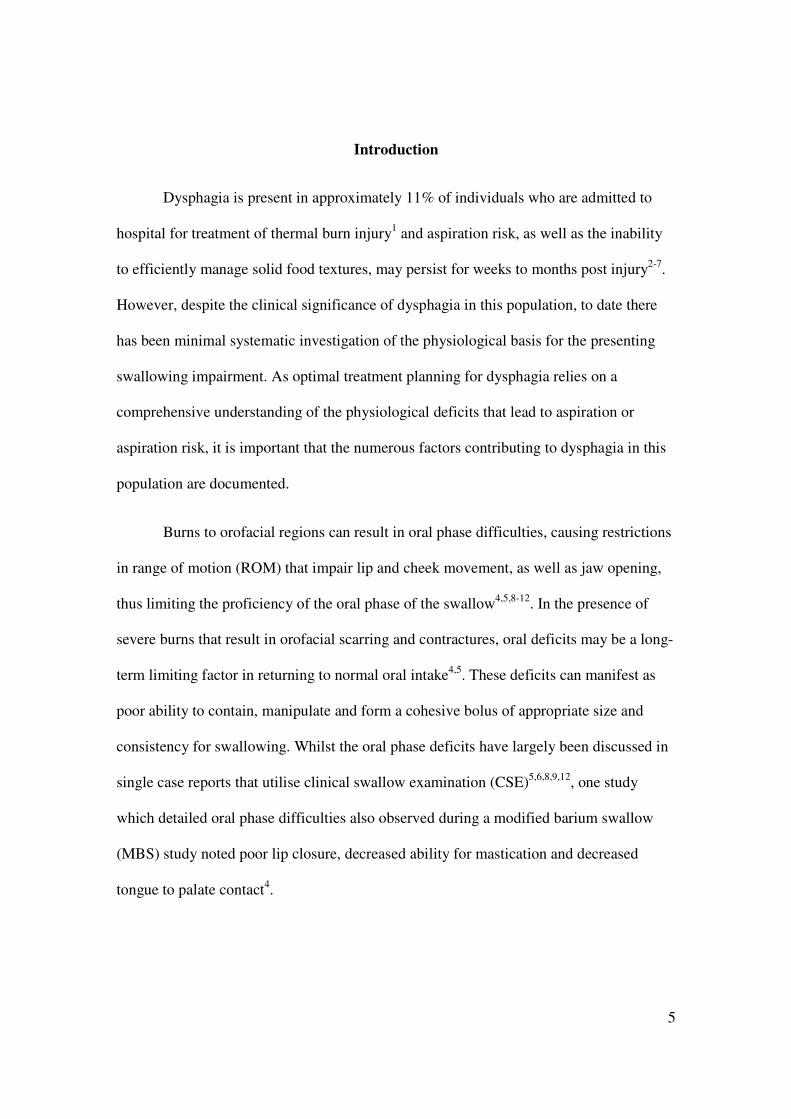
Dysphagia is present in approximately 11% of individuals who are admitted to
hospital for treatment of thermal burn injury1 and aspiration risk, as well as the inability
to efficiently manage solid food textures, may persist for weeks to months post injury2-7
.
However, despite the clinical significance of dysphagia in this population, to date there
has been minimal systematic investigation of the physiological basis for the presenting
swallowing impairment. As optimal treatment planning for dysphagia relies on a
comprehensive understanding of the physiological deficits that lead to aspiration or
aspiration risk, it is important that the numerous factors contributing to dysphagia in this
population are documented.
Burns to orofacial regions can result in oral phase difficulties, causing restrictions
in range of motion (ROM) that impair lip and cheek movement, as well as jaw opening,
thus limiting the proficiency of the oral phase of the swallow4,5,8-12
. In the presence of
severe burns that result in orofacial scarring and contractures, oral deficits may be a long-
term limiting factor in returning to normal oral intake4,5. These deficits can manifest as
poor ability to contain, manipulate and form a cohesive bolus of appropriate size and
consistency for swallowing. Whilst the oral phase deficits have largely been discussed in
single case reports that utilise clinical swallow examination (CSE)5,6,8,9,12
, one study
which detailed oral phase difficulties also observed during a modified barium swallow
(MBS) study noted poor lip closure, decreased ability for mastication and decreased
tongue to palate contact4.
6
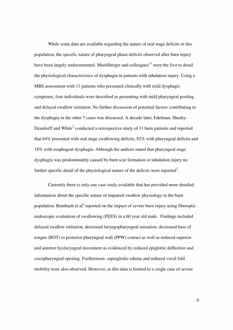
While some data are available regarding the nature of oral stage deficits in this
population, the specific nature of pharyngeal phase deficits observed after burn injury
have been largely undocumented. Muehlberger and colleagues13 were the first to detail
the physiological characteristics of dysphagia in patients with inhalation injury. Using a
MBS assessment with 11 patients who presented clinically with mild dysphagic
symptoms, four individuals were described as presenting with mild pharyngeal pooling
and delayed swallow initiation. No further discussion of potential factors contributing to
the dysphagia in the other 7 cases was discussed. A decade later, Edelman, Sheehy-
Deardorff and White2 conducted a retrospective study of 11 burn patients and reported
that 64% presented with oral stage swallowing deficits, 82% with pharyngeal deficits and
18% with esophageal dysphagia. Although the authors stated that pharyngeal stage
dysphagia was predominantly caused by burn scar formation or inhalation injury no
further specific detail of the physiological nature of the deficits were reported2.
Currently there is only one case study available that has provided more detailed
information about the specific nature of impaired swallow physiology in the burn
population. Rumbach et al4 reported on the impact of severe burn injury using fiberoptic
endoscopic evaluation of swallowing (FEES) in a 60 year old male. Findings included
delayed swallow initiation, decreased laryngopharyngeal sensation, decreased base of
tongue (BOT) to posterior pharyngeal wall (PPW) contact as well as reduced superior
and anterior hyolaryngeal movement as evidenced by reduced epiglottic deflection and
cricopharyngeal opening. Furthermore, supraglottic edema and reduced vocal fold
mobility were also observed. However, as this data is limited to a single case of severe

7
dysphagia post burn it is uncertain how closely this information may be extrapolated to
the wider clinical population with burn injury.
Due to the limited information that currently exists, the aim of the current study is
to investigate and describe the acute characteristics of swallowing dysfunction in an adult
cohort post thermal burn injury with dysphagia by means of a clinical and instrumental
examination. By sampling from a prospective clinical cohort across a range of injury
severities, it is intended that the current data will provide a more representative view of
the range of swallowing deficits which may be present in this population. Ultimately this
data will provide clinicians with detailed information regarding dysphagia characteristics
in the thermal burn population to help inform assessment, treatment and rehabilitation
planning.
Methods
Participant Population
Participants included 19 adults (14 males, 5 females), ranging in age from 18 to
85 years (M = 47.95, SD = 20.81) with thermal burn injury, with or without inhalation
injury, who presented for management at the Royal Brisbane and Women’s Hospital
specialised burns unit, Australia, over a 24 month period (August 2007- July 2009). The
mean total body surface area (TBSA) affected was 31.87% (SD = 17.06, range = 4-66.5),
with 58% (n = 11) having concomitant inhalation injury and 74% (n = 14) having burns
to the head and neck. Over 85% of participants required periods of mechanical
ventilation, with six participants requiring a tracheostomy during the course of their

8
hospital admission. No participant had existing neurological or structural impairment that
could influence swallowing behaviour or a prior history of swallowing disorders, as
determined by medical chart review, multidisciplinary team discussion, and patient
report. Biographical data including burn aetiology, severity, subsequent medical
management and dysphagia severity prior to and post FEES are detailed for each
participant in table 1.
/insert Table 1 near here/
Participants in this cohort diagnosed with orofacial burns (n=14) had received
multidisciplinary and interdisciplinary management for contracture prevention from the
point of hospital admission as per Rumbach et al11
. In addition, prior to the current
investigation, all participants had undergone at least one prior CSE by an experienced
speech pathologist. The initial referral to speech language pathology (SLP) for dysphagia
assessment had occurred on average 16 days post admission (SD = 15 days, range = 1-45
days) once the medical officer in charge had determined the patient to be medically stable
and suitable for oral intake. Intervention for suspected aspiration risk as determined from
these initial CSEs was limited to compensatory management via texture modifications as
participants were deemed not medically stable to participate in active rehabilitation11
.
Referral to the current study occurred only once there was agreement by the
multidisciplinary team (including but not limited to the medical officer, speech
pathologist, occupational therapist, physiotherapist, psychologist, and dietician) that
patients were suitable to undergo an instrumental examination of swallowing to assess
aspiration risk and facilitate active rehabilitation planning. Instrumental assessment
therefore occurred at various stages of recovery for each patient, at a mean of 37 days

9
(SD = 22 days, range = 5-80 days) post injury, and at a mean of 21 days (SD = 14 days,
range = 4-53 days) post initial assessment by SLP. At the time of participation in this
study, four individuals were tracheostomised, with the cuff deflated and a one-way
speaking valve in situ. An additional two individuals had been decannulated 48 hours
prior to assessment.
Procedure
Prior to the study, reliability training in clinical decision making (i.e., dysphagia
absence/presence and optimal oral intake) following a CSE was undertaken by the
primary rating clinician that involved completing 40 simultaneous CSEs with a second
experienced clinician. Percent exact agreement for clinical decisions regarding
recommendations for (1) optimal food textures to minimise aspiration risk and (2) fluid
consistency to minimise aspiration risk for each patient was 100% between the two
clinicians.
Once deemed suitable for FEES and recruited into this study, all participants
underwent a CSE performed by an experienced speech-language pathologist, conducted
no more than one day prior to a FEES assessment. This enabled information on both oral
and pharyngeal stage deficits to be compiled. The CSE consisted of a patient interview,
visual examination of the oromusculature, oromotor examination, perceptual evaluation
of voice quality, and a series of oral intake trials of fluids and foods that also included a
water swallow test14-17 when appropriate. All participants were trialled with the
fluids/foods considered to be least normal first (i.e., extremely thick fluids, puree diet),
with progression towards normal dietary consistencies and textures (i.e., thin fluids,

10
general diet), during oral intake trials if appropriate. Considerations for conducting a CSE
with burned individuals, as outlined by Rumbach et al11 were followed, with each
assessment requiring some variation depending on patient presentation. From this, an
initial rating of dysphagia severity was conducted using a purpose-built rating scale6 that
ranged from 1= normal swallow status (normal diet) to 4 = severe dysphagia (nil by
mouth; small amounts only of oral intake of full texture and consistency restriction). A
purpose built severity scale is necessary for this clinical population as most existing
dysphagia severity scales factor the need for alternative or supplemental feeding into the
severity rating. However, supplemental feeding in the burns population is often not
related to dysphagia but rather management of the hypermetabolic response to burn
injury, hence most published severity scales cannot be applied to this clinical population.
The FEES procedure was then conducted at the bedside as per protocol18
under
the direction of an otorhinolaryngologist (ENT) and treating speech pathologist. All
images were viewed online using an Olympus Viser OTV-37 digital processor scope with
an Olympus Visera CLV-S40 light source attached to a dysphagia swallow workstation
(DSW; Kay Pentax). The images were recorded using the DSW, with audio captured by a
lapel microphone attached to the patient’s collar. Participants were seated upright, and the
scope, was passed through the nostril in order to view the nasopharynx, hypopharynx and
larynx during non swallow and swallow tasks. Passing of the scope was undertaken by an
ENT and no local anaesthesia was used during the procedure.
The initial stage of the FEES involved examination of the structural integrity,
symmetry, range, speed and precision/timing/coordination of the velum, BOT,
pharyngeal muscles and larynx were assessed through observation of the structures at

11
rest, during phonation and a dry swallow. Following this, the observable features of the
swallow were documented with the presentation of boluses of different food and fluid
textures and consistencies as appropriate for each patient. Dietary consistencies trialled
were consistent with the Australian standards for texture modified food and fluids19
and
the range included smooth puree, minced and moist, soft and normal food consistencies
as well as extremely thick (level 900), moderately thick (level 400), mildly thick (level
150) and thin (regular) fluids. All food and fluid was dyed green to allow for easy
visualisation of the bolus path. Consistent with the CSE procedure, all participants were
trialled with the fluids/foods considered to be least normal first (i.e., extremely thick
fluids, puree diet), with progression towards normal dietary consistencies and textures
(i.e., thin fluids, general diet), during oral intake trials under FEES. Fluid trials preceded
food trials and each participant received at least two trials of each consistency presented.
Larger bolus volumes of food and fluid of approximately 20mls20
were used in this study
in comparison to volumes typically used in research (5-10ml boluses21-24
) to better
evaluate aspiration risk with more typical meal bolus sizes. Liquids were presented using
a spoon, straw or cup, depending on the patient’s ability to self-feed. The participant was
instructed to take one sip at a time. Continuous drinking was also assessed on thin fluids
if appropriate. Foods were presented via spoon, or as a whole entity (e.g.,
marshmallow/biscuit) and the participant was instructed to take a normal size bite.
Suitability for progression to the next food or fluid texture/consistency was based on (a)
the safety of food/fluid intake and (b) the efficiency of fluid/food intake. Compensatory
strategies were trialled where appropriate. However, swallowing ability without the use
of compensatory measures (other than texture modification) was used in all subsequent
12
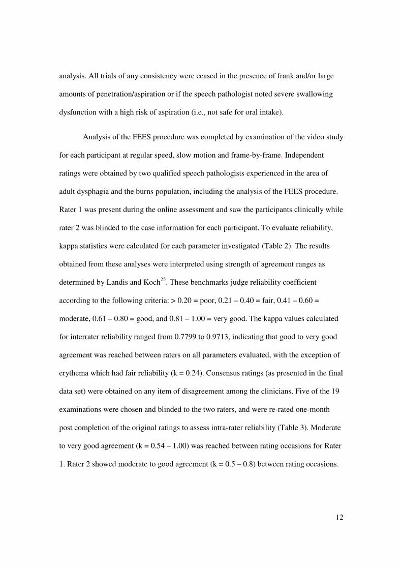
analysis. All trials of any consistency were ceased in the presence of frank and/or large
amounts of penetration/aspiration or if the speech pathologist noted severe swallowing
dysfunction with a high risk of aspiration (i.e., not safe for oral intake).
Analysis of the FEES procedure was completed by examination of the video study
for each participant at regular speed, slow motion and frame-by-frame. Independent
ratings were obtained by two qualified speech pathologists experienced in the area of
adult dysphagia and the burns population, including the analysis of the FEES procedure.
Rater 1 was present during the online assessment and saw the participants clinically while
rater 2 was blinded to the case information for each participant. To evaluate reliability,
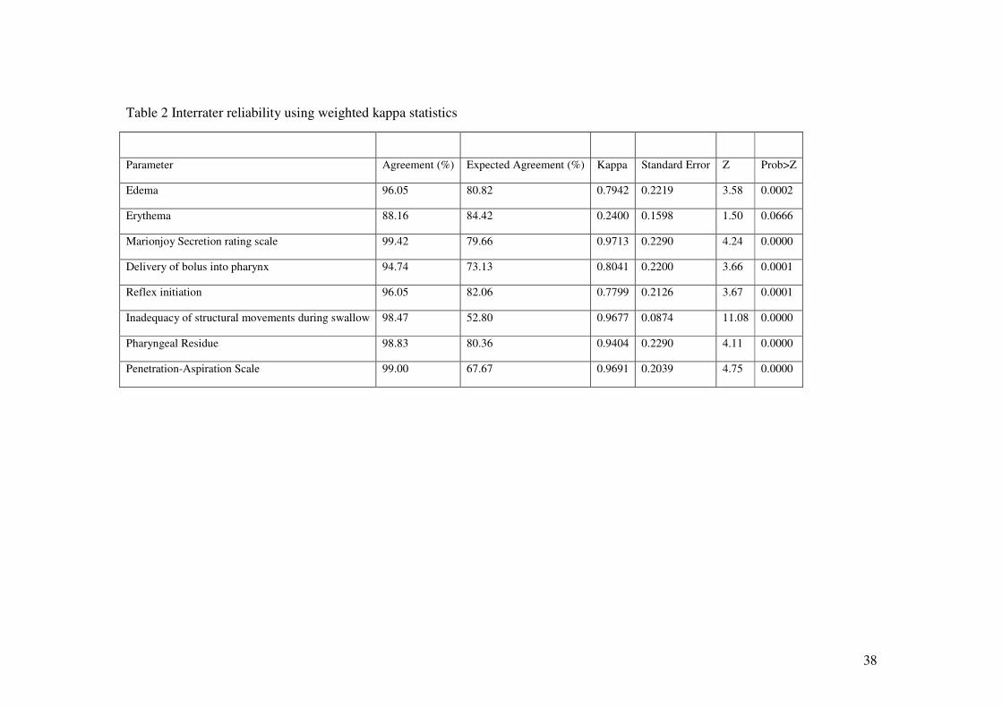
kappa statistics were calculated for each parameter investigated (Table 2). The results
obtained from these analyses were interpreted using strength of agreement ranges as
determined by Landis and Koch25
. These benchmarks judge reliability coefficient
according to the following criteria: > 0.20 = poor, 0.21 – 0.40 = fair, 0.41 – 0.60 =
moderate, 0.61 – 0.80 = good, and 0.81 – 1.00 = very good. The kappa values calculated
for interrater reliability ranged from 0.7799 to 0.9713, indicating that good to very good
agreement was reached between raters on all parameters evaluated, with the exception of
erythema which had fair reliability (k = 0.24). Consensus ratings (as presented in the final
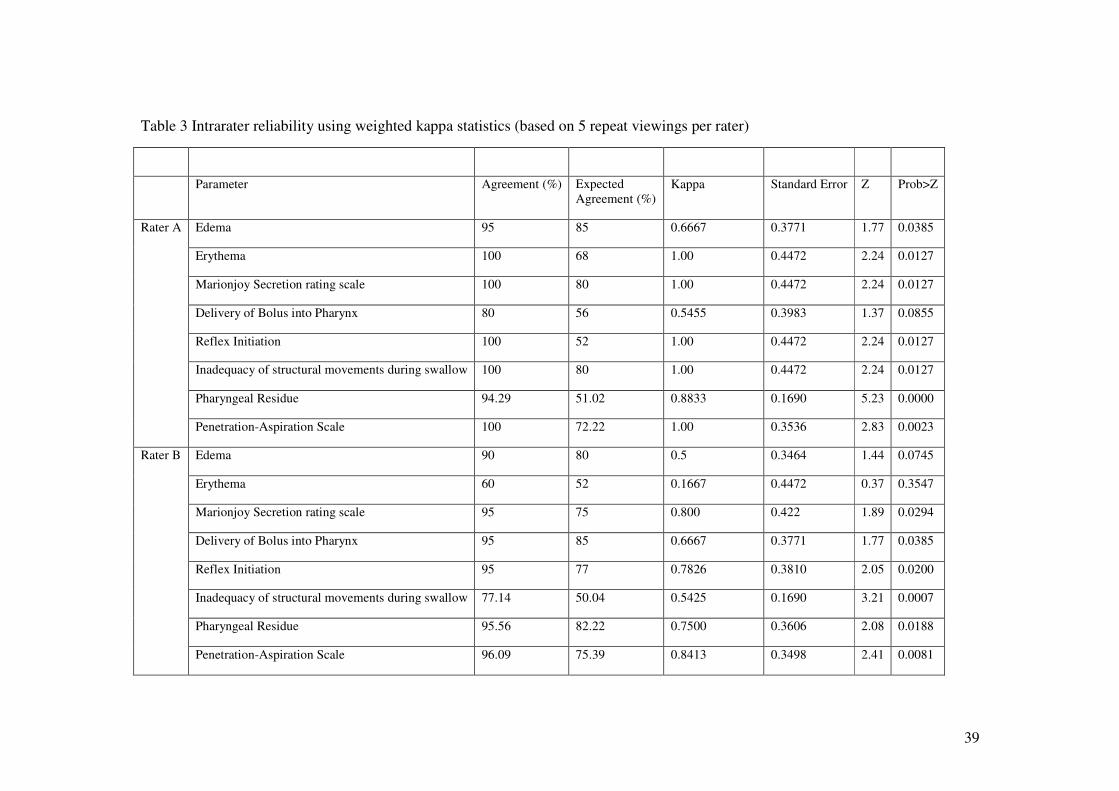
data set) were obtained on any item of disagreement among the clinicians. Five of the 19
examinations were chosen and blinded to the two raters, and were re-rated one-month
post completion of the original ratings to assess intra-rater reliability (Table 3). Moderate
to very good agreement (k = 0.54 – 1.00) was reached between rating occasions for Rater
1. Rater 2 showed moderate to good agreement (k = 0.5 – 0.8) between rating occasions.

13
/insert Tables 2 and 3 near here/
The analysis parameters used in this study were based on Langmore’s26 original
rating form. Ratings were made of the structural integrity and physiological function of
the velum, tongue, pharynx and larynx (as appropriate). The presence of any
laryngotracheal pathology (including edema and erythema) was noted. Any edema or
erythema of tissues affecting the structural integrity of the hypopharynx and larynx were
rated using a purpose-built scale that ranged from 1 (no edema/erythema) to 3 (all tissues
edematous/widespread erythema). Adequacy of saliva management was then rated
according to the Marionjoy Secretion Rating Scale27. This is a 5-level rating scale with
ratings ranging from normal (thin, clear secretions with less than 10% pooling in
pyriform fossae or valleculae) to profound (secretions present on vocal folds).
Appearance of secretions (colour and viscosity), patient response to secretions (i.e., are
attempts to spontaneously clear secretions being made), affect of spontaneous or cued
swallows on secretion reduction, and the frequency of spontaneous swallows were also
noted to determine adequacy of saliva management prior to the commencement of oral
intake trials. Swallow frequency was classified as reduced if spontaneous swallows were
observed to be less than 1 per minute. Sensation testing was informally conducted via
patient response to the presence and light touch of the endoscope on the lateral
pharyngeal walls and tip of the epiglottis. The patient was deemed to have reduced
sensation if there was no response to the presence and light taps of the scope in the
pharynx and an absent or reduced response to residue or to an event of silent aspiration.
The presence of the laryngeal adductor reflex (i.e., brief closure of the true vocal cords),

14
an involuntary airway protection reflex, was also examined by light touch of the scope to
the arytenoid epithelium.
For the food and fluid trials, each bolus for each consistency trialled was
individually rated. Any parameter observed during at least one swallow trial per
consistency denoted the characteristic as present for that consistency. Parameters for
analysis included observable characteristics of the oral and pharyngeal stage, oral and
pharyngeal transit times and other markers indicating poor coordination or inefficiency of
the swallow. Specifically, the oral phase of the swallow trials was examined through
visual inspection of the acceptance, containment and manipulation of the bolus, oral
transit time and an informal rating of delivery of the bolus into the pharynx on a 4-point
scale (see appendix A). Ratings ranged from normal (prompt delivery of bolus) to severe
impairment (large amount of leakage during oral preparation with minimal mastication of
bolus and no attempt at a transfer). At the completion of each bolus, the oral cavity was
examined for the presence of oral residue and was rated as being absent or present, with
location of any residue being noted.
The pharyngeal phase was examined through determining the adequacy of
structural movements during the swallow as per Langmore26
. This included investigation
of bolus driving and clearing forces (i.e., BOT movement, pharyngeal longitudinal and
constrictor movement), and valving forces (i.e., velopharyngeal and laryngeal valves).
Observations regarding presence of residue after and between swallows were also
recorded including information on: location of residue, amount of residue, patient
15
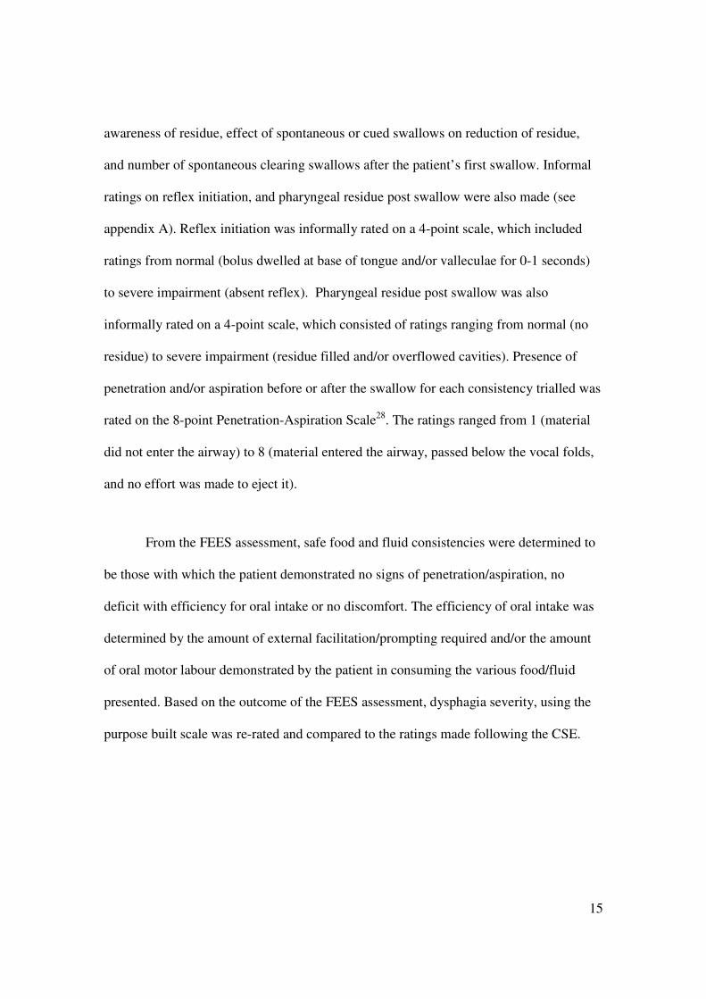
awareness of residue, effect of spontaneous or cued swallows on reduction of residue,
and number of spontaneous clearing swallows after the patient’s first swallow. Informal
ratings on reflex initiation, and pharyngeal residue post swallow were also made (see
appendix A). Reflex initiation was informally rated on a 4-point scale, which included
ratings from normal (bolus dwelled at base of tongue and/or valleculae for 0-1 seconds)
to severe impairment (absent reflex). Pharyngeal residue post swallow was also
informally rated on a 4-point scale, which consisted of ratings ranging from normal (no
residue) to severe impairment (residue filled and/or overflowed cavities). Presence of
penetration and/or aspiration before or after the swallow for each consistency trialled was
rated on the 8-point Penetration-Aspiration Scale28. The ratings ranged from 1 (material
did not enter the airway) to 8 (material entered the airway, passed below the vocal folds,
and no effort was made to eject it).
From the FEES assessment, safe food and fluid consistencies were determined to
be those with which the patient demonstrated no signs of penetration/aspiration, no
deficit with efficiency for oral intake or no discomfort. The efficiency of oral intake was
determined by the amount of external facilitation/prompting required and/or the amount
of oral motor labour demonstrated by the patient in consuming the various food/fluid
presented. Based on the outcome of the FEES assessment, dysphagia severity, using the
purpose built scale was re-rated and compared to the ratings made following the CSE.

16
Results
At the time of referral for FEES assessment, CSE classified 52.5% (10/19) of the
cohort as having severe dysphagia, 31.5% (6/19) had a moderate impairment, and 16%
(3/19) had a mild impairment (Table 1). However, subsequently FEES assessment
revealed normal swallow function in 6 individuals (participants 2, 3, 4, 6, 13, and 17 –
Table 1), and these participants were excluded from all subsequent data analysis. Data
from this point is represented as a portion of the total remaining cohort of 13 individuals,
unless specifically stated otherwise.
Oromotor examination revealed that 77% of the remaining cohort of 13 presented
with a weak or absent voluntary cough and 100% a dysphonic vocal quality. Poor
dentition was also prevalent, with 54% of the cohort having absent teeth and/or teeth with
advanced decay. Decreased lip strength and ROM were evident in 100% of cases. Jaw
strength was also reduced in 54% of individuals. Nil deficits in tongue function were
observed in any participant. Two individuals presented with severe orofacial scarring and
contracture formation and presented with severely reduced and asymmetrical orofacial
ROM (Table 1). During swallow trials, this degree of scarring and contracture formation
caused inadequate labial closure, increased time for oral preparation/bolus formation and
diffuse oral residue that required manual removal from the lateral sulci by the clinician
for these 2 individuals. No other participant had difficulties controlling oral boluses, yet
increased oral preparation time was required for the majority of individuals’ trialled on
more solid textures (e.g., biscuit). Tightness at the oral commissures, alongside poor
dentition, advanced age and reduced jaw strength, lead to fatigue and discomfort in one
17
individual; thus limiting their ability to be upgraded to a normal and varied texture diet
although aspiration risk has resolved.
FEES assessment of the structural integrity and function of the velopharynx,
hypopharynx and larynx prior to the commencement of food and fluid trials are displayed
in Table 4. Laryngotracheal pathology, as confirmed by an otorhinolaryngologist, was
highly prevalent across the cohort, with 77% (n = 10) displaying edema, with
approximately one third of the cohort exhibiting concurrent tissue granulation, ulceration
and/or edema (Table 4). Laryngeal dysfunction was also prevalent in the cohort, with up
to 69% (n = 9) of the population presenting with one or more difficulties contributing to
altered vocal quality (Table 4). No participants presented with visible nasopharyngeal,
hypopharyngeal, laryngeal or tracheal strictures.
/insert Table 4 near here/
Impaired secretion management was also prevalent, with over 75% of the cohort
presenting with pooling of secretions in the pharynx, suggesting a reduced awareness or
ability to clear secretions. Aspiration of secretions was observed in two individuals with
severe dysphagia. Two individuals who were unaware of their secretions could clear
them with cued swallows. Sensation assessment revealed minimal or nil response to
contact by the scope in 77% (n = 10) of cases. Of note, 70% of individuals with
reductions in both secretion management and sensation had been identified as having
burn injury to the airway on admission to hospital.
The characteristics of the swallow mechanism observed during bolus trials via
FEES are displayed in table 5.The information gained from the FEES on the oral phase
18
was limited to judgement on bolus delivery into the pharynx (Table 5). Over 50% of the
cohort had mild or moderate deficits with bolus delivery on both thickened and thin
fluids. For those 8 individuals trialled on solids, 75% presented with mild or moderate
deficits with bolus delivery.
/insert Table 5 near here/
A mild to moderate delay in swallow reflex initiation was noted in 85% of the
cohort for all fluid consistencies trialled. For food textures, two participants demonstrated
a mild to moderate delay in triggering the swallow reflex. Inadequate structural
movements during the swallow were evident across bolus types for the entire cohort
(Table 5), with up to 92% of the cohort displaying weak BOT function, inadequate
pharyngeal squeeze, and decreased epiglottal inversion(i.e., reduced whiteout at the
height of the swallow).
Pharyngeal residue was also a consistent characteristic, being present in at least
one consistency trialled for all individuals. Distribution of residue throughout the pharynx
varied considerably for each participant for each bolus type. Lodging of pharyngeal
residue that remained above the airway across fluid boluses was most commonly located
at the BOT (47%), valleculae (79%), lateral channels (74%), pyriforms (68%), along the
pharyngeal walls (47%) and at the posterior cricoid region (53%). Pharyngeal residue
was located on the true vocal cords and subglottic shelf in greater than 60% of
individuals trialled on thin fluids. Mild-moderate pharyngeal residue was also an issue for
75% of individuals trialled on solid textures, with residue being localised to the BOT,
valleculae, lateral channels and pyriforms.

19
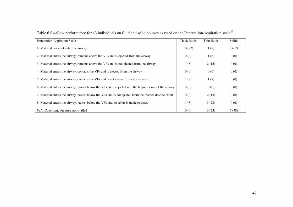
Laryngeal penetration (Penetration-Aspiration scale levels 2-5) occurred in 38%
(n = 5) of the cohort on thick or thin fluids boluses, with aspiration (Penetration-
Aspiration scale levels 6-8) being identified for 54% (n = 7) of patients, predominantly
on thin fluid bolus trials (Table 6). Sixty percent (n = 6/10) of penetration/aspiration
events were silent (participants 10,11,12,13,14, and 18) i.e., the patient had no response
to the material entering the airway and made no effect to eject the material. No
penetration or aspiration was observed on trials of solid textures. Aspiration most
commonly occurred after the swallow (83% of those who demonstrated aspiration on one
or more fluid consistencies; n = 5). Timing of aspiration after the swallow was immediate
in one case, delayed due to spill-over of residue in four cases, and a combination of both
immediate and delayed in one individual. The overall rating for dysphagia severity at the
conclusion of FEES for each participant is detailed in table 1. For those individuals with
ongoing dysphagia, 38% (n = 5) of participants had their dysphagia severity reduced as a
result of their diet status being upgraded following instrumental assessment.
/insert Table 6 near here/
Discussion
Clinical and instrumental examination of swallowing dysfunction in an adult
cohort in the acute stages post thermal burn demonstrated a combination of oral and
pharyngeal deficits which are in agreement with the handful of previously reported single
case studies4,5,8,9. Although both clinical and instrumental assessments have their
strengths and limitations, in this clinical population employing a combination of both
clinical and instrumental evaluations of swallow function proved imperative to accurately

20
identify the multifactorial deficits contributing to aspiration risk. There were a level of
disagreement observed between the CSE and FEES findings, with CSE results failing to
accurately discern the true severity of the dysphagia in all cases. Specifically, 32% (n =
6/19) diagnosed as dysphagia per CSE had no dysphagia identified on FEES, and 58% (n
= 11/19) of patients were incorrectly rated as having a more severe dysphagia on CSE
than on FEES. Consequently, without instrumental evaluation, some patients would have
potentially continued to receive modified texture and consistency diets unnecessarily,
continuing the financial burden associated with ongoing need for altered diets and
negatively impacting on return to normal oral intake for the patient. However, whilst
FEES was able to identify those pharyngeal stage deficits contributing to aspiration risk,
it was unable to discern oral stage deficits that may limit overall ability for oral intake. As
videofluoroscopy is generally unsuitable for use in the acute burns population11
, the oral
stage assessment conducted as part of the CSE proved a valuable part of the total
assessment process in determining oromotor status and functioning during the oral phase
of the swallow in this population. Considering the presence of oral and pharyngeal stage
deficits in this clinical group, information obtained from both clinical and instrumental
assessment results should be reviewed in combination in order to provide a
comprehensive picture of all swallowing issues.
Oromotor dysfunction in terms of weakness, while prevalent, was largely
unobtrusive to swallow function in the majority of participants at the time of FEES in the
current study. Long periods of muscle disuse brought on by extended
intubation/ventilation durations in this population may be an attributing factor to
weakness29. Furthermore, edema of the orofacial structures for those with facial burns

21
and large body burns (>20% TBSA) may have hindered oromotor functioning within the
first week post injury. These factors coupled with pain from unhealed burns may impact
upon strength and range of oromotor movement willingly demonstrated by individuals
during CSE, rendering the movements to be rated as suboptimal yet functional for oral
intake as seen in the current study. Difficulties with the oral phase of the swallow at the
time of FEES manifested solely in individuals with orofacial scarring and contractures,
thus indicating that oral phase deficits may not persist past the time required for healing
to occur for superficial and superficial partial thickness burns and may be limited to those
with deep partial and full thickness facial burns that require surgical intervention and
long healing times. This finding coincides with the body of research that links orofacial
contractures and long-term oral dysphagia4,5. Nasopharyngeal, hypopharyngeal, laryngeal
or tracheal strictures were not present in the cohort studied and thus did not contribute to
inadequate structural movements during the pharyngeal phase of the swallow. It is
feasible to hypothesise that laryngeal anchoring may occur in patients with contractures
on the anterior neck, however this was not observed in this cohort. Strictures after
thermal burn seem relatively uncommon and reports are sparse within the literature30
, yet
are well documented after chemical ingestion injuries31,32.
Numerous pharyngeal stage deficits have been found to be closely associated with
increased risk of aspiration in other dysphagic populations, including pooling of saliva33
,
laryngotracheal pathology34-38
, delayed swallow trigger39
, impaired sensation40
, reduced
airway protection41,42
, and the presence of pharyngeal residue39,43
. In the current study all
of these features were found to be prevalent, indicating that this is a clinical cohort at risk
of aspiration due to multifactorial causes. Even across the spectrum of dysphagia
22
severity, participants in the current group presented with multiple factors contributing to
aspiration risk. In addition, the potential for aspiration to lead to pulmonary compromise
in this population was found to be further compounded by the presence of poor cough
strength and advanced tooth decay. Most participants had weakened cough strength
indicating the potential inability to successfully clear aspirated secretions. Furthermore
the high presence of poor dentition is significant, considering the known relationship
between the number of decayed teeth, missing teeth and poorly fitted dentures and an
increased incidence of aspiration pneumonia44-49
. Poor oral hygiene creates a hospitable
environment for growth of pathogenic organisms in the mouth which further predisposes
the patient to pneumonia following the aspiration of contaminated oral secretions49-52.
Hence, there are a number of co-existing factors placing this clinical population at high
risk for aspiration and possibly the development of aspiration pneumonia. For this reason,
it is critical that a comprehensive instrumental assessment is conducted alongside the
CSE to accurately identify all potential risk factors.
Decreased laryngopharyngeal sensation was observed in 77% of the current
cohort and was potentially contributed to by prolonged periods of ETT intubation and
ventilation and, in some cases, concomitant inhalation injury. Predisposition for poor
sensation in the burn population has lead to the prediction of greater incidence of silent
aspiration11
, which was a concern in the current cohort in 60% of participants who had
penetration/aspiration verified during FEES. The potential for silent aspiration and its
clinical implications further compounds the need for instrumental assessment of swallow
function post thermal burn injury to detect physiological dysfunction that cannot be
23
reliably determined via CSE. FEES is particularly suited to the burn population and may
be the preferred procedure during the acute phase following burn injury due to its ability
to assess sensory impairment, edema, erythema and ulceration of pharyngeal and
laryngeal structures caused by concomitant inhalation injury, and its ability to be carried
out at bedside when mobility and positioning are compromised by pain, splints and
contractures11
. Furthermore, FEES is repeatable, which caters for the long recovery
process for burn patients and allows for assessment of vocal cord integrity and function,
which can be affected by endotracheal intubation and inhalation injury53
.
Laryngotracheal pathology was a prevalent feature across the cohort studied, with
over a large proportion of cohort demonstrating some degree of anatomical alteration in
the hypopharynx and larynx. This high incidence of structural abnormality is comparable
to the findings of Clayton and colleagues10
who conducted a retrospective study of
patients with severe burns who required tracheostomy, and found laryngotracheal
abnormalities in 100% of participants. In light of their data, Clayton et al10
proposed that
the actual incidence of laryngotracheal abnormality in this population is currently
underestimated. The current data would concur, and lends evidence to support that
laryngotracheal pathology is a highly prevalent feature in this clinical population. It is
postulated that such pathological changes may be caused by the often long periods of
intubation by either an endotracheal tube or tracheostomy tube, and/or as a consequence
of mucosal damage caused by inhalation injury54-57
. In the current cohort, such possible
causal factors were common with >50% experiencing inhalation injury, 85% undergoing
prolonged durations of intubation (i.e., 5 or more days with ETT), and 46% requiring a

24
tracheostomy tube. A high incidence of laryngeal pathology within the cohort is an
important finding, as it could conceivably contribute to the observed aspiration risk.
Pharyngeal edema was a significant issue for the majority of the cohort and its
presence has the potential to disrupt sensation and efficiency of the swallow. Widespread
edema (i.e., beyond the location of the burn injury) is an acute physiological response to
burns larger than 30%58
and is considered an early reaction which resolves within three to
four days post injury in most cases58,59
. Pharyngeal edema that persists past the initial
injury phase, once fluid resuscitation has been achieved, may be contributed to by the
presence of long term NGT placement, necessary for the management of the
hypermetabolic response post burn injury. Further understanding of the impact of
widespread edema on swallowing and its patterns of resolution is needed. A prospective
study using repeated FEES assessment would provide valuable insight into this issue.
Aspiration of saliva is a common feature in patients that are ventilator or
tracheostomy dependent29,37,60,61, and the presence of pooled saliva is reported to be
associated with greater likelihood of aspiration of fluids33. It has previously been
postulated that increased salivary secretions may be precipitated in this population by
concomitant inhalation injury, with poor cilia functioning and sloughing of and damage
to the mucosa causing increased secretion production and decreased sensation11,56,62
.
Although a large proportion of the cohort (70%) displayed poor ability to manage
secretions in combination with poor sensation and inhalation injury, this study fails to
provide statistical evidence to support this theory. Investigation via FEES at earlier and
multiple intervals post injury may provide a different insight into the effects of inhalation
injury on secretion management.
25
Premature spillage (mild to moderate deficits in delivery of the bolus into the
pharynx), a parameter related to the presence of lingual dysfunction63 was observed in the
current cohort. Although all participants were rated on the CSE as having tongue function
within normal limits, it is possible that some mild degree of generalised oromotor
weakness coupled with reduction in sensation may account for poor bolus containment
prior to swallow initiation in this population. Furthermore, a delayed swallow trigger was
a frequent deficit. Delayed reflex initiation has been recognised as being strongly
associated with increased risk of aspiration42
, with risk of aspiration increasing
proportionally to the time the bolus dwells in the pharynx before the initiation of the
swallow64. As no participants in the current cohort were identified as having any
neurological deficits, it is likely that failure to trigger a swallow promptly is solely
attributable to the fact that the participant is unaware of the position of the bolus in the
pharynx. While this may increase the possibility of aspiration prior to the swallow, this
was not evident within the current cohort.
Specific pharyngeal phase deficits that have been reported in literature to date for
patients with burn injury include decreased BOT to PPW movement, reduced pharyngeal
clearance and suboptimal hyolaryngeal movement4,9,13. Similar characteristics were
observed across the cohort; a large proportion presented with reduced BOT to PPW
contact and abnormal pharyngeal longitudinal and constrictor movement. This may lead
to a build up of residue outside the larynx, contributing to increased aspiration risk.
Distribution of residue was vastly different for each participant; acknowledgement of
residue location is imperative in determining the point of breakdown in the process of
bolus clearance26. Residue was widely dispersed throughout the velopharynx,

26
hypopharynx and larynx, suggesting either a delayed movement or reduced force in one
laryngeal structure or all muscles involved in performing a swallow26. The presence of
pharyngeal residue was observed to lead to aspiration after the swallow as the result of
spill-over into and through the laryngeal inlet for 83% of individuals who aspirated
during the assessment. Langmore26
noted that the overall severity of residue relates
directly to the sensitivity/response of the patient to the existence of the residue. In the
burn population altered sensation was a notable feature with 77% of patients registering
no response to light touch from the scope on assessment.
The current data would suggest that the deficits contributing to dysphagia post
burn relate primarily to disruption to the structure and function of the swallowing
mechanism, largely due to edema and orofacial contracture formation. This information
has implications for rehabilitation, as it appears the swallow does not necessarily need to
be retrained at a neurological level, but requires compensatory management of oral and
pharyngeal deficits through postural adjustment, training of airway protection strategies
and the use of texture and consistency modification to minimise aspiration risk coupled
with early and ongoing intervention to minimise/prevent any oral contractures5,65,66. Due
to the medically fragile nature of this population and the fact that recovery patterns for
the physiological deficits identified by the current study, such as edema, is unknown,
regular monitoring of swallow function via both CSE and FEES is recommended to
optimise the rehabilitation process.

27
Conclusion
Currently there is very little literature investigating the nature of the swallowing
impairment post thermal burn injury. Findings of this study identified generalised
oromotor weakness was observed upon CSE in the majority of the cohort, with functional
deficits of the oral phase reserved for the few individuals with severe dysphagia and
orofacial contractures. Observable physiological deficits on FEES that were identified as
being prominent across the cohort included: laryngotracheal pathology, decreased ability
to manage secretions, delayed swallow initiation, decreased laryngopharyngeal sensation,
diffuse pharyngeal residue and a risk of silent aspiration. The need for the introduction
and routine utilisation of instrumental assessment of swallowing function in some cases
post thermal burn injury has been highlighted due to the multifaceted nature for the
dysphagia and the possibility for silent aspiration, a feature undetectable using clinical
beside evaluation of swallowing. This study has provided a first detailed report of the
physiological effects of burn injury on swallow function in a group of patients with
varying degrees of dysphagia. The data presented in this paper facilitates a greater
understanding of the physiological underpinnings of dysphagia in the burn population, as
well as promotes the inclusion of SLP services and the use of instrumental evaluations of
swallowing with this population. Further research however is needed through systematic
routine FEES assessment of a large prospective cohort in order to examine physiological
changes and recovery over time.

28
Acknowledgements
The authors gratefully acknowledge funding support for this study from the Royal
Brisbane and Women’s Hospital Foundation. We acknowledge the assistance of the
Royal Brisbane and Women’s Hospital Professor Stuart Pegg Adult Burns Unit in the
recruitment of participants for this study. Special thanks to the Royal Brisbane and
Women’s Hospital ENT department for their assistance with the assessments. Finally, the
authors wish to acknowledge the 19 individuals that participated in this study for the
generous gift of their time, patience and courage.
29
References
1. Rumbach AF, Ward EC, Cornwell PL, Bassett LV, Khan A, Muller MJ:
Incidence and predictive factors for dysphagia following thermal burn injury: A
prospective cohort study. J Burn Care Res, in press.
2. Edelman DA, Sheehy-Deardorff DA, White MT: Bedside assessment of
swallowing is predictive of an abnormal barium swallow examination. J Burn
Care Res 29: 89-96, 2008.
3. McKinnon DuBose C, Groher MG, Carnaby Mann GS, Mozingo DW:Pattern of
dysphagia recovery after thermal burn injury. J Burn Care Rehabil 26: 233-237,
2005.
4. Rumbach AF, Ward EC, Cornwell PL, Bassett LV, Muller MJ: The challenges of
dysphagia management and rehabilitation after extensive thermal burn injury: A
complex case. J Burn Care Res 30: 899-903, 2009.
5. Rumbach AF, Ward EC, Cornwell PL, Bassett LV, Spermon ML, Plaza AL,
Muller MJ: Dysphagia management and rehabilitation: an interdisciplinary and
multidisciplinary collaborative. J Med Speech Lang Pathol 19: 25-34, 2011.
6. Rumbach AF, Ward EC, Cornwell PL, Bassett LV, Muller MJ: Clinical
progression and outcome of dysphagia following thermal burn injury: a
prospective cohort study, J Burn Care Res, in press.
7. Ward EC, Uriarte M, Conroy A-L: Duration of dysphagic symptoms and
swallowing outcomes after thermal burn injury. J Burn Care Rehabil 22:441-453,
2001.

30
8. Clayton NA, Kennedy PJ: Management of firecracker induced oropharyngeal
burns: a case report. Advances in Speech-Language Pathology 9: 265-270, 2007.
9. Clayton NA, Ledgard JP, Haertsch PA, Kennedy P, Maitz P: Rehabilitation of
speech and swallowing after burns reconstructive surgery of the lips and nose. J
Burn Care Res 30: 1039-1045, 2009.
10. Clayton N, Kennedy P, Maitz P: The severe burns patient with tracheostomy:
Implications for management of dysphagia, dysphonia and laryngotracheal
pathology. Burns 36: 850-855, 2010.
11. Rumbach AF, Ward EC, McKinnon DuBose C, Clayton NA: Burn Injury. In:
Ward EC, Morgan AT (eds.): Dysphagia post trauma. San Francisco: Plural
Publishing, pp 151-199.
12. Wust KJ: A modified dynamic mouth splint for burn patients. J Burn Care Res 27:
86-92, 2006.
13. Muehlberger T, Kunar D, Munster A, Couch M: Efficacy of fiberoptic
laryngoscopy in the diagnosis of inhalation injuries. Arch Otolaryngol Head Neck
Surg 124: 1003–1007, 1998.
14. DePippo KL, Holas MA, Reding MJ: Validation for the 3-oz water swallow test
for aspiration following stroke. Arch Neurol 49: 1259-1261, 1992.
15. Garon BR, Engle M, Ormiston C: Reliability of the 3-oz water swallow test
utilising cough reflex as sole indicator of aspiration. Neurorehabilitation and
Neural Repair, 9: 139-143, 1995.
16. Suiter DM, Leder SB: Clinical utility of the 3-ounce water swallow test.
Dysphagia 23: 244-250, 2008.
31
17. Wu M-C, Chang Y-C, Wang T-G, Lin LC: Evaluating swallowing dysfunction
using a 100-ml water swallowing test. Dysphagia 19: 43-47, 2004.
18. Langmore SE, Schatz K, Olsen N: Fiberoptic endoscopic examination of
swallowing safety: a new procedure. Dysphagia 2: 216-219, 1988.
19. Dieticians Association of Australian and the Speech Pathology Association of
Australia Limited: Texture-modified foods and thickened fluids as used for
individuals with dysphagia: Australian standardised labels and definitions.
Nutrition and Dietetics 64: S53-S76, 2007.
20. Adnerhill I, Ekberg O, Groher ME: Determining normal bolus size for thin
liquids. Dysphagia 4: 1-3, 1989.
21. Kelly AM, Leslie P, Beale T, Payten C, Drinnan MJ: Fiberopic endoscopic
evaluation of swallowing and videofluoroscopy: does examination type influence
perception of pharyngeal residue severity?. Clin Otolaryngol 31: 425-432, 2006.
22. McGowan SL, Gleeson M, Smith M, Hirsch N, Shuldham CM: A pilot study of
fiberoptic endsoscopic evaluation of swallowing in patients with cuffed
tracheostomies in neurological intensive care. Neurocritical Care 6:90-93, 2007.
23. Robbins J, Levine R, Maser A, Rosenbek JC, Kempster GB: Swallowing after
unilateral stroke of the cerebral cortex. Arch Phys Med Rehabil 74: 1295-1300,
1993.
24. Tracy JF, Logemann JA, Kahrilas PJ, Jacob P, Kobara M, Krugler C: Preliminary
observations on the effects of age on oropharyngeal deglutition. Dysphagia 4: 90-
94, 1989.

32
25. Landis JR, Koch GG:The measurement of observer agreement for categorical
data. Psychol Bull 86: 420-428, 1979.
26. Langmore SE: Fiberoptic evaluation and treatment of swallowing disorders. New
York: Thieme, 2001.
27. Donzelli J, Brady S, Wesling M, Craney M: Predictive value of accumulated
oropharyngeal secretions for aspiration during video nasal endoscopic evaluation
of the swallow. Ann Otol Rhinol Laryngol 112:469-475, 203.
28. Rosenbek JC, Robbins JA, Roecker EB, Coyle JL, Wood JL: A Penetration-
Aspiration Scale. Dysphagia 11:93-98, 1996.
29. DeVita MA, Spierer-Runback L: Swallowing disorders in patients with prolonged
orotracheal intubation or tracheostomy tubes. Crit Care Med 18:1328-1330, 1990.
30. Gaissert HA, Lofgren RH, Grillo HC: Upper airway compromise after inhalation
injury: Complex strictures of the larynx and trachea and their management.
Annals of Surgery 218:672-678, 1993.
31. Hoseok I, Shim YM, Son YI, Kim K, Choi YS: Pharyno-colostomy with
supraglottic partial laryngectomy in caustic oropharyngeal stricture. Annals of
Thoracic Surgery 81: 712-714, 2006.
32. Shikowitz MJ, Levy J, Villano D, Graver ML, Pochaczevsky P: Speech and
swallowing rehabilitation following devastating caustic ingestion: Techniques and
indicators for success. Laryngoscope 106: 1-12, 1996.
33. Murray J, Langmore SE, Ginsberg S, Dostie A: The significance of accumulated
oropharyngeal secretions and swallowing frequency in predicting aspiration.
Dysphagia 11: 99-103, 1996.

33
34. Colice GL: Resolution of laryngeal injury following translaryngeal intubation.
Am Rev Respir Dis 145: 361-364, 1992.
35. Colice GL, Stukel TA, Dain B: Laryngeal complications of prolonged intubation.
Chest 96: 877-884, 1989.
36. Stauffer JL, Olson DE, Petty TL: Complications and consequences of
endotracheal intubation and tracheotomy. Am J Med 10: 65-76, 1981.
37. Tolep K, Getch CL, Criner GJ: Swallowing dysfunction in patients receiving
prolonged mechanical ventilation. Chest 109:167-172, 1996.
38. Whited RE: A prospective study of laryngotracheal sequelae in long-term
intubation. Laryngoscope 94: 367-377, 1984.
39. Perlman AL, Booth BM, Grayhack JP: Videofluoroscopic predictors of aspiration
in patients with oropharyngeal dysphagia. Dysphagia 9: 90-95, 1994.
40. Aviv JE, Kim T, Goodhart K, Kaplan S, Thomson J, Diamond B, Close LG:
FEESST: a new bedside endoscopic test of the motor and sensory components of
swallowing. Ann Otol Rhinol Laryngol 107: 378-387, 1998.
41. Kahrilas PJ, Lin S, Rademaker AW, Logemann JA: Impaired deglutitive airway
protection: a videofluoroscopic analysis of severity and mechanism.
Gasteroenterology, 113: 1457-1464, 1997.
42. Lundy DS, Smith C, Colangelo L, Sullivan PA, Logemann JA, Lazarus CL,
Newman LA, Murry T, Lombard L, Gaziano J: Aspiration: cause and
implications. Otolaryngol Head Neck Surg 120: 474-478, 1999.
43. Eisenhuber E, Schima W, Schober E, Pokieser P, Stadler A, Scharitzer M,
Oschatz E: Videofluroscopic assessment of patients with dysphagia: pharyngeal
34
retention is a predictive factor for aspiration. Am J Roentgenol 178: 393-398,
2002.
44. Bartlett JG, Finegold SM. Anaerobic infections of the lung and pleural space. Am
Rev Respir Dis 110: 56-77, 1974.
45. Langmore SE, Terpenning MS, Schork A, Chen Y, Murray JD, Lopatin D,
Losche WJ. Predictors of aspiration pneumonia: How important is dysphagia?
Dysphagia 13:69-81, 1998.
46. Mojon P, Budtz-Jorgensen E, Michel JP et al. Oral health and history of
respiratory tract infection in frail institutionalized elders. Gerodontology 14:9-16,
1997.
47. Mojon P, Rentsch A, Budtz-Jorgensen E et al. Effects of an oral health program
on selected clinical parameters and salivary bacteria in a long-term care facility.
Eur J Oral Sci 106: 827-834, 1998.
48. Scannapieco FA, Papandontos GD, Dunford RG. Associations between oral
conditions and respiratory disease in a national sample survey population. Ann
Periodontol 3: 251-256, 1998.
49. Terpenning MS, Taylor GW, Lopatin DE, Kinder C, Dominguez L, Loesche WJ.
Aspiration pneumonia: Dental and oral risk factors in an older veteran population.
J Am Geriat Soc 49: 557-563, 2001.
50. Abe S, Ishihara K, Adachi M, Okuda K. Oral hygiene evaluation for effective oral
care in preventing pneumonia in dentate elderly. Arch Gerontol Geriatr 43: 53-64,
2006.
35
51. Bartlett JG, Gorbach SL, Finegold SM. The bacteriology of aspiration pneumonia.
Am J Med 56: 202-207, 1974.
52. Yoneyama T, Yoshida M, Ohrui T, Mukaiyama H, Okamoto H, Hoshiba K, et al.
Oral care reduces pneumonia in older patients in nursing homes. J Am Geriatr Soc
50: 430-434, 2002.
53. Leder SB, Sasaki CT, Burrell MI: Fiberoptic endoscopic evaluation of dysphagia
to identify silent aspiration. Dysphagia 18: 19-21, 1998.
54. Clement P, Hans S, de Mones E, Sigston E, Laccourreye O, Brasnu D: Dilatation
for assisted ventilation-induced laryngotracheal stenosis. Laryngoscope 115:
1595-1598, 2005.
55. Flexon PB, Cheney ML, Montgomery WW, Turner PA: Management of patients
with glottic and subglottic stenosis resulting from thermal burns. Ann Otol Rhinol
Laryngol 98: 27-30, 1989.
56. Gaissert HA, Lofgren RH, Grillo HC: Upper airway compromise after inhalation
injury: Complex strictures of the larynx and trachea and their management. Ann
Surg 218: 672-678, 1993.
57. Goldsmith T: Evaluation and treatment of swallowing disorders following
endotracheal intubation and tracheostomy. Int Anesthesiol Clin 38: 219-242,
2000.
58. Cook D: Pathophysiology of burns. In: Bosworth Bousfield C (ed): Burn trauma:
Management and nursing care. London: Whurr, pp 1-12.
59. Pankhurst S, Pochkhanawala T: Wound Care. In: Bosworth Bousfield C (ed):
Burn trauma: Management and nursing care. London: Whurr, pp 81-108..

36
60. Elpern EH, Scott MG, Petro L, Ries MH: Pulmonary aspiration in mechanically
ventilated patients with tracheostomies. Chest 105: 563-566, 1994.
61. Leder SB: Incidence and type of aspiration in acute care patients requiring
mechanical ventilation via a new tracheostomy. Chest 122: 1721-1726, 2002.
62. Cohen MA, Guzzardi LJ: Inhalation of products of combustion. Ann Emerg Med
12: 628–632, 1983.
63. Logemann JA: Manual for the videofluorographic study of swallowing. Austion
TX: Pro-Ed, 1993.
64. Arvedson JC, Lefton-Greif MA: Pediatric videofluoroscopic swallow studies. San
Antonio TX: Communication Skill Builders, 1998.
65. Australian and New Zealand Burn Association: Report: Burns Workforce Survey
2007 (Allied Health). Brisbane, Australia, in preparation.
66. Clayton N, Whitney S, O’Loughlin G, Crouch A, Patterson M: Speech Pathology.
In D. Edgar (Ed.), Burn survivor rehabilitation: Principles and guidelines for the
allied health professional (pp. 74–88). Brisbane, Australia: Australian and New
Zealand Burn Association, 2005.

37
Table 1 Biographical information for 19 participants who underwent FEES assessment for investigation of dysphagia post thermal
burn injury
Participant
number
Gender Age Burn
Etiology
%
TBSA
Inhalation
injury
Facial
burns
Duration
of ETT
(days)
Ventilation
duration
(days)�
Tracheostomy
In Situ during
admission
Initial
referral to
SLP (days
from
admission)
Duration
to FEES
(days from
admission)
Dysphagia
severity #–
Pre FEES
Dysphagia
severity# –
Post FEES
1 M 54 Flame 20 Y Y∞ 21 21 N 24 38 Severe Moderate~
2 F 35 Flame 20 N N 13 13 N 1 41 Moderate Resolved
3 M 58 Flash 4 Y Y 5 5 N 6 12 Mild Resolved
4 F 39 Flame 37 N Y 0 0 N 1 5 Mild Resolved
5 M 60 Flame 53.5 Y Y∞ 13 41 Y^ 44 80 Severe Severe~
6 M 27 Flame 29 Y Y 15 15 N 16 29 Moderate Resolved
7 M 38 Flame 18 Y Y 5 5 N 6 14 Moderate Moderate~
8 F 18 Scald 22 N Y 15 15 N 4 30 Severe Moderate~~
9 M 54 Combination 24.5 Y Y 17 17 N 19 31 Severe Moderate~~
10 F 31 Scald 40 N N 5 5* Y^ 6 59 Severe Severe~~
11 M 43 Flame 51 Y Y 24 41 Y 45 66 Moderate Moderate~~
12 M 64 Flame 39 N Y 23 36 Y^ 38 65 Severe Severe~
13 M 64 Flame 20 N N 0 0 N 1 12 Severe Severe~~
14 M 80 Flame 30 N N 42 3 N 9 25 Severe Severe~~
15 M 85 Combination 11 Y Y 12 12 N 13 24 Moderate Resolved
16 M 30 Flame 66.5 Y Y 20 36 Y^ 36 64 Severe Moderate~
17 M 18 Flame 63 Y Y 4 17 Y 17 54 Mild Resolved
18 F 32 Flame 22 Y Y 13 13 N 14 18 Severe Severe~~
19 M 81 Combination 35 N N 1 1 N 3 27 Moderate Mild
Note: M = Male, F = Female, ETT = endotracheal intubation, SLP = speech-language pathology, TBSA = total body surface area, Y =
yes, N = no, ∞ orofacial scarring and contractures present �Ventilatory support provided at initial hospital admission only; *
additional period of ventilatory support required during inpatient stay ^ tracheostomy tube in situ at time of FEES; #
Dysphagia
severity as per purpose-built scale for the burn population based on levels of diet restriction only; ~ penetration observed;
~~ aspiration
observed.
38
Table 2 Interrater reliability using weighted kappa statistics
Parameter Agreement (%) Expected Agreement (%) Kappa Standard Error Z Prob>Z
Edema 96.05 80.82 0.7942 0.2219 3.58 0.0002
Erythema 88.16 84.42 0.2400 0.1598 1.50 0.0666
Marionjoy Secretion rating scale 99.42 79.66 0.9713 0.2290 4.24 0.0000
Delivery of bolus into pharynx 94.74 73.13 0.8041 0.2200 3.66 0.0001
Reflex initiation 96.05 82.06 0.7799 0.2126 3.67 0.0001
Inadequacy of structural movements during swallow 98.47 52.80 0.9677 0.0874 11.08 0.0000
Pharyngeal Residue 98.83 80.36 0.9404 0.2290 4.11 0.0000
Penetration-Aspiration Scale 99.00 67.67 0.9691 0.2039 4.75 0.0000
39
Table 3 Intrarater reliability using weighted kappa statistics (based on 5 repeat viewings per rater)
Parameter Agreement (%) Expected
Agreement (%)
Kappa Standard Error Z Prob>Z
Edema 95 85 0.6667 0.3771 1.77 0.0385
Erythema 100 68 1.00 0.4472 2.24 0.0127
Marionjoy Secretion rating scale 100 80 1.00 0.4472 2.24 0.0127
Delivery of Bolus into Pharynx 80 56 0.5455 0.3983 1.37 0.0855
Reflex Initiation 100 52 1.00 0.4472 2.24 0.0127
Inadequacy of structural movements during swallow 100 80 1.00 0.4472 2.24 0.0127
Pharyngeal Residue 94.29 51.02 0.8833 0.1690 5.23 0.0000
Rater A
Penetration-Aspiration Scale 100 72.22 1.00 0.3536 2.83 0.0023
Edema 90 80 0.5 0.3464 1.44 0.0745
Erythema 60 52 0.1667 0.4472 0.37 0.3547
Marionjoy Secretion rating scale 95 75 0.800 0.422 1.89 0.0294
Delivery of Bolus into Pharynx 95 85 0.6667 0.3771 1.77 0.0385
Reflex Initiation 95 77 0.7826 0.3810 2.05 0.0200
Inadequacy of structural movements during swallow 77.14 50.04 0.5425 0.1690 3.21 0.0007
Pharyngeal Residue 95.56 82.22 0.7500 0.3606 2.08 0.0188
Rater B
Penetration-Aspiration Scale 96.09 75.39 0.8413 0.3498 2.41 0.0081

40
Table 4 Anatomic-Physiologic data collected during FEES for 13 individuals post burn
Parameter Presence of Characteristic N (%)
Reduced velopharyngeal closure 2 (15) Reduced BOT and pharyngeal muscle movement 8 (62) Altered structural appearance of the hypopharynx and larynx Laryngeal granuloma (unilateral or bilateral) Laryngeal ulceration (unilateral or bilateral) Edema Erythema
5 (38) 3 (23)
10 (77) 5 (38)
Altered laryngeal function Decreased TVC movement (unilateral or bilateral) Glottic gap on phonation FVC/AP constriction
7 (54) 7 (54) 4 (31)
Secretion management: Marionjoy secretion rating scale26
Normal Mild Moderate Severe Profound
3 (23) 6 (46) 0 (0) 2 (15) 2 (15)
Note: AP = anterior-posterior; BOT = base of tongue; FVC = false vocal cord; TVC = true vocal cord

41
Table 5 Oral and pharyngeal phase swallowing characteristics observed during FEES for 13 individuals post thermal burn
Rating/Presence of characteristic per bolus type
N (%)
Parameter
Thick fluids Thin fluids Solids
Oral Phase
Delivery of bolus into pharynx
Normal
Mild
Moderate
N/A
2 (15)
8 (62)
3 (23)
0 (0)
2 (15)
5 (38)
3 (23)
3 (23)
2 (15)
5 (38)
1 (8)
5 (38)
Pharyngeal Phase
Reflex Initiation
Normal
Mild
Moderate
N/A
6 (46)
5 (38)
2 (15)
0 (0)
5 (38)
4 (31)
1 (8)
3 (23)
6 (46)
1 (8)
1 (8)
5 (38)
Inadequacy of structural movements during swallow
BOT
Pharyngeal constrictors
Epiglottal inversion
Velar elevation
Arytenoid tilt
TVC/FVC adduction
12 (92)
11 (85)
9 (69)
1 (8)
1 (8)
5 (38)
8 (62)
8 (62)
6 (46)
1 (8)
0 (0)
3 (23)
6 (46)
6 (46)
4 (31)
1 (8)
0 (0)
3 (23)
Pharyngeal residue
Normal
Mild
Moderate
Severe
N/A
2 (15)
7 (54)
2 (15)
2 (15)
0 (0)
3 (23)
5 (38)
1 (8)
1 (8)
3 (23)
2 (15)
5 (38)
1 (8)
0 (0)
5 (38)
Note: BOT = base of tongue; FVC = false vocal cord; N/A = not applicable/not trialled; TVC = true vocal cords
42
Table 6 Swallow performance for 13 individuals on fluid and solid boluses as rated on the Penetration-Aspiration scale27
Penetration-Aspiration Scale Thick fluids Thin fluids Solids
1: Material does not enter the airway
2: Material enters the airway, remains above the VFs and is ejected from the airway
3: Material enters the airway, remains above the VFs and is not ejected from the airway
4: Material enters the airway, contacts the VFs and is ejected from the airway
5: Material enters the airway, contacts the VFs and is not ejected from the airway
6: Material enters the airway, passes below the VFs and is ejected into the larynx or out of the airway
7: Material enters the airway, passes below the VFs and is not ejected from the trachea despite effort
8: Material enters the airway, passes below the VFs and no effort is made to eject.
N/A: Consistency/texture not trialled
10 (77)
0 (0)
1 (8)
0 (0)
1 (8)
0 (0)
0 (0)
1 (8)
0 (0)
1 (8)
1 (8)
2 (15)
0 (0)
1 (8)
0 (0)
2 (15)
3 (23)
3 (23)
8 (62)
0 (0)
0 (0)
0 (0)
0 (0)
0 (0)
0 (0)
0 (0)
5 (38)
43
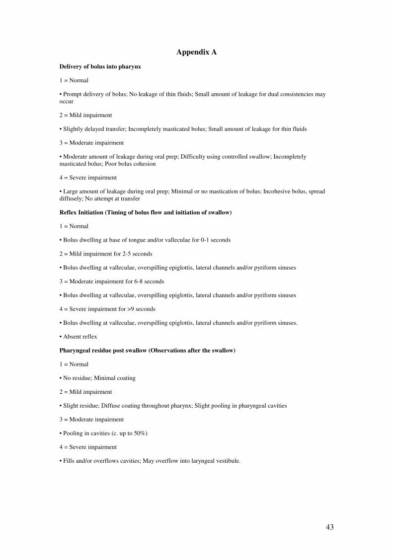
Appendix A
Delivery of bolus into pharynx
1 = Normal
• Prompt delivery of bolus; No leakage of thin fluids; Small amount of leakage for dual consistencies may
occur
2 = Mild impairment
• Slightly delayed transfer; Incompletely masticated bolus; Small amount of leakage for thin fluids
3 = Moderate impairment
• Moderate amount of leakage during oral prep; Difficulty using controlled swallow; Incompletely
masticated bolus; Poor bolus cohesion
4 = Severe impairment
• Large amount of leakage during oral prep; Minimal or no mastication of bolus; Incohesive bolus, spread
diffusely; No attempt at transfer
Reflex Initiation (Timing of bolus flow and initiation of swallow)
1 = Normal
• Bolus dwelling at base of tongue and/or valleculae for 0-1 seconds
2 = Mild impairment for 2-5 seconds
• Bolus dwelling at valleculae, overspilling epiglottis, lateral channels and/or pyriform sinuses
3 = Moderate impairment for 6-8 seconds
• Bolus dwelling at valleculae, overspilling epiglottis, lateral channels and/or pyriform sinuses
4 = Severe impairment for >9 seconds
• Bolus dwelling at valleculae, overspilling epiglottis, lateral channels and/or pyriform sinuses.
• Absent reflex
Pharyngeal residue post swallow (Observations after the swallow)
1 = Normal
• No residue; Minimal coating
2 = Mild impairment
• Slight residue; Diffuse coating throughout pharynx; Slight pooling in pharyngeal cavities
3 = Moderate impairment
• Pooling in cavities (c. up to 50%)
4 = Severe impairment
• Fills and/or overflows cavities; May overflow into laryngeal vestibule.