sequence and expression pattern of the germ line marker ... · pdf filesequence and expression...
TRANSCRIPT
Sequence and expression pattern of the germ line marker vasain honey bees and stingless bees
Érica Donato Tanaka1,2 and Klaus Hartfelder3
1Departamento de Biologia, Faculdade de Filosofia, Ciências e Letras de Ribeirão Preto,
Universidade de São Paulo, Ribeirão Preto, SP, Brazil.2Institut de Biologia Evolutiva, Consejo Superior de Investigaciones Científicas,
Universitat Pompeu Fabra, Barcelona, Spain.3Departamento de Biologia Celular e Molecular e Bioagentes Patogênicos,
Faculdade de Medicina de Ribeirão Preto, Universidade de São Paulo, Ribeirão Preto, SP, Brazil.
Abstract
Queens and workers of social insects differ in the rates of egg laying. Using genomic information we determined thesequence of vasa, a highly conserved gene specific to the germ line of metazoans, for the honey bee and fourstingless bees. The vasa sequence of social bees differed from that of other insects in two motifs. By RT-PCR weconfirmed the germ line specificity of Amvasa expression in honey bees. In situ hybridization on ovarioles showedthat Amvasa is expressed throughout the germarium, except for the transition zone beneath the terminal filament. Adiffuse vasa signal was also seen in terminal filaments suggesting the presence of germ line cells. Oocytes showedelevated levels of Amvasa transcripts in the lower germarium and after follicles became segregated. In previ-tellogenic follicles, Amvasa transcription was detected in the trophocytes, which appear to supply its mRNA to thegrowing oocyte. A similar picture was obtained for ovarioles of the stingless bee Melipona quadrifasciata, except thatAmvasa expression was higher in the oocytes of previtellogenic follicles. The social bees differ in this respect fromDrosophila, the model system for insect oogenesis, suggesting that changes in the sequence and expression patternof vasa may have occurred during social evolution.
Key words: Apis mellifera, Melipona quadrifasciata, vasa, social insect, oogenesis.
Received: January 27, 2009; Accepted: April 24, 2009.
Introduction
The decision of who is to reproduce and who is not isa key question in the evolution of sociality. In the highlyeusocial Hymenoptera, this decision has become fixed intoa system of reproductive division of labor between twomorphologically distinct female phenotypes, the queen andworker. Honey bees (Apini) and stingless bees (Melipo-nini) belong to the subfamily Apinae (Michener, 2000).Both are highly eusocial, but they differ considerably intheir life history characteristics (Sakagami, 1982; Impera-triz-Fonseca and Kleinert, 1998). Besides exhibiting amuch more flexible age polyethism (Waldschmidt andCampos, 1997) than Apis mellifera, a major difference be-tween stingless bees and honey bees concerns ovary devel-opment and worker reproduction (Hartfelder et al., 2006).
When a honey bee queen emerges from the broodcell, each of her two ovaries consists of 180-200 ovarioles.
These are activated soon after mating, allowing her to layup to 2,000 eggs per day. In contrast, adult workers haveonly 2-10 ovarioles in each ovary (Snodgrass, 1956). In thepresence of the queen, only very few workers show signs ofprogressive oogenesis (Visscher and Dukas, 1995), an ex-ception being a mutant phenotype, the so-called anarchisticworkers (Barron and Oldroyd, 2001).
Queens and workers of stingless bees do not exhibitsuch drastic differences in ovariole number and, in terms ofgonad size, the two castes differ primarily in the length of theovariole filaments (Hartfelder and Engels, 1992; Cruz-Lan-dim et al., 1998). Interestingly, worker reproduction instingless bees covers the entire spectrum, from full fertility inthe presence of the queen (Imperatriz-Fonseca and Kleinert,1998) to complete sterility due to ovary degeneration duringmetamorphosis (Boleli et al., 1999). This marked variation inovary structure and reproductive biology makes social bees aninteresting group for comparative studies on oogenesis.
Recent studies on oogenesis progress and control inApis mellifera have raised several questions concerning thepresence of putative germ-line stem cells in the terminal fil-
Genetics and Molecular Biology, 32, 3, 582-593 (2009)Copyright © 2009, Sociedade Brasileira de Genética. Printed in Brazilwww.sbg.org.br
Send correspondence to Érica Donato Tanaka. Institut de BiologiaEvolutiva, Consejo Superior de Investigaciones Científicas, Univer-sitat Pompeu Fabra, Passeig Marítim de la Barceloneta 37-49,E-08003 Barcelona, Spain. E-mail: [email protected].
Research Article
ament (Gutzeit et al., 1993; Tanaka and Hartfelder, 2004),on programmed cell death during oogenesis (Tanaka et al.,2006), and on the control of vitellogenesis via expression ofthe vitellogenin receptor encoding gene (Guidugli-Lazza-rini et al., 2008). These studies generally compared honeybee oogenesis with corresponding processes in the fruit fly,Drosophila melanogaster, which also has a polytrophicmeroistic type of ovary and for which the genetics of germcell determination and differentiation are much better un-derstood (for reviews see, King, 1970; de Cuevas et al.,1997; Gilboa and Lehmann, 2004).
Of specific interest to the determination of the germline is the vasa gene product. Vasa protein, an ATP-dependent DEAD-box RNA helicase (Hay et al., 1988; Pyle,2008), is a conserved germ line-specific marker for mostmetazoan phyla (Fujiwara et al., 1994; Gruidl et al., 1996;Olsen et al., 1997; Shibata et al., 1999; Mochizuki et al.,2001; Extavour and Akam, 2003; Sagawa et al., 2005;Rebscher et al., 2007). In Drosophila oogenesis, Vasa pro-tein surrounds germ cell nuclei in the germarium and in earlyegg chambers. Later on (stage 10), vasa mRNA appears atfirst homogeneously distributed throughout the oocyte cyto-plasm before the protein becomes asymmetrically segre-gated to the posterior pole (Hay et al., 1988; Liang et al.,1994; Sano et al., 2002; Findley et al., 2003). In the grass-hopper Schistocerca gregaria, which has a panoistic typeovary, vasa mRNA, as well as Vasa protein are highly ex-pressed in the early stages of oogenesis, with levels rapidlyfalling in maturing oocytes (Chang et al., 2002).
Due to the highly conserved association of vasa ex-pression with germ line development and gametogenesis inmetazoans (Extavour, 2007), efforts were made to identify abona fide vasa ortholog in the sequenced honey bee genome(Dearden et al., 2006; The Honey Bee Genome SequencingConsortium, 2006). Since this computationally predictedvasa sequence revealed a divergence in one of the character-istic Vasa motifs, being a change from ARKF to IVKF(Dearden et al., 2006), the question became whether thischange may be specific to the honey bee, as part of its specialsocial biology, or whether it may have preceded social evo-lution in the corbiculate bees. Furthermore, we were inter-ested to see whether vasa gene function, and especially itslocalization in the germ cells may differ between honey beesand stingless bees, as part of their divergence in worker re-production. In the present study we sequenced vasa
orthologs for four species of stingless bees and, by RT-PCRand in situ hybridization with a honey bee vasa probe, wecompared vasa expression patterns in Apis mellifera ovarieswith those of the stingless bee Melipona quadrifasciata.
Materials and Methods
Bees
Apis mellifera queens and workers were obtainedfrom Africanized honey bee stocks. Worker larvae and pu-
pae were collected directly from brood frames and staged(Michelette and Soares, 1993). Queens were reared follow-ing standard apicultural procedures. Pharate adult queensand workers were allowed to hatch in an incubator (34 °C,80% r.h.). Newly emerged workers were paint-marked andreintroduced into queenless hives, from where they wereretrieved two weeks later when their ovaries were expectedto be active (Makert et al., 2006). Queens that were to beaged without mating (virgin queens) were caged individu-ally in plastic queen cages and were kept in hives until theyreached the appropriate age. Other queens were paint-marked after hatching and were shortly thereafter intro-duced into queenless hives where they were allowed tomate naturally. Once they started to lay eggs they were re-trieved for ovary analysis.
Melipona quadrifasciata queens and workers werecollected from laboratory colonies. To obtain virgin queensof known age, combs with pharate-adult brood were re-moved and transferred to an incubator (28 °C, 80% r.h.) forup to 16 h. Newly emerged queens were kept in Petridishes, each accompanied by five workers, and were main-tained in the incubator until they reached the adequate agefor analysis. These bees received sugar water, fermentedpollen paste and water ad libitum. Specimens of the otherthree stingless bee species, Melipona scutellaris,
Scaptotrigona postica [frequently also referred to asScaptotrigona aff. depilis, due to uncertainties in the taxon-omy of this genus, (Camargo and Pedro, 2007)] andFrieseomelitta varia were also collected from brood framesof laboratory colonies, as described for M. quadrifasciata.
Sequencing and bioinformatics analysis
For experimental confirmation of the computatio-nally predicted honey bee vasa ortholog (GB14804-PA)(Dearden, 2006; The Honey Bee Genome Sequencing Con-sortium, 2006) we designed gene-specific primers aligningto five regions in the predicted coding sequence (V7F0:5’-ATGGCTGATGACTGGGGT-3’; V7R1: 5’-CCATAACTACGTCCACCTTC-3’; V7F2: 5’-GAGGAAAGTTGTCTGCTGG-3’; V7R2: 5’-CCAGCAGACAACTTTCCTC-3’; V7F3: 5’-GCCGTTTTCTTATCCGAG-3’;V7R3: 5’-CTCGGATAAGAAAACGGC-3’; V7R4: 5’-CCGGTTCTT TGCTACG-3’) and one in the predicted3’UTR region (V7R5: 5’-GAAACAAAGCTTACTACCCTG-3’). RT-PCR conditions were as follows: 95 °C for5 min, followed by 35 cycles of 94 °C for 30 s, 50 °C for30 s, 72 °C for 30 s, and a final extension step at 72 °C for7 min. The amplification products of expected size(V7F0+V7R1: 100 bp, V7F0+V7R2: 900 bp,V7F2+V7R3: 500 bp, V7F2+V7R4: 900 bp, V7F3+V7R4:500 bp, V7F3+V7R5: 700 bp) were ligated into pGEM-TEasy vector (Promega) for transformation of competentDH5�E. coli cells.
To identify vasa orthologs in the four stingless beespecies, Melipona quadrifasciata, Melipona scutellaris,
Tanaka and Hartfelder 583
Frieseomelitta varia and Scaptotrigona postica, we usedprimer combinations originally designed to amplify Apis
mellifera vasa (V7F0+V7R2, V7F2+V7R3, V7F3+V7R4).DNA sequencing was performed by the dideoxy sequenc-ing method, using a BigDye terminator v3.0 Cycle Se-quencing Ready Reaction (Applied Biosystems) in an ABIPrism 310 Genetic Analyzer (Applied Biosystems).
For annotation of the honey bee vasa gene, the CDSassembled from the RT-PCR products was aligned againstthe genome sequence (Amel. 4.0, The Honey Bee GenomeSequencing Consortium, 2006) using a LINUX-based Ar-temis platform (version 7.1, The Sanger Institute). Se-quence homology searches were performed by BLAST(blastx algorithm) to retrieve putative vasa orthologs of twoother hymenopteran species, Copidosoma floridanum
(GenBank accession number AAT11555.1) and Nasonia
vitripennis (GenBank accession numberXM_001603906.1). The vasa orthologs of these two spe-cies and those of the honey bee and the four stingless beespecies obtained in this study were used in ClustalW multi-ple alignments, with D. melanogaster vasa as an outgroup.The ClustalW results were transformed to Mega 3.1 format(Kumar et al., 2004). The Neighbor joining procedure(Saitou and Nei, 1987) was used for tree construction,which was evaluated by 1000 bootstrap repetitions.
RT-PCR analysis of vasa expression in Apismellifera ovaries
RNA was extracted using TRIzol reagent (Invitro-gen) from pools of 15-20 ovary pairs each. First-strandcDNA was synthesized using an oligo(dT)12-18 primer(Invitrogen) and Superscript II (Invitrogen) reverse tran-scriptase. RT-PCR reactions were carried out using theAmvasa primers V7F2 and V7R3 at 94 °C for 2 min fol-lowed by 32 cycles of 94 °C for 30 s, 55 °C for 30 s, 72 °Cfor 30 s, and a final extension step at 72 °C for 7 min. Fornormalization we amplified a cytoplasmatic actin genefragment (GenBank accession number AB023025), an en-dogenous control gene appropriate for quantitative gene ex-pression analysis in honey bees (Lourenço et al., 2008).The number of amplification cycles was adjusted to avoidsaturation. The Amvasa amplification products were ana-lyzed by electrophoresis in 1% agarose gels containingethidium bromide.
For the analysis of vasa expression in honey bee tis-sues other than the ovaries we prepared first-strand cDNAfrom brain, fat body, midgut, thorax (dorsal tegument) andabdomen (dorsal tegument) from newly emerged queensand workers. Additional samples were prepared fromspermathecae of queens and from hypopharyngeal glandsof workers, both of which are organs of caste-specific func-tions.
In situ hybridization
For in situ localization of vasa transcripts in honeybee and stingless bee ovaries, sense and antisense probeswere synthesized using Amvasa-specific primers contain-ing a T7 promoter sequence (underlined) at the respective5’-ends (ISH-F1: 5’-TAATACGACTCACTATAGGGCGAGGTAGAGGTCATGGTAAAGGAG-3’ and ISH-R1:5’-TAATACGACTCACTATAGGGCGAGAGGCACATTATCTCCACTCAC-3’), in combination with corre-sponding primers lacking the T7 sequence (ISH-F2 andISH-R2). Amplification parameters were: 94 °C for 2 min,followed by 40 cycles of 94 °C for 40 s, 57 °C for 40 s,72 °C for 40 s, and a final extension step at 72 °C for 7 min.PCR reactions with these primers generated a product of331 bp. The antisense and sense products were produced bythe primer combinations ISH-F2 + ISH-R1 and ISH-F1+ISH-R2, respectively.
Aliquots of the amplification products were checkedon agarose gels, purified (Wizard® SV Gel and PCRClean-Up System, Promega) and quantified spectrophoto-metrically. RNA probes were generated by in vitro tran-scription from the T7 promoter using the DIG RNALabelling Kit (SP6/T7) (Roche Applied Science). The tran-scription products were precipitated by addition of 1 �Lammonium acetate (10M) and 20 �L isopropanol. Aftercentrifugation, the pellet was washed in 70% ethanol. Theethanol-free probes were resuspended in 50 �L hybridiza-tion buffer (50% formamide, 4 X SSC, 1 X Denhardt’s so-lution, 250 �g/mL yeast extract, 250 �g/mL salmon spermDNA, 50 �g/mL de heparin, 0,1% Tween-20, 5% dextranesulfate) before storage at -20 °C.
Ovariole whole mounts were prepared for A.
mellifera workers kept in queenright and queenless condi-tions, for mated and unmated queens, as well as forphysogastric and virgin queens of M. quadrifasciata. Afterdissection of the ovaries, the ovarioles were separated inhoney bee tissue culture medium (Rachinsky and Hart-felder, 1998) and the ovariole sheath was removed fromeach ovariole by means of watchmaker’s forceps (number5, Dumont). Fixation of the ovarioles and the subsequenthybridization and detection reactions were performed fol-lowing the protocol optimized by Osborne and Dearden(2005) for in situ hybridization studies on honey bees.
Results
Sequence characteristics of Apis mellifera andMelipona quadrifasciata vasa genes
The Apis mellifera vasa gene spans an Open ReadingFrame (ORF) of 1,893 bp, divided into 3 exons that are sep-arated by 2 small introns (Figure 1A). The product se-quenced in our study from overlapping RT-PCR fragmentsaligned 100% with the computationally predicted Apis
mellifera vasa gene sequence (GenBank accession number
584 vasa gene structure and expression in social bees

ABC41341). Conceptual translation resulted in a predictedprotein of 630 amino acids (AmVasa). Sequence similarityscores obtained by BLASTP analysis showed that AmVasawas 55 to 61% identical to the Vasa proteins of members ofother insect orders, including the orthopterans Schistocerca
gregaria and Gryllus bimaculatus, the lepidopteranBombyx mori, the coleopteran Tribolium castaneum, andthe dipterans Aedes aegypti , Anopheles gambiae, Culex
pipiens quinquefasciatus and Drosophila melanogaster
(Supplementary Material, Figure S1).Apis mellifera Vasa contains all the nine conserved
domains characteristic for the DEAD-box helicase family(GxxxPxxIQ, AxTGxGKT, PTRELA, TPGR, DEAD,SAT, GG, ARGLD, HRIGRTGR) and two of the con-served domains of the Vasa subfamily (GIVGxA,ExEExW) (Supplementary Material, Figure S1). AmVasa,however, differs in two potentially important aspects fromthe Vasa proteins of other insect species. First, the Vasa-specific ARKF domain presented a change in the first twoamino acids, being IVKF in AmVasa. Second, the argini-ne-glycine-glycine (RGG) repeat, which is frequentlyfound in N-terminal regions of many Vasa-related proteins,was represented in AmVasa by a single RGG copy only.The computationally predicted CDS encoding AmVasa(Dearden, 2006) was, thus, fully confirmed by assembly ofRT-PCR fragments, including the modifications in the twoabove mentioned motifs.
For the stingless bee, Melipona quadrifasciata, con-ceptual translation of the vasa ortholog (partial cds,GenBank accession number FJ161962) resulted in a pre-dicted protein (MqVasa) of 624 amino acid residues show-ing 98% sequence identity to Apis mellifera Vasa protein. Asimilarly high degree of identity as that between the vasa
genes of A. mellifera and M. quadrifasciata was also ob-tained for the vasa orthologs of the three other stingless beespecies, Melipona scutellaris (GenBank accession numberEF601038), Scaptotrigona postica (GenBank accessionnumber EF601037) and Frieseomelitta varia (GenBank ac-cession number FJ161963). In the molecular phylogenytree constructed by Neighbor Joining (Figure 1B) the Vasasequences of the five bee species came out as a well sup-ported branch, separated from the parasitic wasps Nasonia
vitripennis and Copidosoma floridanum, both belonging toChalcidoidea. Interestingly, the two Melipona species werealso well separated from the two trigonine species S.
postica and F. varia.
In the ClustalW alignments of the hymenopteranVasa proteins, with Drosophila as outgroup (Supplemen-tary Material - Figure S2), the following observations con-cerning modifications in characteristic Vasa motifs arenoteworthy: (1) in the GxxxPxxIQ domain the five bee spe-cies are all GYKKPTPVQK, differing from Nasonia andCopidosoma in the third and fourth position, (2) in thePTRELA domain, Copidosoma is identical to Drosophila,differing from Nasonia and the bees, which are allPTRELT, (3) in the GIVGxA domain, Nasonia andCopidosoma are GIVGSA, whereas the bees andDrosophila are GIVGGA.
Amvasa expression in the postembryonic gonad andin adult somatic tissues
Using specific primers we confirmed the presence ofAmvasa transcripts in the ovaries of both castes of thehoney bee throughout larval, pupal and imaginal develop-ment. Judging from band intensity, normalized againstactin levels in the respective cDNA samples, the expressionlevels of vasa in ovaries of workers appear to be lower thanin queens (Figure 2A). This observation is consistent withthe differing dynamics of ovary development for the twocastes (Hartfelder and Steinbrück, 1997). Amvasa tran-script levels in the two castes showed little apparent modu-lation, suggesting that the gene is continuously expressedduring postembryonic development of the gonads.
We next investigated whether Amvasa expression isgonad-specific. This analysis was conducted using RNAextracts from different body parts and organs of adulthoney bee queens and workers. Apart from the expected ex-pression in ovaries we could detect a faint but neverthelessclear Amvasa signal in dorsal fat body of queens (Figure2B). Cloning and sequencing of this fragment confirmedthat it was a bona fide vasa signal. We did not detect corre-sponding transcripts in fat body of workers, indicating pos-
Tanaka and Hartfelder 585
Figure 1 - The vasa genes of highly eusocial bees. (A) Schematic diagramof the genomic region encoding the vasa gene of the honey bee Apis
mellifera. The open arrowheads on the left and right indicate the start andstop codons, respectively. Exons are represented as boxes and their re-spective sizes are shown. Intron length was scaled to provide a general es-timate of vasa gene size. (B) Molecular phylogeny for hymenopteran Vasaprotein sequences. Vasa proteins of two species of parasitic wasps,Nasonia vitripennis and Copidosoma floridanum were aligned with Vasaprotein sequences of the stingless bees Melipona scutellaris, Melipona
quadrifasciata, Scaptotrigona postica and Frieseomelitta varia, as well aswith Apis mellifera Vasa protein. Drosophila melanogaster Vasa was in-cluded as outgroup. ClustalW alignment results (Supplementary MaterialFigure S2) were used as input for tree construction by Neighbor joining(NJ). Results of 1,000 bootstrap repetitions are shown adjacent to the re-spective branches.
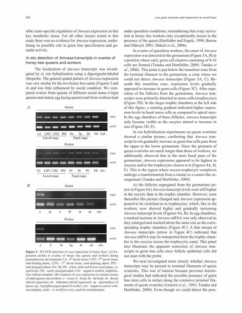
sible caste-specific regulation of Amvasa expression in thiskey metabolic tissue. For all other tissues tested in thisstudy there was no evidence for Amvasa expression, under-lining its possible role in germ line specification and go-nadal activity.
In situ detection of Amvasa transcripts in ovaries ofhoney bee queens and workers
The localization of Amvasa transcripts was investi-gated by in situ hybridization using a digoxigenin-labeledriboprobe. The general spatial pattern of Amvasa expressionwas very similar for the two honey bee castes (Figures 3 and4) and was little influenced by social condition. We com-pared ovaries from queens of different social status (virginqueens and mated, egg laying queens) and from workers kept
under queenless conditions, remembering that ovary activa-tion in honey bee workers only exceptionally occurs in thepresence of the queen (Hartfelder and Engels, 1998; Barronand Oldroyd, 2001; Makert et al., 2006).
In ovaries of queenless workers, the onset of Amvasa
expression was detected in the germarium (Figure 3A, B) ata position where early germ cell clusters consisting of 8-16cells are formed (Tanaka and Hartfelder, 2004; Tanaka et
al., 2006). This point is just below the transition zone fromthe terminal filament to the germarium, a zone where wecould not detect Amvasa transcripts (Figure 3A, C). Be-neath this transition zone, expression levels graduallyappeared to increase in germ cells (Figure 3C). After sepa-ration of the follicles from the germarium, Amvasa tran-scripts were primarily detected in nurse cells (trophocytes)(Figure 3D). In the larger trophic chambers at the left sideof this figure, a staining gradient indicated higher expres-sion levels in basal nurse cells as compared to apical ones.In the egg chambers of these follicles, Amvasa transcriptsonly became visible as the oocytes started to increase insize (Figure 3D, E).
In situ hybridization experiments on queen ovariolesshowed a similar picture, confirming that Amvasa tran-script levels gradually increase as germ line cells pass fromthe upper to the lower germarium. Since the germaria ofqueen ovarioles are much longer than those of workers, weadditionally observed that in the more basal parts of thegermarium, Amvasa expression appeared to be highest inoocytes and/or the trophocytes closest to it (Figures 4A, D,E). This is the region where oocyte-trophocyte complexesundergo a transformation from a cluster to a comet-like ar-rangement (Tanaka and Hartfelder, 2004).
As the follicles segregated from the germarium (ar-row in Figure 4A) Amvasa transcript levels were still higherin the oocytic than in the trophic chamber. However, soonthereafter this picture changed and Amvasa expression ap-peared to be switched on in trophocytes, which, like in theworkers, now showed higher and gradually increasingAmvasa transcript levels (Figures 4A, B). In egg chambers,a marked increase in Amvasa mRNA was only observed asthey enlarged and reached about the same size as the corre-sponding trophic chambers (Figure 4C). A thin stream ofAmvasa transcripts (arrow in Figure 4C) indicated thatAmvasa mRNA may be transported from the trophic cham-ber to the oocytes across the trophocytic canal. This panelalso illustrates the apparent restriction of Amvasa tran-scripts to germ line cells since follicle epithelial cells didnot stain with the probe.
We next investigated more closely whether Amvasa
transcripts may be present in terminal filaments of queenovarioles. This was of interest because previous histolo-gical studies had indicated the possible presence of germline stem cells in niches along the extensive terminal fila-ments of queen ovarioles (Gutzeit et al., 1993; Tanaka andHartfelder, 2004). Even though we could detect the pres-
586 vasa gene structure and expression in social bees
Figure 2 - RT-PCR detection of vasa expression in honey bees. (A) Ex-pression profile in ovaries of honey bee queens and workers duringpostembryonic development. L4 - 4th larval instar; L5F2 - 5th larval instar,mid-feeding phase; L5S2 - 5th larval instar, mid-spinning phase; PP2 -mid-prepupal phase; Pw, Pp, Pb - white, pink and brown-eyed pupae, re-spectively; NE - newly emerged adult. Ctrl- - negative control, amplifica-tion without template. (B) Analysis of vasa expression in somatic tissuesof adult queens and workers: o - ovary; b - brain; fb - fat body; th - thorax(dorsal tegument); ab - abdomen (dorsal tegument); sp - spermatheca ofqueen; hg - hypopharyngeal gland of worker. ctrl- - negative control with-out template. actin - A. mellifera actin, used for normalization.

ence of Amvasa transcripts in terminal filaments (Figure 5),this staining was rather diffuse and not constant. Espe-cially, it did not allow us to distinguish between therounded putative germ line cells and the flattened, disc-likesomatic cells of the filaments. This finding is of interest be-cause, as described above, we also could not detect Amvasa
transcripts in the transition zone from the terminal filamentto the top of the germarium (Figure 3A), where germ cellscould be clearly identified by their general histology andcell division pattern (Tanaka and Hartfelder, 2004). Thisfinding indicates that in the honey bee, vasa may not be amarker appropriate for germ line stem cells, but that vasa
apparently becomes induced in the germ line only aftercystocytes separate from the niche and begin to form thelarge germ cell clusters, typical of the polytrophic meroisticovary type.
In situ detection of vasa transcripts in ovaries of thestingless bee Melipona quadrifasciata
The high degree in sequence identity made it possibleto use the Amvasa probe for in situ hybridization experi-
ments on ovarioles of the stingless bee, M. quadrifasciata.This probe detected vasa transcripts throughout the entirelength of the ovarioles of stingless bee queens. Like in thehoney bee, vasa transcript levels gradually increased alongthe germarium accompanying oogenesis progression (Fig-ure 6A-C). However, as the follicles separated from thegermarium, the timing of vasa expression in the stinglessbee seemed to differ from the pattern observed in the honeybee. In the honey bee, Amvasa transcript levels were ini-tially higher in the oocyte, but subsequently Amvasa ex-pression became strongly turned on in the trophocytes, andonly later did its transcript levels gradually increase againin the oocyte. In contrast to this, we noted that vasa tran-script levels in the stingless bee ovary were continuouslyhigher in the oocytes, already from the start of folliclegrowth (Figure 6D). This difference in the localization of apresumably important component for germ line function isof interest as it may represent a molecular corollary to themorphological differences between A. mellifera and M.
quadrifasciata ovarioles (Tanaka et al., 2009).
Tanaka and Hartfelder 587
Figure 3 - Amvasa mRNA detection by in situ hybridization in ovarioles of queenless honey bee workers. Apical is to the left in all figures. (A, B and C)Amvasa expression visualized in germ line cells throughout the germarium. The Amvasa-negative area at the left of A and C is the transition zone from theterminal filament to the germarium. (D) Amvasa expression in previtellogenic follicles is prominent in the trophic chambers and only gradually increasesin the growing oocytes. (E) In early vitellogenic follicles, Amvasa transcripts in the oocyte show a homogeneous distribution. Negative control with senseprobe (F). g - germarium, o - oocyte, pf - previtellogenic follicle, tc - trophic chamber, tf - terminal filament.
Discussion
Molecular analysis of the vasa orthologs of the
honey bee and stingless bees
The predicted amino acid sequence of the Apis
mellifera Vasa protein contained all the diagnostic motifsof DEAD-box helicases, but differed in two puzzling as-pects from Vasa proteins of other insects. Most notably, theARKF motif, which is considered a diagnostic motif of theVasa family, had changed to IVKF (Chang et al., 2002).Similar alterations in this motif had previously been re-ported for the parasitic wasp, Copidosoma floridanum
(Donnell et al., 2004), the red flour beetle Tribolium
castaneum (Lorenzen et al., 2005) and also for other organ-isms (Sagawa et al., 2005; Ohashi et al., 2007). The func-tion of the ARKF domain is not clear yet, and alterations inthis motif do not seem to interfere with the ATPase or
helicase activities of Vasa proteins (Sagawa et al., 2005;Schröder, 2006; Ohashi et al., 2007).
Another difference between honey bee Vasa and itsorthologs in other insects concerns the reduction in N-terminal RGG repeats. RGG motifs in human hnRNP Uprotein have been shown to bind RNA (Kiledjian and Drey-fuss, 1992). This function could be important in the organi-zation of specific RNA complexes at the posterior egg polewhen polar granules are formed (Hay et al., 1988), eventhough the localization of Vasa itself to the posterior poleappears to be independent of its RNA binding capacity(Liang et al., 1994).
Sequencing of vasa orthologs of four stingless beespecies showed that the change from ARKF to an IVKFmotif and the reduction in RGG repeats is shared by the twotribes Apini and Meliponini. These modifications in an oth-erwise highly conserved protein, thus, appear to predate thesplit between the two tribes of highly eusocial bees. Espe-cially in the light of recent molecular phylogenies of the en-
588 vasa gene structure and expression in social bees
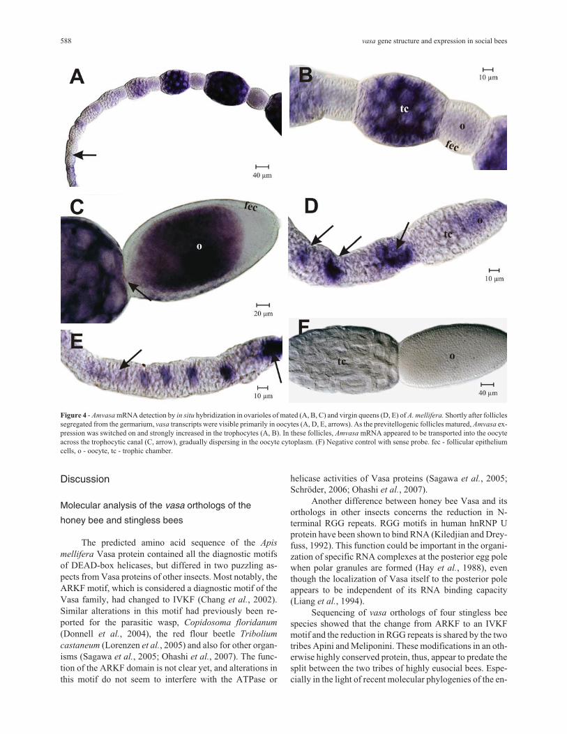
Figure 4 - Amvasa mRNA detection by in situ hybridization in ovarioles of mated (A, B, C) and virgin queens (D, E) of A. mellifera. Shortly after folliclessegregated from the germarium, vasa transcripts were visible primarily in oocytes (A, D, E, arrows). As the previtellogenic follicles matured, Amvasa ex-pression was switched on and strongly increased in the trophocytes (A, B). In these follicles, Amvasa mRNA appeared to be transported into the oocyteacross the trophocytic canal (C, arrow), gradually dispersing in the oocyte cytoplasm. (F) Negative control with sense probe. fec - follicular epitheliumcells, o - oocyte, tc - trophic chamber.
tire clade Apinae (Kawakita et al., 2008; Whitfield et al.,2008), they could actually represent an ancestral trait in thecorbiculate bees, predating the evolution of sociality in thisgroup. Confirming this hypothesis will, however, requiresequencing of vasa orthologs in bumble bees (Bombini)and orchid bees (Euglossini).
The cladogram for the hymenopteran vasa orthologsfurther underlines the high degree in similarity for this genein the two tribes of highly eusocial bees and sets themclearly apart from parasitic wasps. The fact that the two
species of the genus Melipona cluster together is not sur-prising. The split between these and the two other stinglessbee species (S. postica and F. varia), however is of interest.The latter two species are traditionally included within thetrigonines, a grouping of several hundred stingless bee spe-cies (Camargo and Pedro, 2007). The two groups differwith respect to the mechanism of caste determination (Kerr,1950; Hartfelder et al., 2006).
Developmental profiling and tissue specificity ofvasa expression in honey bees
The results of this qualitative analysis showed thatAmvasa is expressed continuously and at apparently con-stant levels in the developing gonads of both queens andworkers during the late larval stages (fourth and fifth instar)and during pupal development. Interestingly, the expres-sion levels seem to be higher in queens than in workers, andthis aspect certainly deserves a closer look using a quantita-tive RT-PCR approach. The observed caste differencesmight be related to the divergence in ovary development,particularly in the fifth instar which is marked by massivecell death in the ovaries of worker larvae (Hartfelder andSteinbrück, 1997; Schmidt-Capella and Hartfelder, 1998,2002), considerably reducing the number of germ line cells.
The above described differences in amino acid se-quence between honey bee Vasa and its orthologs in otherinsects made us ask whether this may be reflected in alteredtissue specificity of vasa expression. Besides the clear andexpected signal obtained for ovaries of adult queens andworkers, we also found evidence for a low, yet clearly de-tectable level of vasa expression in fat body of honey beequeens. Since none of the other tissues showed such a sig-nal, and since it was found in queen fat body only, we con-
Tanaka and Hartfelder 589
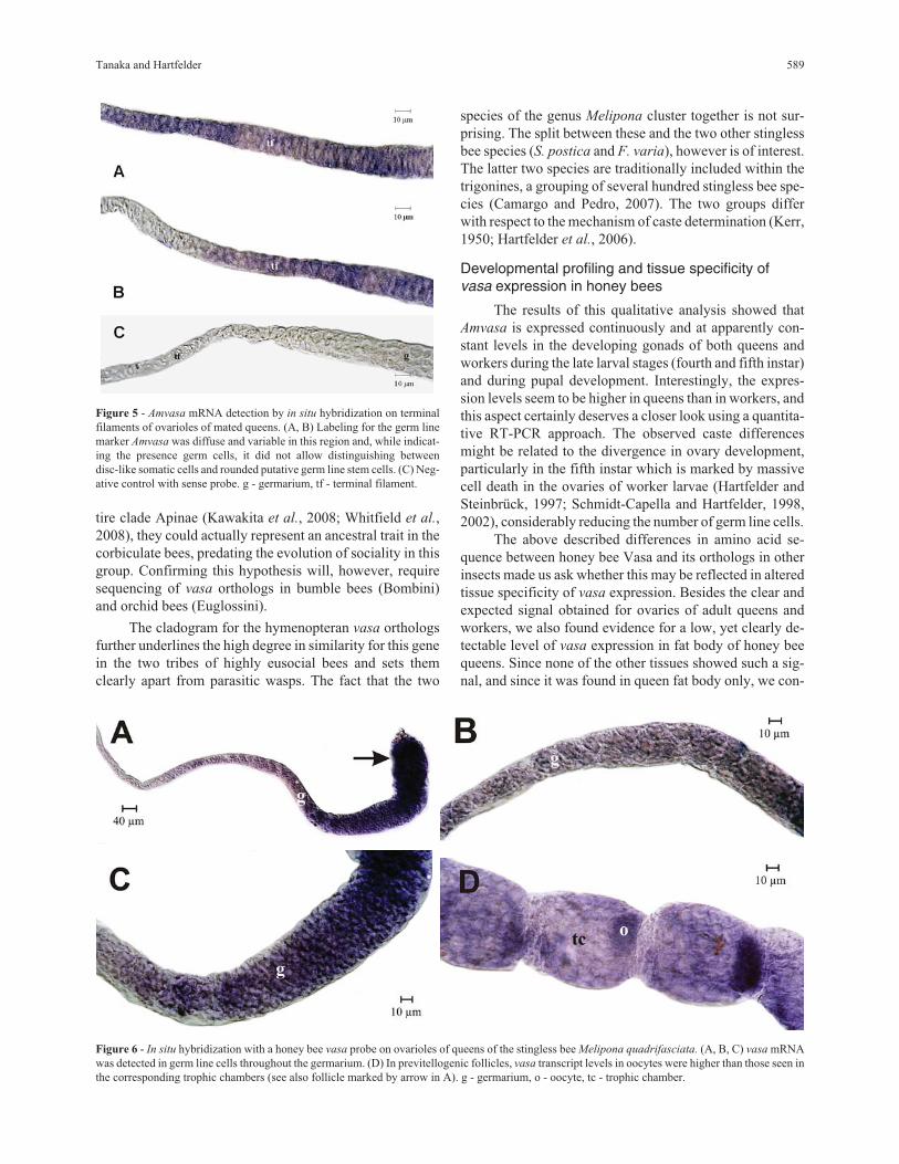
Figure 5 - Amvasa mRNA detection by in situ hybridization on terminalfilaments of ovarioles of mated queens. (A, B) Labeling for the germ linemarker Amvasa was diffuse and variable in this region and, while indicat-ing the presence germ cells, it did not allow distinguishing betweendisc-like somatic cells and rounded putative germ line stem cells. (C) Neg-ative control with sense probe. g - germarium, tf - terminal filament.
Figure 6 - In situ hybridization with a honey bee vasa probe on ovarioles of queens of the stingless bee Melipona quadrifasciata. (A, B, C) vasa mRNAwas detected in germ line cells throughout the germarium. (D) In previtellogenic follicles, vasa transcript levels in oocytes were higher than those seen inthe corresponding trophic chambers (see also follicle marked by arrow in A). g - germarium, o - oocyte, tc - trophic chamber.
clude that this is a relevant non-germ line expression,specific to the fat body of the reproductive caste. Know-ingly, the fat body plays a major role in controlling fertilityof queens and workers by its output of vitellogenin, the ma-jor yolk protein precursor. Honey bee queens are notoriousfor their extremely high hemolymph vitellogenin titers (En-gels, 1974; Hartfelder and Engels, 1998), and the fat body isthe major site of vitellogenin gene expression (Piulachs et
al., 2003). The finding of a vasa expression signal in queenfat body could, thus, provide a link between the previ-tellogenic and vitellogenic phases of follicle development.
Extra-gonadal expression of vasa is, however, neithera novel finding nor a unique one specific to honey bees.Vasa protein and/or vasa mRNA have, for example, al-ready been detected in embryonic somatic cells of Xenopus
laevis (Ikenishi and Tanaka, 2000), in polychaetes(Rebscher et al., 2007), and in ectodermal epithelial cells ofcnidarians (Mochizuki et al., 2001). In honey bee embryos,a group of cells in the mandibular segment has been shownto express Amvasa (Dearden, 2006).
In situ localization of vasa mRNA in the ovaries ofhighly eusocial bees
The detection of Amvasa mRNA throughout thegermarium and in the cytoplasm of nurse cells and oocytesof follicles confirmed the expected association of Amvasa
expression with germ line cells. Besides detecting anoocytic vasa mRNA signal in the lower germarium and inearly follicles as they segregated from the germarium, wealso found evidence for Amvasa expression in cells com-posing the terminal filaments. Divergence from the flymodel of oogenesis (Bastock and St Johnston, 2008) inthese two aspects raises the question as to whether honeybees may differ from Drosophila in pathways involvingVasa function(s). In fly oogenesis, vasa transcription de-pends on the prior localization of oskar mRNA to the poste-rior pole, and Oskar protein then remains associated withthe germ line determining pole plasm (Rongo andLehmann, 1996). In turn, Vasa protein is necessary for thelocalized translation of nanos mRNA (Gavis et al., 1996)and for the localization of other gene products, includinggurken mRNA, which is involved in the establishment ofthe two major oocyte and, consequently, embryonic axes(Tinker et al., 1998). Since in the honey bee genome neithergurken nor oskar orthologs were found (The Honey BeeGenome Sequencing Consortium, 2006), but a clearortholog of nanos, it is quite possible that embryonic axisspecification and germ line determination in the honey beefollows a different route from that established for the fly.Interestingly, gurken, oskar and bicoid are also missing inthe Bombyx mori and Tribolium castaneum genomes(Dearden et al., 2006; Schröder, 2006) and, like in thehoney bee, in neither of these species differentiated polecells were evidenced (Nakao, 1999; Handel et al., 2000). Inthe honey bee, germ line cells seem to be formed by an in-
ductive event late in embryogenesis (Dearden, 2006; Fleigand Sander, 1998), as is the case in most metazoans (Exta-vour and Akam, 2003). Whether and how the early appear-ance of Amvasa mRNA in oocytes and trophocytes may berelated to the determination of germ line cells, needs furtherinvestigation, especially as to how vasa expression may beconnected to downstream events, such as the translation ofnanos mRNA in honey bee eggs.
The detection by in situ hybridization of Amvasa tran-scripts in the terminal filament region is another clear dif-ference to Drosophila, but actually did not come as asurprise. The ovarioles of Apis mellifera queens and work-ers have enormously elongated terminal filaments, andthese house two cell types. Interspersed between the flat-tened somatic cells organized in stack-of-coins arrange-ments are clusters of rounded cells with large nuclei andweakly staining cytoplasm. Being mitotically active (Tana-ka and Hartfelder, 2004), they show a set of characteristicsnormally attributed to primordial germ line cells(Mahowald, 1962; Wolf et al., 1983). In contrast, terminalfilaments in the Drosophila ovary are short and made up ofdisk-like somatic cells only. Apart from tethering togetherthe ovarioles, an important function of the terminal fila-ment is to form a cap structure that, in Drosophila, providesa niche environment around the 2-3 germ line stem cells(Lin and Spradling, 1993; Zhang and Kalderon, 2000; Mor-rison and Spradling, 2008). For sustaining high egg-layingrates, such as observed for honey bee queens, a system of2-3 stem cells in a niche would probably be insufficient.Clusters of germ-line stem cells in elongated terminal fila-ments could, thus, be a solution to this problem. The ratherdiffuse pattern of Amvasa staining over the terminal fila-ments, however, did not permit to definitively distinguishsomatic from putative germ line cells. But while this wouldbe in accordance with epigenetic inductive events thatmight progressively separate germ line from somatic cells(Extavour, 2007), it is still unclear as to how such germ linecells in the terminal filament would make their way downinto the germarium.
The spatial pattern of vasa expression observed inhoney bee ovarioles was very similar to that seen in thestingless bee Melipona quadrifasciata. Also in this species,vasa transcript levels were observed to gradually increasealong the apical-basal axis of the germarium, and vasa
mRNA was also detected in early follicles and seen to accu-mulate in oocytes. These findings are of interest because,together with the close vasa sequence similarity for the twogroups of highly eusocial bees, they provide a unifyingtheme for oogenesis in these bees, inspite of their differ-ences in ovariole structure (Tanaka et al., 2009) and in re-productive biology. This difference resides primarily in theovarian activity of stingless bee workers which may pro-duce both reproductive and trophic eggs (for review seeHartfelder et al., 2006).
590 vasa gene structure and expression in social bees
Our findings on vasa expression in social bees are inaccordance with a recent study on vasa and nanos expres-sion in ants (Khila and Abouheif, 2008), where Vasa pro-tein was also detected in oocytes which were still in thegermarium and in follicles that had separated from it. In antspecies where workers produce two types of eggs, as dostingless bee workers, nanos mRNA appeared concentratedin a spot that colocalized with Vasa protein. In eggs with re-productive potential, this spot stayed associated with theposterior oocyte pole. In contrast, Vasa protein, and conse-quently also nanos mRNA, apparently failed to localize tothe posterior pole in the non-viable trophic eggs. Khila andAbouheif (2008) consider that this mislocalization of twocrucial posterior pole components observed in trophic eggscould represent a developmental mechanism that generatesreproductive constraint in the worker caste and, thus, wouldrepresent an important factor in social evolution in theHymenoptera. Obviously, this is an interesting hypothesiswith wide ranging implications on the expression patternsof genes underlying developmental patterning mecha-nisms. The question is, how and to what extent these genesmay have also been co-opted to generate evolutionarily sta-ble differences in the reproductive potential of females inthe caste phenotypes of other social Hymenoptera, such aswasps and bees.
Acknowledgments
We thank Luis Roberto Aguiar and Weyder CristianoSantana for assistance in maintaining the bee colonies. Fi-nancial support was provided by the Fundação de Amparo àPesquisa do Estado de São Paulo (FAPESP grants99/00719-6 and 02/11729-7).
References
Barron AB and Oldroyd BP (2001) Social regulation of ovary ac-tivation in ‘anarchistic’ honey bees (Apis mellifera). BehavEcol Sociobiol 49:214-219.
Bastock R and St Johnston D (2008) Drosophila oogenesis. CurrBiol 18:1082-1087.
Boleli IC, Simões ZLP and Bitondi MMG (1999) Cell death inovarioles causes permanent sterility in Frieseomelitta varia
workers bees. J Morphol 242:271-282.
Camargo JMF and Pedro SRM (2007) Meliponini Lepeletier,1936. In: Moure JS, Urban D and Melo GAR (eds) Cata-logue of Bees (Hymenoptera, Apoidea) in the NeotropicalRegion. Sociedade Brasileira de Entomologia, Curitiba, pp272-578.
Chang C, Dearden PK and Akam M (2002) Germ line develop-ment in the grasshopper Schistocerca gregaria. Dev Biol252:100-118.
Cruz-Landim C, Reginato RD and Imperatriz-Fonseca VL (1998)Variation in ovariole number in Meliponinae(Hymenoptera, Apidae) queen’s ovaries, with comments onovary development and caste differentiation. Pap AvulsosZool S Paulo 40:289-296.
de Cuevas M, Lilly MA and Spradling AC (1997) Germline cystformation in Drosophila. Annu Rev Genet 31:405-428.
Dearden PK (2006) Germ cell development in the honeybee (Apis
mellifera): Vasa and Nanos expression. BMC Dev Biol6:1-14.
Dearden PK, Wilson MJ, Sablan L, Osborne PW, Havler M,McNaughton E, Kimura K, Milshina NV, Hasselmann M,Gempre T, et al. (2006) Patterns of conservation and changein honey bee developmental genes. Genome Res 16:1376-1384.
Donnell DM, Corley LS, Chen G and Strand MR (2004) Caste de-termination in a polyembryonic wasp involves inheritanceof germ cells. Proc Natl Acad Sci USA 101:10095-10100.
Engels W (1974) Occurrence and significance of vitellogenins infemale castes of social Hymenoptera. Am Zool 14:1229-1237.
Extavour C (2007) Evolution of the bilaterian germ line: Lineageorigin and modulation of specification mechanisms. IntegrComp Biol 47:770-785.
Extavour C and Akam ME (2003) Mechanisms of germ cell speci-fication across the metazoans: Epigenesis and preformation.Development 130:5869-5884.
Findley SD, Tamanaha M, Clegg NJ and Ruohola-Baker H (2003)Maelstrom, a Drosophila spindle-class gene, encodes a pro-tein that colocalizes with Vasa and RDE1/AGO1 homolog,Aubergine, in nuage. Development 130:859-871.
Fleig R and Sander K (1998) Honeybee morphogenesis, embry-onic cell movements that shape the larval body. Develop-ment 103:525-534.
Fujiwara Y, Komiya T, Kawabata H, Sato M, Fujimoto H, Furu-sawa M and Noce T (1994) Isolation of a DEAD-family pro-tein gene that encodes a murine homologue of Drosophila
vasa and its specific expression in germ cell lineage. ProcNatl Acad Sci USA 91:12258-12262.
Gavis ER, Lunsford L, Bergsten SE and Lehmann R (1996) Aconserved 90 nucleotide element mediates translational re-pression of nanos RNA. Development 122:2791-2800.
Gilboa L and Lehmann R (2004) How different is Venus fromMars? The genetics of germ-line stem cells in Drosophila
females and males. Development 131:4895-4905.Gruidl M, Smith PA, Kuznicki KA, McCrone JS, Kirchner J,
Roussell DL, Strome S and Benett KL (1996) Multiple po-tential germ-line helicases are components of the germ-line-specific P granules of Caenorhabditis elegans. ProcNatl Acad Sci USA 93:13837-13842.
Guidugli-Lazzarini KR, Nascimento AM, Tanaka ED, PiulachsMD, Hartfelder K, Bitondi MMG and Simoes ZLP (2008)Expression analysis of putative vitellogenin and lipophorinreceptors in honey bee (Apis mellifera L.) queens and work-ers. J Insect Physiol 54:1138-1147.
Gutzeit HO, Zissler D and Fleig R (1993) Oogenesis in the honey-bee Apis mellifera: Cytological observations on the forma-tion and differentiation of previtellogenic ovarian follicles.Roux’s Arch Dev Biol 202:181-191.
Handel K, Grünfelder CG, Roth S and Sander K (2000) Tribolium
embryogenesis: A SEM study of cell shapes and movementsfrom blastoderm to serosal closure. Dev Genes Evol210:167-179.
Hartfelder K and Engels W (1992) Allometric and multivariateanalysis of sex and caste polymorphism in the neotropical
Tanaka and Hartfelder 591
stingless bee, Scaptotrigona postica. Insectes Soc 39:251-266.
Hartfelder K and Engels W (1998) Social insect polymorphism:Hormonal regulation of plasticity in development and repro-duction in the honeybee. Curr Topics Dev Biol 40:45-77.
Hartfelder K, Makert GR, Judice CC, Pereira GAG, Santana WC,Dallacqua R and Bitondi MMG (2006) Physiological andgenetic mechanisms underlying caste development, repro-duction and division of labor in stingless bees. Apidologie37:144-163.
Hartfelder K and Steinbrück G (1997) Germ cell cluster formationand cell death are alternatives in caste-specific differentia-tion of the larval honey bee ovary. Invertebr Reprod Dev31:237-250.
Hay B, Jan LY and Jan YN (1988) A protein component ofDrosophila polar granules is encoded by vasa and has exten-sive sequence similarity to ATP-dependent helicases. Cell55:577-587.
Ikenishi K and Tanaka T (2000) Spatio-temporal expression ofXenopus vasa homolog, XVLG1, in oocytes and embryos:The presence of XVLG1 RNA in somatic cells as well asgermline cells. Dev Growth Diff 42:95-103.
Imperatriz-Fonseca VL and Kleinert ADP (1998) Worker repro-duction in the stingless bee species Friesella schrottkyi
(Hymenoptera, Apidae, Meliponinae). Entomol Gener Appl23:169-175.
Kawakita A, Ascher JS, Sota T, Kato M and Roubik DW (2008)Phylogenetic analysis of the corbiculate bee tribes based on12 nuclear protein-coding genes (Hymenoptera, Apoidea,Apidae). Apidologie 39:163-175.
Kerr WE (1950) Genetic determination of castes in the genusMelipona. Genetics 35:143-152.
Khila A and Abouheif E (2008) Reproductive constraint is a de-velopmental mechanism that maintains social harmony inadvanced ant societies. Proc Natl Acad Sci USA105:17884-17889.
Kiledjian M and Dreyfuss G (1992) Primary structure and bindingactivity of the hnRNP U protein: Binding RNA throughRGG box. EMBO J 11:2655-2664.
King RC (1970) Ovarian Development in Drosophila
melanogaster. Academic Press, New York, 227 pp.Kumar S, Tamura K and Nei M (2004) MEGA3: Integrated soft-
ware for Molecular Evolutionary Genetics Analysis and se-quence alignment. Brief Bioinform 5:150-163.
Liang L, Diehl-Jones W and Lasko PF (1994) Localization ofVasa protein to the Drosophila pole plasm is independent ofits RNA-binding and helicase activities. Development120:1201-1211.
Lin H and Spradling AC (1993) Germline stem cell division andegg chamber development in transplanted Drosophila ger-maria. Dev Biol 159:140-152.
Lorenzen MD, Doyungan Z, Savard J, Snow K, Crumly LR,Shippy TD, Stuart JJ, Brown SJ and Beeman RW (2005) Ge-netic linkage maps of the red flour beetle, Tribolium
castaneum, based on bacterial artificial chromosomes andexpressed sequence tags. Genetics 170:741-747.
Lourenço AP, Mackert A, Cristino AS and Simoes ZLP (2008)Validation of reference genes for gene expression studies inthe honey bee, Apis mellifera, by quantitative real-timeRT-PCR. Apidologie 39:372-385.
Mahowald AP (1962) Fine structure of pole cell and polar gran-ules in Drosophila melanogaster. J Exp Zool 151:201-215.
Makert GR, Paxton RJ and Hartfelder K (2006) Ovariole number -a predictor of differential success among worker subfamiliesin queenless honeybee (Apis mellifera L.) colonies. BehavEcol Sociobiol 60:815-825.
Michelette ERD and Soares AEE (1993) Characterization of prei-maginal developmental stages in Africanized honey beeworkers (Apis mellifera L.). Apidologie 24:431-440.
Michener CD (2000) The Bees of the World. John Hopkins Uni-versity Press, Baltimore, 913 pp.
Mochizuki K, Nishimiya-Fujisawa C and Fujisawa T (2001) Uni-versal occurrence of the vasa-related genes among metazo-ans and their germline expression in Hydra. Dev Genes Evol211:299-308.
Morrison SJ and Spradling AC (2008) Stem cells and niches:Mechanisms that promote stem cell maintenance throughoutlife. Cell 132:598-611.
Nakao H (1999) Isolation and characterization of a Bombyx vasa-like gene. Dev Genes Evol 209:312-316.
Ohashi H, Umeda N, Hirazawa N, Ozaki Y, Miura C and Miura T(2007) Expression of vasa (vas)-related genes in germ cellsand specific interference with gene functions by double-stranded RNA in the monogenean Neobenedenia girellae.Int J Parasitol 37:515-523.
Olsen LC, Aasland R and Fjose A (1997) A vasa-like gene inzebrafish identifies putative primordial germ cells. MechDev 66:95-105.
Osborne PW and Dearden PK (2005) Non-radioactive in situ hy-bridization to honeybee embryos and ovaries. Apidologie36:113-118.
Piulachs MD, Guidugli KR, Barchuk AR, Cruz J, Simoes ZLP andBelles X (2003) The vitellogenin gene of the honey bee, Apis
mellifera: Structural analysis of the cDNA and expressionstudies. Insect Biochem Mol Biol 33:459-465.
Pyle AM (2008) Translocation and unwinding mechanisms ofRNA and DNA helicases. Annu Rev Biophys 37:317-336.
Rachinsky A and Hartfelder K (1998) In vitro biosynthesis of ju-venile hormone in larval honey bees: Comparison of six me-dia. In Vitro Cell Dev Biol - Animal 34:646-648.
Rebscher N, Zelada-González F, Banish TU, Raible F and ArendtD (2007) Vasa unveils a common origin of germ cells and ofsomatic stem cells from the posterior growth zone in thepolychaete Platynereis dumerilii. Dev Biol 306:599-611.
Rongo C and Lehmann L (1996) Regulated synthesis, transportand assembly of the Drosophila germ plasm. Trends Genet12:102-109.
Sagawa K, Yamagata H and Shiga Y (2005) Exploring embryonicgerm line development in the water flea, Daphnia magna, byzinc-finger-containing VASA as a marker. Gene Expr Pat-terns 5:669-678.
Saitou N and Nei M (1987) The neighbor-joining method: A newmethod for reconstructing phylogenetic trees. Mol Biol Evol4:406-425.
Sakagami SF (1982) Stingless bees. In: Hermann HR (ed) SocialInsects. Academic Press, New York, pp 361-423.
Sano H, Nakamura A and Kobayashi S (2002) Identification of atranscriptional regulatory region for germline-specific ex-pression of vasa gene in Drosophila melanogaster. MechDev 112:129-139.
592 vasa gene structure and expression in social bees
Schmidt-Capella IC and Hartfelder K (1998) Juvenile hormoneeffect on DNA synthesis and apoptosis in caste-specific dif-ferentiation of the larval honey bee (Apis mellifera L.)ovary. J Insect Physiol 44:385-391.
Schmidt-Capella IC and Hartfelder K (2002) Juvenile-hormone-dependent interaction of actin and spectrin is crucial forpolymorphic differentiation of the larval honey bee ovary.Cell Tissue Res 307:265-272.
Schröder R (2006) vasa mRNA accumulates at the posterior poleduring blastoderm formation in the flour beetle Tribolium
castaneum. Dev Genes Evol 261:277-283.
Shibata N, Umesono Y, Orii H, Sakurai T, Watanabe K and AgataK (1999) Expression of vasa (vas)-related genes in germlinecells and totipotent somatic stem cells of planarians. DevBiol 206:73-87.
Snodgrass RE (1956) Anatomy of the Honey Bee. Cornell Uni-versity Press, Ithaca, 334 pp.
Tanaka ED and Hartfelder K (2004) The initial stages of ooge-nesis and their relation to differential fertility in the honeybee (Apis mellifera) castes. Arthropod Struct Dev 33:431-442.
Tanaka ED, Schmidt-Capella IC and Hartfelder K (2006) Celldeath in the germline - mechanisms and consequences forreproductive plasticity in social bees. Braz J Morph Sci23:15-26.
Tanaka ED, Santana WC and Hartfelder K (2009) Ovariole struc-ture and oogenesis in queens and workers of the stinglessbee Melipona quadrifasciata (Hymenoptera, Apidae, Meli-ponini) kept under different social conditions. Apidologie40:163-177.
The Honey Bee Genome Sequencing Consortium (2006) Insightsinto social insects from the genome of the honeybee Apis
mellifera. Nature 443:931-949.
Tinker R, Silver D and Montell DJ (1998) Requirement for thevasa RNA helicase in gurken mRNA localization. Dev Biol199:1-10.
Visscher PK and Dukas R (1995) Honeybees recognize develop-ment of nestmates ovaries. Anim Behav 49:542-544.
Waldschmidt AM and Campos LAO (1997) Behavioral plasticityof Melipona quadrifasciata (Hymenoptera, Meliponinae).Rev Brasil Biol 58:25-31.
Whitfield JB, Cameron SA, Huson DH and Steel. MA (2008) Fil-tered Z-closure supernetworks for extracting and visualizingrecurrent signal from incongruent gene trees. Syst Biol57:939-947.
Wolf N, Priess J and Hirsh D (1983) Segregation of germlinegranules in early embryos of Caenorhabditis elegans: Anelectron microscopic analysis. J Embryol Exp Morphol73:297-306.
Zhang Y and Kalderon D (2000) Regulation of cell proliferationand patterning in Drosophila oogenesis by Hedgehog sig-naling. Development 127:2165-2176.
Supplementary Material
The following online material is available for this ar-ticle:
Figure S1 - ClustalW alignment of insect Vasa pro-tein sequences.
Figure S2 - ClustalW alignment of hymenopteranVasa protein sequences, with Drosophila Vasa asoutgroup.
Associate Editor: André Luiz Paranhos Perondini
License information: This is an open-access article distributed under the terms of theCreative Commons Attribution License, which permits unrestricted use, distribution, andreproduction in any medium, provided the original work is properly cited.
Tanaka and Hartfelder 593
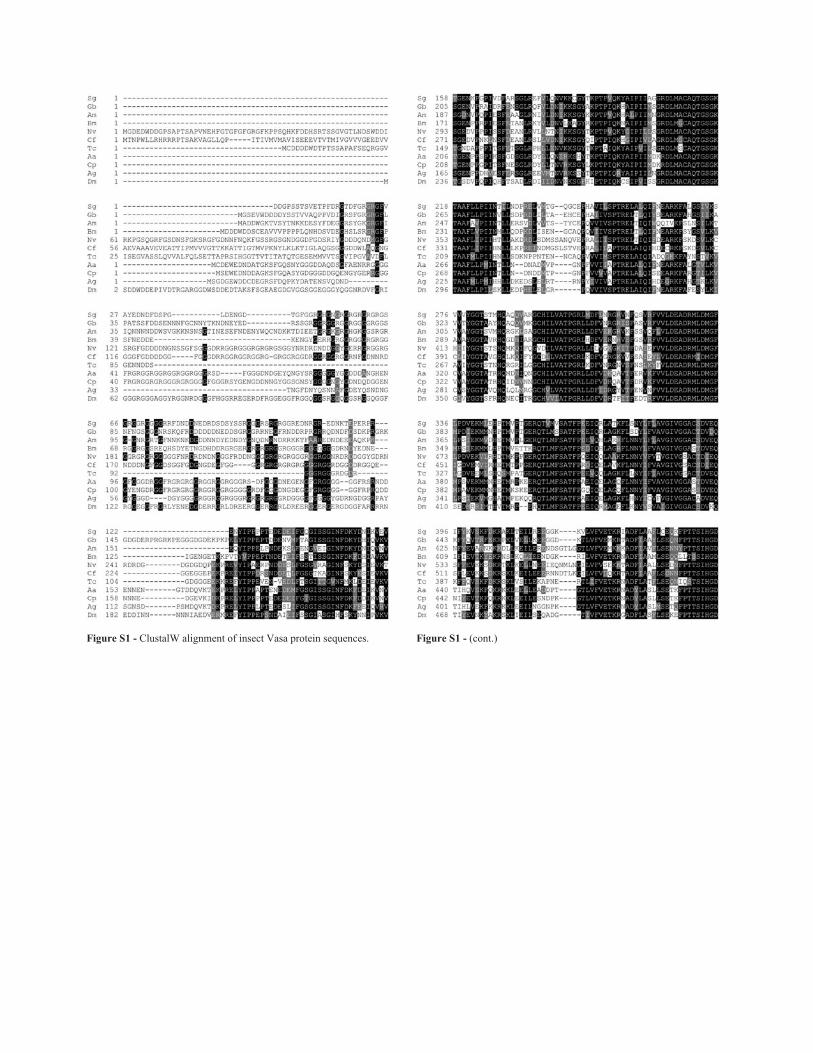
Figure S1 - ClustalW alignment of insect Vasa protein sequences. Figure S1 - (cont.)
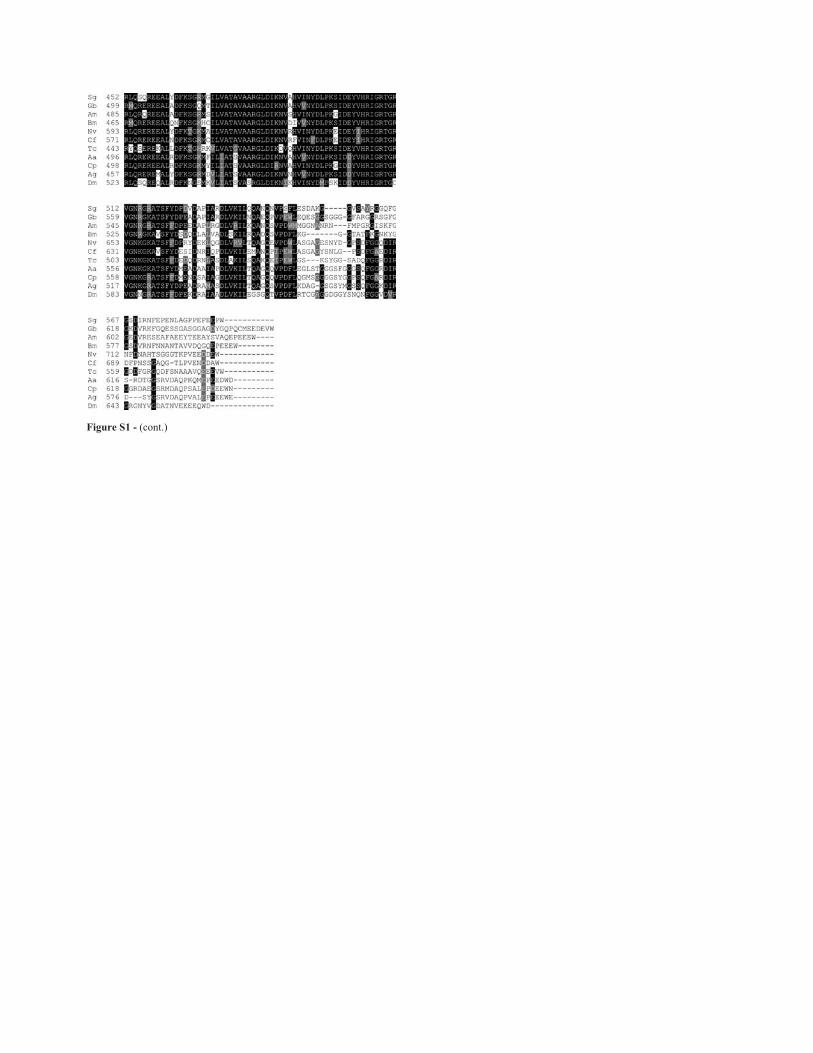
Figure S1 - (cont.)
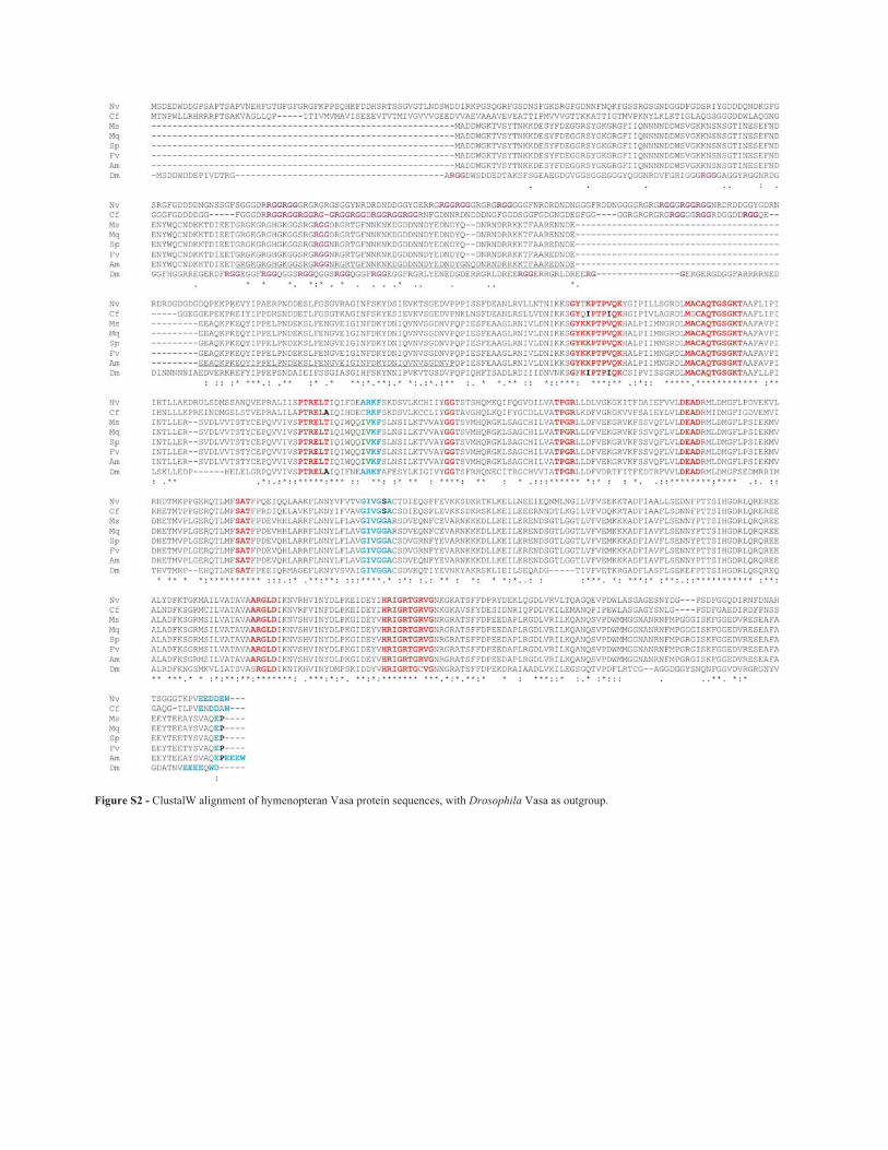
Figure S2 - ClustalW alignment of hymenopteran Vasa protein sequences, with Drosophila Vasa as outgroup.