sequence-tagged site (sts) content mapping of human
TRANSCRIPT
Sequence-tagged Site (STS) Content Mapping of
Human Chromosomes: Theoretical Considerations
and Early Experiences Eric D. Green1, 2 and Philip Green 1
Departments of 1Genetics, 2pathology, and 2Internal Medicine, Washington University School of Medicine, St. Louis, Missouri 63110
M a j o r goals of the Human Genome Project include the isolation of the entire h u m a n genome in overlapping clones and the development of physi- cal maps of the cloned DNA. ~1) Con- struction of these physical maps in- volves specifying the coordinates of DNA-based landmarks, whose nature, to a great extent, has reflected the ex- per imental tools that are available for their detection wi th in complex sources of DNA. The advent of polymerase chain reaction (PCR) technology has facilitated the evolution of a new type of landmark-- the sequence-tagged site (STS)--for constructing physical maps of chromosomes. Here we review the basic concepts that guide the use of STSs as DNA landmarks, discuss a strategy for developing clone-based STS-content maps of chromosomes, and describe the role that such approaches are playing in the early phases of the Human Genome Project.
OVERVIEW OF STS-CONTENT MAPPING
The process of developing long-range physical maps of complex genomes in- volves the identification and localiza- t ion of landmarks wi th in recombinant DNA clones. In the typical case, this process can be viewed as occurring in four steps: (1) f ragmenting the genome into smaller pieces (e.g., by restriction digestion); (2) isolating and purifying each fragment (i.e., cloning); (3) exam- in ing the individual DNA fragments for the presence of c o m m o n land- marks; and (4) constructing physical maps of the cloned DNA based on in- formation about the relative positions
of the landmarks. Recent technological advances provide powerful new tools for carrying out these interrelated ac- tivities. In particular, the development of yeast artificial chromosomes (YACs) as a c loning system in conjunct ion with the utilization of PCR for the detection of physical landmarks has yielded powerful approaches for the physical mapping of entire genomes.
YAC cloning provides the means to isolate segments of DNA that are large (100-1000 kb) yet exper imental ly manageable. (2-5) The implementa t ion of YAC cloning has been greatly facili- tated in recent years by the develop- ment of efficient methods to con- struct (6-13) and screen04-20) YAC li- braries as well as the es tabl ishment of effective approaches to manipu- late(21-28) and characterize (23,26-33) YAC clones, inc luding a number of dif- ferent strategies for isolating their in- sert ends. (2,30,31,34-43) In their short history, YACs have been used to isolate and map contiguous stretches of DNA that are significantly larger than those isolated using bacterial c loning sys- tems (21,35,44-51) and to play important roles in the identif ication of genes causing several h u m a n genetic dis- eases, such as neuroflbromatosis, (52) fragile X syndrome, t53-57t and familial adenomatous polyposis coli. (48,49) Based on the experience to date, YACs are generally considered the best avail- able c loning system for constructing long-range physical maps of DNA.
The most serious problem thus far associated with YAC cloning is the presence of clones conta in ing two or more unrelated pieces of DNA (i.e., seg-
ments arising from different regions of the genome of origin). Such chimeric YACs have been detected either by ob- vious differences between the physical map of the YAC and that of the corres- ponding region of the source genome or by discrepant chromosomal origins of the two insert ends (21,34,35,39,46) The frequency of chimeric clones seems to vary widely among different libraries and may vary from locus to locus in the same library. For example, in one library derived from total h u m a n DNA, C6,1°,17) 40-60% of the YAC clones are estimated to be chimeric, (34) while in another library made from a h u m a n - h a m s t e r hybrid cell line, the fraction of chimeric YACs is estimated at 10-20%3581 The mechanism(s) by which such anomalous YACs form remains an area of investigation, ~34t as is the development of methods to el iminate their formation. One pro- posed mechan i sm of chimeric YAC formation involves the recombinat ion in the transformed yeast cell of repeti- tive sequences (e.g., Alu) located wi th in unrelated DNA molecules. This model is based on the detailed charac- terization of one chimeric YAC (34) and the collective experiences with libraries derived from h u m a n - r o d e n t hybr id cell lines, which seem to contain fewer chimeric clones. In the latter cases, the presence of a background of irrelevant rodent DNA likely minimizes the fre- quencies with which recombinat ion events can occur between unrelated human-YAC clones. A much less fre- quent problem with YAC cloning is that created by DNA segments that are unclonable or unstable. In some cases
1:77-90©1991 by Cold Spring Harbor Laboratory Press ISSN 1054-9803/91 $3.00 PCR Methods and Applications 77
Cold Spring Harbor Laboratory Press on January 28, 2018 - Published by genome.cshlp.orgDownloaded from
in w h i c h ins tabi l i ty has been en- countered , stable over lapp ing YACs c o n t a i n i n g the p rob lemat ic DNA have been isolated. (s8)
The d e v e l o p m e n t of YAC c lon ing has largely occurred in parallel wi th tha t of PCR technology . For m a n y ap- pl icat ions, PCR has been used as the f ront - l ine tool for bo th isola t ing (14,1s) and characterizing(21,35,39,46,50) YAC
clones in efforts to cons t ruct physical maps of DNA. These early studies he lped to mot iva t e the proposal to use STSs as the l andmarks in physical maps of DNA, (s9) a l t h o u g h the case in favor of STSs t ranscends any part icular map- p ing me thod . By def in i t ion , an STS is a shor t s tretch of DNA tha t can be spe-
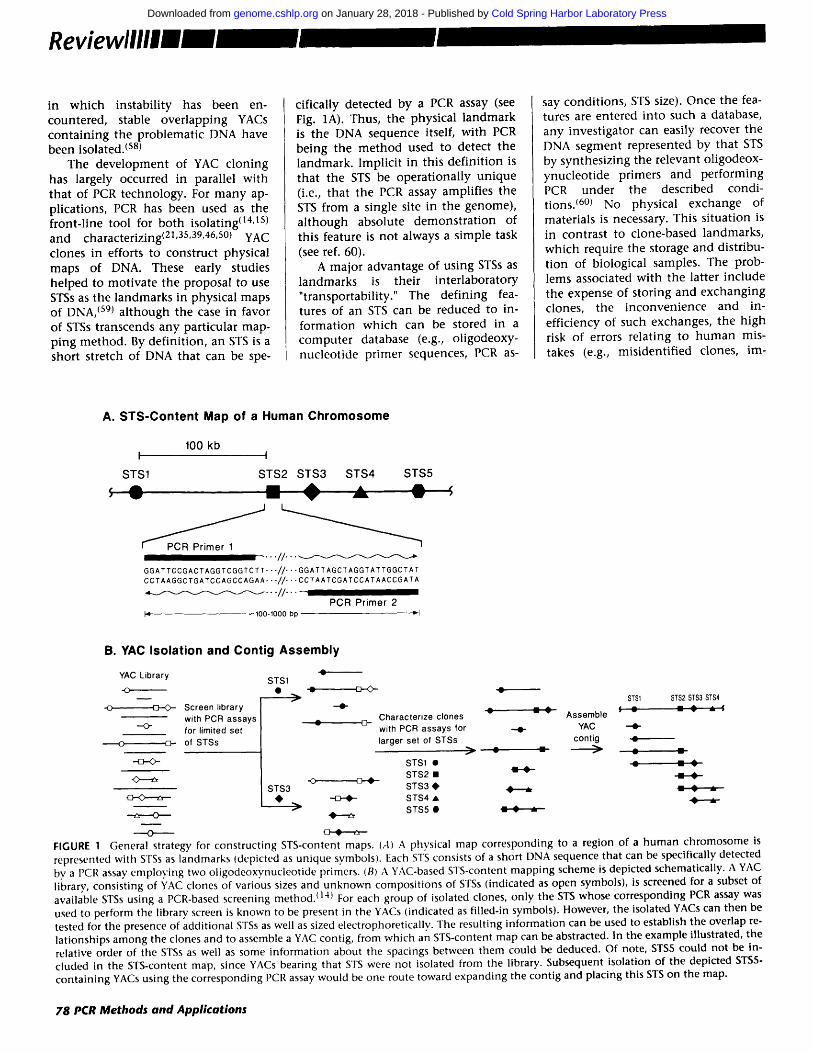
cifically detected by a PCR assay (see Fig. 1A). Thus, the physical l a n d m a r k is the DNA sequence itself, wi th PCR be ing the m e t h o d used to detect the landmark . Implic i t in this de f in i t ion is tha t the STS be ope ra t iona l ly un ique (i.e., tha t the PCR assay amplif ies the STS from a single site in the genome) , a l t h o u g h absolute d e m o n s t r a t i o n of this feature is no t always a s imple task (see ref. 60).
A major advantage of using STSs as l andmarks is thei r in te r labora tory " transportabi l i ty ." The def in ing fea- tures of an STS can be reduced to in- fo rma t ion wh ich can be stored in a compu te r database (e.g., o l igodeoxy- nuc leo t ide pr imer sequences, PCR as-
say condi t ions , STS size). Once the fea- tures are entered in to such a database, any invest igator can easily recover the DNA segment represented by tha t STS by synthes iz ing the re levant ol igodeox- ynuc leo t ide pr imers and pe r fo rming PCR unde r the described condi- t ions. (6°) No physical exchange of materials is necessary. This s i tua t ion is in cont ras t to c lone-based landmarks , wh ich require the storage and distribu- t ion of biological samples. The prob- lems associated wi th the latter inc lude the expense of s tor ing and exchang ing clones, the i n c o n v e n i e n c e and in- eff ic iency of such exchanges, the h igh risk of errors re lat ing to h u m a n mis- takes (e.g., mis ident i f ied clones, ira-
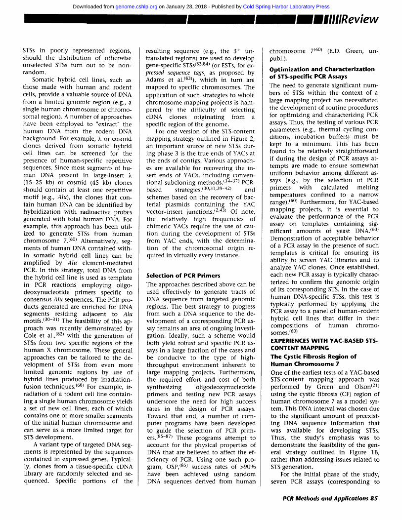
A. STS-Content Map of a Human Chromosome
100 kb I '" I
STS1 STS2 STS3 STS4 STS5 A
I~ PCR Primer 1 . . . . / / . • . ~
GGATTCCGACTAGGTCGGTCTT" • " / / " • . G G A T T A G C T A G G T A T T G G C T A T CCTAAGGCTGATCCAGCCAGAA. • - / / " • • C C T A A T C G A T C C A T A A C C G A T A ~ . • . / / . . . .
PCR Primer 2 I ~ ~ 100-1000 bp -'-I
B. YAC Isolation and Contig Assembly
YAC L i b r a r y
- o [3---<>- S c r e e n l i b r a r y
w i t h P C R a s s a y s - -O-
f o r l im i t ed set
~ - o f S T S s
-'43-0-
¢
STS1
f STSl STS2 STS3 STS4 - -O- ¢ _ - ¢ ~ ¢ - ¢ _- :
C h a r a c t e r i z e c l o n e s A s s e m b l e
¢ [3- w i t h P C R a s s a y s f o r - . - I - YAC - . t -
l a r g e r s e t o f S T S s c o n t i g ¢
/
STS1 • ¢ = ¢
STS2 • -0
STS3 STS3 • ~ • ~ STS4 •
. . .r S T S 5 •
- - - o FIGURE 1 General strategy, for constructing STS-content maps. (A) A physical map corresponding to a region of a human chromosome is represented with STSs as landmarks (depicted as unique symbols). Each STS consists of a short DNA sequence that can be specifically detected bv a PCR assay employing two oligodeoxvnucleotide primers. (B) A YAC-based STS-content mapping scheme is depicted schematically. A YAC library, consisting of YAC clones of various sizes and unknown compositions of STSs (indicated as open symbols), is screened for a subset of available STSs using a PCR-based screening method./14) For each group of isolated clones, only the STS whose corresponding PCR assay was used to perform the library screen is known to be present in the YACs (indicated as filled-in symbols). However, the isolated YACs can then be tested for the presence of additional STSs as well as sized electrophoretically. The resulting information can be used to establish the overlap re- lationships among the clones and to assemble a YAC contig, from which an STS-content map can be abstracted. In the example illustrated, the relative order of the STSs as well as some information about the spacings between them could be deduced. Of note, STS5 could not be in- cluded in the STS-content map, since YACs bearing that STS were not isolated from the library. Subsequent isolation of the depicted STS5- containing YACs using the corresponding PCR assay would be one route toward expanding the contig and placing this STS on the map.
78 PCR Methods and Applications
Cold Spring Harbor Laboratory Press on January 28, 2018 - Published by genome.cshlp.orgDownloaded from
proper storage), clone instability, and, most importantly, the rapid obsoles- cence of particular vector-host sys- tems. The ability to access the DNA landmarks on which the physical maps are based without dependence on stored biological materials is a key no- tion behind the proposal to use STSs as the "common language" in physical maps of DNA. (59)
The fundamental features of a YAC- based STS-content mapping strategy are highlighted in Figure 1. Dispersed across a large DNA interval (e.g., a hu- man chromosome) reside numerous unique DNA sequences (i.e., STSs), each of which can be detected by a specific PCR assay (Fig. 1A). One route toward defining the relative order and spacings of such STSs is schematically depicted in Figure lB. A YAC library contains clones bearing one or more of the indicated STSs. Using appropriate PCR assays, YACs containing specific STSs can be readily isolated.(14) The subsequent characterization of the resulting YACs includes both testing for a larger number of STSs and elec- trophoretic assessment of YAC size. Knowledge of the STS content and size of each YAC can be used to construct a clone map, which is a collection of clones aligned in an overlapping man- ner (a set of contiguous, overlapping clones is also known as a contig). This information can in turn be used to es- tablish an STS-content map indicating the relative locations of the various STSs within the contig. Incorporation of contig maps on STS-content maps of the whole chromosome will require, in addition, the use of long-range meth- ods (such as linkage mapping and radiation hybrid mapping) to order and orient the contigs along the chromosome.
In the following, we propose a strategy for developing STS-content maps of whole chromosomes and dis- cuss some of its theoretical properties. We then review relevant practical ex- perience, with particular emphasis on activities in the Washington University Human Genome Center, where this strategy is being applied to construct physical maps of human chromosomes 7 and X. Several alternative physical mapping strategies have been success- fully used by other investigators, as reviewed recently by Evans. (61)
A STRATEGY FOR CONSTRUCTING STS-CONTENT MAPS OF WHOLE CHROMOSOMES
Conceptual Basis of Strategy The development of high-resolution physical maps typically involves the assembly of overlapping clones into a contig, from which a map can be derived. Two classical strategies have been used to produce such overlapping clone maps: (1) fingerprinting, in which a characteristic sequence-dependent pattern is generated for each randomly selected clone (e.g., by sizing restric- tion fragments) and clone overlaps are inferred based on shared parts of the pattern; and (2) chromosome walking, in which unique sequence probes are used to obtain clones, screening the clone library with an initial probe, and then, iteratively, with probes derived from the ends of clones obtained in the previous screen, until the region of interest is fully isolated. Chromosome walking has the advantages of being targeted to the region of interest and of being able to detect small overlaps be- tween clones, thereby allowing the construction of larger contigs. How- ever, this approach has generally been considered unsuitable for mapping large genomic regions, because each walking step depends upon the suc- cessful completion of the preceding one and requires both probe genera- tion and library screening.
The STS-content mapping strategy (Fig. 1B) represents a hybrid approach that can be used for constructing over- lapping YAC maps of large, targeted genomic regions, such as whole hu- man chromosomes (the details of this strategy were originally presented by P. Green at the 1990 "Genome Mapping and Sequencing" meeting at Cold Spring Harbor Laboratory). In this ap- proach, S T S s randomly located throughout the region to be mapped fill three roles: (1) a set of random STSs serve as anchor points for multiple, short chromosome walks; (2) STSs near the ends of the growing YAC contigs serve as surrogate ends for expanding the contigs by walking; and (3) the STS contents of the YACs are used to estab- lish overlaps among the YACs (i.e., the STSs in a YAC constitute its "finger- print" for use in constructing contig maps, as illustrated in Fig. 1B). Two
disadvantages of chromosome walking, the complete dependence on clone end isolation and the need for multiple se- quential steps, are largely avoided. As with other targeted mapping ap- proaches, numerous library screens are required; however, their number is minimized by prescreening the pre- viously isolated YACs for the presence of newly generated STSs and perform- ing library screens either with STSs ab- sent from the previously isolated YACs or with surrogate-end STSs. Thus, li- brary screens are only performed with STSs likely to add a significant amount of new DNA to the clone map. More- over, a single library can, in principle, be used for mapping any genomic region.
Because contig assembly is based on STSs already known to be located on the chromosome being mapped, this strategy, unlike pure fingerprint- based approaches, is relatively insensi- tive to the presence of chimeric YACs. A portion of a YAC that lies on another chromosome is not detected with any of the STS-specific PCR assays, and therefore will not cause spurious mergers of non-overlapping contigs. (Chimeric YACs can affect the distance estimates between STSs, however.) Of course, chimeric YACs containing two discontinuous segments from the same chromosome can cause problems, but these should be manageable if they are not too frequent (see Prospects and Limitations, below).
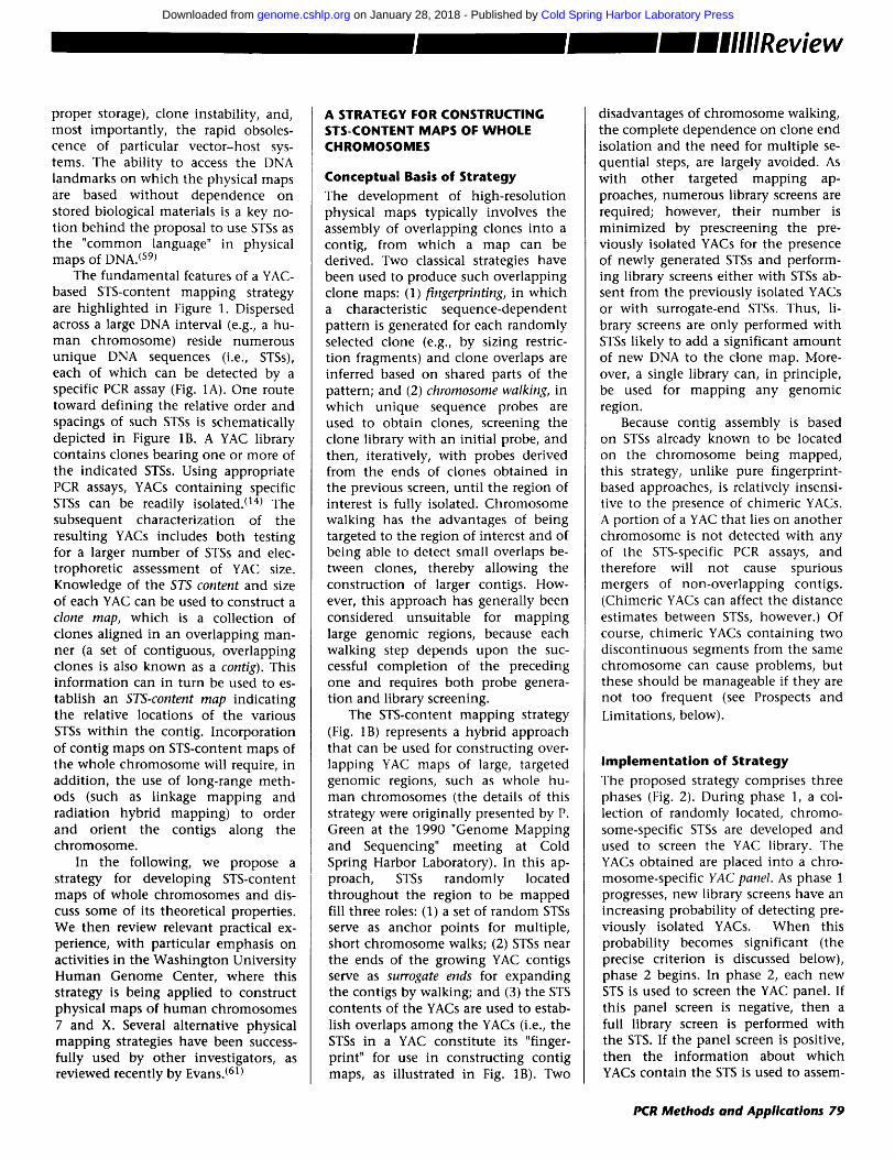
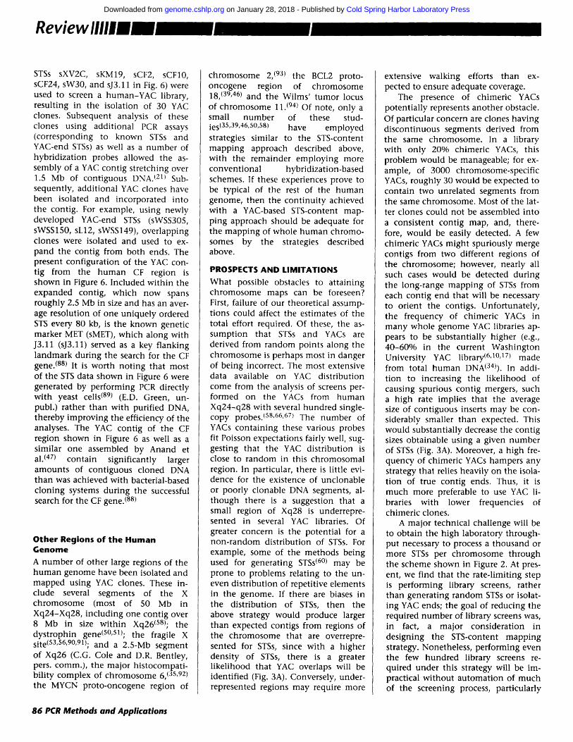
Implementat ion of Strategy The proposed strategy comprises three phases (Fig. 2). During phase 1, a col- lection of randomly located, chromo- some-specific STSs are developed and used to screen the YAC library. The YACs obtained are placed into a chro- mosome-specific YAC panel. As phase 1 progresses, new library screens have an increasing probability of detecting pre- viously isolated YACs. When this probability becomes significant (the precise criterion is discussed below), phase 2 begins. In phase 2, each new STS is used to screen the YAC panel. If this panel screen is negative, then a full library screen is performed with the STS. If the panel screen is positive, then the information about which YACs contain the STS is used to assem-
PCR Methods and Applications 79
Cold Spring Harbor Laboratory Press on January 28, 2018 - Published by genome.cshlp.orgDownloaded from
PHASE
~ Develop Chromosome-Specific S T S |
PHASE 2 ~'
l@,,,~-~, I Develop Chromosome-Specific STS ..... -
I~i:~ Screen Chromosome-Specific YAC Pane ! ~'~,~i sTs Absent : % STS Present
~lScreen YAC Librar~ : I " us'e 'Information' to ' ........ ..... I Construct STS-Content I
Add New lclones t o I I Maps of YAC Contigs I YAC Panel
PHASE 3 '~
FIGURE 2 Experimental strategy for con- structing STS-content maps of whole chromosomes. The general aspects of the three phases of an STS-content mapping project are outlined, with details provided in the text.
ble the YACs into contigs and to con- struct STS-content maps.
In essence, phases 1 and 2 initiate mult iple chromosome walks from ran- dom locations on the chromosome. Phase 3, which begins when the YAC panel contains most of the DNA in the chromosome, aims to extend the exist- ing contigs, both by ident i fying any undetected overlaps among the isola- ted YACs and by obtaining addit ional overlapping YACs from the library. The contig maps developed in phases 1 and 2 are utilized in a directed m a n n e r for selecting the STSs to be used for library screens. First, surrogate ends, STSs which have previously been used to screen the YAC panel but not the full library and which are known from the STS-content maps to lie near the ends of contigs, are retested against the YAC panel. (This retesting is necessary be- cause the YAC panel is cont inuously
expanding, so it will usually contain clones added subsequent to the initial panel testing with that STS.) Some- times a new panel screen with a sur- rogate end will detect a previously un- known overlap between two contigs, thereby allowing the contigs to be merged. If no new overlap is detected, then the STS is used for a library screen.
An obvious modif icat ion of this scheme is to use true ends (developed from the YACs at the ends of contigs) instead of surrogate ends as the STSs in phase 3. True ends are somewhat more likely to reveal overlaps and detect new YACs. However, the generation of STSs from true ends is likely to involve more effort, since it generally requires isolating both insert ends of several terminal YACs and testing them against other YACs in the contig to determine which YAC end represents the true contig end. Moreover, the presence of chimeric YACs implies that in some instances the true contig end will, in fact, not be at the end of a YAC insert. The decision about the type of "end" to use in phase 3 will ul t imately depend upon the relative costs and the desired contig sizes, as discussed below.
In describing this model for devel- oping Sl"S-content maps, we have con- sidered all the mappable landmarks to be STSs, implying that all mapping data would be obtained using PCR as the analytical technique. In actual practice, this will likely not be the case. For example, any single-copy hybridi- zation probe (e.g., X, cosmid, or cDNA clones) can be viewed as a "potential" STS and used in the identical fashion for mapping as has been illustrated in Figures 1 and 2 for true STSs, the major difference being the mode of detection (hybridization for probe sequences vs. PCR for STSs). Of note, the conversion of hybridizat ion probes (potential STSs) into functional PCR assays is rela- tively straightforward. Thus, for the screening of YAC libraries and panels described above, either hybridizat ion detection of probe sequences or PCR detection of STSs can be utilized and the resulting informat ion used in an identical manne r for constructing physical maps.
The choice between PCR- and hy- bridization-based protocols is really an experimental issue rather than a map-
ping one. In practice, we envision a mixture of experimental activities, with the overall efficiency, accuracy, and cost of each process dictating the extent of its use. We also expect that such a mixture will change throughout the course of a project. For example, in our opin ion h igh- throughput screen- ing of YAC libraries can more efficient- ly be performed by PCR than by hy- bridization. (14) PCR assay development would therefore likely predominate during phase 1, when large numbers of library screens are being performed. In contrast, in phases 2 and 3, most of the analyses are being performed on relatively small numbers of YACs clones (the YAC panel), which may be more efficiently analyzed by hybridiza- tion. Overall, there is an incentive to tip the balance toward PCR assay de- ve lopment because of the desirability of using true STSs as landmarks. (59,6°) An addit ional factor which may favor the emphasis of PCR is the existence of duplicated segments of DNA wi th in a chromosome. Evolutionarily older du- plications are likely to have diverged over time, and PCR offers better pros- pects than does hybridizat ion for dis- c r iminat ing between different copies. Such duplications may turn out to be relatively c o m m o n in the h u m a n ge- nome, with examples including pseu- dogenes, gene families, and several large regions of the X chromo- some. (58,62) These considerations are also relevant to the important issue of how best to integrate chromosomal STS-content maps with existing long- range maps developed using other methods, such as linkage mapping and radiation hybrid mapping. We envi- sion performing this integration by in- corporating the markers from the long- range maps into the STS-content maps. This can be done either early in the project, by generating STSs from the markers and using them to screen the YAC library, or later, by using the markers to screen the YAC panel by hybridization. The former approach is likely to be less efficient, since it re- quires the generation of DNA se- quences from templates cloned into a diversity of vectors; moreover, the number of such markers available for individual chromosomes is small rela- tive to the needs of the overall project (see below).
80 PCR Methods and Applications
Cold Spring Harbor Laboratory Press on January 28, 2018 - Published by genome.cshlp.orgDownloaded from
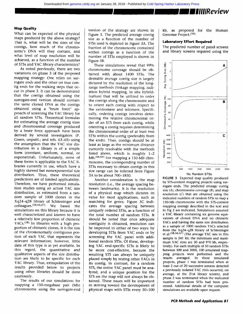
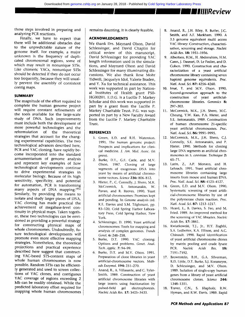
Map Quality What can be expected of the physical maps produced by the above strategy? That is, what will be the sizes of the contigs, how much of the chromo- some's DNA will they contain, and what level of map resolution will be achieved, as a funct ion of the number of STSs and YAC library characteristics?
As noted previously, there are two variations on phase 3 of the proposed mapp ing strategy: One relies on sur- rogate ends and the other on true con- tig ends for the walking steps that oc- cur in phase 3. It can be demonstrated that the contigs obtained using the surrogate-end version should contain the same cloned DNA as the contigs obtained using a "brute force" ap- proach of screening the YAC library for all random STSs. Theoretical formulas for est imating the average contig sizes and chromosomal coverage produced by a brute force approach have been derived by several investigators (P. Green, unpubl. ; and refs. 63-65) using the assumption that the YAC size dis- t r ibution in a library is of a simple form (constant, uniform, normal, or exponential) . Unfortunately, none of these forms is applicable to the YAC li- braries currently in use, which have a h ighly skewed but nonexponent ia l size distribution. Thus, these theoretical predictions are of l imited applicability. Therefore, we have performed simula- t ion studies using an actual YAC size distribution, as estimated from a ran- dom sample of 1000 clones in the Xq24-q28 library of Schlessinger and colleagues. (58,66,67) We based the simulat ions on this library because it is well characterized and known to have a relatively low proportion of chimeric YACs. (s8) In libraries with a h igh pro- port ion of chimeric clones, it is the size of the chromosomal ly contiguous por- t ion of each YAC that represents the relevant information; however, little data of this type is as yet available. In this regard, the quanti tat ive and qualitative aspects of the size distribu- tion are likely to be specific for each YAC library. Thus, extrapolation of the results provided below to projects using other libraries should be done cautiously.
The results of our simulat ions for mapp ing a 150-megabase pair (Mb) chromosome using the surrogate-end
version of the strategy are shown in Figure 3. The predicted average contig size as a function of the number of STSs used is depicted in Figure 3A. The fraction of the chromosome contained wi th in contigs as a funct ion of the number of STSs employed is shown in Figure 3B.
These simulat ions reveal that 99% chromosome coverage should be ob- tained with about 1400 STSs. The desirable average contig size is largely dictated by the resolution of the long- range methods (linkage mapping, radi- ation hybrid mapping, in situ hybrid- ization) that will be utilized to order the contigs along the chromosome and to orient each contig with respect to the centromere and telomere. Specifi- cally, ordering contigs involves deter- min ing the relative chromosomal or- der of an STS from each contig, while orienting a contig requires determining the chromosomal order of at least two STSs wi th in the contig (preferably from the ends). Thus, contigs should be at least as large as the m i n i m u m distance currently resolvable with the methods listed above, which is roughly 1-2 Mb. (68'69) For mapp ing a 150-Mb chro- mosome, the corresponding number of STSs necessary to obtain contigs in this size range can be inferred from Figure 3A to be about 700-1850.
Another consideration is the map resolution (i.e., the average spacing be- tween landmarks). It is the resolution of a physical map which dictates its value for most applications, such as searching for genes. Figure 3C indi- cates the average spacing between uniquely ordered STSs, as a funct ion of the total number of random STSs. It should be noted that once adequate contiguity is obtained, resolution can be improved in either of two ways: by developing STSs from YAC ends or by screening the YAC panel with addi- t ional random STSs. Of these, develop- ing YAC end-specific STSs is likely to be more cost-effective, because the resulting STS can always be uniquely placed s imply by testing other YACs in its contig. In contrast, for a random STS, the entire YAC panel must be ana- lyzed, and a unique position for the STS on the map will not always be ob- tained. These issues will be important in striving toward the development of physical maps with STSs every 50-100
kb, as proposed for the Human Genome Project. (s9)
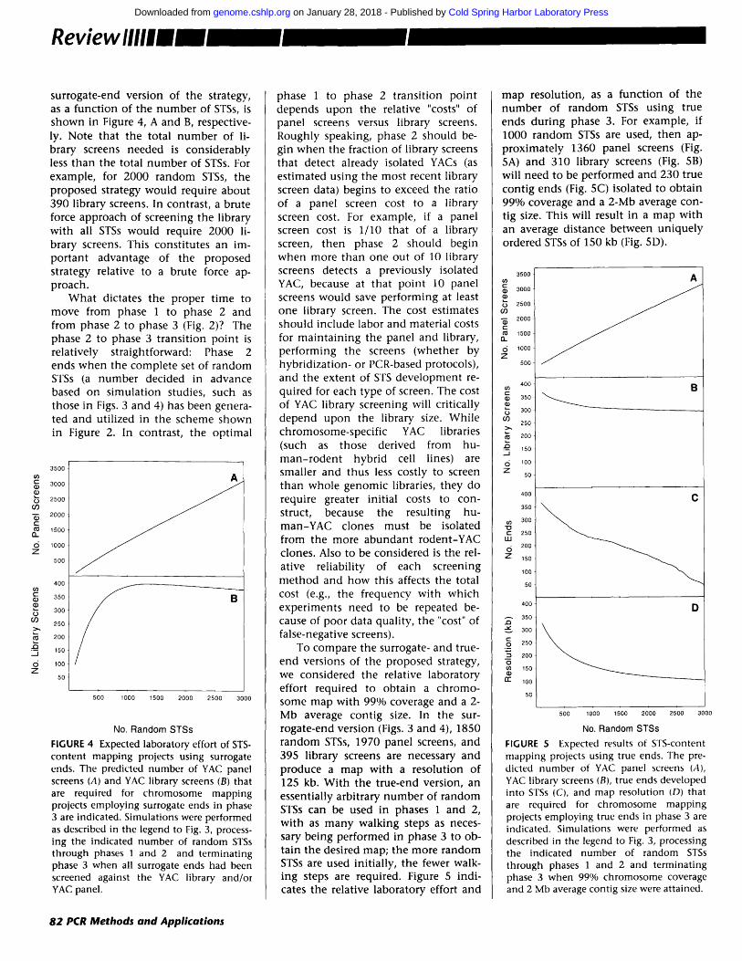
Laboratory Effort Required The predicted number of panel screens and library screens required using the
8
(.9 6
o E ~- 2
o
140
~. 120
~ 1 0 0
o ~_ 80
~ 60
g: 40 2 0
A
/
500 1000 1500 2000 2500 3000
No. Random STSs FIGURE 3 Expected map quality produced by El'S-content mapping projects using sur- rogate ends. The predicted average contig size (A), chromosome coverage (B), and map resolution (C) that are obtained using the indicated numbers of random STSs to map a 150-Mb chromosome with the STS-content mapping strategy described in the text and in Fig 2 are indicated. Simulations assumed a YAC library containing six genome equi- valents of cloned DNA and no chimeric YACs and having the same size distribution as a sample of 1000 random YACs selected from the Xq24-q28 library of Schlessinger et al. (58,66,67) (The average YAC size in this sample is 241 kb; the min imum and maxi- mum YAC sizes are 30 and 970 kb, respec- tively). For each multiple of 50 random STSs between 100 and 3000, 100 simulated map- ping projects were performed and the results averaged. In these simulated projects, phase 1 was terminated when at least 2 out of 20 successive screens detected a previously isolated YAC (this occurred, on average, at the 21st library screen), and phase 2 was terminated when the indicated number of random STSs had been pro- cessed. Additional details of the computer simulations are available upon request.
PCR Methods and Applications 81
Cold Spring Harbor Laboratory Press on January 28, 2018 - Published by genome.cshlp.orgDownloaded from
Review llll|ll
surrogate-end version of the strategy, as a function of the number of STSs, is shown in Figure 4, A and B, respective- ly. Note that the total number of li- brary screens needed is considerably less than the total number of STSs. For example, for 2000 random STSs, the proposed strategy would require about 390 library screens. In contrast, a brute force approach of screening the library with all STSs would require 2000 li- brary screens. This constitutes an im- por tant advantage of the proposed strategy relative to a brute force ap- proach.
Wha t dictates the proper t ime to move from phase 1 to phase 2 and from phase 2 to phase 3 (Fig. 2)? The phase 2 to phase 3 transit ion point is relatively straightforward: Phase 2 ends when the complete set of random STSs (a number decided in advance based on simulation studies, such as those in Figs. 3 and 4) has been genera- ted and utilized in the scheme shown in Figure 2. In contrast, the optimal
3500
3000 O
2500 O')
2000 c-
(~ 1500 a -
6 1ooo Z
5o0
400
350
300
250
200
• ~ 150 ._l
6 lOO Z 5O
500 1000 1500 2000 2500 3000
No. Random STSs
FIGURE 4 Expected laboratory effort of STS- content mapping projects using surrogate ends. The predicted number of YAC panel screens (A) and YAC library screens (B) that are required for chromosome mapping projects employing surrogate ends in phase 3 are indicated. Simulations were performed as described in the legend to Fig. 3, process- ing the indicated number of random STSs through phases 1 and 2 and terminating phase 3 when all surrogate ends had been screened against the YAC library and/or YAC panel.
phase 1 to phase 2 transit ion point depends upon the relative "costs" of panel screens versus library screens. Roughly speaking, phase 2 should be- gin when the fraction of library screens that detect already isolated YACs (as estimated using the most recent library screen data) begins to exceed the ratio of a panel screen cost to a library screen cost. For example, if a panel screen cost is 1/10 that of a library screen, then phase 2 should begin when more than one out of 10 library screens detects a previously isolated YAC, because at that point 10 panel screens would save performing at least one library screen. The cost estimates should include labor and material costs for main ta in ing the panel and library, performing the screens (whether by hybridization- or PCR-based protocols), and the extent of STS development re- quired for each type of screen. The cost of YAC library screening will critically depend upon the library size. While chromosome-specific YAC libraries (such as those derived from hu- m a n - r o d e n t hybrid cell lines) are smaller and thus less costly to screen than whole genomic libraries, they do require greater initial costs to con- struct, because the resulting hu- man-YAC clones must be isolated from the more abundan t rodent-YAC clones. Also to be considered is the rel- ative reliability of each screening method and how this affects the total cost (e.g., the frequency with which experiments need to be repeated be- cause of poor data quality, the "cost" of false-negative screens).
To compare the surrogate- and true- end versions of the proposed strategy, we considered the relative laboratory effort required to obtain a chromo- some map with 99% coverage and a 2- Mb average contig size. In the sur- rogate-end version (Figs. 3 and 4), 1850 random STSs, 1970 panel screens, and 395 library screens are necessary and produce a map with a resolution of 125 kb. With the true-end version, an essentially arbitrary number of random STSs can be used in phases 1 and 2, with as m a n y walking steps as neces- sary being performed in phase 3 to ob- tain the desired map; the more random STSs are used initially, the fewer walk- ing steps are required. Figure 5 indi- cates the relative laboratory effort and
map resolution, as a funct ion of the number of random STSs using true ends during phase 3. For example, if 1000 random STSs are used, then ap- proximately 1360 panel screens (Fig. 5A) and 310 library screens (Fig. 5B) will need to be performed and 230 true contig ends (Fig. 5C) isolated to obtain 99% coverage and a 2-Mb average con- tig size. This will result in a map with an average distance between uniquely ordered STSs of 150 kb (Fig. 5D).
500
300
500
D00
500
O00
500
400 i
3 5 0 '
300 -
250 ~
200 ~
150
1 0 0
50-
400
350
300
250
200-
150
100
5O
4 0 0
35G
300
250
2OO
150
100
5C
A
C
500 1000 1500 2000 2500 3000
No. Random STSs
FIGURE 5 Expected results of STS-content mapping projects using true ends. The pre- dicted number of YAC panel screens (A), YAC library, screens (B), true ends developed into STSs (C), and map resolution (D) that are required for chromosome mapping projects employing true ends in phase 3 are indicated. Simulations were performed as described in the legend to Fig. 3, processing the indicated number of random STSs through phases 1 and 2 and terminating phase 3 when 99% chromosome coverage and 2 Mb average contig size were attained.
82 PCR Methods and Applications
Cold Spring Harbor Laboratory Press on January 28, 2018 - Published by genome.cshlp.orgDownloaded from
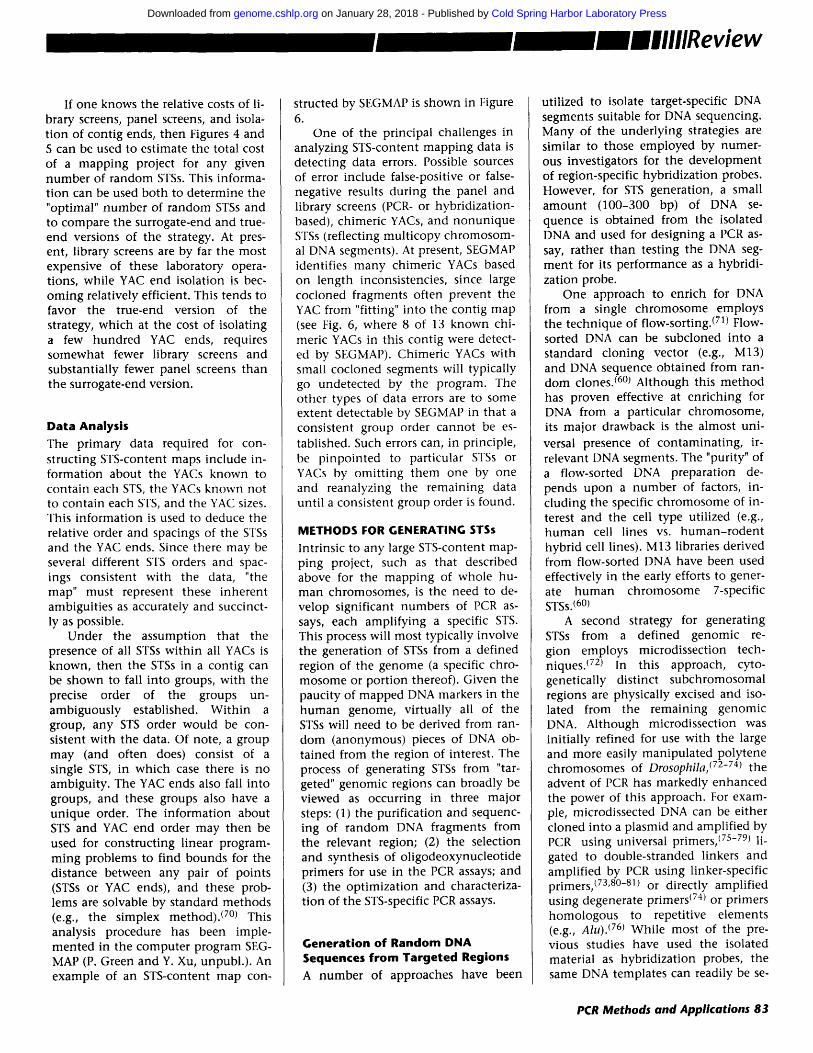
If one knows the relative costs of li- brary screens, panel screens, and isola- t ion of contig ends, then Figures 4 and 5 can be used to estimate the total cost of a mapping project for any given number of random STSs. This informa- t ion can be used both to determine the "optimal" number of random STSs and to compare the surrogate-end and true- end versions of the strategy. At pres- ent, library screens are by far the most expensive of these laboratory opera- tions, while YAC end isolation is bec- oming relatively efficient. This tends to favor the true-end version of the strategy, which at the cost of isolating a few hundred YAC ends, requires somewhat fewer library screens and substantially fewer panel screens than the surrogate-end version.
Data Analysis The primary data required for con- structing STS-content maps include in- formation about the YACs known to contain each STS, the YACs known not to contain each STS, and the YAC sizes. This informat ion is used to deduce the relative order and spacings of the STSs and the YAC ends. Since there may be several different STS orders and spac- ings consistent with the data, "the map" must represent these inherent ambiguit ies as accurately and succinct- ly as possible.
Under the assumption that the presence of all STSs wi th in all YACs is known, then the STSs in a contig can be shown to fall into groups, with the precise order of the groups un- ambiguously established. Wi th in a group, any STS order would be con- sistent with the data. Of note, a group may (and often does) consist of a single STS, in which case there is no ambiguity. The YAC ends also fall into groups, and these groups also have a unique order. The informat ion about STS and YAC end order may then be used for constructing linear program- ming problems to find bounds for the distance between any pair of points (STSs or YAC ends), and these prob- lems are solvable by standard methods (e.g., the simplex method). (7°) This analysis procedure has been imple- mented in the computer program SEG- MAP (P. Green and Y. Xu, unpubl.) . An example of an STS-content map con-
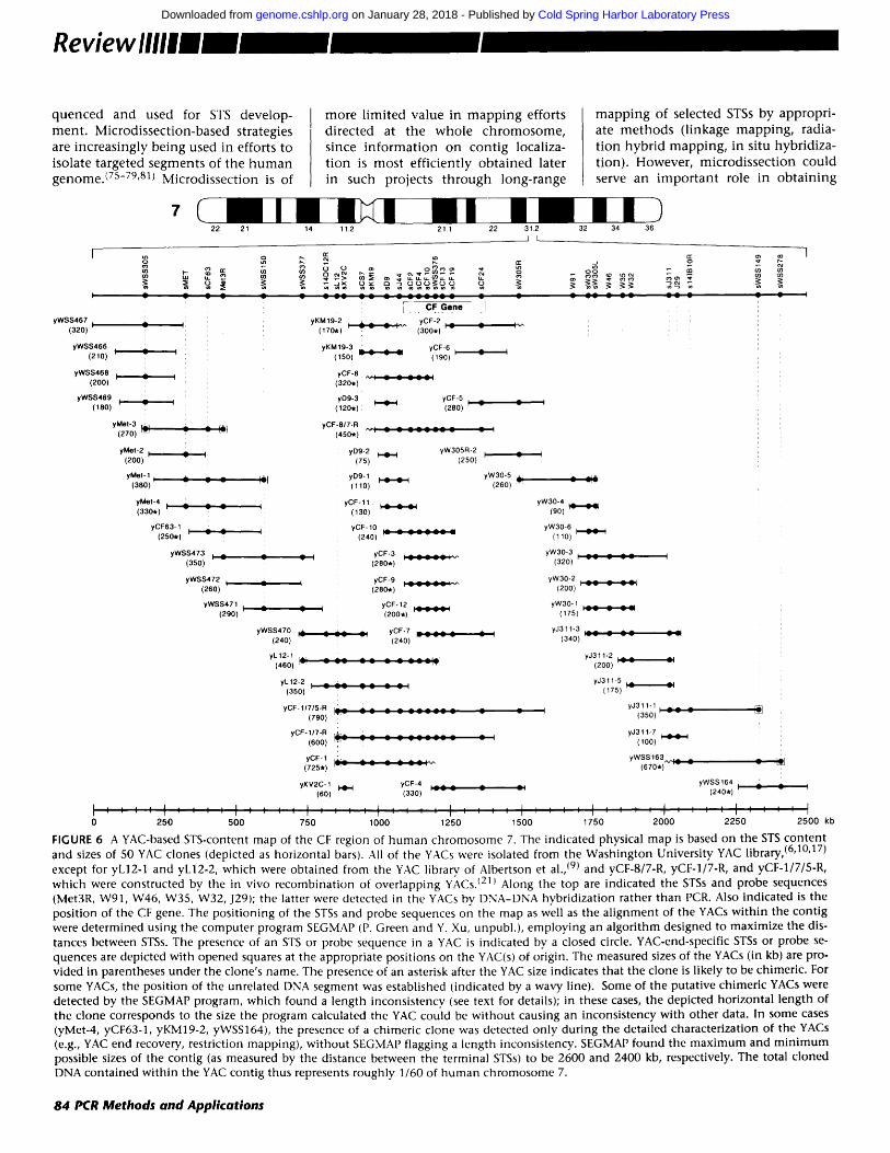
structed by SEGMAP is shown in Figure 6.
One of the principal challenges in analyzing STS-content mapp ing data is detecting data errors. Possible sources of error include false-positive or false- negative results during the panel and library screens (PCR- or hybridization- based), chimeric YACs, and nonun ique STSs (reflecting mult icopy chromosom- al DNA segments). At present, SEGMAP identifies m a n y chimeric YACs based on length inconsistencies, since large cocloned fragments often prevent the YAC from "fitting" into the contig map (see Fig. 6, where 8 of 13 known chi- meric YACs in this contig were detect- ed by SEGMAP). Chimeric YACs with small cocloned segments will typically go undetected by the program. The other types of data errors are to some extent detectable by SEGMAP in that a consistent group order cannot be es- tablished. Such errors can, in principle, be p inpointed to particular STSs or YACs by omit t ing them one by one and reanalyzing the remaining data unti l a consistent group order is found.
METHODS FOR GENERATING STSs
Intrinsic to any large STS-content map- ping project, such as that described above for the mapp ing of whole hu- m a n chromosomes, is the need to de- velop significant numbers of PCR as- says, each ampli fying a specific STS. This process will most typically involve the generation of STSs from a defined region of the genome (a specific chro- mosome or portion thereof). Given the paucity of mapped DNA markers in the h u m a n genome, virtually all of the STSs will need to be derived from ran- dom (anonymous) pieces of DNA ob- tained from the region of interest. The process of generating STSs from "tar- geted" genomic regions can broadly be viewed as occurring in three major steps: (1) the purification and sequenc- ing of random DNA fragments from the relevant region; (2) the selection and synthesis of ol igodeoxynucleotide primers for use in the PCR assays; and (3) the opt imizat ion and characteriza- t ion of the STS-specific PCR assays.
Generation of Random DNA Sequences from Targeted Regions A number of approaches have been
utilized to isolate target-specific DNA segments suitable for DNA sequencing. Many of the under ly ing strategies are similar to those employed by numer- ous investigators for the development of region-specific hybridizat ion probes. However, for STS generation, a small amoun t (100-300 bp) of DNA se- quence is obtained from the isolated DNA and used for designing a PCR as- say, rather than testing the DNA seg- ment for its performance as a hybridi- zation probe.
One approach to enrich for DNA from a single chromosome employs the technique of flow-sorting. (71) Flow- sorted DNA can be subcloned into a standard cloning vector (e.g., M13) and DNA sequence obtained from ran- dom clones. (6°1 Although this method has proven effective at enr iching for DNA from a particular chromosome, its major drawback is the almost uni- versal presence of contaminat ing, ir- relevant DNA segments. The "purity" of a flow-sorted DNA preparation de- pends upon a number of factors, in- cluding the specific chromosome of in- terest and the cell type utilized (e.g., h u m a n cell lines vs. h u m a n - r o d e n t hybrid cell lines). M13 libraries derived from flow-sorted DNA have been used effectively in the early efforts to gener- ate h u m a n chromosome 7-specific STSs.(60)
A second strategy for generating STSs from a defined genomic re- gion employs microdissection tech- niques. (72) In this approach, cyto- genetically distinct subchromosomal regions are physically excised and iso- lated from the remaining genomic DNA. Although microdissection was init ially refined for use with the large and more easily manipula ted polytene chromosomes of Drosophila, (72-74) the advent of PCR has markedly enhanced the power of this approach. For exam- ple, microdissected DNA can be either cloned into a plasmid and amplif ied by PCR using universal primers, (75-79) li- gated to double-stranded linkers and amplif ied by PCR using linker-specific primers,(73,8°-81) or directly amplif ied using degenerate primers (74) or primers homologous to repetitive elements (e.g., A/u). (76) While most of the pre- vious studies have used the isolated material as hybridizat ion probes, the same DNA templates can readily be se-
PCR Methods and Applications 83
Cold Spring Harbor Laboratory Press on January 28, 2018 - Published by genome.cshlp.orgDownloaded from
Reviewllllll
q u e n c e d and used for STS develop-
m e n t . Microdissec t ion-based strategies are increas ing ly be ing used in efforts to isolate ta rgeted segment s of the h u m a n g e n o m e . ~75-79,8~) Microd issec t ion is of
m o r e l imi ted va lue in m a p p i n g efforts
d i rec ted at the w h o l e c h r o m o s o m e , since i n f o r m a t i o n on con t ig localiza- t ion is mos t eff ic ient ly ob t a ined later in such projects t h r o u g h long- range
m a p p i n g of selected STSs by appropr i - ate m e t h o d s ( l inkage m a p p i n g , radia- t ion hybr id m a p p i n g , in situ hybr id iza- t ion). However , mic rod i s sec t ion cou ld serve an i m p o r t a n t role in o b t a i n i n g
yWSS467 I
( 320 )
yWSS466 ,
( 210 ) '
yWSS468 ,
( 200 ) "
yWSS469 ,
(180) ""
y M e t - 2 ,~
( 200 )
y M e t - 1 ,
( 380 )
7 22 21 14 11.2 21.1 22 31.2 32 34 36
.J L
o { e e _,x o ~ ?, o ~= ~ e
y M e l - 3 r~
( 270 ) ~
t~
l
yWSS470 ,.~
( 240 ) ' -
' L . . C F G e n e . ]
yKM19-2 , .~ ~ ~ i v . yCF -2 , ~
( 170 . ) , ( 300 * ) '
yKM19 -3 yCF-6 , =
( 150 ) . . . . ( 190 )
yCF -8
( 320 * )
yD9 -3 , _ yCF-5 , i
( 120 . ) ' - ( 280 ) ' "
yCF -817 .R • v , : = : ( 45o * )
yD9 -2 = yW305R-2 I
( 75 ) ( 250 )
yD9-1 _ _ . - - ,
( 11o )
yCF -11 . . . .
( 130 ) . . . .
yCF -10
( 240 )
= : yCF -3 ; ===C~ . : v .
( 280 * )
( yCF -9 . . . . . . :_j .
( 260 * )
= yCF . 12 . . . . . .
( 200 . ) . . . . . .
, ~ A , yCF -7
-- " - - " ' ( 240 )
v ~p,
yMe t -4 .~. ... _
( 330 . )
yCF63 -1 . -- .~
( 26o . ) ' - -
ywss473 , _ ,
( 350 )
yWSS472 I
( 260 )
yWSS471 ,
( 290 ) '
e o o ~
v v
yW30-5 i i ( 260 )
- - - - L - - ,
yW30-4 . . . .
(90 ) . . . .
yW30 -6 , . ~ . ,
( 110 ) . . . .
yW30 -3 I ( 320 )
yW30 -2 . . . . . . .
( 200 ) . . . . . . .
yW30 -1 . . . . . .
( 175 ) . . . . . .
y J311 .3 . . . . . .
( 340 ) . . . . . .
A ~ v - ,
yL12 -1 r,.~ ( 460 ) t-- . . . . . . . . . . . t - r
yL12 -2 , = ~ .= . . . . . .
( 350 ) ' - " . . . . . .
yCF-11715-R ,AL... A ~ i ,"r, . . . . . . . . . v v =
( 790 )
yCF- 1 /7 -R = :
(600) ~ . . . . . . . . - -
yCF- 1 '
( 725 * ) ~-J-
y J311 -2 , A _
( 200 ) ' - -
y J311 -5 , A
( 175 ) ' - -
y J311 .1 . . . .
( 350 ) . . . .
y J311 .7 . . . .
( lOO) . . . .
yWSS 163.,,..r : = =
( 670 * )
yXV2C-1 H~d yCF-4 ; =~ ~
(60 ) ( 330 )
I , , • , I . . . . 1 . . . . I . . . . I . . . . I . . . . 1 , , , , l , , , , I , I . . . . ! . . . . 1 . . . . 1 . . . . ! . . . . I . . . . I . . . . I . . . . I
0 250 500 750 1000 1250 1500 1750 2000
yWSS 164 I -~ ( 240 * )
' ' ' I ' '
2250 2500 kb
FIGURE 6 A YAC-based STS-content map of the CF region of human chromosome 7. The indicated physical map is based on the STS content and sizes of 50 YAC clones (depicted as horizontal bars). All of the YACs were isolated from the Washington University YAC library, (6'10'17) except for yL12-1 and yL12-2, which were obtained from the YAC library, of Albertson et al., (9) and yCF-8/7-R, yCF-1/7-R, and yCF-1/7/5-R, which were constructed by the in vivo recombination of overlapping YACs. I211 Along the top are indicated the STSs and probe sequences (Met3R, W91, W46, W35, W32, J29); the latter were detected in the YACs by DNA-DNA hybridization rather than PCR. Also indicated is the position of the CF gene. The positioning of the STSs and probe sequences on the map as well as the alignment of the YACs within the contig were determined using the computer program SEGMAP (P. Green and Y. Xu, unpubl.), employing an algorithm designed to maximize the dis- tances between STSs. The presence of an STS or probe sequence in a YAC is indicated by a closed circle. YAC-end-specific STSs or probe se- quences are depicted with opened squares at the appropriate positions on the YAC(s) of origin. The measured sizes of the YACs (in kb) are pro- vided in parentheses under the clone's name. The presence of an asterisk after the YAC size indicates that the clone is likely to be chimeric. For some YACs, the position of the unrelated DNA segment was established (indicated by a wavy line). Some of the putative chimeric YACs were detected by the SEGMAP program, which found a length inconsistency (see text for details); in these cases, the depicted horizontal length of the clone corresponds to the size the program calculated the YAC could be without causing an inconsistency with other data. In some cases (yMet-4, yCF63-1, yKM19-2, yWSS164), the presence of a chimeric clone was detected only during the detailed characterization of the YACs (e.g., YAC end recovery, restriction mapping), without SEGMAP flagging a length inconsistency. SEGMAP found the maximum and minimum possible sizes of the contig (as measured by the distance between the terminal STSs) to be 2600 and 2400 kb, respectively. The total cloned DNA contained within the YAC contig thus represents roughly 1/60 of human chromosome 7.
84 PCR Methods and Applications
Cold Spring Harbor Laboratory Press on January 28, 2018 - Published by genome.cshlp.orgDownloaded from
STSs in poorly represented regions, should the distr ibution of otherwise unselected STSs turn out to be non- random.
Somatic hybrid cell lines, such as those made with h u m a n and rodent cells, provide a valuable source of DNA from a l imited genomic region (e.g., a single h u m a n chromosome or chromo- somal region). A number of approaches have been employed to "extract" the h u m a n DNA from the rodent DNA background. For example, k or cosmid clones derived from somatic hybrid cell lines can be screened for the presence of human-specif ic repetitive sequences. Since most segments of hu- man DNA present in large-insert ~. (15-25 kb) or cosmid (45 kb) clones should contain at least one repetitive motif (e.g., Alu), the clones that con- tain h u m a n DNA can be identified by hybridizat ion with radioactive probes generated with total h u m a n DNA. For example, this approach has been util- ized to generate STSs from h u m a n chromosome 7. (60) Alternatively, seg- ments of h u m a n DNA contained with- in somatic hybrid cell lines can be amplif ied by Alu element-mediated PCR. In this strategy, total DNA from the hybrid cell line is used as template in PCR reactions employing oligo- deoxynucleot ide primers specific to consensus Alu sequences. The PCR pro- ducts generated are enriched for DNA segments residing adjacent to Alu motifs. (3°-31~ The feasibility of this ap- proach was recently demonstrated by Cole et al., (82) with the generation of STSs from two specific regions of the h u m a n X chromosome. These general approaches can be tailored to the de- ve lopment of STSs from even more l imited genomic regions by use of hybrid lines produced by irradiation- fusion techniques. (68) For example, ir- radiation of a rodent cell l ine contain- ing a single h u m a n chromosome yields a set of new cell lines, each of which contains one or more smaller segments of the initial h u m a n chromosome and can serve as a more l imited target for STS development.
A variant type of targeted DNA seg- ments is represented by the sequences contained in expressed genes. Typical- ly, clones from a tissue-specific cDNA library are randomly selected and se- quenced. Specific portions of the
resulting sequence (e.g., the 3 ' un- translated regions) are used to develop gene-specific STSs (83,84) (or ESTs, for ex- pressed sequence tags, as proposed by Adams et a1.(83)), which in turn are mapped to specific chromosomes. The application of such strategies to whole chromosome mapp ing proiects is ham- pered by the difficulty of selecting cDNA clones originating from a specific region of the genome.
For one version of the STS-content mapp ing strategy outl ined in Figure 2, an important source of new STSs dur- ing phase 3 is the true ends of YACs at the ends of contigs. Various approach- es are available for recovering the in- sert ends of YACs, inc luding conven- tional subcloning methods, (34-37) PCR- based strategies, (3°,31,38-42~ and schemes based on the recovery of bac- terial plasmids conta in ing the YAC vector-insert junctions. (2,43) Of note, the relatively high frequencies of chimeric YACs require the use of cau- tion during the development of STSs from YAC ends, with the determina- t ion of the chromosomal origin re- quired in virtually every instance.
Selection of PCR Primers The approaches described above can be used effectively to generate tracts of DNA sequence from targeted genomic regions. The best strategy to progress from such a DNA sequence to the de- ve lopment of a corresponding PCR as- say remains an area of ongoing investi- gation. Ideally, such a scheme would both yield robust and specific PCR as- says in a large fraction of the cases and be conducive to the type of high- th roughput env i ronment inherent to large mapping projects. Furthermore, the required effort and cost of both synthesizing oligodeoxynucleotide primers and testing new PCR assays underscore the need for h igh success rates in the design of PCR assays. Toward that end, a number of com- puter programs have been developed to guide the selection of PCR prim- ers. (8s-87) These programs at tempt to account for the physical properties of DNA that are believed to affect the ef- ficiency of PCR. Using one such pro- gram, OSP, (Ss) success rates of >90% have been achieved using random DNA sequences derived from h u m a n
chromosome 7 (60) (E.D. Green, un- publ.).
Optimization and Characterization of STS-specific PCR Assays The need to generate significant num- bers of STSs wi th in the context of a large mapping project has necessitated the development of routine procedures for opt imizing and characterizing PCR assays. Thus, the testing of various PCR parameters (e.g., thermal cycling con- ditions, incubat ion buffers) must be kept to a m i n i m u m . This has been found to be relatively straightforward if during the design of PCR assays at- tempts are made to ensure somewhat uniform behavior among different as- says (e.g., by the selection of PCR primers with calculated mel t ing temperatures confined to a narrow range). (6°) Furthermore, for YAC-based mapping projects, it is essential to evaluate the performance of the PCR assay on templates conta in ing sig- nif icant amounts of yeast DNA. (6°) Demonstrat ion of acceptable behavior of a PCR assay in the presence of such templates is critical for ensuring its ability to screen YAC libraries and to analyze YAC clones. Once established, each new PCR assay is typically charac- terized to confirm the genomic origin of its corresponding STS. In the case of h u m a n DNA-specific STSs, this test is typically performed by applying the PCR assay to a panel of h u m a n - r o d e n t hybrid cell lines that differ in their composit ions of h u m a n chromo- somes.(60)
EXPERIENCES WITH YAC-BASED STS- CONTENT MAPPING The Cystic Fibrosis Region of Human Chromosome 7 One of the earliest tests of a YAC-based STS-content mapp ing approach was performed by Green and Olson (21) using the cystic fibrosis (CF) region of h u m a n chromosome 7 as a model sys- tem. This DNA interval was chosen due to the significant amoun t of preexist- ing DNA sequence informat ion that was available for developing STSs. Thus, the study's emphasis was to demonstrate the feasibility of the gen- eral strategy out l ined in Figure 1B, rather than addressing issues related to STS generation.
For the initial phase of the study, seven PCR assays (corresponding to
PCR Methods and Applications 85
Cold Spring Harbor Laboratory Press on January 28, 2018 - Published by genome.cshlp.orgDownloaded from
ReviewllllUl
STSs sXV2C, sKM19, sCF2, sCFIO, sCF24, sW30, and sJ3.11 in Fig. 6) were used to screen a human-YAC library, resulting in the isolation of 30 YAC clones. Subsequent analysis of these clones using additional PCR assays (corresponding to known STSs and YAC-end STSs) as well as a number of hybridization probes allowed the as- sembly of a YAC contig stretching over 1.5 Mb of contiguous DNA. ~21) Sub- sequently, additional YAC clones have been isolated and incorporated into the contig. For example, using newly developed YAC-end STSs (sWSS305, sWSS150, sL12, sWSS149), overlapping clones were isolated and used to ex- pand the contig from both ends. The present configuration of the YAC con- tig from the human CF region is shown in Figure 6. Included within the expanded contig, which now spans roughly 2.5 Mb in size and has an aver- age resolution of one uniquely ordered STS every 80 kb, is the known genetic marker MET (sMET), which along with J3.11 (sJ3.11) served as a key flanking landmark during the search for the CF gene. (88) It is worth noting that most of the STS data shown in Figure 6 were generated by performing PCR directly with yeast cells (89) (E.D. Green, un- publ.) rather than with purified DNA, thereby improving the efficiency of the analyses. The YAC contig of the CF region shown in Figure 6 as well as a similar one assembled by Anand et al. (47) contain significantly larger amounts of contiguous cloned DNA than was achieved with bacterial-based cloning systems during the successful search for the CF gene.(88)
Other Regions of the Human Genome
A number of other large regions of the human genome have been isolated and mapped using YAC clones. These in- clude several segments of the X chromosome (most of 50 Mb in Xq24-Xq28, including one contig over 8 Mb in size within Xq26(58); the dystrophin gene(S°,Sl); the fragile X site(S3,S6,9°,91); and a 2.5-Mb segment of Xq26 (C.G. Cole and D.R. Bentley, pets. comm.), the major histocompati- bility complex of chromosome 6, (3s,92) the MYCN proto-oncogene region of
chromosome 2, (93) the BCL2 proto- oncogene region of chromosome 18, (39'46) and the Wilms' tumor locus of chromosome 11. (94) Of note, only a small number of these stud- ies (3s,39,46,sO,s8) have employed strategies similar to the STS-content mapping approach described above, with the remainder employing more conventional hybridization-based schemes. If these experiences prove to be typical of the rest of the human genome, then the continuity achieved with a YAC-based STS-content map- ping approach should be adequate for the mapping of whole human chromo- somes by the strategies described above.
PROSPECTS AND LIMITATIONS
What possible obstacles to attaining chromosome maps can be foreseen? First, failure of our theoretical assump- tions could affect the estimates of the total effort required. Of these, the as- sumption that STSs and YACs are derived from random points along the chromosome is perhaps most in danger of being incorrect. The most extensive data available on YAC distribution come from the analysis of screens per- formed on the YACs from human Xq24-q28 with several hundred single- copy probes. 158,66,67) The number of YACs containing these various probes fit Poisson expectations fairly well, sug- gesting that the YAC distribution is close to random in this chromosomal region. In particular, there is little evi- dence for the existence of unclonable or poorly clonable DNA segments, al- though there is a suggestion that a small region of Xq28 is underrepre- sented in several YAC libraries. Of greater concern is the potential for a non-random distribution of STSs. For example, some of the methods being used for generating STSs (60) may be prone to problems relating to the un- even distribution of repetitive elements in the genome. If there are biases in the distribution of STSs, then the above strategy would produce larger than expected contigs from regions of the chromosome that are overrepre- sented for STSs, since with a higher density of STSs, there is a greater likelihood that YAC overlaps will be identified (Fig. 3A). Conversely, under- represented regions may require more
extensive walking efforts than ex- pected to ensure adequate coverage.
The presence of chimeric YACs potentially represents another obstacle. Of particular concern are clones having discontinuous segments derived from the same chromosome. In a library with only 20% chimeric YACs, this problem would be manageable; for ex- ample, of 3000 chromosome-specific YACs, roughly 30 would be expected to contain two unrelated segments from the same chromosome. Most of the lat- ter clones could not be assembled into a consistent contig map, and, there- fore, would be easily detected. A few chimeric YACs might spuriously merge contigs from two different regions of the chromosome; however, nearly all such cases would be detected during the long-range mapping of STSs from each contig end that will be necessary to orient the contigs. Unfortunately, the frequency of chimeric YACs in many whole genome YAC libraries ap- pears to be substantially higher (e.g., 40-60% in the current Washington University YAC library (6,1°,17) made from total human DNA(34)). In addi- tion to increasing the likelihood of causing spurious contig mergers, such a high rate implies that the average size of contiguous inserts may be con- siderably smaller than expected. This would substantially decrease the contig sizes obtainable using a given number of STSs (Fig. 3A). Moreover, a high fre- quency of chimeric YACs hampers any strategy that relies heavily on the isola- tion of true contig ends. Thus, it is much more preferable to use YAC li- braries with lower frequencies of chimeric clones.
A major technical challenge will be to obtain the high laboratory through- put necessary to process a thousand or more STSs per chromosome through the scheme shown in Figure 2. At pres- ent, we find that the rate-limiting step is performing library screens, rather than generating random STSs or isolat- ing YAC ends; the goal of reducing the required number of library screens was, in fact, a major consideration in designing the STS-content mapping strategy. Nonetheless, performing even the few hundred library screens re- quired under this strategy will be im- practical without automation of much of the screening process, particularly
86 PCR Methods and Applications
Cold Spring Harbor Laboratory Press on January 28, 2018 - Published by genome.cshlp.orgDownloaded from
those steps involved in preparing and analyzing PCR reactions.
Finally, we have to expect that there will be additional obstacles due to the unpredictable nature of the genome itself. For example, a major unknown is the frequency of dupli- cated chromosomal regions, some of which may result in nonunique STSs. Like chimeric YACs, nonun ique STSs should be detected if they do not occur too frequently, because they will usual- ly prevent the assembly of consistent contig maps.
SUMMARY
The magni tude of the effort required to complete the h u m a n genome proiect will require constant refinements of the tools available for the large-scale study of DNA. Such improvements must include both the development of more powerful technologies and the reformulat ion of the theoretical strategies that account for the chang- ing experimental capabilities. The two technological advances described here, PCR and YAC cloning, have rapidly be- come incorporated into the standard a rmamen ta r ium of genome analysis and represent key examples of how technological developments cont inue to drive experimental strategies in molecular biology. Because of its high sensitivity, specificity, and potential for automat ion, PCR is t ransforming m a n y aspects of DNA mapping. (9st Similarly, by providing the means to isolate and study larger pieces of DNA, YAC cloning has made practical the achievement of megabase-level con- t inuity in physical maps. Taken togeth- er, these two technologies can be envi- sioned as providing a powerful strategy for constructing physical maps of whole chromosomes. Undoubtedly, fu- ture technological developments will p romote even more effective mapping strategies. Nonetheless, the theoretical projections and practical experience described here suggest that construct- ing YAC-based STS-content maps of whole h u m a n chromosomes is now possible. Random STSs can be efficient- ly generated and used to screen collec- tions of YAC clones, and contiguous YAC coverage of regions exceeding 2 Mb can be readily obtained. While the predicted laboratory effort required for mapp ing whole h u m a n chromosomes
remains daunt ing, it is clearly feasible.
ACKNOWLEDGMENTS
We thank Drs. Maynard Olson, David Schlessinger, and David Chaplin for critical review of this manuscript , David Schlessinger for providing YAC length informat ion used in the simula- tions, and Maynard Olson and David Schlessinger for m a n y i l luminat ing dis- cussions. We also thank Rose Mohr Tidwell, Jacquelyn Idol, Valerie Braden, and Y. Xu for technical assistance. This work was supported in part by Nation- al Institutes of Health grant P50- HG00201. E.D.G. is a Lucille P. Markey Scholar and this work was supported in part by a grant from the Lucille P. Markey Charitable Trust. P.G. was sup- ported in part by a New Faculty Award from the Lucille P. Markey Charitable Trust.
REFERENCES
1. Green, E.D. and R.H. Waterston. 1991. The human genome project: Prospects and implications for clini- cal medicine. J. Am. Med. Assoc. (in press).
2. Burke, D.T., G.F. Carle, and M.V. Olson. 1987. Cloning of large segments of exogenous DNA into yeast by means of artificial chromo- some vectors. Science 236:806-812.
3. Hieter, P., C. Connelly, J. Shero, M.K. McCormick, S. Antonarakis, W. Pavan, and R. Reeves. 1990. Yeast artificial chromosomes: Promises kept and pending. In Genome analysis (ed. K.E. Davies and S.M. Tilghman), pp. 83-120, Cold Spring Harbor Labora- tory Press, Cold Spring Harbor, New York.
4. Schlessinger, D. 1990. Yeast artificial chromosomes: Tools for mapping and analysis of complex genomes. Trends
Genet. 6: 248-258. 5. Burke, D.T. 1990. YAC cloning:
Options and problems. Genet. Anal.
Tech. Applic. 7: 94-99. 6. Burke, D.T. and M.V. Olson. 1991.
Preparation of clone libraries in yeast artificial-chromosome vectors. Meth-
ods Enzymol. 194: 251-270. 7. Anand, R., A. Villasante, and C. Tyler-
Smith. 1989. Construction of yeast artificial chromosome libraries with large inserts using fractionation by pulsed-field gel electrophoresis. Nucleic Acids Res. 17: 3425-3433.
8. Anand, R., J.H. Riley, R. Butler, J.C. Smith, and A.F. Markham. 1990. A 3.5 genome equivalent multi access YAC library: Construction, characteri- sation, screening and storage. Nucleic
Acids Res. 18: 1951-1956. 9. Albertsen, H.M., H. Abderrahim, H.M.
Cann, J. Dausset, D. Le Paslier, and D. Cohen. 1990. Construction and cha- racterization of a yeast artificial chromosome library containing seven haploid genome equivalents. Proc.
Natl. Acad. Sci. 87: 4256-4260. 10. Imai, T. and M.V. Olson. 1990.
Second-generation approach to the construction of yeast artificial- chromosome libraries. Genomics 8: 297-303.
11. McCormick, M.K., J.H. Shero, M.C. Cheung, Y.W. Kan, P.A. Hieter, and S.E. Antonarakis. 1989. Construction of human chromosome 21-specific yeast artificial chromosomes. Proc.
Natl. Acad. Sci. 86: 9991-9995. 12. McCormick, M.K., J.H. Shero, C.J.
Connelly, S.E. Antonarakis, and P. Hieter. 1990. Methods for cloning large DNA segments as artificial chro- mosomes in S. cerevisiae. Technique 2:
65-71. 13. Latin, Z., A.P. Monaco, and H.
Lehrach. 1991. Yeast artificial chro- mosome libraries containing large inserts from mouse and human DNA. Proc. Natl. Acad. Sci. 88: 4123-4127.
14. Green, E.D. and M.V. Olson. 1990. Systematic screening of yeast artifi- cial-chromosome libraries by use of the polymerase chain reaction. Proc.
Natl. Acad. Sci. 87: 1213-1217. 15. Heard, E., B. Davies, S. Feo, and M.
Fried. 1989. An improved method for the screening of YAC libraries. Nucleic Acids Res. 17: 5861.
16. Kwiatkowski, T.J., Jr., H.Y. Zoghbi, S.A. Ledbetter, K.A. Ellison, and A.C. Chinault. 1990. Rapid identification of yeast artificial chromosome clones by matrix pooling and crude lysate PCR. Nucleic Acids Res. 18:
7191-7192. 17. Brownstein, B.H., G.A. Silverman,
R.D. Little, D.T. Burke, S.J. Korsmeyer, D. Schlessinger, and M.V. Olson. 1989. Isolation of single-copy human genes from a library of yeast artificial chromosome clones. Science 244: 1348-1351.
18. Traver, C.N., S. Klapholz, R.W. Hyman, and R.W. Davis. 1989. Rapid
PCR Methods and Applications 87
Cold Spring Harbor Laboratory Press on January 28, 2018 - Published by genome.cshlp.orgDownloaded from
Review lllllll
screening of a human genomic library in yeast artificial chromo- somes for single-copy sequences. Proc. Natl. Acad. Sci. 86: 5898-5902.
19. Lai, E. and C. Cantrell. 1989. Rapid colony screening of YAC libraries by using alginate as matrix support. Nucleic Acids Res. 17: 8008.
20. Mendez, M.J., S. Klapholz, B.H. Brownstein, and R.M. Gemmill. 1991. Rapid screening of a YAC library by pulsed-field gel Southern blot analy- sis of pooled YAC clones. Genomics lO: 661-665.
21. Green, E.D. and M.V. Olson. 1990. Chromosomal region of the cystic fibrosis gene in yeast artificial chromosomes: A model for human genome mapping. Science 250: 94-98.
22. Silverman, G.A., E.D. Green, R.L. Young, J.I. Jockel, P.H. Domer, and S.J. Korsmeyer. 1990. Meiotic recombination between yeast artifi- cial chromosomes yields a single clone containing the entire BCL2 protooncogene. Proc. Natl. Acad. Sci. 87: 9913-9917.
23. Pavan, W.J., P. Hieter, and R.H. Reeves. 1990. Generation of deletion derivatives by targeted transforma- tion of human-derived yeast artificial chromosomes. Proc. Natl. Acad. Sci.
87: 1300-1304. 24. Srivastava, A.K. and D. Schlessinger.
1991. Vectors for inserting selectable markers in vector arms and human DNA inserts of yeast artificial chromosomes (YACs). Gene (in press).
25. Cellini, A., R.M. Lacatena, and G.P. Tocchini-Valentini. 1991. Detection of homologous recombination be- tween yeast artificial chromosomes with overlapping inserts. Nucleic Acids
Res. 19: 997-1000. 26. Pavan, w.J., P. Hieter, and R.H.
Reeves. 1990. Modification and transfer into an embryonal carcinoma cell line of a 360-kilobase human- derived yeast artificial chromosome. Mol. Cell. Biol. 10: 4163-4169.
27. Reeves, R.H., W.J. Pavan, and P. Hieter. 1990. Modification and manipulation of mammalian DNA cloned as YACs. Genet. Anal. Techn. Appl. 7:107-113.
28. Campbell, C., R. Gulati, A.K. Nandi, K. Floy, P. Hieter, and R.S. Kucher- lapati. 1991. Generation of a nested series of interstitial deletions in yeast
artificial chromosomes carrying
human DNA. Proc. Natl. Acad. Sci. 88: 5744-5748.
29. Nelson, D.L. 1990. Current methods for YAC clone characterization. Genet.
Anal. Techn. Appl. 7: 100-106. 30. Nelson, D.L., A. Ballabio, M.F.
Victoria, M. Pieretti, R.D. Bies, R.A. Gibbs, J.A. Maley, A.C. Chinault, T.D. Webster, and C.T. Caskey. 1991. Alu- primed polymerase chain reaction for regional assignment of 110 yeast arti- ficial chromosome clones from the human X chromosome: Identifica- tion of clones associated with a disease locus. Proc. Natl. Acad. Sci. 88:
6157-6161. 31. Nelson, D . L . 1991. Interspersed
repetitive sequence polymerase chain reaction (IRS PCR) for generation of human DNA fragments from complex sources. Methods: Companion Methods
Enzymol. 2: 60-74. 32. Monaco, A.P. , V.M.S. Lain, G.
Zehetner, G.G. Lennon, C. Douglas, D. Nizetic, P.N. Goodfellow, and H. Lehrach. 1991. Mapping irradiation hybrids to cosmid and yeast artificial chromosome libraries by direct hy- bridization of AIu-PCR products. Nucleic Acids Res. 19: 3315-3318.
33. Pieretti, M., R. Tonlorenzi, and A. Ballabio. 1991. Rapid assembly of lambda phage contigs within YAC clones. Nucleic Acids Res. 19:
2795-2796. 34. Green, E.D., H.C. Riethman, J.E.
Dutchik, and M.V. Olson. 1991. Detection and characterization of chimeric yeast artificial-chromosome clones. Genomics (in press).
35. Bronson, S.K., J. Pei, P. Taillon-Miller, M.J. Chorney, D.E. Geraghty, and D.D. Chaplin. 1991. Isolation and characterization of yeast artificial chromosome clones linking the HLA- B and HLA-C loci. Proc. Natl. Acad.
Sci. 88: 1676-1680. 36. Huxley, C., Y. Hagino, D.
Schlessinger, and M.V. Olson. 1991. The human HPRT gene on a yeast artificial chromosome is functional when transferred to mouse cells by cell fusion. Genomics 9: 742-750.
37. Gnirke, A., T.S. Barnes, D. Patterson, D. Schild, T. Featherstone, and M.V. Olson. 1991. Cloning and in vivo expression of the human GART gene using yeast artificial chromosomes. EMBO ]. 10: 1629-1634.
38. Ochman, H., M.M. Medhora, D.
Garza, and D.L. Hartl. 1990. Amplifi- cation of flanking sequences by inverse PCR. In PCR protocols: A guide
to methods and applications (ed. M.A. Innis, D.H. Gelfand, J.J. Sninsky, and T.J. White), pp. 219-227, Academic
Press, San Diego. 39. Silverman, G.A. , R.D. Ye, K.M.
Pollock, J.E. Sadler, and S.J. Korsmeyer. 1989. Use of yeast artificial chromosome clones for mapping and walking within human chromosome segment 18q21.3. Proc. Natl. Acad. Sci. 86: 7485-7489.
40. Riley, J., R. Butler, D. Ogilvie, R. Finniear, D. Jenner, S. Powell, R. Anand, J.C. Smith, and A.F. Markham. 1990. A novel, rapid method for the isolation of terminal sequences from yeast artificial chromosome (YAC) clones. Nucleic
Acids Res. 18: 2887-2890. 41. Breukel, C., J. Wijnen, C. Tops, H. v/d
Klift, H. Dauwerse, and P. Meera Khan. 1990. Vector-Alu PCR: A rapid step in mapping cosmids and YACs. Nucleic Acids Res. 18: 3097.
42. Lagerstrom, M., J. Parik, H. Malmgren, J. Stewart, U. Pettersson, and U. Landegren. 1991. Capture PCR: Efficient amplification of DNA fragments adjacent to a known sequence in human and YAC DNA. PCR Methods Applic. 1: 111-119.
43. Hermanson, G.G., M.F. Hoekstra, D.L. McElligott, and G.A. Evans. 1991. Rescue of end fragments of yeast artificial chromosomes by homolo- gous recombination in yeast. Nucleic
Acids Res. (in press). 44. Coulson, A., R. Waterston, J. Kiff, J.
Sulston, and Y. Kohara. 1988. Genome linking with yeast artificial chromosomes. Nature 335: 184-186.
45. Garza, D., J.W. Ajioka, D.T. Burke, and D.L. Hartl. 1989. Mapping the Drosophila genome with yeast artifi- cial chromosomes. Science 246: 641-646.
46. Silverman, G.A., J.I. Jockel, P.H. Domer, R.M. Mohr, P. Taillon-Miller, and S.J. Korsmeyer. 1991. Yeast artificial chromosome cloning of a two-megabase-size contig within chromosomal band 18q21 establishes physical linkage between BCL2 and plasminogen activator inhibitor type-
2. Genomics 9: 219-228. 47. Anand, R., D.J. Ogilvie, R. Butler, J.H.
Riley, R.S. Finniear, S.J. Powell, J.C.
88 PCR Methods and Applications
Cold Spring Harbor Laboratory Press on January 28, 2018 - Published by genome.cshlp.orgDownloaded from
Smith, and A.F. Markham. 1991. A yeast artificial chromosome contig encompassing the cystic fibrosis locus. Genomics 9" 124-130.
48. Joslyn, G., M. Carlson, A. Thliveris, H. Albertsen, L. Gelbert, W. Samowitz, J. Groden, J. Stevens, L. Spirio, M. Robertson, L. Sargeant, K. Krapcho, E. Wolff, R. Butt, J.P. Hughes, J. Warrington, J. McPherson, J. Wasmuth, D. Le Paslier, H. Abderrahim, D. Cohen, M. Leppert, and R. White. 1991. Identification of deletion mutations and three new genes at the familial polyposis locus. Cell 66: 601-613.
49. Kinzler, K.W., M.C. Nilbert, L.-K. Su, B. Vogelstein, T.M. Bryan, D.B. Levy, K.J. Smith, A.C. Preisinger, P. Hedge, D. McKechnie, R. Finniear, A. Markham, J. Groffen, M.S. Boguski, S.F. Altschul, A. Horii, H. Ando, Y. Miyoshi, Y. Miki, I. Nishisho, and Y. Nakamura. 1991. Identification of FAP locus genes from chromosome 5q21. Science 253: 661-665.
50. Coffey, A.J., R.G. Roberts, E.D. Green, C.G. Cole, R. Anand, F. Giannelli, and D.R. Bentley, in preparation.
51. Monaco, A.P . , A . P . Walker, I. Millwood, Z. Latin, and H. Lehrach, in preparation.
52. Wallace, M.R., D.A. Marchuk, L.B. Andersen, R. Letcher, H.M. Odeh, A.M. Saulino, J.W. Fountain, A. Brereton, J. Nicholson, A.L. Mitchell, B.H. Brownstein, and F.S. Collins. 1990. Type 1 neurofibromatosis gene: Identification of a large transcript disrupted in three NF1 patients. Science 249: 181-186.
53. Heitz, D., F. Rousseau, D. Devys, S. Saccone, H. Abderrahim, D. Le Paslier, D. Cohen, A. Vincent, D. Toniolo, G. Della Valle, S. Johnson, D. Schlessinger, I. Oberle, and J.L. Mandel. 1991. Isolation of sequences that span the fragile X and identifi- cation of a fragile X-related CpG island. Science 251:1236-1239.
54. Kremer, E.J., M. Pritchard, M. Lynch, S. Yu, K. Holman, E. Baker, S.T. Warren, D. Schlessinger, G.R. Suther- land, and R.I. Richards. 1991. Map- ping of DNA instability at the fragile X to a trinucleotide repeat sequence p(CCG)n. Science 252:1711-1714.
55. Yu S., M. Pritchard, E. Kremer, M. Lynch, J. Nancarrow, E. Baker, K. Holman, J.C. Mulley, S.T. Warren, D.
Schlessinger, G.R. Sutherland, and R.I. Richards. 1991. Fragile X genotype characterized by an unstable region of DNA. Science 252: 1179-1181.
56. Verkerk A.J.M.H., M. Pieretti, J.S. Sutcliffe, Y.-H. Fu, D.P.A. Kuhl, A. Pizzuti, O. Reiner, S. Richards, M.F. Victoria, F. Zhang, B.E. Eussen, G.-J.B. van Ommen, L.A.J. Blonden, G.J. Riggins, J.L. Chastain, C.B. Kunst, H. Galjaard, C.T. Caskey, D.L. Nelson, B.A. Oostra, and S.T. Warren. 1991. Identification of a gene (FMR-I) containing a CGG repeat coincident with a breakpoint cluster region exhibiting length variation in fragile X syndrome. Cell 65: 905-914.
57. Oberle, I., F. Rousseau, D. Heitz, C. Kretz, D. Devys, A. Hanauer, J. Boue, M.F. Bertheas, and J.L. Mandel. 1991. Instability of a 550-base pair DNA segment and abnormal methylation in fragile X syndrome. Science 252: 1097-1102.
58. Schlessinger, D., R.D. Little, D. Freije, F. Abidi, I. Zucchi, G. Porta, G. Pilia, R. Nagaraja, S.K. Johnson, J.-Y. Yoon, A. Srivastava, J. Kere, G. Palmieri, A. Ciccodicola, V. Montanaro, G. Romano, A. Casamassimi, and M. D'Urso. 1991. Yeast artificial chromosome-based genome map- ping: Some lessons from Xq24-q28. Genomics (in press).
59. Olson, M., L. Hood, C. Cantor, and D. Botstein. 1989. A common lan- guage for physical mapping of the human genome. Science 245: 1434-1435.
60. Green, E.D., R.M. Mohr, J.R. Idol, M. Jones, J.M. Buckingham, L.L Deaven, R.K. Moyzis, and M.V. Olson. 1991. Systematic generation of sequence- tagged sites for physical mapping of human chromosomes: Application to the mapping of human chromosome 7 using yeast artificial chromosomes. Genomics (in press).
61. Evans, G.A. 1991. Combinatoric strategies for genome mapping. BioEssays 13: 39-44.
62. Freije, D. and D. Schlessinger, in preparation.
63. Torney, D.C. 1991. Mapping using unique sequences. J. Mol. Biol. 217: 259-264.
64. Barillot, E., J. Dausset, and D. Cohen. 1991. Theoretical analysis of a physical mapping strategy using
random single-copy landmarks. Proc. Natl. Acad. Sci. 88: 3917-3921.
65. Arratia, R., E.S. Lander, S. Tavare, and M.S. Waterman. 1991. Genomic map- ping by anchoring random clones: A mathematical analysis. Genomics (in press).
66. Abidi, F.E., M. Wada, R.D. Little, and D. Schlessinger. 1990. Yeast artificial chromosomes containing human Xq24-Xq28 DNA: Library construc- tion and representation of probe sequences. Genomics 7: 363-376.
67. Wada, M., R.D. Little, F. Abidi, G. Porta, T. Labella, T. Cooper, G. Della Valle, M. D'Urso, and D. Schlessinger. 1990. Human Xq24-Xq28: Ap- proaches to mapping with yeast artificial chromosomes. Am. J. Hum. Genet. 46: 95-106.
68. Cox, D.R., M. Burmeister, E. Roydon Price, S. Kim, and R.M. Myers. 1990. Radiation hybrid mapping: A somatic cell genetic method for constructing high-resolution maps of mammalian chromosomes. Science 250: 245-250.
69. Lichter, P., C.-J.C. Tan, K. Call, G. Hermanson, G.A. Evans, D. Hous- man, and D.C. Ward. 1990. High resolution mapping of human chro- mosome 11 by in situ hybridization with cosmid clones. Science 247: 64-69.
70. Danzig, G.B. 1963. Linear program- ming and extensions. Princeton Uni- versity Press, Princeton.
71. Deaven, L.L, M.A. Van Dilla, M.F. Bartholdi, A.V. Carrano, L.S. Cram, J.C. Fuscoe, J.W. Gray, C.E. Hildebrand, R.K. Moyzis, and J. Perlman. 1986. Construction of human chromosome-specific DNA libraries from flow-sorted chromo- somes. Cold Spring Harbor Symp. Quant. Biol. 51: 159-167.
72. Saunders, R.D.C. 1990. Short cuts for genomic walking: Chromosome microdissection and the polymerase chain reaction. BioEssays 12: 245-248.
73. Johnson, D.H. 1990. Molecular cloning of DNA from specific chromosomal regions by micro- dissection and sequence-independent
amplification of DNA. Genomics 6: 243-251.
74. Wesley, C.S., M. Ben, M. Kreitman, N. Hagag, and W.F. Eanes. 1990. Cloning regions of the Drosophila genome by microdissection of
PCR Methods and Applications 89
Cold Spring Harbor Laboratory Press on January 28, 2018 - Published by genome.cshlp.orgDownloaded from
83. Adams, Gocayne,
polytene chromosome DNA and PCR with nonspecific primer. Nucleic Acids
Res. 18: 599-603. 75. Ludecke, H.-J., G. Senger, U.
Claussen, and B. Horsthemke. 1989. Cloning defined regions of the human genome by microdissection of banded chromosomes and enzymatic amplification. Nature 338: 348-350.
76. Djabali, M., C. Nguyen, I. Biunno, B.A. Oostra, M.-G. Mattei, J.E. Ikeda, and B.R. Jordan. 1991. Laser micro- dissection of the fragile X region: Identification of cosmid clones and of conserved sequences in this region. Genomics 10: 1053-1060.
77. MacKinnon, R.N., M.C. Hirst, M.V. Bell, J.E.V. Watson, U. Claussen, H.-J. Ludecke, G. Senger, B. Horsthemke, and K.E. Davies. 1990. Microdissec- tion of the fragile X region. Am. ].
Hum. Genet. 47: 181-187. 78. Buiting, K., M. Neumann, H.-J.
Ludecke, G. Senger, U.Claussen, J. Antich, E. Passarge, and B. Horsthemke. 1990. Microdissection of the Prader-Wiili syndrome chromosome region and identifica- tion of potential gene sequences. Genomics 6: 521-527.
79. Davis, L.M, G. Senger, H.-J. Ludecke, U. Claussen, B. Horsthemke, S.S. Zhang, B. Metzroth, K. Hohenfellner, B. Zabel, and T.B. Shows. 1990. Somatic cell hybrid and long-range physical mapping of 11p13 microdis- sected genomic clones. Proc. Natl.
Acad. Sci. 87: 7005-7009. 80. Saunders, R.D.C., D.M. Glover, M.
Ashburner, I. Siden-Kiamos, C. Louis, M. Monastirioti, C. Savakis, and F. Kafatos. 1989. PCR amplification of DNA microdissected from a single polytene chromosome band: A com- parison with conventional micro- cloning. Nucleic Acids Res. 17:
9027-9037. 81. Kao, F.-t. and J.-w Yu. 1991.
Chromosome microdissection and cloning in human genome and genet- ic disease analysis. Proc. Natl. Acad. Sci. 88: 1844-1848.
82. Cole, C.G., P.N. Goodfellow, M. Bobrow, and D.R. Bentley. 1991. Generation ~f novel sequence tagged sites (STSs) f~om discrete chromosom- al regions using AIu-PCR. Genomics 10: 816-826.
M.D., J.M. Kelley, J.D. M. Dubnick, M.H.
Polymeropoulos, H. Xiao, C.R. Merril, A. Wu, B. Olde, R.F. Moreno, A.R. Kerlavage, W.R. McCombie, and J.C. Venter. 1991. Complementary DNA sequencing: Expressed sequence tags and human genome project. Science
252: 1651-1656. 84. Wilcox, A.S., A.S. Khan, J.A. Hopkins,
and J.M. Sikela. 1991. Use of 3 ' untranslated sequences of human cDNAs for rapid chromosome assign- ment and conversion to STSs: Impli- cations for an expression map of the genome. Nucleic Acids Res. 19: 1837-1843.
85. Hillier, L. and P. Green. 1991. OSP: A computer program for choosing PCR and DNA sequencing primers. PCR
Methods Applic. 1: 124-128. 86. Lowe, T., J. Sharefkin, S.Q. Yang, and
C.W. Dieffenbach. 1990. A computer program for selection of oligonucleo- tide primers for polymerase chain reactions. Nucleic Acids Res. 18: 1757-1761.
87. Rychlik, W. and R.E. Rhoads. 1989. A computer program for choosing optimal oligonucleotides for filter hybridization, sequencing, and in vitro amplification of DNA. Nucleic
Acids Res. 17: 8543-8551. 88. Rommens, J.M., M.C. lannuzzi, B.-5.
Kerem, M.L. Drumm, G. Melmer, M. Dean, R. Rozmahel, J.L. Cole, D. Kennedy, N. Hidaka, M. Zsiga, M. Buchwald, J.R. Riordan, L.-C. Tsui, and F.S. Collins. 1989. Identification of the cystic fibrosis gene: Chromo- some walking and jumping. Science
245: 1059-1065. 89. Huxley, C., E.D. Green, and I.
Dunham. 1990. Rapid assessment of S. cerevisiae mating type by PCR. Trends Genet. 6: 236.
90. Dietrich, A., P. Kioschis, A.P. Monaco, B. Gross, B. Korn, S.V. Williams, D. Sheer, D. Heitz, I. Oberle, D. Toniolo, S.T. Warren, H. Lehrach, and A. Poustka. 1991. Molecular cloning and analysis of the fragile X region in man, Nucleic Acids Res. 19: 2567-2572.
91. Hirst, M.C., K. Rack, Y. Nakahori, A. Roche, M.V. Bell, G. Flynn, Z. Christadoulou, R.N. MacKinnon, M. Francis, A.J. Littler, R. Anand, A.-M. Poustka, H. Lehrach, D. Schlessinger, M. D'Urso, V.J. Buckle, and K.E. Davies. 1991. A YAC contig across the fragile X site defines the region of
fragility. Nucleic Acids Res. 19: 3283-3288.
92. Ragoussis, J., A. Monaco, I. Mock- ridge, E. Kendall, R.D. Campbell, and J. Trowsdale. 1991. Cloning of the HLA class II region in yeast artificial chromosomes. Proc. Natl. Acad. Sci.
88: 3753-3757. 93. Schneider, S.S., B.A. Zehnbauer, B.
Vogelstein, and G.M. Brodeur. 1991. Yeast artificial chromosome (YAC) vector cloning of the MYCN ampli- fied domain in neuroblastomas. Adv.
Neuroblastoma Res. 3: 71-76. 94. Bonetta, L., S.E. Kuehn, A. Huang,
D.J. Law, L.M. Kalikin, M. Koi, A.E. Reeve, B.H. Brownstein, H. Yeger, B.R.G. Williams, and A.P. Feinberg. 1990. Wilms tumor locus on 11p13 defined by multiple CpG island- associated transcripts. Science 250: 994-997.
95. Rose, E.A. 1991. Applications of the polymerase chain reaction to genome analysis. FASEB J. 5: 46-54.
90 PCR Methods and Applications
Cold Spring Harbor Laboratory Press on January 28, 2018 - Published by genome.cshlp.orgDownloaded from
10.1101/gr.1.2.77Access the most recent version at doi:1991 1: 77-90 Genome Res.
E D Green and P Green chromosomes: theoretical considerations and early experiences.Sequence-tagged site (STS) content mapping of human
References
http://genome.cshlp.org/content/1/2/77.full.html#ref-list-1
This article cites 82 articles, 34 of which can be accessed free at:
License
ServiceEmail Alerting
click here.top right corner of the article or
Receive free email alerts when new articles cite this article - sign up in the box at the
http://genome.cshlp.org/subscriptionsgo to: Genome Research To subscribe to
Copyright © Cold Spring Harbor Laboratory Press
Cold Spring Harbor Laboratory Press on January 28, 2018 - Published by genome.cshlp.orgDownloaded from