sirna conjugate delivery system
TRANSCRIPT
8/6/2019 siRNA Conjugate Delivery System
http://slidepdf.com/reader/full/sirna-conjugate-delivery-system 1/10
REVIEWS
siRNA Conjugate Delivery Systems
Ji Hoon Jeong,† Hyejung Mok,‡ Yu-Kyoung Oh,§ and Tae Gwan Park*,‡
College of Pharmacy, Sungkyunkwan University, Suwon 440-746, South Korea, Department of Biological Sciences, KoreaAdvanced Institute of Science and Technology, Daejeon 305-701, South Korea, and School of Life Sciences and Biotechnology,
Korea University, Anam-dong, Seungbuk-gu, Seoul, South Korea. Received July 4, 2008; Revised Manuscript Received August 18, 2008
Small interfering RNA (siRNA) has been chemically conjugated to a variety of bioactive molecules, lipids, polymers,peptides, and inorganic nanostructured materials to enhance their pharmacokinetic behavior, cellular uptake, targetspecificity, and safety. To efficiently deliver siRNAs to the target cells and tissues, many different siRNAbioconjugates were synthesized and characterized, and their gene silencing efficiencies were tested in vitro andin vivo. In this review, recent developments of siRNA bioconjugates are summarized.
I. INTRODUCTION
Synthetic siRNAs have been considered a new class of nucleicacid therapeutics for treatment of various infectious and geneticdiseases including cancer (1). RNA interference (RNAi), acellular post-transcriptional gene silencing mechanism, can beinduced by double-stranded siRNA consisting of 21-25 nucle-otides that degrades a target mRNA in a highly sequence specificmanner (2–4). Despite the high therapeutic potential of siRNA,its application in clinical settings is still limited mainly due tothe lack of efficient delivery systems (5). Clinically acceptablesiRNA delivery systems should be carefully designed to improvethe stability of siRNA after administration into the body, todeliver siRNA specifically to the desired tissue site, and tofacilitate the cellular uptake of siRNA within target cells (6 )
Anumberofnonviraldeliverycarriers,includingliposomes(7,8),lipids (9, 10), polymers (11, 12), peptides (13), virus-basedvectors (14), and pressurized hydrodynamic injection (15), havebeen suggested for improved intracellular delivery of siRNA.Various kinds of cationic species can form nanosized polyelec-trolyte complexes with negatively charged siRNA by ionicinteractions. The resulting complexes can provide excellentprotection of siRNA from attack by extracellular nucleases andallow facile cellular uptake via an endocytic pathway. Amongthe carriers, a wide array of cationic polymers with differentarchitectures and functionalities can also be molecularly engi-neered for further modifications to provide the carriers withtargeted delivery, biodegradability, and prolonged circulationproperties (16 ). However, many cationic agents used forcondensing siRNAs have often exhibited severe cytotoxicity,limiting clinical applications.
Recently, it has been reported that direct conjugation of smalldrug molecules, aptamers, lipids, peptides, proteins, or polymersto siRNA could improve in vivo pharmacokinetic behavior of siRNA (17 ). Such siRNA bioconjugates, either with or without
forming nanocomplexes with cationic carriers, could signifi-cantly enhance biological half-life with a concomitant increaseof delivery efficiency to the target tissue while maintainingsufficient gene silencing activity. This short review focuses onthe recent development of various siRNA conjugate deliverysystems for in vitro and in vivo applications.
II. PREPARATION OF siRNA CONJUGATES
A. Site of Conjugation and Chemical Modification. SincesiRNA is a hybridized product of two complementary strands(sense and antisense), there are four terminal ends for potentialconjugation sites. After cellular uptake, an antisense strand of
siRNA, a strand having a complementary sequence to a targetmRNA, is incorporated into the RISC to initiate the RNAimechanism. This characteristic strand bias could be greatlyinfluenced by chemical modification or conjugation of siRNA.Previous observations showed that the integrity of the 5′-terminus of the antisense strand, rather than that of the3′-terminus, is important for the initiation of an RNAimechanism (18–20). Therefore, the 3′- and 5′-terminus of thesense strand and the 3′-terminus of the antisense strand areprimarily considered as potential sites for conjugation withminimal influence on RNAi activity. Conjugation strategies of several siRNA conjugates in the literature are summarized inTable 1. Either the 3′- or 5′-terminus of the sense strand isgenerally used for conjugation. Also, most of the conjugates
employ cleavable linkages including acid-labile and reduciblebonds between siRNA and the conjugation partner for facilitat-ing the release of intact siRNA inside cells. The acid-labile anddisulfide linkages are expected to be cleaved in the acidicendosome compartments and the reductive cytosolic space,respectively. Some siRNA conjugates use the action of theendogenous Dicer, which can process a premature double-stranded RNA to generate an active siRNA (17, 21)
To improve the nuclease resistance of siRNA and enhanceits stability in biological fluids, siRNA itself is chemicallymodified. Chemical modification of the phosphorothioate linkage(backbone phosphate group O f S) or the boranophosphatelinkage (backbone phosphate group O f BH3) has beenpopularly used, since it is considered a simple and effective
* Corresponding author. Tae Gwan Park, Ph.D. Tel: +82-42-869-2621; fax: +82-42-869-2610; E-mail: [email protected].
† Sungkyunkwan University.‡ Korea Advanced Institute of Science and Technology.§ Korea University.
Bioconjugate Chem. 2009, 20, 5–14 5
10.1021/bc800278e CCC: $40.75 2009 American Chemical Society Published on Web 11/17/2008

8/6/2019 siRNA Conjugate Delivery System
http://slidepdf.com/reader/full/sirna-conjugate-delivery-system 2/10
8/6/2019 siRNA Conjugate Delivery System
http://slidepdf.com/reader/full/sirna-conjugate-delivery-system 3/10
method to increase the nuclease resistance of siRNA (22–25).Modification of the 2′-hydroxyl group of the pentone sugar, suchas 2′-O-methyl (22, 26 ), 2′-O-(2-methoxyethyl) (26 ), 2′-deoxy-2′floro (27 ), 2′-deoxy-2′floro- β-D-arabinonucleic acid (FANA)(28), and a methylene linkage between the 2′ and 4′ positionsof the ribose (locked nucleic acid, LNA) (29), has also beenemployed for the enhanced nuclease resistance of siRNA, sincethe 2′ modifications apparently did not interfere with the actionof the intracellular RNAi machinery (18). The replacement of
oxygen linked to the 4′
carbon of the ribose with sulfur and themodification of terminal nucleotides of siRNA have also beenattempted to enhance resistance to enzymatic degradation (30, 31).However, it should be noted that, in order to fully maintain itssilencing activity of siRNA, extensive chemical modificationsof siRNA are not desirable (32)
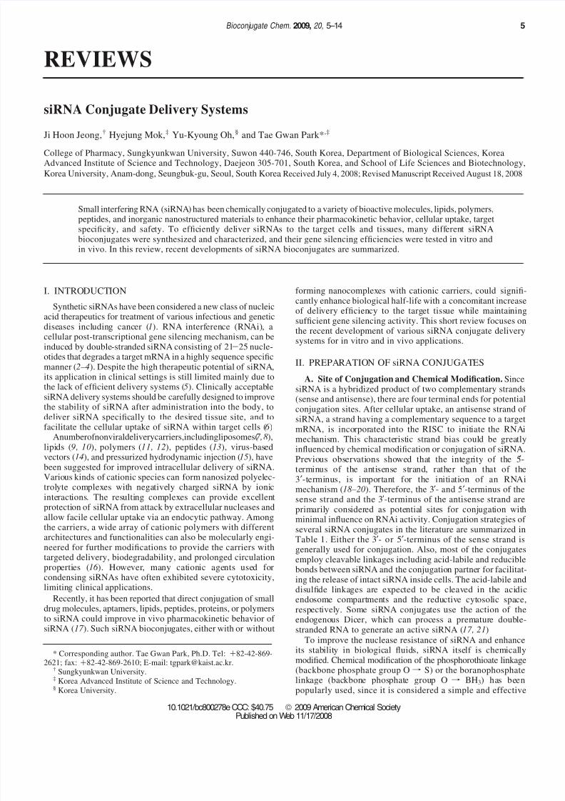
B. Lipophile-siRNA Conjugates. Cholesterol was co-valently conjugated to siRNA for systemic delivery (33).Cholesterol was conjugated to the 3′-terminus of the sense strandof siRNA via a pyrrolidone linkage. The cholesterol-siRNAconjugate (chol-siRNA) could induce intracellular RNAi withoutthe significant loss of gene silencing activity. In addition, theconjugate exhibited significantly higher cellular transfer ef-ficiency in cultured cells without the aid of transfection agents.
In animal experiments, the significant silencing of apoliporoteinB (apoB) gene, which encodes a protein essential for cholesterolmetabolism, was observed in the liver and the jejunum afterintravenous administration of the chol-siRNA conjugate. Thesilencing of the apoB gene resulted in a decreased plasma apoBprotein level and consequently a reduction in the total levels of cholesterol. The conjugation of cholesterol to siRNA improvedin vivo pharmacokinetic behaviors of siRNA as well. Intrave-nously administered chol-siRNA was distributed to varioustissues including the liver, heart, lungs, kidneys, and fat tissuesand was detected in the tissues even 24 h after injection ( 33).In contrast, no detectable naked siRNA could be observed inthe tissues 24 h after intravenous injection. In addition to thechol-siRNA conjugate, a series of lipophilic siRNA conjugates,
including siRNA conjugates with bile acids and lipids, weresynthesized (Figure 1) (34, 35). They interacted with lipoproteinsin the blood serum, such as high-density lipoprotein (HDL) andlow-density lipoprotein (LDL), lipoprotein receptors, and trans-membrane proteins, influencing tissue distribution and uptakebehaviors of the siRNA conjugates (34). The degree of hydrophobicity, which directly relates to the length of the alkylchain, seemed to be a major determinant for the affinity of siRNA-fatty acid conjugates to lipoproteins. The siRNAconjugates with higher affinity to lipoproteins, i.e., the ones withlonger fatty acid chains, showed enhanced gene silencingcapabilities, suggesting that lipoproteins may facilitate thecellular uptake of the conjugates. When systemically adminis-tered, chol-siRNA bound to HDL demonstrated ca. five times
higher cleavage of the target RNA transcript (apoB) in mouse,compared to unbound chol-siRNA at the same concentration.This suggests that the association of lipoproteins may furtherimprove the pharmacokinetic properties of lipophilic siRNAconjugates. The association of lipoprotein also changed tissuedistribution profiles of the conjugates. The lipophilic siRNAconjugates bound to LDL were mainly distributed to the liverby the action of LDL receptors, while the conjugates bound toHDL were taken up by various tissues, including the liver, gut,kidneys, and steroidogenic organs. The high affinity of HDL tothe scavenger receptor SR-BI plays an important role in theuptake of HDL-bound siRNA conjugates. Further optimizationmay be possible by modifying strategies for the formation of preassembly between the lipophilic siRNA conjugates and
lipoproteins or by identifying new lipophilic conjugate partnersthat lead to more favorable interaction with serum lipoproteins.
Although the animal results are encouraging, since the studyshowed the feasibility of using siRNAs as therapeutics, therequirement of a relatively high siRNA dose (50 mg/kg) remainsto be solved for clinical applications.
Another lipophile-siRNA conjugate, R-tocopherol (vitaminE)-siRNA, was introduced for systemic siRNA delivery to theliver (17 ). Lipophilic vitamin E was covalently conjugated tothe 5′-terminus of the antisense strand of 27/29-mer siRNA,which was partially modified with 2′-O-methylated ribose andphosphorothioate linkage. After intracellular delivery, the 27/ 29-mer siRNA was to be processed by the action of Dicer (36 )to generate 21/21-mer siRNA, which caused the simultaneousrelease of the vitamin E moiety. The intravenous administrationof the conjugate achieved a significant reduction of the targetprotein (apoB) in the liver without any induction of inflamma-tory interferons, such as interferon-R and interferon- β (17 ).
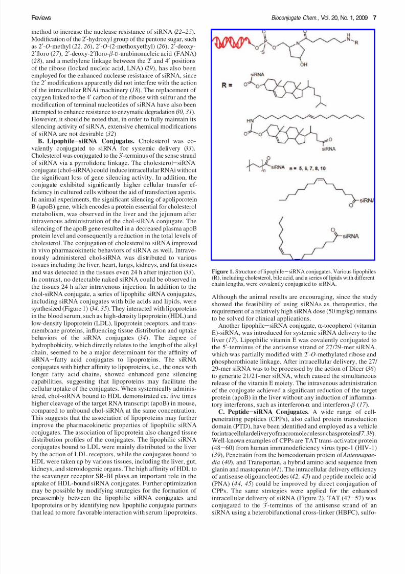
C. Peptide-siRNA Conjugates. A wide range of cell-penetrating peptides (CPPs), also called protein transductiondomain (PTD), have been identified and employed as a vehicleforintracellulardeliveryofmacromoleculessuchasproteins(37,38).Well-known examples of CPPs are TAT trans-activator protein(48-60) from human immunodeficiency virus type-1 (HIV-1)(39), Penetratin from the homeodomain protein of Antennapae-dia (40), and Transportan, a hybrid amino acid sequence fromglanin and mastoparan (41). The intracellular delivery efficiencyof antisense oligonucleotides (42, 43) and peptide nucleic acid(PNA) (44, 45) could be improved by direct conjugation of CPPs. The same strategies were applied for the enhancedintracellular delivery of siRNA (Figure 2). TAT (47-57) wasconjugated to the 3′-terminus of the antisense strand of ansiRNA using a heterobifunctional cross-linker (HBFC), sulfo-
Figure 1. Structure of lipophile-siRNA conjugates. Various lipophiles(R), including cholesterol, bile acid, and a series of lipids with differentchain lengths, were covalently conjugated to siRNA.
Reviews Bioconjugate Chem., Vol. 20, No. 1, 2009 7
8/6/2019 siRNA Conjugate Delivery System
http://slidepdf.com/reader/full/sirna-conjugate-delivery-system 4/10
succinimidyl4-( p-maleimidophenyl)butyrate(46 ).TheTAT-siRNAconjugate demonstrated a dramatic improvement in the intra-cellular delivery of siRNA. The extent of cellular uptake showeda direct relationship with the amount of conjugate used for thetransfection and the time elapsed after transfection. Cellularuptake of siRNA by the conjugate was as highly efficient asthe commercial Lipofectamine formulation. The TAT-siRNAconjugate also successfully elicited the effective silencing of the target gene. Although TAT was conjugated to the antisense
strand of siRNA via a noncleavable thioether linkage, thepresence of the peptide did not seem to seriously interfere withthe downstream process for the induction of RNAi, includingthe incorporation of TAT-siRNA into the RNA-inducedsilencing complex (RISC). The 5′-terminus of the antisensestrand of siRNA was considered crucial for induction of theRNAi mechanism (19, 20). Many research groups used acleavable linkage, such as the disulfide linkage, between CPPand siRNA to minimize potential reduction of RNAi activityby the presence of highly charged peptides. The disulfide linkagewas employed for the conjugation of CPPs, including Penetratinand Transportan, to the 5′-terminus of the sense strand of siRNA.This delivery system was designed to improve the intracellulardelivery of siRNA by the action of CPP and to release intact
siRNA by reductive cleavage of the disulfide linkage betweenthe CPP and the siRNA in the reductive cytoplasmic environ-ment. The reductive release of intact siRNA from the conjugatemay further facilitate the incorporation of siRNA into RISC byeliminating the potential interference of CPP. The CPP-siRNAconjugates, in which the disulfide linkage between the CPP andsiRNA was formed by oxidation of thiol groups in the presenceof oxidant, diamide, exhibited efficient suppression of the targetreporter gene expression, such as luciferase or green fluorescenceprotein (GFP), in a variety of cell lines (47 ). The gene silencingefficacy of the conjugates was comparable to those of a cationiclipid-based formulation. Similar conjugates of Penetratin 1peptide to siRNA, which also contain a disulfide linkagebetween the two components, were used for the delivery of
siRNA to primary mammalian neuronal cells (48). The conjugateshowed efficient down-regulation of the target genes, includingsuperoxide dismutase (SOD) variants and caspases, in theneuronal cells without apparent cytotoxicity. However, theconjugation of CPP to siRNA did not appear to improve siRNAstability. The conjugates of CPP and siRNA showed similardegradation profiles to those of naked siRNA after exposure tomouse bronchoalveolar lavage (BAL) fluid (49). Besides, somepeptide-siRNA conjugates elicited undesirable immune re-sponses. Although intratracheal delivery of siRNA usingCPP-siRNA conjugates successfully suppressed the expressionof a target gene, p38 MAP kinase, the siRNA conjugate of Penetratin but not TAT(48-60) caused elevated secretion of inflammatory cytokines, such as interferon-R, tumor necrosis
factor-R, and interleukin-12, after intratracheal distillation,suggesting the induction of innate immunity (49)
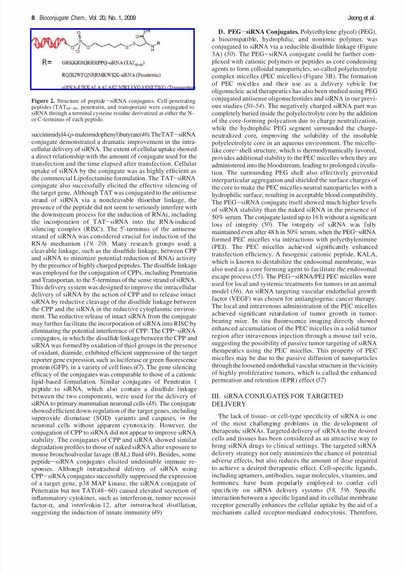
D. PEG-siRNA Conjugates. Poly(ethylene glycol) (PEG),
a biocompatible, hydrophilic, and nonionic polymer, was
conjugated to siRNA via a reducible disulfide linkage (Figure
3A) (50). The PEG-siRNA conjugate could be further com-
plexed with cationic polymers or peptides as core condensing
agents to form colloidal nanoparticles, so-called polyelectrolyte
complex micelles (PEC micelles) (Figure 3B). The formation
of PEC micelles and their use as a delivery vehicle for
oligonucleic acid therapeutics has also been studied using PEG
conjugated antisense oligonucleotides and siRNA in our previ-ous studies (50–54). The negatively charged siRNA part was
completely buried inside the polyelectrolyte core by the addition
of the core-forming polycation due to charge neutralization,
while the hydrophilic PEG segment surrounded the charge-
neutralized core, improving the solubility of the insoluble
polyelectrolyte core in an aqueous environment. The micelle-
like core-shell structure, which is thermodynamically favored,
provides additional stability to the PEC micelles when they are
administered into the bloodstream, leading to prolonged circula-
tion. The surrounding PEG shell also effectively prevented
interparticular aggregation and shielded the surface charges of
the core to make the PEC micelles neutral nanoparticles with a
hydrophilic surface, resulting in acceptable blood compatibility.
The PEG-siRNA conjugate itself showed much higher levelsof siRNA stability than the naked siRNA in the presence of
50% serum. The conjugate lasted up to 16 h without a significant
loss of integrity (50). The integrity of siRNA was fully
maintained even after 48 h in 50% serum, when the PEG-siRNA
formed PEC micelles via interactions with polyethylenimine
(PEI). The PEC micelles achieved significantly enhanced
transfection efficiency. A fusogenic cationic peptide, KALA,
which is known to destabilize the endosomal membrane, was
also used as a core forming agent to facilitate the endosomal
escape process (55). The PEG-siRNA/PEI PEC micelles wereused for local and systemic treatments for tumors in an animal
model (56 ). An siRNA targeting vascular endothelial growth
factor (VEGF) was chosen for antiangiogenic cancer therapy.The local and intravenous administration of the PEC micelles
achieved significant retardation of tumor growth in tumor-
bearing mice. In situ fluorescence imaging directly showed
enhanced accumulation of the PEC micelles in a solid tumor
region after intravenous injection through a mouse tail vein,
suggesting the possibility of passive tumor targeting of siRNA
therapeutics using the PEC micelles. This property of PEC
micelles may be due to the passive diffusion of nanoparticles
through the loosened endothelial vascular structure in the vicinityof highly proliferative tumors, which is called the enhanced
permeation and retention (EPR) effect (57 )
III. siRNA CONJUGATES FOR TARGETED
DELIVERY
The lack of tissue- or cell-type specificity of siRNA is one
of the most challenging problems in the development of
therapeutic siRNAs. Targeted delivery of siRNA to the desired
cells and tissues has been considered as an attractive way to
bring siRNA drugs to clinical settings. The targeted siRNA
delivery strategy not only minimizes the chance of potential
adverse effects, but also reduces the amount of dose required
to achieve a desired therapeutic effect. Cell-specific ligands,
including aptamers, antibodies, sugar molecules, vitamins, and
hormones, have been popularly employed to confer cell
specificity on siRNA delivery systems (58, 59). Specific
interaction between a specific ligand and its cellular membrane
receptor generally enhances the cellular uptake by the aid of amechanism called receptor-mediated endocytosis. Therefore,
Figure 2. Structure of peptide-siRNA conjugates. Cell-penetratingpeptides (TAT48-60, penetratin, and transportan) were conjugated tosiRNA through a terminal cysteine residue derivatized at either the N-or C-terminus of each peptide.
8 Bioconjugate Chem., Vol. 20, No. 1, 2009 Jeong et al.
8/6/2019 siRNA Conjugate Delivery System
http://slidepdf.com/reader/full/sirna-conjugate-delivery-system 5/10
increasing specific cellular uptake by targeted delivery cansignificantly improve therapeutic efficacy with a much lowerdose.
A. Ligand Peptide-siRNA Conjugates. For the receptor-ligand mediated delivery of siRNA, a ligand peptide for thetargeted receptor was conjugated to siRNA. A carboxylic acidgroup of peptide mimetic of IGF1, D-(Cys-Ser-Lys-Cys) wasactivated and conjugated to an amine group of the 5 ′-sensestrand of siRNA (60). After the annealing reaction, the genesilencing effect by an IGF1-siRNA conjugate was analyzedwithout the use of a transfection reagent. About 60% of thetarget IRS1 (insulin receptor substrate 1) gene expression wasinhibited by the IGF1-siRNA conjugate, which was similar tothat from the chol-siRNA conjugate.
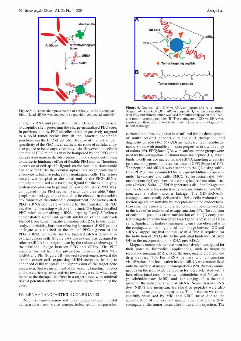
B. Antibody-siRNA Conjugates. Antibody-mediated tar-geted drug delivery systems have attracted much attention dueto their superior stability during systemic circulation and highselectiveness toward a target protein on the cell surface. APEGylated immunoliposome-based brain targeting system waspreviously introduced, in which two antibodies were im-mobilized on the surface of liposomes (61). One monoclonalantibody was to target a transferring receptor expressed in theblood brain barrier (BBB), and the other antibody was to targetinsulin receptors expressed in brain cancer. The binding of transferrin receptor antibody may induce the transcytosis of theimmunoliposome across the BBB. Similarly, the monoclonalantibody targeting the transferrin receptor at the BBB wasdirectly conjugated to siRNA via a biotin-streptavidin linkage(62). The siRNA biotinylated at either terminus (3′ or 5′) of the sense strand was coupled to the streptavidin-conjugatedantibody (Figure 5). The intravenous administration of theantibody-siRNA conjugate led to the efficient suppression of a reporter gene expression in a rat model bearing intracraniallytransplanted brain tumor.
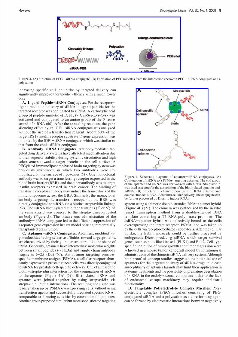
C. Aptamer-siRNA Conjugates. Aptamers, modified oli-gonucleotides having selective affinities toward target proteins,are characterized by their globular structure, like the shape of tRNA. Generally, aptamers have intermediate molecular weightsbetween small peptides (∼1 kDa) and single chain antibodyfragments (∼25 kDa) (63). An aptamer targeting prostate-specific membrane antigen (PSMA), a cellular receptor abun-dantly expressed in prostate cancer cells, was directly conjugatedto siRNA for prostate cell-specific delivery. Chu et al. used thebiotin-streptavidin interaction for the conjugation of siRNAto the aptamer (Figure 4A) (64). Biotinylated siRNA andaptamer were joined together by using streptavidin viastreptavidin-biotin interactions. The resulting conjugate wasreadily taken up by PSMA overexpressing cells without usingtransfection agents and successfully mediated specific RNAi,comparable to silencing activities by conventional lipoplexes.Another group proposed similar but more sophisticated targeting
system using a chimeric double-stranded RNA-aptamer hybrid(Figure 4B) (21). The chimera was synthesized by the in vitrorunoff transcription method from a double-stranded DNAtemplate containing a T7 RNA polymerase promoter. ThedsRNA-aptamer hybrid was selectively bound to the cellsoverexpressing the target receptor, PSMA, and was taken upby the cells via receptor-mediated endocytosis. After the cellularuptake, the hybrid molecule could be further processed byendogenous Dicer, producing siRNA which target survivalgenes, such as polo-like kinase 1 (PLK1) and Bcl-2. Cell-typespecific inhibition of tumor growth and tumor regression wereachieved in a mouse tumor xenograft model by intratumoraladministration of the chimeric siRNA delivery system. Althoughboth proof-of-concept studies suggested the potential use of aptamers for the targeted delivery of siRNA drugs, nucleasesusceptibility of aptamer ligands may limit their application insystemic treatments and the possibility of premature degradationof siRNA in the endolysosomal compartment due to the lack of endosomal escape machinery may require additionalfunctionality.
D. Targetable Polyelectrolyte Complex Micelles. Poly-electrolyte complex (PEC) micelles consisting of PEG-conjugated siRNA and a polycation as a core forming agentcan be formed by electrostatic interactions between negatively
Figure 3. (A) Structure of PEG-siRNA conjugate. (B) Formation of PEC micelles from the interactions between PEG-siRNA conjugate and apolycation.
Figure 4. Schematic diagrams of aptamer-siRNA conjugates. (A)Conjugation of siRNA to a PSMA-targeting aptamer. The end groupof the aptamer and siRNA was derivatized with biotin. Streptavidinwas used as a core for the association of the biotinylated aptamer andsiRNA. (B) Structure of chimeric conjugate of RNA aptamer anddouble-stranded siRNA. After intracellular delivery, the conjugate canbe further processed by Dicer to induce RNAi.
Reviews Bioconjugate Chem., Vol. 20, No. 1, 2009 9
8/6/2019 siRNA Conjugate Delivery System
http://slidepdf.com/reader/full/sirna-conjugate-delivery-system 6/10
charged siRNA and polycation. The PEG segment acts as ahydrophilic shell protecting the charge-neutralized PEC core.In previous studies, PEC micelles could be passively targetedto a solid tumor region through the loosened endothelial junctions via the EPR effect (56 ). Because of the lack of cellspecificity of the PEC micelles, the main route of cellular entryis expected to be adsorptive endocytosis. However, the cellularcontact of PEC micelles may be hampered by the PEG shellthat prevents nonspecific adsorption of blood components owingto the steric-hindrance effect of flexible PEG chains. Therefore,decoration of cell-specific ligands on the micelle surface wouldnot only facilitate the cellular uptake via receptor-mediatedendocytosis, but also reduce it by nontargeted cells. The lactose
moiety was coupled to the distal end of the PEG-siRNAconjugate and used as a targeting ligand for the asialoglyco-protein receptors on hepatoma cells (65, 66 ). An siRNA wasconjugated to the PEG segment via an acid-cleavable β-thio-propionate linkage and expected to be cleaved in the acidicenvironment of the endosomal compartment. The lactosylated-PEG-siRNA conjugate was used for the formation of PECmicelles by interacting with poly(L-lysine). The ligand installedPEC micelles containing siRNA targeting RecQL5 helicasedemonstrated significant growth inhibition of the spheroidsformed from human hepatocarcinoma cells (66 ). In our recentstudy, a luteinizing hormone-releasing hormone (LHRH) peptideanalogue was attached to the end of PEG segment of thePEG-siRNA conjugate for the targeted siRNA delivery to
ovarian cancer cells (Figure 7A) The system was designed torelease siRNA in the cytoplasm by the reductive cleavage of the disulfide linkage between PEG and siRNA. The PECmicelles formed from the interaction between LHRH-PEG-siRNA and PEI (Figure 7B) showed selectiveness toward theovarian cancer cells expressing LHRH receptors, leading toenhanced cellular uptake and suppression of the target geneexpression. Surface installment of cell-specific targeting moietiesonto the carriers gives selectivity toward target cells, which mayincrease the therapeutic effect in a target tissue with minimalrisk of potential adverse effect by reducing the amount of thedose.
IV. siRNA-NANOPARTICLE CONJUGATES
Recently, various nanosized imaging agents (quantum dotnanoparticles, iron oxide nanoparticles, gold nanoparticles,
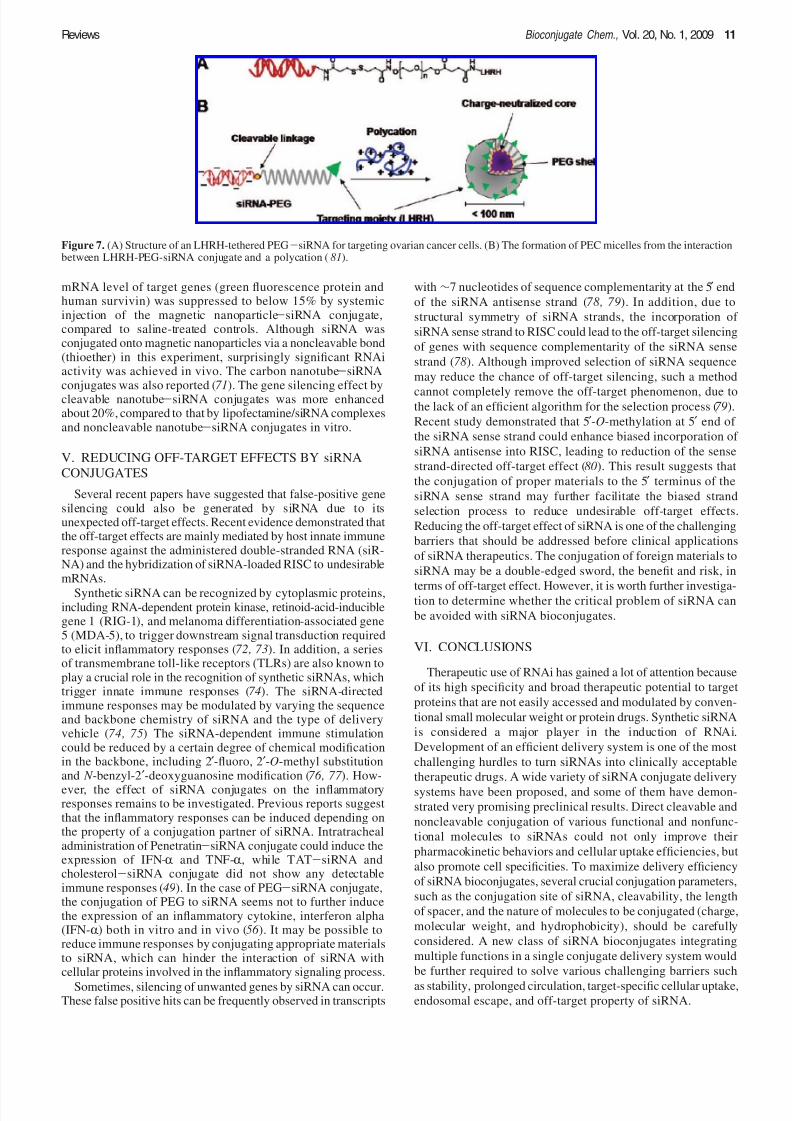
carbon nanotubes, etc.) have been utilized for the developmentof multifunctional nanoparticles for dual therapeutic anddiagnostic purposes (67, 68). QDs are fluorescent semiconductornanocrystals with tunable emission properties in a wide rangeof colors (69). PEGylated QDs with surface amine groups wereused for the conjugation of a tumor targeting peptide (F3), whichbinds to cell-surface nucleolin, and siRNA targeting a reportergene encoding green fluorescence protein (GFP) (Figure 6) (67 ).The peptide and siRNA was attached to the QD using sulfo-LC-SPDP (sulfosuccinimidyl 6-(3′-[2-pyridyldithio]-propiona-mido) hexanoate) and sulfo-SMCC (sulfosuccinimidyl 4-( N -maleimidomethyl) cyclohexane-1-carboxylate) as heterofunctionalcross-linkers. Sulfo-LC-SPDP generates a disulfide linkage thatcan be cleaved in the reductive cytoplasm, while sulfo-SMCCgenerates a stable thioether linkage. The QD-siRNA/F3conjugate successfully delivered to HeLa cells without trans-fection agents presumably by receptor-mediated endocytosis.However, the gene silencing effect could not be observed dueto the lack of an endosomal escape function ( 67 ). The additionof cationic liposomes after transfection of the QD conjugateled to significant reduction of the target gene expression in HeLacells. Significantly higher silencing efficiency was observed withthe conjugate containing a disulfide linkage between QD andsiRNA, suggesting that the release of siRNA is required forthe induction of RNAi due to the potential hindrance of largeQD in the incorporation of siRNA into RISC.
Magnetic nanoparticles have been intensively investigated fortheir potential biomedical applications, such as magneticresonance imaging (MRI), hyperthermia, magnetofection, anddrug delivery (70). For siRNA delivery with concomitantvisualization of its localization in vivo, siRNA was immobilizedonto the surface of magnetic nanoparticles (68). Primary aminegroups on the iron oxide nanoparticles were activated with aheterofunctional cross-linker, m-maleimidobenzoyl-N-hydrox-ysuccinimide ester (MBS), and then conjugated to the thiolgroup of the antisense strand of siRNA. Near-infrared Cy5.5dye (NIRF) and membrane translocation peptides were alsocoated onto magnetic nanoparticles. Tumor tissues were suc-cessfully visualized by MRI and NIRF image due to theaccumulation of the resultant magnetic nanoparticle-siRNAconjugate at the tumor tissue after intravenous injection. The
Figure 5. A schematic representation of antibody-siRNA conjugate.Biotinylated siRNA was coupled to streptavidin-conjugated antibody.
Figure 6. Quantum dot (QD)-siRNA conjugate. (A) A schematicdiagram of a targetable QD-siRNA conjugate. Quantum dot modifiedwith PEG and primary amine was used for further conjugation of siRNA
and tumor targeting peptide. (B) The conjugate of QD-siRNA wassynthesized through a reducible disulfide linkage or a nondegradablethioether linkage.
10 Bioconjugate Chem., Vol. 20, No. 1, 2009 Jeong et al.
8/6/2019 siRNA Conjugate Delivery System
http://slidepdf.com/reader/full/sirna-conjugate-delivery-system 7/10
mRNA level of target genes (green fluorescence protein andhuman survivin) was suppressed to below 15% by systemicinjection of the magnetic nanoparticle-siRNA conjugate,compared to saline-treated controls. Although siRNA wasconjugated onto magnetic nanoparticles via a noncleavable bond(thioether) in this experiment, surprisingly significant RNAiactivity was achieved in vivo. The carbon nanotube-siRNAconjugates was also reported (71). The gene silencing effect bycleavable nanotube-siRNA conjugates was more enhancedabout 20%, compared to that by lipofectamine/siRNA complexesand noncleavable nanotube-siRNA conjugates in vitro.
V. REDUCING OFF-TARGET EFFECTS BY siRNA
CONJUGATES
Several recent papers have suggested that false-positive genesilencing could also be generated by siRNA due to itsunexpected off-target effects. Recent evidence demonstrated thatthe off-target effects are mainly mediated by host innate immuneresponse against the administered double-stranded RNA (siR-NA) and the hybridization of siRNA-loaded RISC to undesirablemRNAs.
Synthetic siRNA can be recognized by cytoplasmic proteins,including RNA-dependent protein kinase, retinoid-acid-induciblegene 1 (RIG-1), and melanoma differentiation-associated gene5 (MDA-5), to trigger downstream signal transduction requiredto elicit inflammatory responses (72, 73). In addition, a seriesof transmembrane toll-like receptors (TLRs) are also known toplay a crucial role in the recognition of synthetic siRNAs, whichtrigger innate immune responses (74). The siRNA-directedimmune responses may be modulated by varying the sequenceand backbone chemistry of siRNA and the type of deliveryvehicle (74, 75) The siRNA-dependent immune stimulationcould be reduced by a certain degree of chemical modificationin the backbone, including 2′-fluoro, 2′-O-methyl substitutionand N -benzyl-2′-deoxyguanosine modification (76, 77 ). How-
ever, the effect of siRNA conjugates on the inflammatoryresponses remains to be investigated. Previous reports suggestthat the inflammatory responses can be induced depending onthe property of a conjugation partner of siRNA. Intratrachealadministration of Penetratin-siRNA conjugate could induce theexpression of IFN-R and TNF-R, while TAT-siRNA andcholesterol-siRNA conjugate did not show any detectableimmune responses (49). In the case of PEG-siRNA conjugate,the conjugation of PEG to siRNA seems not to further inducethe expression of an inflammatory cytokine, interferon alpha(IFN-R) both in vitro and in vivo (56 ). It may be possible toreduce immune responses by conjugating appropriate materialsto siRNA, which can hinder the interaction of siRNA withcellular proteins involved in the inflammatory signaling process.
Sometimes, silencing of unwanted genes by siRNA can occur.These false positive hits can be frequently observed in transcripts
with ∼7 nucleotides of sequence complementarity at the 5′ end
of the siRNA antisense strand (78, 79). In addition, due to
structural symmetry of siRNA strands, the incorporation of
siRNA sense strand to RISC could lead to the off-target silencing
of genes with sequence complementarity of the siRNA sense
strand (78). Although improved selection of siRNA sequence
may reduce the chance of off-target silencing, such a method
cannot completely remove the off-target phenomenon, due tothe lack of an efficient algorithm for the selection process (79).
Recent study demonstrated that 5′-O-methylation at 5′ end of
the siRNA sense strand could enhance biased incorporation of
siRNA antisense into RISC, leading to reduction of the sense
strand-directed off-target effect (80). This result suggests that
the conjugation of proper materials to the 5′ terminus of the
siRNA sense strand may further facilitate the biased strand
selection process to reduce undesirable off-target effects.
Reducing the off-target effect of siRNA is one of the challenging
barriers that should be addressed before clinical applications
of siRNA therapeutics. The conjugation of foreign materials to
siRNA may be a double-edged sword, the benefit and risk, in
terms of off-target effect. However, it is worth further investiga-tion to determine whether the critical problem of siRNA can
be avoided with siRNA bioconjugates.
VI. CONCLUSIONS
Therapeutic use of RNAi has gained a lot of attention because
of its high specificity and broad therapeutic potential to target
proteins that are not easily accessed and modulated by conven-
tional small molecular weight or protein drugs. Synthetic siRNA
is considered a major player in the induction of RNAi.
Development of an efficient delivery system is one of the most
challenging hurdles to turn siRNAs into clinically acceptable
therapeutic drugs. A wide variety of siRNA conjugate delivery
systems have been proposed, and some of them have demon-
strated very promising preclinical results. Direct cleavable and
noncleavable conjugation of various functional and nonfunc-
tional molecules to siRNAs could not only improve their
pharmacokinetic behaviors and cellular uptake efficiencies, but
also promote cell specificities. To maximize delivery efficiency
of siRNA bioconjugates, several crucial conjugation parameters,
such as the conjugation site of siRNA, cleavability, the length
of spacer, and the nature of molecules to be conjugated (charge,
molecular weight, and hydrophobicity), should be carefully
considered. A new class of siRNA bioconjugates integrating
multiple functions in a single conjugate delivery system would
be further required to solve various challenging barriers such
as stability, prolonged circulation, target-specific cellular uptake,endosomal escape, and off-target property of siRNA.
Figure 7. (A) Structure of an LHRH-tethered PEG-siRNA for targeting ovarian cancer cells. (B) The formation of PEC micelles from the interactionbetween LHRH-PEG-siRNA conjugate and a polycation (81).
Reviews Bioconjugate Chem., Vol. 20, No. 1, 2009 11

8/6/2019 siRNA Conjugate Delivery System
http://slidepdf.com/reader/full/sirna-conjugate-delivery-system 8/10
ACKNOWLEDGMENT
This work was supported by the Ministry of Health andWelfare, and the National Research Laboratory program fromthe Ministry of Education, and Science and Technology,Republic of Korea.
LITERATURE CITED
(1) Elbashir, S. M., Harborth, J., Lendeckel, W., Yalcin, A., Weber,K., and Tuschl, T. (2001) Duplexes of 21-nucleotide RNAsmediate RNA interference in cultured mammalian cells. Nature411, 494–8.
(2) McManus, M. T., and Sharp, P. A. (2002) Gene silencing inmammals by small interfering RNAs. Nat. ReV. Genet. 3, 737–47.
(3) Hannon, G. J., and Rossi, J. J. (2004) Unlocking the potentialof the human genome with RNA interference. Nature 431, 371–8.
(4) Caplen, N. J., Parrish, S., Imani, F., Fire, A., and Morgan, R. A.(2001) Specific inhibition of gene expression by small double-stranded RNAs in invertebrate and vertebrate systems. Proc. Natl. Acad. Sci. U.S.A. 98, 9742–7.
(5) Jeong, J. H., Kim, S. W., and Park, T. G. (2007) Molecular
design of functional polymers for gene therapy. Prog. Polym.Sci. 32, 1239–1274.
(6) Dorsett, Y., and Tuschl, T. (2004) siRNAs: applications infunctional genomics and potential as therapeutics. Nat. ReV. Drug DiscoVery 3, 318–29.
(7) Landen, C. N., Jr., Chavez-Reyes, A., Bucana, C., Schmandt,R., Deavers, M. T., Lopez-Berestein, G., and Sood, A. K. (2005)Therapeutic EphA2 gene targeting in vivo using neutral liposomalsmall interfering RNA delivery. Cancer Res. 65, 6910–8.
(8) Zimmermann, T. S., Lee, A. C., Akinc, A., Bramlage, B.,Bumcrot, D., Fedoruk, M. N., Harborth, J., Heyes, J. A., Jeffs,L. B., John, M., Judge, A. D., Lam, K., McClintock, K., Nechev,L. V., Palmer, L. R., Racie, T., Rohl, I., Seiffert, S., Shanmugam,S., Sood, V., Soutschek, J., Toudjarska, I., Wheat, A. J.,Yaworski, E., Zedalis, W., Koteliansky, V., Manoharan, M.,Vornlocher, H. P., and MacLachlan, I. (2006) RNAi-mediatedgene silencing in non-human primates. Nature 441, 111–4.
(9) Santel, A., Aleku, M., Keil, O., Endruschat, J., Esche, V.,Durieux, B., Loffler, K., Fechtner, M., Rohl, T., Fisch, G.,Dames, S., Arnold, W., Giese, K., Klippel, A., and Kaufmann,J. (2006) RNA interference in the mouse vascular endotheliumby systemic administration of siRNA-lipoplexes for cancertherapy. Gene Ther. 13, 1360–70.
(10) Yano, J., Hirabayashi, K., Nakagawa, S., Yamaguchi, T.,Nogawa, M., Kashimori, I., Naito, H., Kitagawa, H., Ishiyama,K., Ohgi, T., and Irimura, T. (2004) Antitumor activity of smallinterfering RNA/cationic liposome complex in mouse modelsof cancer. Clin. Cancer Res. 10, 7721–6.
(11) Kim, S. H., Mok, H., Jeong, J. H., Kim, S. W., and Park,
T. G. (2006) Comparative evaluation of target-specific GFP genesilencing efficiencies for antisense ODN, synthetic siRNA, andsiRNA plasmid complexed with PEI-PEG-FOL conjugate. Bio-conjugate Chem. 17 , 241–4.
(12) Urban-Klein, B., Werth, S., Abuharbeid, S., Czubayko, F.,and Aigner, A. (2005) RNAi-mediated gene-targeting throughsystemic application of polyethylenimine (PEI)-complexed siR-NA in vivo. Gene Ther. 12, 461–6.
(13) Mok, H., and Park, T. G. (2008) Self-crosslinked and reduciblefusogenic peptides for intracellular delivery of siRNA, Biopoly-mers.
(14) Grimm, D., Streetz, K. L., Jopling, C. L., Storm, T. A., Pandey,K., Davis, C. R., Marion, P., Salazar, F., and Kay, M. A. (2006)Fatality in mice due to oversaturation of cellular microRNA/ short hairpin RNA pathways. Nature 441, 537–41.
(15) Song, E., Lee, S. K., Wang, J., Ince, N., Ouyang, N., Min, J.,Chen, J., Shankar, P., and Lieberman, J. (2003) RNA interference
targeting Fas protects mice from fulminant hepatitis. Nat. Med.9, 347–51.
(16) Park, T. G., Jeong, J. H., and Kim, S. W. (2006) Current statusof polymeric gene delivery systems. Ad V. Drug DeliVery ReV.58, 467–86.
(17) Nishina, K., Unno, T., Uno, Y., Kubodera, T., Kanouchi, T.,Mizusawa, H., and Yokota, T. (2008) Efficient in vivo deliveryof siRNA to the liver by conjugation of alpha-tocopherol. Mol.Ther. 16 , 734–40.
(18) Chiu, Y. L., and Rana, T. M. (2003) siRNA function in RNAi:
a chemical modification analysis. RNA 9, 1034–48.(19) Khvorova, A., Reynolds, A., and Jayasena, S. D. (2003)
Functional siRNAs and miRNAs exhibit strand bias. Cell 115,209–16.
(20) Schwarz, D. S., Hutvagner, G., Du, T., Xu, Z., Aronin, N.,and Zamore, P. D. (2003) Asymmetry in the assembly of theRNAi enzyme complex. Cell 115, 199–208.
(21) McNamara, J. O., II, Andrechek, E. R., Wang, Y., Viles, K. D.,Rempel, R. E., Gilboa, E., Sullenger, B. A., and Giangrande,P. H. (2006) Cell type-specific delivery of siRNAs with aptamer-siRNA chimeras. Nat. Biotechnol. 24, 1005–15.
(22) Amarzguioui, M., Holen, T., Babaie, E., and Prydz, H. (2003)Tolerance for mutations and chemical modifications in a siRNA. Nucleic Acids Res. 31, 589–95.
(23) Braasch, D. A., Jensen, S., Liu, Y., Kaur, K., Arar, K., White,M. A., and Corey, D. R. (2003) RNA interference in mammaliancells by chemically-modified RNA. Biochemistry 42, 7967–75.
(24) Braasch, D. A., Paroo, Z., Constantinescu, A., Ren, G., Oz,O. K., Mason, R. P., and Corey, D. R. (2004) Biodistribution of phosphodiester and phosphorothioate siRNA. Bioorg. Med.Chem. Lett. 14, 1139–43.
(25) Hall, A. H., Wan, J., Shaughnessy, E. E., Ramsay Shaw, B.,and Alexander, K. A. (2004) RNA interference using borano-phosphate siRNAs: structure-activity relationships. Nucleic Acids Res. 32, 5991–6000.
(26) Prakash, T. P., Allerson, C. R., Dande, P., Vickers, T. A.,Sioufi, N., Jarres, R., Baker, B. F., Swayze, E. E., Griffey, R. H.,and Bhat, B. (2005) Positional effect of chemical modificationson short interference RNA activity in mammalian cells. J. Med.
Chem. 48, 4247–53.(27) Blidner, R. A., Hammer, R. P., Lopez, M. J., Robinson, S. O.,and Monroe, W. T. (2007) Fully 2′-deoxy-2′-fluoro substitutednucleic acids induce RNA interference in mammalian cell culture.Chem. Biol. Drug Des. 70, 113–22.
(28) Dowler, T., Bergeron, D., Tedeschi, A. L., Paquet, L., Ferrari,N., and Damha, M. J. (2006) Improvements in siRNA propertiesmediated by 2′-deoxy-2′-fluoro-beta-D-arabinonucleic acid(FANA). Nucleic Acids Res. 34, 1669–75.
(29) Elmen, J., Thonberg, H., Ljungberg, K., Frieden, M., West-ergaard, M., Xu, Y., Wahren, B., Liang, Z., Orum, H., Koch,T., and Wahlestedt, C. (2005) Locked nucleic acid (LNA)mediated improvements in siRNA stability and functionality. Nucleic Acids Res. 33, 439–47.
(30) Hoshika, S., Minakawa, N., Kamiya, H., Harashima, H., and
Matsuda, A. (2005) RNA interference induced by siRNAsmodified with 4′-thioribonucleosides in cultured mammaliancells. FEBS Lett. 579, 3115–8.
(31) Czauderna, F., Fechtner, M., Dames, S., Aygun, H., Klippel,A., Pronk, G. J., Giese, K., and Kaufmann, J. (2003) Structuralvariations and stabilising modifications of synthetic siRNAs inmammalian cells. Nucleic Acids Res. 31, 2705–16.
(32) Rossi, J. J. (2004) Medicine: a cholesterol connection in RNAi. Nature 432, 155–6.
(33) Soutschek, J., Akinc, A., Bramlage, B., Charisse, K., Constien,R., Donoghue, M., Elbashir, S., Geick, A., Hadwiger, P.,Harborth, J., John, M., Kesavan, V., Lavine, G., Pandey, R. K.,Racie, T., Rajeev, K. G., Rohl, I., Toudjarska, I., Wang, G.,Wuschko, S., Bumcrot, D., Koteliansky, V., Limmer, S., Mano-haran, M., and Vornlocher, H. P. (2004) Therapeutic silencingof an endogenous gene by systemic administration of modifiedsiRNAs. Nature 432, 173–8.
12 Bioconjugate Chem., Vol. 20, No. 1, 2009 Jeong et al.
8/6/2019 siRNA Conjugate Delivery System
http://slidepdf.com/reader/full/sirna-conjugate-delivery-system 9/10
(34) Wolfrum, C., Shi, S., Jayaprakash, K. N., Jayaraman, M.,Wang, G., Pandey, R. K., Rajeev, K. G., Nakayama, T., Charrise,K., Ndungo, E. M., Zimmermann, T., Koteliansky, V., Mano-haran, M., and Stoffel, M. (2007) Mechanisms and optimizationof in vivo delivery of lipophilic siRNAs. Nat. Biotechnol. 25,1149–57.
(35) Lorenz, C., Hadwiger, P., John, M., Vornlocher, H. P., andUnverzagt, C. (2004) Steroid and lipid conjugates of siRNAs toenhance cellular uptake and gene silencing in liver cells. Bioorg. Med. Chem. Lett. 14, 4975–7.
(36) Kim, D. H., Behlke, M. A., Rose, S. D., Chang, M. S., Choi,S., and Rossi, J. J. (2005) Synthetic dsRNA Dicer substratesenhance RNAi potency and efficacy. Nat. Biotechnol. 23, 222–6.
(37) Lindgren, M., Hallbrink, M., Prochiantz, A., and Langel, U.(2000) Cell-penetrating peptides. Trends Pharmacol. Sci. 21, 99–103.
(38) Prochiantz, A. (2000) Messenger proteins: homeoproteins,TAT and others. Curr. Opin. Cell Biol. 12, 400–6.
(39) Vives, E., Brodin, P., and Lebleu, B. (1997) A truncated HIV-1Tat protein basic domain rapidly translocates through the plasmamembrane and accumulates in the cell nucleus. J. Biol. Chem.272, 16010–7.
(40) Prochiantz, A. (1996) Getting hydrophilic compounds into
cells: lessons from homeopeptides. Curr. Opin. Neurobiol. 6 ,629–34.
(41) Pooga, M., Hallbrink, M., Zorko, M., and Langel, U. (1998)Cell penetration by transportan. Faseb J. 12, 67–77.
(42) Astriab-Fisher, A., Sergueev, D., Fisher, M., Shaw, B. R., andJuliano, R. L. (2002) Conjugates of antisense oligonucleotideswith the Tat and antennapedia cell-penetrating peptides: effectson cellular uptake, binding to target sequences, and biologicactions. Pharm. Res. 19, 744–54.
(43) Turner, J. J., Arzumanov, A. A., and Gait, M. J. (2005)Synthesis, cellular uptake and HIV-1 Tat-dependent trans-activation inhibition activity of oligonucleotide analogues dis-ulphide-conjugated to cell-penetrating peptides. Nucleic Acids Res. 33, 27–42.
(44) Turner, J. J., Ivanova, G. D., Verbeure, B., Williams, D.,
Arzumanov, A. A., Abes, S., Lebleu, B., and Gait, M. J. (2005)Cell-penetrating peptide conjugates of peptide nucleic acids(PNA) as inhibitors of HIV-1 Tat-dependent trans-activation incells. Nucleic Acids Res. 33, 6837–49.
(45) Tripathi, S., Chaubey, B., Ganguly, S., Harris, D., Casale,R. A., and Pandey, V. N. (2005) Anti-HIV-1 activity of anti-TAR polyamide nucleic acid conjugated with various membranetransducing peptides. Nucleic Acids Res. 33, 4345–56.
(46) Chiu, Y. L., Ali, A., Chu, C. Y., Cao, H., and Rana, T. M.(2004) Visualizing a correlation between siRNA localization,cellular uptake, and RNAi in living cells. Chem. Biol. 11, 1165–75.
(47) Muratovska, A., and Eccles, M. R. (2004) Conjugate forefficient delivery of short interfering RNA (siRNA) into mam-
malian cells. FEBS Lett. 558, 63–8.(48) Davidson, T. J., Harel, S., Arboleda, V. A., Prunell, G. F.,
Shelanski, M. L., Greene, L. A., and Troy, C. M. (2004) Highlyefficient small interfering RNA delivery to primary mammalianneurons induces MicroRNA-like effects before mRNA degrada-tion. J. Neurosci. 24, 10040–6.
(49) Moschos, S. A., Jones, S. W., Perry, M. M., Williams, A. E.,Erjefalt, J. S., Turner, J. J., Barnes, P. J., Sproat, B. S., Gait,M. J., and Lindsay, M. A. (2007) Lung delivery studies usingsiRNA conjugated to TAT(48-60) and penetratin reveal peptideinduced reduction in gene expression and induction of innateimmunity. Bioconjugate Chem. 18, 1450–9.
(50) Kim, S. H., Jeong, J. H., Lee, S. H., Kim, S. W., and Park,T. G. (2006) PEG conjugated VEGF siRNA for anti-angiogenicgene therapy. J. Controlled Release 116 , 123–9.
(51) Jeong, J. H., Kim, S. H., Kim, S. W., and Park, T. G. (2005)Polyelectrolyte complex micelles composed of c-raf antisense
oligodeoxynucleotide-poly(ethylene glycol) conjugate and poly-(ethylenimine): effect of systemic administration on tumorgrowth. Bioconjugate Chem. 16 , 1034–7.
(52) Jeong, J. H., Kim, S. W., and Park, T. G. (2003) A newantisense oligonucleotide delivery system based on self-as-sembled ODN-PEG hybrid conjugate micelles. J. Controlled Release 93, 183–91.
(53) Jeong, J. H., Kim, S. W., and Park, T. G. (2003) Novelintracellular delivery system of antisense oligonucleotide by self-assembled hybrid micelles composed of DNA/PEG conjugate
and cationic fusogenic peptide. Bioconjugate Chem. 14, 473–9.(54) Oishi, M., Nagatsugi, F., Sasaki, S., Nagasaki, Y., and
Kataoka, K. (2005) Smart polyion complex micelles for targetedintracellular delivery of PEGylated antisense oligonucleotidescontaining acid-labile linkages. ChemBioChem 6 , 718–25.
(55) Lee, S. H., Kim, S. H., and Park, T. G. (2007) IntracellularsiRNA delivery system using polyelectrolyte complex micellesprepared from VEGF siRNA-PEG conjugate and cationic fuso-genic peptide. Biochem. Biophys. Res. Commun. 357 , 511–6.
(56) Kim, S. H., Jeong, J. H., Lee, S. H., Kim, S. W., and Park,T. G. (2008) Local and systemic delivery of VEGF siRNA usingpolyelectrolyte complex micelles for effective treatment of cancer. J. Controlled Release 129, 107–16.
(57) Maeda, H., Wu, J., Sawa, T., Matsumura, Y., and Hori, K.
(2000) Tumor vascular permeability and the EPR effect inmacromolecular therapeutics: a review. J. Controlled Release65, 271–84.
(58) Wu, G., Yang, W., Barth, R. F., Kawabata, S., Swindall, M.,Bandyopadhyaya, A. K., Tjarks, W., Khorsandi, B., Blue, T. E.,Ferketich, A. K., Yang, M., Christoforidis, G. A., Sferra, T. J.,Binns, P. J., Riley, K. J., Ciesielski, M. J., and Fenstermaker,R. A. (2007) Molecular targeting and treatment of an epidermalgrowth factor receptor-positive glioma using boronated cetux-imab. Clin. Cancer Res. 13, 1260–8.
(59) Ikeda, Y., and Taira, K. (2006) Ligand-targeted delivery of therapeutic siRNA. Pharm. Res. 23, 1631–40.
(60) Cesarone, G., Edupuganti, O. P., Chen, C. P., and Wickstrom,E. (2007) Insulin receptor substrate 1 knockdown in humanMCF7 ER+ breast cancer cells by nuclease-resistant IRS1 siRNA
conjugated to a disulfide-bridged D-peptide analogue of insulin-like growth factor 1. Bioconjugate Chem. 18, 1831–40.
(61) Zhang, Y., Zhang, Y. F., Bryant, J., Charles, A., Boado, R. J.,and Pardridge, W. M. (2004) Intravenous RNA interference genetherapy targeting the human epidermal growth factor receptorprolongs survival in intracranial brain cancer. Clin. Cancer Res.10, 3667–77.
(62) Xia, C. F., Zhang, Y., Zhang, Y., Boado, R. J., and Pardridge,W. M. (2007) Intravenous siRNA of brain cancer with receptortargeting and avidin-biotin technology. Pharm. Res. 24, 2309–16.
(63) Hicke, B. J., and Stephens, A. W. (2000) Escort aptamers: adelivery service for diagnosis and therapy. J. Clin. InVest. 106 ,923–8.
(64) Chu, T. C., Twu, K. Y., Ellington, A. D., and Levy, M. (2006)Aptamer mediated siRNA delivery. Nucleic Acids Res. 34, e73.
(65) Oishi, M., Nagasaki, Y., Itaka, K., Nishiyama, N., andKataoka, K. (2005) Lactosylated poly(ethylene glycol)-siRNAconjugate through acid-labile beta-thiopropionate linkage toconstruct pH-sensitive polyion complex micelles achievingenhanced gene silencing in hepatoma cells. J. Am. Chem. Soc.127 , 1624–5.
(66) Oishi, M., Nagasaki, Y., Nishiyama, N., Itaka, K., Takagi,M., Shimamoto, A., Furuichi, Y., and Kataoka, K. (2007)Enhanced growth inhibition of hepatic multicellular tumorspheroids by lactosylated poly(ethylene glycol)-siRNA conjugateformulated in PEGylated polyplexes. ChemMedChem 2, 1290–1297.
(67) Derfus, A. M., Chen, A. A., Min, D. H., Ruoslahti, E., andBhatia, S. N. (2007) Targeted quantum dot conjugates for siRNAdelivery. Bioconjugate Chem. 18, 1391–6.
Reviews Bioconjugate Chem., Vol. 20, No. 1, 2009 13
8/6/2019 siRNA Conjugate Delivery System
http://slidepdf.com/reader/full/sirna-conjugate-delivery-system 10/10
(68) Medarova, Z., Pham, W., Farrar, C., Petkova, V., and Moore,A. (2007) In vivo imaging of siRNA delivery and silencing intumors. Nat. Med. 13, 372–7.
(69) Akerman, M. E., Chan, W. C., Laakkonen, P., Bhatia, S. N.,and Ruoslahti, E. (2002) Nanocrystal targeting in vivo. Proc. Natl. Acad. Sci. U.S.A. 99, 12617–21.
(70) Gupta, A. K., and Gupta, M. (2005) Synthesis and surfaceengineering of iron oxide nanoparticles for biomedical applica-tions. Biomaterials 26 , 3995–4021.
(71) Kam, N. W., Liu, Z., and Dai, H. (2005) Functionalization of
carbon nanotubes via cleavable disulfide bonds for efficientintracellular delivery of siRNA and potent gene silencing. J. Am.Chem. Soc. 127 , 12492–3.
(72) Sioud, M. (2007) RNA interference and innate immunity. Ad V. Drug DeliVery ReV. 59, 153–63.
(73) Kato, H., Takeuchi, O., Sato, S., Yoneyama, M., Yamamoto,M., Matsui, K., Uematsu, S., Jung, A., Kawai, T., Ishii, K. J.,Yamaguchi, O., Otsu, K., Tsujimura, T., Koh, C. S., Reis e Sousa,C., Matsuura, Y., Fujita, T., and Akira, S. (2006) Differentialroles of MDA5 and RIG-I helicases in the recognition of RNAviruses. Nature 441, 101–5.
(74) Judge, A. D., Sood, V., Shaw, J. R., Fang, D., McClintock,K., and MacLachlan, I. (2005) Sequence-dependent stimulationof the mammalian innate immune response by synthetic siRNA. Nat. Biotechnol. 23, 457–62.
(75) Judge, A., and MacLachlan, I. (2008) Overcoming the innateimmune response to small interfering RNA. Hum. Gene Ther.19, 111–24.
(76) Judge, A. D., Bola, G., Lee, A. C., and MacLachlan, I. (2006)Design of noninflammatory synthetic siRNA mediating potentgene silencing in vivo. Mol. Ther. 13, 494–505.
(77) Puthenveetil, S., Whitby, L., Ren, J., Kelnar, K., Krebs, J. F.,and Beal, P. A. (2006) Controlling activation of the RNA-dependent protein kinase by siRNAs using site-specific chemicalmodification. Nucleic Acids Res. 34, 4900–11.
(78) Jackson, A. L., Bartz, S. R., Schelter, J., Kobayashi, S. V.,Burchard, J., Mao, M., Li, B., Cavet, G., and Linsley, P. S. (2003)
Expression profiling reveals off-target gene regulation by RNAi. Nat. Biotechnol. 21, 635–7.
(79) Jackson, A. L., Burchard, J., Schelter, J., Chau, B. N., Cleary,M., Lim, L., and Linsley, P. S. (2006) Widespread siRNA “off-target” transcript silencing mediated by seed region sequencecomplementarity. RNA 12, 1179–87.
(80) Chen, P. Y., Weinmann, L., Gaidatzis, D., Pei, Y., Zavolan,M., Tuschl, T., and Meister, G. (2008) Strand-specific 5′-O-methylation of siRNA duplexes controls guide strand selectionand targeting specificity. RNA 14, 263–74.
(81) Kim, S. H., Jeong, J. H., Lee, S. H., Kim, S. W., and Park,T. G. (2008) LHRH receptor-mediated delivery of siRNA usingpolyelectrolyte complex micelles self-assembled from siRNA-PEG-LHRH conjugate and PEI. Bioconjugate Chem. 19,
2156–2162.
BC800278E
14 Bioconjugate Chem., Vol. 20, No. 1, 2009 Jeong et al.