bsa nanoparticles for sirna delivery: coating effects on
TRANSCRIPT

Chapman UniversityChapman University Digital Commons
Pharmacy Faculty Articles and Research School of Pharmacy
2012
BSA Nanoparticles for siRNA Delivery: CoatingEffects on Nanoparticle Properties, Plasma ProteinAdsorption, and In Vitro siRNA DeliveryHaran YogasundaramUniversity of Alberta
Markian Stephan BahniukUniversity of Alberta
Harsh-Deep SinghUniversity of Alberta
Hamidreza Montazeri AliabadiChapman University, [email protected]
Hasan UludagUniversity of Alberta
See next page for additional authorsFollow this and additional works at: http://digitalcommons.chapman.edu/pharmacy_articles
Part of the Medical Genetics Commons, Nanomedicine Commons, Other Chemicals and DrugsCommons, Pharmaceutical Preparations Commons, and the Pharmaceutics and Drug DesignCommons
This Article is brought to you for free and open access by the School of Pharmacy at Chapman University Digital Commons. It has been accepted forinclusion in Pharmacy Faculty Articles and Research by an authorized administrator of Chapman University Digital Commons. For more information,please contact [email protected].
Recommended CitationYogasundaram, Haran, Markian Stephan Bahniuk, Harsh-Deep Singh, Hamidreza Montezari Aliabadi, Hasan Uludaǧ, and Larry DavidUnsworth. "BSA Nanoparticles for siRNA Delivery: Coating Effects on Nanoparticle Properties, Plasma Protein Adsorption, and InVitro siRNA Delivery." International journal of biomaterials 2012 (2012). doi: 10.1155/2012/584060

BSA Nanoparticles for siRNA Delivery: Coating Effects on NanoparticleProperties, Plasma Protein Adsorption, and In Vitro siRNA Delivery
CommentsThis article was originally published in International Journal of Biomaterials, volume 2012, in 2012. DOI:10.1155/2012/584060
Creative Commons License
This work is licensed under a Creative Commons Attribution 3.0 License.
CopyrightThe authors
AuthorsHaran Yogasundaram, Markian Stephan Bahniuk, Harsh-Deep Singh, Hamidreza Montazeri Aliabadi, HasanUludag, and Larry David Unsworth
This article is available at Chapman University Digital Commons: http://digitalcommons.chapman.edu/pharmacy_articles/147

Hindawi Publishing CorporationInternational Journal of BiomaterialsVolume 2012, Article ID 584060, 10 pagesdoi:10.1155/2012/584060
Research Article
BSA Nanoparticles for siRNA Delivery: Coating Effects onNanoparticle Properties, Plasma Protein Adsorption, andIn Vitro siRNA Delivery
Haran Yogasundaram,1, 2 Markian Stephan Bahniuk,1, 2 Harsh-Deep Singh,1
Hamidreza Montezari Aliabadi,1 Hasan Uludag,1, 3 and Larry David Unsworth1, 2, 3
1 Department of Chemical and Materials Engineering, Faculty of Engineering, University of Alberta, Edmonton, AB, Canada T6G 2G62 National Research Council, National Institute for Nanotechnology, Edmonton, AB, Canada T6G 2M93 Department of Biomedical Engineering, Faculty of Medicine and Dentistry, University of Alberta, Edmonton, AB, Canada T6G 2G6
Correspondence should be addressed to Hasan Uludag, [email protected] and Larry David Unsworth, [email protected]
Received 12 April 2012; Accepted 21 June 2012
Academic Editor: Esmaiel Jabbari
Copyright © 2012 Haran Yogasundaram et al. This is an open access article distributed under the Creative Commons AttributionLicense, which permits unrestricted use, distribution, and reproduction in any medium, provided the original work is properlycited.
Developing vehicles for the delivery of therapeutic molecules, like siRNA, is an area of active research. Nanoparticles composedof bovine serum albumin, stabilized via the adsorption of poly-L-lysine (PLL), have been shown to be potentially inert drug-delivery vehicles. With the primary goal of reducing nonspecific protein adsorption, the effect of using comb-type structures ofpoly(ethylene glycol) (1 kDa, PEG) units conjugated to PLL (4.2 and 24 kDa) on BSA-NP properties, apparent siRNA release rate,cell viability, and cell uptake were evaluated. PEGylated PLL coatings resulted in NPs with ζ-potentials close to neutral. Incubationwith platelet-poor plasma showed the composition of the adsorbed proteome was similar for all systems. siRNA was effectivelyencapsulated and released in a sustained manner from all NPs. With 4.2 kDa PLL, cellular uptake was not affected by the presenceof PEG, but PEG coating inhibited uptake with 24 kDa PLL NPs. Moreover, 24 kDa PLL systems were cytotoxic and this cytotoxicitywas diminished upon PEG incorporation. The overall results identified a BSA-NP coating structure that provided effective siRNAencapsulation while reducing ζ-potential, protein adsorption, and cytotoxicity, necessary attributes for in vivo application of drug-delivery vehicles.
1. Introduction
Short interfering RNA (siRNA) is extremely promising forthe therapeutic treatment of a myriad of diseases; however,its clinical application has hitherto been hindered by anapparent inability to control its delivery. The use of NPbased drug delivery vehicles presents several advantages overconventional delivery stratagems, including the fact that theymay be used for precise tissue targeting, remain in bloodfor a prolonged time, and be immediately injected into thesystemic circulation. Furthermore, favorable tissue responseshave been observed for decreasing particle sizes [1] and amultitude of covalent and noncovalent modifications of NPsurfaces can be achieved, aspects that facilitate the design ofmore effective carriers. In particular, BSA-based NPs have
many advantageous qualities [2]: presence of a hydrophobiccore facilitating delivery of hydrophobic drugs, a naturalabundance in plasma, relative stability and inertness inbiochemical pathways, availability, and a relatively benign invivo biological fate [3]. Unlike NPs fabricated from syntheticpolymers, it is thought that the natural protein removalmechanisms will result in a reduced overall toxicity relatedto the application of BSA NPs [3]. That said, an importantstep in facilitating the localization of these NPs at the siteof interest involves both decreasing their removal from thecirculation (i.e., decreasing opsonization) as well as ensuringthat any targeting moiety remains able to interact withthe cellular site of interest. Inhibiting nonspecific proteinadsorption will then be central to both of these effects.A common strategy for preventing protein adsorption at

2 International Journal of Biomaterials
the tissue-material interface is to incorporate end-tetheredPEG to the surfaces of biomaterials. It has been well estab-lished that the presence of end-tethered PEG can preventparticulate aggregation, reduce interactions with plasmaproteins [4], minimize reticuloendothelial system clearance,and prolong blood circulation time of a host of NPs [5–7].
PEGylation of surfaces has been shown to impede non-specific protein adsorption [8], where both the presenceand conformation of end-tethered PEG play a critical role[9, 10]. It is noteworthy that not only is the amount ofplasma protein adsorbed at the tissue-biomaterial interfaceimportant in obfuscating an engineered surface but also thecomposition of the protein layer itself is critical, as this mayultimately direct host responses. NP opsonization has beencorrelated to surface properties, including hydrophilicity,roughness, ζ-potential, and surface chemistry [11]. Recentresults [12] have shown that systemic administration ofBSA NPs, stabilized with polyethyleneimine-graft-PEG withbisphosphonic acid attached for bone targeting, showedno beneficial effects associated with the polymer coating.Although the reason for this result was not fully elucidated,it was postulated that the biodistribution of the NPs may beaffected by the presence of the adsorbed protein corona tothe PEG modified NPs; it is hoped that further analyzingthe adsorbed protein composition to these PEG modifiedBSA NPs may clarify this point. Other previous work [13]has looked specifically at the use of positively charged poly-L-lysine (PLL) as a coating polymer that stabilizes the NPused for the apparent release of siRNA from BSA NPs. Itwas observed that, for low concentrations of PLL, varyingthe size of the PLL used for coating resulted in minimaleffect on the net release of siRNA from the NPs. In furtherwork [14], the release of a model drug from BSA NPs couldbe controlled from ∼5 to 90% over 14 days, depending onthe nature of coating designed to display differential stabilityagainst endogenous enzymes.
In this study, we continued the development of BSA NPsby exploring the role of PEG coating by employing comblikestructures of PEG-conjugated PLLs and compared systemsstabilized via unmodified PLL. Specifically, PLLs of 4.2 and24 kDa were utilized to understand the effect molecularweight may have on critical issues related to NP stabilization,siRNA encapsulation and passive release kinetics, plasmaprotein adsorption, cytotoxicity, and cellular incorporation.Conjugates of PLLs with 1 kDa PEG were synthesized so asto determine if any direct effect on NP stabilization as wellas siRNA encapsulation and passive release kinetics mightbe altered. The cellular uptake of the NPs and the plasmaprotein adsorption profile were assessed, investigating therole of PEG coating on these features. Our results identifiedspecific types of BSA NPs that provided adequate siRNArelease and cellular uptake with relatively low amounts ofprotein adsorption and cytotoxicity.
2. Materials and Methods
BSA and HBr salt of PLLs of different MWs (4.2 and 24 kDa)were purchased from Sigma-Aldrich (St. Louis, MO, USA)
and used without further purification. The sodium dodecylsulfate (SDS) was obtained from J. T. Baker (Phillipsburg,NJ, USA). FAM-labelled siRNA (double stranded, 21 basepairs) was purchased from Ambion Inc. (Austin, TX, USA).EDTA/trypsin (10X; Invitrogen, Carlsbad, CA, USA) wasdiluted 1 : 10 with Hank’s Buffered Salt Solution (HBSS;Invitrogen) to 0.05 g/L concentration before use. Dulbecco’sModified Eagle Medium (DMEM; high glucose), and pen-icillin/streptomycin (10000 U/mL/10 mg/mL) solution wereobtained from Invitrogen. Fetal bovine serum (FBS) wasfrom PAA Laboratories (Etobicoke, Ontario, Canada). So-dium phosphate, monobasic, monohydrate sodium phos-phate, and sodium chloride laboratory-grade reagents werepurchased from EMD Chemical Inc. (Darmstadt, Germany).Ethanol was purchased from Fischer Scientific (Ottawa, On-tario, Canada). The N-hydroxysuccinimide ester of 1 kDa(mPEG-NHS) was obtained from Creative PEG works, NC,USA. The 3,3′,5,5′-tetramethylbenzidine substrate (TMBS)was obtained from Promega (Madison, WI, USA). Thedialysis tubing of various MW cutoffs was obtained fromSpectrum Laboratories Inc. (Rancho Dominguez, CA, USA).
2.1. PEG Conjugation to PLL. Previously reported methodswere used to conjugate PLL to 1 kDa PEG [15]. Briefly, PLLwas dissolved in 0.1 M phosphate buffer (PB, pH 7.4) to afinal concentration of 2.4 mg/mL. mPEG-NHS was dilutedto 18 mM using 0.1 M PB, added to the PLL solution, andreacted for 3 hrs at room temperature. Solution was dialyzed2 days against MilliQ (18Ω) water, at 4◦C; MilliQ water beingchanged twice a day. Dialysate containing 4.2 and 24 kDaPLL was freeze-dried and used to reconstitute the polymersolutions at desired concentrations.
2.2. NP Preparation. BSA NPs were formed via a coacerva-tion method as detailed elsewhere [13]. Briefly, 250 μL of10 mg/mL BSA in water was added dropwise to an equalvolume of 10 mM NaCl in water at room temperature. After15 min stirring (600 rpm, room temperature), ethanol wasadded dropwise to a final volume ratio of 6 : 1, ethanol toBSA solution. The mixture was stirred for 3 hrs to form NPs.To stabilize formed NPs, native PLLs (0.3 mg/mL) or PEG-conjugated PLLs (0.3 mg PLL equivalents/mL) in deionizedwater were introduced dropwise to the equal volume of NPssuspension under constant stirring. Stirring was continuedfor 1 hr at room temperature to ensure time for the PLLand PEG-PLL conjugates to adsorb to the BSA NP surface.Suspensions of coated NPs were dialyzed against ddH2Ofor 3 days (12 hrs between solution replacements). Whereindicated, the amount of coating on the surface was deter-mined by using the FITC-labeled PLL or PEG-PLL. The poly-mer labeling was achieved by incubating 10 μL of 0.1 mMFITC solution (in DMSO) with 1 mL of PLL or PEG-PLL(2 mg/mL in 100 mM phosphate buffer, pH = 7.4) for 1 hrat room temperature. Ethanol (9 mL) was then added to thissolution. The solution then was centrifuged at room tem-perature (3000 rpm) for 15 minutes, and the supernatant,with unconjugated FITC, was removed. The pellet formedduring this process was further washed with 5 mL of ethanol

International Journal of Biomaterials 3
and centrifuged at 3000 rpm for 15 min [13]. The solidsobtained were air-dried under vacuum for 5 hrs and storedin the dark at 4◦C until used. The coating of FITC-labeledpolymer was determined using fluorescence measurements.The coated NPs suspension was then diluted with phosphatebuffer (pH = 7.4) by 100% and centrifuged (15,000 rpm,1 hr) and fluorescence (λEX: 485 nm; λEM: 527 nm) of thesupernatant was analyzed using a multiwall plate reader(Thermo Labsystems, Franklin, MA, USA). A calibrationcurve generated was used to calculate the coating efficiencyas (1 − FITC-polymersupernatant/(FITC-polymersupernatant +FITC-polymerpellet))× 100%.
2.3. Particle Sizing and ζ-Potential. Mean particle size of thecoated and uncoated NPs were determined using dynamiclight scattering (Zetasizer 3000 HS, Malvern InstrumentsLtd., UK) with a 633 nm He-Ne laser at a scattering angleof 90◦. Uncoated BSA NPs were used directly for themeasurements while the coated BSA NPs were diluted 1 : 2with PB (10 mM, pH 7.4). Intensity measurements wereused to determine the NP size. The ζ-potential of the NPswas determined by measuring their electrophoretic mobilityusing the same instrument at 25◦C.
2.4. Plasma Incubation and Elution. Human blood plasmawas obtained from Canadian Blood Services, where Cana-dian Blood Services obtained written informed consentfrom all volunteers for the collection and distribution ofhuman blood products for research purposes. Human bloodproducts were then shipped to our lab and used followingresearch ethics procedures as approved by the Universityof Alberta Research Ethics Board (Institutional), CanadianBlood Services Research Ethics Board (Federal), and theNational Research Council of Canada Research Ethics Board(Federal). NP solutions (500 μL) were combined with anequal volume of undiluted human plasma and incubated atroom temperature for 2 hrs with gentle rocking. The sampleswere then centrifuged at 13000 rpm for 10 minutes and thesupernatants discarded. Pellets were resolubilized in 1 mLof 0.15 M phosphate buffered saline (PBS; pH 7.4) for 30minutes and the procedure was repeated two times. Thesesamples were spun down and pellets were solubilized in 2%w/v SDS in 0.15 M PBS for one hr, with rocking at roomtemperature in order to elute adsorbed plasma proteins.To separate the NPs from the eluted plasma proteins, thesamples were centrifuged at 13000 rpm for 10 minutes andthe supernatants collected and characterized using SDS-PAGE and immunoblotting analysis.
2.5. SDS-PAGE and Immunoblotting. Reduced SDS-PAGEand immunoblotting techniques were used to evaluate andto identify eluted proteins as described previously [9]. Allelectrophoretic apparatus were purchased from Bio-Rad(Hercules, CA). Briefly, samples were separated on 12% SDS-PAGE gels and then transferred onto a 0.2 μm immunoblotPVDF membrane. The membrane was cut into strips fortotal protein staining using colloidal gold (Bio-Rad) andfor immunoblotting. Primary and secondary antibodies (see
Table S1 in Supplementary Materials available online atdoi:10.1155/2012/584060) were used without further purifi-cation at concentrations of 1 : 1000. To visualize protein-antibody complexes, 350 μL of stabilized TMBS substrate wasincubated with membrane strips for 10 minutes at roomtemperature, with rocking. The colour-developing reactionwas then quenched for 10 minutes using 2 mL of MilliQwater.
2.6. Preparation of siRNA Loaded BSA NPs and Release Stud-ies. The coacervation technique previously described wasemployed to prepare BSA NPs encapsulating siRNA. Briefly,500 μL of aqueous solution of BSA (10 mg/mL) was addedto an equal volume of 10 mM NaCl solution under stirring(600 rpm) in glass vials. The stirring was continued for 15minutes at room temperature. To achieve siRNA encapsu-lation, siRNA solution (20 μL of 0.15 mg/mL) was added tothis binary solution and stirred at 600 rpm for 1 hr at roomtemperature. The siRNA (scrambled) used for encapsulationwas either unlabeled or labeled with FAM. NPs were thenformed by adding ethanol dropwise (final volume ratio ofethanol to starting BSA solution = 6). Stirring was continuedfor 3 hrs at room temperature after the complete additionof ethanol. To coat these siRNA loaded BSA NPs with PLL’s,500 μL of an aqueous polymer solution was added dropwiseto an equal volume of BSA NPs suspension under constantshaking of 500 rpm. Shaking was continued for 1 hr.
To obtain encapsulation efficiency, NPs containing FAM-labeled siRNA were centrifuged at 15000 rpm for 30 minutes.The FAM-labeled siRNA in the supernatant and pelletwere determined using a plate reader (λEX: 485 nm, λEM:527 nm) and a calibration curve based on known concen-trations of FAM-labeled siRNA. The encapsulation efficiencywas calculated as (FAM-siRNApellet/(FAM-siRNApellet +FAM-siRNAsupernatant))× 100%. FAM-labeled siRNA wasalso used to study the apparent release kinetics of siRNA fromBSA NPs coated with different polymers. Polymer concen-tration of 0.3 mg/mL was used to coat BSA NPs and releasevalues were normalized to 0% for day 0, as described previ-ously [13]. The suspensions were incubated at 37◦C in PBSunder shaking and aliquots were taken at predetermined timepoints and centrifuged at 15000 rpm for 30 minutes. ThesiRNA in the supernatant and pellet were determined usinga calibration curve.
2.7. Cell Uptake Studies. To assess cellular uptake of NPs,human breast cancer MDA-231 cells were used. Two setsof NPs were prepared for cell uptake; (i) uncoated andcoated BSA NPs with no siRNA, and (ii) uncoated andcoated BSA NPs loaded with FAM-labeled siRNA. The siRNAencapsulation was achieved as described above, except that20 μL of siRNA solution (0.15 mg/mL) was used for FAM-labeled siRNA encapsulation. 1000 μL of aqueous polymersolution was added to 2000 μL of BSA NPs suspension (finalconcentration: 0.3 mg/mL) under shaking for 1 hr to achievecoating. The suspensions were then dialyzed against DMEMfor 24 hrs with two changes in dialysis solution.
For uptake, a monolayer of MDA-MB-231 cells wereseeded in 24-well plates and allowed to attach for 24 hrs
4 International Journal of Biomaterials
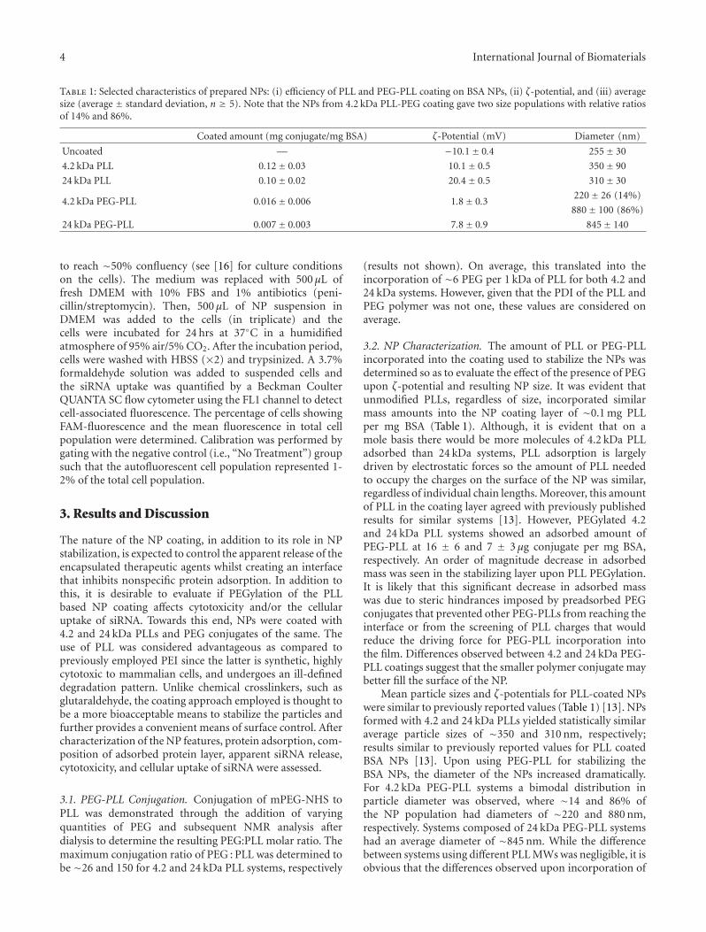
Table 1: Selected characteristics of prepared NPs: (i) efficiency of PLL and PEG-PLL coating on BSA NPs, (ii) ζ-potential, and (iii) averagesize (average ± standard deviation, n ≥ 5). Note that the NPs from 4.2 kDa PLL-PEG coating gave two size populations with relative ratiosof 14% and 86%.
Coated amount (mg conjugate/mg BSA) ζ-Potential (mV) Diameter (nm)
Uncoated — −10.1± 0.4 255± 30
4.2 kDa PLL 0.12± 0.03 10.1± 0.5 350± 90
24 kDa PLL 0.10± 0.02 20.4± 0.5 310± 30
4.2 kDa PEG-PLL 0.016± 0.006 1.8± 0.3220± 26 (14%)
880± 100 (86%)
24 kDa PEG-PLL 0.007± 0.003 7.8± 0.9 845± 140
to reach ∼50% confluency (see [16] for culture conditionson the cells). The medium was replaced with 500 μL offresh DMEM with 10% FBS and 1% antibiotics (peni-cillin/streptomycin). Then, 500 μL of NP suspension inDMEM was added to the cells (in triplicate) and thecells were incubated for 24 hrs at 37◦C in a humidifiedatmosphere of 95% air/5% CO2. After the incubation period,cells were washed with HBSS (×2) and trypsinized. A 3.7%formaldehyde solution was added to suspended cells andthe siRNA uptake was quantified by a Beckman CoulterQUANTA SC flow cytometer using the FL1 channel to detectcell-associated fluorescence. The percentage of cells showingFAM-fluorescence and the mean fluorescence in total cellpopulation were determined. Calibration was performed bygating with the negative control (i.e., “No Treatment”) groupsuch that the autofluorescent cell population represented 1-2% of the total cell population.
3. Results and Discussion
The nature of the NP coating, in addition to its role in NPstabilization, is expected to control the apparent release of theencapsulated therapeutic agents whilst creating an interfacethat inhibits nonspecific protein adsorption. In addition tothis, it is desirable to evaluate if PEGylation of the PLLbased NP coating affects cytotoxicity and/or the cellularuptake of siRNA. Towards this end, NPs were coated with4.2 and 24 kDa PLLs and PEG conjugates of the same. Theuse of PLL was considered advantageous as compared topreviously employed PEI since the latter is synthetic, highlycytotoxic to mammalian cells, and undergoes an ill-defineddegradation pattern. Unlike chemical crosslinkers, such asglutaraldehyde, the coating approach employed is thought tobe a more bioacceptable means to stabilize the particles andfurther provides a convenient means of surface control. Aftercharacterization of the NP features, protein adsorption, com-position of adsorbed protein layer, apparent siRNA release,cytotoxicity, and cellular uptake of siRNA were assessed.
3.1. PEG-PLL Conjugation. Conjugation of mPEG-NHS toPLL was demonstrated through the addition of varyingquantities of PEG and subsequent NMR analysis afterdialysis to determine the resulting PEG:PLL molar ratio. Themaximum conjugation ratio of PEG : PLL was determined tobe ∼26 and 150 for 4.2 and 24 kDa PLL systems, respectively
(results not shown). On average, this translated into theincorporation of ∼6 PEG per 1 kDa of PLL for both 4.2 and24 kDa systems. However, given that the PDI of the PLL andPEG polymer was not one, these values are considered onaverage.
3.2. NP Characterization. The amount of PLL or PEG-PLLincorporated into the coating used to stabilize the NPs wasdetermined so as to evaluate the effect of the presence of PEGupon ζ-potential and resulting NP size. It was evident thatunmodified PLLs, regardless of size, incorporated similarmass amounts into the NP coating layer of ∼0.1 mg PLLper mg BSA (Table 1). Although, it is evident that on amole basis there would be more molecules of 4.2 kDa PLLadsorbed than 24 kDa systems, PLL adsorption is largelydriven by electrostatic forces so the amount of PLL neededto occupy the charges on the surface of the NP was similar,regardless of individual chain lengths. Moreover, this amountof PLL in the coating layer agreed with previously publishedresults for similar systems [13]. However, PEGylated 4.2and 24 kDa PLL systems showed an adsorbed amount ofPEG-PLL at 16 ± 6 and 7 ± 3 μg conjugate per mg BSA,respectively. An order of magnitude decrease in adsorbedmass was seen in the stabilizing layer upon PLL PEGylation.It is likely that this significant decrease in adsorbed masswas due to steric hindrances imposed by preadsorbed PEGconjugates that prevented other PEG-PLLs from reaching theinterface or from the screening of PLL charges that wouldreduce the driving force for PEG-PLL incorporation intothe film. Differences observed between 4.2 and 24 kDa PEG-PLL coatings suggest that the smaller polymer conjugate maybetter fill the surface of the NP.
Mean particle sizes and ζ-potentials for PLL-coated NPswere similar to previously reported values (Table 1) [13]. NPsformed with 4.2 and 24 kDa PLLs yielded statistically similaraverage particle sizes of ∼350 and 310 nm, respectively;results similar to previously reported values for PLL coatedBSA NPs [13]. Upon using PEG-PLL for stabilizing theBSA NPs, the diameter of the NPs increased dramatically.For 4.2 kDa PEG-PLL systems a bimodal distribution inparticle diameter was observed, where ∼14 and 86% ofthe NP population had diameters of ∼220 and 880 nm,respectively. Systems composed of 24 kDa PEG-PLL systemshad an average diameter of ∼845 nm. While the differencebetween systems using different PLL MWs was negligible, it isobvious that the differences observed upon incorporation of

International Journal of Biomaterials 5
Table 2: BSA NP plasma adsorption conditions and results as evaluated with immunoblots.
Fragment size (kDa) Fragment nameSystem
4.2 PLL 24 PLL 4.2 PEG-PLL 24 PEG-PLL
Fibrinogen
48 γ ∗∗∗ ∗∗ ∗∗ ∗∗∗
56 β ∗∗∗ ∗∗ ∗∗∗ ∗∗∗
68 α ∗∗∗ ∗∗∗ ∗∗∗ ∗∗∗
<48 Cleavage ∗ 0 ∗ ∗∗
Albumin 66 ∗∗∗ ∗∗∗ ∗∗∗ ∗∗∗
C342 Activation ∗∗ ∗ ∗∗ ∗∗
70 β ∗ ∗∗ ∗∗ ∗∗
115 α 0 0 0 0
Apolipoprotein A-1 27 0+ 0+ 0 0
Plasminogen25 0 0 0 0
60 0+ ∗ 0 0
Plasma proteins eluted with 1.0 mL of 2% SDS in PBS; 0 indicates zero band intensity, while 0+ indicates trace band intensity and ∗∗∗indicates highestintensity bands. Proteins shown in Table 1 but absent from this table were not observed in immunoblotting (zero band intensity throughout).
PEG were not. This large difference may be a direct result ofthe steric hindrances imposed by adsorbed PEG-PLL leadingto lower amount of conjugate being incorporated into thestabilizing coating. With less conjugated PEG-PLL filling thesurface, a larger NP may form.
The ζ-potentials of all four types of NPs were positive,suggesting sufficient PLL or PEG-PLL adsorbed to offsetthe inherent negative ζ-potential of the BSA NP. The ζ-potential for the 4.2 and 24 kDa PLL systems were ∼10and 20 mV, respectively. The ζ-potentials for similar systemswere found to plateau, with respect to increasing PLLconcentration, around these values, suggesting that the NPsurfaces were nearly saturated [13]. The PEG-PLL coatinginstead appeared to reduce the ζ-potential, as the 4.2 and24 kDa PEG-PLL constructs had ζ-potentials of ∼1.8 and7.8 mV, respectively. Low average ζ-potentials for the PEG-PLL coated NPs seemed to suggest the presence of PEG,as PEG should result in a less charged surface as well aspossibly screening ζ-potentials. It is interesting that therewas a higher ζ-potential for the 24 versus 4.2 kDa PEG-PLLsystem given that the 4.2 kDa system adsorbed more PEG-PLL material. It may be that the more 4.2 kDa PEG-PLLmolecules result in a more compressed PEG layer that shieldsthe ζ-potentials of the PLL and thus lower the ζ-potential.Whereas the 24 kDa PEG-PLL film has more flexible PEGchains (i.e., mushroom regime) that may allow for more ofthe ζ-potential to be measured. The literature has shown thatPEI-PEG systems observed a ζ-potential plateau at ∼14 mV,which was greater than that observed herein [12]. A weakpositive charge (ζ-potential < +5 mV) has been suggested forminimally adsorbing surfaces [17, 18] and PEG-coated NPsprepared in this study fulfill this feature, and surface PEGscould further improve the stability for such low ζ-potentialNPs. Moreover, this low ζ-potential may also mediate NPaggregation; however, previous studies have shown that NPsizes are highly dependent on coating properties [13].
3.3. Protein Adsorption to NP Systems. The adsorption ofproteins at the NP-blood interface is crucial to several
important aspects of drug delivery, where a decrease in theamount of adsorbed protein may lead to an increase in theeffectiveness of incorporating tethered targeting moleculeson the NP surface. Moreover, decreasing protein adsorptionmay lead to increased circulation times by decreasing opson-ization and potential host responses to the NP. Thus, in orderto understand how the differences in the coating affects bothnonspecific protein adsorption as well as the compositionof the adsorbed layer, 4.2 and 24 kDa PLL and PEGylatedversions of these PLLs were evaluated using platelet poorplasma adsorption, where adsorbed proteins were elutedfrom the surface using a 2% SDS incubation. As there isno way to accurately control the total surface area of NPsin solution, or to accurately estimate it, conducting a totalprotein analysis would not be indicative of the amount ofadsorbed protein per surface area. Moreover, as it has beenshown previously that 4.2 and 24 kDa PLL systems do notleak more than 1% of the BSA incorporated into the formedNPs within 2 hrs, all eluted proteins are most likely fromadsorbed protein [13].
Since immunoblot analysis is qualitative for detectingprotein levels, and more informative for determining proteinpresence, equal volumes of eluted protein solution wereloaded (50 μL), being a commonly employed technique. Theresults of the immunoblot analysis for the adsorbed plasmaproteins on NPs are summarized in Table 2. The presence ofhigh levels of fibrinogen and fibrinogen fragments suggestsactive coagulation in all samples except for the 24 kDa PLLNPs. It is possible that fibrinogen might have been lesseasily eluted from these surfaces. Adsorbed fibrinogen haspreviously been shown to activate platelets and induce theaccumulation of phagocytes [19, 20].
High intensity bands for human serum BSA were ob-served for all formulations. BSA adsorption in these systemsis not unusual as the surfaces were formed with polymers thatbind avidly to BSA. BSA is an unreactive protein that displaysanticell adhesion and provides “passivation” properties, sothat the presence of BSA in such great quantities on all of

6 International Journal of Biomaterials
the NP systems is promising from a biocompatibility stand-point [21, 22].
Complement activation is a response against foreign sur-faces with important implications for biocompatibility ofadministered agents [23]. Complement activation pathwaysare triggered by a variety of stimuli but ultimately serve tocause opsonization through the activation of C3 [24]. C3is composed of α (115 kDa) and β (70 kDa) peptide chains.If complement is activated and the C3 cleaved, a 42-kDafragment is created. The 42 kDa C3 fragment and the 70-kDa β-fragment were both present in relatively significantquantities on all NP systems. The presence of the 42-kDa C3fragment indicated complement activation. PLL-coated NPsappeared to adsorb less 70 kDa C3. While this may be thecase, it is important to consider that 70 kDa C3 may simplybe less readily eluted from these surfaces than the others.The 115-kDa α C3 fragment was not observed on any of thesystems. It is possible that the NPs did generate this fragment,but it did not adsorb to the NP surfaces.
Trace levels of apolipoprotein A-1 were found on bothPLL-coated NP systems, while none was observed on PEG-PLL coated NPs. The presence of apolipoprotein A-1 impliesanti-inflammatory activity. The literature states that HDLssuch as apolipoprotein A-1 are also capable of endothelialprotection, including the control of cell proliferation, theinhibition of apoptosis, the modulation of the secretoryfunctions, the regulation of coagulation, fibrinolysis, andplatelet adhesion, and the inhibition of inflammatory pro-cesses. Similar to the apolipoprotein A-1 case, 60 kDa plas-minogen was found in moderate quantities on the 24 kDaPLL system, with minor amounts in 4.2 kDa PLL coatedNPs and none on PEG-PLL coated NPs. This data suggeststhat PEG can effectively block plasminogen adsorption inthese NP systems. Plasminogen binding is likely to befacilitated due to the epsilon amines of the PLL chains [25].The presence of surface-localized plasminogen may actuallyenhance the biocompatibility characteristics of the NPs, asit is the precursor for plasmin, which has the potential forclot dissolution [25]. Further studies are needed to discern ifthe surface-adsorbed plasminogen indeed can be convertedto plasmin within blood while localized to the NP surface.
In addition to these five proteins, sixteen other proteinswere screened without detection, even in trace amounts,for any of the NP formulations. These fifteen includehigh molecular weight kininogen (HMWK), low molecu-lar weight kininogen (LMWK), factor I, fibronectin, α1-antitrypsin, thrombin, prothrombin, protein C, vitronectin,protein S, prekallikrein, antithrombin, immunoglobulin G(IgG), factor XII, factor XI, and α2-macroglobulin. Thelack of the contact phase coagulation proteins of theintrinsic clotting cascade, prekallikrein, HMWK, factor XIand factor XII (Table 2) implied that the NPs should notbe procoagulant. HMWK has been shown to both enhancebiocompatibility through its anticell adhesion properties, aswell as to hinder it by acting as a cofactor for the contactphase of coagulation; therefore, it is unclear whether thepresence of this protein on the surfaces is desirable [26,27]. Further along the cascade, the absence of prothrombinand thrombin reinforces the inference that the systems are
noncoagulant [28]. Fibrinogen was detected in significantquantities (Table 2), so the absence of thrombin is especiallyimportant to prevent fibrin formation.
The anticoagulation pathway was also monitored via theimmunoblots. Protein C, a significant component of an-ticoagulation, was not observed in significant quantities.Protein S, a cofactor for Protein C, and vitronectin, an in-direct inhibitor of plasminogen conversion to plasmin, werealso not detected, indicating that the proteins controllinganticoagulation were not present. Two proteins involved inboth coagulation and anticoagulation pathways, α2-ma-croglobulin and antithrombin, were investigated, but notdetected again. Antithrombin is an uncharged serine pro-tease inhibitor that is responsible for limiting irregularclotting [28]. Due to the absence of thrombin, the absenceof α2-macroglobulin is inconsequential for the coagulationpathway. α2-Macroglobulin inhibits plasmin in the anticoag-ulation pathway, but was not detected. Antithrombin, whichhas a variety of targets in both the coagulation and anticoag-ulation pathways, was not observed in any of the NP systems.
The absence of other proteins not involved in clotting orfibrinolysis cascades is informative. Lack of IgG adsorptionsuggests a lack of reactivity by the circulating antibodies andno subsequent stimulation of the immune response. The lackof IgG also indicates that the possible complement activationseen (based on the presence of C3 fragments; Table 2) occursvia the alternative pathway only. The α1-antitrypsin is animportant serine proteases in the body [29]. α1-Antitrypsinhas a charge of −12 at a pH of 7.0, so its adsorption to thepositively charged NPs would be expected. However, it wasnot detected in the immunoblot analysis. It is possible thatother negatively charged proteins are preferentially adsorbedto the surface of the NPs, neutralizing its charge.
3.4. siRNA Encapsulation Efficiency and Release. siRNA en-capsulation as a function of coating conditions was explored(Figure 1). Respective encapsulation efficiencies ranged from16±2% to 53±7% for uncoated and 4.2 kDa PEG-PLL coatedNPs. Statistically significant differences in encapsulationefficiency from uncoated NPs were observed for NPs with4.2 kDa PLL (P < 0.005), 4.2 kDa PEG-PLL (P < 0.05), and24 kDa PEG-PLL (P < 0.05). In almost all cases, the useof a coating increased the encapsulation efficiency. Previousstudies have shown that when PEI was used to stabilizepoly(D,L-lactide-co-glycolide) NPs, the encapsulation effi-ciency increased from∼43–80% [30]. The cationic polymerspresumably sequester the siRNA from freely diffusing duringthe fabrication process and help to retain the therapeuticagent within the NPs. Using PEG-substituted PLL for coatingresulted in increased encapsulation efficiency for both 4.2and 24 kDa PLL systems. Furthermore, the 4.2 kDa PLLsystems, with or without PEG, exhibited nearly double theencapsulation efficiencies of their corresponding 24 kDa PLLsystems (Figure 1). Excluding the 24 kDa PLL systems (whichdid not give statistical significance from uncoated NPs), theseresults demonstrate that encapsulation efficiency can becontrolled through varying PLL size and the incorporationof PEGylated PLL moieties.

International Journal of Biomaterials 7
0
10
20
30
40
50
60
En
caps
ula
tion
effi
cien
cy (
%)
BSA
BSA
/PLL
(4.
2 kD
a)
BSA
/PLL
(24
kD
a)
BSA
/PE
G-P
LL (
4.2
kDa)
BSA
/PE
G-P
LL (
24 k
Da)
∗∗
∗∗∗ ∗
Figure 1: Encapsulation efficiencies of siRNA in various NPs. Thevalue in parentheses represents the molecular weight of the PLL inkDa. For statistical comparison via double-sided t-tests, one asterisk(∗) represents P < 0.05 and data represent average ±1 SD, n > 5.
The siRNA release from the NPs coated with PLL andPEG-PLL conjugates was monitored over 7 days (Figure 2).It should be noticed that all systems studied had a minimalburst effect, which may suggest the incorporation of thesiRNA within the NPs studied. The highest release wasobserved in the 4.2 kDa PLL coated NPs, which had a Day7 release of 93± 1%, while the lowest release was observed inthe 24 kDa PLL coated NPs, with a Day 7 release of 33± 1%.Despite the similar adsorbed mass incorporated into thestabilizing layer for both 4.2 and 24 kDa PLLs, the releaseprofile of siRNA was drastically different. This may be anindication that the 24 kDa PLL coatings form a more stableNP which may impede both the breakup of the NP and/orthe diffusive release of siRNA. However, previous work in ourlab has shown that 4.2 and 24 kDa PLL stabilized NPs yieldsimilar stabilities at the 0.3 mg/mL condition [13]; thus, is itlikely that the differences observed are most likely due to anincrease in resistance to diffusive forces leading to a slowerrelease profile for the 24 kDa PLL systems.
Interestingly, the effect of incorporating PEG into theNP coating had a different effect upon siRNA release for4.2 and 24 kDa PLL systems. After 7 days it was observedthat 4.2 kDa coated NPs showed a decrease in siRNA releasefrom 93 ± 1% to 62 ± 25% (P < 0.05) upon incorporatingPEG, whereas for 24 kDa an increased release from 33 ± 1to 43 ± 12% occurred upon PEG presence; the latter trendwas not statistically significant. These data may coincidewith the discussion regarding the density of the PEG-PLLlayers highlighted by the ζ-potential studies. Namely, that the4.2 kDa PLL layer adsorbed more PEG-PLL than the 24 kDasystem, yet had a lower net charge that may suggest a denserPEG layer that shielded some of the 4.2 kDa PLL charge.Thus, it is probable that the large 24 kDa PEG-PLL conjugatewas not as able to fill the surface an impede passive siRNArelease as compared to the 4.2 kDa conjugate. Taken together,
0
10
20
30
40
50
60
70
80
90
100
0 2 4 6 8
Cu
mu
lati
ve s
iRN
A r
elea
se (
%)
Time (days)
BSA/PLL (4.2 kDa)BSA/PEG-PLL (4.2 kDa)
(a)
0
10
20
30
40
50
60
70
80
90
100
0 2 4 6 8
Cu
mu
lati
ve s
iRN
A r
elea
se (
%)
Time (days)
BSA/PLL (24 kDa)BSA/PEG-PLL (24 kDa)
(b)
Figure 2: Cumulative siRNA release profile for 4.2 kDa (a) and24 kDa (b) PLL-based coatings, over seven days. Trend lines areprovided as a guide to the eye only. Data points represent an average±1 SD, n ≥ 3.
these results indicated that, based upon the presence/absenceof PEG and the size of PLL, siRNA release from BSA NPscan be controlled over a range of ∼20% to 90% over 7days. Previously, mPEG-PGLA-PLL coated NPs [31] yieldeda similar release profile, including ∼85% release after 7 days.Related in vivo work using solid lipid NPs [32] had similarrelease profiles except for the fact that they observed an initialburst release of∼20% that was not observed herein. Throughmodifying NP creation parameters in this lipid study, theoverall release could be varied from ∼70–90% over a periodof 7 days. An experiment involving PLGA-PLL NPs (withadsorbed PEG to improve circulation time) found a plateauin the release profile at ∼55% after 7 days [33].

8 International Journal of Biomaterials
0
20
40
60
80
100
120
140
Cel
l con
cen
trat
ion
(co
un
ts/m
L)
BSA
BSA
/PLL
(4.
2 kD
a)
BSA
/PLL
(24
kD
a)
BSA
/PE
G-P
LL (
4.2
kDa)
BSA
/PE
G-P
LL (
24 k
Da)
Figure 3: Cell concentrations after exposure to blank NPs (�)and FAM-siRNA-containing NPs (�). The value in parenthesesrepresents the molecular weight of the PLL in kDa. The NPscoated in PEG-PLL (4 kDa) and containing FAM-siRNA showed thegreatest cell concentrations. Data represent average ±1 SD, n > 5.
3.5. Cellular Uptake. siRNA uptake was investigated in orderto ascertain the delivery potential of the NPs as a function ofcoating properties. Flow cytometry was used to detect siRNAuptake based on NPs containing FAM-labeled siRNA, as wellas the cell counts from the cultures exposed to the NPs.The latter is representative of cell survival upon incubationwith the NPs containing no siRNA or FAM-labeled siRNA(Figure 3). Uncoated NPs and NPs coated with 4.2 kDaPLL had similar cell counts, irrespective of the presence orabsence of encapsulated siRNA. Coating with 24 kDa PLLcaused a dramatic drop in cell numbers, clearly indicating thetoxicity of this type of coating irrespective of the presence orabsence of encapsulated siRNA (Figure 3). After coating withPEG-substituted PLLs, there was little toxicity for the 24 kDaPLL for blank NPs and siRNA-containing NPs >20-foldincrease in toxicity. With 4 kDa PLL, using PEG-substitutedPLL gave better cells counts in the absence of siRNA butsomehow reduced cell counts in the presence of siRNA (P <0.05 between the two groups). No other system showed sucha difference with and without siRNA (based on paired t-test). The encapsulated siRNA was nonspecific and was notexpected on its own to cause cell toxicity. It is possiblethat it might have resulted in nonspecific effects once insidethe cells since the molecule is highly charged and it mightinteract with cationic molecules critical for cell survival (suchas histones, etc.), ultimately disrupting the normal cellularphysiology. This issue needs to be further explored in futurestudies.
The siRNA uptake is summarized in Figure 4 as themean uptake (Figure 4(a)) or the percentage of cells positivefor siRNA (Figure 4(b)). The mean fluorescence of the cellsexposed to NPs without FAM-siRNA was not statisticallydifferent among the NPs (as expected), and representedthe background readings (i.e., normal autofluorescence).Compared to uncoated NPs, cells exposed to coated NPs
0
100
200
300
400
500
600
700
Mea
n s
iRN
A u
ptak
e (a
.u.)
BSA
BSA
/PLL
(4.
2 kD
a)
BSA
/PLL
(24
kD
a)
BSA
/PE
G-P
LL (
4.2
kDa)
BSA
/PE
G-P
LL (
24 k
Da)
(a)
0
10
20
30
40
50
60
70si
RN
A u
ptak
e (%
cells
)
BSA
BSA
/PLL
(4.
2 kD
a)
BSA
/PLL
(24
kD
a)
BSA
/PE
G-P
LL (
4.2
kDa)
BSA
/PE
G-P
LL (
24 k
Da)
(b)
Figure 4: Data summarizing the mean uptake and percent cellularuptake of labeled and unlabeled siRNA. (a) Mean (+1 SD) FAMfluorescence of the cells exposed to NPs without siRNA (�) andwith siRNA (�). BSA NPs coated with 24 kDa PLL showed thegreatest cellular uptake. (b) Mean (+1 SD) siRNA-positive cellswhen the cells were exposed to NPs without siRNA (�) and withsiRNA (�). BSA NPs coated with 24 kDa PLL showed the greatestvalue of siRNA-positive cell population. The value in parenthesesrepresents the molecular weight of the PLL in kDa.
containing FAM-siRNA all had greater fluorescence than thebackground (P < 0.01 for NPs coated with 4 kDa PLL, 24 kDaPLL and 4 kDa PEG-PLL), except the NPs coated with 24 kDaPEG-PLL. The latter did not show any evidence of increaseduptake based on mean fluorescence of the cells. Although it isunknown why modification of the 24 kDa PLL NPs with PEG

International Journal of Biomaterials 9
resulted in an insignificant amount of uptake (compared tocontrols), it is possible that the large average diameter of∼800 nm may prohibit cellular uptake. It was clear that theNPs coated with 24 kDa PLL had the highest cellular deliveryof FAM-labeled siRNA. This was consistent with toxicityresults that indicated highest toxicity (i.e., cell interaction)with this type of NPs. While the presence of PEG did notaffect uptake with 4.2 kDa PLL, an apparent dramatic effectof PEG was evident with the 24 kDa PLL. The protein-repellent properties of PEG presumably prevented binding tocell surfaces, which is critical for internalization and siRNAuptake. This result was also in line with toxicity results, wherethe cells displayed much more tolerance to 24 kDa PEG-PLLcoated NPs.
Figure 4(b) summarizes that uptake of BSA NPs amongthe cell population exposed to the NPs. Note that the uptakewas minimal in the absence of coating (i.e., pure BSANPs) and for NPs containing no FAM-labeled siRNA, whichserved as the background control (1–3% siRNA-positive cellpopulation). The only exception to this observation was theNPs coated with the 24 kDa PLL; a high percentage of cells(14.4%) became auto-fluorescent that yielded significantlyhigh proportion of “apparently” siRNA-positive cells. Wepreviously made such an observation when NPs impartedcertain toxicity on the cells [34]. For example, when cellsare exposed to blank NPs with no reporter genes such asGFP, they display GFP-like fluorescence (with similar exci-tation/emission characteristics to FAM) even in the absenceof a reporter gene. It is not surprising that the most toxicformulation in this study behaved in this way as well.
With NPs coated by 4.2 kDa PLL and 4.2 kDa PEG-PLL, ∼16 and 17% of the cells, respectively, yielded siRNA-positive cells, clearly indicating the beneficial effect of thiscoating on the cellular delivery of the NPs. With NPs coatedby 24 kDa PLL, 62.2% of the cells yielded siRNA-positivecells, but using the same MW PLL with PEG substitutionabolished the uptake totals (note the lack of difference incell uptake for between siRNA-positive and siRNA-negativeNPs). Considering the auto-fluorescence obtained in the cellsexposed the 24 kDa PLL coated NPs, we expect the uptaketo be closer to ∼48% in this case. PEG obviously plays asignificant role in this case, preventing the uptake of theNPs. This is in line with previous protein adsorption results,which indicated relatively less binding of plasma proteins tothe NPs. It must be noted that the uptake values reportedamong the cell population should be considered as a relativemeasure to compare different NP formulations and not takenat absolute values. It is possible to significantly alter themeasured values depending on the siRNA loading in NPs;with higher fluorescing NPs, higher rates of uptake could beobtained for the same formulations.
4. Conclusions
The stabilizing coating used on BSA NPs was expected tohave significant implications on the physical characteristicsof the formed NPs, blood plasma interactions, siRNAencapsulation, and cellular uptake. It was observed that theuse of PEG increased average NP size and polymer coating on
the NPs. In addition, PEG coatings were found to decreasenonspecific protein adsorption from human plasma as wellas decrease the cytotoxicity of certain NPs (i.e., ones coatedwith highly toxic 24 kDa PLL). This result was likely not dueto size, but rather attributed to inherently higher toxicity ofthe high MW PLL. Although an extensive array of adsorbedproteins was found on the NP surfaces, proteins for bothpassivating the NPs as well as activating the foreign bodyreactions were noted. The benefit of a PEGylated NPs wasnot clear in this respect and further evaluation (in vitro orin vivo) will be necessary to fully reveal the biocompatibilityof the NPs. For the NPs coated with 4.2 kDa PLL, PEG alsoincreased the siRNA encapsulation efficiency, maintained asimilar cellular uptake, and delayed siRNA release over aperiod of 7 days. These desirable results suggest that NPengineering could be possible by controlling PLL size andthe use of PEG to prevent the removal from the blood-stream without hindering the efficiency of drug delivery.It is thought that the fundamental knowledge acquired inthis study will further the design of coating strategies forcontrolling the formation and biological interactions of BSANPs in circulation for the express purpose of delivering drugsvia systemic administration.
Abbreviations
BSA: Bovine serum BSAFAM: 5-CarboxyfluoresceinMW: Molecular weightNP: NanoparticlePEG: Poly(ethylene glycol)PLL: Poly-L-lysinesiRNA: Short interfering RNA.
Authors’ Contribution
H. Yogasundaram and M. S. Bahniuk are equally contribut-ed.
Acknowledgments
This work was supported by Discovery Grants provided toHasan Uludag and Larry David Unsworth by the NaturalSciences and Engineering Research Council of Canada(NSERC), and an operating grant provided to Larry DavidUnsworth by the National Research Council-National Insti-tute of Nanotechnology (NRC-NINT). The authors thankChemical and Materials Engineering Department (Univer-sity of Alberta, Canada) for the infrastructure and personnelsupport.
References
[1] H. Harashima, K. Sakata, K. Funato, and H. Kiwada, “En-hanced hepatic uptake of liposomes through complementactivation depending on the size of liposomes,” Pharmaceu-tical Research, vol. 11, no. 3, pp. 402–406, 1994.
[2] C. Weber, J. Kreuter, and K. Langer, “Desolvation process andsurface characteristics of HSA-nanoparticles,” InternationalJournal of Pharmaceutics, vol. 196, no. 2, pp. 197–200, 2000.

10 International Journal of Biomaterials
[3] G. Wang and H. Uludag, “Recent developments in nanopar-ticle-based drug delivery and targeting systems with emphasison protein-based nanoparticles,” Expert Opinion on Drug De-livery, vol. 5, no. 5, pp. 499–515, 2008.
[4] L. D. Unsworth, H. Sheardown, and J. L. Brash, “Protein-resistant polyethylene oxide-grafted surfaces: chain density-dependent multiple mechanisms of action,” Langmuir, vol. 24,no. 5, pp. 1924–1929, 2008.
[5] K. S. Soppimath, T. M. Aminabhavi, A. R. Kulkarni, and W.E. Rudzinski, “Biodegradable polymeric nanoparticles as drugdelivery devices,” Journal of Controlled Release, vol. 70, no. 1-2,pp. 1–20, 2001.
[6] J. Milton Harris and R. B. Chess, “Effect of pegylation on phar-maceuticals,” Nature Reviews Drug Discovery, vol. 2, no. 3, pp.214–221, 2003.
[7] D. E. Owens and N. A. Peppas, “Opsonization, biodistri-bution, and pharmacokinetics of polymeric nanoparticles,”International Journal of Pharmaceutics, vol. 307, no. 1, pp. 93–102, 2006.
[8] P. Kingshott, H. Thissen, and H. J. Griesser, “Effects of cloud-point grafting, chain length, and density of PEG layers oncompetitive adsorption of ocular proteins,” Biomaterials, vol.23, no. 9, pp. 2043–2056, 2002.
[9] L. D. Unsworth, H. Sheardown, and J. L. Brash, “Polyethyleneoxide surfaces of variable chain density by chemisorption ofPEO-thiol on gold: adsorption of proteins from plasma stud-ied by radiolabelling and immunoblotting,” Biomaterials, vol.26, no. 30, pp. 5927–5933, 2005.
[10] L. D. Unsworth, H. Sheardown, and J. L. Brash, “Proteinresistance of surfaces prepared by sorption of end-thiolatedpoly(ethylene glycol) to gold: effect of surface chain density,”Langmuir, vol. 21, no. 3, pp. 1036–1041, 2005.
[11] M. Bonomini, B. Pavone, V. Sirolli et al., “Proteomics char-acterization of protein adsorption onto hemodialysis mem-branes,” Journal of Proteome Research, vol. 5, no. 10, pp. 2666–2674, 2006.
[12] G. Wang, C. Kucharski, X. Y. Lin, and H. Uludag, “Bisphos-phonate-coated BSA nanoparticles lack bone targeting aftersystemic administration,” Journal of Drug Targeting, vol. 18,no. 8, pp. 611–626, 2010.
[13] H. D. Singh, G. Wang, H. Uludag, and L. D. Unsworth, “Poly-L-lysine-coated albumin nanoparticles: stability, mechanismfor increasing in vitro enzymatic resilience, and siRNA releasecharacteristics,” Acta Biomaterialia, vol. 6, no. 11, pp. 4277–4284, 2010.
[14] H. D. Singh, I. Bushnak, and L. D. Unsworth, “Engineeredpeptides with enzymatically cleavable domains for controllingthe release of model protein drug from “soft” nanoparticles,”Acta Biomaterialia, vol. 8, no. 2, pp. 636–645, 2012.
[15] S. Zhang, J. E. Wright, N. Ozber, and H. Uludag, “Theinteraction of cationic polymers and their bisphosphonatederivatives with hydroxyapatite,” Macromolecular Bioscience,vol. 7, no. 5, pp. 656–670, 2007.
[16] H. M. Aliabadi, B. Landry, P. Mahdipoor, and H. Uludag,“Induction of apoptosis by survivin silencing through siRNAdelivery in a human breast cancer cell line,” MolecularPharmaceutics, vol. 8, no. 5, pp. 1821–1830, 2011.
[17] N. Cao, D. Cheng, S. Zou, H. Ai, J. Gao, and X. Shuai,“The synergistic effect of hierarchical assemblies of siRNAand chemotherapeutic drugs co-delivered into hepatic cancercells,” Biomaterials, vol. 32, no. 8, pp. 2222–2232, 2011.
[18] A. J. Convertine, D. S. W. Benoit, C. L. Duvall, A. S. Hoffman,and P. S. Stayton, “Development of a novel endosomolytic
diblock copolymer for siRNA delivery,” Journal of ControlledRelease, vol. 133, no. 3, pp. 221–229, 2009.
[19] T. M. Massa, M. L. Yang, J. Y. C. Ho, J. L. Brash, andJ. P. Santerre, “Fibrinogen surface distribution correlates toplatelet adhesion pattern on fluorinated surface-modified pol-yetherurethane,” Biomaterials, vol. 26, no. 35, pp. 7367–7376,2005.
[20] L. Tang and J. W. Eaton, “Natural responses to unnaturalmaterials: a molecular mechanism for foreign body reactions,”Molecular Medicine, vol. 5, no. 6, pp. 351–358, 1999.
[21] G. A. Skarja, R. L. Kinlough-Rathbone, D. W. Perry, F. Rubens,and J. L. Brash, “A cone-and-plate device for the investigationof platelet biomaterial interactions,” Journal of BiomedicalMaterials Research, vol. 34, no. 4, pp. 427–438, 1997.
[22] S. Thakurta and A. Subramanian, “Evaluation of in situalbumin binding surfaces: a study of protein adsorption andplatelet adhesion,” Journal of Materials Science: Materials inMedicine, vol. 22, no. 1, pp. 137–149, 2011.
[23] L. Tang, L. Liu, and H. B. Elwing, “Complement activationand inflammation triggered by model biomaterial surfaces,”Journal of Biomedical Materials Research, vol. 41, no. 2, pp.333–340, 1998.
[24] H. Molina, “Complement and immunity,” Rheumatic DiseaseClinics of North America, vol. 30, no. 1, pp. 1–18, 2004.
[25] J. L. Brash, “Exploiting the current paradigm of blood-material interactions for the rational design of blood-compati-ble materials,” Journal of Biomaterials Science, Polymer Edition,vol. 11, no. 11, pp. 1135–1146, 2000.
[26] S. Asakura, R. W. Hurley, K. Skorstengaard, I. Ohkubo, andD. F. Mosher, “Inhibition of cell adhesion by high molecularweight kininogen,” Journal of Cell Biology, vol. 116, no. 2, pp.465–476, 1992.
[27] R. G. Flemming, R. A. Proctor, and S. L. Cooper, “Bacterialadhesion to functionalized polyurethanes,” Journal of Biomate-rials Science, Polymer Edition, vol. 10, no. 6, pp. 679–697, 1999.
[28] E. W. Davie and J. D. Kulman, “An overview of the structureand function of thrombin,” Seminars in Thrombosis andHemostasis, vol. 32, no. 1, pp. 3–15, 2006.
[29] P. G. W. Gettins, “Serpin structure, mechanism, and function,”Chemical Reviews, vol. 102, no. 12, pp. 4751–4803, 2002.
[30] Y. Patil and J. Panyam, “Polymeric nanoparticles for siRNAdelivery and gene silencing,” International Journal of Pharma-ceutics, vol. 367, no. 1-2, pp. 195–203, 2009.
[31] J. Du, Y. Sun, Q. S. Shi et al., “Biodegradable nanoparticles ofmPEG-PLGA-PLL Triblock Copolymers as Novel Non-ViralVectors for Improving siRNA Delivery and Gene Silencing,”International Journal of Molecular Sciences, vol. 13, no. 1, pp.516–533, 2012.
[32] T. Lobovkina, G. B. Jacobson, E. Gonzalez-Gonzalez et al., “Invivo sustained release of siRNA from solid lipid nanoparticles,”ACS Nano, vol. 5, no. 12, pp. 9977–9983, 2011.
[33] J. Zhou, T. R. Patel, M. Fu, J. P. Bertram, and W. M.Saltzman, “Octa-functional PLGA nanoparticles for targetedand efficient siRNA delivery to tumors,” Biomaterials, vol. 33,no. 2, pp. 583–591, 2011.
[34] M. Abbasi, H. Uludag, V. Incani, C. Y. M. Hsu, and A.Jeffery, “Further investigation of lipid-substituted poly(L-lysine) polymers for transfection of human skin fibroblasts,”Biomacromolecules, vol. 9, no. 6, pp. 1618–1630, 2008.

Submit your manuscripts athttp://www.hindawi.com
ScientificaHindawi Publishing Corporationhttp://www.hindawi.com Volume 2014
CorrosionInternational Journal of
Hindawi Publishing Corporationhttp://www.hindawi.com Volume 2014
Polymer ScienceInternational Journal of
Hindawi Publishing Corporationhttp://www.hindawi.com Volume 2014
Hindawi Publishing Corporationhttp://www.hindawi.com Volume 2014
CeramicsJournal of
Hindawi Publishing Corporationhttp://www.hindawi.com Volume 2014
CompositesJournal of
NanoparticlesJournal of
Hindawi Publishing Corporationhttp://www.hindawi.com Volume 2014
Hindawi Publishing Corporationhttp://www.hindawi.com Volume 2014
International Journal of
Biomaterials
Hindawi Publishing Corporationhttp://www.hindawi.com Volume 2014
NanoscienceJournal of
TextilesHindawi Publishing Corporation http://www.hindawi.com Volume 2014
Journal of
NanotechnologyHindawi Publishing Corporationhttp://www.hindawi.com Volume 2014
Journal of
CrystallographyJournal of
Hindawi Publishing Corporationhttp://www.hindawi.com Volume 2014
The Scientific World JournalHindawi Publishing Corporation http://www.hindawi.com Volume 2014
Hindawi Publishing Corporationhttp://www.hindawi.com Volume 2014
CoatingsJournal of
Advances in
Materials Science and EngineeringHindawi Publishing Corporationhttp://www.hindawi.com Volume 2014
Smart Materials Research
Hindawi Publishing Corporationhttp://www.hindawi.com Volume 2014
Hindawi Publishing Corporationhttp://www.hindawi.com Volume 2014
MetallurgyJournal of
Hindawi Publishing Corporationhttp://www.hindawi.com Volume 2014
BioMed Research International
MaterialsJournal of
Hindawi Publishing Corporationhttp://www.hindawi.com Volume 2014
Nano
materials
Hindawi Publishing Corporationhttp://www.hindawi.com Volume 2014
Journal ofNanomaterials