site-specific phosphorylation of myosin binding protein-c
TRANSCRIPT

Site-specific phosphorylation of myosin bindingprotein-C coordinates thin and thick filamentactivation in cardiac muscleSaraswathi Ponnama,b, Ivanka Sevrievaa,b, Yin-Biao Suna,b, Malcolm Irvinga,b, and Thomas Kampourakisa,b,1
aRandall Centre for Cell and Molecular Biophysics, King’s College London, SE1 1UL London, United Kingdom; and bBritish Heart Foundation Centre ofResearch Excellence, King’s College London, SE1 1UL London, United Kingdom
Edited by Richard L. Moss, University of Wisconsin, Madison, WI, and accepted by Editorial Board Member Yale E. Goldman June 20, 2019 (received for reviewFebruary 20, 2019)
The heart’s response to varying demands of the body is regulatedby signaling pathways that activate protein kinases which phos-phorylate sarcomeric proteins. Although phosphorylation of car-diac myosin binding protein-C (cMyBP-C) has been recognized as akey regulator of myocardial contractility, little is known about itsmechanism of action. Here, we used protein kinase A (PKA) and Ce(PKCe), as well as ribosomal S6 kinase II (RSK2), which have differ-ent specificities for cMyBP-C’s multiple phosphorylation sites, toshow that individual sites are not independent, and that phos-phorylation of cMyBP-C is controlled by positive and negative reg-ulatory coupling between those sites. PKA phosphorylation ofcMyBP-C’s N terminus on 3 conserved serine residues is hierar-chical and antagonizes phosphorylation by PKCe, and vice versa.In contrast, RSK2 phosphorylation of cMyBP-C accelerates PKAphosphorylation. We used cMyBP-C’s regulatory N-terminal do-mains in defined phosphorylation states for protein–protein in-teraction studies with isolated cardiac native thin filaments andthe S2 domain of cardiac myosin to show that site-specific phos-phorylation of this region of cMyBP-C controls its interactionwith both the actin-containing thin and myosin-containing thickfilaments. We also used fluorescence probes on the myosin-associated regulatory light chain in the thick filaments and ontroponin C in the thin filaments to monitor structural changes inthe myofilaments of intact heart muscle cells associated withactivation of myocardial contraction by the N-terminal regionof cMyBP-C in its different phosphorylation states. Our resultssuggest that cMyBP-C acts as a sarcomeric integrator of multiplesignaling pathways that determines downstream physiologicalfunction.
cardiac muscle regulation | myosin binding protein-C | phosphorylation
Contraction of cardiac muscle is initiated by activation of theactin-containing thin filaments, but is modulated by struc-
tural changes in the myosin-containing thick filaments. Calciumbinding to troponin induces an azimuthal movement of tropo-myosin on the surface of the thin filaments which allows myosinhead domains from the neighboring thick filaments to stronglyattach to actin (1). Subsequently, small conformational changesin the actin-attached myosin catalytic domain are amplified bythe essential and regulatory light chain-containing myosin lightchain domain or “lever arm” associated with the release ofAdenosine 5′-triphosphate hydrolysis products (2, 3). This “workingstroke” produces piconewton-scale force and nanometer-scale dis-placement of the thin filaments toward the center of the sarcomere.Heart muscle contractility is also regulated by posttranslational
modifications of sarcomeric proteins, including phosphorylation ofthe regulatory components of the thick filaments (4). Phosphor-ylation of these components has been widely implicated in theregulation of cardiac output, and altered phosphorylation levelshave been frequently associated with heart failure (5), furtherunderlining their functional significance. In the current study, wefocused on phosphorylation of cardiac myosin binding protein-C
(cMyBP-C), a thick filament-associated protein with importantregulatory functions in both healthy and diseased states of theheart. The functional significance of cMyBP-C phosphorylation ishighlighted by the fact that ablation of either cMyBP-C or itsphosphorylation leads to pathological hypertrophy in animal mod-els, suggesting that cMyBP-C phosphorylation is essential for nor-mal heart function (6, 7).cMyBP-C is localized to 9 transverse stripes in the central
region of each half-thick filament, called the C-zone, via inter-actions of its C-terminal anchoring region with the myosin tailsand titin (Fig. 1A), closely matching the ∼43-nm periodicity ofthe myosin head domains. In contrast, interactions of its regu-latory N-terminal domains are less well defined, and binding sitesfor both myosin and actin have been identified in vitro (8).Myosin interactions of cMyBP-C’s N terminus are generally asso-ciated with an inhibitory effect on contractility, and both structuraland functional studies suggest that cMyBP-C stabilizes the thickfilament OFF state by tethering myosin head domains to the sur-face of the thick filament backbone (9, 10). In contrast, cMyBP-C’sN-terminal domains have also been shown to bind actin and acti-vate the thin filament presumably by moving tropomyosin awayfrom its blocked position, which increases the calcium sensitivity ofits regulatory units (11, 12).
Significance
Phosphorylation of cardiac myosin binding protein-C (cMyBP-C) isa key regulator of myocardial contractility, and dephosphorylationof cMyBP-C is associated with heart failure. However, the molec-ular mechanisms underlying contractile regulation by cMyBP-Cphosphorylation are poorly understood. We describe the kinasespecificity of the multiple phosphorylation sites on cMyBP-C andshow that they are interdependent and have distinct effects onthe structure of the thin and thick filaments. The results lead to amodel of regulation by cMyBP-C phosphorylation through alteredaffinity of cMyBP-C’s N terminus for thin and thick filaments,as well as their structures and associated regulatory states. Im-pairment of these mechanisms is likely to underlie the functionaleffects of mutations in filament proteins associated withcardiomyopathy.
Author contributions: T.K. designed research; S.P. and T.K. performed research; I.S. andY.-B.S. contributed new reagents/analytic tools; S.P., M.I., and T.K. analyzed data; and M.I.and T.K. wrote the paper.
The authors declare no conflict of interest.
This article is a PNAS Direct Submission. R.L.M. is a guest editor invited by theEditorial Board.
This open access article is distributed under Creative Commons Attribution License 4.0 (CC BY).1To whom correspondence may be addressed. Email: [email protected].
This article contains supporting information online at www.pnas.org/lookup/suppl/doi:10.1073/pnas.1903033116/-/DCSupplemental.
Published online July 15, 2019.
www.pnas.org/cgi/doi/10.1073/pnas.1903033116 PNAS | July 30, 2019 | vol. 116 | no. 31 | 15485–15494
BIOCH
EMISTR
Y
Dow
nloa
ded
by g
uest
on
Nov
embe
r 21
, 202
1
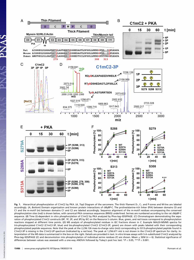
Fig. 1. Hierarchical phosphorylation of C1mC2 by PKA. (A, Top) Diagram of the sarcomere. The thick filament D-, C-, and P-zones and M-line are labeledaccordingly. (A, Bottom) Domain organization and known protein interactions of cMyBP-C. The proline/alanine-rich linker (P/A) between domains C0 andC1 and the m-motif (m) between domains C1 and C2 are labeled accordingly. Sequence alignment of the m-motif residues encompassing the conservedphosphorylation sites (red) is shown below, with canonical PKA consensus sequences (RRXS) underlined. Serines are numbered according to the rat cMyBP-Csequence. (B) Time (t)-dependent in vitro phosphorylation of C1mC2 by PKA analyzed by Phos-tag–SDS/PAGE. (C) Chromatogram demonstrating the sepa-ration of phosphorylated C1mC2 constructs (0P, 1P, 2P, and 3P) by IEC on the Resource S column. Blue, green, and red traces correspond to phosphorylationreactions stopped at different time points. (D) MS analysis of phosphorylated residues in IEC fractions shown in C. Example MALDI-MS/MS spectra fortris-phosphorylated C1mC2 (C1mC2-3P, blue) and bis-phosphorylated C1mC2 (C1mC2-2P, green) are shown with peaks labeled with their correspondingphosphorylated peptide sequences. Note that the peak at the 2,236.126 mass-to-charge ratio (m/z) corresponding to S313-phosphorylated peptide found inC1mC2-3P is missing in the C1mC2-2P spectrum (indicated by a red box). The peak at 1,256.671 m/z is not shown in the C1mC2-2P spectrum for clarity. In-terpretation of the MS data is summarized in the box on the right. Details are provided in text. In vitro kinase assays with S-A–substituted C1mC2 analyzed byPhos-tag–SDS/PAGE (E) and deconvolution into phosphorylation of individual serine residues (F) are shown. Mean ± SEM (n = 6). Statistical significance ofdifferences between values was assessed with a one-way ANOVA followed by Tukey’s post hoc test: *P < 0.05, ***P < 0.001.
15486 | www.pnas.org/cgi/doi/10.1073/pnas.1903033116 Ponnam et al.
Dow
nloa
ded
by g
uest
on
Nov
embe
r 21
, 202
1

Both the inhibitory and activating interactions of cMyBP-C arebelieved to be controlled by its phosphorylation state. The car-diac specific “m-motif” between domains C1 and C2 contains aseries of conserved serine residues that are phosphorylated byseveral protein kinases in vivo and in vitro (13) (Fig. 1A). Pro-tein kinase A (PKA) has been identified as the primary kinaseacting upon cMyBP-C (14), mediating some of the effects ofβ-adrenergic stimulation on myocardial function, such as in-creased cross-bridge kinetics, decreased calcium sensitivity, andaccelerated relaxation (15, 16). More recently, other proteinkinases have been shown to phosphorylate cMyBP-C with dis-tinct specificities for m-motif serines, suggesting that eachphosphorylation site might have distinct regulatory effects (13).However, the detailed function and mechanism underlying
cMyBP-C phosphorylation at individual sites remain poorly un-derstood, likely due to the complexity of its interaction withother sarcomeric components, which is further compounded bythe multiplicity of signaling pathways acting upon cMyBP-C andits associated phosphorylation states (17). Current mechanistichypotheses of the effects of cMyBP-C phosphorylation arelargely based on in vivo experiments in animal models (6, 18) ortissues derived from those animals (19) in which phosphorylationlevels and associated regulatory mechanisms cannot be con-trolled at the molecular level. Moreover, serine-to-aspartatesubstitutions have been frequently used as a convenient modelfor phosphorylation, although recent studies showed that thesesubstitutions only partially recapitulate the effects of phosphor-ylation (20). Conversely, in vitro experiments with fully phos-phorylated proteins or proteins containing serine-to-aspartatesubstitutions do not recapitulate the in vivo complexity of cMyBP-C’sinteractions and its dynamic phosphorylation (21–23).The aim of the present work was to understand the molecular
mechanism of cMyBP-C phosphorylation and its structural andfunctional effects on both thin and thick filament-based regula-tion in heart muscle cells. We investigated the relationship ofindividual cMyBP-C phosphorylation sites and potential cross-talkusing several protein kinases known to phosphorylate cMyBP-C.We utilized cMyBP-C fragments with well-characterized phos-phorylation states to show that site-specific phosphorylation hasdistinct effects on its interaction with and regulation of the thinand thick filaments, combining in vitro biochemical binding assayswith in situ structural measurements in intact heart muscle cells.The results show that cMyBP-C acts as a sarcomeric integrator ofdifferent signaling pathways to determine downstream physiologicaleffects, and that the functional effects of cMyBP-C phosphorylationcan only be understood by combining thin and thick filament-basedmechanisms into an integrated model of contractile regulation.
ResultsHierarchical Phosphorylation of cMyBP-C by PKA. To elucidate theregulatory function of cMyBP-C phosphorylation, we phosphory-lated a recombinant fragment of rat cMyBP-C containing domainsC1, the phosphorylatable m-motif, and C2 (C1mC2; Fig. 1A) withthe catalytic subunit of PKA in vitro (Fig. 1B). The C1mC2 frag-ment is a convenient model for studying the function of full-lengthcMyBP-C, particularly with respect to its effects on both thin andthick filament structure (11, 12, 20) and its regulation by phos-phorylation (20, 24, 25). Although rodent and human C1mC2 havea high sequence identity (>92%), suggesting a conserved molec-ular function, species-specific effects cannot be excluded (26).Incubation of C1mC2 for 30 to 60 min with PKA resulted
in a mixture of unphosphorylated, monophosphorylated, bis-phosphorylated, and tris-phosphorylated protein, which could beclearly separated by both Phos-tag and sodium dodecyl sulfatepolyacrylamide gel electrophoresis (SDS/PAGE) (Fig. 1B).Individual C1mC2 phospho-species were isolated by stopping thekinase reaction at different time points and separating thephosphorylated proteins by ion-exchange chromatography (IEC)
(Fig. 1C). The phosphorylation level and homogeneity (>95%)of each IEC fraction (e.g., 1P, 2P, 3P) were confirmed by elec-trospray ionization (ESI) mass spectrometry (MS) (SI Appendix,Table S1). Tetrakis-phosphorylated C1mC2 was only observedafter prolonged incubation with PKA at 30 °C, suggesting thatthe fourth site is a poor substrate for PKA.Phosphorylated amino acid residues in each IEC fraction were
identified by proteolytic digestion followed by phospho-peptideenrichment and matrix-assisted laser desorption ionization (MALDI)MS and ESI-MS (SI Appendix, Fig. S1) (details are provided in SIAppendix, Supplementary Information Methods). Analysis of theIEC fraction corresponding to monophosphorylated C1mC2(C1mC2-1P) revealed specific phosphorylation on a single serineresidue, S288, in agreement with previous studies suggesting thatS288 in the cardiac-specific insertion is the initial PKA phos-phorylation site (14). Bis-phosphorylated C1mC2 (C1mC2-2P)contained phosphorylated serine residues only in positions 288and 279, and tris-phosphorylated C1mC2 (C1mC2-3P) containedphosphorylated serines only in positions 288, 279, and 313. As anexample, the MALDI-MS spectrum of C1mC2-3P is shown in Fig.1D, with peaks corresponding to identified phospho-peptides la-beled accordingly. The peak at a 2,236.126 mass-to-charge ratiocorresponding to the S313-phosphorylated peptide is missing inthe MALDI-MS spectrum of the bis-phosphorylated C1mC2(C1mC2-2P) (Fig. 1 D, Inset, Top Left, red box), suggesting thatS313 is phosphorylated after S288 and S279. In contrast, phos-phorylation of S308 by PKA was only observed after prolongedincubation, suggesting that this serine is a poor substrate for PKAin vitro. Thus, our results suggest that PKA phosphorylation of thecardiac-specific m-motif follows a concerted hierarchical mecha-nism in the sequence of S288, followed by S279, followed by S313(Fig. 1 D, Inset, Top Right).To further test this conclusion, we prepared serine-to-alanine
(S-A) substitutions of each individual PKA site in C1mC2, andanalyzed their phosphorylation profiles by Phos-tag–SDS/PAGE(Fig. 1E). Total phosphate incorporation after 60 min of in-cubation was strongly reduced in S288A-substituted C1mC2(1.25 ± 0.08 mol of inorganic phosphate [Pi]/mol [mean ± SEM];n = 6; SI Appendix, Fig. S2) compared with wild type (2.94 ±0.18 mol of Pi/mol [mean ± SEM]; n = 6). S279A- and S313A-substituted C1mC2 showed intermediate levels of phosphorylation,although S279A had a stronger inhibitory effect than S313A (1.6 ±0.11 and 2.17 ± 0.12 mol of Pi/mol, respectively), in agreement withthe hierarchical phosphorylation sequence proposed above.Next, we analyzed the effects of S-A substitutions on phos-
phate incorporation by PKA at each individual phosphorylationsite (Fig. 1F). Consistent with the hierarchical model, phos-phorylation of S288 was not significantly affected by substitutionof either S279 or S313 by alanine. Similarly, S279 phosphoryla-tion was not inhibited by substitution of either S288 or S313 byalanine, although in the native fragment, this residue is phos-phorylated after S288. This suggests that unphosphorylatedS288 may be involved in intramolecular interactions that preventaccess to S279, and that substitution of S288 by alanine abolishesthis interaction. If so, substitution of S288 by alanine mighttherefore partially mimic phosphorylation of this site. In con-trast, S313 phosphorylation was inhibited by substitution of ei-ther S288 or S279 to alanine, further supporting the proposalthat this residue is phosphorylated downstream of S288 and S279.Contrary to the effects of S-A substitutions described above, PKAphosphorylation of S308 was greatly increased in C1mC2-S313A(∼50%) compared with wild-type control (∼10%), suggesting thatphosphorylation of S313 per se has an inhibitory effect on phos-phorylation of S308.
cMyBP-C Phosphorylation by Non-PKA Kinases Reveals RegulatoryCoupling of Phosphorylation Sites. Although PKA is consideredto be the primary kinase acting upon cMyBP-C in vivo, several
Ponnam et al. PNAS | July 30, 2019 | vol. 116 | no. 31 | 15487
BIOCH
EMISTR
Y
Dow
nloa
ded
by g
uest
on
Nov
embe
r 21
, 202
1

other kinases have been shown to be able to phosphorylate specificresidues within the cardiac-specific m-motif (13). Moreover, thesekinases have been frequently shown to be activated during heartdisease, underlining their functional significance.Ribosomal S6 kinase II (RSK2) has been suggested to spe-
cifically phosphorylate serine residues in the cardiac-specific m-motif insertion in response to activation of the mitogen-activatedprotein kinase/extracellular signal-regulated kinase pathway(27), and we used site-specific S-A substitution and MS to con-firm these results (Fig. 2A and SI Appendix, Fig. S3). RSK2primarily phosphorylated rat C1mC2 at S288 in vitro, althoughwe observed phosphorylation of other residues at higher enzyme-to-substrate ratios or after prolonged incubation. In contrast,α-adrenergic stimulation leads to an increase in cMyBP-Cphosphorylation primarily via stimulation of protein kinase Ce(PKCe) (28, 29) [and protein kinase D (PKD) (30)] activity. Weused site-directed mutagenesis, MS, and Western blot analysis toconfirm that PKCe specifically phosphorylates S308 in C1mC2 invitro (Fig. 2 B and C and SI Appendix, Fig. S3). Phosphorylationof S288 was not a prerequisite for phosphorylation of S308 byPKCe as shown previously (31).Next, we investigated potential cross-talk between cMyBP-C–
mediated phosphorylation by RSK2, PKCe, and PKA using pu-rified C1mC2 fragments site-specifically phosphorylated by RSK2and PKCe as substrates in PKA kinase assays. As expected fromthe sequential phosphorylation of C1mC2 described above, S288phosphorylation by RSK2 significantly accelerated phosphoryla-tion of S279 by PKA, but had no effect on phosphorylation ofresidues downstream of S279 (i.e., S313) (Fig. 2 D and E). Incontrast, phosphorylation of S308 by PKCe decreased the rate of
PKA phosphorylation of S313, suggesting that S308P is a negativeregulator of cMyBP-C PKA phosphorylation of S313, consistentwith previous results in isolated cardiomyocytes (32). Moreover,both S288 by RSK2 and S308 by PKCe phosphorylation reducedoverall phosphate incorporation into C1mC2 by PKA by ∼1 molof Pi/mol of C1mC2 (Fig. 2F). Although this is expected forphosphorylation of S288 as a main PKA site, S308 is primarilyphosphorylated by PKCe and PKD, further confirming the an-tagonistic effects of PKA- and PKCe/PKD-mediated phosphory-lation of cMyBP-C.In summary, the multiple cMyBP-C phosphorylation sites are
not independent, and exhibit positive and negative regulatorycoupling with partially overlapping specificity for multiple kinases.
Site-Specific Phosphorylation Controls Thick and Thin FilamentBinding of C1mC2. To characterize the effects of site-specificcMyBP-C phosphorylation on its proposed interactions with boththe myosin-containing thick and actin-containing thin filaments(12, 20), we determined the binding affinities of C1mC2 in itsdifferent phosphorylation states for isolated myosin S2 and na-tive thin filaments (NTFs) in vitro. The affinity of C1mC2 for itsbinding site on myosin, the first 126 amino acids of myosin S2(S2Δ), was characterized by microscale thermophoresis (MST).As previously shown, unphosphorylated C1mC2 binds S2Δ in abiphasic manner, corresponding to high- and low-affinity bindingsites with dissociation constants (Kds) of 20.7 ± 3.5 μmol/L(mean ± SEM; n = 6) and >400 μmol/L, respectively (20).Moreover, the Kd for myosin S2Δ of λ-protein phosphatase–treated native full-length cMyBP-C isolated from rat ventriculartissue was 24.2 ± 3.9 μmol/L (mean ± SEM; n = 4) (SI Appendix,
Fig. 2. C1mC2 phosphorylation by RSK2 and PKCe. (A) RSK2-mediated phosphorylation of S288 in rat C1mC2 was confirmed by in vitro kinase (IVK) assaysusing wild-type and S288A- substituted C1mC2 as well as MALDI-MS phosphorylation site profiling. m/z, mass-to-charge ratio. (B) PKCe phosphorylation ofS308 in rat C1mC2was confirmed by both IVK assays using phosphoablated C1mC2-S308D and ESI-MS. (C) Western blot analysis of RSK2- and PKCe-phosphorylatedC1mC2 using PKA site (RRXpS)-specific and S288 (pS288)-specific antibodies. Note that PKCe-phosphorylated C1mC2 is not recognized by either of the antibodies,confirming S308 as the main phosphorylation site. (D) PKA IVK assays with unphosphorylated, S288-phosphorylated, or S308-phosphorylated C1mC2 analyzed byPhos-tag–SDS/PAGE. t, time. Analysis of individual site phosphorylation (E) and total level of phosphate incorporation by PKA (F) are shown. Mean ± SEM (n = 3 to9). Statistical significance of differences between values was assessed with a one-way ANOVA followed by Tukey’s post hoc test: *P < 0.05, **P < 0.01.
15488 | www.pnas.org/cgi/doi/10.1073/pnas.1903033116 Ponnam et al.
Dow
nloa
ded
by g
uest
on
Nov
embe
r 21
, 202
1

Fig. S4), in good agreement with the results obtained forrecombinant rat C1mC2.Monophosphorylation of either S308 or S288 slightly reduced
C1mC2’s affinity for myosin S2Δ as indicated by an increase inKd to ∼30 μmol/L (Fig. 3A and SI Appendix, Fig. S5 and TableS2). In contrast, both PKA bis-phosphorylation (S288 and S279)and tris-phosphorylation (S288, S279, and S313) completelyabolished S2Δ binding, indicating that either bis-phosphorylationor phosphorylation of S279 per se controls the cMyBP-C–myosinS2 interaction. We addressed this question by phosphorylatingC1mC2-S288A with PKA and isolating the monophosphorylatedspecies (C1mC2-S288A-1P) using IEC. Both unphosphorylatedand monophosphorylated C1mC2-S288A bind myosin S2Δ witha Kd similar to that measured for the wild-type protein (Kd of∼20 μmol/L), suggesting that bis-phosphorylation is necessaryand sufficient to abolish cMyBP-C–myosin S2 interaction (Fig.3B and SI Appendix, Table S2). To further test this conclusion,we sequentially phosphorylated C1mC2 with RSK2 and PKCe,and measured the affinity of the bis-phosphorylated C1mC2(S288 and S308) for myosin S2Δ (SI Appendix, Fig. S6). TheRSK2/PKCe bis-phosphorylated C1mC2 did not bind to myosinS2Δ, further supporting the hypothesis that bis-phosphorylationper se, independent of the phosphorylation site combination,abolishes cMyBP-C–myosin S2 interaction. These results areconsistent with the largely ionic nature of cMyBP-C’s main in-teraction site in the m-motif and myosin S2 (20).We measured the affinity of C1mC2 in its different phos-
phorylation states for isolated bovine cardiac NTFs using high-velocity cosedimentation. Unphosphorylated C1mC2 (0P) bindsNTFs in the absence of calcium (pCa 9) in a saturable mannerwith a Kd of ∼20 μmol/L and a maximal binding capacity (Bmax)of ∼1, indicating stoichiometric binding of C1mC2 to actin (Fig.3 C–F and SI Appendix, Fig. S5 and Table S2). Addition of cal-cium (pCa 4.5) had no effect on C1mC2 binding to NTFs asindicated by identical Kd and Bmax values (Fig. 3 D–F and SIAppendix, Fig. S5A and Table S2). Strikingly, both PKA mono-phosphorylation (S288) and bis-phosphorylation (S288 andS279), as well as monophosphorylation of S308 by PKCe, showedno change in the affinity of C1mC2 for NTFs (Kd ∼ 20 μmol/L),but decreased Bmax to ∼0.5, suggesting a lower binding capacityof C1mC2 in the partially phosphorylated states. As before, fullcalcium activation of NTFs had no additional effect on either Kdor Bmax. However, PKA tris-phosphorylation (3P) significantlydecreased C1mC2’s affinity for NTF with an estimated Kd of∼60 μmol/L independent of [Ca2+]. Addition of calcium increasedBmax from ∼0.5 to ∼1 for C1mC2-3P, suggesting an interplaybetween tris-phosphorylation of C1mC2, thin filament binding,and calcium activation (23).These results suggest that cMyBP-C phosphorylation regulates
contractility partly via differential modulation of its affinity formyosin and actin binding sites, so that phosphorylation leads to aredistribution of cMyBP-C’s N-terminal domains from theirmyosin binding sites in the thick filaments to their actin bindingsites in the thin filaments. Monophosphorylation weakens andbis-phosphorylation abolishes thick filament binding, and only tris-phosphorylation affects C1mC2 binding to regulated thin filaments.
Site-Specific Phosphorylation of C1mC2 Controls Its Effect on Thin andThick Filament Structure. Next, we used a bifunctional rhodamineprobe attached to the E-helix of cardiac troponin C (cTnC-E) tomonitor structural changes in the thin filaments of demembra-nated ventricular muscle cells associated with the activating ef-fect of C1mC2 described previously (12). UnphosphorylatedC1mC2 activates the force and thin filament structure of ven-tricular trabeculae in the absence of Ca2+ (pCa 9) with a half-maximal effective concentration (EC50) of ∼20 μmol/L (Fig. 4A,filled red circles). Maximum isometric force at [C1mC2] = 40μmol/L is only ∼60% of that measured during Ca2+ activation in
the absence of C1mC2 (Fig. 4B), although the level of thin fil-ament activation as reported by the cTnC probe is significantlyhigher, suggesting that exogenous C1mC2 has both activatingand inhibitory effects on contractility.PKA phosphorylation of S288 reduced the activating effect of
40 μmol/L C1mC2 on active force to ∼30% of that measuredduring control conditions (Fig. 4B, light blue, 1P), to less than10% after PKA bis-phosphorylation (C1mC2-2P, dark blue,2P), and active force was completely abolished for PKA tris-phosphorylated C1mC2 (C1mC2-3P) (Fig. 4B, purple). In con-trast, S308 phosphorylation by PKCe did not inhibit the activatingeffect of C1mC2 on force generation (Fig. 4B, yellow), suggestingdifferent regulatory functions of phosphorylation of S288 and S308.The effects on isometric force described above are mirrored by
those on the thin filament structure as monitored by the cTnC-Eprobe orientation. C1mC2 “superactivates” the thin filament asindicated by a change in the order parameter <P2>, which issignificantly larger than that observed for calcium activation
Fig. 3. Effects of site-specific phosphorylation on binding of C1mC2 tomyosin S2Δ and NTFs. (A) Normalized MST curves for C1mC2 and its differentphosphorylation states titrated against myosin S2Δ. (B) NormalizedMST bindingcurves for C1mC2-S288A (brown) and C1mC2-S288A-1P (black) titrated againstmyosin S2Δ. NTF cosedimentation data for C1mC2 and its different phosphor-ylation states in the absence (C, pCa 9) and presence (D, pCa 4.5) of 32 μmol/LCaCl2 are shown. Calculated Kd (E) and Bmax (F) values for the NTF binding as-says in the absence (filled circles) and presence (open circles) of CaCl2 are shown.Mean ± SEM (n = 4 to 10). Statistical significance of difference was assessedwitha one-way ANOVA followed by Tukey’s post hoc test: *P < 0.05, **P < 0.01.
Ponnam et al. PNAS | July 30, 2019 | vol. 116 | no. 31 | 15489
BIOCH
EMISTR
Y
Dow
nloa
ded
by g
uest
on
Nov
embe
r 21
, 202
1

alone (∼120%) (Fig. 4C). PKA monophosphorylation reducedthe activating effect to ∼30% of that measured during controlconditions in the absence of C1mC2, in agreement with the forcedata. In contrast, S308 monophosphorylated C1mC2 showed anintermediate effect on thin filament activation, corresponding to∼70% of the control value (Fig. 4C, yellow). Incubation ofventricular trabeculae with either 40 μmol/L PKA bis- or tris-phosphorylated C1mC2 had no significant effect on thin filamentstructure in the absence of Ca2+.These results are in stark contrast to the NTF binding data
described above, suggesting that although phosphorylation ofcMyBP-C has only minor effects on its binding to the thin fila-ment, it significantly alters thin filament regulation. Recentelectron microscopy studies demonstrated that N-terminal do-mains of cMyBP-C bind polymorphically to isolated actin fila-ments, and that only a subset of binding modes can directlyinterfere with tropomyosin’s position and induce the ON state ofthe thin filament (33, 34). The comparison suggests that cMyBP-C phosphorylation regulates thin filament activation by alteringthe equilibrium between binding states that affect tropomyosin’sposition on actin and those that do not.Structural changes in the thick filament associated with the
activation of ventricular trabeculae by C1mC2 in its differentphosphorylation states were monitored using a bifunctional sul-forhodamine probe cross-linking helices B and C in the myosinregulatory light chain (BSR-cRLC-BC) (35). BSR-cRLC-BC islocalized close to the myosin S1/S2 junction and is mainly sen-sitive to the regulatory state of the thick filament; the orderparameter <P2> from this probe increases upon calcium acti-vation. In contrast to its effect on thin filament structure de-scribed above, C1mC2 incubation leads to a partial activation ofthe thick filament structure corresponding to ∼70% of thatmeasured during control activations (pCa 4.5; Fig. 4A, orangeopen circle). Moreover, C1mC2 activated the thick filament witha significantly lower EC50 than that measured for force or thinfilament activation (∼10 μmol/L; Fig. 4A, open red circles).Monophosphorylation of either S288 by PKA or S308 by
PKCe showed no significant reduction in the activating effect of40 μmol/L C1mC2 on the thick filament structure as reported bythe BC probe orientation (Fig. 4D), in contrast to the strongreduction in thin filament activation and isometric force pro-duction associated with phosphorylation of S288 (Fig. 4 B and C;1P). Thus, although S288 phosphorylation and S308 phosphor-ylation have similar effects on the regulatory state of the thickfilament, they have very different effects on the regulatory stateof the thin filament. In agreement with their effects on force andthin filament structure, both C1mC2-2P and C1mC2-3P had nosignificant effect on thick filament structure as measured by theRLC BC probe orientation, consistent with the abolished bindingof C1mC2 to myosin S2Δ after PKA bis-phosphorylation andtris-phosphorylation (Fig. 3A and SI Appendix, Table S2).The comparison of the effects of C1mC2 in its different
phosphorylation states on the myosin head conformation with theMST binding data described above further suggests that C1mC2has a direct activating effect on the thick filament and that theactivating effect is, in turn, controlled by its phosphorylation-dependent affinity for myosin S2Δ.
Site-Specific Phosphorylation of C1mC2 Controls Actomyosin ATPaseActivity. We further investigated the functional consequences ofsite-specific C1mC2 phosphorylation on actomyosin interactionsby measuring the NTF-stimulated adenosinetriphosphatase(ATPase) activity of isolated bovine myosin S1 in the presence ofC1mC2 in its different phosphorylation states using a colori-metric assay (Fig. 5A).Unphosphorylated C1mC2 activates the NTF-stimulated my-
osin S1 ATPase in the absence of calcium in a concentration-dependent manner (Fig. 5B, red) and data points were fitted to
an activation/inhibition model as previously described (36). Lowconcentrations activate phosphate production with an EC50of ∼1 μmol/L and a Hill coefficient of ∼1.5, suggesting cooper-ative activation of NTFs by C1mC2. In contrast, [C1mC2] >2 μmol/L inhibits the ATPase activity with a half-maximalinhibitory concentration (IC50) of ∼3 μmol/L. PKA mono-phosphorylation (Fig. 5B, light blue) and bis-phosphorylation(Fig. 5B, dark blue) of C1mC2 increased the EC50 of the acti-vating effect further to ∼2 and ∼5 μmol/L, respectively, and tris-phosphorylated C1mC2 did not activate actomyosin S1 ATPasewithin the concentration range tested (Fig. 5B, purple). In fact,C1mC2-2P and C1mC2-3P showed an inhibitory effect on theactomyosin S1-ATPase in the concentration range of 1 to4 μmol/L, presumably by competing with myosin S1 for bindingsites on actin. In contrast, during full calcium activation (Fig. 5C,pCa 4), C1mC2 largely inhibits myosin S1-NTF ATPase activitywith an IC50 of ∼2 μmol/L and a maximal inhibition of ∼50% (Fig.5C, red), similar to the values measured in the absence of Ca2+ and[C1mC2] > 2 μmol/L. PKA phosphorylation of C1mC2 decreasedthe amplitude of inhibition without affecting its IC50.The direct comparison with the NTF binding data (Fig. 3 and
SI Appendix, Table S2) further supports the conclusion thatC1mC2 interacts with NTF in different binding modes and thatphosphorylation controls the distribution between states with in-hibitory and activating effects. Surprisingly, [C1mC2-3P] < 2 μmol/L
Fig. 4. Effects of site-specific phosphorylation of C1mC2 on active forceand thin and thick filament structure in ventricular trabeculae. (A)Concentration-dependent effect of C1mC2 on force generation (blacksquares) and thin (red filled circles) and thick (red open circles) filament struc-ture in ventricular trabeculae. TnC and RLC probe orientations are expressed asthe order parameter <P2>, which is +1 for a probe dipole orientation parallelto the filament axis and −0.5 for a perpendicular orientation. <P2> for fullcalcium activation is shown in orange on the left axis. (B) Isometric force ofventricular trabeculae in the presence of 40 μmol/L C1mC2 in its differentphosphorylation states. ACT, activating solution (pCa 4.5); REL, relaxing solu-tion (pCa 9). TnC (C) and RLC (D) probe orientations of ventricular trabeculae inthe presence of 40 μmol/L C1mC2 in its different phosphorylation states areshown. Mean ± SEM, with the number of trabeculae (n), is indicated in eachpanel. Statistical significance of differences between groups was assessed witha one-way ANOVA followed by Tukey’s post hoc test: *P < 0.05, **P < 0.01,***P < 0.001. ns, not significant.
15490 | www.pnas.org/cgi/doi/10.1073/pnas.1903033116 Ponnam et al.
Dow
nloa
ded
by g
uest
on
Nov
embe
r 21
, 202
1
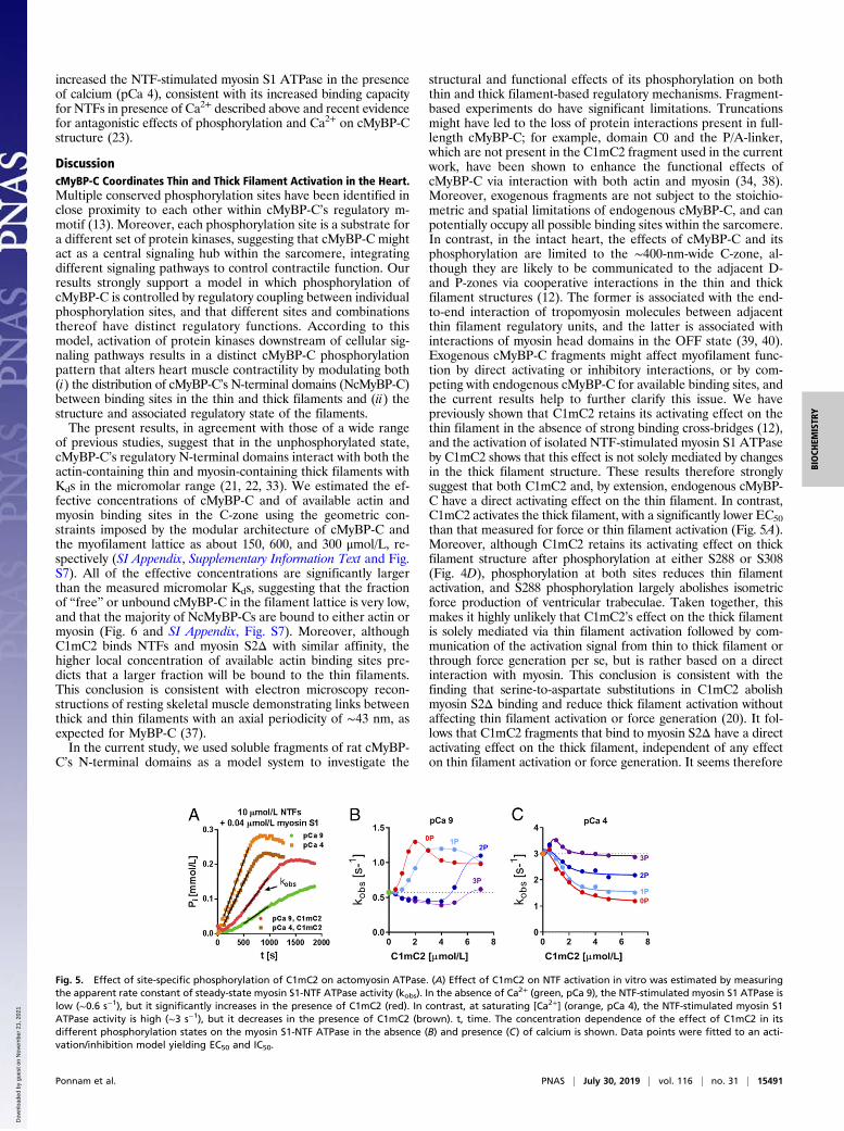
increased the NTF-stimulated myosin S1 ATPase in the presenceof calcium (pCa 4), consistent with its increased binding capacityfor NTFs in presence of Ca2+ described above and recent evidencefor antagonistic effects of phosphorylation and Ca2+ on cMyBP-Cstructure (23).
DiscussioncMyBP-C Coordinates Thin and Thick Filament Activation in the Heart.Multiple conserved phosphorylation sites have been identified inclose proximity to each other within cMyBP-C’s regulatory m-motif (13). Moreover, each phosphorylation site is a substrate fora different set of protein kinases, suggesting that cMyBP-C mightact as a central signaling hub within the sarcomere, integratingdifferent signaling pathways to control contractile function. Ourresults strongly support a model in which phosphorylation ofcMyBP-C is controlled by regulatory coupling between individualphosphorylation sites, and that different sites and combinationsthereof have distinct regulatory functions. According to thismodel, activation of protein kinases downstream of cellular sig-naling pathways results in a distinct cMyBP-C phosphorylationpattern that alters heart muscle contractility by modulating both(i) the distribution of cMyBP-C’s N-terminal domains (NcMyBP-C)between binding sites in the thin and thick filaments and (ii) thestructure and associated regulatory state of the filaments.The present results, in agreement with those of a wide range
of previous studies, suggest that in the unphosphorylated state,cMyBP-C’s regulatory N-terminal domains interact with both theactin-containing thin and myosin-containing thick filaments withKds in the micromolar range (21, 22, 33). We estimated the ef-fective concentrations of cMyBP-C and of available actin andmyosin binding sites in the C-zone using the geometric con-straints imposed by the modular architecture of cMyBP-C andthe myofilament lattice as about 150, 600, and 300 μmol/L, re-spectively (SI Appendix, Supplementary Information Text and Fig.S7). All of the effective concentrations are significantly largerthan the measured micromolar Kds, suggesting that the fractionof “free” or unbound cMyBP-C in the filament lattice is very low,and that the majority of NcMyBP-Cs are bound to either actin ormyosin (Fig. 6 and SI Appendix, Fig. S7). Moreover, althoughC1mC2 binds NTFs and myosin S2Δ with similar affinity, thehigher local concentration of available actin binding sites pre-dicts that a larger fraction will be bound to the thin filaments.This conclusion is consistent with electron microscopy recon-structions of resting skeletal muscle demonstrating links betweenthick and thin filaments with an axial periodicity of ∼43 nm, asexpected for MyBP-C (37).In the current study, we used soluble fragments of rat cMyBP-
C’s N-terminal domains as a model system to investigate the
structural and functional effects of its phosphorylation on boththin and thick filament-based regulatory mechanisms. Fragment-based experiments do have significant limitations. Truncationsmight have led to the loss of protein interactions present in full-length cMyBP-C; for example, domain C0 and the P/A-linker,which are not present in the C1mC2 fragment used in the currentwork, have been shown to enhance the functional effects ofcMyBP-C via interaction with both actin and myosin (34, 38).Moreover, exogenous fragments are not subject to the stoichio-metric and spatial limitations of endogenous cMyBP-C, and canpotentially occupy all possible binding sites within the sarcomere.In contrast, in the intact heart, the effects of cMyBP-C and itsphosphorylation are limited to the ∼400-nm-wide C-zone, al-though they are likely to be communicated to the adjacent D-and P-zones via cooperative interactions in the thin and thickfilament structures (12). The former is associated with the end-to-end interaction of tropomyosin molecules between adjacentthin filament regulatory units, and the latter is associated withinteractions of myosin head domains in the OFF state (39, 40).Exogenous cMyBP-C fragments might affect myofilament func-tion by direct activating or inhibitory interactions, or by com-peting with endogenous cMyBP-C for available binding sites, andthe current results help to further clarify this issue. We havepreviously shown that C1mC2 retains its activating effect on thethin filament in the absence of strong binding cross-bridges (12),and the activation of isolated NTF-stimulated myosin S1 ATPaseby C1mC2 shows that this effect is not solely mediated by changesin the thick filament structure. These results therefore stronglysuggest that both C1mC2 and, by extension, endogenous cMyBP-C have a direct activating effect on the thin filament. In contrast,C1mC2 activates the thick filament, with a significantly lower EC50than that measured for force or thin filament activation (Fig. 5A).Moreover, although C1mC2 retains its activating effect on thickfilament structure after phosphorylation at either S288 or S308(Fig. 4D), phosphorylation at both sites reduces thin filamentactivation, and S288 phosphorylation largely abolishes isometricforce production of ventricular trabeculae. Taken together, thismakes it highly unlikely that C1mC2’s effect on the thick filamentis solely mediated via thin filament activation followed by com-munication of the activation signal from thin to thick filament orthrough force generation per se, but is rather based on a directinteraction with myosin. This conclusion is consistent with thefinding that serine-to-aspartate substitutions in C1mC2 abolishmyosin S2Δ binding and reduce thick filament activation withoutaffecting thin filament activation or force generation (20). It fol-lows that C1mC2 fragments that bind to myosin S2Δ have a directactivating effect on the thick filament, independent of any effecton thin filament activation or force generation. It seems therefore
Fig. 5. Effect of site-specific phosphorylation of C1mC2 on actomyosin ATPase. (A) Effect of C1mC2 on NTF activation in vitro was estimated by measuringthe apparent rate constant of steady-state myosin S1-NTF ATPase activity (kobs). In the absence of Ca2+ (green, pCa 9), the NTF-stimulated myosin S1 ATPase islow (∼0.6 s−1), but it significantly increases in the presence of C1mC2 (red). In contrast, at saturating [Ca2+] (orange, pCa 4), the NTF-stimulated myosin S1ATPase activity is high (∼3 s−1), but it decreases in the presence of C1mC2 (brown). t, time. The concentration dependence of the effect of C1mC2 in itsdifferent phosphorylation states on the myosin S1-NTF ATPase in the absence (B) and presence (C) of calcium is shown. Data points were fitted to an acti-vation/inhibition model yielding EC50 and IC50.
Ponnam et al. PNAS | July 30, 2019 | vol. 116 | no. 31 | 15491
BIOCH
EMISTR
Y
Dow
nloa
ded
by g
uest
on
Nov
embe
r 21
, 202
1
more likely that C1mC2’s activating effect on the thick filament isdue to a direct competition with the endogenous cMyBP-C, whichstabilizes the thick filament OFF state via interactions with theS2 region of myosin, although an alternative mechanism involvinga direct effect of C1mC2 on the myosin head domains cannot beexcluded by the present results. Therefore, endogenous cMyBP-Cand exogenous C1mC2 have the same effect on the thin filamentbut opposite effects on the thick filament.We may therefore consider a dephosphorylated state of
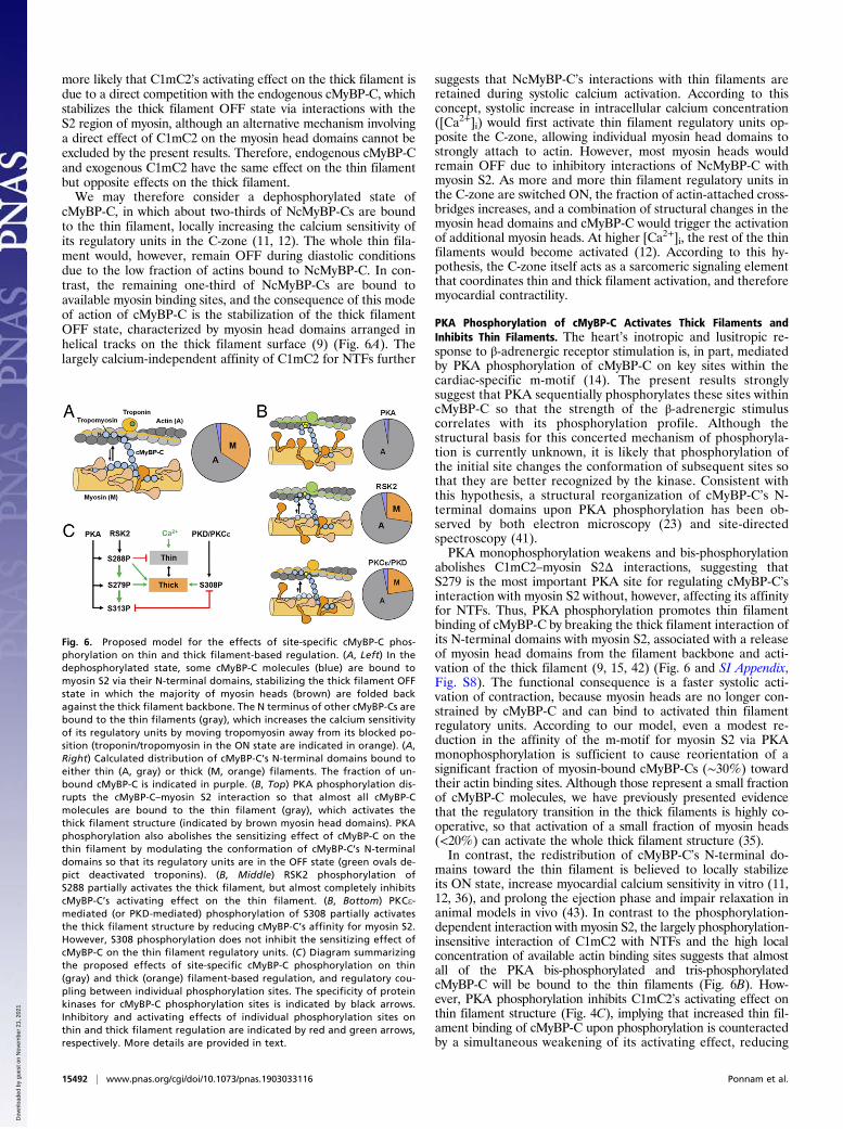
cMyBP-C, in which about two-thirds of NcMyBP-Cs are boundto the thin filament, locally increasing the calcium sensitivity ofits regulatory units in the C-zone (11, 12). The whole thin fila-ment would, however, remain OFF during diastolic conditionsdue to the low fraction of actins bound to NcMyBP-C. In con-trast, the remaining one-third of NcMyBP-Cs are bound toavailable myosin binding sites, and the consequence of this modeof action of cMyBP-C is the stabilization of the thick filamentOFF state, characterized by myosin head domains arranged inhelical tracks on the thick filament surface (9) (Fig. 6A). Thelargely calcium-independent affinity of C1mC2 for NTFs further
suggests that NcMyBP-C’s interactions with thin filaments areretained during systolic calcium activation. According to thisconcept, systolic increase in intracellular calcium concentration([Ca2+]i) would first activate thin filament regulatory units op-posite the C-zone, allowing individual myosin head domains tostrongly attach to actin. However, most myosin heads wouldremain OFF due to inhibitory interactions of NcMyBP-C withmyosin S2. As more and more thin filament regulatory units inthe C-zone are switched ON, the fraction of actin-attached cross-bridges increases, and a combination of structural changes in themyosin head domains and cMyBP-C would trigger the activationof additional myosin heads. At higher [Ca2+]i, the rest of the thinfilaments would become activated (12). According to this hy-pothesis, the C-zone itself acts as a sarcomeric signaling elementthat coordinates thin and thick filament activation, and thereforemyocardial contractility.
PKA Phosphorylation of cMyBP-C Activates Thick Filaments andInhibits Thin Filaments. The heart’s inotropic and lusitropic re-sponse to β-adrenergic receptor stimulation is, in part, mediatedby PKA phosphorylation of cMyBP-C on key sites within thecardiac-specific m-motif (14). The present results stronglysuggest that PKA sequentially phosphorylates these sites withincMyBP-C so that the strength of the β-adrenergic stimuluscorrelates with its phosphorylation profile. Although thestructural basis for this concerted mechanism of phosphoryla-tion is currently unknown, it is likely that phosphorylation ofthe initial site changes the conformation of subsequent sites sothat they are better recognized by the kinase. Consistent withthis hypothesis, a structural reorganization of cMyBP-C’s N-terminal domains upon PKA phosphorylation has been ob-served by both electron microscopy (23) and site-directedspectroscopy (41).PKA monophosphorylation weakens and bis-phosphorylation
abolishes C1mC2–myosin S2Δ interactions, suggesting thatS279 is the most important PKA site for regulating cMyBP-C’sinteraction with myosin S2 without, however, affecting its affinityfor NTFs. Thus, PKA phosphorylation promotes thin filamentbinding of cMyBP-C by breaking the thick filament interaction ofits N-terminal domains with myosin S2, associated with a releaseof myosin head domains from the filament backbone and acti-vation of the thick filament (9, 15, 42) (Fig. 6 and SI Appendix,Fig. S8). The functional consequence is a faster systolic acti-vation of contraction, because myosin heads are no longer con-strained by cMyBP-C and can bind to activated thin filamentregulatory units. According to our model, even a modest re-duction in the affinity of the m-motif for myosin S2 via PKAmonophosphorylation is sufficient to cause reorientation of asignificant fraction of myosin-bound cMyBP-Cs (∼30%) towardtheir actin binding sites. Although those represent a small fractionof cMyBP-C molecules, we have previously presented evidencethat the regulatory transition in the thick filaments is highly co-operative, so that activation of a small fraction of myosin heads(<20%) can activate the whole thick filament structure (35).In contrast, the redistribution of cMyBP-C’s N-terminal do-
mains toward the thin filament is believed to locally stabilizeits ON state, increase myocardial calcium sensitivity in vitro (11,12, 36), and prolong the ejection phase and impair relaxation inanimal models in vivo (43). In contrast to the phosphorylation-dependent interaction with myosin S2, the largely phosphorylation-insensitive interaction of C1mC2 with NTFs and the high localconcentration of available actin binding sites suggests that almostall of the PKA bis-phosphorylated and tris-phosphorylatedcMyBP-C will be bound to the thin filaments (Fig. 6B). How-ever, PKA phosphorylation inhibits C1mC2’s activating effect onthin filament structure (Fig. 4C), implying that increased thin fil-ament binding of cMyBP-C upon phosphorylation is counteractedby a simultaneous weakening of its activating effect, reducing
Fig. 6. Proposed model for the effects of site-specific cMyBP-C phos-phorylation on thin and thick filament-based regulation. (A, Left) In thedephosphorylated state, some cMyBP-C molecules (blue) are bound tomyosin S2 via their N-terminal domains, stabilizing the thick filament OFFstate in which the majority of myosin heads (brown) are folded backagainst the thick filament backbone. The N terminus of other cMyBP-Cs arebound to the thin filaments (gray), which increases the calcium sensitivityof its regulatory units by moving tropomyosin away from its blocked po-sition (troponin/tropomyosin in the ON state are indicated in orange). (A,Right) Calculated distribution of cMyBP-C’s N-terminal domains bound toeither thin (A, gray) or thick (M, orange) filaments. The fraction of un-bound cMyBP-C is indicated in purple. (B, Top) PKA phosphorylation dis-rupts the cMyBP-C–myosin S2 interaction so that almost all cMyBP-Cmolecules are bound to the thin filament (gray), which activates thethick filament structure (indicated by brown myosin head domains). PKAphosphorylation also abolishes the sensitizing effect of cMyBP-C on thethin filament by modulating the conformation of cMyBP-C’s N-terminaldomains so that its regulatory units are in the OFF state (green ovals de-pict deactivated troponins). (B, Middle) RSK2 phosphorylation ofS288 partially activates the thick filament, but almost completely inhibitscMyBP-C’s activating effect on the thin filament. (B, Bottom) PKCe-mediated (or PKD-mediated) phosphorylation of S308 partially activatesthe thick filament structure by reducing cMyBP-C’s affinity for myosin S2.However, S308 phosphorylation does not inhibit the sensitizing effect ofcMyBP-C on the thin filament regulatory units. (C ) Diagram summarizingthe proposed effects of site-specific cMyBP-C phosphorylation on thin(gray) and thick (orange) filament-based regulation, and regulatory cou-pling between individual phosphorylation sites. The specificity of proteinkinases for cMyBP-C phosphorylation sites is indicated by black arrows.Inhibitory and activating effects of individual phosphorylation sites onthin and thick filament regulation are indicated by red and green arrows,respectively. More details are provided in text.
15492 | www.pnas.org/cgi/doi/10.1073/pnas.1903033116 Ponnam et al.
Dow
nloa
ded
by g
uest
on
Nov
embe
r 21
, 202
1

calcium sensitivity (15) and facilitating relaxation (44). In contrastto the intermediate effect on the thick filament described above,monophosphorylation is sufficient to reduce the activating effectof C1mC2 to ∼30% of that measured for unphosphorylatedC1mC2 (Fig. 4C), suggesting that S288 is the main regulator ofcMyBP-C’s effect on thin filament activation. Bis-phosphorylationfurther reduces (to ∼10%) and tris-phosphorylation com-pletely abolishes C1mC2’s activating effect on thin filamentstructure as measured by cTnC probe orientation. This nonlinearresponse to cMyBP-C phosphorylation is consistent with the pro-gressive decrease in C1mC2’s ability to activate NTF-stimulatedmyosin S1 ATPase after sequential phosphorylation of the 3 PKAsites (Fig. 5B).Taken together with the hierarchical order of phosphorylation
sites discussed above, these results suggest that β-adrenergicstimulation mainly reduces thin filament calcium sensitivity viaPKA phosphorylation of S288, which subsequently facilitatesphosphorylation of S279 and activation of the thick filament(Fig. 6).Serine 288 is also phosphorylated by RSK2 (Fig. 2A), and it
was previously shown that RSK2-mediated in situ phosphoryla-tion of cMyBP-C in skinned ventricular trabeculae increasescross-bridge kinetics and decreases calcium sensitivity (27),consistent with the S288-mediated partial activation of the thickfilament structure and inhibition of the thin filament structureproposed here (Fig. 6B).In summary, cMyBP-C exists in different regulatory states
depending on its phosphorylation profile, suggesting that cardiacmyofilament function is fine-tuned by the relative distribution ofcMyBP-C between its different phosphorylation states (e.g., 0P,1P, 2P, 3P). According to this model, even moderate changes inbasal levels of cMyBP-C phosphorylation would have significantfunctional consequences for the myocardium.
PKCe Phosphorylation of cMyBP-C Increases Thick Filament Activationwithout Inhibiting the Thin Filament. In contrast, phosphorylationof S308 does not abolish the activating effect of C1mC2 on thinfilament or force development, but similarly reduces its affinityfor myosin S2Δ. Thus, phosphorylation of S308 in cMyBP-C ispredicted to partially activate the thick filament C-zone withoutthe associated deactivation of the thin filament structure (Fig.6B). The consequence would be significantly faster systolic ac-tivation of contraction, since myosin head domains would bereadily available for interaction with the calcium-activated thin fila-ment regulatory units inside the C-zone. Both PKCe and PKD havebeen shown to phosphorylate S308 in vivo and in vitro, suggesting adirect link between α-adrenergic receptor stimulation (45, 46),S308 phosphorylation, and the inotropic response of the heart.Consistent with this idea, PKD phosphorylation of trabeculaefrom transgenic mouse lines expressing S22A/S23A-substitutedcardiac troponin I showed an increase in cross-bridge kinetics,without an associated decrease in calcium sensitivity (30).CamKII has been shown to be an important regulator of
cMyBP-C function, and, recently, CamKII-mediated phosphor-ylation of S308 has been implicated in the positive force-frequency
relation of cardiac muscle (47), suggesting that the molecularpathway described above for S308 phosphorylation by PKCemightalso be triggered by CamKII.
Functional Implications for Pathophysiology of Contractile Regulationin the Heart. The phosphorylation-dependent interactions ofcMyBP-C have important implications for the physiology andpathophysiology of contractile regulation in the heart, and thecurrent results show that cMyBP-C functions as an integrator ofmultiple signaling elements that mediate context-specific functionsof the myocardium in health and disease. Dephosphorylation ofcMyBP-C has been frequently observed during heart failure (17),likely associated with myocardial β-adrenergic receptor desensiti-zation, and the present results suggest that the depressed force-generating capacity and impaired relaxation are, in part, mediatedby dephosphorylated cMyBP-C stabilizing the thick and thin fila-ment OFF and ON states, respectively.Of particular interest in the heart failure setting is the RSK2-
mediated phosphorylation of S288, which partially mimics thestructural and functional effects of β-adrenergic stimulation andprimes S279 for phosphorylation by PKA (Fig. 2D) (and po-tentially by other kinases [e.g., CamKII]). Similar to RSK2,α-adrenergic receptor stimulation has been proposed as an al-ternative pathway to unlock the inotropic reserve of the failingheart (45), and our results suggest that PKD/ PKCe-mediatedphosphorylation of cMyBP-C increases contractile force viadirect activation of the thick filament. Although either RSK2or PKD/PKCe phosphorylation only partially mimics the effects ofβ-adrenergic signaling, phosphorylation by both kinases mimicsPKA bis-phosphorylation and abolishes the C1mC2–myosin S2Δinteraction, suggesting that the combination of both pathwaysmight constitute a more effective heart failure treatment.The concept of modulating the distribution of cMyBP-C’s
N-terminal domains between inhibitory binding sites in the thickfilaments and activating binding sites in the thin filaments has widerimplications for the regulation of cardiac contractility. Other sig-naling pathways such as RLC phosphorylation or length-dependentactivation, the cellular analog of the Frank–Starling law of theheart, might act through a similar mechanism by disrupting cMyBP-C–myosin interactions and favoring binding of the N-terminal do-mains of cMyBP-C to the thin filament. From the perspective ofthe well-known mechanisms of cardiac muscle regulation, thecurrent results therefore require a paradigm shift that integratesboth thin and thick filament-based mechanisms into a single modelof contractile regulation, with a key role for cMyBP-C.
MethodsProtein production and phosphorylation, preparation of cardiac trabeculae,protein exchange protocols, and fluorescence polarization experiments wereperformed according to published protocols. Details of materials andmethods are provided in SI Appendix, Supplementary Information Methods.
ACKNOWLEDGMENTS. We thank David Trentham and Mathias Gautel forhelp and advice. We also thank the British Heart Foundation for financialsupport (Fellowship FS/16/3/31887 to T.K.).
1. A. M. Gordon, E. Homsher, M. Regnier, Regulation of contraction in striated muscle.Physiol. Rev. 80, 853–924 (2000).
2. I. Rayment et al., Three-dimensional structure of myosin subfragment-1: A molecularmotor. Science 261, 50–58 (1993).
3. R. Dominguez, Y. Freyzon, K. M. Trybus, C. Cohen, Crystal structure of a vertebratesmooth muscle myosin motor domain and its complex with the essential light chain:Visualization of the pre-power stroke state. Cell 94, 559–571 (1998).
4. R. J. Solaro, Multiplex kinase signaling modifies cardiac function at the level of sar-comeric proteins. J. Biol. Chem. 283, 26829–26833 (2008).
5. J. van der Velden et al., Increased Ca2+-sensitivity of the contractile apparatus in end-stage human heart failure results from altered phosphorylation of contractile pro-teins. Cardiovasc. Res. 57, 37–47 (2003).
6. S. Sadayappan et al., Cardiac myosin-binding protein-C phosphorylation and cardiacfunction. Circ. Res. 97, 1156–1163 (2005).
7. S. P. Harris et al., Hypertrophic cardiomyopathy in cardiac myosin binding protein-Cknockout mice. Circ. Res. 90, 594–601 (2002).
8. M. Pfuhl, M. Gautel, Structure, interactions and function of the N-terminus of cardiacmyosin binding protein C (MyBP-C): Who does what, with what, and to whom? J.Muscle Res. Cell Motil. 33, 83–94 (2012).
9. R. W. Kensler, R. Craig, R. L. Moss, Phosphorylation of cardiac myosin binding proteinC releases myosin heads from the surface of cardiac thick filaments. Proc. Natl. Acad.Sci. U.S.A. 114, E1355–E1364 (2017).
10. J. W. McNamara et al., Ablation of cardiac myosin binding protein-C disrupts thesuper-relaxed state of myosin in murine cardiomyocytes. J. Mol. Cell. Cardiol. 94, 65–71 (2016).
11. J. Y. Mun et al., Myosin-binding protein C displaces tropomyosin to activate cardiacthin filaments and governs their speed by an independent mechanism. Proc. Natl.Acad. Sci. U.S.A. 111, 2170–2175 (2014).
Ponnam et al. PNAS | July 30, 2019 | vol. 116 | no. 31 | 15493
BIOCH
EMISTR
Y
Dow
nloa
ded
by g
uest
on
Nov
embe
r 21
, 202
1
12. T. Kampourakis, Z. Yan, M. Gautel, Y. B. Sun, M. Irving, Myosin binding protein-Cactivates thin filaments and inhibits thick filaments in heart muscle cells. Proc. Natl.Acad. Sci. U.S.A. 111, 18763–18768 (2014).
13. S. C. Bardswell, F. Cuello, J. C. Kentish, M. Avkiran, cMyBP-C as a promiscuous sub-strate: Phosphorylation by non-PKA kinases and its potential significance. J. MuscleRes. Cell Motil. 33, 53–60 (2012).
14. M. Gautel, O. Zuffardi, A. Freiburg, S. Labeit, Phosphorylation switches specific for thecardiac isoform of myosin binding protein-C: A modulator of cardiac contraction?EMBO J. 14, 1952–1960 (1995).
15. B. A. Colson et al., Myosin binding protein-C phosphorylation is the principal medi-ator of protein kinase A effects on thick filament structure in myocardium. J. Mol.Cell. Cardiol. 53, 609–616 (2012).
16. P. C. Rosas et al., Phosphorylation of cardiac myosin-binding protein-C is a criticalmediator of diastolic function. Circ. Heart Fail. 8, 582–594 (2015).
17. O. Copeland et al., Analysis of cardiac myosin binding protein-C phosphorylation inhuman heart muscle. J. Mol. Cell. Cardiol. 49, 1003–1011 (2010).
18. S. Sadayappan et al., Cardiac myosin binding protein-C phosphorylation in a beta-myosin heavy chain background. Circulation 119, 1253–1262 (2009).
19. M. Kumar et al., Cardiac myosin-binding protein C and troponin-I phosphorylationindependently modulate myofilament length-dependent activation. J. Biol. Chem.290, 29241–29249 (2015).
20. T. Kampourakis, S. Ponnam, Y. B. Sun, I. Sevrieva, M. Irving, Structural and functionaleffects of myosin-binding protein-C phosphorylation in heart muscle are not mim-icked by serine-to-aspartate substitutions. J. Biol. Chem. 293, 14270–14275 (2018).
21. J. F. Shaffer, R. W. Kensler, S. P. Harris, The myosin-binding protein C motif binds to F-actin in a phosphorylation-sensitive manner. J. Biol. Chem. 284, 12318–12327 (2009).
22. M. Gruen, H. Prinz, M. Gautel, cAPK-phosphorylation controls the interaction of theregulatory domain of cardiac myosin binding protein C with myosin-S2 in an on-offfashion. FEBS Lett. 453, 254–259 (1999).
23. M. J. Previs et al., Phosphorylation and calcium antagonistically tune myosin-bindingprotein C’s structure and function. Proc. Natl. Acad. Sci. U.S.A. 113, 3239–3244 (2016).
24. G. Kunst et al., Myosin binding protein C, a phosphorylation-dependent force regu-lator in muscle that controls the attachment of myosin heads by its interaction withmyosin S2. Circ. Res. 86, 51–58 (2000).
25. S. P. Harris, E. Rostkova, M. Gautel, R. L. Moss, Binding of myosin binding protein-C tomyosin subfragment S2 affects contractility independent of a tether mechanism. Circ.Res. 95, 930–936 (2004).
26. J. F. Shaffer, P. Wong, K. L. Bezold, S. P. Harris, Functional differences between the N-terminal domains of mouse and human myosin binding protein-C. J. Biomed. Bio-technol. 2010, 789798 (2010).
27. F. Cuello et al., Novel role for p90 ribosomal S6 kinase in the regulation of cardiacmyofilament phosphorylation. J. Biol. Chem. 286, 5300–5310 (2011).
28. L. Xiao et al., PKCepsilon increases phosphorylation of the cardiac myosin bindingprotein C at serine 302 both in vitro and in vivo. Biochemistry 46, 7054–7061 (2007).
29. R. S. Decker et al., Phosphorylation of contractile proteins in response to alpha- andbeta-adrenergic stimulation in neonatal cardiomyocytes. Transl. Res. 155, 27–34(2010).
30. S. C. Bardswell et al., Distinct sarcomeric substrates are responsible for protein kinaseD-mediated regulation of cardiac myofilament Ca2+ sensitivity and cross-bridge cy-cling. J. Biol. Chem. 285, 5674–5682 (2010).
31. S. Sadayappan et al., A critical function for Ser-282 in cardiac myosin binding protein-C phosphorylation and cardiac function. Circ. Res. 109, 141–150 (2011).
32. A. C. Hinken et al., Protein kinase C depresses cardiac myocyte power output andattenuates myofilament responses induced by protein kinase A. J. Muscle Res. CellMotil. 33, 439–448 (2012).
33. S. P. Harris, B. Belknap, R. E. Van Sciver, H. D. White, V. E. Galkin, C0 and C1 N-terminalIg domains of myosin binding protein C exert different effects on thin filament ac-tivation. Proc. Natl. Acad. Sci. U.S.A. 113, 1558–1563 (2016).
34. C. Risi et al., N-terminal domains of cardiac myosin binding protein C cooperativelyactivate the thin filament. Structure 26, 1604–1611.e4 (2018).
35. T. Kampourakis, Y. B. Sun, M. Irving, Myosin light chain phosphorylation enhancescontraction of heart muscle via structural changes in both thick and thin filaments.Proc. Natl. Acad. Sci. U.S.A. 113, E3039–E3047 (2016).
36. B. Belknap, S. P. Harris, H. D. White, Modulation of thin filament activation of myosinATP hydrolysis by N-terminal domains of cardiac myosin binding protein-C. Bio-chemistry 53, 6717–6724 (2014).
37. P. K. Luther et al., Direct visualization of myosin-binding protein C bridging myosinand actin filaments in intact muscle. Proc. Natl. Acad. Sci. U.S.A. 108, 11423–11428(2011).
38. J. Ratti, E. Rostkova, M. Gautel, M. Pfuhl, Structure and interactions of myosin-bindingprotein C domain C0: Cardiac-specific regulation of myosin at its neck? J. Biol. Chem.286, 12650–12658 (2011).
39. M. E. Zoghbi, J. L. Woodhead, R. L. Moss, R. Craig, Three-dimensional structure ofvertebrate cardiac muscle myosin filaments. Proc. Natl. Acad. Sci. U.S.A. 105, 2386–2390 (2008).
40. H. A. Al-Khayat, R. W. Kensler, J. M. Squire, S. B. Marston, E. P. Morris, Atomic modelof the human cardiac muscle myosin filament. Proc. Natl. Acad. Sci. U.S.A. 110, 318–323 (2013).
41. B. A. Colson, A. R. Thompson, L. M. Espinoza-Fonseca, D. D. Thomas, Site-directedspectroscopy of cardiac myosin-binding protein C reveals effects of phosphorylationon protein structural dynamics. Proc. Natl. Acad. Sci. U.S.A. 113, 3233–3238 (2016).
42. B. A. Colson et al., Protein kinase A-mediated phosphorylation of cMyBP-C increasesproximity of myosin heads to actin in resting myocardium. Circ. Res. 103, 244–251(2008).
43. S. J. van Dijk et al., Point mutations in the tri-helix bundle of the M-domain of cardiacmyosin binding protein-C influence systolic duration and delay cardiac relaxation. J.Mol. Cell. Cardiol. 119, 116–124 (2018).
44. K. S. Gresham, J. E. Stelzer, The contributions of cardiac myosin binding protein C andtroponin I phosphorylation to β-adrenergic enhancement of in vivo cardiac function.J. Physiol. 594, 669–686 (2016).
45. B. C. Jensen, T. D. O’Connell, P. C. Simpson, Alpha-1-adrenergic receptors: Targets foragonist drugs to treat heart failure. J. Mol. Cell. Cardiol. 51, 518–528 (2011).
46. P. M. L. Janssen, B. D. Canan, A. Kilic, B. A. Whitson, A. J. Baker, Human myocardiumhas a robust α1A-subtype adrenergic receptor inotropic response. J. Cardiovasc.Pharmacol. 72, 136–142 (2018).
47. C. W. Tong et al., Phosphoregulation of cardiac inotropy via myosin binding protein-Cduring increased pacing frequency or β1-adrenergic stimulation. Circ. Heart Fail. 8,595–604 (2015).
15494 | www.pnas.org/cgi/doi/10.1073/pnas.1903033116 Ponnam et al.
Dow
nloa
ded
by g
uest
on
Nov
embe
r 21
, 202
1