open access calmodulin binding to recombinant myosin-1c and myosin
TRANSCRIPT
BioMed CentralBMC Biochemistry
ss
Open AcceBMC Biochemistry 2002, 3 xResearch articleCalmodulin binding to recombinant myosin-1c and myosin-1c IQ peptidesPeter G Gillespie*1 and Janet L Cyr1,2Address: 1Oregon Hearing Research Center and Vollum Institute, Oregon Health & Science University, Portland OR 97239, USA and 2Present address: Department of Otolaryngology & Sensory Neuroscience Research Center, West Virginia University School of Medicine, Morgantown WV 26506, USA
E-mail: Peter G Gillespie* - [email protected]; Janet L Cyr - [email protected]
*Corresponding author
AbstractBackground: Bullfrog myosin-1c contains three previously recognized calmodulin-binding IQdomains (IQ1, IQ2, and IQ3) in its neck region; we identified a fourth IQ domain (IQ4), locatedimmediately adjacent to IQ3. How calmodulin binds to these IQ domains is the subject of thisreport.
Results: In the presence of EGTA, calmodulin bound to synthetic peptides corresponding to IQ1,IQ2, and IQ3 with Kd values of 2–4 µM at normal ionic strength; the interaction with an IQ4 peptidewas much weaker. Ca2+ substantially weakened the calmodulin-peptide affinity for all of the IQpeptides except IQ3. To reveal how calmodulin bound to the linearly arranged IQ domains of themyosin-1c neck, we used hydrodynamic measurements to determine the stoichiometry ofcomplexes of calmodulin and myosin-1c. Purified myosin-1c and T701-Myo1c (a myosin-1cfragment with all four IQ domains and the C-terminal tail) each bound 2–3 calmodulin molecules.At a physiologically relevant temperature (25°C) and under low-Ca2+ conditions, T701-Myo1cbound two calmodulins in the absence and three calmodulins in the presence of 5 µM freecalmodulin. Ca2+ dissociated nearly all calmodulins from T701-Myo1c at 25°C; one calmodulin wasretained if 5 µM free calmodulin was present.
Conclusions: We inferred from these data that at 25°C and normal cellular concentrations ofcalmodulin, calmodulin is bound to IQ1, IQ2, and IQ3 of myosin-1c when Ca2+ is low. Thecalmodulin bound to one of these IQ domains, probably IQ2, is only weakly associated. Upon Ca2+
elevation, all calmodulin except that bound to IQ3 should dissociate.
BackgroundMyosin-1c (Myo1c), the myosin previously called my-osin-Iβ, myr 2, or MI-110K [1], is an unconventional my-osin isozyme implicated in nuclear transcription [2],lamellopodia dynamics of motile cells [3,4], brush-borderdynamics of proximal-tubule cells of the kidney [5,6], and
adaptation of mechanoelectrical transduction in haircells, the sensory cells of the inner ear [7]. Myo1c belongsto the myosin-I class, which contains eight members inhumans [8] and mice [9]; the bullfrog genome possessesat least two members [10]. Members of the myosin-I classhave a single globular motor domain, followed by a neck
Published: 26 November 2002
BMC Biochemistry 2002, 3:31
Received: 22 August 2002Accepted: 26 November 2002
This article is available from: http://www.biomedcentral.com/1471-2091/3/31
© 2002 Gillespie and Cyr; licensee BioMed Central Ltd. This is an Open Access article: verbatim copying and redistribution of this article are permitted in all media for any purpose, provided this notice is preserved along with the article's original URL.
Page 1 of 17(page number not for citation purposes)
BMC Biochemistry 2002, 3 http://www.biomedcentral.com/1471-2091/3/31
region and a relatively short (30–40 kD) tail domain (Fig.1A). This latter domain is highly basic and binds to acidicphospholipids [11]. Like all biochemically characterizedunconventional myosins, Myo1c binds calmodulin lightchains in its neck region [12]; this region also interactswith non-calmodulin receptors in hair cells [13].
Unconventional myosins contain from one to several IQdomains, which are calmodulin-binding motifs that ad-here to the general consensus sequence IQX3RGX3R [14].Calmodulin, which can bind up to four Ca2+ ions, gener-ally binds IQ domains in the Ca2+-free conformation; in-teraction of Ca2+-bound calmodulin to other proteinsoccurs through alternative binding motifs [14].
Myo1c contains three readily recognized IQ motifs of 23amino acids each (Fig. 1C; refs. [10,15–18]). PurifiedMyo1c apparently includes 2–3 calmodulins per Myo1cheavy chain [19–21]; calmodulin supplementation canincrease the stoichiometry to as many as 4 calmodulinsper Myo1c [21]. Unfortunately, the lack of appropriatequantitation standards for the Myo1c heavy chain inthose experiments limits the reliability of these values.
How Ca2+ and calmodulin regulate Myo1c or indeed anymyosin-I is unclear. Although Ca2+ increases ATPase activ-ity of most myosin-I isozymes, in vitro motility is usuallyblocked under identical conditions [11]. Ca2+ dissociatesone or more calmodulins from the myosin-calmodulincomplex, which apparently elevates ATPase activity andinhibits motility [11]. In conventional myosin, lightchains related to calmodulin appear to be essential for sta-bilization of the myosin lever arm [22], a domain that isvital for efficient conversion of chemical energy into me-chanical work [23]. Calmodulin probably plays a similarlever-arm stabilizing role for Myo1c; Ca2+-induced cal-modulin release would reverse the stabilization and in-hibit motility.
To better understand the regulation of Myo1c activity bycalmodulin, we sought to more accurately determine howcalmodulin binds to Myo1c by measuring the Ca2+-de-pendence of calmodulin binding to individual Myo1c IQpeptides. In addition, to examine the consequences of cal-modulin binding to adjacent IQ domains, we measuredhydrodynamic properties of recombinant Myo1c-calmod-ulin complexes, under differing conditions of Ca2+, cal-modulin, and temperature. These measurements allowedus to determine the molecular mass and hence stoichiom-etry of the Myo1c complex. Our results indicate that IQ1,IQ2, and IQ3 have calmodulin bound when the concen-tration of Ca2+ is low, and that increased Ca2+ induces re-lease of calmodulin from IQ1 and IQ2.
ResultsSequence analysis of IQ domainsExamination of the primary sequence of the bullfrogMyo1c neck region reveals an exact repeat of five aminoacids located both in the IQ3 region (YRNQP; residues761–765) and at residues 786–790. Alignment of the res-idues surrounding the repeat revealed reasonable homol-ogy with the three known IQ domains, with particularsimilarity to IQ3, suggesting that this region may be afourth IQ domain (Fig. 1C). Although the pair of aminoacids (LM; residues 782 and 783) that align with the RGof the IQ consensus motif are not conserved, the first pairof amino acids (IR; residues 777 and 778) that align withthe consensus IQ adhere to the consensus better thanthose of IQ3. Because of the sequence similarity to IQ3and because this peptide binds calmodulin (albeit weakly;see below), we refer to this domain as IQ4.
IQ – Alexa-calmodulin interaction on plastic platesTo investigate the calmodulin-binding properties of eachMyo1c IQ domain, we measured interaction of a fluores-cently labeled calmodulin (Alexa-calmodulin) with indi-vidual Myo1c IQ peptides that had been conjugated towells of a plastic plate. We used an IQ peptide from neu-romodulin [24] as a positive control; calmodulin binds tothis site, with its interaction reduced by high ionicstrength [24,25]. As a negative control, we used a peptide(PVP), corresponding to the 25 amino acids of Myo1c im-mediately following IQ4. Indeed, Alexa-calmodulinbound to wells derivatized with the neuromodulin IQpeptide and did not bind to the PVP peptide (Fig. 2A).
Substantial amounts of Alexa-calmodulin bound to wellsderivatized with IQ1, IQ2, and IQ3; by contrast, relativelylittle bound to IQ4-coated wells under these conditions(Fig. 2A). As has been noted for the neuromodulin IQ do-main [25], increasing the KCl concentration reducedbinding to each IQ peptide. Although the data shown inFig. 2A were obtained at room temperature, we saw a sim-ilar rank order of binding – albeit with lower total Alexa-calmodulin bound – at 4°C (data not shown).
To confirm the approximate binding strength reported bythis assay, we used free IQ peptides to prevent Alexa-cal-modulin binding to an IQ3-derivatized plate. Because theIQ peptides strongly quenched Alexa-calmodulin fluores-cence when bound, we corrected fluorescence measure-ments using an identical assay in an underivatized plate.Although this quenching correction introduced substan-tial scatter into the data, we found that the apparent affin-ities for binding of peptides to Alexa-calmodulin followedthe order IQ3 > IQ1 � IQ2 > IQ4 (Fig. 2B).
Page 2 of 17(page number not for citation purposes)
BMC Biochemistry 2002, 3 http://www.biomedcentral.com/1471-2091/3/31
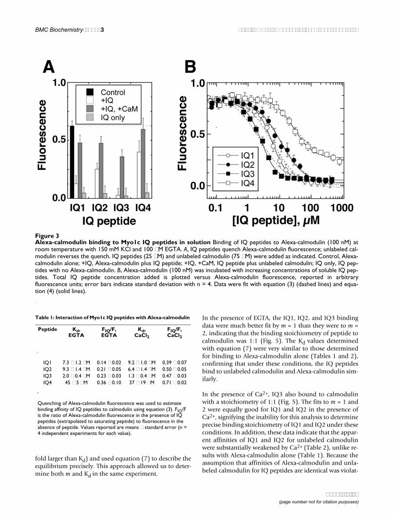
IQ – calmodulin interaction by quenching of Alexa-cal-modulin fluorescenceAs noted above, Alexa-calmodulin fluorescence wasquenched upon binding to an IQ peptide (Fig. 3A). Be-cause an excess of unlabeled calmodulin was able to re-verse 70–95% of the quench (Fig. 3A), we inferred thatmost Alexa-calmodulin bound to the same site as unla-beled calmodulin. We used this fluorescence-intensityquench empirically to measure the affinity of each IQ pep-tide for Alexa-calmodulin (Fig. 3B). In some experiments,Ca2+ was held at <30 nM by chelation with 100 µM EGTA;in other experiments, we added 25 µM exogenous CaCl2in the absence of EGTA. These two concentrations mimicthe low- and high-Ca2+ conditions that Myo1c may en-counter in hair cells when the transduction channel isclosed or open [26]. In the presence of 100 µM EGTA, Kdvalues followed the order IQ3 < IQ1 � IQ2 << IQ4. Al-though the data were fit somewhat better with a modifiedHill equation that with a standard bimolecular-bindingisotherm (Fig. 3B), the physiological significance of Hillcoefficients >1 is uncertain, particularly given the 1:1 pep-
tide:calmodulin stoichiometry (see below). Ca2+ had onlymodest effects on the affinity of the Myo1c IQ peptides forAlexa-calmodulin (Table 1).
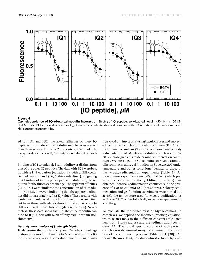
Despite only minor effects on binding affinity, Ca2+ didinfluence the calmodulin-peptide complex, as signaled bychanges in Alexa-calmodulin fluorescence. Changes influorescence intensity during manipulation of a single pa-rameter, like Ca2+ concentration, should report conforma-tional changes in Alexa-calmodulin. For example, thefluorescence intensity of free Alexa-calmodulin in solu-tion was ~15% lower in 25 µM CaCl2 than in 100 µMEGTA (left-hand limits in Fig. 4). Because the dye moietyitself is not Ca2+ sensitive [27], the Ca2+-dependent fluo-rescent change reflects changes in the dye's surroundingenvironment, probably signaling the compact-to-openstructural change seen when Ca2+ binds to calmodulin[28]. In contrast to the reduction of free Alexa-calmodulinfluorescence by Ca2+, fluorescence of Alexa-calmodulinwhen saturated by IQ peptides was 1.5- to 2-fold greater in25 µM CaCl2 than in 100 µM EGTA (Fig. 4; Table 1). Thus,
Figure 1Myo1c constructs and IQ-domain sequences A, domain structure of Myo1c, indicating amino acids encompassed by full-length Myo1c and T701-Myo1c constructs. B, SDS-PAGE of 15 pmol each of purified full-length Myo1c and T701-Myo1c con-structs co-expressed with calmodulin, and calmodulin alone (CaM; 45 pmol) on a 18% acrylamide gel. Molecular mass markersindicated on left. C, aligned bullfrog Myo1c IQ domains. The IQ motif positions are in bold and the repeat present in IQ3 andIQ4 is underlined. Numbers indicate amino-acid residue positions.
Page 3 of 17(page number not for citation purposes)
BMC Biochemistry 2002, 3 http://www.biomedcentral.com/1471-2091/3/31
when Alexa-calmodulin was bound to IQ peptides, Ca2+
induced a conformational change that was substantiallydifferent from that seen in the peptide-free state.
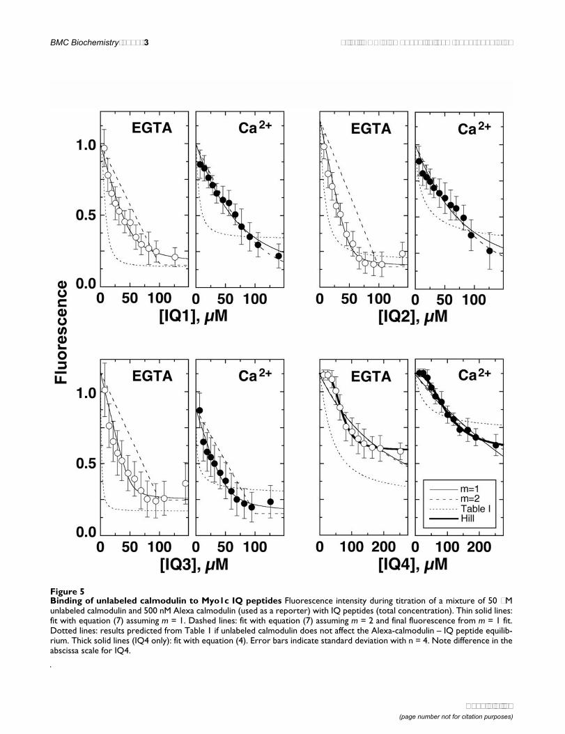
IQ – calmodulin interaction under stoichiometric-titration conditionsTo determine the affinities of the Myo1c IQ peptides forunlabeled calmodulin, we used Alexa-calmodulin as a re-porter (Alexa-calmodulin : unlabeled calmodulin ratio of1:100) in our binding studies. This approach assumes thatAlexa-calmodulin is functionally equivalent to unlabeledcalmodulin.
We determined affinities by fitting the IQ-peptide concen-tration vs. fluorescence quench data with an appropriatemodel. If the IQ peptides bound only Alexa-calmodulinand not unlabeled calmodulin, the Kd and FIQ/F values ofTable 1 would have described the fit to the concentration-quench plots. The line derived from these values did not
fit the data (Fig. 5), indicating that, as expected, unlabeledcalmodulin binds to the Myo1c IQ peptides.
These experimental conditions resembled a stoichiomet-ric titration, where the total concentration of calmodulinwas higher than the Kd values for IQ1, IQ2, and IQ3. Un-der true stoichiometric-titration conditions (fixed concen-tration of receptor at 100-fold or more than the Kd,varying the ligand concentration up to and beyond the re-ceptor concentration), almost all of the added IQ peptidewould bind tightly to calmodulin and linearly decreasethe fluorescence; at the point where the IQ-peptide con-centration exceeds the calmodulin concentration multi-plied by the peptide:calmodulin stoichiometry (m), aplateau in the fluorescence intensity would be reached.Because the relatively weak affinities observed here makesuch true stoichiometric titration impractical, we used anintermediate concentration of calmodulin (50 µM, ~10-
Figure 2Alexa-calmodulin binding to Myo1c IQ peptides on plates A, Alexa-calmodulin (50 nM) was incubated in a 96-well platecovalently bound with Myo1c IQ peptides, a positive-control IQ peptide (NM), or a negative-control peptide (PVP); bindingwas carried out at room temperature in the presence of low (150 mM) or high (400 mM) KCl and in the presence of 100 µMEGTA. We measured unbound Alexa-calmodulin concentration by fluorescence, then inferred the amount bound. Averageddata from three independent experiments (error bars indicate standard error). B, competition with free IQ peptides. Followingincubation of 50 nM Alexa-calmodulin and free IQ peptides in the presence of 100 µM EGTA and 150 mM KCl in wells of anIQ3-derivatized plastic plate, unbound Alexa-calmodulin was measured; error bars indicate standard deviation with n = 6 (sin-gle experiment). To correct for quenching of Alexa-calmodulin fluorescence by IQ peptides, each sample was standardizedagainst an identical reaction in an underivatized plate. Data were fit with equation (4) with the Hill coefficient set at 2.
Page 4 of 17(page number not for citation purposes)
BMC Biochemistry 2002, 3 http://www.biomedcentral.com/1471-2091/3/31
fold larger than Kd) and used equation (7) to describe theequilibrium precisely. This approach allowed us to deter-mine both m and Kd in the same experiment.
In the presence of EGTA, the IQ1, IQ2, and IQ3 bindingdata were much better fit by m = 1 than they were to m =2, indicating that the binding stoichiometry of peptide tocalmodulin was 1:1 (Fig. 5). The Kd values determinedwith equation (7) were very similar to those determinedfor binding to Alexa-calmodulin alone (Tables 1 and 2),confirming that under these conditions, the IQ peptidesbind to unlabeled calmodulin and Alexa-calmodulin sim-ilarly.
In the presence of Ca2+, IQ3 also bound to calmodulinwith a stoichiometry of 1:1 (Fig. 5). The fits to m = 1 and2 were equally good for IQ1 and IQ2 in the presence ofCa2+, signifying the inability for this analysis to determineprecise binding stoichiometry of IQ1 and IQ2 under theseconditions. In addition, these data indicate that the appar-ent affinities of IQ1 and IQ2 for unlabeled calmodulinwere substantially weakened by Ca2+ (Table 2), unlike re-sults with Alexa-calmodulin alone (Table 1). Because theassumption that affinities of Alexa-calmodulin and unla-beled calmodulin for IQ peptides are identical was violat-
Figure 3Alexa-calmodulin binding to Myo1c IQ peptides in solution Binding of IQ peptides to Alexa-calmodulin (100 nM) atroom temperature with 150 mM KCl and 100 µM EGTA. A, IQ peptides quench Alexa-calmodulin fluorescence; unlabeled cal-modulin reverses the quench. IQ peptides (25 µM) and unlabeled calmodulin (75 µM) were added as indicated. Control, Alexa-calmodulin alone; +IQ, Alexa-calmodulin plus IQ peptide; +IQ, +CaM, IQ peptide plus unlabeled calmodulin; IQ only, IQ pep-tides with no Alexa-calmodulin. B, Alexa-calmodulin (100 nM) was incubated with increasing concentrations of soluble IQ pep-tides. Total IQ peptide concentration added is plotted versus Alexa-calmodulin fluorescence, reported in arbitraryfluorescence units; error bars indicate standard deviation with n = 4. Data were fit with equation (3) (dashed lines) and equa-tion (4) (solid lines).
Table 1: Interaction of Myo1c IQ peptides with Alexa-calmodulin
Peptide Kd, EGTA
FIQ/F, EGTA
Kd, CaCl2
FIQ/F, CaCl2
IQ1 7.3 ± 1.2 µM 0.14 ± 0.02 9.2 ± 1.0 µM 0.39 ± 0.07IQ2 9.3 ± 1.4 µM 0.21 ± 0.05 6.4 ± 1.4 µM 0.50 ± 0.05IQ3 2.0 ± 0.4 µM 0.23 ± 0.03 1.3 ± 0.4 µM 0.47 ± 0.03IQ4 45 ± 5 µM 0.36 ± 0.10 37 ± 19 µM 0.71 ± 0.02
Quenching of Alexa-calmodulin fluorescence was used to estimate binding affinity of IQ peptides to calmodulin using equation (3). FIQ/F is the ratio of Alexa-calmodulin fluorescence in the presence of IQ peptides (extrapolated to saturating peptide) to fluorescence in the absence of peptide. Values reported are means ± standard error (n = 4 independent experiments for each value).
Page 5 of 17(page number not for citation purposes)
BMC Biochemistry 2002, 3 http://www.biomedcentral.com/1471-2091/3/31
ed for IQ1 and IQ2, the actual affinities of these IQpeptides for unlabeled calmodulin may be even weakerthan those reported in Table 2. By contrast, Ca2+ had onlya very modest effect on IQ3 affinity for unlabeled calmod-ulin.
Binding of IQ4 to unlabeled calmodulin was distinct fromthat of the other IQ peptides. The data with IQ4 were bestfit with a Hill equation (equation 4), with a Hill coeffi-cient of greater than 2 (Fig. 5, thick solid lines), suggestingthat binding of two peptides per calmodulin may be re-quired for the fluorescence change. The apparent affinities(~100 µM) were similar to the concentration of calmodu-lin (50 µM), however, indicating that the apparent affini-ties did not accurately reflect Kd values. These results witha mixture of unlabeled and Alexa-calmodulin were differ-ent from those with Alexa-calmodulin alone, where IQ4Hill coefficients were close to 1 (data not shown). Never-theless, these data show that unlabeled calmodulin canbind to IQ4, albeit with weak affinity and uncertain stoi-chiometry.
Hydrodynamic analysis of full-length Myo1cTo determine the stoichiometry and Ca2+-dependent reg-ulation of calmodulin binding to Myo1c with all four IQmotifs, we co-expressed calmodulin and full-length bull-
frog Myo1c in insect cells using baculoviruses and subject-ed the purified Myo1c-calmodulin complexes (Fig. 1B) tohydrodynamic analysis (Table 3). We carried out velocitysedimentation of Myo1c-calmodulin complexes on 5–20% sucrose gradients to determine sedimentation coeffi-cients. We measured the Stokes radius of Myo1c-calmod-ulin complexes using gel filtration on Superdex 200 undertemperature and buffer conditions identical to those ofthe velocity-sedimentation experiments (Table 3). Al-though most experiments used 400 mM KCl (which pre-vented adsorption to the gel-filtration matrix), weobtained identical sedimentation coefficients in the pres-ence of 150 or 250 mM KCl (not shown). Velocity-sedi-mentation and gel-filtration experiments were carried outat 4°C, the temperature used for Myo1c purification, aswell as at 25°C, a physiologically relevant temperature fora bullfrog.
To calculate the molecular mass of Myo1c-calmodulincomplexes, we applied the modified Svedberg equation,which relates mass to the diffusion constant (calculatedhere from Stokes radius) and the sedimentation coeffi-cient [29]. The partial specific volume of each proteincomplex was determined using the amino-acid composi-tion of the constituent proteins (Table 3; ref. [30]). Al-though the uncertainty in calmodulin stoichiometry leads
Figure 4Ca2+-dependence of IQ-Alexa-calmodulin interaction Binding of IQ peptides to Alexa-calmodulin (50 nM) in 100 µMEGTA or 25 µM CaCl2 as described for Fig. 3; error bars indicate standard deviation with n = 6. Data were fit with a modifiedHill equation (equation (4)).
Page 6 of 17(page number not for citation purposes)
BMC Biochemistry 2002, 3 http://www.biomedcentral.com/1471-2091/3/31
Figure 5Binding of unlabeled calmodulin to Myo1c IQ peptides Fluorescence intensity during titration of a mixture of 50 µMunlabeled calmodulin and 500 nM Alexa calmodulin (used as a reporter) with IQ peptides (total concentration). Thin solid lines:fit with equation (7) assuming m = 1. Dashed lines: fit with equation (7) assuming m = 2 and final fluorescence from m = 1 fit.Dotted lines: results predicted from Table 1 if unlabeled calmodulin does not affect the Alexa-calmodulin – IQ peptide equilib-rium. Thick solid lines (IQ4 only): fit with equation (4). Error bars indicate standard deviation with n = 4. Note difference in theabscissa scale for IQ4.
Page 7 of 17(page number not for citation purposes)
BMC Biochemistry 2002, 3 http://www.biomedcentral.com/1471-2091/3/31
to ambiguity in this calculation, the calculated partial spe-cific volumes were so close (e.g., 0.734 for one and 0.731for three calmodulins per Myo1c complex) that the pre-cise value did not significantly affect the final molecular-mass value.
Full-length Myo1c bound ~3 calmodulins per Myo1c at4°C in the presence of EGTA or CaCl2 (Table 3). One ofthe bound calmodulins was only weakly associated, as el-evation of the temperature to 25°C induced the release of1 mole of calmodulin in the presence of EGTA. WhenCa2+ was elevated to 25 µM at 25°C, however, we couldnot detect substantial full-length Myo1c in solution after
sucrose-gradient centrifugation or gel filtration, suggest-ing that the protein had aggregated.
Hydrodynamic analysis of T701-Myo1cBecause the size of full-length Myo1c (125 kD, includingpurification and detection tags) is much larger than cal-modulin (16.7 kD), we improved our ability to determinestoichiometry from molecular mass by examining a small-er (45 kD) neck-tail recombinant fragment of Myo1c. Thisconstruct, T701-Myo1c, contained amino acids 701–1028of bullfrog Myo1c, including all four IQ domains, the en-tire C-terminal tail, and N-terminal purification andepitope tags (Fig. 1A,1B).
T701-Myo1c bound 2.5 moles of calmodulin per mole ofheavy chain at 4°C in the presence of 100 µM EGTA (Fig.6; Table 4). As with full-length Myo1c, elevation of theanalysis temperature to 25°C induced the release of ~0.7mole of calmodulin. In contrast to the results seen withfull-length Myo1c, elevation of the CaCl2 concentration to25 µM at 4°C also induced the release of ~0.7 mole of cal-modulin. The amount of T701-calmodulin complex re-covered on sucrose gradients or by gel filtration decreasedsubstantially when the CaCl2 concentration was elevatedto 25 µM at 25°C, signaling the formation of aggregates,as seen with the full-length complex. Furthermore, the cal-culated calmodulin stoichiometry of the observed T701-calmodulin complex under these conditions was only~0.3 mole of calmodulin per mole of Myo1c, reinforcingthe suggestion that Ca2+ induced the dissociation of mostcalmodulins at 25°C and that this loss of light chains re-sulted in aggregation.
Table 2: Interaction of Myo1c IQ peptides with unlabeled calmodulin
Peptide Apparent Kd, EGTA
Apparent Kd, CaCl2
IQ1 4.0 ± 0.8 µM 56 ± 13 µMIQ2 4.2 ± 2.0 µM 70 ± 15 µMIQ3 1.7 ± 0.7 µM 3.2 ± 1.1 µM
Quenching of Alexa-calmodulin fluorescence was used to estimate binding affinity of IQ peptides to calmodulin using equation (7). Values reported are means ± standard error (n = 3 to 4 independent experi-ments for each value). IQ4 affinities were not accurately measured under conditions of this assay, but were >100 µM.
Table 3: Hydrodynamic analysis of full-length Myo1c
Property Temperature Full-length Myo1c 100 µM EGTA
Full-length Myo1c 25 µM CaCl2
Sedimentation coefficient 4°C 6.70 ± 0.07 S (n = 5) 6.90 ± 0.08 S (n = 4)25°C 6.28 ± 0.21 S (n = 8) *
Stokes radius 4°C 6.06 ± 0.08 nm (n = 7) 6.07 ± 0.05 nm (n = 4)25°C 5.78 ± 0.11 nm (n = 5) *
Molecular mass 4°C 168 ± 4 kD 4°C: 173 ± 3 kD25°C 150 ± 7 kD ND
Frictional ratio 4°C 1.7 ± 0.1 1.7 ± 0.125°C 1.7 ± 0.1 ND
Calmodulins per molecule 4°C 2.5 ± 0.2 2.9 ± 0.225°C 1.5 ± 0.4 ND
* Very low levels of protein detected. Full-length Myo1c was co-expressed with calmodulin, purified, and subjected to sucrose-gradient centrifugation or gel-filtration analysis. Myo1c was detected by ELISA (sucrose gradients) or by absorption at 280 nm (gel filtration). Molecular mass was determined using the Stokes-Einstein equa-tion; the number of calmodulins was estimated by subtracting the mass of the Myo1c heavy chain from the estimated molecular mass, then dividing the remainder by the mass of calmodulin (16.7 kD). Values reported are mean ± standard deviation. ND, not determined.
Page 8 of 17(page number not for citation purposes)
BMC Biochemistry 2002, 3 http://www.biomedcentral.com/1471-2091/3/31
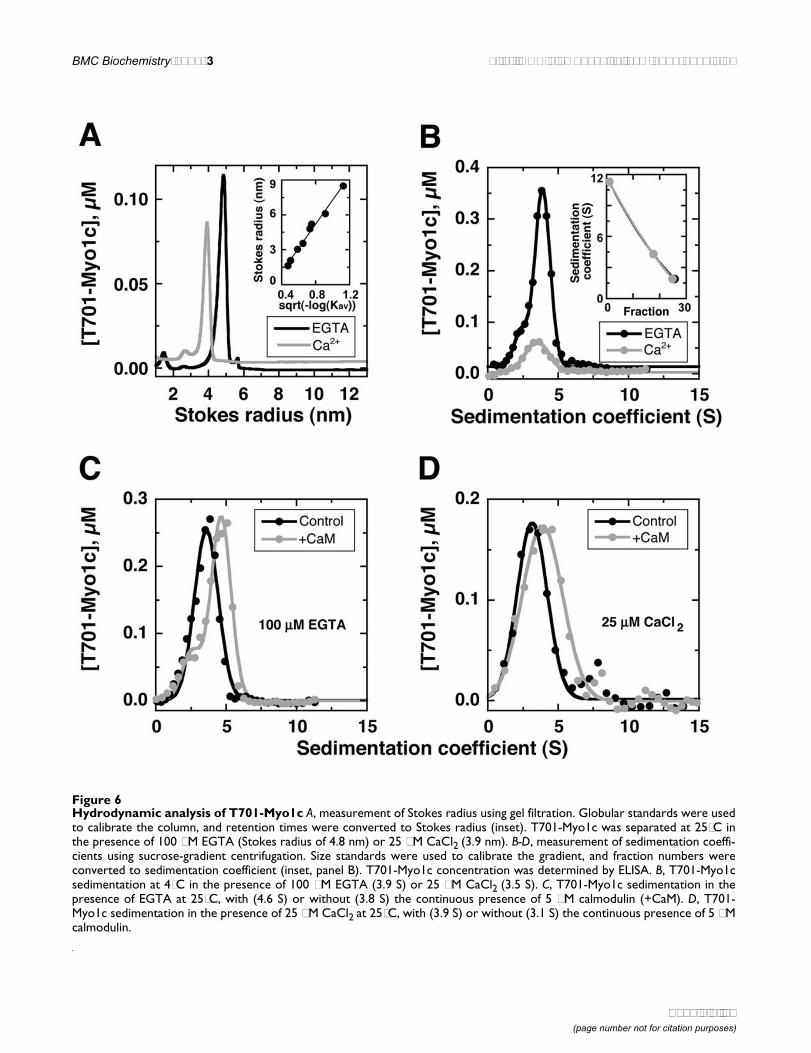
Figure 6Hydrodynamic analysis of T701-Myo1c A, measurement of Stokes radius using gel filtration. Globular standards were usedto calibrate the column, and retention times were converted to Stokes radius (inset). T701-Myo1c was separated at 25°C inthe presence of 100 µM EGTA (Stokes radius of 4.8 nm) or 25 µM CaCl2 (3.9 nm). B-D, measurement of sedimentation coeffi-cients using sucrose-gradient centrifugation. Size standards were used to calibrate the gradient, and fraction numbers wereconverted to sedimentation coefficient (inset, panel B). T701-Myo1c concentration was determined by ELISA. B, T701-Myo1csedimentation at 4°C in the presence of 100 µM EGTA (3.9 S) or 25 µM CaCl2 (3.5 S). C, T701-Myo1c sedimentation in thepresence of EGTA at 25°C, with (4.6 S) or without (3.8 S) the continuous presence of 5 µM calmodulin (+CaM). D, T701-Myo1c sedimentation in the presence of 25 µM CaCl2 at 25°C, with (3.9 S) or without (3.1 S) the continuous presence of 5 µMcalmodulin.
Page 9 of 17(page number not for citation purposes)
BMC Biochemistry 2002, 3 http://www.biomedcentral.com/1471-2091/3/31
We could not prevent the release of calmodulin at 25°C bysaturating T701-Myo1c with excess calmodulin immedi-ately prior to centrifugation (preloading). In EGTA, thesedimentation coefficient of calmodulin-preloaded T701-Myo1c measured at 25°C (3.85 ± 0.07 S; n = 2) was nearlyidentical to that measured without preloading (3.83 S; Ta-ble 4). Likewise, the sedimentation coefficient of calmod-ulin-preloaded T701-Myo1c measured at 25°C and in25 µM CaCl2 (2.80 ± 0.42 S; n = 2) was similar to thatmeasured without preloading (3.13 S; Table 4).
By contrast, we could prevent the temperature-dependentloss of calmodulin by carrying out sedimentation in thecontinuous presence of 5 µM calmodulin (Fig. 6C,6D; Ta-ble 5). Although gel-filtration analysis was impracticalwith this high calmodulin concentration, we assumedthat the Stokes radius of T701-Myo1c in the presence ofcalmodulin was identical to the value obtained in the ab-
sence. Sedimentation at 25°C in EGTA gradients supple-mented with 5 µM calmodulin resulted in the retention of~3 calmodulins per T701-Myo1c. In 25 µM CaCl2, supple-mentation with 5 µM calmodulin resulted in ~1 calmod-ulin bound per T701-Myo1c. In addition, protein loss dueto aggregation was minimal under these conditions.
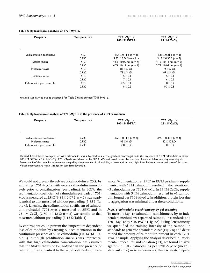
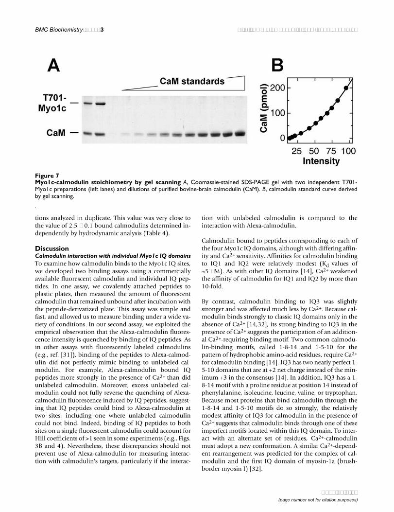
Myo1c-calmodulin stoichiometry by gel scanningTo measure Myo1c-calmodulin stoichiometry by an inde-pendent method, we separated calmodulin standards andT701-Myo1c by SDS-PAGE (Fig. 7A). Using densitometry,we quantified the staining intensity of the calmodulinstandards to generate a standard curve (Fig. 7B) and deter-mined the amount of calmodulin present in each T701-Myo1c sample. Applying the analysis described in Experi-mental Procedures and equation (13), we found an aver-age of 2.6 ± 0.2 calmodulins per T701-Myo1c (mean ±standard error) in six experiments, three separate prepara-
Table 4: Hydrodynamic analysis of T701-Myo1c.
Property Temperature T701-Myo1c 100 µM EGTA
T701-Myo1c 25 µM CaCl2
Sedimentation coefficient 4°C 4.64 ± 0.11 S (n = 4) 4.27 ± 0.21 S (n = 3)25°C 3.83 ± 0.06 S (n = 11) 3.13 ± 0.30 S (n = 7)
Stokes radius 4°C 4.52 ± 0.06 nm (n = 4) 4.19 ± 0.11 nm (n = 6)25°C 4.74 ± 0.13 nm (n = 6) 3.78 ± 0.07 nm (n = 6)
Molecular mass 4°C 87 ± 5 kD 74 ± 6 kD25°C 75 ± 3 kD 49 ± 5 kD
Frictional ratio 4°C 1.5 ± 0.1 1.5 ± 0.125°C 1.7 ± 0.1 1.6 ± 0.2
Calmodulins per molecule 4°C 2.5 ± 0.1 1.8 ± 0.225°C 1.8 ± 0.2 0.3 ± 0.3
Analysis was carried out as described for Table 3 using purified T701-Myo1c.
Table 5: Hydrodynamic analysis of T701-Myo1c in the presence of 5 µM calmodulin
Property Temperature T701-Myo1c 100 µM EGTA
T701-Myo1c 25 µM CaCl2
Sedimentation coefficient 25°C 4.68 ± 0.11 S (n = 2) 3.95 ± 0.33 S (n = 4)Molecular mass 25°C 92 ± 4 kD 62 ± 12 kD
Calmodulins per molecule 25°C 2.8 ± 0.2 1.0 ± 0.7
Purified T701-Myo1c co-expressed with calmodulin, was subjected to sucrose-gradient centrifugation in the presence of 5 µM calmodulin in either 100 µM EGTA or 25 µM CaCl2. T701-Myo1c was detected by ELISA. We estimated molecular mass and hence stoichiometry by assuming that Stokes radii of the complexes were unchanged by the presence of calmodulin, an assumption that might have led to an underestimate of the mass. Values reported are mean ± range or standard deviation.
Page 10 of 17(page number not for citation purposes)
BMC Biochemistry 2002, 3 http://www.biomedcentral.com/1471-2091/3/31
tions analyzed in duplicate. This value was very close tothe value of 2.5 ± 0.1 bound calmodulins determined in-dependently by hydrodynamic analysis (Table 4).
DiscussionCalmodulin interaction with individual Myo1c IQ domainsTo examine how calmodulin binds to the Myo1c IQ sites,we developed two binding assays using a commerciallyavailable fluorescent calmodulin and individual IQ pep-tides. In one assay, we covalently attached peptides toplastic plates, then measured the amount of fluorescentcalmodulin that remained unbound after incubation withthe peptide-derivatized plate. This assay was simple andfast, and allowed us to measure binding under a wide va-riety of conditions. In our second assay, we exploited theempirical observation that the Alexa-calmodulin fluores-cence intensity is quenched by binding of IQ peptides. Asin other assays with fluorescently labeled calmodulins(e.g., ref. [31]), binding of the peptides to Alexa-calmod-ulin did not perfectly mimic binding to unlabeled cal-modulin. For example, Alexa-calmodulin bound IQpeptides more strongly in the presence of Ca2+ than didunlabeled calmodulin. Moreover, excess unlabeled cal-modulin could not fully reverse the quenching of Alexa-calmodulin fluorescence induced by IQ peptides, suggest-ing that IQ peptides could bind to Alexa-calmodulin attwo sites, including one where unlabeled calmodulincould not bind. Indeed, binding of IQ peptides to bothsites on a single fluorescent calmodulin could account forHill coefficients of >1 seen in some experiments (e.g., Figs.3B and 4). Nevertheless, these discrepancies should notprevent use of Alexa-calmodulin for measuring interac-tion with calmodulin's targets, particularly if the interac-
tion with unlabeled calmodulin is compared to theinteraction with Alexa-calmodulin.
Calmodulin bound to peptides corresponding to each ofthe four Myo1c IQ domains, although with differing affin-ity and Ca2+ sensitivity. Affinities for calmodulin bindingto IQ1 and IQ2 were relatively modest (Kd values of~5 µM). As with other IQ domains [14], Ca2+ weakenedthe affinity of calmodulin for IQ1 and IQ2 by more than10-fold.
By contrast, calmodulin binding to IQ3 was slightlystronger and was affected much less by Ca2+. Because cal-modulin binds strongly to classic IQ domains only in theabsence of Ca2+ [14,32], its strong binding to IQ3 in thepresence of Ca2+ suggests the participation of an addition-al Ca2+-requiring binding motif. Two common calmodu-lin-binding motifs, called 1-8-14 and 1-5-10 for thepattern of hydrophobic amino-acid residues, require Ca2+
for calmodulin binding [14]. IQ3 has two nearly perfect 1-5-10 domains that are at +2 net charge instead of the min-imum +3 in the consensus [14]. In addition, IQ3 has a 1-8-14 motif with a proline residue at position 14 instead ofphenylalanine, isoleucine, leucine, valine, or tryptophan.Because most proteins that bind calmodulin through the1-8-14 and 1-5-10 motifs do so strongly, the relativelymodest affinity of IQ3 for calmodulin in the presence ofCa2+ suggests that calmodulin binds through one of theseimperfect motifs located within this IQ domain. To inter-act with an alternate set of residues, Ca2+-calmodulinmust adopt a new conformation. A similar Ca2+-depend-ent rearrangement was predicted for the complex of cal-modulin and the first IQ domain of myosin-1a (brush-border myosin I) [32].
Figure 7Myo1c-calmodulin stoichiometry by gel scanning A, Coomassie-stained SDS-PAGE gel with two independent T701-Myo1c preparations (left lanes) and dilutions of purified bovine-brain calmodulin (CaM). B, calmodulin standard curve derivedby gel scanning.
Page 11 of 17(page number not for citation purposes)
BMC Biochemistry 2002, 3 http://www.biomedcentral.com/1471-2091/3/31
In support of this view, we observed evidence for Ca2+-de-pendent conformational changes in calmodulin whilebound to IQ peptides. When Alexa-calmodulin wasbound to Myo1c IQ peptides, its fluorescence was higherin the presence of Ca2+ than in its absence, suggesting thatthat Ca2+-bound Alexa-calmodulin binds to the IQ pep-tides in a different conformation than does Ca2+-free Al-exa-calmodulin. For example, in the absence of Ca2+,Alexa-calmodulin may bind to IQ peptides in a morecompact conformation, quenching fluorescence by bury-ing dye moieties in a less polar environment. Althoughthe Ca2+-induced conformational change could be a prop-erty of Alexa-calmodulin rather than calmodulin itself, theCa2+-dependent changes in affinity of calmodulin for IQ1and IQ2 (Table 2) and calmodulin's likely shift to a newbinding site on IQ3 suggests that the conformationalchange is probably also a property of authentic calmodu-lin.
Calmodulin also bound to a newly identified domain,IQ4. Because the affinity of calmodulin for IQ4 is veryweak, calmodulin should only occupy IQ4 in subcellularlocations with a low Ca2+ concentration and a high levelof free calmodulin. For example, a small population ofMyo1c molecules with calmodulin bound to IQ4 shouldbe present in the stereocilia of inner-ear hair cells, whichcontain ~35 µM free calmodulin [33]. Although most tis-sues contain less free calmodulin [34], concentrations inother individual organelles can reach the millimolar range[35]. On the other hand, the weak affinity of this IQ do-main for calmodulin suggests that IQ4 may play anotherrole, such as interacting with another protein.
Calmodulin interaction with Myo1cThe binding affinities of calmodulin for the individual IQpeptides do not reflect exactly the affinities of calmodulinfor the IQ domains within Myo1c. For example, despitemicromolar Kd values for calmodulin-IQ peptide interac-tions, calmodulin remains bound to Myo1c during longgel-filtration or centrifugation experiments, even at na-nomolar Myo1c concentrations (Fig. 6). This result sug-gests that calmodulin binds to some of Myo1c's fourtandem IQ domains substantially more strongly than tothe individual peptides. For example, other regions ofMyo1c could constrain the IQ domains in conformationsthat are substantially more (or less) favorable for calmod-ulin binding than the population of conformationsadopted by a soluble IQ peptide. Moreover, calmodulinbinding to Myo1c could be influenced by interactionswith adjacent calmodulin molecules or to the Myo1c heador tail domains.
To examine calmodulin binding to IQ domains in Myo1c,we determined the molecular mass (and hence calmodu-lin:Myo1c stoichiometry) and shape of Myo1c under the
appropriate conditions of temperature and Ca2+. Al-though analytical ultracentrifugation is more commonlyused to measure molecular size of protein-protein com-plexes [36], we instead used classic hydrodynamic meth-ods of velocity sedimentation on sucrose gradients toobtain sedimentation coefficients and gel filtration to ob-tain Stokes' radius. One advantage of this approach wasthat by detecting Myo1c using a sensitive ELISA method,we were able to use very low concentrations of Myo1c.Furthermore, we were able to carry out sedimentation inthe presence of a high concentration of calmodulin, a ma-nipulation that prevents Myo1c detection in a standardanalytical ultracentrifugation experiment. A disadvantageof this approach was the need for high concentrations ofsucrose, which in rare conditions can substantially affectthe hydrodynamic properties of a protein [37]; neverthe-less, changes in Myo1c size were observed both in velocitysedimentation (in the presence of sucrose) and in gel fil-tration (in its absence). Another disadvantage of our clas-sic approach to molecular-mass determination was thatthe gel filtration and velocity sedimentations were doneon different time scales (~1 hour vs. 15–18 hours). If cal-modulin slowly dissociated during the analysis (which inboth assays diluted Myo1c well below 1 µM), the degreeof dissociation would be larger in the velocity sedimenta-tion experiments than in the gel filtration experiments.Nevertheless, our approach was validated by the demon-stration that the number of calmodulins per T701-Myo1cwas identical in hydrodynamic and gel-scanning experi-ments, at least in EGTA at 4°C.
Because T701-Myo1c mimicked properties of the full-length protein (except under low-temperature, high-Ca2+
conditions), we exploited the neck-tail construct for amore detailed analysis of calmodulin binding. As expect-ed from the large Ca2+-dependent weakening of calmod-ulin affinity for IQ1 and IQ2 (Fig. 5; Table 2), Ca2+
decreased the number of calmodulins bound to T701-Myo1c at high ionic strength. When Ca2+ was low at 25°C,each T701-Myo1c had about two bound calmodulins,with a third bound if the calmodulin concentrationreached 5 µM. At this calmodulin concentration, IQ do-mains 1, 2, and 3 are likely occupied by calmodulin.When Ca2+ is high at 25°C, all but one calmodulin disso-ciated from T701-Myo1c in the presence of 5 µM free cal-modulin. The strong affinity of IQ3 for Ca2+-calmodulinsuggests that the remaining calmodulin was bound to thisIQ domain.
How many calmodulins are bound to Myo1c in the cell atincreased Ca2+ concentrations? The elevated ionicstrength used for the hydrodynamic analysis probablyweakened the affinity of the calmodulin for IQ3 (Fig. 2A),requiring 5 µM free calmodulin to maintain occupancy ofthat site. We therefore infer that at a physiologically signif-
Page 12 of 17(page number not for citation purposes)
BMC Biochemistry 2002, 3 http://www.biomedcentral.com/1471-2091/3/31
icant temperature and at a cellular ionic strength, Ca2+
triggers release of calmodulins from IQ1 and IQ2 fromT701-Myo1c, leaving only IQ3 occupied. Although theseresults contrast with those reported for mammalianMyo1c, where only one of three calmodulins is releasedby Ca2+ [19,38], our T701 construct lacks Myo1c's motordomain. It is entirely plausible that even in the presenceof Ca2+, calmodulin remains bound to IQ1, albeit in a dif-ferent conformation and dependent on interactions withthe myosin head. Our results therefore suggest that Ca2+
either induces the release of calmodulin from IQ1 or caus-es it to change its interaction with Myo1c substantially.
Of the three calmodulins bound to Myo1c, one of thesebinds relatively weakly at 25°C, even in EGTA. To whichIQ domain does this weakly bound light chain bind? Al-though calmodulin binds to IQs 1–3 with approximatelythe same strength in the presence of EGTA, we suggest thatthe readily released calmodulin is likely to be that boundto IQ2. To bind three calmodulins, IQs 1–3, each of whichare only 23 amino acids long, must be arranged withoutkinks [32]; this arrangement may produce unfavorablestrain on each of the calmodulin molecules. Release ofcalmodulin from IQ2 would relieve all of that strain; re-lease from IQs 1 or 3 would not. Strain relief also may ac-celerate calmodulin release in the presence of Ca2+;because Ca2+ apparently rearranges the three-dimensionalinteraction of calmodulin with IQ3, binding of an adja-cent calmodulin – on IQ2 – might be destabilized evenmore [32].
Despite the loss of calmodulin from T701-Myo1c inducedby elevation of the temperature from 4°C to 25°C, the fric-tional ratio (a measure of the protein's asymmetry) in-creased (Table 4). The neck-tail region of Myo1c thusappears to adopt a compact structure at 4°C, becomingmore extended at 25°C. Less calmodulin may be releasedat lower temperatures because the Myo1c tail may bind toand stabilize calmodulin's interaction with the Myo1cneck.
Implications for Myo1c activityThe Ca2+-dependent change in interaction of calmodulinwith IQ1, the IQ domain closest to the motor domain, hasimportant implications for Myo1c mechanochemicalfunction. Although Ca2+ increases Myo1c ATPase activity,the ion completely halts in vitro motility [19]. Ca2+-de-pendent changes in conformation may prevent amplifica-tion of a small converter-domain movement into a largemotor step. In the presence of an external force, as is seenby Myo1c during an excitatory mechanical stimulus in ahair cell [39], Ca2+ (which enters the cell through opentransduction channels), should permit Myo1c to gothrough its ATPase cycle, binding and unbinding from ac-tin, but the altered interaction of calmodulin and IQ1
may prevent force production by the motor. We predictthat Ca2+ will decrease the stiffness of a Myo1c-actin inter-action, preventing coupling of the energy released by ATPhydrolysis to the swing of the neck [40]. This behavior willassist Myo1c in its role of adaptation in hair cells, wherethe motor reduces force applied to the hair cell's transduc-tion channel.
A limitation of our experiments is the restriction of Myo1cbinding to a single type of light chain, calmodulin. Otherlight chains can interact with IQ domains, including es-sential light chain isoforms [41] and calmodulin-like pro-tein [42]. Although purified bovine adrenal Myo1c doesnot appear to have alternative associated light chains [12],we can not rule out the possibility that other light chainsbind in a cellular context. Nevertheless, purified recom-binant full-length Myo1c associated with calmodulinlight chains exhibited actin-activated ATPase activity andmotility in vitro [43], indicating that calmodulin can func-tion as a Myo1c light chain.
That Myo1c does not bind calmodulin tightly is, at firstglance, surprising. Weak calmodulin binding may, how-ever, permit access of IQ domains to intracellular Myo1creceptors. Accordingly, we have found that a Myo1c frag-ment containing only IQs 1–3, partially complexed withcalmodulin, binds avidly to hair-cell receptors; excess cal-modulin blocks this interaction, probably by binding toan unoccupied IQ site on the Myo1c fragment [13]. IQ2 ishighly conserved between species, leading us to proposethat hair-cell receptors interact through this region [13].Because Myo1c-interacting proteins in hair cells and else-where may interact through IQ domains, regulation ofcalmodulin binding to Myo1c – for example, by Ca2+ –likely affects coupling of the motor protein to its cargo.
ConclusionsUnder low Ca2+ conditions and normal ionic strength,calmodulin binds moderately tightly to three Myo1c IQdomains, IQ1, IQ2, and IQ3. IQ4 will only be occupiedwhen the calmodulin concentration is very high. Whenlinearly arranged in the Myo1c molecule, at least one cal-modulin (most likely that bound to IQ2) is bound lesstightly, probably due to steric constraints. Upon bindingCa2+, calmodulin bound to IQ2 dissociates; that bound toIQ1 either dissociates or changes its conformation suffi-ciently that chemomechanical coupling cannot ensue.
MethodsPeptide – calmodulin interaction on platesBullfrog Myo1c IQ peptides were synthesized (GenemedSynthesis, South San Francisco, CA) with N-terminalcysteine residues: IQ1 (residues 698–720), CRKHSIAT-FLQARWRGYHQRQKFL; IQ2 (721–743), CHM-KHSAVEIQSWWRGTIGRRKAA; IQ3 (744–766),
Page 13 of 17(page number not for citation purposes)
BMC Biochemistry 2002, 3 http://www.biomedcentral.com/1471-2091/3/31
CKRKWAVDVVRRFIKGFIYRNQPR; and IQ4 (767–791;native cysteine at residue 767), CTENEYFLDYIRYSFLMT-LYRNQPK. Peptide concentrations were measured by de-termining optical density at 280 nm, using calculatedmolar extinction coefficients of 7090 (IQ1), 11500 (IQ2),7090 (IQ3), and 5240 M-1 cm -1 (IQ4). We also synthe-sized a negative-control peptide ("PVP") corresponding toamino acids 792–816 of frog Myo1c (SV-LDKSWPVPPPSLREASELLREMC; native C816) and a pos-itive control IQ-peptide ("NM") corresponding to aminoacids 29–52 of bovine neuromodulin with an added C-terminal cysteine (KAHKAATKIQASFRGHITRKKLKC)[24].
For measuring interaction of calmodulin with peptidesconjugated to plastic plates, we incubated 10 µM peptidein phosphate-buffered saline (PBS; 137 mM NaCl,2.7 mM KCl, 4.3 mM Na2HPO4, 1.4 mM KH2PO4, pH7.4) overnight at room temperature in a maleimide-deri-vatized 96-well plate (Pierce, Rockford, IL). Peptide waspresent in large excess over free binding sites(25–50 pmol) on the plates. To remove unconjugatedpeptides, plates were washed with PBS; unreacted siteswere saturated by incubating with 10 µg/ml cysteine for1 hour. We then incubated the peptide-conjugated plateswith 50 nM Alexa Fluor 488 calmodulin (Alexa-calmodu-lin; Molecular Probes, Eugene, OR) in 100 µl of a solutionthat contained 150 or 400 mM KCl, 1 mM MgCl2, 100 µMethylene glycol-bis(β-aminoethylether)-N,N,N',N'-tetraacetic acid (EGTA) or 25 µM CaCl2, and 15 mM 2-[4-(2-hydroxyethyl)-1-piperazinyl] ethanesulfonic acid(HEPES) at pH 7.5. According to the manufacturer, Alexa-calmodulin had two dye moieties per calmodulin mole-cule; the modified residues were likely Lys-75 and Lys-94,the most reactive of calmodulin's lysine residues [44]. Af-ter incubation for 2 hours at room temperature, we trans-ferred 50 µl of the solution to another 96-well plate andmeasured fluorescence (excitation 485 nm; emission,520 nm) using a BMG Labtechnologies Fluorostar 403microplate fluorometer (Durham, NC). Under the assayconditions, the inner-filter effect (absorption of excitationor emission photons by the sample) was negligible. Fromthis measurement, we calculated the amount of calmodu-lin bound to the conjugated peptides. In some experi-ments, we also included 0.1–100 µM unconjugated IQpeptide; in that case, we carried out duplicate control re-actions in underivatized 96-well plates to correct for fluo-rescence quenching exerted by IQ peptides.
Peptide – calmodulin interaction by fluorescence quenchWe used empirically observed changes in the fluorescenceintensity of Alexa-calmodulin, large in magnitude, tomeasure binding of IQ peptides to calmodulin. Peptidesand 50–500 nM Alexa-calmodulin were mixed in 96- or384-well microtiter plates with 150 mM KCl, 1 mM
MgCl2, 100 µM EGTA or 25 µM CaCl2, 0.5 mg/ml bovineserum albumin, and 15 mM HEPES at pH 7.5; in some ex-periments we added 50–75 µM bovine-brain calmodulin.Total volume varied from 10 µl (384-well plates) to100 µl (96-well plates). After 1–2 hours at room tempera-ture, fluorescence was read directly.
When IQ peptides bound to Alexa-calmodulin, the fluo-rescence intensity was reduced as the quantum yield de-creased (fluorescence quenching). We assumed that twofluorescent species were present, Alexa-calmodulin andIQ peptide-bound Alexa-calmodulin, and that the fluores-cence intensity (I) was a linear combination of the fluores-cence of the two species:
I = fCaM ICaM +fCaM-IQ ICaM-IQ (1)
where fCaM and fCaM-IQ are the mole fractions of the twocomponents and ICaM and ICaM-IQ are their fluorescenceintensities. Because the quantum yield of Alexa-calmodu-lin is reduced when IQ peptides bind, ICaM-IQ <ICaM. Thefraction of peptide bound is:
To calculate Kd, we fit the data with a bimolecular-bindingisotherm:
where [IQ] was the free IQ-peptide concentration added.Because we used concentrations of Alexa-calmodulin inour experiments that were much less than the Kd, we ap-proximated [IQ] using the total IQ peptide concentration.
In other cases, however, the binding data were fit betterwith a modified Hill equation:
where h is the Hill coefficient. A value for h greater thanone suggests the fluorescence change arose from a morecomplex equilibrium than just one peptide binding percalmodulin.
To carry out stoichiometric-titration experiments (cal-modulin concentration greater than the Kd), we used alow concentration of Alexa-calmodulin as a reporter andadded an excess of unlabeled calmodulin. For simplicityin analysis, we assumed that Alexa-calmodulin behavedidentically to calmodulin, and thus this calmodulin mix-ture was equivalent to a decrease in specific activity (fluo-rescence quench) of calmodulin. We then solved thebimolecular-binding isotherm to enable us to plot the to-
fI I
I ICaM-IQCaM-IQ
CaM CaM-IQ= −
−−
( )1 2
I I IK
Id
= −( ) ⋅ − [ ][ ] +
+ ( )− −CaM CaM IQ CaM IQ
IQ
IQ1 3
I I IK
Ih
hdh
= −( ) ⋅ − [ ][ ] +
+ ( )− −CaM CaM IQ CaM IQIQ
IQ1 4
Page 14 of 17(page number not for citation purposes)
BMC Biochemistry 2002, 3 http://www.biomedcentral.com/1471-2091/3/31
tal ligand concentration (T) added versus fluorescence in-tensity (I). The concentration of peptide bound (B) was:
B = m [CaM] fCaM-IQ (5)
where m is the number of binding sites per calmodulinand [CaM] is the fixed concentration of calmodulin. Thefree concentration of IQ peptide (F) was T – B. We substi-tuted the expression for B in equations (2) and (5) into:
Note that n [CaM] is the maximum amount of IQ peptidethat can bind (Bmax). We then solved equation (6) for flu-orescence intensity using Mathematica 4.0 (Wolfram Re-search, Champaign, IL):
For m = 1, the only free parameters were Kd and ICaM-IQ.We were forced to include ICaM-IQ as one of the fit param-eters; the limited solubility of IQ peptides in the assay so-lution prevented us from using very high peptideconcentrations that would independently establish its val-ue by producing a plateau in the T vs. I plot. We then usedthe value of ICaM-IQ determined from the m = 1 fit and refitthe data for m = 2, using Kd as the only free parameter. Tojudge the stoichiometry, we compared by eye the effective-ness of the fit under the two conditions.
Baculovirus constructsUsing methods described previously for rat Myo1c [43],we cloned full-length bullfrog Myo1c into the baculovirustransfer vector pBlueBacHis2B (Invitrogen, Carlsbad, CA),introducing an N-terminal hexahistidine tag for purifica-tion and a DLYDDDDK epitope tag for antibody detec-tion. Baculoviruses were generated, purified, andcharacterized using standard techniques [43,45].
Protein expression and purificationBullfrog Myo1c or its neck-tail fragment (Fig. 1A,1B) wereco-expressed with Xenopus calmodulin in Sf9 cells usingmethods described previously [43]. Xenopus calmodulin isidentical to all other sequenced vertebrate calmodulins,including bovine calmodulin [46]; we presume that bull-frog calmodulin is also identical. Recombinant proteinswere partially purified by centrifugation of an Sf9-cell ex-tract and Ni2+-nitrilotriacetic acid chromatography [43];further purification was achieved using gel filtration at4°C on a 25-ml Superdex 200 HR 10/30 column run at 0.5ml/min in 400 mM KCl, 1 mM MgCl2, 100 µM EGTA, 15mM HEPES pH 7.5 with an AKTA-FPLC system (Amer-sham Pharmacia Biotech, Piscataway, NJ). The concentra-tion of each purified recombinant protein was calculatedby measuring absorption at 280 nm and using extinction
coefficients calculated from the appropriate aminoacid se-quence using the ExPASy ProtParam tool [http://www.ex-pasy.ch/tools/protparam.html], assuming 2.5calmodulins per full-length Myo1c (53,619 M-1cm-1) orT701 fragment (65,565 M-1cm-1). We typically obtained100–300 µg of recombinant protein from ~109 Sf9 cells.Full-length Myo1c had NH4Cl-activated ATPase activity[12] of 1.8 ± 0.7 s-1, with a Km for ATP of 0.3 ± 0.1 mM.Actin activated basal Mg2+-ATPase activity ~15-fold. Cal-modulin was purified from bovine brain (Pel-Freez, Rog-ers, AR) by isoelectric precipitation and phenyl-agarose(Sigma, St. Louis, MO) chromatography [47]; its concen-tration was measured assuming a molar extinction coeffi-cient of 3030 M-1 cm -1 at 276 nm [48].
Gel filtrationStokes radii of Myo1c and T701-Myo1c were measured us-ing gel filtration on a 25-ml Superdex 200 HR 10/30 col-umn at either 4°C or room temperature (23–25°C).Columns were run at 0.5 ml/min in 400 mM KCl, 1 mMMgCl2, 15 mM HEPES pH 7.5, and either 100 µM EGTAor 25 µM CaCl2; 5–20 µg of recombinant protein was ap-plied to the column. Columns were calibrated using 20–200 µg each of globular proteins of known Stokes radii(thyroglobulin, 8.50 nm; ferritin, 6.10 nm; catalase, 5.22nm; aldolase, 4.81 nm; bovine serum albumin, 3.55 nm;ovalbumin, 3.05 nm; chymotrypsinogen, 2.09 nm; andRNase A, 1.64 nm; all obtained from Amersham Pharma-cia Biotech). Proteins were detected by absorption at 280nm.
Velocity sedimentation on sucrose gradientsSedimentation coefficients of full-length and T701-Myo1cwere measured using linear 5–20% sucrose gradients in11.5 ml of 400 mM KCl, 1 mM MgCl2, 15 mM HEPES pH7.5, 0.2 mM phenylmethylsulfonyl fluoride, 10 µM leu-peptin, 10 µM pepstatin, and either 100 µM EGTA or 25µM CaCl2. Gradients were calibrated with 2–20 µg inter-nal standards of known sedimentation coefficients (cata-lase, 11.3 S; bovine serum albumin, 4.31 S; lysozyme,1.91 S; all obtained from Sigma-Aldrich). After centrifuga-tion at 33,000–40,000 rpm in an SW 41 rotor for 15–18hours at 4°C or 25°C, gradients were fractionated from thebottom into ~30 fractions. Calibration proteins were lo-cated using a Bradford protein assay [49]; Myo1c-contain-ing fractions were located by ELISA [43] using an antibodyagainst the Myo1c tail (mT2/M2; ref. [50]) or against theDLYDDDDK epitope tag (anti-Xpress; Invitrogen). To de-termine the location of protein peaks, plots of fractionnumber versus the levels of Myo1c or calibration proteinswere fit with either one, two, or three Gaussian curves.
For calmodulin preloading of T701-Myo1c, 10 µM puri-fied calmodulin was incubated with 1 µM Myo1c in a so-
Bm
FFCaM Kd[ ] =
+( )6
Im
I m I m I TI
I
= − [ ] − − [ ] − [ ] −( +− − −1
2 CaMK CaM CaMd CaM IQ CaM IQ CaM CaM IQ
CCaM IQ CaM d d dK K CaM CaM K CaM− −( ) + [ ] + [ ] + − [ ] +
( )I m m T mT T2 2 2 2
72 2 2
Page 15 of 17(page number not for citation purposes)
BMC Biochemistry 2002, 3 http://www.biomedcentral.com/1471-2091/3/31
lution containing either 100 µM EGTA or 25 µM CaCl2 for60 min at room temperature prior to centrifugation.
Determination of molecular massWe used the modified Svedberg equation for molecular-mass determination:
where M = molecular mass, η = viscosity of the medium,N = Avogadro's number, a = Stokes radius, s = sedimenta-tion coefficient, = partial specific volume, and ρ = den-sity of the medium. Partial specific volume was calculatedfrom the composition of Myo1c or T701-Myo1c, alongwith the appropriate number of calmodulins, by sum-ming the partial specific volumes of each amino acid [30].We used η = 1.002 × 10⁻² g cm-1 s-1 and ρ = 0.998 g cm-3.
Errors in molecular mass were propagated from standarddeviations for Stokes radius and sedimentation coefficientmeasurements. To calculate error in calmodulin stoichi-ometry, we used the conservative assumption that all errorin the molecular-mass measurement was due to variabili-ty in the number of calmodulins.
The frictional ratio was determined from:
where f is the frictional coefficient of the Myo1c-calmod-ulin complex and f0 is the frictional coefficient of a sphereof equal volume. Accordingly, the frictional ratio of aglobular protein will be 1; that of an elongated proteinwill be >1.
Stoichiometry determination by gel scanningT701-Myo1c and bovine-brain calmodulin were separat-ed by sodium dodecyl sulfate gel electrophoresis (SDS-PAGE) and stained with Coomassie blue R250. Gels werescanned with a flatbed scanner; calmodulin was quanti-fied using analysis of the resulting images with NIH Imageversion 1.62. The concentration of the T701-Myo1c heavychain was determined by measuring absorbance at 280nm, although the analysis was complicated by the uncer-tain calmodulin stoichiometry (p). To circumvent thisproblem, we solved several simultaneous equations for p.The molar extinction coefficient of the T701-Myo1c/cal-modulin complex (∈T701-CaM) is given by:
∈T701-CaM = ∈T701 + p·∈CaM (10)
where ∈T701 is the extinction coefficient of the T701-Myo1c heavy chain alone (57,990 M-1 cm-1), ∈CaM is the
extinction coefficient of calmodulin (2560 M-1cm-1), andp is the calmodulin:T701 stoichiometry. The concentra-tion of T701-Myo1c heavy chain is given by:
where A280 is the absorbance of the complex at 280 nmfor a 1 cm pathlength. Finally,
p·[T701HC] = [CaM] (12)
where [CaM] is the calmodulin concentration determinedby gel scanning. Solving for p:
Other methodsWe measured the free Ca2+ concentrations in our solu-tions using spectrofluorometry with Calcium Green-2(Molecular Probes). SDS-PAGE was carried out with 18%acrylamide Criterion gels (Bio-Rad Laboratories; Her-cules, CA); gels were stained with Coomassie blue R250.
Authors' contributionsAuthor 1 (PGG) conceived of the experimental approach,carried out many of the experiments, supervised the tech-nician who performed the remainder, developed themethods for analysis, analyzed and interpreted the data,and wrote the manuscript. Author 2 (JLC) contributed tothe development of the experimental approach, helpedanalyze and interpret the data, and edited the manuscript.
AcknowledgementsWe thank Dr. Serge Jean for producing the original full-length bullfrog Myo1c baculovirus construct. Weiyi Zhao provided excellent technical sup-port; Drs. Susan Gillespie and Kevin Nusser provided additional support and advice. Research was supported by NIH grant DC02368.
References1. Gillespie PG, Albanesi JP, Bahler M, Bement WM, Berg JS, Burgess DR,
Burnside B, Cheney RE, Corey DP, Coudrier E, de Lanerolle P, Ham-mer JA, Hasson T, Holt JR, Hudspeth AJ, Ikebe M, Kendrick-Jones J,Korn ED, Li R, Mercer JA, Milligan RA, Mooseker MS, Ostap EM, PetitC, Pollard TD, Sellers JR, Soldati T, Titus MA: Myosin-I nomencla-ture. J Cell Biol 2001, 155:703-704
2. Pestic-Dragovich L, Stojiljkovic L, Philimonenko AA, Nowak G, Ke Y,Settlage RE, Shabanowitz J, Hunt DF, Hozak P, de Lanerolle P: A my-osin I isoform in the nucleus. Science 2000, 290:337-341
3. Wang FS, Wolenski JS, Cheney RE, Mooseker MS, Jay DG: Functionof myosin-V in filopodial extension of neuronal growthcones. Science 1996, 273:660-663
4. Diefenbach TJ, Latham VM, Yimlamai D, Liu CA, Herman IM, Jay DG:Myosin 1c and myosin IIB serve opposing roles in lamellipo-dial dynamics of the neuronal growth cone. J Cell Biol 2002,158:1207-1217
5. Boyd-White J, Srirangam A, Goheen MP, Wagner MC: Ischemia dis-rupts myosin Ibeta in renal tubules. Am J Physiol Cell Physiol 2001,281:C1326-1335
6. Wagner MC, Molitoris BA: ATP depletion alters myosin Iβ cel-lular location in LLC-PK1 cells. Am J Physiol 1997, 272:C1680-1690
MNasv
=−
( )61
8πη
ρ
v
ff
avM N0
13
3 49=
( )π
T701A280
HCT701-CaM
= ( )ε
11
p = [ ] ⋅− [ ] ⋅
( )CaM
A CaMT701
CaM
εε280
13
Page 16 of 17(page number not for citation purposes)
BMC Biochemistry 2002, 3 http://www.biomedcentral.com/1471-2091/3/31
7. Holt JR, Gillespie SK, Provance DW, Shah K, Shokat KM, Corey DP,Mercer JA, Gillespie PG: A chemical-genetic strategy implicatesmyosin-1c in adaptation by hair cells. Cell 2002, 108:371-381
8. Berg JS, Powell BC, Cheney RE: A millennial myosin census. MolBiol Cell 2001, 12:780-794
9. Dumont RD, Zhao Y-d, Holt JR, Bähler M, Gillespie PG: Myosin-Iisozymes in neonatal rodent auditory and vestibular epithe-lia. J Assoc Res Otolaryngol 2002
10. Solc CK, Derfler BH, Duyk GM, Corey DP: Molecular cloning ofmyosins from bullfrog saccular macula: a candidate for thehair cell adaptation motor. Auditory Neurosci 1994, 1:63-75
11. Sokac AM, Bement WM: Regulation and expression of metazo-an unconventional myosins. Int Rev Cytol 2000, 200:197-304
12. Barylko B, Wagner MC, Reizes O, Albanesi JP: Purification andcharacterization of a mammalian myosin I. Proc Natl Acad Sci US A 1992, 89:490-494
13. Cyr JL, Dumont RA, Gillespie PG: Myosin-1c interacts with hair-cell receptors through its calmodulin-binding IQ domains. JNeurosci 2002, 22:2487-2495
14. Rhoads AR, Friedberg F: Sequence motifs for calmodulin recog-nition. Faseb J 1997, 11:331-340
15. Reizes O, Barylko B, Li C, Südhof TC, Albanesi JP: Domain struc-ture of a mammalian myosin I. Proc Natl Acad Sci U S A 1994,91:6349-6353
16. Sherr EH, Joyce MP, Greene LA: Mammalian myosin Iα, Iβ, andIγ: new widely expressed genes of the myosin I family. J CellBiol 1993, 120:1405-1416
17. Ruppert C, Godel J, Muller RT, Kroschewski R, Reinhard J, Bähler M:Localization of the rat myosin I molecules myr 1 and myr 2and in vivo targeting of their tail domains. J Cell Sci 1995,108:3775-3786
18. Metcalf AB, Chelliah Y, Hudspeth AJ: Molecular cloning of a my-osin Iβ isozyme that may mediate adaptation by hair cells ofthe bullfrog's internal ear. Proc Natl Acad Sci U S A 1994, 91:11821-11825
19. Zhu T, Sata M, Ikebe M: Functional expression of mammalianmyosin I beta: analysis of its motor activity. Biochemistry 1996,35:513-522
20. Coluccio LM: Differential calmodulin binding to three myosin-1 isoforms from liver. J Cell Sci 1994, 107:2279-2284
21. Chacko S, Jacob SS, Horiuchi KY: Myosin I from mammaliansmooth muscle is regulated by caldesmon-calmodulin. J BiolChem 1994, 269:15803-15807
22. Rayment I, Rypniewski RW, Schmidt-Base K, Smith R, Tomchick DR,Benning MM, Winkelmann DA, Wessenberg G, Holden HM: Three-dimensional structure of myosin subfragment-1: a molecularmotor. Science 1993, 261:50-58
23. Uyeda TQ, Abramson PD, Spudich JA: The neck region of the my-osin motor domain acts as a lever arm to generate move-ment. Proc Natl Acad Sci U S A 1996, 93:4459-4464
24. Chapman ER, Au D, Alexander KA, Nicolson TA, Storm DR: Char-acterization of the calmodulin binding domain of neuromod-ulin. Functional significance of serine 41 and phenylalanine42. J Biol Chem 1991, 266:207-213
25. Alexander KA, Cimler BM, Meier KE, Storm DR: Regulation of cal-modulin binding to P-57. A neurospecific calmodulin bindingprotein. J Biol Chem 1987, 262:6108-6113
26. Lumpkin EA, Hudspeth AJ: Regulation of free Ca2+ concentra-tion in hair-cell stereocilia. J Neurosci 1998, 18:6300-6318
27. Panchuk-Voloshina N, Haugland RP, Bishop-Stewart J, Bhalgat MK,Millard PJ, Mao F, Leung WY: Alexa dyes, a series of new fluores-cent dyes that yield exceptionally bright, photostable conju-gates. J Histochem Cytochem 1999, 47:1179-1188
28. Jurado LA, Chockalingam PS, Jarrett HW: Apocalmodulin. PhysiolRev 1999, 79:661-682
29. Siegel LM, Monty KJ: Determination of molecular weights andfrictional ratios of proteins in impure systems by use of gelfiltration and density gradient centrifugation. Application tocrude preparations of sulfite and hydroxylamine reductases.Biochim Biophys Acta 1966, 112:346-362
30. Prakash V, Timasheff SN: Calculation of partial specific volumesof proteins in 8 M urea solution. Methods Enzymol 1985, 117:3-60
31. Olwin BB, Storm DR: Preparation of fluorescent labeled cal-modulins. Methods Enzymol 1983, 102:148-157
32. Houdusse A, Silver M, Cohen C: A model of Ca2+-free calmodu-lin binding to unconventional myosins reveals how calmodu-lin acts as a regulatory switch. Structure 1996, 4:1475-1490
33. Walker RG, Hudspeth AJ, Gillespie PG: Calmodulin and calmod-ulin-binding proteins in hair bundles. Proc Natl Acad Sci U S A1993, 90:2807-2811
34. Kakiuchi S, Yasuda S, Yamazaki R, Teshima Y, Kanda K, Kakiuchi R,Sobue K: Quantitative determinations of calmodulin in thesupernatant and particulate fractions of mammalian tissues.J Biochem (Tokyo) 1982, 92:1041-1048
35. Mooseker MS: Organization, chemistry, and assembly of thecytoskeletal apparatus of the intestinal brush border. AnnuRev Cell Biol 1985, 1:209-241
36. Rivas G, Stafford W, Minton AP: Characterization of heterolo-gous protein-protein interactions using analytical ultracen-trifugation. Methods 1999, 19:194-212
37. Cann JR, Coombs RO, Howlett GJ, Jacobsen MP, Winzor DJ: Effectsof molecular crowding on protein self-association: a poten-tial source of error in sedimentation coefficients obtained byzonal ultracentrifugation in a sucrose gradient. Biochemistry1994, 33:10185-10190
38. Zhu T, Beckingham K, Ikebe M: High affinity Ca2+ binding sites ofcalmodulin are critical for the regulation of myosin Iβ motorfunction. J Biol Chem 1998, 273:20481-20486
39. Gillespie PG, Corey DP: Myosin and adaptation by hair cells.Neuron 1997, 19:955-958
40. Howard J, Spudich JA: Is the lever arm of myosin a molecularelastic element? Proc Natl Acad Sci U S A 1996, 93:4462-4464
41. Espindola FS, Suter DM, Partata LB, Cao T, Wolenski JS, Cheney RE,King SM, Mooseker MS: The light chain composition of chickenbrain myosin-Va: calmodulin, myosin-II essential light chains,and 8-kDa dynein light chain/PIN. Cell Motil Cytoskeleton 2000,47:269-281
42. Rogers MS, Strehler EE: The tumor-sensitive calmodulin-likeprotein is a specific light chain of human unconventional my-osin X. J Biol Chem 2001, 276:12182-12189
43. Gillespie PG, Gillespie SK, Mercer JA, Shah K, Shokat KM: Engineer-ing of the myosin-Iβ nucleotide-binding pocket to create se-lective sensitivity to N6-modified ADP analogs. J Biol Chem1999, 274:31373-31381
44. Mann D, Vanaman TC: Specific chemical modification as aprobe of calmodulin function. Methods Enzymol 1987, 139:417-433
45. O'Reilly DR, Miller LK, Luckow VA: Baculovirus Expression Vec-tors: A Laboratory Manual New York: Oxford University Press 1994
46. Toutenhoofd SL, Strehler EE: The calmodulin multigene familyas a unique case of genetic redundancy: multiple levels ofregulation to provide spatial and temporal control of cal-modulin pools? Cell Calcium 2000, 28:83-96
47. Gopalakrishna R, Anderson WB: Ca2+-induced hydrophobic siteon calmodulin: application for purification of calmodulin byphenyl-Sepharose affinity chromatography. Biochem BiophysRes Commun 1982, 104:830-836
48. Wallace RW, Tallant EA, Cheung WY: Assay of calmodulin byCa2+-dependent phosphodiesterase. Methods Enzymol 1983,102:39-47
49. Bradford MM: A rapid and sensitive method for the quantita-tion of microgram quantities of protein utilizing the princi-ple of protein-dye binding. Anal Biochem 1976, 72:248-254
50. Wagner MC, Barylko B, Albanesi JP: Tissue distribution and sub-cellular localization of mammalian myosin I. J Cell Biol 1992,119:163-170
Page 17 of 17(page number not for citation purposes)