skepinone-l is a selective p38 mitogen-activated protein kinase inhibitor
TRANSCRIPT

nature CHeMICaL BIOLOGY | vol 8 | february 2012 | www.nature.com/naturechemicalbiology 141
brief communicationpuBLIsHed OnLIne: 25 deCeMBer 2011 | dOI: 10.1038/nCHeMBIO.761
Until now, a lack of inhibitors with high potency and selectivity in vivo has hampered investigation of the p38 mitogen-activated protein kinase (MAPK) signaling pathway. We describe the design of skepinone-L, which is, to our knowledge, the first ATP-competitive p38 MAPK inhibitor with excellent in vivo efficacy and selectivity. Therefore, skepinone-L is a valuable probe for chemical biology research, and it may foster the development of a unique class of kinase inhibitors.
p38 MAPK is a key enzyme in a vast number of inflammatory processes. The stress-induced activation of p38 MAPK is largely understood; thus, current studies focus on the crucial role of p38 MAPK in processes such as cancer1, angiogenesis2 and apoptosis3. Highly potent selective p38 MAPK inhibitors could facilitate inves-tigations within the complex network of p38 MAPK signaling. Many ATP-competitive and partially allosteric p38 MAPK inhibitors con-tain several hydrophobic aromatic rings. These rings are required for interaction with different regions of the ATP-binding pocket (for example, the hinge region, the hydrophobic regions I and II or the DFG motif) of the kinase. Most of these inhibitors tend to be large compounds with molecular weights >500 Da, which might be detri-mental to in vivo potency. Indeed, the majority of p38 MAPK inhibi-tors are moderately potent in physiologically relevant whole-blood assays4 and require high dosing in vivo to achieve effective plasma concentrations. Another disadvantage of many p38 MAPK inhibitors is their ATP-competitive binding mode. This feature makes selectivity challenging because the ATP-binding site is highly conserved among a variety of proteins. Indeed, most of these inhibitors, including 4-[4-(4-fluorophenyl)-2-(4-methylsulfinylphenyl)-1H-imidazol-5-yl]pyridine (SB203580; 1) and the partially allosteric 1-(5-tert-butyl-2-p-tolyl-2H-pyrazol-3-yl)-3(4-(2-morpholin-4-yl-ethoxy)naphthalen-1- yl)urea (BIRB796; 2) have poor selectivity5. Nevertheless, 1 is one of the most commonly used probes for p38 MAPK inhibition in vari-ous assays that address disease processes including cancer, inflam-mation, angiogenesis and apoptosis1–3. The results of those studies might be ambiguous because inhibitor effects could have been caused by signaling pathways unrelated to p38 MAPK. These observations emphasize the need for selective, potent p38 MAPK inhibitors for use as probes in chemical biology research. Herein, we report the design and biological evaluation of skepinone-L, a dibenzosuberone-type p38 MAPK inhibitor that shows outstanding selectivity and high in vivo potency.
We reasoned that we could exploit two primary differences between p38α MAPK and other kinases to improve the selectiv-ity of a p38α MAPK inhibitor6,7—first, the presence of a small
gatekeeper residue, and second, the possibility to undergo the so-called glycine flip6,7. Owing to a small gatekeeper residue (Thr106), effective inhibitors of p38α MAPK can include aro-matic rings connected in a direct, linear fashion6. Compounds with directly linked aromatic ring systems make good use of the small gatekeeper by forming a tight complementary surface with the hinge region of the enzyme (Fig. 1)6. In p38α MAPK, a small L-glycine (Gly110) is directly adjacent to the linker residue (Met109), and this arrangement exists only in 9.2% of all kinases6. L-glycine is able to undergo rotation, which replaces the position of the L-glycine carbo-nyl oxygen with the L-glycine NH group (Fig. 1). Thus, compounds can form two hydrogen bonds instead of one. Compounds that bind the L-glycine–flipped form of the p38α MAPK dramatically lose their potency when the L-glycine is replaced with L-aspartate, which disables the flip8. The initial lead compound of our design was the 2-(2-aminophenyl-amino)-10,11-dihydro-dibenzo[a,d]- cyclohepten-5-one (3, half-maximal inhibitory concentration (IC50), 0.10 μM, Fig. 1a)9. This scaffold was able to form a tight, complemen-tary surface to the linker strand, thereby exploiting the small gate-keeper (because aromatic rings of 3 are connected in a linear fashion), and also induce an L-glycine flip with the carbonyl oxygen. Therefore, it should have potential advantages in selectivity over the existing lead inhibitor compounds9. A selectivity screen against 402 kinases with 3 confirmed the selectivity of the compound (http://www.discoverx.com) (Supplementary Results, Supplementary Table 2), and an X-ray structure of 3 in complex with p38α MAPK verified the predicted binding mode (Fig. 1b). However, the lead structures revealed high lipophilicity with limited in vivo efficacy (IC50 of 3 in the whole-blood assay = 3.8 μM). Our earlier work, which focused on aza analogs to produce more hydrophilic com-pounds, failed because of a loss of potency in the cell-free p38α MAPK activity assay10.
To search for possible ways to increase hydrophilicity, we docked 2-(2,4-difluorophenyl-amino)-10,11-dihydro-dibenzo[a,d]-cyclohepten-5- one (4)10 in the p38α MAPK active center using the Induced Fit Docking protocol from Schrödinger (Schrödinger Suite 2008 Induced Fit Docking protocol, Glide version 5.0 and Prime version 1.7, Schrödinger LLC, 2005; Fig. 1a). We introduced a flexible, linear dihydroxypropoxy moiety in the 7-position of the dibenzosuberone core because the computational calculation indicated that this moiety was well incorpo-rated into hydrophobic region II and that its OH groups might form additional hydrogen bonds, resulting in an overall more potent compound (skepinone-L; 5). Furthermore, the dihydroxypropoxy residue seems to be neither cytotoxic nor prone to rapid clearance in vivo11.
skepinone-L is a selective p38 mitogen-activated protein kinase inhibitorsolveigh C Koeberle1,2, Johannes romir3, stefan Fischer1, andreas Koeberle4, Verena schattel1, Wolfgang albrecht5, Christian Grütter6, Oliver Werz4, daniel rauh6, thilo stehle3,7* & stefan a Laufer1*
1Institute of Pharmacy, university of Tübingen, Tübingen, Germany. 2Institute of age research, fritz lipmann Institute, Jena, Germany. 3Interfaculty Institute of biochemistry, university of Tübingen, Tübingen, Germany. 4Institute of Pharmacy, university of Jena, Jena, Germany. 5C-a-I-r biosciences GmbH, Tübingen, Germany. 6faculty of Chemistry – Chemical biology, Technische universität Dortmund, Dortmund, Germany. 7Department of Pediatrics, vanderbilt university School of Medicine, Nashville, Tennessee, uSa. *e-mail: [email protected] or [email protected]
npg
© 2
012
Nat
ure
Am
eric
a, In
c. A
ll rig
hts
rese
rved
.

142 nature CHeMICaL BIOLOGY | vol 8 | february 2012 | www.nature.com/naturechemicalbiology
brief communication NATUre cheMicAL bioLogy dOI: 10.1038/nCHeMBIO.761
Skepinone-L was potent in the cell-free p38α MAPK activity assay (Supplementary Methods), with an IC50 of 5 nM. We rea-soned that, in addition to the gain in potency in this cell-free assay, skepinone-L might also benefit from higher hydrophilicity in cel-lular models. Indeed, in the whole-blood assay (Supplementary Methods), the potency of skepinone-L (IC50 = 40 nM) had improved overall by 95-fold compared to that of its parent compound 3. To verify the binding mode, we determined an X-ray structure of p38α MAPK in complex with skepinone-L (Fig. 1b). The X-ray structure confirmed that skepinone-L combined a linear binding mode that exploits the small gatekeeper with a carbonyl oxygen to induce the peptide flip. Furthermore, an additional hydrogen bond was formed between the terminal OH of the 2,3-dihydroxypropoxy moiety and the backbone carbonyl of Gly110. This additional interaction could explain the superior binding affinity of skepinone-L compared to that of the unsubstituted derivatives 3 and 4.
To characterize its selectivity, we tested skepinone-L in two dif-ferent selectivity screens: the Ambit screen (Ambit Bioscience, 402 kinases; Fig. 1c and Supplementary Table 2), which determined direct binding of skepinone-L to the kinases, and the ProQinase selectivity panel (ProQinase GmbH, 333 kinases; Supplementary Table 3), which analyzed the effect of the inhibitor on kinase activ-ity. Skepinone-L had a Kd value of 1.5 nM (Ambit) for p38α MAPK. At 1,000 nM, skepinone-L did not in a relevant manner bind any of the 402 kinases of the Ambit screen except p38α MAPK and p38β MAPK. Comparable results were obtained from the ProQinase screen.
To investigate the cellular potency of skepinone-L, we treated HeLa cells with anisomycin and determined that the phosphory-lation of heat shock protein 27 (HSP27) through the p38 MAPK pathway could function as a marker of p38 MAPK activity (Supplementary Methods). Skepinone-L showed concentration-dependent inhibition of HSP27 (Ser82) phosphorylation with a cellular IC50 of approximately 25 nM (Fig. 1d).
We further investigated the effect of skepinone-L on cytokine release by activated human peripheral blood mononuclear cells (hPBMCs) (Supplementary Methods and Supplementary Tables 7 and 8). Skepinone-L considerably reduced concentrations of tumor necrosis factor-α (TNF-α), interleukin-1β (IL-1β) and IL-10 (IC50 = 30–50 nM; high responder), which are all well known to be regu-lated by p38 MAPK12–14. Moreover, the production of IL-6, IL-8, IL-13, IL-17α and interferon-γ (IFN-γ) was clearly inhibited, in agreement with literature describing the role of p38 MAPK in their induction12,14–19. However, skepinone-L did not affect the release of IL-4, IL-5, monocyte chemotactic protein (MCP)-1 and IFN-γ– induced protein 10 (high responder), although studies using the nonselective p38 MAPK inhibitor SB203580 have suggested the involvement of the p38 MAPK pathway2,15,19–21. Another study sug-gested an essential role of c-Jun N-terminal kinase (JNK), but not p38 MAPK, for the expression of MCP-1 (ref. 22), emphasizing even more the need for selective tools in research of the p38 MAPK pathway.
To compare the cellular selectivity of the p38 MAPK inhibitors skepinone-L, BIRB796 and SB203580, we determined the phos-phorylation of HSP27, c-Jun and cAMP response element–binding
OH
NHMet109
O
Thr106
ONH
Gly110 O
O
NH
R1
R2
O
NH
F
F
O
OHHO
Smallgatekeeper
Hydrophobicregion I
Glycineflip
Hydrophobicregion II
3: R1 = NH2; R2 = H
4: R1 = F; R2 = F
5Skepinone-L
Thr106
a
c d
b
W2W1 W1
W3Asp168
Met109Gly110
120
Percentage control
Sign
al o
f p-H
SP27
det
ecte
d (%
)
0%
10%
20%30%40%
100
80
60
40
20
01 × 10–5 0.01 10
Concentration of 5 (µM)
Thr106Phe169
Met109Gly110
53
Figure 1 | binding mode, selectivity profile and cellular efficacy of dibenzosuberone-type p38a MAPK inhibitors. (a) Structures of 3, 4 and skepinone-l (5). (b) Crystal structure of p38α MaPK in complex with 3 (left) and skepinone-l (right) at resolutions of 1.9 Å and 2.7 Å, respectively. The ligands are shown with their experimental electron density map (2Fo – Fc) contoured at 2σ (3) and 1σ (skepinone-l). The carbonyl group of the pinone core of 3 and skepinone-l forms two hydrogen bonds with the amide nitrogens of Met109 and Gly110. The peptide bond of Gly110 flips to accommodate ligand binding. The secondary amine group of 3 engages, via a water (W)-mediated hydrogen bond, the Tyr35 main-chain carbonyl and the asp168 carboxylate groups, whereas the secondary amine of skepinone-l forms water-mediated hydrogen bonds with the backbone amide of Phe169 and the side chain of the catalytic lys53. a water-bridged hydrogen bond links the primary amine of 3 with the amide group of asp168. The terminal oH of the 2,3-dihydroxypropoxy moiety of skepinone-l forms an additional hydrogen bond with the backbone carbonyl of Gly110. (c) Kinome dendrogram of skepinone-l. at a concentration of 1 μM, skepinone-l only displaced the control compound in a relevant manner when binding p38α MaPK and p38β MaPK. Kinase data was generated with the KINoMescan screening platform. The kinase dendrogram was adapted and is reproduced with permission from Science (ref. 25) and Cell Signaling Technology Inc. (http://www.cellsignal.com/). (d) Percentage of phosphorylated HSP27 plotted against increasing concentrations of skepinone-l (IC50 ± s.d., 25 ± 9 nM, n = 3).
npg
© 2
012
Nat
ure
Am
eric
a, In
c. A
ll rig
hts
rese
rved
.
nature CHeMICaL BIOLOGY | vol 8 | february 2012 | www.nature.com/naturechemicalbiology 143
brief communicationNATUre cheMicAL bioLogy dOI: 10.1038/nCHeMBIO.761
protein (CREB) (specific substrates for p38 MAPK, JNK and ERK) in TNF-α– and phorbol-12-myristate-13-acetate–stimulated human THP-1 monocytes (Supplementary Methods and Supplementary Figs. 2 and 3). Whereas skepinone-L exclusively interfered with p38 MAPK–dependent HSP27 phosphor ylation (Ser82), BIRB796 also suppressed JNK-mediated c-Jun phosphorylation (Ser63)23, as sug-gested from the kinase selectivity profile5, and SB203580 inhibited ERK-dependent CREB phosphorylation (Ser133)24.
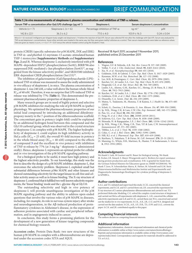
The inhibition of galactosamine (Gal)/lipopolysaccharide (LPS)-induced TNF-α release was measured to assess the orally administered in vivo efficacy of skepinone-L in mice. The plasma concentration of skepinone-L was 240 nM, a value well above the human whole-blood IC50 of 40 nM. Therefore, it was no surprise that LPS-induced TNF-α release was inhibited by 77% (Table 1). This demonstrated the pre-eminent pharmacodynamic properties of skepinone-L.
Many research groups are in need of highly potent and selective p38 MAPK inhibitors for studying the role of p38 MAPK in (patho)physiology. We optimized the pharmacodynamic properties of our initial lead compound by introducing an additional dihydroxy-propoxy moiety in the 7-position of the dibenzosuberone scaffold. The concomitant gain in potency (eight-fold) could be explained by an additional hydrogen bond between this compound and the Gly110 carbonyl group, which was observed in the crystal structure of skepinone-L in complex with p38 MAPK. The higher hydrophi-licity of skepinone-L could explain its high inhibitory activity in HeLa cells (IC50 = 25 nM), the overall 95-fold increase in potency in the whole-blood assay (IC50 = 40 nM) compared to the effects of compound 3 and the excellent in vivo potency with inhibition of TNF-α release by 77% (at 3 mg kg−1 skepinone-L administered orally). Hence, skepinone-L represents an optimal probe for cellular and in vivo investigation of the p38 MAPK signaling pathway.
For a biological probe to be useful, it must have high potency and the highest selectivity possible. To our knowledge, this study was the first to describe the design of a p38 MAPK inhibitor, skepinone-L, that overcomes the selectivity problem. Skepinone-L exploited small but substantial differences between p38α MAPK and other kinases and showed outstanding selectivity for the target kinase in cell-free and cel-lular activity assays as well as in kinase binding. The X-ray structure of skepinone-L confirmed that it fulfilled two well-known selectivity require-ments, the ‘linear binding’ mode and the L-glycine–flip at Gly110.
The outstanding selectivity and high in vivo potency of skepinone-L will provide unambiguous investigation of the p38 MAPK signaling pathway and the ability to address open-ended questions that involve the various roles of p38 MAPK in diseases, including, for example, its role in nervous system injury after stroke and neurodegeneration, in the Aβ-induced production of proin-flammatory cytokines in Alzheimer’s disease, in the expression of adhesion proteins associated with cardiac and peripheral inflam-mation, and in angiogenesis induced in cancer.
In conclusion, this study forms a promising platform for the development of a new generation of kinase-targeting compounds for chemical biology research.
Accession codes. Protein Data Bank: two new structures of the human p38 MAPK in complex with a dibenzosuberone are depos-ited under the accession codes 3ZYA and 3QUE.
received 18 april 2011; accepted 1 november 2011; published online 25 December 2011
references1. Wagner, E.F. & Nebreda, A.R. Nat. Rev. Cancer 9, 537–549 (2009).2. Veillat, V. et al. J. Clin. Endocrinol. Metab. 95, E403–E412 (2010).3. Xie, N. et al. Neuropharmacology 59, 444–451 (2010).4. Goldstein, D.M. & Gabriel, T. Curr. Top. Med. Chem. 5, 1017–1029 (2005).5. Karaman, M.W. et al. Nat. Biotechnol. 26, 127–132 (2008).6. Lee, M.R. & Dominguez, C. Curr. Med. Chem. 12, 2979–2994 (2005).7. Karcher, S.C. & Laufer, S.A. Curr. Top. Med. Chem. 9, 655–676 (2009).8. Fitzgerald, C.E. et al. Nat. Struct. Biol. 10, 764–769 (2003).9. Laufer, S.A., Ahrens, G.M., Karcher, S.C., Hering, J.S. & Niess, R. J. Med.
Chem. 49, 7912–7915 (2006).10. Karcher, S.C. & Laufer, S.A. J. Med. Chem. 52, 1778–1782 (2009).11. Goldstein, D.M. et al. J. Med. Chem. 49, 1562–1575 (2006).12. Dinarello, C.A. Semin. Dial. 22, 256–259 (2009).13. Mutou, Y., Tsukimoto, M., Homma, T. & Kojima, S. J. Health Sci. 56, 675–683
(2010).14. Schett, G., Zwerina, J. & Firestein, G. Ann. Rheum. Dis. 67, 909–916 (2008).15. Ip, W.K., Wong, C.K. & Lam, C.W. Clin. Exp. Immunol. 145, 162–172 (2006).16. Pan, S.L. et al. Shock 30, 496–502 (2008).17. Peng, H. et al. J. Biol. Chem. 286, 24508–24518 (2011).18. Schieven, G.L. Curr. Top. Med. Chem. 9, 1038–1048 (2009).19. Shimada, H. & Rajagopalan, L.E. J. Biol. Chem. 285, 12536–12542 (2010).20. Ip, W.K., Wong, C.K., Wang, C.B., Tian, Y.P. & Lam, C.W. Immunopharmacol.
Immunotoxicol. 27, 371–393 (2005).21. Tibbles, L.A. et al. J. Virol. 76, 1559–1568 (2002).22. Cekic, C. et al. J. Biol. Chem. 284, 31982–31991 (2009).23. Hill, R.J. et al. J. Pharmacol. Exp. Ther. 327, 610–619 (2008).24. Kurihara, Y., Nakahara, T. & Furue, M. Cell. Immunol. 270, 25–31 (2011).25. Manning, G., Whyte, D.B., Martinez, R., Hunter, T. & Sudarsanam, S. Science
6, 1912–1934 (2002).
acknowledgmentsWe thank S. Luik, M Goettert and K. Bauer for biological testing. We thank M. Fecker, J.R. Simard, S. Mayer-Wrangowski and A. Richters for expert assistance during protein production and crystallization. D.R. is grateful for funds from the German Federal Ministry for Education (grant no. BMBF 01GS08104). We thank T. Joos, N. Schneiderhan-Marra, K. Hefner, M. Schmohl and G.M. Stein (Naturwissenschaftliches und Medizinisches Institut and Experimentelle und Diagnostische Immunologie Reutlingen) for cytokine profiling of skepinone-L in hPMBCs.
author contributionsS.A.L. and T.S. initiated and supervised this study, S.C.K. conceived the chemical experiments, and S.C.K. and S.F. carried them out, J.R. conceived the experiment for X-ray structure of 2-amino-phenylamino-dibenzosuberone and carried it out. V.S. performed Molecular Modeling. C.G. solved the complex crystal structure of skepinone-L and carried out HeLa cell experiments with skepinone-L. A.K. conceived the cellular selectivity experiments and A.K and S.C.K. carried them out; W.A. conceived and carried out the studies for in vivo experiments. S.C.K., A.K., J.R., C.G. and W.A. designed and carried out the data analysis. S.C.K., A.K., S.F., D.R., T.S., O.W. and S.A.L. cowrote the paper. S.C.K. and J.R. contributed equally to this paper.
Competing financial interestsThe authors declare no competing financial interests.
additional informationSupplementary information, chemical compound information and chemical probe information is available online at http://www.nature.com/naturechemicalbiology/. Reprints and permissions information is available online at http://www.nature.com/reprints/index.html. Correspondence and requests for materials should be addressed to T.S. or S.A.L.
Table 1 | In vivo measurements of skepinone-L plasma concentration and inhibition of TNF-a release.serum tnF-a concentration after Gal/Lps challenge (pg ml−1) skepinone-L serum skepinone-L concentration
vehicle (n = 7) Skepinone-l (n = 6) Percentage inhibition of TNf-α ng ml−1 μM
142.8 ± 22.1 36.3 ± 6.2 77.0 ± 4.0 103.9 ± 16.3 0.24 ± 0.04Mice (n = 6) received 3 milligrams per kilogram body weight oral skepinone-l 1 h before the injection of Gal/lPS. Serum was collected 90 min after the challenge with Gal/lPS. Serum was analyzed for TNf-α and skepinone-l concentrations. Seven other animals were treated in the same way, but they were given methyl cellulose as a control. Their serum measurements were taken as reference values. Data are given as means ± s.e.m.; n = 6 or 7. P < 0.01 versus the vehicle-treated control using the Student’s t-test.
npg
© 2
012
Nat
ure
Am
eric
a, In
c. A
ll rig
hts
rese
rved
.