small molecule ligands for enhanced intracellular delivery of lipid nanoparticle formulations of...
TRANSCRIPT
BASIC SCIENCE
Nanomedicine: Nanotechnology, Biology, and Medicine9 (2013) 665–674
Original Article
Small molecule ligands for enhanced intracellular delivery of lipidnanoparticle formulations of siRNA
Yuen Yi C. Tam, PhDa,⁎, Sam Chen, BSca, Josh Zaifman, PhDb, Ying K. Tam, PhDc,Paulo J.C. Lin, PhDa, Steven Ansell, PhDc, Michel Roberge, PhDa,
Marco A. Ciufolini, PhDb, Pieter R. Cullis, PhDa
aDepartment of Biochemistry and Molecular Biology, University of British Columbia, Vancouver, British Columbia, CanadabDepartment of Chemistry, University of British Columbia, Vancouver, British Columbia, Canada
cAlCana Technologies, Vancouver, British Columbia, Canada
Received 9 July 2012; accepted 20 November 2012
nanomedjournal.com
Abstract
Gene silencing activity of lipid nanoparticle (LNP) formulations of siRNA requires LNP surface factors promoting cellular uptake. Thisstudy aimed to identify small molecules that enhance cellular uptake of LNP siRNA systems, then use them as LNP-associated ligands toimprove gene silencing potency. Screening the Canadian Chemical Biology Network molecules for effects on LNP uptake into HeLa cellsfound that cardiac glycosides like ouabain and strophanthidin caused the highest uptake. Cardiac glycosides stimulate endocytosis on bindingto plasma membrane Na+/K+ ATPase found in all mammalian cells, offering the potential to stimulate LNP uptake into various cell types. APEG-lipid containing strophanthidin at the end of PEG (STR-PEG-lipid) was synthesized and incorporated into LNP. Compared to non-liganded systems, STR-PEG-lipid enhanced LNP uptake in various cell types. Furthermore, this enhanced uptake improved marker genesilencing in vitro. Addition of STR-PEG-lipid to LNP siRNA may have general utility for enhancing gene silencing potency.
From the Clinical Editor: In this study, the authors identified small molecules that enhance cellular uptake of lipid nanoparticle siRNAsystems, then used them as LNP-associated ligands to improve gene silencing potency.© 2013 Elsevier Inc. All rights reserved.
Key words: Lipid nanoparticle; Small molecule ligand; Cardiac glycosides; High-throughput screening; Gene silencing; siRNA; Targeting; Targeted delivery
The use of small interfering RNA (siRNA) to silence disease-associated genes has considerable therapeutic promise.1–4 How-ever, realizing the potential of siRNA therapeutics requiressophisticated systems to deliver siRNA into target cells in vivo.This is because “naked” siRNA molecules are rapidly brokendown in biological fluids, do not accumulate in target tissuesand cannot penetrate target cell membranes to reach intracellularsites of activity even if they get there. Lipid nanoparticle (LNP)formulations of siRNA (LNP siRNA) have demonstrated signifi-cant potential for overcoming these problems for delivery of siRNAto hepatocytes following intravenous (i.v.) injection.5–8 Recent
Supported by Canadian Institutes for Health Research (CIHR) underUniversity-Industry grant FRN59836, Tekmira Pharmaceuticals and Alny-lam Pharmaceuticals.
Conflict of interest statement: PRC has a financial interest in TekmiraPharmaceuticals and receives grant funding from Alnylam Pharmaceuticals.
⁎Corresponding author:E-mail address: [email protected] (Y.Y.C. Tam).
1549-9634/$ – see front matter © 2013 Elsevier Inc. All rights reserved.http://dx.doi.org/10.1016/j.nano.2012.11.006
Please cite this article as: Tam YYC, et al, Small molecule ligands for enhaNanomedicine: NBM 2013;9:665-674, http://dx.doi.org/10.1016/j.nano.2012.11
improvements in the cationic lipid component of LNP siRNAsystems have resulted in effective gene silencing in hepatocytesat dose levels as low as 0.02 μg siRNA/kg body weight inmouse models.8
The potency of LNP siRNA systems for gene silencing ofhepatocyte genes is dependent, at least in part, on associationwith apolipoprotein E (ApoE) following i.v. administration.9,10
The presence of the ApoE on the LNP facilitates uptake intohepatocytes via the LDL and scavenger receptors on hepatocytesand little gene silencing activity is observed in ApoE knock-outmice. The requirement for Apo E emphasizes the vital role offactors on the LNP surface that bind to and trigger uptake intotarget cells and indicates that unless an LNP is able to bind aserum protein that facilitates uptake into the cell of interest, orhas ligands on the LNP surface that promote uptake, little genesilencing activity can be expected. This results in an interestingopportunity. Because a given siRNA is so specific for silencing aparticular gene once it arrives in the cell cytoplasm, moregeneralized uptake into non-target tissue, which may not expressthat gene, could be of little consequence. As a result, LNP-
nced intracellular delivery of lipid nanoparticle formulations of siRNA..006
666 Y.Y.C. Tam et al / Nanomedicine: Nanotechnology, Biology, and Medicine 9 (2013) 665–674
associated ligands that encourage promiscuous uptake into awide variety of cells could well be of considerable utility in targetvalidation and possibly therapeutic applications of siRNA.
Targeting ligands such as antibodies, antibody fragments andpeptides against specific cell surface receptors have been used totarget liposomes to specific cells.11–14 However, there are manyissues associated with use of such ligands, including induction ofimmune responses to the targeting ligand, cost, as well asformulation and characterization issues, among others. Smallmolecule targeting ligands conjugated to lipid anchors in LNPoffer important potential advantages, notably reduced immuno-genicity as well as much improved ease of ligand manufactureand formulation into LNP. For example, anisamide, whichpossesses high affinity for sigma receptors can be coupled to aPEG-lipid which is easily formulated into LNP and has beenshown to increase delivery of LNP to prostate and lung cancercells that over-express sigma receptors.15–18
In this work we conducted a screen of 800 molecules fromthe Canadian Chemical Biology Network library to identifysmall molecules that can enhance cellular uptake of LNP in apotentially general manner. This resulted in the identification ofcardiac glycosides as a class of compounds that enhance LNPuptake into human cervical cancer HeLa cells by binding to anextracellular region of the Na+/K+ ATPase in the cell plasmamembrane. Because Na+/K+ ATPase is expressed in allmammalian cells, it offers a potentially general target tostimulate uptake into a wide variety of cells. We synthesizeda poly(ethylene)glycol (PEG) lipid containing a cardiacglycoside (strophanthidin, STR) at the distal end of the PEGmoiety and show that LNP siRNA systems containing the STR-PEG-lipid exhibit enhanced uptake in a variety of cell typesderived from different tissues and improved gene silencingproperties in vitro.
Materials and methods
Materials
1,2-distearoyl-sn-glycero-3-phosphocholine (DSPC) waspurchased from Avanti Polar Lipids (Alabaster, AL), whereascholesterol was obtained from Sigma (St Louis, MO). 1,2-dilinoleyloxy-keto-N,N-dimethyl-3-aminopropane (DLinK-DMA), 2,2-Dilinoleyl-4-(2-dimethylaminoethyl)-[1,3]-dioxo-lane (DLinK-C2-DMA)and polyethylene glycol-dimyristol glyc-erol (PEG-s-DMG) were provided by Tekmira PharmaceuticalsCorporation (Burnaby, BC). The fluorescently-labeled lipid 3,3′-dioctadecyl-5,5′-di(4-sulfophenyl)-oxacarbocyanine, sodiumsalt (SPDiO) was purchased from Invitrogen (Burlington, ON).The small molecule library used in this study was obtained fromthe Canadian Chemical Biology Network. Ouabain andstrophanthidin were purchased from Sigma.
Cell culture
All cells were incubated at 37 °C with 5% CO2 unlessindicated otherwise. The human cervix carcinoma cells (HeLa),carcinomic alveolar basal epithelial cells (A549), hepatocellularcarcinoma cells (Hep3B), exocrine pancreatic carcinoma cells
(PANC1), prostate adenocarcinoma cells (PC3) and mousepancreatic beta cells (MIN6) were cultured in DMEM supple-mented with 10% fetal bovine serum (FBS), 2 mM L-glutamineand 0.1 mM non-essential amino acids (NEAA). The humanovarian clear cell carcinoma cells (TOV21G, JHOC-5 and JHOC-9), androgen-sensitive prostate adenocarcinoma (LNCaP), breastductal carcinoma (MCF7), and breast adenocarcinoma (MDA-MB-231) were cultured in RPMI 1640 Medium supplementedwith 10% FBS, 2 mM L-glutamine and 0.1 mM NEAA. All cellculture reagents were obtained from Invitrogen.
Cell lines stably expressing shRNA targeted to ATP1A1(shATP1A1) or shRNA directed to a negative control sequence(shScramble) were constructed by transfection of shATP1A1plasmid or shScramble plasmid (SABiosciences, MD) accordingto the manufacturer's instructions.
Preparation of siRNA-LNP
siRNA-Cy3 targeting mouse factor VII mRNA was obtainedfrom Alnylam Pharmaceuticals (Cambridge, MA). siRNA (5′-TGGCCAAGGTCATCCATGA-3′) directed to glyceraldehyde3-phosphate dehydrogenase (siGAPDH) was purchased fromDharmacon (Thermo Scientific, Pittsburg, PA). siRNA with arandom sequence of low GC content (siScramble) was purchasedfrom Invitrogen. siRNA was encapsulated in LNP using thepreformed vesicle (PFV) procedure as previously described.8
Briefly, lipids weremixed together in 30% ethanol and the mixturewas slowly added to 50 mM citrate or acetate buffer, pH 4.0 underrapid vortexing followed by extrusion through two stacks of 80 nmpolycarbonate filters at ~300 psi. The siRNA solution was thenslowly added to the liposome dispersion equivalent of ten timessiRNA amount under vortexing. The mixture was subsequentlyincubated at 31 °C for 30 min and dialyzed twice in 1× PBS for18 h to remove ethanol. siRNA-Cy3 was encapsulated in LNPconsisting of DLinK-DMA/DSPC/cholesterol/PEG-s-DMG/SPDiO at a molar ratio of 40/10/39.8/10/0.2 whereas siGAPDHand siScramble were encapsulated at a molar ratio of 40/18.8/40/1/0.2 for in vitro experiments using ouabain. The LNP compositionused for subsequent in vitro uptake and knockdown experimentswas DLinK-C2-DMA/DSPC/Cholesterol/PEG-s-DMG/STR-PEG or DSPE-PEG/SPDiO at a molar ratio of 40/14.8/40/4/1/0.2 and 40/17.3/40/1.5/1/0.2, respectively. All LNPswere ~80 nmin diameter measured by dynamic light scattering with intensitymode (Nicomp, Port Richey, FL). LNPs containing DSPE-PEGand STR-PEG have zeta-potential of 1.91 and 0.08 mV at pH 7.4,respectively as measured by a Zetasizer NanoZS (Malvern,Worcestershire, UK).
Small molecules and LNP treatment on 96-well plate
Cells were seeded at 8000 cells/well in 96-well ViewPlates(PerkinElmer, Shelton, CT) in 100 μl of medium and wereallowed to grow overnight. Fresh medium containing LNP wasadded the next day. Small molecules were either added manuallyor pinned from 1000-fold stocks in DMSO using a pinning robotequipped with 0.4 mm pins (BioRobotics, Cambridge, UK).Cells were incubated for 24 h. Cells were then washed once inPBSCM (1× PBS containing 1 μM MgCl2 and 0.1 μM CaCl2),fixed in 3% paraformaldehyde containing 500 ng/ml Hoechst
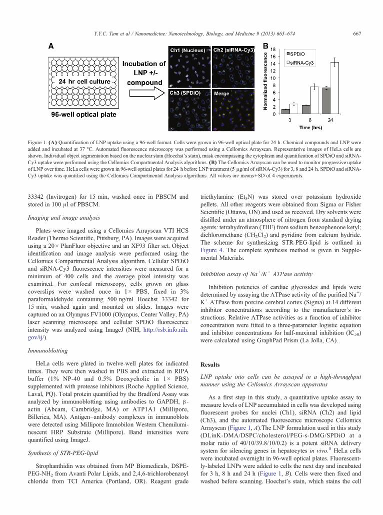
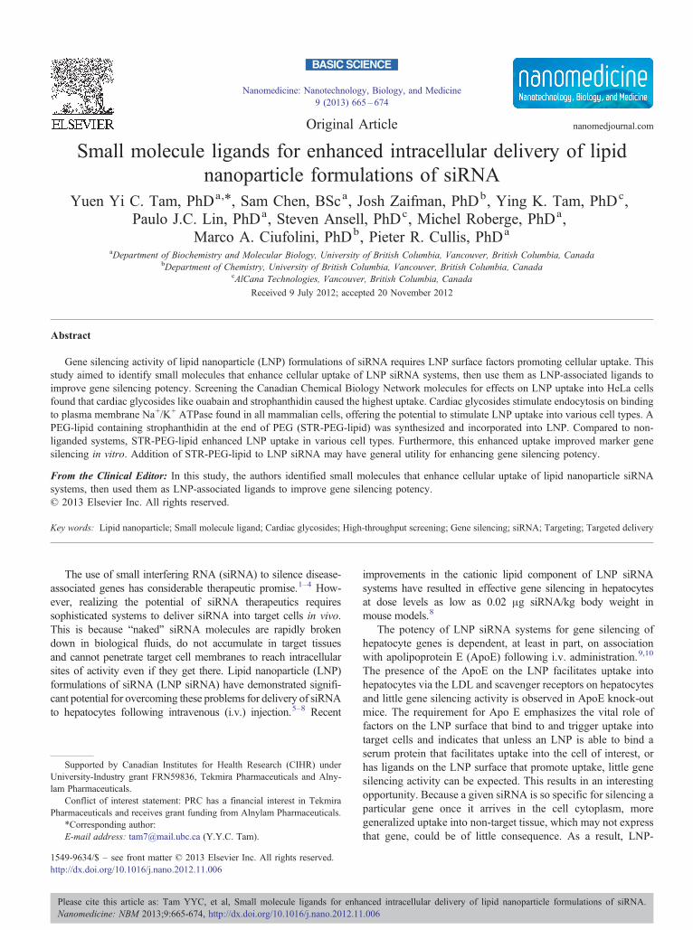
Figure 1. (A) Quantification of LNP uptake using a 96-well format. Cells were grown in 96-well optical plate for 24 h. Chemical compounds and LNP wereadded and incubated at 37 °C. Automated fluorescence microscopy was performed using a Cellomics Arrayscan. Representative images of HeLa cells areshown. Individual object segmentation based on the nuclear stain (Hoechst's stain), mask encompassing the cytoplasm and quantification of SPDiO and siRNA-Cy3 uptake were performed using the Cellomics Compartmental Analysis algorithms. (B) The Cellomics Arrayscan can be used to monitor progressive uptakeof LNP over time. HeLa cells were grown in 96-well optical plates for 24 h before LNP treatment (5 μg/ml of siRNA-Cy3) for 3, 8 and 24 h. SPDiO and siRNA-Cy3 uptake was quantified using the Cellomics Compartmental Analysis algorithms. All values are means±SD of 4 experiments.
667Y.Y.C. Tam et al / Nanomedicine: Nanotechnology, Biology, and Medicine 9 (2013) 665–674
33342 (Invitrogen) for 15 min, washed once in PBSCM andstored in 100 μl of PBSCM.
Imaging and image analysis
Plates were imaged using a Cellomics Arrayscan VTI HCSReader (Thermo Scientific, Pittsburg, PA). Images were acquiredusing a 20× PlanFluor objective and an XF93 filter set. Objectidentification and image analysis were performed using theCellomics Compartmental Analysis algorithm. Cellular SPDiOand siRNA-Cy3 fluorescence intensities were measured for aminimum of 400 cells and the average pixel intensity wasexamined. For confocal microscopy, cells grown on glasscoverslips were washed once in 1× PBS, fixed in 3%paraformaldehyde containing 500 ng/ml Hoechst 33342 for15 min, washed again and mounted on slides. Images werecaptured on an Olympus FV1000 (Olympus, Center Valley, PA)laser scanning microscope and cellular SPDiO fluorescenceintensity was analyzed using ImageJ (NIH, http://rsb.info.nih.gov/ij/).
Immunoblotting
HeLa cells were plated in twelve-well plates for indicatedtimes. They were then washed in PBS and extracted in RIPAbuffer (1% NP-40 and 0.5% Deoxycholic in 1× PBS)supplemented with protease inhibitors (Roche Applied Science,Laval, PQ). Total protein quantified by the Bradford Assay wasanalyzed by immunoblotting using antibodies to GAPDH, β-actin (Abcam, Cambridge, MA) or ATP1A1 (Millipore,Billerica, MA). Antigen–antibody complexes in immunoblotswere detected using Millipore Immobilon Western Chemilumi-nescent HRP Substrate (Millipore). Band intensities werequantified using ImageJ.
Synthesis of STR-PEG-lipid
Strophanthidin was obtained from MP Biomedicals, DSPE-PEG-NH2 from Avanti Polar Lipids, and 2,4,6-trichlorobenzoylchloride from TCI America (Portland, OR). Reagent grade
triethylamine (Et3N) was stored over potassium hydroxidepellets. All other reagents were obtained from Sigma or FisherScientific (Ottawa, ON) and used as received. Dry solvents weredistilled under an atmosphere of nitrogen from standard dryingagents: tetrahydrofuran (THF) from sodium benzophenone ketyl;dichloromethane (CH2Cl2) and pyridine from calcium hydride.The scheme for synthesizing STR-PEG-lipid is outlined inFigure 4. The complete synthesis method is given in Supple-mental Materials.
Inhibition assay of Na+/K+ ATPase activity
Inhibition potencies of cardiac glycosides and lipids weredetermined by assaying the ATPase activity of the purified Na+/K+ ATPase from porcine cerebral cortex (Sigma) at 14 differentinhibitor concentrations according to the manufacturer's in-structions. Relative ATPase activities as a function of inhibitorconcentration were fitted to a three-parameter logistic equationand inhibitor concentrations for half-maximal inhibition (IC50)were calculated using GraphPad Prism (La Jolla, CA).
Results
LNP uptake into cells can be assayed in a high-throughputmanner using the Cellomics Arrayscan apparatus
As a first step in this study, a quantitative uptake assay tomeasure levels of LNP accumulated in cells was developed usingfluorescent probes for nuclei (Ch1), siRNA (Ch2) and lipid(Ch3), and the automated fluorescence microscope CellomicsArrayscan (Figure 1, A).The LNP formulation used in this study(DLinK-DMA/DSPC/cholesterol/PEG-s-DMG/SPDiO at amolar ratio of 40/10/39.8/10/0.2) is a potent siRNA deliverysystem for silencing genes in hepatocytes in vivo.8 HeLa cellswere incubated overnight in 96-well optical plates. Fluorescent-ly-labeled LNPs were added to cells the next day and incubatedfor 3 h, 8 h and 24 h (Figure 1, B). Cells were then fixed andwashed before scanning. Hoechst's stain, which stains the cell
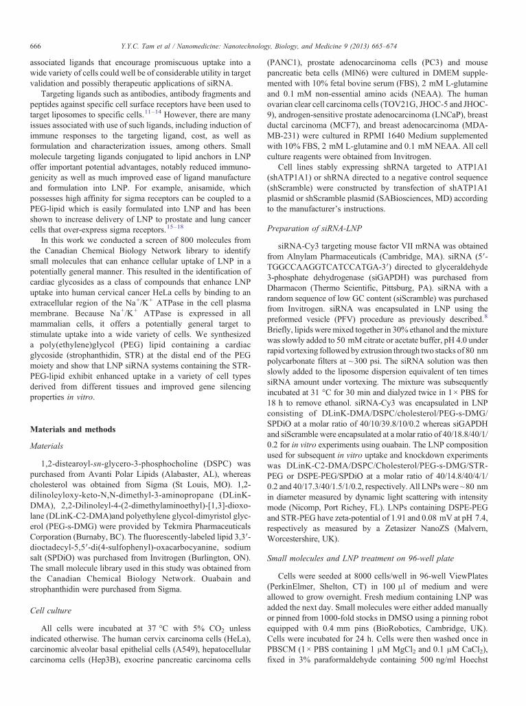
Figure 2. (A) Normalized uptake of fluorescently labelled LNP systems intoHeLa cells following incubation with small molecules that are known drugs.HeLa cells were incubated with 800 small molecule drugs and 5 μg/ml ofsiRNA-Cy3-LNPfor 24 h as described under Methods. The fluorescenceresulting from the accumulated SPDiO-labeled LNP following incubationwith each individual compound was normalized to the LNP SPDiOfluorescence in cells untreated with any compound. The small moleculesare sorted so that those giving rise to the highest LNP accumulation are on theleft and the lowest on the right. (B) Effects of different cardiac glycosides onLNP uptake. HeLa cells were incubated with 50 μg/ml (lipid concentration)of empty LNP and each of 9 cardiac glycosides at 0.15 μM, 1.5 μM and15 μM for 24 h. Cardiac glycosides on the x-axis are arranged by theiraffinity for the Na+/K+ ATPase, from the weakest (helveticoside) to thestrongest (Proscillaridin A).19 Cellular LNP SPDiO fluorescence in thepresence of individual compounds was normalized to the LNP SPDiOfluorescence in cells untreated with any cardiac glycoside. All values aremeans±SD of 4 experiments.
Figure 3. (A) Ouabain induces LNP uptake into HeLa cells. Confocalmicrographs of HeLa cells treated with 10 μg/ml of siGAPDH-LNP and0 nM or 30 nM of ouabain for 24 h. Cell nuclei were stained with Hoechst'sdye in blue. SPDiO fluorescence is shown in green. (B) LNP siRNA-inducedGAPDH knockdown is increased in the presence of 30 nM ouabain.Cellswere treated with or without 10 μg/ml of siGAPDH-LNP or siScramble-LNPin the presence of 0 nM or 30 nM of ouabain for 24 h. LNP and ouabain wereremoved and cells were further incubated in fresh medium for 48 h. Equalportions of protein samples were analyzed by immunoblotting to GAPDHand β-actin which served as a loading control.
668 Y.Y.C. Tam et al / Nanomedicine: Nanotechnology, Biology, and Medicine 9 (2013) 665–674
nuclei, was used to form the nuclear mask (blue line in Ch1,Figure 1, A) to identify valid objects or cells.
Cellular siRNA was monitored by the Cy3 fluorophore whichwas conjugated to the 3′ end of the siRNA sense strand (Ch2).The distribution of LNP lipid was reported by the fluorescentlipid, SPDiO (Ch3). A cellular mask (green line in Ch2 or Ch3,Figure 1, A), which was slightly larger than the nuclear mask butstayed within the cell boundary, was used to delineate the areafrom which Ch2 or Ch3 cytological features were measured. Atleast 400 cells were scored per well, and the average fluorescenceintensity per pixel in each channel was measured for each cellregion. Fluorescence values were normalized to untreated cells(Figure 1, B). A progressive time-dependent increase inintracellular SPDiO or siRNA-Cy3 fluorescence was observed
following incubation of the cells with the fluorescently labeledLNP siRNA systems, indicating that LNP siRNA was taken upby cells in increasing amounts over time. This was taken asbaseline behavior and the effect of added small molecules wasassessed relative to this baseline.
Cardiac glycosides enhance uptake of LNP into HeLa cells
The influence of 800 compounds from the CanadianChemical Biology Network collection of small molecules onLNP uptake into HeLa cells was assessed. Compounds wereintroduced into the 96-well optical plates containing cells andLNP using a pinning robot. Approximately 7.5 μM to 10 μM ofeach compound was transferred by each pin into each well.Approximately half of the molecules led to enhanced LNPuptake into HeLa cells as evidenced by increased cellular SPDiOfluorescence levels compared to control whereas the remainderdecreased LNP uptake (Figure 2, A). Interestingly, among theseven molecules that led to the most uptake, three belonged tothe cardiac glycoside family.
Cardiac glycosides are a diverse family of molecules that havebeen used to treat heart failure for many years.20–22 They bind tothe Na+/K+ ATPase on the plasma membrane thereby leading toan increase in intracellular Ca2+ concentrations and enhancedcontractility for cardiac tissue. The binding site is on theextracellular side of the α-subunit of the enzyme. Binding ofcardiac glycosides to theATPase inhibits the enzyme and can alsoact as a signal transducer.23–25 As important, for the purposes ofthis study, is the observation that binding of cardiac glycosides
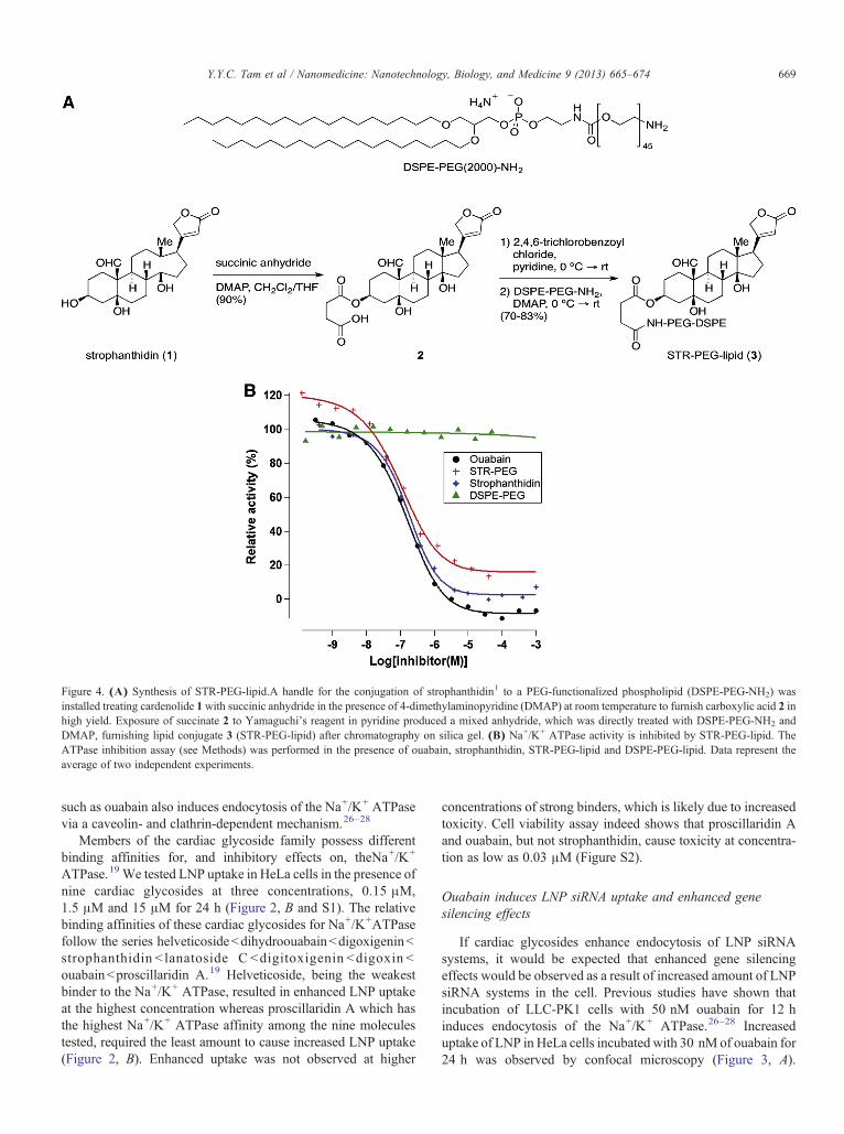
Figure 4. (A) Synthesis of STR-PEG-lipid.A handle for the conjugation of strophanthidin1 to a PEG-functionalized phospholipid (DSPE-PEG-NH2) wasinstalled treating cardenolide 1with succinic anhydride in the presence of 4-dimethylaminopyridine (DMAP) at room temperature to furnish carboxylic acid 2 inhigh yield. Exposure of succinate 2 to Yamaguchi's reagent in pyridine produced a mixed anhydride, which was directly treated with DSPE-PEG-NH2 andDMAP, furnishing lipid conjugate 3 (STR-PEG-lipid) after chromatography on silica gel. (B) Na+/K+ ATPase activity is inhibited by STR-PEG-lipid. TheATPase inhibition assay (see Methods) was performed in the presence of ouabain, strophanthidin, STR-PEG-lipid and DSPE-PEG-lipid. Data represent theaverage of two independent experiments.
669Y.Y.C. Tam et al / Nanomedicine: Nanotechnology, Biology, and Medicine 9 (2013) 665–674
such as ouabain also induces endocytosis of the Na+/K+ ATPasevia a caveolin- and clathrin-dependent mechanism.26–28
Members of the cardiac glycoside family possess differentbinding affinities for, and inhibitory effects on, theNa+/K+
ATPase.19 We tested LNP uptake in HeLa cells in the presence ofnine cardiac glycosides at three concentrations, 0.15 μM,1.5 μM and 15 μM for 24 h (Figure 2, B and S1). The relativebinding affinities of these cardiac glycosides for Na+/K+ATPasefollow the series helveticosidebdihydroouabainbdigoxigeninbstrophanthidin b lanatoside C bdigitoxigenin bdigoxin bouabainbproscillaridin A.19 Helveticoside, being the weakestbinder to the Na+/K+ ATPase, resulted in enhanced LNP uptakeat the highest concentration whereas proscillaridin A which hasthe highest Na+/K+ ATPase affinity among the nine moleculestested, required the least amount to cause increased LNP uptake(Figure 2, B). Enhanced uptake was not observed at higher
concentrations of strong binders, which is likely due to increasedtoxicity. Cell viability assay indeed shows that proscillaridin Aand ouabain, but not strophanthidin, cause toxicity at concentra-tion as low as 0.03 μM (Figure S2).
Ouabain induces LNP siRNA uptake and enhanced genesilencing effects
If cardiac glycosides enhance endocytosis of LNP siRNAsystems, it would be expected that enhanced gene silencingeffects would be observed as a result of increased amount of LNPsiRNA systems in the cell. Previous studies have shown thatincubation of LLC-PK1 cells with 50 nM ouabain for 12 hinduces endocytosis of the Na+/K+ ATPase.26–28 Increaseduptake of LNP in HeLa cells incubated with 30 nM of ouabain for24 h was observed by confocal microscopy (Figure 3, A).
670 Y.Y.C. Tam et al / Nanomedicine: Nanotechnology, Biology, and Medicine 9 (2013) 665–674
Quantification of cellular SPDiO fluorescence showed that cellstreated with 30 nM of ouabain contained 2.5 times higher levelsof LNP than untreated cells. No significant cell death wasobserved when cells were treated at this concentration of ouabain.
The gene silencing potencies of LNP siRNA systems weredetermined in the presence or absence of ouabain using GAPDHas a target gene. GAPDH was chosen as a target gene since it isubiquitously expressed at high levels in all cell types. LNP siRNAsystems containing siGAPDH or the negative control siScramblewere incubated with HeLa cells for 24 h in the presence orabsence of ouabain. Because GAPDH has a relatively long half-life of ~38 h,29 cells were further incubated in plain medium for48 h before protein expression was analyzed. As shown inFigure 3, B, expression of GAPDH was substantially reducedonly in cells treated with siGAPDH-LNP and ouabain. The genesilencing effect was strictly due to enhanced LNP uptake causedby ouabain as no changes in GAPDH expression were observedin cells not treated with ouabain or treated with LNP containingsiScramble (Figure 3, B).
Incorporation of a targeting lipid containing a cardiacglycoside increases LNP uptake in cell lines of various origins
The LNP systems employed here have a small positive chargewhich encourages association with negatively charged cellsurfaces. The increased LNP uptake caused by free ouabain isattributed to increased plasma membrane turnover accompany-ing the increased endocytosis of Na+/K+ ATPase. A more directway of stimulating uptake would be to incorporate a targetinglipid containing a cardiac glycoside into the LNP itself. For easeof synthesis, strophanthidin (STR), a relatively simple cardiacglycoside was chosen to test this approach. STR was conjugatedto the distal end of a 2000 MW polyethylene glycol (PEG) lipidwith distearyl (C18) fatty acid chains (Figure 4, A) as indicatedin Materials and Methods and Supplementary Materials.
It is important to show that the STR incorporated into thePEG-lipid (STR-PEG-lipid) maintains an ability to bind to andinhibit the Na+/K+ ATPase. The inhibitory activity of the STR-PEG-lipid on the Na+/K+ ATPase was directly measured usingan ATPase inhibition assay30,31 (Figure 4, B). Purified Na+/K+
ATPase was incubated with different concentrations of inhibitor(10−10 M to 10−3 M) at pH 7.8 at 37 °C. The inhibitorconcentration for half-maximal inhibition, IC50, was 0.13 μMfor ouabain and 0.18 μM for strophanthidin. STR-PEG-lipidexhibited an IC50 of 0.33 μM which is fully consistent with amaintained ability of the PEG-associated STR ligand to bind tothe Na+/K+ ATPase. The limited increase in the IC50 can beattributed to inhibitory steric effects due to the presence of theconjugated PEG-lipid. Unmodified PEG-lipid was not able toinhibit the enzyme at any concentration tested.
The resulting STR-PEG-lipid could be easily formulatedinto LNP siRNA systems by simply including it as one of the
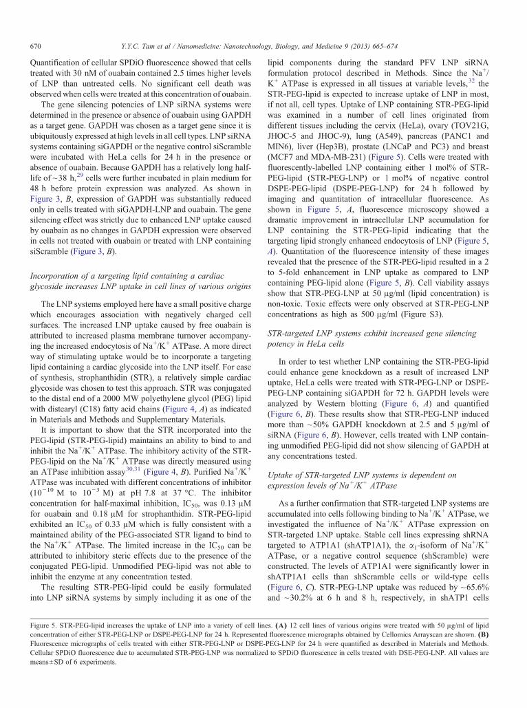
Figure 5. STR-PEG-lipid increases the uptake of LNP into a variety of cell lineconcentration of either STR-PEG-LNP or DSPE-PEG-LNP for 24 h. RepresentedFluorescence micrographs of cells treated with either STR-PEG-LNP or DSPE-Cellular SPDiO fluorescence due to accumulated STR-PEG-LNP was normalizedmeans±SD of 6 experiments.
lipid components during the standard PFV LNP siRNAformulation protocol described in Methods. Since the Na+/K+ ATPase is expressed in all tissues at variable levels,32 theSTR-PEG-lipid is expected to increase uptake of LNP in most,if not all, cell types. Uptake of LNP containing STR-PEG-lipidwas examined in a number of cell lines originated fromdifferent tissues including the cervix (HeLa), ovary (TOV21G,JHOC-5 and JHOC-9), lung (A549), pancreas (PANC1 andMIN6), liver (Hep3B), prostate (LNCaP and PC3) and breast(MCF7 and MDA-MB-231) (Figure 5). Cells were treated withfluorescently-labelled LNP containing either 1 mol% of STR-PEG-lipid (STR-PEG-LNP) or 1 mol% of negative controlDSPE-PEG-lipid (DSPE-PEG-LNP) for 24 h followed byimaging and quantitation of intracellular fluorescence. Asshown in Figure 5, A, fluorescence microscopy showed adramatic improvement in intracellular LNP accumulation forLNP containing the STR-PEG-lipid indicating that thetargeting lipid strongly enhanced endocytosis of LNP (Figure 5,A). Quantitation of the fluorescence intensity of these imagesrevealed that the presence of the STR-PEG-lipid resulted in a 2to 5-fold enhancement in LNP uptake as compared to LNPcontaining PEG-lipid alone (Figure 5, B). Cell viability assaysshow that STR-PEG-LNP at 50 μg/ml (lipid concentration) isnon-toxic. Toxic effects were only observed at STR-PEG-LNPconcentrations as high as 500 μg/ml (Figure S3).
STR-targeted LNP systems exhibit increased gene silencingpotency in HeLa cells
In order to test whether LNP containing the STR-PEG-lipidcould enhance gene knockdown as a result of increased LNPuptake, HeLa cells were treated with STR-PEG-LNP or DSPE-PEG-LNP containing siGAPDH for 72 h. GAPDH levels wereanalyzed by Western blotting (Figure 6, A) and quantified(Figure 6, B). These results show that STR-PEG-LNP inducedmore than ~50% GAPDH knockdown at 2.5 and 5 μg/ml ofsiRNA (Figure 6, B). However, cells treated with LNP contain-ing unmodified PEG-lipid did not show silencing of GAPDH atany concentrations tested.
Uptake of STR-targeted LNP systems is dependent onexpression levels of Na+/K+ ATPase
As a further confirmation that STR-targeted LNP systems areaccumulated into cells following binding to Na+/K+ ATPase, weinvestigated the influence of Na+/K+ ATPase expression onSTR-targeted LNP uptake. Stable cell lines expressing shRNAtargeted to ATP1A1 (shATP1A1), the α1-isoform of Na+/K+
ATPase, or a negative control sequence (shScramble) wereconstructed. The levels of ATP1A1 were significantly lower inshATP1A1 cells than shScramble cells or wild-type cells(Figure 6, C). STR-PEG-LNP uptake was reduced by ~65.6%and ~30.2% at 6 h and 8 h, respectively, in shATP1 cells
s. (A) 12 cell lines of various origins were treated with 50 μg/ml of lipidfluorescence micrographs obtained by Cellomics Arrayscan are shown. (B)
PEG-LNP for 24 h were quantified as described in Materials and Methods.to SPDiO fluorescence in cells treated with DSE-PEG-LNP. All values are
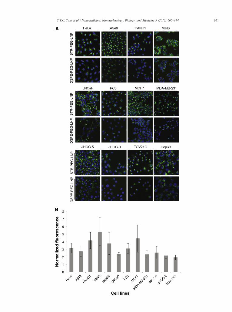
Figure 6. (A) LNP siRNA-induced GAPDH knockdown is increased whenSTR-PEG-lipid is incorporated into LNP siGAPDH systems. HeLa cellswere treated with STR-PEG-LNP or DSPE-PEG-LNP encapsulatingsiGAPDH at various siRNA concentrations for 72 h. Equal portions ofprotein samples were analyzed by immunoblotting to GAPDH and β-actinwhich served as a loading control. (B) Quantitation of GAPDH levels.GAPDH and β-actin intensities in western blots were quantified. GAPDHlevels were normalized to that of β-actin and reported relative to the untreatedcontrol group. (C) ATP1A1 expression is reduced in HeLa cell line stablytransfected with shATP1A1 plasmid. Cells stably transfected with or withoutshATP1A1 or shScramble plasmid were lysed. Equal portions of proteinsamples were analyzed by immunoblotting to ATP1A1 and β-actin. (D) LNPuptake is dependent on expression of ATP1A1. Cells stably transfected withshATP1A1 or shScramble plasmid were treated with STR-PEG-LNP at25 μg/ml of lipid concentration for 6 or 8 h. Confocal images were collectedand SPDiO fluorescence was quantified using ImageJ. All values are means±SD of 3 experiments.
672 Y.Y.C. Tam et al / Nanomedicine: Nanotechnology, Biology, and Medicine 9 (2013) 665–674
compared to shScramble cells (Figure 6, D). These results areagain consistent with an STR-PEG-LNP uptake process thatoccurs via binding tothe Na+/K+ ATPase.
Discussion
In this study we have identified a class of small molecules(cardiac glycosides) that enhance LNP uptake into a variety ofcell types, have shown that incorporation of a representativecardiac glycoside (strophanthidin) into LNP siRNA systems viaa PEG-lipid anchor results in enhanced uptake and genesilencing in vitro and have shown that uptake of this targetedLNP system is dependent on the expression of the ubiquitouscell-surface receptor, the Na+/K+ ATPase. Here we discuss theutility of the small molecule Cellomics screen for identifyingtargeting ligands and the potential of the strophanthidin-targeted LNP siRNA systems for target validation andtherapeutic applications.
This study illustrates the considerable utility of developingscreens for identifying small molecules that enhance LNPuptake into target cells. We focused our screening efforts onsmall molecules that are known drugs because the proteintarget, the binding affinities and the structure-activity relation-ships are often known. There is an obvious need for asecondary screen as many of the molecules increasing uptakemay act on an intracellular receptor, which would not makethem suitable as extracellular targeting ligands. In the case ofthe cardiac glycosides the secondary screen was straightfor-ward, as it is well-known that they bind to an extracellularregion of the Na+/K+ ATPase which is ubiquitously expressedon all mammalian cells.21
From a synthetic perspective, a targeting lipid incorporating acardiac glycoside can be assembled by tethering an appropriatelyfunctionalized member of the family to commercially availableactivated PEG-lipids. It is known that cardiac glycosides bind tothe active site of the ATPase with the unsaturated lactone deep inthe binding pocket,19,33 indicating that a logical place forconjugation would be through a functional group distant from thelactone, such as the C3-hydroxyl that is common to all membersof this family; however, several members have a glycosidicgroup at this position complicating such functionalization. Of thetested cardiac glycosides, strophanthidin, digitoxigenin anddigoxigenin possess free hydroxy groups at C3, with stro-phanthidin being the most affordable (Figure S1). Selectiveacylation of STR at C3 provided a handle for conjugation andprovided a viable synthesis of the desired PEG-lipid conjugate. Itis expected that further screening of small molecules librarieswill lead to identification of other small molecule ligands thatenhance cell uptake either through cell surface receptors specificto particular cells or to receptors, such as the Na+/K+ ATPase,that promote relatively non-specific uptake.
The results presented support accumulation of the non-targeted LNP siRNA systems through endocytosis as fluores-cently-labelled LNPs appeared in punctate structures in HeLacells (Figures 1, A and 3, A) and in other cells tested (Figure 5,A), in agreement with previous observations that (non-targeted)LNPs co-localize with the early endosomal marker in bothRaw264.7 and primary antigen presenting cells.34,35 Theobservation that more non-targeted LNPs were internalizedupon ouabain treatment (Figure 3, A) is also consistent with anendocytotic mechanism. Ouabain has been shown to induceendocytosis of the Na+/K+ ATPase via a caveolin- and clathrin-
673Y.Y.C. Tam et al / Nanomedicine: Nanotechnology, Biology, and Medicine 9 (2013) 665–674
dependent mechanism.26–28 Indeed, confocal microscopy sug-gested that both non-targeted and targeted LNPs reside inendosomal compartments containing transferrin, a marker forclathrin-mediated endocytosis (Figure S4). Furthermore, subcel-lular fractionation showed that a significant amount of the Na+/K+
ATPase was enriched in the endosomal fraction in cells treatedwith 30 nM of ouabain (data not shown).
The in vivo applications of LNP siRNA systems for silencingtarget genes are currently primarily limited to silencing liver(hepatocyte) target genes. The high potency of LNP siRNA forsuch applications is due in large part to the association of ApoEto the LNP following i.v. administration, which facilitateshepatocyte uptake.9,10 In order to access other tissues alternativeuptake ligands will need to be incorporated into the LNP. Asnoted previously, small molecule targeting ligands tethered to alipid anchor in the LNP have considerable advantages comparedto the use of peptide, protein or other larger ligands due to cost,ease of manufacturing and reduced immunogenicity amongstother factors. The incorporation of targeting ligands such asstrophanthidin offers the possibility to stimulate relatively non-specific uptake and enhanced gene silencing in a variety of tissuetypes due to the ubiquitous expression of Na+/K+ ATPase. Whilethe uptake of LNP into cells may be relatively non-specific, thehighly specific nature of the siRNA cargo suggests considerableutility for target validation and possibly therapeutic applicationsin a broad range of diseases. For example, an LNP siRNA systemdesigned to silence a tumour oncogene may be expected inducefew “off target” effects if introduced into non-tumour tissue.Furthermore, the enhanced LNP uptake due to the targetingligands can reduce the amount of drug used which translates intolower possible side effects and costs associated with the drug.
In summary, this study describes a screening approach toidentify small molecule ligands to stimulate uptake of LNPsiRNA systems into cells. We show that by screening smallmolecules that are known drugs additional benefits can beachieved in terms of knowledge of target proteins andstructure–activity relationships that facilitate the design ofligand-PEG lipids that exhibit selective target binding proper-ties and straightforward incorporation into LNP systems. In theparticular case of the STR-PEG-lipid we demonstrate that thepresence of this targeting agent in LNP siRNA systems resultsin enhanced uptake and gene silencing in a variety of cell typesand may be of utility for targeted validation and possiblytherapeutic applications when access to tissues other thanhepatocytes is required.
Acknowledgments
Dr. Karen Lam is gratefully acknowledged for helpfulcomments and discussion. We also thank Ms. Joslyn Quick fortechnical support.
Appendix A. Supplementary data
Supplementary data to this article can be found online athttp://dx.doi.org/10.1016/j.nano.2012.11.006.
References
1. Davidson BL, McCray Jr PB. Current prospects for RNA interference-based therapies. Nat Rev Genet 2011;12:329-40.
2. Dorsett Y, Tuschl T. siRNAs: applications in functional genomics andpotential as therapeutics. Nat Rev Drug Discov 2004;3:318-29.
3. de Fougerolles A, Vornlocher HP, Maraganore J, Lieberman J.Interfering with disease: a progress report on siRNA-based therapeutics.Nat Rev Drug Discov 2007;6:443-53.
4. Pecot CV, Calin GA, Coleman RL, Lopez-Berestein G, Sood AK. RNAinterference in the clinic: challenges and future directions. Nat RevCancer 2011;11:59-67.
5. Zimmermann TS, Lee AC, Akinc A, Bramlage B, Bumcrot D, FedorukMN, et al. RNAi-mediated gene silencing in non-human primates. Na-ture 2006;441:111-4.
6. Morrissey DV, Lockridge JA, Shaw L, Blanchard K, Jensen K, BreenW,et al. Potent and persistent in vivo anti-HBV activity of chemicallymodified siRNAs. Nat Biotechnol 2005;23:1002-7.
7. Akinc A, Zumbuehl A, Goldberg M, Leshchiner ES, Busini V, HossainN, et al. A combinatorial library of lipid-like materials for delivery ofRNAi therapeutics. Nat Biotechnol 2008;26:561-9.
8. Semple SC, Akinc A, Chen J, Sandhu AP, Mui BL, Cho CK, et al.Rational design of cationic lipids for siRNA delivery. Nat Biotechnol2010;28:172-6.
9. Yan X, Kuipers F, Havekes LM, Havinga R, Dontje B, Poelstra K, et al.The role of apolipoprotein E in the elimination of liposomes from bloodby hepatocytes in the mouse. Biochem Biophys Res Commun 2005;328:57-62.
10. Akinc A, Querbes W, De S, Qin J, Frank-Kamenetsky M, JayaprakashKN, et al. Targeted delivery of RNAi therapeutics with endogenous andexogenous ligand-based mechanisms. Mol Ther 2010;18:1357-64.
11. Sapra P, Allen TM. Ligand-targeted liposomal anticancer drugs. ProgLipid Res 2003;42:439-62.
12. Cressman S, Dobson I, Lee JB, Tam YY, Cullis PR. Synthesis of alabeled RGD-lipid, its incorporation into liposomal nanoparticles, andtheir trafficking in cultured endothelial cells. Bioconjug Chem 2009;20:1404-11.
13. Di Paolo D, Ambrogio C, Pastorino F, Brignole C, Martinengo C,Carosio R, et al. Selective therapeutic targeting of the anaplasticlymphoma kinase with liposomal siRNA induces apoptosis and inhibitsangiogenesis in neuroblastoma. Mol Ther 2011;19:2201-12.
14. Di Paolo D, Brignole C, Pastorino F, Carosio R, Zorzoli A, Rossi M,et al. Neuroblastoma-targeted nanoparticles entrapping siRNA specifi-cally knockdown ALK. Mol Ther 2011;19:1131-40.
15. Banerjee R, Tyagi P, Li S, Huang L. Anisamide-targeted stealthliposomes: a potent carrier for targeting doxorubicin to human prostatecancer cells. Int J Cancer 2004;112:693-700.
16. Li SD, Huang L. Targeted delivery of antisense oligodeoxynucleotideand small interference RNA into lung cancer cells. Mol Pharm 2006;3:579-88.
17. Chen Y, Sen J, Bathula SR, Yang Q, Fittipaldi R, Huang L. Novelcationic lipid that delivers siRNA and enhances therapeutic effect in lungcancer cells. Mol Pharm 2009;6:696-705.
18. Li SD, Chono S, Huang L. Efficient oncogene silencing and metastasisinhibition via systemic delivery of siRNA. Mol Ther 2008;16:942-6.
19. Paula S, Tabet MR, Ball Jr WJ. Interactions between cardiac glycosidesand sodium/potassium-ATPase: three-dimensional structure-activityrelationship models for ligand binding to the E2-Pi form of the enzymeversus activity inhibition. Biochemistry 2005;44:498-510.
20. Kelly RA. Cardiac glycosides and congestive heart failure. Am J Cardiol1990;65:10E-6E [discussion 22E-3E].
21. Schoner W, Scheiner-Bobis G. Endogenous and exogenous cardiacglycosides: their roles in hypertension, salt metabolism, and cell growth.Am J Physiol Cell Physiol 2007;293:C509-36.
22. Prassas I, Diamandis EP. Novel therapeutic applications of cardiacglycosides. Nat Rev Drug Discov 2008;7:926-35.
674 Y.Y.C. Tam et al / Nanomedicine: Nanotechnology, Biology, and Medicine 9 (2013) 665–674
23. Xie Z, Askari A. Na(+)/K(+)-ATPase as a signal transducer. Eur JBiochem 2002;269:2434-9.
24. Kometiani P, Liu L, Askari A. Digitalis-induced signaling by Na+/K+−ATPase in human breast cancer cells.Mol Pharmacol 2005;67:929-36.
25. Aizman O, Aperia A. Na, K-ATPase as a signal transducer. Ann N YAcad Sci 2003;986:489-96.
26. Chibalin AV, Katz AI, Berggren PO, Bertorello AM. Receptor-mediatedinhibition of renal Na(+)-K(+)-ATPase is associated with endocytosis ofits alpha- and beta-subunits. Am J Physiol 1997;273:C1458-65.
27. Liu J, Liang M, Liu L, Malhotra D, Xie Z, Shapiro JI. Ouabain-inducedendocytosis of the plasmalemmal Na/K-ATPase in LLC-PK1 cellsrequires caveolin-1. Kidney Int 2005;67:1844-54.
28. Liu J, Kesiry R, Periyasamy SM, Malhotra D, Xie Z, Shapiro JI.Ouabain induces endocytosis of plasmalemmal Na/K-ATPase in LLC-PK1 cells by a clathrin-dependent mechanism. Kidney Int 2004;66:227-41.
29. Franch HA, Sooparb S, Du J, Brown NS. A mechanism regulatingproteolysis of specific proteins during renal tubular cell growth. J BiolChem 2001;276:19126-31.
30. Giotta GJ. Native (Na-+/K-+)-dependent adenosine triphosphatase hastwo trypsin-sensitive sites. J Biol Chem 1975;250:5159-64.
31. Breier A, Sulova Z, Vrbanova A. Ca(2+)-induced inhibition of sodiumpump: noncompetitive inhibition in respect of magnesium and sodiumcations. Gen Physiol Biophys 1998;17:179-88.
32. Su AI, Wiltshire T, Batalov S, Lapp H, Ching KA, Block D, et al. A geneatlas of the mouse and human protein-encoding transcriptomes. ProcNatl Acad Sci U S A 2004;101:6062-7.
33. Ogawa H, Shinoda T, Cornelius F, Toyoshima C. Crystal structure of thesodium-potassium pump (Na+, K+−ATPase) with bound potassium andouabain. Proc Natl Acad Sci U S A 2009;106:13742-7.
34. Basha G, Novobrantseva TI, Rosin N, Tam YY, Hafez IM, Wong MK,et al. Influence of cationic lipid composition on gene silencing propertiesof lipid nanoparticle formulations of siRNA in antigen-presenting cells.Mol Ther 2011;19:2186-200.
35. Lin PJ, TamYY, Hafez I, Sandhu A, Chen S, CiufoliniMA, et al. Influenceof cationic lipid composition on uptake and intracellular processing of lipidnanoparticle formulations of siRNA. Nanomedicine 2012 [Epub ahead ofprint] In press.