species deletions from microbiome consortia …species deletions from microbiome consortia reveal...
TRANSCRIPT
Species Deletions from Microbiome Consortia Reveal KeyMetabolic Interactions between Gut Microbes
Natalia Gutiérrez,a Daniel Garridoa
aDepartment of Chemical and Bioprocess Engineering, School of Engineering, Pontificia Universidad Catolica de Chile, Santiago, Chile
ABSTRACT The gut microbiome is a complex microbial community that plays a keyrole in human health. Diet is an important factor dictating gut microbiome composi-tion. This is mediated by multiple microbe-microbe interactions that result in the fer-mentation of nondigestible carbohydrates and the production of short-chain fattyacids. Certain species play key metabolic roles in the microbiome, and their disap-pearance could result in dysbiosis. In this work, a synthetic consortium of 14 gut mi-crobes was studied during the utilization of prebiotic inulin in batch bioreactors. Fer-mentations were repeated leaving one species out every time, in order to evaluatethe impact of their elimination on the system. Substrate consumption, microbialcomposition, and metabolite production were determined. Single deletions never re-sulted in a complete loss of bacterial growth or inulin consumption, suggestingfunctional redundancy. Deletions of Bacteroides dorei and Lachnoclostridium clostrid-ioforme resulted in lower biomass and higher residual inulin. The absence of B. doreiimpacted the abundance of the other 10 species negatively. Lachnoclostridium sym-biosum, a butyrate producer, appeared to be the most sensitive species to deletions,being stimulated by the presence of Escherichia coli, Bifidobacterium adolescentis, B.dorei, and Lactobacillus plantarum. Conversely, bioreactors without these species didnot show butyrate production. L. clostridioforme was observed to be essential forpropionate production, and B. dorei for lactate production. Our analysis identifiedspecific members that were essential for the function of the consortium. In conclu-sion, species deletions from microbial consortia could be a useful approach to iden-tify relevant interactions between microorganisms and defining metabolic roles inthe gut microbiome.
IMPORTANCE Gut microbes associate, compete for, and specialize in specific meta-bolic tasks. These interactions are dictated by the cross-feeding of degradation orfermentation products. However, the individual contribution of microbes to thefunction of the gut microbiome is difficult to evaluate. It is essential to understandthe complexity of microbial interactions and how the presence or absence of spe-cific microorganisms affects the stability and functioning of the gut microbiome. Theexperimental approach of this study could be used for identifying keystone species,in addition to redundant functions and conditions that contribute to community sta-bility. Redundancy is an important feature of the microbiome, and its reductioncould be useful for the design of microbial consortia with desired metabolic proper-ties enhancing the tasks of the keystone species.
KEYWORDS bioreactor, keystone species, microbiome, butyrate, metabolicinteraction
The gut microbiome is a complex microbial community characterized by a high celldensity, reaching numbers similar to somatic cells (1). Dominant phyla are Firmic-
utes and Bacteroidetes, which could reach up to 90% of the total bacteria (2). Other
Citation Gutiérrez N, Garrido D. 2019. Speciesdeletions from microbiome consortia revealkey metabolic interactions between gutmicrobes. mSystems 4:e00185-19. https://doi.org/10.1128/mSystems.00185-19.
Editor John F. Rawls, Duke University School ofMedicine
Copyright © 2019 Gutiérrez and Garrido. Thisis an open-access article distributed under theterms of the Creative Commons Attribution 4.0International license.
Address correspondence to Daniel Garrido,[email protected].
Species deletions from microbiomeconsortia revealed the metabolic role ofkeystone gut microbes
Received 12 March 2019Accepted 26 June 2019Published
RESEARCH ARTICLENovel Systems Biology Techniques
July/August 2019 Volume 4 Issue 4 e00185-19 msystems.asm.org 1
16 July 2019
on July 4, 2020 by guesthttp://m
systems.asm
.org/D
ownloaded from

phyla with minor but significant representation are Proteobacteria, Actinobacteria, andVerrucomicrobia (2).
The impact of dietary substrates on the gut microbiome is one of the mostremarkable and best studied (3, 4). Certain components not absorbed in the smallintestine reaching the colon, such as complex carbohydrates and partially degradedproteins, are accessed by gut microbes. Some of these substrates are consideredprebiotics and are of great interest in order to modify the gut microbiome forpromoting health (5, 6). Fermentation of these macromolecules results in the produc-tion of short-chain fatty acids (SCFA), such as acetate, propionate, and butyrate (7).These molecules display several physiological effects on the host (8, 9), such asinfluencing the stability and physiology of gut environment and serving as an energysource for colonocytes (10). These SCFA represent up to 90% of total acids and have anapproximate molar ratio ranging from 75:15:10 to 40:40:20, respectively (11, 12).Among several roles, butyrate helps to maintain the integrity of the mucosa, protectingagainst cellular inflammation and promoting the removal of dysfunctional cells (13, 14).
The influence of diet on gut microbiome composition and diversity is probablymediated by metabolic interactions between gut microbes (15, 16). These interactionsare the basis of complex ecological networks, characterized by cooperative and com-petitive relationships (17). These could in turn modulate the activity and stability of thegut microbiome. Gut microbes play distinct metabolic roles specializing in the degra-dation of complex polysaccharides, the fermentation of simple monomers, or theproduction of essential metabolic intermediates (16, 18). Cross-feeding is a strong forceguiding microbiome composition, where certain intermediates produced or released byone microbe are utilized by another. These metabolites derive from the degradation ofcomplex macromolecules, resulting in simple monomers, or from the production offermentation products such as SCFA or amino acids (19).
The metabolic functions assigned to the gut microbiome are remarkably moreconserved than microbial composition, suggesting redundant functionalities (20). How-ever, microbial diversity in the gut microbiome has been shown in several studies to bean important parameter associated with health. In the gut microbiome, certain specieshave been shown to display a large impact on the structure and function of thecommunity (21). These keystone species could produce unique metabolites connectingtwo populations or providing essential metabolites for the host. Therefore, they have acrucial role in maintaining the organization of communities due to their functionalcapabilities and their biotic interactions with other members of the community (22, 23).
Conditions resulting in the loss of keystone species could lead to a dysbiotic statethat impairs the integrity of the gut ecosystem (24, 25). Dysbiosis has been linked tocertain disorders such as atopic dermatitis and obesity (26) and inflammatory boweldiseases (IBD) (27). Studies have associated IBD with a decrease in the abundance ofbutyrate-producing species such as Faecalibacterium prausnitzii and Roseburia sp. (10,28). Interestingly, their reincorporation into the microbiome restores the production ofbutyrate and consequently promotes the recovery of gut homeostasis (28). This high-lights their pivotal importance in the stability and function of the gut microbiome.
To understand the role of the gut microbiome in health and even target themicrobiome as a therapeutic target, it is important to define better the metabolic rolesperformed by gut microbes and their metabolic interactions (29). Recent studies haveshown that paired cocultures of gut microbes could explain the behavior of more-complex communities (17, 30). However, it is not well known how individual speciescould affect the stability and performance of larger microbiome consortia. In this study,we evaluated a species deletion approach to synthetic microbiome consortia, in orderto evaluate the impact of single species on the microbial composition and the meta-bolic function of the microbial community.
RESULTSGeneral properties of the microbial consortium. The setup of the study is shown
in Fig. 1 We included 14 microorganisms frequently found in the gut microbiome
Gutiérrez and Garrido
July/August 2019 Volume 4 Issue 4 e00185-19 msystems.asm.org 2
on July 4, 2020 by guesthttp://m
systems.asm
.org/D
ownloaded from
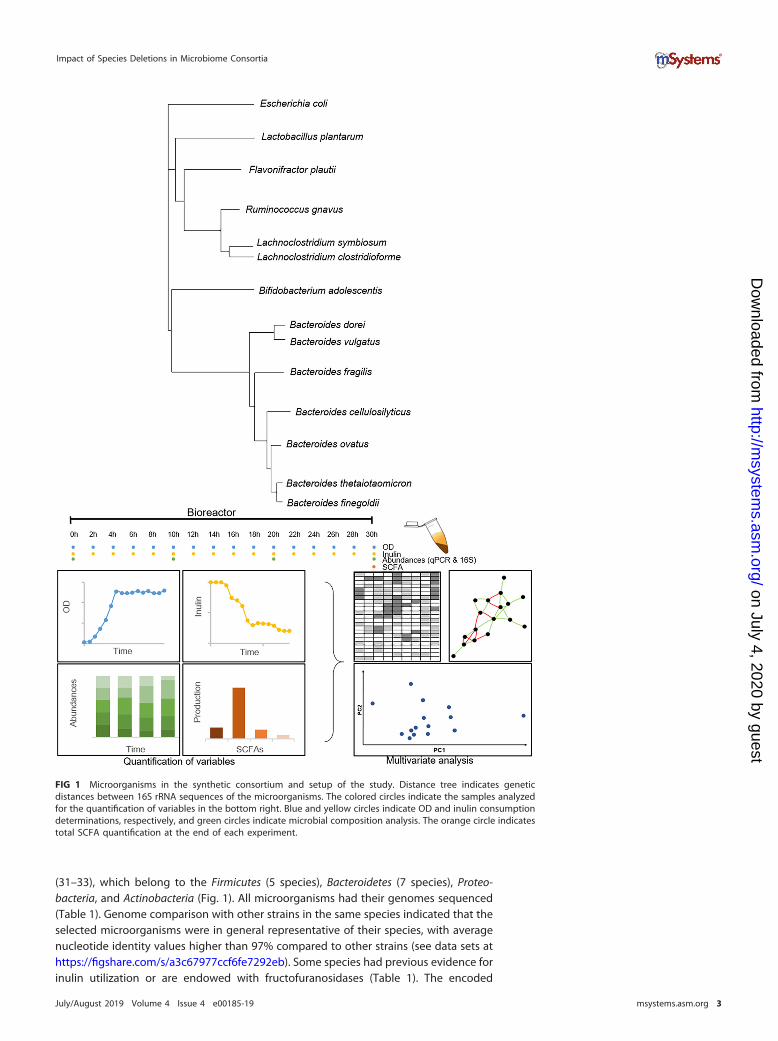
(31–33), which belong to the Firmicutes (5 species), Bacteroidetes (7 species), Proteo-bacteria, and Actinobacteria (Fig. 1). All microorganisms had their genomes sequenced(Table 1). Genome comparison with other strains in the same species indicated that theselected microorganisms were in general representative of their species, with averagenucleotide identity values higher than 97% compared to other strains (see data sets athttps://figshare.com/s/a3c67977ccf6fe7292eb). Some species had previous evidence forinulin utilization or are endowed with fructofuranosidases (Table 1). The encoded
FIG 1 Microorganisms in the synthetic consortium and setup of the study. Distance tree indicates geneticdistances between 16S rRNA sequences of the microorganisms. The colored circles indicate the samples analyzedfor the quantification of variables in the bottom right. Blue and yellow circles indicate OD and inulin consumptiondeterminations, respectively, and green circles indicate microbial composition analysis. The orange circle indicatestotal SCFA quantification at the end of each experiment.
Impact of Species Deletions in Microbiome Consortia
July/August 2019 Volume 4 Issue 4 e00185-19 msystems.asm.org 3
on July 4, 2020 by guesthttp://m
systems.asm
.org/D
ownloaded from
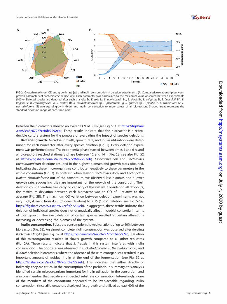
potential for SCFA production was deduced from their genomes (Table 1), indicatingthat the consortium was able to produce lactate, acetate, propionate, or butyrate. Twospecies had the potential to produce butyrate: Lachnoclostridium symbiosum andLachnoclostridium clostridioforme.
Reproducibility of the culture system. We first evaluated the reproducibility of theculture system, culturing the 14 microorganisms in a batch bioreactor using inulin ascarbon source. Microorganisms were inoculated at similar initial concentrations. Cul-turing the consortium with all species (designated All) showed a coefficient of variation(CV) of 3.3% in optical density (OD) and only 3.4% in the final inulin amount left (seeFig. S1A at https://figshare.com/s/a3c67977ccf6fe7292eb). The relative abundances ofeach species showed an average CV of 18.6% in the final composition of the replicates(see Fig. S1B at https://figshare.com/s/a3c67977ccf6fe7292eb). The initial composition
TABLE 1 Description of the strains used in this study and genomic features important for the study
Phylum, species,and strain
Genomesize (Mbp);%GC
Inulin utilizationand fructofuranosidase(locus tag[s])
SCFA productionCulturemediuma Sporulation Reference(s)Lactate Acetate Propionate Butyrate
FirmicutesLactobacillus
plantarumATCC 8014
3.23;44.53
Yes; Ga0133059_101166 Yes MRS 50
RuminococcusgnavusCC55_001C
3.18;43.10
No; HMPREF1201_02241,HMPREF1201_01812,HMPREF1201_01164
Yes Yes RCM Yes 67
Flavonifractorplautii1_3_50AFAA
4.32;60.53
No Yes RCM Yes 68
LachnoclostridiumsymbiosumWAL-14673
4.86;48.17
No; HMPREF9475_00225 Yes RCM Yes 55
Lachnoclostridiumclostridioforme2_1_49FAA
5.46;48.93
No; HMPREF9467_03179 Yes RCM Yes 69
BacteroidetesBacteroides
dorei 5_1_36/D45.53;
41.52Yes; BSEG_04148 Yes Yes Yes BHI 48
Bacteroidesvulgatus ATCC 8482
5.16;42.20
No Yes Yes BHI 53
BacteroidesfinegoldiiCL09T03C10
5.12;42.50
No; HMPREF1057 Yes Yes RCM 53
BacteroidesfragilisCL03T12C07
5.21;43.44
Yes; HMPREF1067_02001 Yes Yes Yes BHI 70
Bacteroidesovatus 3_8_47FAA
6.55;41.97
Yes; HMPREF1017_04501 Yes Yes BHI 53
BacteroidescellulosilyticusCL02T12C19
7.68;43.05
No; HMPREF1062_02875 Yes Yes Yes BHI 53, 71
BacteroidesthetaiotaomicronVPI-5482
6.29;48.86
Yes; BT1760 Yes Yes Yes RCM 72
ProteobacteriaEscherichia
coli K-12 MG16554.64;
50.79No Yes Yes LB/PCA 73
ActinobacteriaBifidobacterium
adolescentisATCC 15703
2.09;59.18
Yes; BAD_1150,BAD_1325
Yes Yes MRS-Cys 74
aMRS, de Man, Rogosa, and Sharpe medium; PCA, plate count agar.
Gutiérrez and Garrido
July/August 2019 Volume 4 Issue 4 e00185-19 msystems.asm.org 4
on July 4, 2020 by guesthttp://m
systems.asm
.org/D
ownloaded from
between the bioreactors showed an average CV of 8.1% (see Fig. S1C at https://figshare.com/s/a3c67977ccf6fe7292eb). These results indicate that the bioreactor is a repro-ducible culture system for the purpose of evaluating the impact of species deletions.
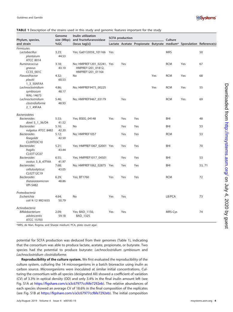
Bacterial growth. Microbial growth, growth rate, and inulin utilization were deter-mined for each bioreactor after every species deletion (Fig. 2). Every deletion experi-ment was performed once. The exponential phase started between times 4 and 6 h, andall bioreactors reached stationary phase between 12 and 14 h (Fig. 2B; see also Fig. S2at https://figshare.com/s/a3c67977ccf6fe7292eb). Escherichia coli and Bacteroidesthetaiotaomicron deletions resulted in the highest biomass and growth rates obtained,indicating that these microorganisms contribute negatively to these parameters in thewhole consortium (Fig. 2). In contrast, when leaving Bacteroides dorei and Lachnoclos-tridium clostridioforme out of the consortium, we observed less biomass and a lowergrowth rate, suggesting they are important for the growth of the consortium. Theirdeletion could therefore free carrying capacity of the system. Considering all dropouts,the maximum deviation between each bioreactor was an OD of 1 relative to theaverage (Fig. 2B). The maximum OD variation between deletion experiments was notvery high; it went from 4.23 (B. dorei deletion) to 7.36 (E. coli deletion; see Fig. S2 athttps://figshare.com/s/a3c67977ccf6fe7292eb). In aggregate, these results indicate thatdeletion of individual species does not dramatically affect microbial consortia in termsof total growth. However, deletion of certain species resulted in certain alterationsincreasing or decreasing the biomass of the system.
Inulin consumption. Substrate consumption showed variations of up to 40% betweenbioreactors (Fig. 2B). An almost complete inulin consumption was observed after deletingBacteroides fragilis (see Fig. S2 at https://figshare.com/s/a3c67977ccf6fe7292eb). Deletionof this microorganism resulted in slower growth compared to all other replicates(Fig. 2A). These results indicate that B. fragilis in this system interferes with inulinconsumption. The opposite was observed in L. clostridioforme, B. thetaiotaomicron, andB. dorei deletion bioreactors, where the absence of these microorganisms resulted in animportant amount of residual inulin at the end of the fermentation (see Fig. S2 athttps://figshare.com/s/a3c67977ccf6fe7292eb). This indicates that either directly orindirectly, they are critical in the consumption of the prebiotic. In summary, this analysisidentified certain microorganisms important for inulin utilization in the consortium andalso one member that negatively impacted substrate consumption. Interestingly, noneof the members of the consortium appeared to be irreplaceable regarding inulinconsumption, since all bioreactors displayed fast growth and utilized at least 40% of the
FIG 2 Growth (maximum OD and growth rate [�]) and inulin consumption in deletion experiments. (A) Comparative relationship betweengrowth parameters of each bioreactor (see key). Each parameter was normalized to the maximum value observed between experiments(100%). Deleted species are denoted after each triangle: Ec, E. coli; Ba, B. adolescentis; Bd, B. dorei; Bv, B. vulgatus; Bf, B. finegoldii; Bfr, B.fragilis; Bc, B. cellulosilyticus; Bo, B. ovatus; Bt, B. thetaiotaomicron; Lp, L. plantarum; Rg, R. gnavus; Fp, F. plautii; Ls, L. symbiosum; Lc, L.clostridioforme. (B) Average of growth (blue) and inulin consumption (orange) values of all bioreactors. Shaded areas represent thestandard deviation range of each time point.
Impact of Species Deletions in Microbiome Consortia
July/August 2019 Volume 4 Issue 4 e00185-19 msystems.asm.org 5
on July 4, 2020 by guesthttp://m
systems.asm
.org/D
ownloaded from
inulin. This suggests functional redundancy in the metabolism of the consortium(Table 1).
Microbial composition of bioreactors. The microbial composition of each con-
sortium was determined by quantitative PCR (qPCR) (Fig. 3). Cell copy numberswere in general correlated with OD values (see Fig. S3 at https://figshare.com/s/a3c67977ccf6fe7292eb). In general, the dominant species were Ruminococcus gnavus,E. coli, B. dorei, L. symbiosum, and Lactobacillus plantarum. L. plantarum showed aconsiderable increase in the absence of B. dorei and L. symbiosum, reaching 50% of thetotal abundance and suggesting a competitive relationship between these microbes(Fig. 3D and N). Bifidobacterium adolescentis was not particularly dominant in anybioreactor, which was unexpected considering that it is a prominent inulin utilizer (34,35). One remarkable exception was the E. coli deletion bioreactor, where B. adolescentisincreased its relative abundance to more than 60% (Fig. 3B).
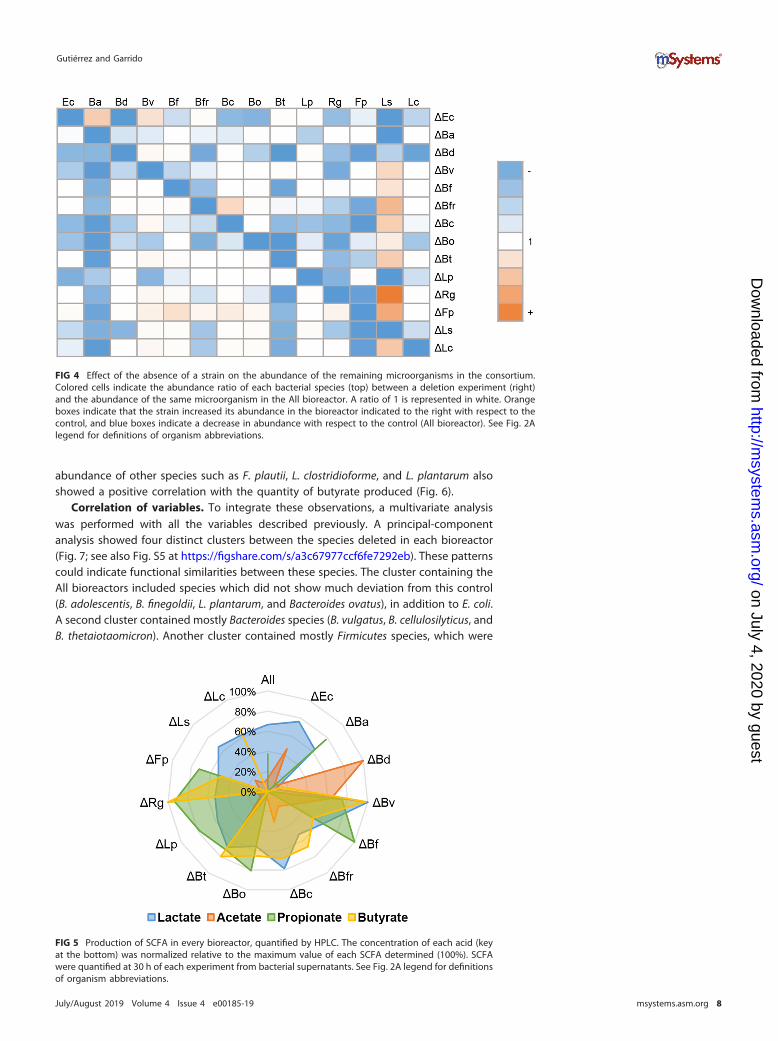
To quantify the effect of the absence of one microorganism on the abundance ofother species in the consortium, an abundance ratio was calculated for each deletedmicroorganism relative to the abundance of the same strain in the All bioreactor(Fig. 4). Deletions of B. adolescentis and Bacteroides finegoldii from the consortium didnot alter the abundance of any other species. While Bacteroides vulgatus and B.finegoldii were mostly unaffected by any dropout, B. adolescentis and Flavonifractorplauti showed reduced abundances in the absence of almost any other member.Finally, deletion of B. dorei from the consortium caused the most deleterious effect onthe abundance of 10 microorganisms (Fig. 4), indicating a key role of B. dorei in thisconsortium.
Certain species benefited from the absence of others. The abundance of L. symbio-sum was stimulated in the absence of eight species of the consortium but alsonegatively impacted by the absence of four others (E. coli, B. adolescentis, B. dorei, andL. plantarum). It could be hypothesized that the latter four are important for L.symbiosum growth since their removal impacted L. symbiosum abundance negatively.This also indicates that L. symbiosum is the most sensitive member relative to thepresence or absence of other microorganisms in this consortium.
Production of metabolites. The concentrations of lactate, acetate, propionate, and
butyrate were determined at the end of each run and normalized to the maximum valueobserved (Fig. 5; see also Fig. S4 at https://figshare.com/s/a3c67977ccf6fe7292eb). Theproduction of SCFA in the All consortium was 83.9 mM lactate, 21.5 mM acetate, 5.9 mMpropionate, and no butyrate.
The maximum production of lactate was reached in the B. vulgatus deletion biore-actor. Deletion of this microorganism also allowed the bioreactor to achieve the highesttotal SCFA concentration (see Fig. S4 at https://figshare.com/s/a3c67977ccf6fe7292eb).In contrast, deletion of B. dorei resulted in the minimal lactate concentration observed,concomitant with the maximum production of acetate observed. This indicates that B.dorei is essential for lactate production, and when it is absent, the consortium switchesto high acetate production.
The maximum propionate concentration was observed in the B. finegoldii deletionbioreactor (16.1 mM), and the concomitant loss of six species did not result in theproduction of propionate (Fig. 5). Three of these were propionate-producing Bacte-roides species (B. dorei, B. fragilis, and Bacteroides cellulosilyticus). Three others (E. coli, L.symbiosum, and L. clostridioforme) could contribute indirectly to propionate production.
Finally, the higher concentration of butyrate was observed in R. gnavus and B.vulgatus deletion bioreactors (10.5 mM [Fig. 5]; see also Fig. S4 at https://figshare.com/s/a3c67977ccf6fe7292eb). This suggests that their presence has a negative impact onthe production of the acid. In contrast, deletions of L. symbiosum, E. coli, B. adolescentis,B. dorei, and L. plantarum resulted in a lack of butyrate. A linear regression analysis ofbutyrate production and microbial abundance indicated that the major contribution tothe production of this SCFA was attributed to L. symbiosum (Fig. 6). However, the
Gutiérrez and Garrido
July/August 2019 Volume 4 Issue 4 e00185-19 msystems.asm.org 6
on July 4, 2020 by guesthttp://m
systems.asm
.org/D
ownloaded from
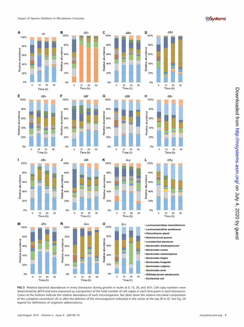
FIG 3 Relative bacterial abundances in every bioreactor during growth in inulin at 0, 10, 20, and 30 h. Cell copy numbers weredetermined by qPCR and were expressed as a proportion of the total number of cell copies in each time point in each bioreactor.Colors at the bottom indicate the relative abundance of each microorganism. Bar plots show the relative microbial compositionof the complete consortium (A) or after the deletion of the microorganism indicated in the center at the top (B to O). See Fig. 2Alegend for definitions of organism abbreviations.
Impact of Species Deletions in Microbiome Consortia
July/August 2019 Volume 4 Issue 4 e00185-19 msystems.asm.org 7
on July 4, 2020 by guesthttp://m
systems.asm
.org/D
ownloaded from
abundance of other species such as F. plautii, L. clostridioforme, and L. plantarum alsoshowed a positive correlation with the quantity of butyrate produced (Fig. 6).
Correlation of variables. To integrate these observations, a multivariate analysiswas performed with all the variables described previously. A principal-componentanalysis showed four distinct clusters between the species deleted in each bioreactor(Fig. 7; see also Fig. S5 at https://figshare.com/s/a3c67977ccf6fe7292eb). These patternscould indicate functional similarities between these species. The cluster containing theAll bioreactors included species which did not show much deviation from this control(B. adolescentis, B. finegoldii, L. plantarum, and Bacteroides ovatus), in addition to E. coli.A second cluster contained mostly Bacteroides species (B. vulgatus, B. cellulosilyticus, andB. thetaiotaomicron). Another cluster contained mostly Firmicutes species, which were
FIG 4 Effect of the absence of a strain on the abundance of the remaining microorganisms in the consortium.Colored cells indicate the abundance ratio of each bacterial species (top) between a deletion experiment (right)and the abundance of the same microorganism in the All bioreactor. A ratio of 1 is represented in white. Orangeboxes indicate that the strain increased its abundance in the bioreactor indicated to the right with respect to thecontrol, and blue boxes indicate a decrease in abundance with respect to the control (All bioreactor). See Fig. 2Alegend for definitions of organism abbreviations.
FIG 5 Production of SCFA in every bioreactor, quantified by HPLC. The concentration of each acid (keyat the bottom) was normalized relative to the maximum value of each SCFA determined (100%). SCFAwere quantified at 30 h of each experiment from bacterial supernatants. See Fig. 2A legend for definitionsof organism abbreviations.
Gutiérrez and Garrido
July/August 2019 Volume 4 Issue 4 e00185-19 msystems.asm.org 8
on July 4, 2020 by guesthttp://m
systems.asm
.org/D
ownloaded from

important for SCFA production (F. plautii and L. clostridioforme) or controlled inulinutilization (B. fragilis). A fourth cluster was obtained with B. dorei and L. symbiosum,where their deletions from the system resulted in comparable results. Similar observa-tions were also obtained when considering all data points (see Fig. S5 at https://figshare.com/s/a3c67977ccf6fe7292eb), where all bioreactors clustered closely at the beginningof the study, and most of the above associations were also found, including additionaldata points.
While the absence of B. dorei altered several parameters of the community, L.symbiosum abundance was essential for butyrate production (see Fig. S6 at https://figshare.com/s/a3c67977ccf6fe7292eb) and sensitive to the absence of several micro-organisms. Finally, the abundance of L. clostridioforme was positively correlated withpropionate production, that of L. symbiosum was positively correlated with butyrategeneration, and that of L. plantarum was positively correlated with acetate production(see Fig. S6 at https://figshare.com/s/a3c67977ccf6fe7292eb).
FIG 6 Percent contribution of each species to the butyrate production. The estimated contributionsrepresent a linear relationship between the abundance of the species and butyrate production in eachbioreactor. See Fig. 2A legend for definitions of organism abbreviations.
FIG 7 Principal-component analysis and clustering of consortia. Data analysis included as variables microbialspecies abundances, SCFA production, inulin consumption, OD values, and growth rate at the final point (t � 30h). Colors represent the clusters formed by the deletion of each microorganism. See Fig. 2A legend for definitionsof organism abbreviations.
Impact of Species Deletions in Microbiome Consortia
July/August 2019 Volume 4 Issue 4 e00185-19 msystems.asm.org 9
on July 4, 2020 by guesthttp://m
systems.asm
.org/D
ownloaded from
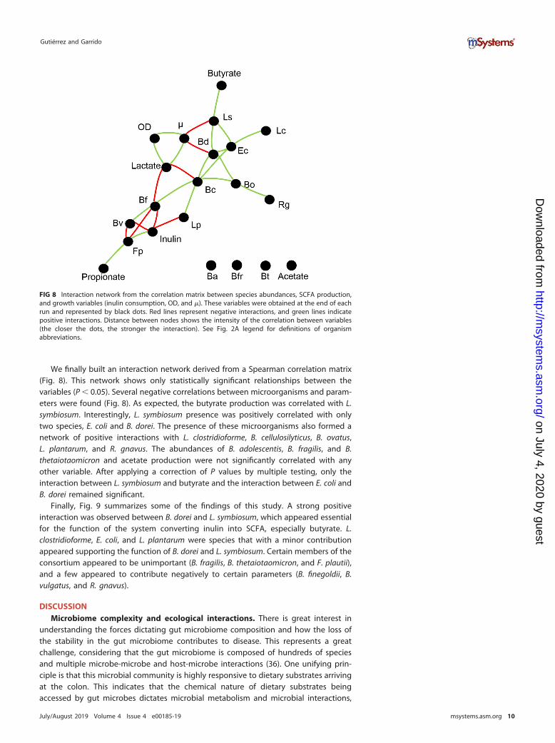
We finally built an interaction network derived from a Spearman correlation matrix(Fig. 8). This network shows only statistically significant relationships between thevariables (P � 0.05). Several negative correlations between microorganisms and param-eters were found (Fig. 8). As expected, the butyrate production was correlated with L.symbiosum. Interestingly, L. symbiosum presence was positively correlated with onlytwo species, E. coli and B. dorei. The presence of these microorganisms also formed anetwork of positive interactions with L. clostridioforme, B. cellulosilyticus, B. ovatus,L. plantarum, and R. gnavus. The abundances of B. adolescentis, B. fragilis, and B.thetaiotaomicron and acetate production were not significantly correlated with anyother variable. After applying a correction of P values by multiple testing, only theinteraction between L. symbiosum and butyrate and the interaction between E. coli andB. dorei remained significant.
Finally, Fig. 9 summarizes some of the findings of this study. A strong positiveinteraction was observed between B. dorei and L. symbiosum, which appeared essentialfor the function of the system converting inulin into SCFA, especially butyrate. L.clostridioforme, E. coli, and L. plantarum were species that with a minor contributionappeared supporting the function of B. dorei and L. symbiosum. Certain members of theconsortium appeared to be unimportant (B. fragilis, B. thetaiotaomicron, and F. plautii),and a few appeared to contribute negatively to certain parameters (B. finegoldii, B.vulgatus, and R. gnavus).
DISCUSSIONMicrobiome complexity and ecological interactions. There is great interest in
understanding the forces dictating gut microbiome composition and how the loss ofthe stability in the gut microbiome contributes to disease. This represents a greatchallenge, considering that the gut microbiome is composed of hundreds of speciesand multiple microbe-microbe and host-microbe interactions (36). One unifying prin-ciple is that this microbial community is highly responsive to dietary substrates arrivingat the colon. This indicates that the chemical nature of dietary substrates beingaccessed by gut microbes dictates microbial metabolism and microbial interactions,
FIG 8 Interaction network from the correlation matrix between species abundances, SCFA production,and growth variables (inulin consumption, OD, and �). These variables were obtained at the end of eachrun and represented by black dots. Red lines represent negative interactions, and green lines indicatepositive interactions. Distance between nodes shows the intensity of the correlation between variables(the closer the dots, the stronger the interaction). See Fig. 2A legend for definitions of organismabbreviations.
Gutiérrez and Garrido
July/August 2019 Volume 4 Issue 4 e00185-19 msystems.asm.org 10
on July 4, 2020 by guesthttp://m
systems.asm
.org/D
ownloaded from

resulting in complex cross-feeding networks. In this study, we analyzed the impact ofspecies deletions on the composition and metabolic function of a synthetic consortiumcomposed of 14 gut microbes during consumption of inulin. Every deletion experimentwas performed once, considering that batch bioreactors showed to be reproducible.However, coculture experiments have shown that gut microbe growth could be verysensitive to the initial concentrations of microorganisms (37). Therefore, the lack ofreplicates is a limitation of this work.
Species deletion is a common approach in ecological theory of food webs (38, 39).Sometimes, the disappearance of one species in a biological community has detrimen-tal effects on the whole community, causing further extinctions of species and impact-ing ecosystem performance and fitness (40). Interestingly, here we never found aspecies deletion causing a catastrophic effect in the community: total growth in thebioreactors always reached OD values higher than 4 and inulin utilization was alwayshigher than 50% of the initial concentration. One possible explanation is that mediumcomposition is directly responsible for the failure to observe larger metabolic depen-dencies between gut microbes. Considering that the substrate was readily consumed,another explanation is functional redundancy in this consortium, where the role of onespecies was readily replaced by another, leading to comparable final states. Redun-dancy is a crucial property of the adult gut microbiome, which has been directlyassociated with microbiome resilience and stability against perturbations (41).
In contrast to these similarities, microbial compositions and metabolic activitieswere markedly different after deletions of single microorganisms (Fig. 5; see also Fig. S4at https://figshare.com/s/a3c67977ccf6fe7292eb). These observations are importantsince it is actually the metabolic activity of the gut microbiome, rather than thecomposition or taxonomy, which has the most profound effect on host physiology (42).B. dorei was the microbe that when absent caused the most substantial change in theconsortium: 10 species showed a decrease in their relative abundance when B. dorei
FIG 9 Working model of the study summarizing major results. Arrows indicate positive relationships. Dashed linesindicate negative relationships, and lines with points indicate a relationship with positive or negative statusunknown. Thick lines indicate that these observations were statistically significant. Orange lines show therelationship between inulin and species that consume it, and blue lines show the relationship between species andthe production of SCFA. See Fig. 2A legend for definitions of organism abbreviations.
Impact of Species Deletions in Microbiome Consortia
July/August 2019 Volume 4 Issue 4 e00185-19 msystems.asm.org 11
on July 4, 2020 by guesthttp://m
systems.asm
.org/D
ownloaded from
was missing. Concomitantly, total biomass was the lowest of all bioreactors, and a clearswitch in SCFA was observed. Therefore, B. dorei could be classified as a keystonespecies in this consortium (40).
Interestingly, metabolic profiles did not necessarily reflect the metabolism of thedominant microbe in each deletion experiment. The deletion of L. plantarum resultedin low acetate concentrations, also showing a positive correlation between the abun-dance of L. plantarum and acetate level. This is intriguing since L. plantarum does notproduce acetate under these conditions. L. plantarum has a facultative heterofermen-tative metabolism, producing large amounts of lactate under anaerobic conditions (43).In addition, we did not observe a decrease in lactate concentration in its absence,indicating that other microorganisms contribute to the lactate pool.
In fecal samples, lactate is not typically detected since it is a cross-feeding metab-olite that is readily fermented by other gut microbes (44, 45). In feces from subjects withinflammatory bowel diseases, lactate could reach up to 100 mM (46). In this study, thelowest lactate production was observed by deleting B. dorei. Bacteroides species arewell known for producing acetate and propionate as end products. However, certainstudies indicate that they could also release lactate to the medium, for example in theabsence of organic nitrogen sources and especially fermentable carbohydrates such asfructans (47, 48). Therefore, we hypothesize that B. dorei is the major lactate producerin the consortium. Interestingly, B. dorei deletion resulted in the highest acetateconcentration obtained and the concomitant dominance of L. plantarum (Fig. 3). Thisobservation could be explained by L. plantarum switching to heterofermentativemetabolism or by another dominant microbe in this consortium, such as E. coli, beingresponsible for the high acetate observed.
Mechanistic evidence for metabolic interactions. Inulin is a well-studied prebiotic
that has been generally associated with the stimulation of Lactobacillus and Bifidobac-terium (35, 49, 50). The impact of this fructan in the gut microbiome is regarded to bemore complex now, stimulating other species such as Anaerostipes and decreasingBilophila (5). In our consortium, at least five species are well known for inulin utilization(B. adolescentis, B. thetaiotaomicron, L. plantarum, B. dorei, and B. ovatus), and severalothers are endowed with fructofuranosidases (L. symbiosum, L. clostridioforme, B. cellu-losilyticus, B. finegoldii, and R. gnavus) (Table 1) (51). Among these, L. plantarum and B.adolescentis displayed a limited representation in the synthetic consortium, and theirgrowth was usually repressed in the presence of other microbes. Their abundanceincreased only after prominent members in the consortium were deleted. B. dorei or L.symbiosum deletion increased L. plantarum growth, and E. coli deletion promoted B.adolescentis. Our working model shows that B. dorei is the dominant microbe accessinginulin and releasing smaller fructans and SCFA, which could support E. coli and L.symbiosum growth. How this combination is able to outcompete L. plantarum or B.adolescentis in vitro is intriguing and could be explained by higher growth rates on thesubstrate or the release of inhibitory molecules.
Our consortium included seven Bacteroides species, including B. dorei, discussedabove. Bacteroides species are important members in the microbiome and are endowedwith a wide array of polysaccharide utilization machineries. They are mostly generalistsable to explore a broad set of nutrients, including proteins (52). Depending on dietaryfibers being fed, Bacteroides species could assume distinct metabolic roles, where somespecies act as primary fermenters accessing complex substrates and releasing smallerdegradation products to other species. For example, it was shown that B. ovatus, whilegrowing in inulin, releases smaller fructans which are utilized by other Bacteroides. B.ovatus also supports the growth of B. vulgatus in gnotobiotic mice (53). These twomicroorganisms showed little representation in this study, which could be explained bya higher inulin-degrading capability of other members of the consortium, such as B.dorei. Certain Bacteroides species are also sensitive to low pH (54), which could limittheir growth in our setup (pH 5.5, simulating the conditions of the proximal colon).
Gutiérrez and Garrido
July/August 2019 Volume 4 Issue 4 e00185-19 msystems.asm.org 12
on July 4, 2020 by guesthttp://m
systems.asm
.org/D
ownloaded from
Lachnoclostridium symbiosum was a prominent member of this consortium produc-ing butyrate. Although the production of butyrate in L. symbiosum occurs preferably viathe 4-aminobutyrate/succinate pathway (55), it can also be synthesized from acetateand lactate (56–58). We hypothesize that acetate is being cross-fed to L. symbiosum,resulting in butyrate production. While this was not demonstrated here, additionalevidence is the presence of homologs of an acetate permease in its genome. Anotherstudy suggested that L. symbiosum could produce butyrate from lactate (59). Clostrid-ium butyricum is able to use both lactate and acetate for butyrate production (60). L.symbiosum was the most sensitive strain with regard to the absence or presence ofother microbes. Deletions of E. coli, B. adolescentis, B. dorei, and L. plantarum resultedin limited growth of L. symbiosum, suggesting they are important for its growth. Arecent report studying the pairwise interactions showed that in a 12-species consor-tium, Faecalibacterium prausnitzii, a keystone species and butyrate producer, was alsothe most sensitive species, receiving the most positive interactions from other microbes(17). We have also previously reported that L. symbiosum, in the presence of B. dorei,produced high concentrations of butyrate which were shown to reduce inflammationmediated by tumor necrosis factor alpha (TNF-�) in a cellular model (48). These positiveinteractions, which are dependent on diet, could be further exploited in order to enrichthe gut microbiome for butyrate-producing species.
Conclusions. In this study, we analyzed the impact of the species deletions in asynthetic consortium of gut microbes. This approach was useful to identify significantmicrobial interactions between microorganisms but also specific microbes playing keymetabolic roles in the community. We also observed emergent interactions whichcannot be detected using paired cocultures. We observed a strong correlation betweenE. coli, B. dorei, and L. symbiosum, and we defined essential species for butyrateproduction. In addition to identifying key roles in the gut microbiome, this approachcould be useful for the design of microbial consortia with desired metabolic properties.
MATERIALS AND METHODSStrains and culture media. Strains used in this study are shown in Table 1, and were obtained from
BEI Resources, the ATCC, or the UC Davis Culture Collection. For routine experiments, microorganismswere cultured in their respective culture media described in Table 1. Luria-Bertani medium (LB; Becton,Dickinson, Franklin Lakes, NJ) was used directly, while reinforced clostridium medium (RCM; Becton,Dickinson, Franklin Lakes, NJ) was supplemented with 0.5 g/liter of L-cysteine (Loba Chemie, India). deMan, Rogosa, and Sharpe medium (MRS; Becton, Dickinson, Franklin Lakes, NJ) was supplemented with0.5 g/liter of L-cysteine, except for L. plantarum. Brain heart infusion (BHI; Becton, Dickinson, FranklinLakes, NJ) was supplemented with 0.5 g/liter of L-cysteine and 0.01 g/liter of hemin. All incubations wereperformed at 37°C for 24 to 48 h in an anaerobic jar (Anaerocult; Merck, Darmstadt, Germany) withanaerobic packs (GasPak EM; Becton, Dickinson, Franklin Lakes, NJ).
Genomic analysis. Genomes were obtained and analyzed using the Integrated Microbial Gene (IMG)(51). Pairwise average nucleotide identity (ANI) (61) was calculated for genomes used in this study,compared to available finished genomes in the same species. A genetic distance tree was obtained basedon the 16S rRNA sequence of each microorganism in IMG. Fructofuranosidase presence in selectgenomes was estimated using the EC number 3.2.1.26. Inulin utilization and acetate and lactateproduction were determined from the literature (Table 1) (16). Propionate and butyrate production wasconfirmed by finding the respective IMG pathways in select genomes. Finally, sporulation was confirmedby the presence of Spo0A homologs and a full set of sporulation genes in select genomes.
Experimental design and bioreactor operation. Sixteen batch experiments were carried out in a250-ml bioreactor connected to a MyControl system (Mini-bio; Applikon Biotechnology, Netherlands).Two of them were inoculated with the complete consortium (All) run in duplicate. The remainingexperiments replicated the same conditions as the control with all strains but leaving one species out;therefore, they were inoculated with 13 strains. The bioreactors were named with Δ followed by anabbreviation of the microorganism left out in each case (Table 1). Microorganisms were cultured usingthe optimized formulation mZMB (62), with a fixed pH of 5.5. Before being inoculated in each experi-ment, microorganisms were grown individually in mZMB supplemented with lactose (20 g/liter) as acarbon source under anaerobic conditions for 48 h at 37°C. After 48 h, the OD was measured, and thevolume required of each culture was considered to use an initial OD630 of 1 for each microorganism.Cultures were centrifuged at 3,000 rpm for 5 min. The supernatant was discarded, and the pellet wasresuspended in the fixed group of mZMB and used to inoculate the reactor. For each bioreactor, mZMBwas supplemented with inulin (20 g/liter) (Piping Rock, Ronkonkoma, NY). Tryptone (Becton, Dickinson,Franklin Lakes, NJ) and L-cysteine were used at 34.2 g/liter and 1 g/liter, respectively, and autoclaveddirectly in the bioreactor in 70 ml of distilled water. The remaining components of mZMB were sterilizedusing 0.22-�m filters and incorporated into the bioreactor after being autoclaved (inulin and 0.005 g/liter
Impact of Species Deletions in Microbiome Consortia
July/August 2019 Volume 4 Issue 4 e00185-19 msystems.asm.org 13
on July 4, 2020 by guesthttp://m
systems.asm
.org/D
ownloaded from
hemin). The bioreactor was inoculated at an initial OD630 of 1 for each microorganism. Foam level wascontrolled by injecting 200 �l of silicone antifoam (polydimethylsiloxane) with the inoculum. To generatean anaerobic environment inside the bioreactor, nitrogen (99.99% purity grade) was injected at thebeginning of the fermentation and remained anaerobic during each experiment. The temperature wasset at 37°C, and stirring was set at 90 rpm. pH was maintained constant throughout the fermentation at5.5, using an automatic injection of 3 M NaOH and 3 M HCl. Samples were taken every 2 h up to 30 h andimmediately centrifuged at 10,000 � g for 2 min. Pellets and supernatants were stored at �20°C. Pelletswere subsequently used for DNA extraction and relative abundance determination, and supernatantswere used for quantification of inulin consumption and production of SCFA.
Substrate consumption. Inulin utilization was quantified using a previously adapted phenol-sulfuricacid method (62, 63). Supernatants from the bioreactors were diluted 1:200 (vol/vol). Inulin standardsbetween 12 and 0.08 �g were prepared. The experiments were performed in 96-well microplatescontaining 50 �l of a cold-diluted sample (4°C), 150 �l of concentrated sulfuric acid (98% H2SO4), and30 �l of 5% phenol. Microplates were heated at 90°C for 5 min and cooled on ice for 5 min. Absorbanceat 490 nm was determined in a Tecan Infinite M200 Pro plate reader (Tecan Trading AG, Grödig, Austria).Measurements were performed in replicates.
Quantification of SCFA by HPLC. Acetic, lactic, propionic, and butyric acid were quantified at theend of each run in the bioreactors by high-performance liquid chromatography (HPLC) according to aprotocol previously reported (64). An ion exchange column of organic acids and carbohydrates, AminexHPX-87H (Bio-Rad, Hercules, CA), was used in a Lachrom L-700 HPLC system (Hitachi, Japan) at 35°C witha flow rate of 0.45 ml/min of 5 mM H2SO4. As standard, concentrations from 30 g/liter to 0.155 g/liter ofeach acid were used.
Determination of relative bacterial abundances. Total DNA extraction from cell pellets wasperformed by an adapted phenol-chloroform-isoamyl protocol (62). DNA was quantified using a Nano-Quant plate in a Tecan Infinite M200 Pro microplate reader and diluted to 10 ng/�l. Bacterial relative andabsolute abundances were determined by qPCR, using a set of species-specific primers based on uniquegenes present in each microorganism (see Table S1 at https://figshare.com/s/a3c67977ccf6fe7292eb).qPCRs and concentration measurements were performed as in the work of Medina et al. (65), withannealing temperatures of 62°C. One exception was R. gnavus qPCR, which was performed at 58°C.Reactions were performed in triplicates, and threshold cycle (CT) values were converted into genomecopy numbers per milliliter as in the work of Medina et al. (65).
Data analysis. The contribution of microorganisms to the butyrate production was calculated as alinear regression of the abundances (variables) to the production butyrate (response). A single contri-bution factor was defined by species. The contribution factor was multiplied by the average of theabundance of the respective bacterium determining its contribution. For the multivariate analysis,principal-component analysis and hierarchical clustering were performed with the ClustVis tool (66). Aninteraction network was built from the Spearman correlation matrix using the ‘corr’ command in Matlaband showing the statistically significant correlations (P value � 0.05). Later, we used the bonf_holmfunction in Matlab (Bonferroni-Holm correction) for multiple testing.
Data availability. Raw data and calculations of average nucleotide identity between microor-ganisms in this study and other strains in the same species are available at https://figshare.com/s/a3c67977ccf6fe7292eb.
ACKNOWLEDGMENTSWe thank Veronica Ortuzar, Rodrigo Santibañez, and Pamela Thomson for their
support in this study.This work was funded by FONDEF IDEA ID16i10045.
REFERENCES1. Sender R, Fuchs S, Milo R. 2016. Revised estimates for the number of
human and bacteria cells in the body. PLoS Biol 14:e1002533-14. https://doi.org/10.1371/journal.pbio.1002533.
2. Zhu L, Qin S, Zhai S, Gao Y, Li L. 2017. Inulin with different degrees ofpolymerization modulates composition of intestinal microbiota in mice.FEMS Microbiol Lett 364:fnx075. https://doi.org/10.1093/femsle/fnx075.
3. Kuo S. 2013. The interplay between fiber and the intestinal microbiomein the inflammatory response. Adv Nutr 4:16 –28. https://doi.org/10.3945/an.112.003046.
4. Lahner E, Annibale B. 2017. Dietary fiber and gut microbiota modula-tion—a possible switch from disease to health for subjects with diver-ticular disease? JSM Nutr Disord 1:1001.
5. Makki K, Deehan EC, Walter J, Bäckhed F. 2018. The impact of dietaryfiber on gut microbiota in host health and disease. Cell Host Microbe23:705–715. https://doi.org/10.1016/j.chom.2018.05.012.
6. Singh RK, Chang HW, Yan D, Lee KM, Ucmak D, Wong K, Abrouk M,Farahnik B, Nakamura M, Zhu TH, Bhutani T, Liao W. 2017. Influence ofdiet on the gut microbiome and implications for human health. J TranslMed 15:1–17. https://doi.org/10.1186/s12967-017-1175-y.
7. Conlon MA, Bird AR. 2014. The impact of diet and lifestyle on gutmicrobiota and human health. Nutrients 7:17– 44. https://doi.org/10.3390/nu7010017.
8. Sun M, Wu W, Liu Z, Cong Y. 2017. Microbiota metabolite short chainfatty acids, GPCR, and inflammatory bowel diseases. J Gastroenterol52:1– 8. https://doi.org/10.1007/s00535-016-1242-9.
9. Li M, van Esch B, Wagenaar GTM, Garssen J, Folkerts G, Henricks P. 2018.Pro- and anti-inflammatory effects of short chain fatty acids on immuneand endothelial cells. Eur J Pharmacol 831:52–59. https://doi.org/10.1016/j.ejphar.2018.05.003.
10. Morrison DJ, Preston T. 2016. Formation of short chain fatty acids by thegut microbiota and their impact on human metabolism. Gut Microbes7:189 –200. https://doi.org/10.1080/19490976.2015.1134082.
11. Bergman EN. 1990. Energy contributions of volatile fatty acids from thegastrointestinal tract in various species. Physiol Rev 70:567–590. https://doi.org/10.1152/physrev.1990.70.2.567.
12. Macfarlane S, Macfarlane GT. 2003. Regulation of short-chain fattyacid production. Proc Nutr Soc 62:67–72. https://doi.org/10.1079/PNS2002207.
Gutiérrez and Garrido
July/August 2019 Volume 4 Issue 4 e00185-19 msystems.asm.org 14
on July 4, 2020 by guesthttp://m
systems.asm
.org/D
ownloaded from
13. Chung WSF, Meijerink M, Zeuner B, Holck J, Louis P, Meyer AS, Wells JM,Flint HJ, Duncan SH. 2017. Prebiotic potential of pectin and pecticoligosaccharides to promote anti-inflammatory commensal bacteria inthe human colon. FEMS Microbiol Ecol 93:fix127. https://doi.org/10.1093/femsec/fix127.
14. Fung KYC, Cosgrove L, Lockett T, Head R, Topping DL. 2012. A reviewof the potential mechanisms for the lowering of colorectal oncogen-esis by butyrate. Br J Nutr 108:820 – 831. https://doi.org/10.1017/S0007114512001948.
15. Sonnenburg JL, Bäckhed F. 2016. Diet-microbiota interactions as mod-erators of human metabolism. Nature 535:56 – 64. https://doi.org/10.1038/nature18846.
16. Sung J, Kim S, Cabatbat JJT, Jang S, Jin YS, Jung GY, Chia N, Kim PJ. 2017.Global metabolic interaction network of the human gut microbiota forcontext-specific community-scale analysis. Nat Commun 8:15393.https://doi.org/10.1038/ncomms15393.
17. Venturelli OS, Carr AC, Fisher G, Hsu RH, Lau R, Bowen BP, Hromada S,Northen T, Arkin AP. 2018. Deciphering microbial interactions in syn-thetic human gut microbiome communities. Mol Syst Biol 14:e8157.https://doi.org/10.15252/msb.20178157.
18. Faust K, Raes J. 2012. Microbial interactions: from networks to models.Nat Rev Microbiol 10:538 –550. https://doi.org/10.1038/nrmicro2832.
19. Deehan EC, Duar RM, Armet AM, Perez-Muñoz ME, Jin M, Walter J. 2017.Modulation of the gastrointestinal microbiome with nondigestible fer-mentable carbohydrates to improve human health. Microbiol Spectr5(5):BAD-0019-2017. https://doi.org/10.1128/microbiolspec.BAD-0019-2017.
20. Bauer E, Thiele I. 2018. From network analysis to functional metabolicmodeling of the human gut microbiota. mSystems 3:e00209-17. https://doi.org/10.1128/mSystems.00209-17.
21. Gibson TE, Bashan A, Cao HT, Weiss ST, Liu YY. 2016. On the origins andcontrol of community types in the human microbiome. PLoS ComputBiol 12:e1004688-21. https://doi.org/10.1371/journal.pcbi.1004688.
22. Trosvik P, de Muinck EJ. 2015. Ecology of bacteria in the human gastro-intestinal tract—identification of keystone and foundation taxa. Micro-biome 3:44. https://doi.org/10.1186/s40168-015-0107-4.
23. Shetty SA, Hugenholtz F, Lahti L, Smidt H, de Vos WM. 2017. Intestinalmicrobiome landscaping: insight in community assemblage and impli-cations for microbial modulation strategies. FEMS Microbiol Rev 41:182–199. https://doi.org/10.1093/femsre/fuw045.
24. Jordán F. 2009. Keystone species and food webs. Philos Trans R Soc LondB Biol Sci 364:1733–1741. https://doi.org/10.1098/rstb.2008.0335.
25. Hooks KB, O’Malley MA. 2017. Dysbiosis and its discontents. mBio8:e01492-17. https://doi.org/10.1128/mBio.01492-17.
26. Davis CD. 2016. The gut microbiome and its role in obesity. Nutr Today51:167–174. https://doi.org/10.1097/NT.0000000000000167.
27. Nishida A, Inoue R, Inatomi O, Bamba S, Naito Y, Andoh A. 2018. Gutmicrobiota in the pathogenesis of inflammatory bowel disease. Clin JGastroenterol 11:1–10. https://doi.org/10.1007/s12328-017-0813-5.
28. Geirnaert A, Calatayud M, Grootaert C, Laukens D, Devriese S, SmaggheG, De Vos M, Boon N, Van De Wiele T. 2017. Butyrate-producing bacteriasupplemented in vitro to Crohn’s disease patient microbiota increasedbutyrate production and enhanced intestinal epithelial barrier integrity.Sci Rep 7:11450. https://doi.org/10.1038/s41598-017-11734-8.
29. Jordán F, Lauria M, Scotti M, Nguyen T-P, Praveen P, Morine M, Priami C.2015. Diversity of key players in the microbial ecosystems of the humanbody. Sci Rep 5:15920. https://doi.org/10.1038/srep15920.
30. Pinto F, Medina DA, Pérez-Correa JR, Garrido D. 2017. Modeling meta-bolic interactions in a consortium of the infant gut microbiome. FrontMicrobiol 8:2507. https://doi.org/10.3389/fmicb.2017.02507.
31. Kraal L, Abubucker S, Kota K, Fischbach MA, Mitreva M. 2014. Theprevalence of species and strains in the human microbiome: a resourcefor experimental efforts. PLoS One 9:e97279. https://doi.org/10.1371/journal.pone.0097279.
32. Yamada T, Le Paslier D, Pons N, Linneberg A, Guarner F, Bork P, ParkhillJ, Qin J, Zhou Y, Yu C, Kristiansen K, Xu J, Sicheritz-Ponten T, Tap J,Levenez F, Nielsen T, Bertalan M, Li Y, Zhu H, Lepage P, Li S, Liang H,Weissenbach J, Li D, Li J, Li S, Batto J-M, Brunak S, Wang J, Jian M, Li R,Nielsen HB, Manichanh C, Qin N, Wang J, Zhang X, Cao J, Zheng H, XieY, Pedersen O, Pelletier E, Turner K, Yang H, Renault P, Raes J, Li S,Burgdorf KS, Wang B, Doré J, Ehrlich SD, Arumugam M, Hansen T, MendeDR. 2010. A human gut microbial gene catalogue established bymetagenomic sequencing. Nature 464:59 – 65. https://doi.org/10.1038/nature08821.
33. Forster SC, Kumar N, Anonye BO, Almeida A, Viciani E, Stares MD, DunnM, Mkandawire TT, Zhu A, Shao Y, Pike LJ, Louie T, Browne HP, MitchellAL, Neville BA, Finn RD, Lawley TD. 2019. A human gut bacterial genomeand culture collection for improved metagenomic analyses. Nat Biotech-nol 37:186 –192. https://doi.org/10.1038/s41587-018-0009-7.
34. Salazar N, Dewulf EM, Neyrinck AM, Bindels LB, Cani PD, Mahillon J, deVos WM, Thissen JP, Gueimonde M, de los Reyes-Gavilán CG, DelzenneNM. 2015. Inulin-type fructans modulate intestinal Bifidobacterium spe-cies populations and decrease fecal short-chain fatty acids in obesewomen. Clin Nutr 34:501–507. https://doi.org/10.1016/j.clnu.2014.06.001.
35. Vandeputte D, Falony G, Vieira-Silva S, Wang J, Sailer M, Theis S, VerbekeK, Raes J. 2017. Prebiotic inulin-type fructans induce specific changes inthe human gut microbiota. Gut 66:1968 –1974. https://doi.org/10.1136/gutjnl-2016-313271.
36. Proal A, Lindseth IA, Marshall TG. 2017. Microbe-microbe and host-microbe interactions drive microbiome dysbiosis and inflammatory pro-cesses. Discov Med 23:51– 60.
37. D’hoe K, Vet S, Faust K, Moens F, Falony G, Gonze D, Lloréns-Rico V,Gelens L, Danckaert J, De Vuyst L, Raes J. 2018. Integrated culturing,modeling and transcriptomics uncovers complex interactions and emer-gent behavior in a three-species synthetic gut community. Elife7:e37090. https://doi.org/10.7554/eLife.37090.
38. Quince C, Higgs PG, McKane AJ. 2005. Deleting species from model foodwebs. Oikos 110:283–296. https://doi.org/10.1111/j.0030-1299.2005.13493.x.
39. Symstad AJ, Tilman D, Willson J, Knops J. 1998. Species loss and eco-system functioning: effects of species identity and community compo-sition. Oikos 81:389 –397. https://doi.org/10.2307/3547058.
40. Paine RT. 1969. A note on trophic complexity and community stability.Am Nat 103:91–93. https://doi.org/10.1086/282586.
41. Lozupone CA, Stombaugh JI, Gordon JI, Jansson JK, Knight R. 2012.Diversity, stability and resilience of the human gut microbiota. Nature489:220 –230. https://doi.org/10.1038/nature11550.
42. Krishnan S, Alden N, Lee K. 2015. Pathways and functions of gut micro-biota metabolism impacting host physiology. Curr Opin Biotechnol36:137–145. https://doi.org/10.1016/j.copbio.2015.08.015.
43. Quatravaux S, Remize F, Bryckaert E, Colavizza D, Guzzo J. 2006. Exam-ination of Lactobacillus plantarum lactate metabolism side effects inrelation to the modulation of aeration parameters. J Appl Microbiol101:903–912. https://doi.org/10.1111/j.1365-2672.2006.02955.x.
44. Bourriaud C, Robins RJ, Martin L, Kozlowski F, Tenailleau E, Cherbut C,Michel C. 2005. Lactate is mainly fermented to butyrate by humanintestinal microfloras but inter-individual variation is evident. J ApplMicrobiol 99:201–212. https://doi.org/10.1111/j.1365-2672.2005.02605.x.
45. Takahashi K, Terashima H, Kohno K, Ohkohchi N. 2013. A stand-alonesynbiotic treatment for the prevention of D-lactic acidosis in short bowelsyndrome. Int Surg 98:110 –113. https://doi.org/10.9738/CC169.
46. Mayeur C, Gratadoux JJ, Bridonneau C, Chegdani F, Larroque B, KapelN, Corcos O, Thomas M, Joly F. 2013. Faecal D/L lactate ratio is ametabolic signature of microbiota imbalance in patients with shortbowel syndrome. PLoS One 8:e54335. https://doi.org/10.1371/journal.pone.0054335.
47. Ríos-Covián D, Ruas-Madiedo P, Margolles A, Gueimonde M, de LosReyes-Gavilán CG, Salazar N. 2016. Intestinal short chain fatty acids andtheir link with diet and human health. Front Microbiol 7:185. https://doi.org/10.3389/fmicb.2016.00185.
48. Thomson P, Medina DA, Ortúzar V, Gotteland M, Garrido D. 2018.Anti-inflammatory effect of microbial consortia during the utilization ofdietary polysaccharides. Food Res Int 109:14 –23. https://doi.org/10.1016/j.foodres.2018.04.008.
49. Rossi M, Corradini C, Amaretti A, Nicolini M, Pompei A, Zanoni S,Matteuzzi D. 2005. Fermentation of fructooligosaccharides and inulin bybifidobacteria: a comparative study of pure and fecal cultures. ApplEnviron Microbiol 71:6150 – 6158. https://doi.org/10.1128/AEM.71.10.6150-6158.2005.
50. Takemura N, Ozawa K, Kimura N, Watanabe J, Sonoyama K. 2010.Inulin-type fructans stimulated the growth of exogenously administeredLactobacillus plantarum No. 14 in the mouse gastrointestinal tract. BiosciBiotechnol Biochem 74:375–381. https://doi.org/10.1271/bbb.90794.
51. Chen IMA, Markowitz VM, Chu K, Palaniappan K, Szeto E, Pillay M, RatnerA, Huang J, Andersen E, Huntemann M, Varghese N, Hadjithomas M,Tennessen K, Nielsen T, Ivanova NN, Kyrpides NC. 2017. IMG/M: inte-
Impact of Species Deletions in Microbiome Consortia
July/August 2019 Volume 4 Issue 4 e00185-19 msystems.asm.org 15
on July 4, 2020 by guesthttp://m
systems.asm
.org/D
ownloaded from
grated genome and metagenome comparative data analysis system.Nucleic Acids Res 45:D507–D516. https://doi.org/10.1093/nar/gkw929.
52. Vieira-Silva S, Falony G, Darzi Y, Lima-Mendez G, Garcia Yunta R, OkudaS, Vandeputte D, Valles-Colomer M, Hildebrand F, Chaffron S, Raes J.2016. Species-function relationships shape ecological properties of thehuman gut microbiome. Nat Microbiol 1:16088. https://doi.org/10.1038/nmicrobiol.2016.88.
53. Rakoff-Nahoum S, Coyne MJ, Comstock LE. 2014. An ecological networkof polysaccharide utilization among human intestinal symbionts. CurrBiol 24:40 – 49. https://doi.org/10.1016/j.cub.2013.10.077.
54. Walker AW, Duncan SH, McWilliam Leitch EC, Child MW, Flint HJ. 2005.pH and peptide supply can radically alter bacterial populations andshort-chain fatty acid ratios within microbial communities from thehuman colon. Appl Environ Microbiol 71:3692–3700. https://doi.org/10.1128/AEM.71.7.3692-3700.2005.
55. Vital M, Howe A, Tiedje J. 2014. Revealing the bacterial synthesis path-ways by analyzing (meta) genomic data. mBio 5:e00889-14. https://doi.org/10.1128/mBio.00889-14.
56. Van Den Abbeele P, Belzer C, Goossens M, Kleerebezem M, De Vos WM,Thas O, De Weirdt R, Kerckhof FM, Van De Wiele T. 2013. Butyrate-producing Clostridium cluster XIVa species specifically colonize mucinsin an in vitro gut model. ISME J 7:949 –961. https://doi.org/10.1038/ismej.2012.158.
57. Duncan SH, Louis P, Flint HJ. 2004. Lactate-utilizing bacteria, isolatedfrom human feces, that produce butyrate as a major fermentationproduct. Appl Environ Microbiol 70:5810 –5817. https://doi.org/10.1128/AEM.70.10.5810-5817.2004.
58. Morrison DJ, Mackay WG, Edwards CA, Preston T, Dodson B, Weaver LT.2006. Butyrate production from oligofructose fermentation by the hu-man faecal flora: what is the contribution of extracellular acetate andlactate? Br J Nutr 96:570 –577.
59. Lozupone C, Faust K, Raes J, Faith JJ, Frank DN, Zaneveld J, Gordon JI,Knight R. 2012. Identifying genomic and metabolic features that canunderlie early successional and opportunistic lifestyles of humangut symbionts. Genome Res 22:1974 –1984. https://doi.org/10.1101/gr.138198.112.
60. Detman A, Mielecki D, Chojnacka A, Salamon A, Błaszczyk MK, Sikora A.2019. Cell factories converting lactate and acetate to butyrate: Clostrid-ium butyricum and microbial communities from dark fermentation bio-reactors. Microb Cell Fact 18:36. https://doi.org/10.1186/s12934-019-1085-1.
61. Richter M, Rosselló-Móra R. 2009. Shifting the genomic gold standard forthe prokaryotic species definition. Proc Natl Acad Sci U S A 106:19126 –19131. https://doi.org/10.1073/pnas.0906412106.
62. Medina DA, Pinto F, Ovalle A, Thomson P, Garrido D. 2017. Prebioticsmediate microbial interactions in a consortium of the infant gut micro-biome. Int J Mol Sci 18:E2095. https://doi.org/10.3390/ijms18102095.
63. Tuomivaara ST, Yaoi K, O’Neill MA, York WS. 2015. Generation andstructural validation of a library of diverse xyloglucan-derived oligosac-
charides, including an update on xyloglucan nomenclature. CarbohydrRes 402:56 – 66. https://doi.org/10.1016/j.carres.2014.06.031.
64. Mendoza SN, Cañón PM, Contreras Á, Ribbeck M, Agosín E. 2017.Genome-scale reconstruction of the metabolic network in Oenococcusoeni to assess wine malolactic fermentation. Front Microbiol 8:534.https://doi.org/10.3389/fmicb.2017.00534.
65. Medina DA, Pedreros JP, Turiel D, Quezada N, Pimentel F, Escalona A,Garrido D. 2017. Distinct patterns in the gut microbiota after surgical ormedical therapy in obese patients. PeerJ 2017:e3443. https://doi.org/10.7717/peerj.3443.
66. Metsalu T, Vilo J. 2015. ClustVis: a web tool for visualizing clustering ofmultivariate data using principal component analysis and heatmap.Nucleic Acids Res 43:W566 –W570. https://doi.org/10.1093/nar/gkv468.
67. Reichardt N, Duncan SH, Young P, Belenguer A, McWilliam Leitch C,Scott KP, Flint HJ, Louis P. 2014. Phylogenetic distribution of threepathways for propionate production within the human gut microbiota.ISME J 8:1323–1335. https://doi.org/10.1038/ismej.2014.14.
68. Carlier J-P, Bedora-Faure M, K’ouas G, Alauzet C, Mory F. 2010. Proposalto unify Clostridium orbiscindens Winter et al. 1991 and Eubacteriumplautii (Seguin 1928) Hofstad and Aasjord 1982, with description ofFlavonifractor plautii gen. nov., comb. nov., and reassignment of Bacte-roides capillosus to Pseudoflavonifractor capillosus gen. nov., comb.nov. Int J Syst Evol Microbiol 60:585–590. https://doi.org/10.1099/ijs.0.016725-0.
69. Alexander CJ, Citron DM, Brazier JS, Goldstein E. 1995. Identification andantimicrobial resistance patterns of clinical isolates of Clostridium clos-tridioforme, Clostridium innocuum, and Clostridium ramosum comparedwith those of clinical isolates of Clostridium perfringens. J Clin Microbiol33:3209 –3215.
70. Rios-Covian D, Salazar N, Gueimonde M, de Los Reyes-Gavilan CG. 2017.Shaping the metabolism of intestinal Bacteroides population throughdiet to improve human health. Front Microbiol 8:376. https://doi.org/10.3389/fmicb.2017.00376.
71. Robert C, Chassard C, Lawson PA, Bernalier-Donadille A. 2007. Bacte-roides cellulosilyticus sp. nov., a cellulolytic bacterium from the humangut microbial community. Int J Syst Evol Microbiol 57:1516 –1520.https://doi.org/10.1099/ijs.0.64998-0.
72. Adamberg S, Tomson K, Vija H, Puurand M, Kabanova N, Visnapuu T,Ja�gi E, Alamae T, Adamberg K. 2014. Degradation of fructans andproduction of propionic acid by Bacteroides thetaiotaomicron are en-hanced by the shortage of amino acids. Front Nutr 1:21. https://doi.org/10.3389/fnut.2014.00021.
73. Liu H, Kang J, Qi Q, Chen G. 2011. Production of lactate in Escherichia coliby redox regulation genetically and physiologically. Appl Biochem Bio-technol 164:162–169. https://doi.org/10.1007/s12010-010-9123-9.
74. Ramirez-Farias C, Slezak K, Fuller Z, Duncan A, Holtrop G, Louis P. 2009.Effect of inulin on the human gut microbiota: stimulation of Bifidobac-terium adolescentis and Faecalibacterium prausnitzii. Br J Nutr 101:541–550. https://doi.org/10.1017/S0007114508019880.
Gutiérrez and Garrido
July/August 2019 Volume 4 Issue 4 e00185-19 msystems.asm.org 16
on July 4, 2020 by guesthttp://m
systems.asm
.org/D
ownloaded from