strategies for successful recombinant expression of disulfide bond
TRANSCRIPT
BioMed CentralMicrobial Cell Factories
ss
Open AcceReviewStrategies for successful recombinant expression of disulfide bond-dependent proteins in Escherichia coliArio de MarcoAddress: Cogentech, IFOM-IEO Campus for Oncogenomic, via Adamello, 16 - 20139, Milano, Italy
Email: Ario de Marco - [email protected]
AbstractBacteria are simple and cost effective hosts for producing recombinant proteins. However, theirphysiological features may limit their use for obtaining in native form proteins of some specificstructural classes, such as for instance polypeptides that undergo extensive post-translationalmodifications. To some extent, also the production of proteins that depending on disulfide bridgesfor their stability has been considered difficult in E. coli.
Both eukaryotic and prokaryotic organisms keep their cytoplasm reduced and, consequently,disulfide bond formation is impaired in this subcellular compartment. Disulfide bridges can stabilizeprotein structure and are often present in high abundance in secreted proteins. In eukaryotic cellssuch bonds are formed in the oxidizing environment of endoplasmic reticulum during the exportprocess. Bacteria do not possess a similar specialized subcellular compartment, but they have bothexport systems and enzymatic activities aimed at the formation and at the quality control ofdisulfide bonds in the oxidizing periplasm.
This article reviews the available strategies for exploiting the physiological mechanisms of bacterato produce properly folded disulfide-bonded proteins.
BackgroundThe success of recombinant protein expression in E. colidepends mainly on the capability of avoiding unproduc-tive interactions of newly expressed polypeptides. Suchinteractions lead to aggregation of folding intermediatesinstead of yielding native proteins. The efficiency of theprocess can be increased by favoring conditions that stabi-lize folding intermediates and promote the formation ofmature structure. Several strategies may help in preventingprotein aggregation by masking hydrophobic patches ontheir external surfaces. These include the introduction ofchaperone molecules, adding detergents, or co-expressinginteracting sub-units of larger complexes. Once the condi-tions have been optimized for keeping the folding inter-mediates monodispersed, it becomes crucial to speed up
the folding process to reach stable native structures andavoid the accumulation of metastable configurations thatremain potentially prone to aggregation. Foldases and iso-merases may strongly enhance the folding (Fig 1).
The attention of this review will be focused on the techni-cally available solutions to improve the bacterial expres-sion of proteins that rely on disulfide bond formation toreach their native state. Such cys-cys bridges block foldingunits into stable conformations by linking residues in acovalent manner and their formation is necessary for aprotein to achieve its stable tertiary structure.
The equilibrium between reduced and oxidized cysteinesis regulated by the redox conditions of each cell compart-
Published: 14 May 2009
Microbial Cell Factories 2009, 8:26 doi:10.1186/1475-2859-8-26
Received: 2 February 2009Accepted: 14 May 2009
This article is available from: http://www.microbialcellfactories.com/content/8/1/26
© 2009 de Marco; licensee BioMed Central Ltd. This is an Open Access article distributed under the terms of the Creative Commons Attribution License (http://creativecommons.org/licenses/by/2.0), which permits unrestricted use, distribution, and reproduction in any medium, provided the original work is properly cited.
Page 1 of 18(page number not for citation purposes)
Microbial Cell Factories 2009, 8:26 http://www.microbialcellfactories.com/content/8/1/26
ment. In eukaryotic cells, the oxidative environment inwhich disulfide bonds are preferentially formed is theendoplasmic reticulum (ER). Therefore, polypeptidesexpressed in the reducing cytoplasm need to be directed toER to complete their folding. The correct targeting to thesubcellular compartment is mediated by signal peptidesfused to the protein amino terminus that are removedafter the import into the organelle. Prokaryotes share witheukaryotic cells the reducing cytoplasm, but do not havestructures resembling the ER. Instead of it, they possess anoxidizing periplasm to which pro-peptides with an extraN-term export peptide can be translocated. Therefore,eukaryotic protein expression in bacteria periplasm is pos-sible following the substitution of the ER with a bacterialsignal sequence for periplasm translocation.
Alternative strategies consider promoting the formationof disulfide bonds by targeting the nascent polypeptidesto the external medium or by modifying the redox state ofcytoplasm to reach a mild oxidative environment (Figure1). Both overexpression and direct fusion to chaperones,foldases, and stabilizing carriers has been tested forimproving the yields of functional target proteins.
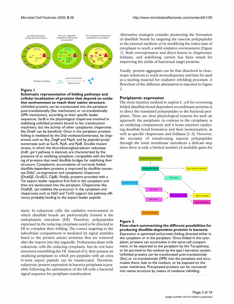
Finally, protein aggregates can be first dissolved in chao-tropic solutions to reach monodispersity and later be usedas a starting material for oxidative refolding processes. Aflowchart of the different alternatives is reported in Figure2.
Periplasmic expressionThe most intuitive method to exploit E. coli for recoveringfolded disulfide-bond dependent recombinant proteins isto direct the translated polypeptides to the bacterial peri-plasm. There are clear physiological reasons for such anapproach: the periplasm, in contrast to the cytoplasm, isan oxidizing compartment and it hosts enzymes catalyz-ing disulfide bond formation and their isomerization, aswell as specific chaperones and foldases [1-3]. However,the necessity of translocating nascent polypeptidesthrough the inner membrane introduces a delicate stepsince there is only a limited number of available gates for
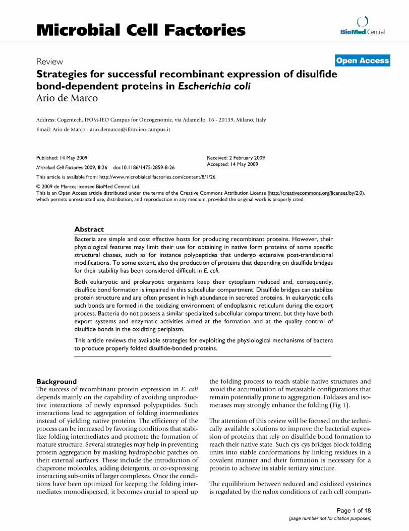
Schematic representation of folding pathways and cellular localization of proteins that depend on oxidative environ-ment to reach their native structureFigure 1Schematic representation of folding pathways and cellular localization of proteins that depend on oxida-tive environment to reach their native structure. Unfolded proteins can be translocated into the periplasm post-translationally (Sec mechanism) or co-translationally (SPR mechanism), according to their specific leader sequences. SecB is the physiological chaperone involved in stabilizing unfolded proteins bound to Sec translocation machinery, but the activity of other cytoplasmic chaperones like DnaK can be beneficial. Once in the periplasm proteins folding is mediated by the Dsb oxidases/isomerases, by chap-erones such as Skp, DegP and FkpA, and by peptidyl-prolyl isomerases such as SurA, PpiA, and PpiB. Double mutant strains, in which the thioredoxin/glutaredoxin reductase (trxB-, gor-) pathway is silenced, are characterized by the presence of an oxidizing cytoplasm, compatible with the fold-ing of proteins that need disulfide bridges for stabilizing their structure. Cytoplasmic accumulation of correctly folded disulfide-dependent proteins is improved by disulfide isomer-ase DsbC co-expression and cytoplasmic chaperone (DnaKJE, GroELS, ClpB). Finally, proteins provided with a Tat export leader sequence first fold in the cytoplasm and then are tanslocated into the periplasm. Chaperones like DnaKJE, can stabilize the precursor in the cytoplasm and chaperones such as SlyD and TorD support the pathway effi-ciency probably binding to the export leader peptide.
Oxidizing environment
Reducing environment
DnaKJESecB
Dsb
Ppi
FkpA
Skp
SurA
DegP
Sec/SRP periplasmicfolding and accumulation
trxB-, gor- dependent cytoplasmic folding and
accumulation
TAT cytoplasmic folding and periplasmic accumulation
Oxidative refolding:
DnaKJE GroELS ClpB
DsbAC Ppi SlyD
Oxidizing environment
Reducing environment
DnaKJEGroELS
SlyDTorD
Oxidizing environment
Oxidizing environment
DnaKJEGroELSClpB
DsbC
Ppi
Flow-chart summarizing the different possibilities for produc-ing disulfide-dependent proteins in bacteriaFigure 2Flow-chart summarizing the different possibilities for producing disulfide-dependent proteins in bacteria. Expression is optimized and protein folding directed either in the cytoplasm or in the periplasm. Once folded in the cyto-plasm, proteins can accumulate in the same cell compart-ment, or be exported to the periplasm by the Tat-pathway, or be secreted to the medium by the type I secretion system. Unfolded proteins can be translocated post-translationaly (Sec) or co-translationaly (SPR) into the periplasm and accu-mulate there, leak to the medium, or be exposed on the outer membrane. Precipitated proteins can be recovered into native structure by means of oxidative refolding.
Expression tuning
Cytoplasmic folding
TAT Export
Periplasmic folding
Type I secretion pathway
Cytoplasmic accumulation
SEC export
SRP export
Medium leakage
Oxidative refolding
Outer membrane
Page 2 of 18(page number not for citation purposes)

Microbial Cell Factories 2009, 8:26 http://www.microbialcellfactories.com/content/8/1/26
reaching the periplasm and metastable precursors mayconsequently accumulate in the cytoplasm.
Modulation of the expression levelConsidering the potential danger of having an over-crowded cytoplasm, some authors proposed that proteinsecretion could be effectively modulated at the transla-tional level by modifying the Shine-Dalgarno sequence[4]. Other authors found that the crucial tuning regionstarts upstream of the Shine-Dalgarno region and spansapproximately twenty nucleotides downstream of the ini-tiation codon (translational initiation region) [5]. Coher-ently, also other parameters influencing the expressionrate have been evaluated in relationship with the produc-tion of disulfide-bond dependent proteins, such asgrowth medium, plasmid origin of replication, andexpression promoters. For instance, the expression fea-tures of tac, uspA, uspB, T7, trc, lacUV5, malK, pm/xylShave been investigated to achieve high product yields bypreventing protein precipitation and cell lysis [6-12].However, data have been collected under different exper-imental conditions and, therefore, they are not compara-ble, preventing the identification of surely preferablesystems.
Choice of the suitable secretion leader peptideIn the absence of an N-terminal signal peptide for peri-plasmic secretion, recombinant polypeptides expressed inbacteria accumulate in the cytoplasm. The fusion to suita-ble leader peptides allows for the translocation ofunfolded precursors into the periplasm by either the Sec(relatively slow, post-translational translocation) or theSRP (fast, co-translational translocation) system [13,14],even in the case of aggregation prone proteins such asPNGase and large molecules like full-length immu-noglobulins [15,16]. The search for optimal leader pep-tides to use in combination with recombinant proteinshas been initially undertaken by comparing the efficiencyof natural signal sequences identified in the precursors ofbacterial periplasmic proteins, including the leader pep-tides from spA, phoA, ribose binding protein, pelB, ompA,ompT, dsbA, torA, torT, and tolT. Furthermore, both syn-thetic sequences and the phage pIII leader peptide wereused [6,9,17-22]. Initially, the approach was not system-atic and no clear preference for any among them wasapparent, although ompT resulted preferable when cou-pled to overexpression of chaperones involved in the sta-bilization of intermediates translocated through the Secexport machinery [23].
However, a wide survey performed by Beckwith and co-workers identified a strong correlation between hydro-phobicity of the leader peptide and export mechanism[24]. Apparently, cotranslational translocation by SRPneeds the presence of highly hydrophobic leader
sequences, even though further unknown biophysical fea-tures may be critical. The physiological necessity of theSRP pathway as an alternative to the post-translationalsecretion meditated by the Sec route is required to avoidpremature folding of the proteins in the cytoplasm. Thebiotechnological implication of these conclusions is thatpoor periplasmic accumulation of rapidly folding recom-binant proteins may be the consequence of their non-pro-ductive cytoplasmic (mis)folding that prevents efficienttranslocation and correct periplasmic folding. Therefore,the choice of the leader peptide may make the differencein terms of secretion efficiency, as demonstrated for ther-modynamic stable proteins [25,26].
Mechanism of protein oxidation in the periplasmSpontaneous protein oxidation in extracytoplasmic com-partments is extremely slow and is incompatible with cellactivity. Therefore, it is necessary that the disulfide bondformation is enzymatically catalyzed. Periplasmic proteinoxidation is regulated by the five members of the Dsb pro-tein system (DsbA, B, C, D, G) [1,2]. With the exceptionof DsbB, these proteins belong to the thioredoxin proteinsuperfamily and are involved in both disulfide-bond for-mation and rearrangement. Specifically, the soluble mon-omer protein DsbA donates its disulfide bond to newlysynthesized polypeptides (Figure 3) that are folding inter-mediates and need disulfide bonds to reach their nativestructure. Interestingly, DsbA binds its substrates byhydrophobic interactions using a chaperone-like recogni-tion mechanism specific for partially unfolded proteins[27]. Such substrate-independent chaperone-like activity
Dsb-dependent protein oxidation and isomerization in the bacterial periplasmFigure 3Dsb-dependent protein oxidation and isomerization in the bacterial periplasm. DsbA couples consecutive cysteines providing the disulfide bond and is re-charged by the inner membrane DsbB. Incorrect disulfides of oxidized proteins are scrambled by DsbC and DsbG. These isomer-ases are kept reduced by the inner membrane DsbD that, in return, is reduced by cytoplasmic thioredoxin.
S S
DsbB DsbD
SH SH
SH SH
Thioredoxin
SH
SHSH
SH S
S
S
S
S
S
S
S
SH SH
DsbA
S S
DsbAD
sbC
/G
Dsb
C/GD
sbC/G
DsbC
/G
SH SHSH SHS SS SPer
ipla
smC
yto
pla
sm
Page 3 of 18(page number not for citation purposes)
Microbial Cell Factories 2009, 8:26 http://www.microbialcellfactories.com/content/8/1/26
seems to have a general stabilizing effect on passengerproteins fused to DsbA mutant deprived of its oxidore-ductase activity and, therefore, it has been successfullyused as fusion partner even for the production of cytoplas-mic proteins [28].
DsbA active site is regenerated by the transfer of electronsto the DsbB integral membrane protein, whose activity iscrucial in maintaining DsbA in its active state (Figure 3)[27,29-32]. The DsbA/DsbB mediated oxidative process isvery efficient, but may result in incorrect cysteine pairingand in trapping the target protein in non-native confor-mations. Therefore, a quality control mechanism for therearrangement of the disulfide bonds was initially postu-lated and later identified. DsbC and DsbG are the isomer-ases that scramble incorrect cystin bridges and, therefore,play a key-role in the periplasmic protein folding process(Figure 3) [33-36]. The active cysteines of DsbC and DsbGmust remain reduced in an oxidizing environment to befunctional. This condition is achieved by the action of theintegral inner membrane DsbD. Such protein constantlyreduces the isomerases by transferring to them the elec-trons made available by the cytoplasmic thioredoxin [37-41]. Similarly to DsbA, both DsbC and DsbG have chap-erone activity. Since misfolding could be a direct conse-quence of incorrect disulfides, DsbC/DsbG chaperoneactivity favors the recognition and interaction with sub-strates necessiting disulfide isomerization [1,42]. Suchhomodimer isomerases are V-shaped and use the structureflexibility of the cleft embodied by the two arms to bindunfolded structures of various sizes. Their adaptabilityallows the recognition of largely heterogeneous substratesand explains the elevated efficiency of bacterial DsbC/DsbG in isomerasing also heterologous proteins [1].
Although the two routes of keeping DsbA oxidized andDsbC/DsbG reduced have been always considered strictlyseparated [1], recent data indicate a possible direct coop-eration between DsbA and DsbC [43], and chimeras ofDsbA and DsbC proved to be able to serve both as oxidaseand as isomerase, thus reconciling the competing path-ways leading to DsbA oxidation and DsbC reduction [44].Furthermore, it has been demonstrated that DsbA can bemutated to become an isomerase by adding a linker withpeptide binding capacity and that provides a dimerisationdomain. These characteristics prevent its immediate oxi-dation by DsbB and allow substrate recognition and bind-ing [45]. Such results will probably simplify thebiotechnological applications of Dsb proteins in vitro.
Use of Dsb proteins for biotechnological purposesThe relevance of the Dsb protein family for the correctfolding of disulfide-bond dependent proteins in the E. colisuggested that boosting such molecular machinery couldresult in increased yields of correctly folded recombinant
constructs. The accumulation of a functional scFv waspositively affected by co-expression of DsbA, B, C, D [46],both DsbAB and DsbCD overexpression improved theproduction of active horseradish peroxidase [47], whilstco-expression of DsbA and DsbC increased the yield offunctional human plasma retinol-binding protein [48].Fusion to DsbA increased the accumulation of few mg/Lculture medium of active bovine enterokinase and humanpro-insulin, a polypeptide stabilized by three disulfidebonds [49,50]. However, in the case of Ragi bifunctionalinhibitor, a protein with five overlapping disulfide bonds,DsbA activity resulted in the accumulation of non-nativeintermediates, whilst the availability of DsbC significantlyincreased the yield of the native protein both in vitro andin vivo [51]. Again, the overexpressoion of DsbA, B, C, Dand DsbCD, but not DsbAB, increased the accumulationof active nerve growth factor beta [52] and DsbC was thekey foldase also for enhancing the production of horse-radish peroxidase and brain-derived neurotrophic factor,although its synergistic effect with DsbA, B, and D wasdetectable [53,54]. These results were confirmed by exper-iments performed with plasminogen activator and a scFv[55,56] and seem to indicate that the isomerase DsbCactivity could be the limiting factor for the correct foldingof at least part of the recombinant proteins expressed inthe E. coli periplasm.
The different responses observed when using eukaryoticsubstrates and DsbA or DsbC overexpression might beexplained by examining the structure of each single sub-strate protein. DsbA couples cysteines as soon as they areavailable and, therefore, only disulfides between consecu-tive residues are formed. As a consequence, DsbC activityis not necessary for proteins in which only disulfide bondsbetween consecutive cysteines are present in the nativestructure [57]. In contrast, bond scrambling is DsbC-dependent. The correlation between the presence of non-consecutive disulfides and DsbC-dependence for achiev-ing the correct folding has been demonstrated foreukaryotic as well as for bacterial proteins [51,58]. How-ever, although the bacterial RNaseI is a natural DsbC sub-strate with three consecutive and one nonconsecutivedisulfide bonds [59], it is possible to artificially tune theperiplasm redox potential to achieve conditions thatallow DsbA to correctly catalyze the bond formationbetween the nonconsecutive residues [60].
The pivotal role of thiol-disulfide oxidoreductases in sup-porting the correct folding of disulfide-bond dependentproteins is conserved in Gram-negative bacterial peri-plasm [61]. Interestingly, functionality seems to beretained also by phylogenetically distant proteins, such asendoplasmic reticulum disulfide isomerase from mam-malian and yeast since, despite the low sequence homol-ogy, a marked structural similarity exists [62-64]. This
Page 4 of 18(page number not for citation purposes)

Microbial Cell Factories 2009, 8:26 http://www.microbialcellfactories.com/content/8/1/26
observation led to successful engineering of humandisulfide isomerase in the E. coli periplasm and even in theGram-positive Bacillus brevis, with consequent functionalrestoration in dsbA mutants and yield improvement offunctional proteins [65,66].
Coexpression of periplasmic chaperones and foldasesThe process leading to correct protein folding in the E. coliperiplasm is mediated by the activity of proteins belong-ing to several classes and that often have partially overlap-ping functions (Fig 1). There are peptidyl-prolylisomerases and chaperones such as SurA, FkpA, PpiA,PpiD, and Skp, and chaperone/proteases such as DegP[67-73]. FkpA and SurA overexpression contributed to anincrease in the accumulation of functional human plasmaretinol-binding protein [48], FkpA, DegP, DsbA, andDsbC rescued the activity of lipase B [74], and both thechaperone and protease activities of DegP were necessaryto enhance the yields of penicillin acylase [71,75].
The prolyl-cis/trans isomerase activity of Ppi apparentlycatalyzes the rate-limiting step of the recombinant anti-body folding represented by the isomerization of proline95 (Kabat numbering) [76]. Its coexpression increased thesolubility of scFvs and Fab fragments [77-79], but thisenzyme was also successfully used as a fusion partner forrecombinant antibody expression [4,80]. Similarly,fusions to FkpA resulted in increased yields of functionalrecombinant antibodies [81,82].
Effect of cytoplasm chaperones on periplasmic protein accumulationThe mechanisms by which cytoplasmic chaperones canhelp periplasmic folding are less clear. In a few cases suchmolecules were directly secreted to the periplasmic space,as was the case of DnaJ and Hsp25 that increased theyields of native plasminogen activator [83]. Probably suchmolecular chaperones had an unspecific stabilizing effecton folding intermediates, but no sound rationale supportsthe strategy of boosting the periplasm with cytoplasmicchaperones, instead of using periplasmic ones. In con-trast, the idea of overexpressing cytoplasmic chaperonesin the cytoplasm might be justified by the considerationthat nascent polypeptides are translocated as unfoldedintermediates into the periplasm by the Sec/SRP systemsand, therefore, their potential aggregation must be pre-vented (Figure 1). SecB is the natural chaperone that bindspolypeptides to be secreted post-translationaly. Appar-ently, SecB possesses specificity for 9-residue sequencemotifs enriched in aromatic and basic residues [84]. Thepresence of these motifs within recombinant proteinsmay considerably influence their chances to be correctlydelivered to the periplasm by the SEC system. However,the overexpression of other chaperones could compensate
for poor SecB binding to non-specific substrates. Theymight unspecifically recognize and stabilize unfoldedpolypeptides in the cytoplasm, as suggested by the obser-vation that chaperone overexpression in the cytoplasmwas beneficial for the accumulation of some periplasmicproteins [85]. Different combinations have been tested,such as DnaKJE (DnaK, DnaJ, GrpE) that increased theaccumulation of functional scFvs against domoic acid andthe mycotoxin deoxynivalenol [56,86]. DnaKJ co-expres-sion prevented the formation of inclusion bodies of CorAand improved the periplasmic accumulation of granulo-cyte-colony stimulating factor [87,88]. In contrast, cyto-plasmic chaperone overexpression failed to improve theyields of horseradish peroxidase [47].
The accumulation rate of SecB, the physiological chaper-one for the Sec system, is apparently efficiently tuned bythe effective amount of potential substrates and, conse-quently, its exogenous overexpression was not considerednecessary [13,89,90]. However, in at least one case higherperiplasmic accumulation of heterologous proteins wasdetected as a consequence of cytoplasmic SecB overexpres-sion [91]. Other authors found that the overexpression ofSecB and DnaKJ was beneficial in promoting recombinantaccumulation of human proteins in the periplasm, butonly in combination with signal sequence optimization[92].
Advantages and limitations of fusion constructsThe problem of stabilizing the folding intermediates hasbeen addressed also by fusing the target polypeptide toprotein carriers. Periplasmic EETI-II, erythropoietin, andscFv production was increased by fusions with maltose-binding protein and immunoglobulin-constant domain,whilst human proinsulin and pepsinogen yields wereenhanced by fusions with ecotin, a trypsin inhibitor thatcould favor target protein accumulation by reducing itsdegradation [93-96]. A double ecotin-ubiquitin tag hasbeen recently engineered. It should allow both the stabili-zation of the passenger proteins and their recovery withtheir authentic amino terminus [12].
It must be underlined that protein fusion always carries aninherent risk of resulting in soluble but incorrectly foldedtarget protein. For instance, fusions of maltose bindingprotein and some scFvs resulted in higher periplasmicyields, but the antibodies were not functional, whilst thefusion to alkaline phosphatase stabilized both productionand activity [97]. Alkaline phosphatase has been oftenused as a fusion partner for recombinant antibodies. It isnot only useful for enhancing the production but, dimer-izing, it increases the avidity of the immunoproteins fortheir substrate and allows their direct detection by enzy-matic assay [97-100].
Page 5 of 18(page number not for citation purposes)

Microbial Cell Factories 2009, 8:26 http://www.microbialcellfactories.com/content/8/1/26
Protein A or single and tandem repeats of its B- and Z-domains were successfully used for expressing and purify-ing a scFv and human proteins such as proinsulin andcoagulation factor VII [9,101-105]. Fusion to barnaseresulted in correct folding of ICK and McoEeTI, but theconstruct was directed to the culture medium [106,107].
Other fusion partners, especially in combination withrecombinant antibodies, were considered not for facilitat-ing folding and increasing yields, but to obtain reagentsmore suitable for the final applications. A biotinylation-consensus sequence has been fused to Fab fragments toobtain streptavidin-affine immunoconstructs [108], GFPhas been fused to scFvs for recovering immunuofluores-cent reagents [109], and fusion to nuclear import/exportsequences have been used to direct scFvs to specific sub-cellular localizations [110].
Effect of chemical chaperonesA positive effect of low molecular weight additives (chem-ical chaperones) supplemented in the culture mediumwas sometimes observed in terms of yields of periplasmicexpressed proteins. Sorbitol addition to the culturemedium resulted in higher accumulation of a functionalscFv [46], glycine betaine and sucrose were beneficial forthe folding of immunotoxin and cytochrome c550[111,112], whilst L-arginine and ethanol increased theyields of human pro-insulin, plasminogen activator, anda scFv [50,83]. Also the supply of reduced glutathione,alone or in combination with DsbC overexpression,increased the accumulation of disulfide-dependent pro-teins [51,83].
Twin-arginine translocase pathwayAlthough the Sec and SRP systems are responsible fortranslocating most of the proteins into the periplasm, bac-teria possess another mechanism -the twin-arginine trans-locase (Tat) pathway- that enables the transport of foldedproteins across the inner plasma membrane (Figure 1)[113]. E. coli genome encodes some tens of proteins witha signal-peptide capable of interacting with Tat machinery[114] and such export route has been exploited for recom-binant expression as well [115,116]. The overexpressionof all the tatABC genes expressing the proteins involved inthe transport increased the translocation of a fluorescentreporter [117]. Also the overexpression of the cytoplasmicchaperone DnaK seems particularly useful in increasingthe efficiency of the Tat pathway at both physiological andprotein overexpression-induced stress conditions[118,119]. Such effect could be a consequence of theDnaK role in facilitating the substrate folding into nativestructures. Another explanation considers that virtually allTat leader peptides contain recognition sequences forDnaK [120,121]. Therefore, Santini et al. [122] proposedthat DnaK sequesters the protein intermediates in the
cytoplasm by masking their leader peptide until the fold-ing is complete.
Other chaperone-like proteins as SlyD and TorD havebeen reported improving Tat efficiency [118,123,124].Interestingly, scFvs correctly folded in the cytoplasm(intrabody mutants or wild type constructs expressed intrxB-gor- cells) were efficiently transported by the Tat sys-tem [125,126].
High throughput selection of suitable clonesProtein accumulation varies from cell to cell inside a pop-ulation and, therefore, strategies for selecting the mostproductive clones are relevant. Similarly, it may be neces-sary to identify, within large bacteria cultures expressingclone libraries, the few cells producing proteins/antibod-ies with desired features. Flow cytometry represents apowerful methodology since it enables a quick and thor-ough screen of large numbers of constructs and it has beensuccessfully applied for selecting proteins, recombinantand full-length antibodies accumulated in the periplasm[127-132]. Several approaches have been described, allreferring to the same principle of labeling proteins accu-mulated in the periplasm to enable the isolation of theproductive bacteria. In the simplest protocol, proteinsaccumulate directly in the the periplasm, otherwiserecombinant antibodies are anchored to the periplasmicside of the inner membrane through a lipoproteindomain, or full-length antibodies, secreted and folded inthe periplasm, are captured through their Fc domain by ananchoring fusion construct consisting of the Z-domain ofthe Protein A and an inner membrane lipoprotein[129,132]. The outer membrane is successively permeabi-lized to form spheroplasts and the recombinant proteins/antibodies are labeled by binding to fluorescent antibod-ies/antigens. Finally, the cultured bacteria are sorted byflow cytometry according to the expression and affinity ofthe produced constructs. Such approach may become aparadigm for efficiently screening tagged proteins andantibodies expressed in the periplasmic space.
Periplasmic inclusion body formationDespite the periplasmic quality control and the observa-tion that the FkpA chaperone activity seems to be effectivein suppressing inclusion body formation [133], proteinscan aggregate also in this cellular compartment. The sim-ple overexpression is already sufficient to induce the accu-mulation of the bacterial beta-lactamase protein inclusionbodies into the periplasm [134,135]. Similarly to whatwas observed in cytoplasmic aggregates, the proteinstrapped in periplasmic inclusion bodies may partially pre-serve their physiological activity [135] and induce stressresponse [136]. There are relatively few papers reportingthe accumulation of heterologous protein inclusion bod-ies in bacterial periplasm [8,75,137-139]. However, the
Page 6 of 18(page number not for citation purposes)

Microbial Cell Factories 2009, 8:26 http://www.microbialcellfactories.com/content/8/1/26
number might be underestimated because cell fractiona-tion is rarely undertaken and negative results are often notcommunicated. From the available information it seemsthat construct instability is often present in engineeredpolypeptides. For instance, both the addition of unpairedcysteines and sub-optimal length or composition of thelinkers connecting the variable domains may be stronglydetrimental for the scFv yields [140,141]. Jeong and Leesystematically tried to alleviate heterologous protein pre-cipitation in the periplasm by analyzing the effect of bac-terial strain, growth temperature, expression vectorfeatures (signal peptide, promoter, codon usage optimiza-tion, linker compositions), and DsbA co-expression, butgeneral conclusions require further studies [8,142].
Induced accumulation of periplasmic proteins into the culture mediumThe recovery of periplasmic proteins is usually achievedby disruption of the outer membrane by osmotic shock.Such purification protocol has the advantage of limitingthe contamination with cytoplasmic material. However,as an alternative it has been also proposed to induce thelysis of the outer membrane to recover the target proteindirectly from the culture medium avoiding a cumbersomepurification step (Figure 2). The most investigated solu-tion is based on the expression of Kil protein, a bacteri-ocin that induces periplasmic protein release throughinduced membrane solubilization [143,144]. Lysis can beinduced by regulating the Kil protein expression with astationary-phase or a temperature inducible promoter[145,146] and the strategy was successfully used torecover from the medium interleukin-2, beta-glucanase,and streptavidin [144,145,147]. Also the co-expression ofthe TolA protein has been proposed for inducing theexternal membrane permeabilization [17].
The accumulation of proteins in the culture medium, andspecifically of recombinant antibodies, expressed forbeing secreted into the periplasm has been often observed[148,149]. The reasons remain unknown and cell lysisdoes not seem to be always involved in the protein leak-age. However, it has been shown that several different fac-tors such as growth conditions, inducer concentration,and aminoacid substitutions can tune the leakage of scFvsand have been proposed as means to regulate scFv parti-tion between periplasm and medium [149,150]. Theinfluence of the expression level on protein leakage fromthe periplasm to the growth medium has been directlydemonstrated comparing different promoters [10].
Cytoplasmic expressionEarly observations indicated that the disulfide bridges inboth β-lactamase and alkaline phosphatase precursorsexpressed in E. coli were formed only after their transloca-tion and processing in the periplasm and that it was pos-
sible to prevent their oxidative folding by trapping theprecursors in the cytoplasm [151,152]. These results wereexplained by the presence of a reducing cytoplasm and anoxidizing periplasm in bacteria. Furthermore, they indi-cated that no protein requiring the formation of disulfidebonds for reaching its native structure can be produced ina functional form in the cytoplasm. The correlationbetween presence of disulfide bonds and native, func-tional structures was exploited by Beckwith and co-work-ers in their pioneering attempt to identify mutations thatenabled the formation of active alkaline phosphatase inbacteria cytoplasm. It turned out that alkaline phos-phatase folded correctly when expressed in bacteria host-ing mutations that blocked the reduction of cysteines inthe cytoplasm by silencing the activity of the thioredoxinreductase [153]. The preliminary model indicated thatNADPH was the source of reducing potential used bythioredoxin reductase to reduce oxidized thioredoxin.However, the data suggested that another thioredoxin-likeprotein could be involved in a parallel reducing route. Thesearch for complementary reducing mechanism(s) led tothe identification of a second thioredoxin and of glutath-ione oxidoreductases, all involved in the mechanismaimed at reducing cytoplasmic cysteines [154,155]. Theapparent redundancy of the reducing machinery is proba-bly due to the necessity of obtaining the maximal electrontransfer efficiency between protein pairs with varyingredox potentials [156]. Under physiological conditionsthe directionality of the electron flux is maintained. How-ever, it was shown that thioredoxin acts as a reductaseonly when it remains constantly reduced by the thiore-doxin reductase activity. In contrast, it can catalyze sub-strate oxidation when exported to periplasm and oxidizedby DsbB [157,158]. Also glutaredoxin 3 can catalyzedisulfide bond formation in the periplasm, but its activitydepends on oxidized glutathione availability rather thanDsbB [159]. A similar process has been already demon-strated in eukaryotic cells [160] and its existence may alsobe hypothesed in the bacterial cytoplasm wherethe envi-ronment becomes oxidized as a consequence of impairedreducing activities.
Summarizing, it is possible to obtain an oxidizing cyto-plasm in a cellular system in which glutaredoxin activity isabrogated by gor- mutation, and both thioredoxin 1 and 2are kept oxidized as a consequence of thioredoxin reduct-ase mutations (Figure 1). Reduced glutathione, necessaryto preserve cell viability, is produced by the disulfidereductase activity of mutated peroxiredoxin AhpC[161,162]. Interestingly, AhpC reductase activity does notsignificantly influence the oxidizing condition of the cyto-plasm in trxB-, gor- cells [162]. Such observation promptedto investigate the biotechnological potential of both sin-gle (trxB-) and double (trxB-, gor-) mutant strains -nowcommercialized with the names of AD494 and Origami
Page 7 of 18(page number not for citation purposes)
Microbial Cell Factories 2009, 8:26 http://www.microbialcellfactories.com/content/8/1/26
(Novagen), respectively- for the cytoplasmic expression ofrecombinant proteins with multiple disulfide bonds intheir native structure. The encouraging data obtained inthe first attempts [163] were further improved by combin-ing cytoplasmic oxidation conditions and cytoplasmicaccumulation of DsbC isomerase (Figure 1) [164].
Recently, a new expression strain has become available.SHuffle (New England BioLab) is trxB-, gor- as the com-mercial Origami (Novagen), but overexpresses cytoplas-mic DsbC and is spectinomycin selectable, allowing thetransformation with the majority of the currently usedvectors. Although no literature is available so far, it isexpected that the SHuffle combination of oxidizing con-ditions and isomerization capability would stronglyimprove the correct folding of disulfide bond-dependentproteins in the E. coli cytoplasm.
The method of expressing disulfide-bond-dependent pro-teins in oxidized cytoplasm was validated by severalgroups and progressively optimized. The collagen prolyl4-hydrolases yield was higher in the cytoplasm of trxB-,gor- bacteria than in the periplasm of the correspondingBL21 wild type strain [165]. The oxidized cytoplasmallowed the accumulation of functional IgG-like extracel-lular domain receptor [166], Ig2 domain of neurolin[167], lipase B [168,169], chitinase [170], and anti-freezeproteins [171]. Furthermore, the correct formation ofdisulfide bonds in the cytoplasmic milieu has beenproved in recombinant oxalate oxidase [172], peanutallergen Ara h 2 [173], and Stereum purpureum endopoly-galacturonase [174].
A specific class of disulfide-bonded proteins: the recombinant antibodiesAs a consequence of the constantly increasing importanceof recombinant antibody expression, a significant efforthas been dedicated for optimizing their production inbacteria. Although few antibodies are stable in theabsence of disulfide bonds and can be directly expressedas intrabodies in the reducing cytoplasm of host cells[175-179], the structure and the functionality of most ofthem strictly depends on correct cys-cys bridges.
Recombinant antibodies in scFv format have one intra-chain disulfide bond for each variable region, those in Fabformat have a further inter chain bond. A scFv fragmentfused to a consensus peptide for obtaining in vivo site-spe-cific biotinylation, a bivalent Fv antibody fragment fusedto molybdopterin synthase to induce its oligomerization,and some Fab fragments were successfully produced inthe cytoplasm of trxB-, gor- cells [180-182]. Camelidaerecombinant antibodies in VHH format possess one intra-chain disulfide bond between framework residues and
may have a further one to constrain the CDR3 loop. Alsothese antibodies and have been successfully produced inthe cytoplasm of trxB-, gor- bacteria [183].
Stabilizing role of chaperones and DsbC isomeraseChaperones and isomerase can facilitate the target proteinfolding by impairing non-productive aggregation of fold-ing intermediates and have been often co-expressed. Thecytoplasmic overexpression of the periplasmic chaperoneSkp and of DsbC isomerase, as well as of the molecularchaperones GroELS and trigger factor, significantlyimproved the yields of recombinant antibodies [184],whilst the cytoplasmic co-expression of Skp and FkpAwere ineffective in rescuing fibrolase venom, in contrast tothe beneficial effect of DsbC [185]. The work of othergroups confirmed that the cytoplasmic co-expression ofDsbC in trxB-, gor-bacteria was advantageous for the func-tional cytoplasmic accumulation of recombinant anti-bodies in both scFv and VHH format [183,186]. In suchconditions the oxidizing environment favors the forma-tion of the cys-cys bridges inside the polypeptide sequenceand the isomerase apparently improves the yields of cor-rectly folded antibodies by exchanging the cysteine resi-dues involved in non-native pairing. The strategy ofcoupling cytoplasm oxidative conditions and isomeraseactivity was optimized by Yuan et al. [187] who overpro-duced thioredoxin fusions of calobin venom in the pres-ence of cytoplasmic trapped DsbC. Co-expression ofDsbC, GroELS and, at lower extent, of trigger factorresulted in higher yields of a functional scFv against thereceptor c-Met [188], whilst the recombinant co-expres-sion of a peptidyl-prolyl isomerase from Pyrococcusimproved yields and functionality of recombinant Fabfragment [189]. A well-documented review addressing thespecific problem of the recombinant antibody expressionin prokaryotes has been published by Arbabi-Ghahroudiet al. [190].
Thioredoxin overexpressionThe cytoplasmic accumulation of thioredoxin as a conse-quence of its recombinant overexpression in wild typebacteria was proposed for increasing the yields of co-expressed eukaryotic proteins without disulfide bonds intheir native structure [191]. Similarly, the precipitation ofeukaryotic disulfide-bond independent proteins in thebacteria cytoplasm was prevented after their fusion tothioredoxin [192]. Cytoplasmic expression of thioredoxinfusions rescued also polypeptides that failed to correctlyfold in the periplasm [187], plant viscotoxins, and por-cine pepsinogen A, although these proteins contain mul-tiple disulfide bridges [193,194]. The reasons as to whythioredoxin can stabilize its passenger proteins in wildtype bacteria is not well understood, although its contri-bution to scFv folding might be due to its chaperone prop-
Page 8 of 18(page number not for citation purposes)

Microbial Cell Factories 2009, 8:26 http://www.microbialcellfactories.com/content/8/1/26
erties [195], since a catalytic cysteine thioredoxin mutantwas still effective in supporting both disulfide bond for-mation and functionality of the target protein [196].
In contrast, there is a logical reason for overexpressingcytoplasmic thioredoxin in double mutant trxB-, gor-bacte-ria since such enzyme can act as an oxidant when it oper-ates in an oxidized milieu [157]. Therefore, it can activelycontribute to maintain oxidizing conditions in the cyto-plasm. Fusions between recombinant scFvs and thiore-doxin 1 expressed in trxB-, gor- bacteria resulted inincreased cytoplasmic yields [196], correct folding of ascFv against the c-Met receptor [188], and of the firstdomain of the multiple Kazal-type inhibitor LEKTI, apolypeptide that contains two disulfide bridges in itsnative structure [197]. Both the His- and GST-fusions ofBSPH1, a protein containing 2 fibronectin type-IIdomains each consisting of 2 disulfide bonds, aggregatedwhen expressed in the cytoplasm of trxB-, gor- bacteria, buttheir fusions with thioredoxin resulted insoluble andactive proteins [198].
Carriers used to improve protein correct foldingOther proteins have been used as fusion partners andshowed positive effects on folding and functionality ofrecombinant proteines accumulated in bacteria cyto-plasm. Carboxyterminus fusions to maltose binding pro-tein stabilized several scFvs independently on thecytoplasm redox conditions [199], although less effi-ciently than thioredoxin [196], whilst fusion to NusA wasstrictly necessary to yield another functional scFv andAPRIL in the cytoplasm of trxB-, gor- cells [200,201]. Sumohas also been proposed as solubilizing carrier fordisulfide-bonded proteins, but the structural quality ofthe recovered proteins has not been investigated[202,203]. However, in a recent report it has been shownthat a scFv against VEGF-165 was soluble and active whenexpressed fused to sumo [204].
Minimal redox conditions compatible with disulfide bond formationAlthough the rational background for the expression ofdisulfide-bond dependent proteins in double mutantstrain trxB-, gor- is generally accepted and such strain hasbeen successfully exploited, alone or in combination withthe expression of foldases/chaperones, the usefulness ofthe single trxB- mutation remains unclear. The glutathioneoxidoreductase is still functional in such bacteria and itsactivity should be sufficient to keep the cytoplasm par-tially reduced [156]. Consequently, disulfide bond forma-tion should be at least partially impaired. In practice, onlyminimal amounts of all expressed scFvs remained solubleand functional in the cytoplasm of trxB- (AD494) cells[109] and the advantage over controls was limited also in
the presence of thioredoxin co-expression [205]. A directcomparison between AD494 and trxB-, gor- bacteria clearlyindicated an advantage in using the double mutant [196].In contrast, the same cells resulted significantly more effi-cient than conventional XL-Blue cells in producing ananti-progesterone scFv [206]. However, data concerningthe oxidation state of this antibody are not reported.Information concerning folding features or functionalityis limited also in other cases in which proteins were suc-cessfully produced in the cytoplasm of AD494 cells. How-ever, at least in the case of the scorpion neurotoxin Lqq-Vand fibronectin II-2 domain from MMP-2, the proteinswere both functional and correctly folded [207,208]. Fur-thermore, the trxB- strain, in combination with DnaKJ co-expression, allowed the production of biological activehuman SPARC in the cytoplasm [209]. In the case of a ser-pin domain, AD494 cells did not improve the totalamount of soluble recombinant protein accumulated inthe cytoplasm with respect to wild type bacteria, but it wascorrectly folded and active, whilst the protein expressed inthe control cells did not form the essential disulfide bonds[210]. GroELS co-expression further increased the serpindomain solubility. In some cases, the combination ofAD494 cells and thioredoxin fusions significantlyimproved the expression of functional target proteins.Leishmania chitinase, the extracellular domain of humanthyrotropin receptor, and the disintegrin domain of jarar-hagin venom were purified, in large amounts, as solubleand active proteins [211-213].
In contrast, it has been reported that a pro-urokinasefusion with thioredoxin expressed in AD494 (DE3) strainaccumulated in inclusion bodies, even when the systemwas supplemented with disulfide isomerase and chaper-onines [214]. However, there is scarce access to negativeresults that would help in understanding the factors regu-lating disulfide bond-dependent protein expression in thecytoplasm of wild type and mutant strains.
Protein engineeringA time-consuming but definitive approach for avoidingtrial-and-error attempts in identifying the optimal cyto-plasmic expression conditions to yield functional proteinswith disulfide bonds is the generation of mutants thatmaintain a stable structure even in the absence of cys-cysbridges [215]. Such protein engineering efforts were par-ticularly successful in the case of recombinant antibodies.Various strategies of mutation and molecular evolutionhave been used to generate disulfide-independent stablescFvs starting from a reduction-sensitive precursor[175,216-218]. Intrabodies were recovered from librariesin which CDR variability was introduced by hypermuta-tion of a naturally disulfide bond-independent VHH orscFv backbone [219,220], and by selecting natural intra-
Page 9 of 18(page number not for citation purposes)

Microbial Cell Factories 2009, 8:26 http://www.microbialcellfactories.com/content/8/1/26
bodies by biopanning [186,220-222]. Such molecules arevaluable tools with great application potentiality for intra-cellular immunization [179,223,224].
The substitution of critical residues in both the frameworkand in the CDRs has also been used to improve the peri-plasmic accumulation of recombinant antibodies[150,225-227].
Outer membrane-bound proteinsExpressing secreted proteins anchored on the external sideof bacteria membranes [228] has been initially consideredto render them accessible for external binders rather thanto produce material for purification purpose. Avirulentbacteria displaying peptides corresponding to pathology-related antigen determinants on their surface weredesigned as vaccine tools [229-231]. They succeeded instimulating immunological reactions [229-234], but theexternally anchored peptides were not thoroughly charac-terized at structural level and, therefore, it cannot be ruledout that immunoresponse was promoted by partiallyunfolded antigen domains.
However, more recent data show that correctly foldedfusions of outer membrane transporters and both scFvand VHH recombinant antibodies were efficiently translo-cated on the outer bacteria surface [235,236]. Once dis-played, anchored proteins can be automatically releasedinto the medium by cleavage of a specific sequence recog-nized by a membrane protease like OmpT [233,237].
Several bacteria transporter proteins have been used todevelop vectors in which they are fused to the proteins ofinterest [238,239], such as FliC [240], pullulanase [241],OprF [242], OprI [243], PhoE [229], MisL [237,244,245],and cytolysin [234].
Exposing the protein to the outer bacteria surface is a par-ticularly valuable approach when the proteins can bedirectly used in such format. For instance, enteric corona-viruses were successfully neutralized in cultured epithelialcells treated with E. coli expressing fusions of IgA proteasebeta domain from Neisseria gonnorrhoeae with specificscFvs [246]. An adhesion domain of intimin has beenused as an anchoring partner for exposing both proteinsand peptide libraries on the surface of the bacteria outermembrane. The resulting bacteria were suitable for cellsorting and used to identify epitope-specific antibodies[247,248].
Type I (extra cellular) secretion pathwayThe type I secretion pathway is used by Gram-negativebacteria to transfer toxins and exoenzymes provided of acarboxyterminus export signal directly from the cyto-plasm to the external medium [249-251]. These proteins
are devoid of disulfide bridges but the work performed byFernandez and co-workers demonstrated that fusions ofthe secreted protein alfa-haemolysin C-domain and anti-body fragments devoid of periplasm export signal peptidewere efficiently secreted in oxidized and functional form[252-254]. Functional alfa-haemolysin fusions of bothscFvs and VHHs were recovered from the culture medium[252-255] and hemolysin-fusion of Shiga toxin B subunit[245] was able to induce host immunoreaction in inocu-lated rabbits.
Oxidative refolding of denatured proteinsDespite the great effort made by several groups for obtain-ing native proteins with correctly folded disulfide bridgesin bacteria, many attempts failed and target proteins accu-mulated as precipitates. This observation prompted othergroups to turn the disappointing results into an opportu-nity of pulling together large amounts of proteins anddeveloping oxidative refolding strategies.
The methodologies available for protein refolding havebeen thoroughly reviewed [256-258] and here onlyparameters that are of specific interest for the disulfidebond-dependent proteins will be briefly presented.
Two common requisites for a refolding buffer are the pres-ence of a redox couple and of alkaline pH. The redox cou-ple (GSH/GSSG, MESNA/diMESNA, heteroaromaticthiols) is necessary to activate the cys residues [259-261],whilst the alkaline pH facilitates the nucleophilic attack-dependent disulfide bond formation [262]. Low tempera-ture generally prevents non-productive contacts of metast-able folding intermediates and can improve the finalyields [263]. Non-productive interactions have been alsosuccessfully limited by modifying the reduced thiolgroups before starting the refolding process. The cysteinesof denatured proteins are first S-sulfonated or trans-formed into mixed disulfides and then the disulfidebonds are formed in the presence of a suitable redox sys-tem [259,264]. Surfactants and polyethylene glycol alsoshowed stabilizing properties [265,266], whilst a dynamicredox environment, in which the conditions pass progres-sively from reductive to oxidative, has been proposed tomaximize disulfide bond shuffling [267]. However, alsosimple dilution was effective for refolding heavy chainsingle domains [268].
An interesting recent paper shows the contradictory effectthat stabilizing molecules may have on the refolding ofdisulfide-bond dependent proteins [269]. L-arginine isknown to slow down the refolding process while sup-pressing hydrophobic interactions. Both these factorshave been usually interpreted as an advantage for efficientrefolding. However, although hydrophobic aggregation isprevented, the accumulation of intermediates with free
Page 10 of 18(page number not for citation purposes)

Microbial Cell Factories 2009, 8:26 http://www.microbialcellfactories.com/content/8/1/26
cysteines leads to the formation of intermoleculardisulfide bonds and the formation of progressively largeroligomers in an arginine-concentration dependent man-ner.
Productive oxidative refolding can be improved by differ-ent means. Chromatography allows separating the targetprotein from contaminants that can interfere during fold-ing and has been successfully used to achieve the refoldingof thioredoxin-scFv fusion proteins [270]. Furthermore,binding unfolded protein to a substrate impairs the phys-ical interaction amongst single molecules and their refold-ing can be performed in a sort of "independent micro-environment" in which aggregation-driving contacts areprevented. Optimization is necessary with respect to saltconcentrations to avoid protein-matrix interactions. On-column refolding was successfully performed by usingmetal affinity chromatography, as in the case of ribonu-clease A [260], chemokines [271], and α-glucosidase[272], ion-exchange substrates, as for bovine serum albu-min [273] and alfa-lactalbumin [274], or by using zeolite[275,276].
Different classes of proteins can contribute to an increasein the refolding yields of native proteins (Figure 1).Thioredoxin supported the productive refold of bothreduced denatured and oxidized but incorrectly paireddisulfides of pancreatic RNase, citrate synthase and alpha-glucosidase [195,277]. The peptidyl-prolyl isomerase wasbeneficial for the refolding of Fab fragments [278], whilstthe disulfide isomerase, alone or in combination withquiescin-sulphydryl oxidase and glutaredoxin, facilitatedthe oxidative folding of ribonuclease A and riboflavinbinding protein [279-281]. The fusion to the chaperoneSlyD had a positive effect on the refolding of the ectodo-main E1 of Rubella virus [282]. DsbA, peptidyl-prolyl-iso-merase, and GroEL minichaperone were immobilized onan agarose gel to refold the scorpion toxin Cn5 [283],immobilized DsbA, DsbC and GroEL minichaperonewere effective in refolding single-chain fragments [284],disulfide isomerase was beneficial for ribonuclease andlysozyme refolding [285], and DnaK in combination withtrigger factor or disulfide isomerase helped the functionalfolding of scFv alone and fused to a toxin domain[286,287]. The availability of ClpB and DnaK/DnaJ/GrpEimproved the refolding efficiency of the cysteine-rich pro-tein gloshedobin [288]. Finally, high-pressure has alsobeen used to prevent the formation of nonnative disulfidebonds [289].
ConclusionOften, when very heterogeneous molecules as the pro-teins are handled, it seems that the only possibleapproach towards optimization of the production process
is the trial-and-error strategy. However, the comprehen-sion of the physiological mechanisms involved in the pro-tein folding and aggregation allowed for more rationalsolutions that simplified the selection of the conditions tobe used in the trial panel (Figure 2).
Robust tools, such as oxidizing mutant strains or plasmidsfor the overexpression of chaperones and foldases, arenow available and several successful expression alterna-tives have been described. There is still no certainty that aspecific disulfide-bond dependent protein will beexpressed in a functional form in bacteria, but a series ofrational approaches can easily be compared. Once opti-mized the construct DNA sequence and the expressionconditions, the main choice is between cytoplasmic andperiplasmic folding and accumulation of the target pro-tein. It will guide the selection of opportune leadersequences and suitable bacteria strains. Finally, the purifi-cation strategy will request further modification if the pro-teins are to be recovered from inclusion bodies, totallysate, periplasmic fractions, or culture medium.
At the end of this review, it becomes apparent that there isa large amount of information missing concerning theaccurate description of the molecular mechanismsinvolved in protein folding, translocation and molecularquality control. There is also a huge gap between the the-oretical knowledge and the results of protein production,as these are only sometimes in agreement with the hypo-thetical expectations and the apparent irrationality ofsome successful conditions may seem as if it were a "mag-ical" activity. In contrast, what we are missing is a coher-ent system for the comparison of experimental data thatremain anecdotal unless precisely annotated. Our under-standing of why some conditions work in one case, butnot in another, will substantially increase when a suffi-cient amount of homogeneous and comparable data willbe available for bioinformatic analyses [290]. Data organ-ization and their direct accessibility would help in extrap-olating the information necessary to improve theidentification of optimal strategies for each specific classof polypeptides. Therefore, the future collective endeavorshould be addressed at rationalizing rather than accumu-lating data. One example to follow for future attemptsmight be the public repository Refold database http://refold.med.monash.edu.au/ that lists the successful meth-ods for refolding of proteins [291].
Competing interestsThe author declares that they have no competing interests.
AcknowledgementsThe author wishes to thank Alicja Gruszka for her critical review of the manuscript.
Page 11 of 18(page number not for citation purposes)

Microbial Cell Factories 2009, 8:26 http://www.microbialcellfactories.com/content/8/1/26
References1. Kadokura H, Katzen F, Beckwith J: Protein disulfide bond forma-
tion in prokaryotes. Annu Rev Biochem 2003, 72:111-135.2. Messens J, Collet J-F: Pathways of disulfide bond formation in
Escherichia coli. Int J Biochem Cell Biol 2006, 38:1050-1062.3. Berndt C, Lillig CH, Holmgren A: Thioredoxins and glutaredox-
ins as facilitators of protein folding. Biochim Biophys Acta 2008,1783:641-650.
4. Mavrangelos C, Thiel M, Adamson PJ, Millard DJ, Nobbs S, Zola H,Nicholson IC: Increased yield and activity of soluble single-chain antibody fragments by combining high-level expres-sion and the Skp peripasmic chaperonin. Protein Expr Purif.2001, 23(2):289-295.
5. Simmons LC, Yansura DG: Translational level is a critical factorfor the secretion of the heterologous proteins in Escherichiacoli. Nat Biotechnol 14:629-634.
6. Obukowicz MG, Gustafson ME, Junger KD, Leimgruber RM, WittwerAJ, Wun TC, Warren TG, Bishop BF, Mathis KJ, McPherson DT, et al.:Secretion of active kringle-2-serine protease in Escherichiacoli. Biochemistry 1990, 29:9737-9745.
7. Prytz I, Sanden AM, Nyström T, Farewell A, Wahlström A, FörbergC, Pragai Z, Barer M, Harwood C, Larsson G: Fed-batch produc-tion of recombinant beta-galactosidase using the universalstress promoters uspA and uspB in high cell density cultiva-tions. Biotechnol Bioeng 2003, 83:595-603.
8. Jeong KJ, Lee SY: Secretory production of human leptin inEscherichia coli. Biotechnol Bioeng 2000, 67:398-407.
9. Mergulhão FJ, Monteiro GA, Larsson G, Bostrom M, Farewell A, Nys-tröm T, Cabral JM, Taipa MA: Evaluation of inducible promoterson the secretion of a ZZ-proinsulin fusion protein inEscherichia coli. Biotechnol Appl Biochem 2003, 38:87-93.
10. Mergulhão FJ, Monteiro GA, Larsson G, Sanden AM, Farewell A, Nys-tröm T, Cabral JM, Taipa MA: Medium and copy number effectson the secretion of human proinsulin in Escherichia coli usingthe universal stress promoters uspA and uspB. Appl MicrobiolBiotechnol 2003, 61:495-501.
11. Sletta H, Nedal A, Aune TE, Hellebust H, Hakvåg S, Aune R, EllingsenTE, Valla S, Brautaset T: Broad-host-range plasmid pJB658 canbe used for industrial-level production of a secreted host-toxic single-chain antibody fragment in Escherichia coli. ApplEnviron Microbiol 2004, 70:7033-7039.
12. Paal M, Heel T, Schneider R, Auer B: A novel Ecotin-Ubiquitin-Tag (ECUT) for efficient, soluble peptide production in theperiplasm of Escherichia coli. Microbial Cell Factories 2009, 8:7.
13. den Blaauwen T, Driessen AJ: Sec-dependent preprotein trans-location in bacteria. Arch Microbiol 1996, 165:1-8.
14. Luirink J, Sinning I: SRP-mediated protein targeting: structureand function revisited. Biochim Biophys Acta 2004, 1694:17-35.
15. Loo T, Patchett ML, Norris GE, Lott JS: Using secretion to solve asolubility problem: High-yield expression in Escherichia coliand purification of the bacterial glycoamidase PNGase F.Prot Expr Purif 2002, 24:90-98.
16. Simmons LC, Reilly D, Klimowski L, Raju TS, Meng G, Sims P, HongK, Shields RL, Damico LA, Rancatore P, Yansura DG: Expression offull-length immunoglobulins in Escherichia coli: rapid and effi-cient production of aglycosylated antibodies. J Immunol Meth-ods 2002, 263:133-147.
17. Wan EW, Baneyx F: TolAIII co-overexpression facilitates therecovery of periplasmic recombinant proteins into thegrowth medium of Escherichia coli. Protein Expr Purif. 1998,14(1):13-22.
18. Oelschlaeger P, Lange S, Schmitt J, Siemann M, Reuss M, Schmid RD:Identification of factors impeding the production of a single-chain antibody fragment in Escherichia coli by comparing invivo and in vitro expression. Appl Microbiol Biotechnol 2003,61:123-132.
19. Malik A, Rudolph R, Söhling B: A novel fusion protein system forthe production of native human pepsinogen in the bacterialperiplasm. Protein Expr Purif. 2006, 47(2):662-671.
20. Sletta H, Tøndervik A, Hakvåg S, Aune TE, Nedal A, Aune R, EvensenG, Valla S, Ellingsen TE, Brautaset T: The presence of N-terminalsecretion signal sequences leads to strong stimulation of thetotal expression levels of three tested medically importantproteins during high-density cultivations of Escherichia coli.Appl Environ Microbiol 2007, 73:906-912.
21. Thie H, Schirrmann T, Paschke M, Dübel S, Hust M: SRP and Secpathway leader peptides for antibody phage display and anti-body fragment production in E. coli. New Biotechnol 2008,25:49-54.
22. Schierle CF, Berkmen M, Huber D, Kumamoto C, Boyd D, BeckwithJ: The DsbA signal sequence directs efficient, cotranslationalexport of passenger proteins to the Escherichia coli periplasmvia the signal recognition particle pathway. J Bacteriol 2003,185:5706-5713.
23. Ignatova Z, Mahsunah A, Georgieva M, Kasche V: Improvement ofpost-translational bottlenecks in the production of penicillinamidase in recombinant Escherichia coli strains. Appl EnvironMicrobiol 2003, 69:1237-1245.
24. Huber D, Boyd D, Xia Y, Olma MH, Gerstein M, Beckwith J: Use ofthioredoxin as a reporter to identify a subset of Escherichiacoli signal sequences that promote signal recognition parti-cle-dependent translocation. J Bacteriol 2005, 187:2983-2991.
25. Steiner D, Forrer P, Stumpp MT, Plückthun A: Signal sequencesdirecting cotranslational translocation expand the range ofproteins amenable to phage display. Nat Biotechnol. 2005,24(7):823-831.
26. Steiner D, Forrer P, Plückthun A: Efficient selection of DARPinswith sub-nanomolar affinities using SRP phage display. J MolBiol 2008, 382:1211-1227.
27. Couprie J, Vinci F, Dugave C, Quemeneur E, Moutiez M: Investiga-tion of the DsbA mechanism through the synthesis and anal-ysis of an irreversible enzyme-ligand complex. Biochemistry2000, 39:6732-6742.
28. Zhang Y, Olsen DR, Nguyen KB, Olson PS, Rhodes ET, MascarenhasD: Expression of eukaryotic proteins in soluble form inEscherichia coli. Protein Expr Purif. 1998, 12(2):159-165.
29. Bardwell JC, McGovern K, Beckwith J: Identification of a proteinrequired for disulfide bond formation in vivo. Cell 1991,67:581-589.
30. Bardwell JC, Lee JO, Jander G, Martin N, Belin D, Beckwith J: A path-way for disulfide bond formation in vivo. Proc Natl Acad Sci USA1993, 90:1038-1042.
31. Missiakas D, Georgopoulos C, Raina S: Identification and charac-terization of the Escherichia coli gene dsbB, whose product isinvolved in the formation of disulfide bonds in vivo. Proc NatlAcad Sci USA 1993, 90:7084-7042.
32. Jander G, Martin N, Beckwith J: Two cysteines in each periplas-mic domain of the membrane protein DsbB are required forits function in protein disulfide bond formation. Embo J 1994,13:5121-5127.
33. Shevchik VE, Condemine G, Robert-Baudouy : Characterization ofDsbC, a periplasmic protein of Erwinia chrysantemi andEscherichia coli with disulfide isomerase activity. Embo J 1994,13:2007-2012.
34. Chen J, Song JL, Zhang S, Wang Y, Cui DF, Wang CC: Chaperoneactivity of DsbC. J Biol Chem 1999, 274:19601-19605.
35. Andersen CL, Matthey-Dupraz A, Missiakas D, Raina S: A newEscherichia coli gene, dsbG, encodes a periplasmic proteininvolved in disulphide bond formation, required for recyclingDsbA/DsbB and DsbC redox proteins. Mol Microbiol 1997,26:121-132.
36. Bessette PH, Coto JJ, Gilbert HF, Georgiou G: In vivo and in vitrofunction of the Escherichia coli periplasmic cysteine oxidore-ductase DsbG. J Biol Chem 1999, 274:7784-7792.
37. Missiakas D, Schwager F, Raina S: Identification and characteriza-tion of a new disulfide isomerase-like protein (DsbD) inEscherichia coli. Embo J 1995, 14:3415-3424.
38. Stewart EJ, Katzen F, Beckwith J: Six conserved cysteines of themembrane protein DsbD are required for the transfer ofelectrons from the cytoplasm to the periplasm of Escherichiacoli. Embo J 1999, 18:5963-5971.
39. Katzen F, Beckwith J: Transmembrane electron transfer by themembrane protein DsbD occurs via a disulfide bond cascade.Cell 2000, 103:769-779.
40. Rietsch A, Bessette P, Georgiou G, Beckwith : Reduction of theperiplasmic disulfide bond isomerase, DsbC, occurs by pas-sage of electrons from cytoplasmic thioredoxin. J Bacteriol1997, 179:6602-6608.
41. Stirnimann CU, Grütter MG, Glockshuber R, Capitani G: nDsbD: aredox interaction hub in the Escherichia coli periplasm. CellMol Life Sci 2006, 63:1642-1648.
Page 12 of 18(page number not for citation purposes)
Microbial Cell Factories 2009, 8:26 http://www.microbialcellfactories.com/content/8/1/26
42. Shao F, Bader MW, Jakob U, Bardwell JC: DsbG, a proteindisulfide isomerase with chaperone activity. J Biol Chem 2000,275:13349-13352.
43. Vertommen D, Depuydt M, Pan J, leverrier P, Knoops L, Szikora JP,Messens J, Bardwell JC, Collet JF: The disulphide isomerase DsbCcooperates with the oxidase DsbA in a DsbD-independentmanner. Mol Microbiol 2008, 67:336-349.
44. Segatori L, Paukstelis PJ, Gilbert HF, Georgiou G: Engineered DsbCchimeras catalyze both protein oxidation and disulfide-bondisomerization in Escherichia coli: reconciling two competingpathways. Proc Natl Acad Sci USA 2004, 101:10018-10023.
45. Arredondo S, Segatori L, Gilbert HF, Georgiou G: De novo designand evolution of artificial disulfide isomerase enzymes anal-ogous to the bacterial DsbC. J Biol Chem 2008, 283:31469-31476.
46. Sandee D, Tungpradabkul S, Kurokawa Y, Fukui K, Takagi M: Com-bination of Dsb co-expression and an addition of sorbitolmarkedly enhanced soluble expression of single-chain Fv inEscherichia coli. Biotechnol Bioeng 2005, 91:418-424.
47. Kondo A, Kohda J, Endo Y, Shiromizu T, Kurokawa Y, Nishihara K,Yanagi H, Yura T, Fukuda H: Improvement of productivity ofactive horseradish peroxidase in Escherichia coli by co-expression of Dsb proteins. J Biosci Bioeng 2000, 90:600-606.
48. Schlapschy M, Grimm S, Skerra A: A system for concomitantoverexpression of four periplasmic folding catalists toimprove secretory protein production in Escherichia coli. ProtEng Des Sel 2006, 19:385-390.
49. Collins-Racie LA, McColgan JM, Grant KL, DiBlasio-Smith EA, McCoyJM, LaVallie ER: Production of recombinant bovine enteroki-nase catalytic subunit in Escherichia coli using the novelsecretory fusion partner DsbA. Biotechnology 1995, 13:982-987.
50. Winter J, Neubauer P, Glockshuber R, Rudolph R: Increased pro-duction of human proinsulin in the periplasmic space ofEscherichia coli by fusion to DsbA. J Biotechnol 2001, 84:175-185.
51. Maskos K, Huber-Wunderlich M, Glockshuber R: DsbA and DsbC-catalyzed oxidative folding of proteins with complexdisulfide bridge patterns in vitro and in vivo. J Mol Biol 2003,325:495-513.
52. Kurokawa Y, Yanagi H, Yura T: Overproduction of bacterial pro-tein disulfide isomerase (DsbC) and its modulator (DsbD)markedly inhances periplasmic production of human nervegrowth factor in Escherichia coli. J Biol Chem 2001,276:14393-14399.
53. Kurokawa Y, Yanagi H, Yura T: Overexpression of proteindisulfide isomerase DsbC stabilizes multiple-disulfide-bonded recombinant protein produced and transported tothe periplasm in Escherichia coli. Appl Environ Microbiol 2000,66:3960-3965.
54. Hoshino K, Eda A, Kurokawa Y, Shimizu N: Production of brain-derived neurotrophic in Escherichia coli by co-expression ofDsb proteins. Biosci Biotechnol Biochem 2002, 66:344-350.
55. Qiu J, Swartz JR, Georgiou G: Expression of active human tissue-type plasminogen activator in Escherichia coli. Appl EnvironMicrobiol 1998, 64:4891-4896.
56. Hu X, O'Hara L, White S, Magner E, Kane M, Wall JG: Optimisationof production of domoic acid-binding scFv antibody frag-ment in Escherichia coli using molecular chaperones andfunctional immobilization on a mesoporous silicate support.Protein Expr Purif. 2007, 52(1):194-201.
57. Joly JC, Swartz JR: In vitro and in vivo redox states of theEscherichia coli periplasmic oxidoreductases DsbA andDsbC. Biochemistry 1997, 36:10067-10072.
58. Berkmen M, Boyd D, Beckwith J: The nonconsecutive disulfidebond of Escherichia coli phytase (AppA) renders it dependenton the protein-disulfide isomerase, DsbC. J Biol Chem 2005,280:11387-11394.
59. Hiniker A, Bardwell JC: In vivo substrate specificity of periplas-mic disulfide oxidoreductases. J Biol Chem 2004,279:12967-12973.
60. Messens J, Collet JF, Van Belle K, Brosens E, Loris R, Wyns L: Theoxidase DsbA folds a protein with a nonconsecutive disulfide.J Biol Chem 2007, 282:31302-31307.
61. Goldstone D, Haebel PW, Katzen F, Bader MW, Bardwell JCA, Beck-with J, Metcalf P: DsbC activation by the N-terminal domain ofDsbD. Proc Natl Acad Sci USA 2001, 98:9551-9556.
62. Martin JL: Thioredoxin – a fold for all reasons. Structure 1995,3:245-250.
63. Ferrari DM, Sling H-D: The protein disulphide-isomerase fam-ily: unraveling a string of folds. Biochem J 1999, 339:1-10.
64. Gruber CW, Cemazar M, Heras B, Martin JL, Craik DJ: Proteindisulfide isomerase: the structure of oxidative folding. TrendsBiochem Sci 2006, 31:455-464.
65. Humphreys DP, Weir N, Mountain A, Lund PA: Human proteindisulfide isomerase functionally complements a dsbA muta-tion and enhances the yield of pectate lyase C in Escherichiacoli. J Biol Chem 1995, 270:28210-28215.
66. Kajino T, Ohto C, Muramatsu M, Obata S, Udaka S, Yamada Y, Taka-hashi H: A protein disulfide isomerase gene fusion expressionsystem that increases the extracellular productivity of Bacil-lus brevis. Appl Environ Microbiol 2000, 66:638-642.
67. Missiakas D, Betton JM, Raina S: New components of proteinfolding in extracytoplasmic compartments of Escherichia coliSurA, FkpA, and Skp/OmpH. Mol Microbiol. 1996,21(4):871-884.
68. Dartigalongue C, Raina S: A new heat-shock gene, ppiD, encodesa peptidyl-prolyl isomerase required for folding of outermembrane proteins in Escherichia coli. Embo J 1998,17:3968-3980.
69. Schäfer U, Beck K, Müller M: Skp, a molecular chaperone ofgram-negative bacteria, is required for the formation of sol-uble periplasmic intermediates of outer membrane pro-teins. J Biol Chem 1999, 274:24567-24574.
70. Sklar JG, Wu T, Kahne D, Silhavy TJ: Defining the roles of theperiplasmic chaperones SurA, Skp, and DegP in Escherichiacoli. Genes Dev 2007, 21:2473-2484.
71. Lin WJ, Huang SW, Chou CP: DegP-coexpression minimizesinclusion-body formation upon overproduction of recom-binant penicillin acylase in Escherichia coli. Biotechnol Bioeng2001, 73:484-492.
72. Miot M, Betton JM: Protein quality control in the bacterial peri-plasm. Microb Cell Fact. 2004, 3(1):4.
73. Narayanan N, Chou CP: Physiological improvement to enhanceEscherichia coli cell-surface display via reducing extracyto-plasmic stress. Biotechnol Prog 2008, 24:293-301.
74. Xu Y, Yasin A, Tang R, Scharer JM, Moo-Young M, Chou CP: Heter-ologous expression of lipase in Escherichia coli is limited byfolding and disulfide bond formation. Appl Microbiol Biotechnol2008, 81(1):79-87. Epub 2008 Aug 29
75. Pan KL, Hsiao HC, Weng CL, Wu MS, Chou CP: Roles of DegP inprevention of protein misfolding in the periplasm upon over-expression of penicillin acylase in Escherichia coli. J Bacteriol2003, 185:3020-3030.
76. Jäger M, Plückthun A: The rate-limiting steps for the folding ofan antibody scFv fragment. FEBS Lett 1997, 418:106-110.
77. Bothmann H, Plücktuhn A: Selection for a periplasmic factorimproving phage display and functional periplasmic expres-sion. Nat Biotechnol 1998, 16:376-380.
78. Hayhurst A, Harris WJ: Escherichia coli Skp chaperone coex-pression improves solubility and phage display of single-chainantibody fragments. Prot Expr Purif 1999, 15(3):336-343.
79. Lin B, Renshaw MW, Autote K, Smith LM, Calveley P, Bowdish KS,Frederickson S: A step-wise approach significantly enhancesprotein yield of a rationally-designed agonist antibody frag-ment in E. coli. Prot Expr Purif 2008, 59:55-63.
80. Hayhurst A, Happe S, Mabry T, Koch Z, Iverson BL, Georgiou G: Iso-lation and expression of recombinant antibody fragments tothe biological warfare pathogen Brucella melitensis. J ImmunolMeth 2003, 276:185-196.
81. Zhang Z, Song LP, Fang M, Wang F, He D, Zhao R, Liu J, Zhou ZY, YinCC, Lin Q, Huang HL: Production of soluble and functionalengineered antibodies in Escherichia coli improved by FkpA.Biotechniques 2003, 35:1032-1042.
82. Padiolleau-Lefevre S, Debat H, Phichith D, Thomas D, Friboulet A,Avalle B: Expression of a functional scFv fragment of an anti-idiotypic antibody with a beta-lactam hydrolytic activity.Immunol Lett 2006, 103:39-44.
83. Schäffner J, Winter J, Rudolph R, Schwarz E: Cosecretion of chap-erones and low-molecular-size medium additives increasesthe yield of recombinant disulfide-bridged proteins. Appl Envi-ron Microbiol 2001, 67:3994-4000.
84. Knoblauch NT, Rudiger S, Schonfeld HJ, Driessen AJ, Schneider-Mer-gener J, Bukau B: Substrate specificity of the SecB chaperone.J Biol Cheml 1999, 274:34219-34225.
Page 13 of 18(page number not for citation purposes)

Microbial Cell Factories 2009, 8:26 http://www.microbialcellfactories.com/content/8/1/26
85. de Marco A, Deuerling E, Mogk A, Tomoyasu T, Bukau B: Chaper-one-based procedure to increase yields of soluble recom-binant proteins produced in E. coli. BMC Biotechnol 2007, 7:32.
86. Choi GH, Lee DH, Min WK, Cho YJ, Kweon DH, Son DH, Park K,Seo JH: Cloning, expression, and characterization of single-chain variable fragment antibody against mycotoxin deoxy-nialenol in recombinant Escherichia coli. Prot Expr Purif 2004,35:84-92.
87. Chen Y, Song J, Sui SF, Wang DN: DnaK and DnaJ facilitated thefolding process and reduced inclusion body formation ofmagnesium transporter CorA overexpressed in Escherichiacoli. Prot Expr Purif 2003, 32:221-231.
88. Perez-Perez J, Martinez-Caja C, Barbero JL, Gutierrez J: DnaK/DnaJsupplementation improves the periplasmic production ofhuman granulocyte-colony stimulating factor in Escherichiacoli. Biochem Biophys Res Commun 1995, 210:524-529.
89. Chou CP, Tseng JH, Kuo BY, Lai KM, Lin MI, Lin HK: Effect of SecBchaperone on production of periplasmic penicillin acylase inEscherichia coli. Biotechnol Prog 1999, 15:439-445.
90. Müller JP: Effects of pre-protein overexpression on SecB syn-thesis in Escherichia coli. FEMS Microbiol Lett 1999, 176:219-227.
91. Fraipont C, Adam M, Nguyen-Disteche M, Keck W, Van Beeumen J,Ayala JA, Granier B, Hara H, Ghuysen JM: Engineering and over-expression of periplasmic forms of the penicillin-binding pro-tein 3 of Escherichia coli. Biochem J 1994, 298:189-195.
92. Berges H, Joseph-Liauzun E, Fayet O: Combined effects of the sig-nal sequence and the major chaperone proteins on theexport of human cytokines in Escherichia coli. Appl EnvironMicrobiol 1996, 62:55-60.
93. Hayhurst A: Improved expression characteristics of single-chain Fv fragments when fused downstream of theEscherichia coli maltose-binding protein or upstream of a sin-gle immunoglobulin-constant domain. Prot Expr Purif 2000,18:1-10.
94. Christmann A, Walter K, Wentzel A, Krätzner R, Kolmar H: Thecysteine knot of a squash-type protease inhibitor as a struc-tural scaffold for Escherichia coli cell surface display of confor-mationally constrained peptides. Prot Eng 1999, 12:797-806.
95. Ahaded A, Winchenne JJ, Cartron JP, Lambin P, Lopez C: The extra-cellular domain of the human erythropoietin receptor:expression as a fusion protein in Escherichia coli, purification,and biological properties. Prep Biochem Biotechnol 1999,29:163-176.
96. Malik A, Jenzsch M, Lübbert A, Rudolph R, Söhling B: Periplasmicproduction of native human proinsulin as a fusion to E. coliecotin. Prot Expr Purif 2007, 55(1):100-111.
97. Martin CD, Rojas G, Mitchell JN, Vincent KJ, Wu J, McCafferty J,Schofield DJ: A simple vector system to improve performanceand utilization of recombinant antibodies. BMC Biotechnol2006, 6:46.
98. Suzuki C, Ueda H, Suzuki E, Nagamune T: Construction, bacterialexpression, and characterization of hapten-specific single-chain Fv and alkaline phosphatase fusion protein. J Biochem1997, 122:322-329.
99. Griep RA, van Twisk C, Kerschbaumer RJ, Harper K, Torrance L,Himmler G, van der Wolf JM, Schots A: pSKAP/S: An expressionvector for the production of single-chain Fv alkaline phos-phatase fusion proteins. Prot Expr Purif 1999, 16(1):63-69.
100. Rau D, Kramer K, Hock B: Single-chain Fv antibody-alkalinephosphatase fusion proteins produced by one-step cloning asrapid detection tools for ELISA. J Immunoassay Immunochem2002, 23:129-143.
101. Kang Y, Yoon JW: Effect of modification of connecting peptideof proinsulin on its export. J Biotechnol 1994, 36:45-54.
102. Nilsson B, Abrahmsen L, Uhlen M: Immobilization and purifica-tion of enzymes with staphylococcal protein A gene fusionvectors. Embo J 1985, 4:1075-1080.
103. Hellebust H, Bergseth S, Orning L: Expression of the second epi-dermal growth factor-like domain of human factor VII inEscherichia coli. J Biotechnol 1998, 66(2-3):203-210.
104. Cocca BA, Seal SN, Radic MZ: Tandem affinity tags for the puri-fication of bivalent anti-DNA single-chain Fv expressed inEscherichia coli. Prot Expr Purif 1999, 17:290-298.
105. Mergulhão FJ, Taipa MA, Cabral JM, Monteiro GA: Evaluation ofbottlenecks in proinsulin secretion by Escherichia coli. J Bio-technol 2004, 109:31-43.
106. Schmoldt HU, Wentzel A, Becker S, Kolmar H: A fusion proteinsystem for the recombinant production of short disulfidebond rich cysteine knot peptides using barnase as a purifica-tion handle. Prot Expr Purif 2005, 39:82-89.
107. Niemann HH, Schmoldt HU, Wentzel A, Kolmar H, Heinz DW: Bar-nase fusion as a tool to determine the crystal structure of thesmall disulfide-rich protein McoEeTI. J Mol Biol 2006, 356:1-8.
108. Sibler A-P, Kempf E, Glacet A, Orfanoudakis G, Bourel D, Weiss E: Invivo biotinylated recombinant antibodies: high efficiency oflabeling and application to the cloning of active anti-humanIgG1 Fab fragments. J Immunol Methods 1999, 224:129-140.
109. Schwalbach G, Sibler A-P, Choulier L, Deryckere F, Weiss E: Pro-duction of fluorescent single-chain antibody fragments inEscherichia coli. Prot Expr Purif 2000, 18(2):121-132.
110. Sibler A-P, Nordhammer A, Masson M, Martineau P, Travé G, WeissE: Nucleocytoplasmic shuttling of antigen in mammaliancells conferred by a soluble versus insoluble single chain anti-body fragment equipped with import/export signals. Exp CellRes 2003, 286:276-287.
111. Barth S, Huhn M, Matthey B, Klimka A, Galinski EA, Engert A: Com-patible-solute-supported peroplasmic expression of func-tional recombinant proteins under stress conditions. ApplEnviron Microbiol 2000, 66:1572-1579.
112. Andrews H, Li Z, Altuve-Blanco A, Rivera M, Burnap RL: Expres-sion, mutagenesis, and characterization of recombinant low-potential cytochrome c550 of photosystem II. Biochemistry2005, 44:6092-6100.
113. Lee PA, Tullman-Ercek D, Georgiou G: The bacterial twin-arginine translocation pathway. Ann Rev Microbiol 2006,60:373-395.
114. Tullman-Ercek D, DeLisa MP, Kawarasaki Y, Iranpour P, Ribnicky B,Palmer T, Georgiou G: Export pathway selectivity of Escherichiacoli twin arginine translocation signal peptide. J Biol Chem2007, 282:8309-8316.
115. Thomas JD, Daniel RA, Errington J, Robinson C: Export of activegreen fluorescent protein to the periplasm by the twin-arginine translocase (Tat) pathway in Escherichia coli. MolMicrobiol 2001, 39:47-53.
116. DeLisa MP, Samuelson P, Palmer T, Georgiou G: Genetic analysisof the twin arginine translocator secretion pathway in bacte-ria. J Biol Chem 2002, 277:29825-29831.
117. Barrett CM, Ray N, Thomas JD, Robinson C, Bolhuis A: Quantita-tive export of a reporter protein, GFP, by the twin-argininetranslocation pathway in Escherichia coli. Biochem Biophys ResCommun 2003, 304:279-284.
118. Graubner W, Schierhorn A, Brüser T: DnaK plays a pivotal rolein Tat targeting of CueO and functions beside SlyD as a gen-eral Tat signal binding chaperones. J Biol Chem 2007,282:7116-7124.
119. Perez-Rodriguez R, Fisher AC, Perlmutter JD, Hicks MG, Chanal A,Santini CL, Wu LF, Palmer T, DeLisa MP: An essential role for theDnaK molecular chaperone in stabilizing over-expressedsubstrate proteins of the bacterial twin-arginine transloca-tion pathway. J Mol Biol 2007, 367:715-730.
120. Oresnik IJ, Ladner CL, Turner RJ: Identification of a twin-arginineleader binding protein. Mol Microbiol 2001, 40:323-331.
121. Fisher AC, DeLisa MP: A little help from my friends: qualitycontrol of presecretory proteins in bacteria. J Bacteriol 2004,186:7467-7473.
122. Santini CL, Ize B, Chanal A, Muller M, Giordano G, Wu LF: A novelsec-independent periplasmic protein translocation pathwayin Escherichia coli. EMBO J 1998, 17:101-112.
123. Li SY, Chang BY, Lin SC: Coexpression of TorD enhances thetransport of GFP via the TAT pathway. J Biotechnol 2006,122:412-421.
124. Han KY, Song JA, Ahn KY, Park JS, Seo HS, Lee J: Solubilization ofaggregation-prone heterologous proteins by covalent fusionof stress-responsive Escherichia coli protein, SlyD. Prot Eng DesSel 2007, 20:543-549.
125. Ribnicky B, Van Blarcom T, Georgiou G: A scFv antibody mutantisolated in a genetic screen for improved export via the twinarginine transporter pathway exhibits faster folding. J Mol Biol2007, 369:631-639.
126. Fisher AC, DeLisa MP: Efficient isolation of soluble intracellularsingle-chain antibodies using the twin-arginine translocationmachinery. J Mol Biol 2009, 385:299-311.
Page 14 of 18(page number not for citation purposes)

Microbial Cell Factories 2009, 8:26 http://www.microbialcellfactories.com/content/8/1/26
127. Georgiou G, Stathopoulos C, Daugherty PS, Nayak AR, Iverson BL,Curtis R III: Display of heterologous proteins on the surface ofmicroorganisms: from the screening of combinatorial librar-ies to live recombinant vaccines. Nat Biotech 1997, 15:29-34.
128. Chen G, Hayhurst A, Thomas JG, Harvey BR, Iverson BL, GeorgiouG: Isolation of high-affinity ligand-binding proteins by peri-plasmic expression with cytometric screening (PECS). NatBiotechnol 2001, 19:537-542.
129. Harvey BR, Georgiou G, Hayhurst A, Jeong KJ, Iverson BL, RogersGK: Anchored periplasmic expression, a versatile technologyfor the isolation of high-affinity antibodies from Escherichiacoli-expressed libraries. Proc Natl Acad Sci USA 2004,101:9193-9198.
130. Harvey BR, Shanafelt AB, Baburina I, Hui R, Vitone S, Iverson BL,Georgiou G: Engineering of recombinant antibody fragmentsmethamphetamine by anchored periplasmic expression. JImmunol Methods. 2006, 308(1-2):43-52.
131. Jung ST, Jeong KJ, Iverson BL, Georgiou G: Binding and enrich-ment of Escherichia coli speroplasts expressing inner mem-brane tethered scFv antibodies on surface immobilizedantigens. Biotechnol Bioeng 2007, 98:39-47.
132. Mazor Y, Van Blarcom T, Mabry R, Iverson BL, Georgiou G: Isolationof engineered, full-length antibodies from librariesexpressed in Escherichia coli. Nat Biotechnol 2007, 25:563-565.
133. Arie JP, Sassoon N, Betton JM: Chaperone function of FkpA, aheat shock prolyl isomerase, in the periplasm of Escherichiacoli. Mol Microbiol 2001, 39:199-210.
134. Georgiou G, Telford JN, Shuler ML, Wilson DB: Localization ofinclusion bodies in Escherichia coli overproducing beta-lacta-mase or alkaline phosphatase. Appl Environ Microbiol 1986,52:1157-1161.
135. Arie JP, Miot M, Sassoon N, Betton JM: Formation of active inclu-sion bodies in the periplasm of Escherichia coli. Mol Microbiol2006, 62:427-437.
136. Hunke S, Betton JM: Temperature effect on inclusion body for-mation and stress response in the periplasm of Escherichiacoli. Mol Microbiol 2003, 50:1579-1589.
137. Liew OW, Choo AB, Too HP: Parameters influencing theexpression of mature glial-cell-line-derived neurotrophic fac-tor in Escherichia coli. Biotechnol Appl Biochem 1997, 25:223-233.
138. Casalvilla R, Dueòas M, Ayala M, Cruz S, Cruz L, Buurman WA,Gavilondo JV: A bacterial single-chain Fv antibody fragmentthat inhibits binding of its parental anti-E-selectin mono-clonal antibody to activated human endothelial cells. J Bio-technol 1999, 72:1-12.
139. Lin YH, Fang WL, Lin WJ, Huang SW, Chou CP: Improving produc-tion of penicillin acylase in Escherichia coli via efficient DegP-mediated processing of precursors in periplasm. Process Bio-chem 2001, 37:23-30.
140. Robinson CR, Sauer RT: Optimizing the stability of single-chainproteins by linker length and composition mutagenesis. ProcNatl Acad Sci USA 1998, 95:5929-5934.
141. Schmiedl A, Breitling F, Winter CH, Queitsch I, Dübel S: Effects ofunpaired cysteines on yield, solubility, and activity of differ-ent recombinant antibody constructs expressed in E. coli. JImmunol Meth 2000, 242(1-2):101-114.
142. Jeong KJ, Lee SY: Secretory production of human colony-stim-ulating factor in Escherichia coli. Prot Expr Purif 2001,32(2):311-318.
143. Nissen-Meyer J, Rogne P, Oppergård C, Haugen HS, Kristiansen PE:Structure-function relationships of the non-lanthionine-con-taining peptide (classII) bacteriocins produced by gram-pos-itive bacteria. Curr Pharm Biotechnol 2009, 10:19-37.
144. Miksch G, Ryu S, Risse JM, Flaschel E: Factors that influence theextracellular expression of streptavidin in Escerichia coliusing a bacteriocin release protein. Appl Microbiol Biotechnol2008, 81:319-326.
145. Beshay U, Miksch G, Friehs K, Flaschel E: Increasing the secretionability of the kil gene for recombinant proteins in Escherichiacoli by using a strong stationary-phase promoter. BiotechnolLett 2007, 29:1893-1901.
146. Steidler L, Fiers W, Remaut E: Efficient specific release of peri-plasmic proteins from Escherichia coli using temperatureinduction of cloned kil gene of pMB9. Biotechnol Bioeng 1994,44:1074-1082.
147. Robbens J, Raeymaekers A, Steidler L, Fiers W, Remaut E: Produc-tion of soluble and active recombinant murine interleukin-2in Escherichia coli: high level expression, Kil-induced release,and purification. Prot Expr Purif 1995, 6(4):481-486.
148. Stollar BD: Bacterial expression of anti-DNA antibodydomains. Methods 1997, 11:12-19.
149. Kipryanov SM, Moldenhauer G, Little M: High level production ofsoluble single chain antibodies in small-scale Escherichia colicultures. J Immunol Methods 1997, 200:69-77.
150. Forsberg G, Forsgren M, Jaki M, Norin M, Sterky C, Enhörning Å,Larsson K, Ericsson M, Björk P: Identification of framework res-idues in a secreted recombinant antibody fragment that con-trol production level and localization in Escherichia coli. J BiolChem 1997, 272:12430-12436.
151. Pollitt S, Zalkin H: Role of primary structure and disulfide bindformation in β-lactamase secretion. J Bacteriol 1983, 153:27-32.
152. Derman AI, Beckwith J: Escherichia coli alkaline phosphatasefails to acquire disulfide bonds when retained in the cyto-plasm. J Bacteriol 1991, 173:7719-7722.
153. Derman AI, Prinz WA, Belin D, Beckwith J: Mutations that allowdisulfide bond formation in the cytoplasm of Escherichia coli.Science 1993, 262:1744-1747.
154. Prinz WA, Åslund F, Holmgren A, Beckwith J: The role of thethioredoxin and glutaredoxin pathways in reducing proteindisulfide bonds in the Escherichia coli cytoplasm. J Biol Chem1997, 272(25):15661-15667.
155. Stewart EJ, Åslund F, Beckwith J: Disufide bond formation in theEscherichia coli cytoplasm: an in vivo role reversal for thethioredoxins. EMBO J 1998, 17:5543-5550.
156. Åslund F, Beckwith J: The thioredoxin superfamily: redundancy,specificity, and gray-area genomics. J Bacteriol 1999,181:1375-1379.
157. Debarbieux L, Beckwith J: The reductive enzyme thioredoxin 1acts as an oxidant when it is exported to the Escherichia coliperiplasm. Proc Natl Acad Sci USA 1998, 95:10751-10756.
158. Jonda S, Huber-Wunderlich M, Glockshuber R, Mossner E: Comple-mentation of DsbA deficiency with secreted thioredoxin var-iants reveals the crucial role of an efficient dithiol oxidant forcatalyzed protein folding in the bacterial periplasm. EMBO J1999, 18:3271-3281.
159. Eser M, Masip L, Kadokura H, Georgiou G, Beckwith J: Disulfidebond formation by exported glutaredoxin indicates glutath-ione's presence in the E. coli periplasm. Proc Natl Acad Sci U S A.2009, 106(5):1572-1577.
160. Østergaard H, Tachibana C, Winther JR: Monitoring disulfidebond formation in the eukaryotic cytosol. J Cell Biol 2004,166:337-345.
161. Yamamoto Y, Ritz D, Planson AG, Jnsson TJ, Faulkner MJ, Boyd D,Beckwith J, Poole LB: Mutant AhpC peroxiredoxins suppressthiol-disulfide redox deficiencies and acquire deglutathio-nylating activity. Mol Cell 2008, 29:36-45.
162. Faulkner MJ, Veeravalli K, Gon S, Georgiou G, Beckwith J: Func-tional plasticity of a peroxidase allows evolution of diversedisulfide-reducing pathways. Proc Natl Acad Sci USA 2008,105:6735-6740.
163. Proba K, Ge L, Plückthun A: Functional antibody single-chainfragments from the cytoplasm of Escherichia coli: influence ofthioredoxin reductase (TrxB). Gene 1995, 159:203-207.
164. Bessette PH, Åslund F, Beckwith J, Georgiou G: Efficient folding ofproteins with multiple disulfide bonds in the Escherichia colicytoplasm. Proc Natl Acad Sci USA 1999, 96:13703-13708.
165. Neubauer A, Neubauer P, Myllyharju J: High-level production ofhuman collagen prolyl 4-hydroxylase in Escherichia coli.Matrix Biol 2005, 24:59-68.
166. Kumano-Kuramochi M, Xie Q, Sakakibara Y, Niimi S, Sekizawa K,Komba S, Machida S: Expression and characterization ofrecombinant C-terminal biotynilated extracellular domainof human receptor for the advanced glycation end products(hsRAGE) in Escherichia coli. J Biochem 2008, 143:229-236.
167. Drees C, Stürmer CA, Möller HM, Fritz G: Expression and purifi-cation of neurolin immunoglobulin domain 2 from Carrassiusauratus (goldfish) in Escherichia coli. Protein Expr Purif. 2008,59(1):47-54.
168. Xu Y, Lewis D, Chou CP: Effect of folding factors in rescuingunstable heterologous lipase B to enhance its overexpres-
Page 15 of 18(page number not for citation purposes)

Microbial Cell Factories 2009, 8:26 http://www.microbialcellfactories.com/content/8/1/26
sion in the periplasm of Escherichia coli. Appl Microbiol Biotechnol2008, 79:1035-44.
169. Larsen MW, Bornscheuer UT, Hult K: Expression of Candidaantartica lipase B in Pichia pastoris and various Escherichiacoli systems. Prot Expr Purif 2008, 62:90-97.
170. Kaomek M, Mizuno K, Fujimura T, Sriyotha P, Cairns JR: Cloning,expression, and characterization of an antifungal chitinasefrom Leucacaena leucocephala de Wit. Biosci Biotechnol Biochem2003, 67:667-676.
171. Bar M, Bar-Ziv R, Scherf T, Fass D: Efficient production of afolded and functional, highly disulfide-bonded-helix anti-freeze protein in bacteria. Prot Expr Purif 2006, 48:243-252.
172. Cassland P, Larsson S, Nilvebrant NO, Jönsson LJ: Heterologousexpression of barley and wheat oxalate oxidase in an E. colitrxB gor double mutant. J Biotechnol 2004, 109:53-62.
173. Lehmann , Hoffmann S, Neudecker P, Suhr M, Becker WM, Rösch P:High-yield expression in Escherichia coli, purification, andcharacterization of properly folded major peanut allergenAra h 2. Prot Expr Purif 2003, 31:250-259.
174. Shimuzu T, Shibata H, Araya T, Nakatsu T, Miyairi K, Okuno T, KatoH: Expression, purification, and crystallization of endopolyg-alacturonidase from a pathogenic fungus, Stereum pur-pureum, in Escherichia coli. Prot Expr Purif 2005, 44:130-135.
175. Martineau P, Jones P, Winter G: Expression of an antibody frag-ment at high levels in the bacterial cytoplasm. J Mol Biol 1998,280:117-127.
176. Duan L, Zhu M, Bagasra O, Pomerantz RJ: Intracellular immuniza-tion against HIV-1 infection of human T lymphocytes: utilityof anti-Rev single-chain variable fragment. J Biol Chem 1995,6:1561-1573.
177. Hassanzadeh Ghassabeh G, Devoogdt N, Ghysen A, De Baetselier P,Muyldermans S, Dambly-Chaudiere C: The regulated expressionof an intrabody produces a mutant phenotype in Drosophila.FEBS Lett 1998, 437:81-86.
178. Nizak C, Monier S, del Nery E, Moutel S, Goud B, Perez F: Recom-binant antibodies to the small GTPase Rab6 as conformationsensors. Science 2003, 300:984-987.
179. Tanaka T, Williams RL, Rabbitts TH: Tumor prevention by a sin-gle antibody domain targeting the interaction of signal trans-duction proteins with RAS. EMBO J 2007, 26:3250-3259.
180. Santala V, Lamminämki U: Production of biotinylated single-chain fragment in the cytoplasm of Escherichia coli. J ImmunolMeth 2004, 284:165-175.
181. Petrov K, Dion M, Hoffmann L, Dintinger T, Defontaine A, Tellier C:Bivalent Fv antibody fragments obtained by substituting theconstant domains of a Fab fragment with heterotetramericmolybdopterin synthase. J Mol Biol 2004, 341:1039-1048.
182. Venturi M, Seifert C, Hunte C: High level production of func-tional antibody Fab fragments in an oxidizing bacterial cyto-plasm. J Mol Biol 2002, 315:1-8.
183. Olichon A, Surrey T: Selection of genetically encoded fluores-cent single-domain antibodies engineered for efficientexpression in Escherichia coli. J Biol Chem 2007,282:36314-36320.
184. Levy R, Weiss R, Chen G, Iverson BL, Georgiou G: Production ofcorrectly folded Fab antibody fragment in the cytoplasm ofEscherichia coli trxB gor mutants via the coexpression ofmolecular chaperones. Protein Expr Purif. 2001, 23(2):338-347.
185. Zhang ST, Shi J, Zhao J, Qi YF, Guo AG: Expression of soluble andfunctional snake venom fibrinolytic enzyme fibrolase via theco-expression of DsbC in Escherichia coli. Prot Pept Lett 2006,13:559-563.
186. Jurado P, Ritz D, Beckwith J, de Lorenzo V, Fernandez LA: Produc-tion of functional single-chain Fv antibodies in the cytoplasmof Escherichia coli. J Mol Biol 2002, 320:1-10.
187. Yuan S, Duan H, Liu C, Liu X, Liu T, Tao H, Zhang Z: The role ofthioredoxin and disulfide isomerase in the expression of thesnake venom thrombin-like enzyme calobin in Escherichiacoli BL21 (DE3). Prot Expr Purif 2004, 38(1):51-60.
188. Heo M-A, Kim S-H, Kim S-Y, Kim Y-J, Chung J, Oh M-K, Lee S-G:Functional expression of single-chain variable fragment anti-body against c-Met in the cytoplasm of Escherichia coli. ProtExpr Purif 2006, 47:203-209.
189. Ideno A, Furutani M, Iba Y, Kurosawa Y, Maruyama T: FK506 bind-ing protein from the hyperthermophilic archaeon Pyrococcus
horikoshii suppresses the aggregation of proteins inEscherichia coli. Appl Environ Microbiol 2002, 68:464-469.
190. Arbabi-Ghahroudi M, Tanha J, MacKenzie R: Prokaryotic expres-sion of antibodies. Cancer Metastasis Rev 2005, 24:501-519.
191. Yasukawa T, Kanei-Ishii C, Maekawa T, Fujimoto J, Yamamoto T, IshiiS: Increase of solubility of foreign proteins in Escherichia coliby coproduction of the bacterial thioredoxin. J Biol Chem 1995,270:25328-25331.
192. LaVallie ER, DiBlasio EA, Kovacic S, Grant KL, Schendel PF, McCoyJM: A thioredoxin gene fusion expression system that cir-cumvents inclusion body formation in the E. coli cytoplasm.Biotechnology 1993, 11:187-193.
193. Tanaka T, Yada RY: Expression of soluble cloned porcine pep-sinogen A in Escherichia coli. Biochem J 1996, 315:443-446.
194. Bogomolovas J, Simon B, Sattler M, Stier G: Screening of fusionpartners for high yield expression and purification of bioac-tive viscotoxins. Protein Expr Purif. 2008, 64(1):16-23. Epub 2008Oct 17.
195. Kern R, Malki A, Holmgren A, Richarme G: Chaperone propertiesof Escherichia coli thioredoxin and thioredoxin reductase. Bio-chem J 371:965-972.
196. Jurado P, de Lorenzo V, Fernandez LA: Thioredoxin fusionsincrease folding of single chain Fv antibodies in the cyto-plasm of Escherichia coli: evidence that chaperone activity isthe prime effect of thioredoxin. J Mol Biol 2006, 357:49-61.
197. Lauber T, Marx UC, Schulz A, Kreutzmann P, Rösch P, Hoffmann S:Accurate disulfide formation in Escherichia coli: overexpres-sion and characterization of the first domain (HF6478)of themultiple Kazal-type inhibitor LEKTI. Prot Expr Purif 2001,22:108-112.
198. Levebvre J, Boileau G, Manjunath P: Recombinant expression andaffinity purification of a novel epididymal human sperm-binding protein, BSPH1. Mol Hum Reprod 2008, 15:105-14.
199. Bach H, Mazor Y, Shaky S, Shoham-Lev , Berdichevsky Y, Gutnick DL,Benhar I: Escherichia coli maltose-binding protein as a molec-ular chaperone for recombinant intracellular cytoplasmicsingle-chain antibodies. J Mol Biol 2001, 312:79-93.
200. Zheng L, Baumann U, Reymond J-L: Production of a functionalcatalytic antibody scFv-NusA fusion protein in bacterialcytoplasm. J Biochem 2003, 133:577-581.
201. Wang SL, Cai YF, Lin QP, Sheng XL, Shui Y, Zhang SQ: Molecularcloning in vitro expression and bioactivity of dog A prolifer-ation-inducing ligand (APRIL). Vet Immunol Immunopathol 2008,128:407-12.
202. Zuo X, Mattern MR, Tan R, Li S, Hall J, Sterner DE, Shoo J, Tran H,Lim P, Sarafianos SG, Kazi L, Navas-Martin S, Weiss SR, Butt TR:Expression and purification of SARS coronavirus proteinsusing SUMO-fusions. Prot Expr Purif 2005, 42:100-110.
203. Assadi-Porter FM, Patry S, Markley JL: Efficient and rapid proteinexpression and purification of small high disulfide containingsweet protein brazzein in E. coli. Prot Expr Purif 2008,58:263-268.
204. Ye T, Lin Z, Lei H: High-level expression and characterizationof an anti-VEGF165 single-chain variable fragment (scFv) bysmall ubiquitin-related modifier fusion in Escherichia coli.Appl Microbiol Biotechnol 2008, 81:311-317.
205. Kim S, Cheung LH, Zhang W, Rosenblum MG: Improved expres-sion of a soluble single chain antibody fusion protein contain-ing tumor necrosis factor in Escherichia coli. Appl MicrobilBiotechnol 2007, 77:99-106.
206. He M, Hamon M, Liu H, Kang A, Taussig MJ: Functional expressionof a single-chain anti-progesterone antibody fragment in thecytoplasm of a mutant Escherichia coli. Nucl Acids Res 1995,23:4009-4010.
207. Banerjee S, Curto EV, Beckman M, Brown GB, Zhong J, Krishna NR:Expression of functional scorpion neurotoxin Lqq-V in E. coli.Peptides 2006, 27:49-54.
208. Peisley AA, Gooley PR: High-level expression of a soluble andfunctional fibronectin type II domain from MMP-2 in theEscherichia coli cytoplasm for solution NMR studies. ProteinExpr Purif. 2007, 53(1):124-131.
209. Schneider EL, Thomas JG, Basuk JA, Sage EH, Baneyx F: Manipulat-ing the aggregation and oxidation of human SPARC in thecytoplasm of Escherichia coli. Nat Biotechnol 1997, 15:581-585.
Page 16 of 18(page number not for citation purposes)
Microbial Cell Factories 2009, 8:26 http://www.microbialcellfactories.com/content/8/1/26
210. Lamark T, Ingebrigtsen M, Bjørnstad C, Melkko T, Mollnes TE,Nielsen EW: Expression of active human C1 inhibitor serpindomain in Escherichia coli. Protein Expr Purif. 2001, 22(2):349-358.
211. Razek-Desouky A, Specht CA, Soong L, Vinetz JM: Leishmaniadonovani: expression and characterization of Escherichia coli-expressed recombinant chitinase LdCHT1. Exp Parasitol 2001,99:220-225.
212. Bobovnikova Y, Graves PN, Vlase H, Davies TF: Characterizationof soluble, disulfide-bond stabilized, prokaryoticallyexpressed human thyrotropin receptor ectodomain. Endo-crinology 1997, 138:588-593.
213. Moura-da-Silva AM, Linica A, Della-Casa MS, Kamiguti AS, Ho PL,Crampton JM, Theakston RD: Jararhagin ECD-containing disin-tegrin domain: expression in Escherichia coli and inhibition ofthe platelet-collagen interaction. Arch Biochem Biophys 1999,369:295-301.
214. Sun AL, Hua ZC, Yao J, Yang YH, Yin DQ: Fusion expression ofhuman pro-urokinase with E. coli thioredoxin. Biochem Mol BiolInt 1998, 46:479-486.
215. Sanders RW, Hsu ST, van Anken E, Liscaljet IM, Dankers M, BontjerI, Land A, Braakman I, Bonvin AM, Berkout B: Evolution rescuesfolding of human immunodeficiency virus-1 envelope glyco-protein GP120 lacking a conserved disulfide bond. Mol Biol Cell2008, 19:4707-4716.
216. Proba K, Wörn A, Honegger A, Plückthun A: Antibody scFv frag-ments without disulfide bonds made by molecular evolution.J Mol Biol 1998, 275:245-253.
217. Martineau P, Betton JM: In vitro folding and thermodynamic sta-bility of an antibody fragment selected in vivo for highexpression levels in Escherichia coli cytoplasm. J Mol Biol 1999,292:921-929.
218. Desiderio A, Franconi R, Lopez M, Villani ME, Viti F, Chiaraluce R,Consalvi V, Neri D, Benvenuto E: A semi-synthetic repertoire ofintrinsically stable antibody fragments derived from a single-framework scaffold. J Mol Biol 2001, 310:603-615.
219. Saerens D, Pellis M, Loris R, Pardon E, Dumoulin M, Matagne A, WynsL, Muyldermans S, Conrath K: Identification of a universal VHHframework to graft non-canonical antigen-binding loops ofcamel single domain antibodies. J Mol Biol 2005, 352:597-607.
220. Philibert P, Stoessel A, Wang W, Sibler A-P, Bec N, Larroque C, SavenJG, Courtete J, Weiss E, Martineau P: A focused antibody libraryfor selecting scFvs expressed at high levels in the cytoplasm.BMC Biotechnol 2007, 7:81.
221. Jobling SA, Jarman C, Teh MM, Hlmberg N, Blake C, Verhoeyen ME:Immunomodulation of enzyme function in plants by single-domain antibody fragments. Nat Biotechnol 2003, 21:77-80.
222. Rothbauer U, Zolghadr K, Tillib S, Nowak D, Schermelleh L, Gahl A,Backmann N, Conrath K, Muyldermans S, Cardoso MC, Leonhardt H:Targeting and tracing antigens in live cells with fluorescentnanobodies. Nat Methods 2006, 3:887-889.
223. Biocca S, Pierandrei-Amaldi P, Campioni N, Cattaneo A: Intracellu-lar immunization with cytosolic recombinant antibodies. Bio-technology 1994, 12:396-399.
224. Marasco WA: Intrabodies: turning the humoral immune sys-tem outside in for intracellular immunization. Gene 1996,4(1):11-15.
225. Knappik A, Plckthun A: Engineered turns of a recombinant anti-body improve its in vivo folding. Protein Eng 1995, 8:81-89.
226. Ulrich HD, Patten PA, Yang PL, Romesberg FE, Schultz PG: Expres-sion studies of catalytic antibodies. Proc Natl Acad Sci USA 1995,92:11907-11911.
227. Honegger A: Engineering antibodies for stability and efficientfolding. Handb Exp Pharmacol 2008, 181:47-68.
228. Wernerus H, Ståhl S: Biotechnological applications for surface-engineered bacteria. Biotechnol Appl Biochem 2004, 40:209-228.
229. Agterberg M, Adriaanse H, Barteling S, van Maanen K, Tommassen J:Protection of guinea-pigs against foot-and-mouth diseasevirus by immunization with PhoE-FMDV hybrid protein. Vac-cine 1990, 8:438-440.
230. Schorr J, Knapp B, Hundt E, Kupper HA, Amman E: Surface expres-sion of maliaria antigens in Salmonella typhimurium: induc-tion of serum antibody response upon oral vaccination ofmice. Vaccine 1991, 9:675-681.
231. Dunne M, Al-Ramadi BK, Barthold SW, Flavel RA, Fikrig E: Oral vac-cination with attenuated Salmonella typhimurium strain
expressing Borrelia burgdorferi OspA prevents murine lymeborreliosis. Infect Immun 1995, 63:1611-1614.
232. Agterberg M, Adriaanse H, Lankhof H, Meloen R, Tommassen J:Outer membrane PhoE protein of Escherichia coli as a car-rier for foreign antigenic determinants: immunogenicity ofepitopes of foot-and-mouth disease virus. Vaccine 1990,8:85-91.
233. Luria-Perez R, Cedillo-Barron L, Santos-Argumedo L, Ortiz-Navar-rete VF, Ocaña-Mondragon A, Gonzalez-Bonilla CR: A fusogenicpeptide expressed on the surface of Salmonella enterica elic-its CTL responses to a dengue virus epitope. Vaccine 2007,25:50-71.
234. Chinchilla M, Pasetti MF, Medina-Moreno S, Wang JY, Gomez-DuarteOG, Stout R, Levine MM, Galen JE: Enhanced immunity to Plas-modium falciparum circumsporozoite protein (PfCSP) byusing Salmonella enterica serovar Thypi expressing PfCSP anda PfCSP-encoding DNA vaccine in a heterologous prime-boost strategy. Infect Immun 2007, 75:3769-3779.
235. Daugherty PS, Olsen MJ, Iverson BL, Georgiou G: Development ofan optimized expression system for the screening of anti-body libraries displayed on the Escherichia coli surface. ProtEngineer 1999, 12:613-621.
236. Veiga E, de Lorenzo V, Fernandez LA: Structural tolerance of bac-terial autotransporters for folded passenger proteindomains. Mol Microbiol 2004, 52:1069-1080.
237. Ruiz-Olvera P, Ruiz-Perez F, Sepulveda NV, Santiago-Machuca A, Mal-donado-Rodriguez R, Garcia-Elorriaga G, Gonzalez-Bonilla C: Dis-play and release of the Plasmodium falciparumcircumsporozoite protein using the autotransporter MisL ofSalmonella enterica. Plasmid 2003, 50:12-27.
238. Charbit A, Molla A, Saurin W, Hofnung M: Versatility of a vectorfor expressing foreigner polypeptides at the surface ofGram-negative bacteria. Gene 1988, 70:181-189.
239. Francisco JA, Earhart CF, Georgiou G: Transport and anchoringof β-lactamase to the external surface of Escherichia coli. ProcNat Acad Sci USA 1992, 89:2713-2717.
240. Westerlund-Wikström B: Peptide display on bacterial flagella:principles and applications. Int J Med Microbiol 2000,290:223-230.
241. Kornacker MG, Pugsley AP: The normally periplasmic enzymeβ-lactamase is specifically and efficiently translocatedthrough the Escherichia coli outer membrane when it is fusedto the cell-surface enzyme pullulanase. Mol Microbiol 1990,4:1101-1109.
242. Wong RSY, Wirtz RA, Hancock REW: Pseudomonas aeruginosaouter membrane protein OprF as an expression vector forforeign epitopes: the effects of positioning and length on theantigenicity of the epitope. Gene 1995, 158:55-60.
243. Cote-Sierra J, Jongert E, Bredan A, Gautam DC, Parkhouse M, Cor-nelis P, De Baetselier P, Revets H: A new membrane-bound OprIlipoprotein expression vector. High production of heterolo-gous fusion proteins in gram (-) bacteria and the implicationsfor oral vaccination. Gene 1998, 221:25-34.
244. Ruiz-Perez F, Leon-Kempis R, Santiago-Machica A, Ortega-Pierres G,Barry E, Levine M, Gonzalez-Bonilla C: Expression of the Plasmo-dium falciparum immunodominant epitope (NANP4) on thesurface of Salmonella enterica using the autotransporterMisL. Infect Immun 2002, 70:3611-3620.
245. Zhu C, Ruiz-Perez F, Yang Z, Mao Y, Hackethal VL, Greco KM, ChoyW, Davis K, Butterton JR, Boedeker EC: Delivery of heterologousprotein antigens via hemolysin or autotransporter systemsby an attenuated Ier mutant of rabbit enteropathogenicEscherichia coli. Vaccine 2006, 24:3821-3831.
246. Veiga E, de Lorenzo V, Fernandez LA: Neutralization of entericcoronaviruses with Escherichia coli cells expressing single-chain Fv-autotransporter fusions. J Virol 2003, 77:13396-13398.
247. Wentzel A, Christmann A, Adams T, Kolmar H: Display of passen-ger proteins on the surface of Escherichia coli K-12 by theenterohemorragic E. coli intimin EaeA. J Bacteriol 2001,183:7273-7284.
248. Christmann A, Wentzel A, Meyer C, Meyers G, Kolmar H: Epitopemapping and affinity purification of monospecific antibodiesby Escherichia coli cell surface display of gene-derived ran-dom peptide libraries. J Immunol Methods. 2001, 257(1-2):163-173.
Page 17 of 18(page number not for citation purposes)

Microbial Cell Factories 2009, 8:26 http://www.microbialcellfactories.com/content/8/1/26
249. Cavalieri S, Bohach G, Snyder I: Escherichia coli haemolysin: char-acteristic and probable role in pathogenicity. Microb Rev 1984,48:326-343.
250. Koronakis V, Koronakis E, Hughes C: Isolation and analysis of theC-terminal signal directing export of Escherichia coli haemo-lysin protein across both bacterial membranes. EMBO J 1989,8:595-605.
251. Gentschev I, Dietrich G, Goebel W: The E. coli alpha-haemolysinsecretion system and its use in vaccine development. TrendsMicrobiol 2002, 10:39-45.
252. Fernandez LA, Sola I, Enjuanes L, de Lorenzo V: Specific secretionof active single-chain Fv antibodies into the supernatants ofEscherichia coli cultures by use of the hemolysin system. ApplEnviron Microbiol 2000, 66:5024-5029.
253. Fernandez LA, de Lorenzo V: Formation of disulfide bonds dur-ing secretion of proteins through the periplasmic-independ-ent type I pathway. Mol Microbiol 2001, 40:332-346.
254. Fraile S, Muoz A, de Lorenzo V, Fernandez LA: Secretion of pro-teins with dimerization capacity by haemolysin type I trans-port system of Escherichia coli. Mol Microbiol 2004, 53:1109-1121.
255. Sugamata Y, Shiba T: Improved secretory production of recom-binant proteins by random mutagenesis of hlyB, an alpha-hemolysin transporter from Escherichia coli. Appl Environ Micro-biol 2005, 71:656-662.
256. Lilie H, Schwarz E, Rudolph R: Advances in refolding of proteinsproduced in E. coli. Curr Opin Biotechnol 1998, 9:157-163.
257. Tsumoto K, Ejima D, Kumagai I, Arakawa T: Practical considera-tions in refolding proteins from inclusion bodies. Prot Expr Purif2003, 28(1):1-8.
258. Vallejo LF, Rinas U: Strategies for the recovery of active pro-teins through refolding of bacterial inclusion body proteins.Microbiol Cell Factories 2004, 3(1):11.
259. Rufdolph R, Lilie H: In vitro folding of inclusion body proteins.Faseb J 1996, 10:49-56.
260. Bastings MM, van Baal I, Meijer EW, Merkx M: One-step refoldingand purification of disulfide-containing proteins with a C-ter-minal MESNA thioester. BMC Biotechnol 2008, 8:76.
261. Patil G, Rudolph R, Lange C: In vitro-refolding of a single-chain Fvfragment in the presence of heteroaromatic thiols. J Biotech-nol 2008, 134:218-221.
262. Tobbell DA, Middleton BJ, Raines S, Needham MR, Taylor IW, Bev-eridge JY, Abbott WM: Identification of in vitro folding condi-tions for procathepsin S and cathepsin S using fractionalfactorial screens. Prot Expr Purif 2002, 24(2):242-254.
263. De Bernardez Clark E, Schwarz E, Rudolph R: Inhibition of aggre-gation side reactions during in vitro protein folding. MethodsEnzymol 1999, 309:217-236.
264. Mukhopadhyay A: Reversible protection of disulfide bonds fol-lowed by oxidative folding renders recombinant hCGβ highlyimmunogenic. Vaccine 2000, 18:1802-1810.
265. Cleland JL, Builder SE, Swartz JR, Winkler M, Chang JY, Wang DI: Pol-yethylene glycol enhanced protein refolding. Biotechnology1992, 10:1013-1019.
266. Wetlaufer DB, Xie Y: Control of aggregation in protein refold-ing: a variety of surfactants promote renaturation of car-bonic anhydrase II. Protein Sci. 1995, 4(8):1535-1543.
267. Lu D, Liu Z: Dynamic redox environment-intensified disulfidebond shuffling for protein refolding in vitro: Molecular simu-lation and experimental validation. J Phys Chem B 2008,112(47):15127-33.
268. Reiter Y, Schuck P, Boyd LF, Plaskin D: An antibody single-domainphage display library of a native heavy chain variable region:isolation of functional single-domain VH molecules with aunique interface. J Mol Biol 1999, 290:685-698.
269. Chen J, Liu Y, Wang Y, Ding H, Su Z: Different effects of L-arginine on protein refolding: suppressing aggregates ofhydrophobic interaction, not covalent binding. Biotechnol Prog2008, 24:1365-1372.
270. Gu Z, Weidenhaupt M, Ivanova N, Pavlov M, Xu B, Su Z-G, Janson J-C: Chromatographic methods for the isolation of, and refold-ing of proteins from, Escherichia coli inclusion bodies. ProtExpr Purif 2002, 25:174-179.
271. Veldkamp CT, Peterson FC, Hayes PL, Mattmiller JE, Haugner JC 3rd,de la Cruz N, Volkman BF: On-column refolding of recombinantchemokines for NMR studies and biological assays. Prot ExprPurif 2007, 52:202-209.
272. Stempfer G, Höll-Neugebauer B, Rudolph R: Improved refoldingof an immobilized fusion protein. Nat Biotechnol. 1996,14(3):329-334.
273. Langenhof M, Leong SS, Pattenden LK, Middelberg AP: Controlledoxidative protein refolding using an ion-exchange column. JChromatogr 2005, 1069:195-201.
274. Machold C, Schlegl R, Buchinger W, Jungbauer A: Continuousmatrix assisted refolding of alpha-lactalbumin by ionexchange chromatography with recycling of aggregatescombined with ultradiafiltration. J Chromatogr A 2005,1080:29-42.
275. Chiku H, Kawai A, Ishibashi T, Takehara M, Yanai T, Mizukami F, Sak-aguchi K: A novel protein refolding method using a zeolite.Anal Biochem 2006, 348:307-314.
276. Nara TY, Togashi H, Sekikawa C, Kawakami M, Yaginuma N, Sak-aguchi K, Mizukami F, Tsunoda T: Use of zeolite to refold adisulfide-bonded protein. Colloids Surf B Biointerfaces 2009,68:68-73.
277. Pigiet VP, Schuster BJ: Thioredoxin-catalyzed refolding ofdisulfide-containing proteins. Proc Natl Acad Sci USA 1986,83:7643-7647.
278. Lilie H, Lang K, Rudolph R, Buchner J: Prolyl isomerases catalyzeantibody folding in vitro. Prot Sci 1993, 2:1490-1496.
279. Shin H-C, Scheraga HA: Catalysis of the oxidative folding ofbovine pancreatic ribonuclease A by protein disulfide iso-merase. J Mol Biol 2000, 300:995-1003.
280. Ruoppolo M, Lundström-Ljung J, Talamo F, Pucci P, Marino G: Effectof glutaredoxin and protein disulfide isomerase on the glu-tathione-dependent folding of ribonuclease A. Biochemistry1997, 36:12259-12267.
281. Rancy PC, Thorpe C: Oxidative protein folding in vitro: a studyof the cooperation between quiescin-sulphydryl oxidase andprotein disulfide isomerase. Biochem 2009, 48:424-432.
282. Scholz C, Thirault L, Schaarschmidt P, Zarnt T, Faatz E, Engel AM,Upmeier B, Bollhagen R, Eckert B, Schmid FX: Chaperone-aided invitro renaturation of an engineered E1 envelope protein fordetection of anti-Rubella virus IgG antibodies. Biochemistry.2008, 47(14):4276-4287.
283. Altamirano MM, Garcia C, Possani LD, Fersht AR: Oxidative refold-ing chromatography: folding of the scorpion toxin Cn5. NatBiotech 1999, 17:187-191.
284. Tsumoto K, Umetsu M, Yamada H, Ito T, Misawa S, Kumagai I:Immobilized oxidoreductase as an additive for refoldinginclusion bodies: application to antibody fragment. Protein Eng2003, 16:535-541.
285. Morjana NA, Gilbert HF: Catalysis of protein folding by agarose-immobilized protein disulfide isomerase. Prot Expr Purif 1994,5:144-148.
286. Ying BW, Taguchi H, Ueda H, Ueda T: Chaperone-assisted foldingof a single-chain antibody in a reconstituted translation sys-tem. Biochem Biophys Res Commun 2004, 320:1359-1364.
287. Buchner J, Brinkmann U, Pastan I: Renaturation of a single-chainimmunotoxin facilitated by chaperones and protein disulfideisomerase. Biotechnology 1992, 10:682-685.
288. Nian R, Tan L, Yoo IK, Choe WS: Chaperone-assisted columnrefolding of gloshedobin with the use of refolding cocktail. JChromatogr A 2008, 1214:47-58.
289. Seefeldt MB, Ouyang J, Froland WA, Carpenter JF, Randolph TW:High-pressure refolding of bikunin: efficacy and thermody-namics. Protein Sci 2004, 13:2639-2650.
290. de Marco A: Minimal information: an urgent need to assessthe functional reliability of recombinant proteins used in bio-logical experiments. Microb Cell Fact 2008, 7:20.
291. Buckle AM, Devlin GL, Jodun RA, Fulton KF, Faux N, Whisstock JC,Bottomley SP: The matrix refolded. Nature Meth 2005, 2:3.
Page 18 of 18(page number not for citation purposes)