structural basis for the nuclear import of the human...
TRANSCRIPT

957Research Article
IntroductionThe human androgen receptor (AR) plays a key role in developmentof male secondary sexual characteristics, and in the developmentand growth of the prostate (Quigley et al., 1995). Abnormalities ofthe AR gene or protein are associated with a wide range of clinicalconditions, including androgen-insensitivity syndrome (AIS)(Brinkmann, 2001; Gottlieb et al., 1999; Quigley et al., 1995),prostate cancer (Heinlein and Chang, 2004; Linja and Visakorpi,2004; Gottlieb et al., 1999; Stanford et al., 1997; Giovannucci etal., 1997), male breast cancer (Lobaccaro et al., 1993; Wooster etal., 1992), and spinal and bulbar muscular atrophy (Kennedydisease) (La Spada et al., 1991). Furthermore, modest increases inAR protein levels play a crucial role in the development of hormonerefractory or hormone-independent prostate cancer (Chen et al.,2004).
In common with other members of the nuclear hormone-receptorfamily of transcription factors, the AR is constructed from a seriesof functional domains (Fig. 1). All receptors of this class have acommon architecture based on a C-terminal ligand-binding domain(LBD), a N-terminal transactivation domain (TAD) and a centralDNA-binding domain (DBD) that is linked to the LBD by a hingeregion (Laudet and Gronemeyer, 2002; Brinkmann et al., 1989). Inits unliganded state, the AR is located in the cytoplasm, complexedwith Hsp90 and several other proteins (Fang et al., 1996; Georgetet al., 2002; Kuil et al., 1995; Pratt et al., 2004). Ligand bindingalters the conformation of the LBD (Wurtz et al., 1996; Kallenbergeret al., 2003; Schaufele et al., 2005), and results in activation of theAR and its translocation to the nucleus (Simental et al., 1991; Jensteret al., 1993; Zhou et al., 1994; Georget et al., 1997; Georget et al.,
1998; Tyagi et al., 2000), where it recognises and binds to androgenresponse elements (Shaffer et al., 2004) and activates thetranscription of a range of target genes (Shang et al., 2002).
The nuclear import of the androgen receptor is crucial for itsfunction. Although the import receptors for AR have not beeninvestigated specifically, import is generally thought to be mediatedthrough the classical pathway that employs importin-α and importin-β (Gorlich and Kutay, 1999; Macara, 2001; Stewart, 2006; Stewart,2007), consistent with the finding that importin-α binds to theglucocroticoid receptor (Savory et al., 1999). In the classicalnuclear-protein-import pathway, a positively charged nuclearlocalization signal (NLS) sequence motif (which is formed fromeither a single or bipartite cluster of Lys and Arg residues) isrecognised in the cytoplasm by importin-α that serves as an adaptorto the nuclear transport factor importin-β. The importin-α–importin-β cargo import complex then moves through nuclear-pore complexes(NPCs) to the nucleus where it is dissociated by the Ras familyGTPase Ran, thus releasing the AR; thereafter the importins arerecycled to the cytoplasm to enable a further import cycle. Earlyimmunostaining studies (Jenster et al., 1993; Simental et al., 1991;Zhou et al., 1994) demonstrated the importance of specific basicresidues for nuclear localisation of the AR after androgen exposure,and these findings have been corroborated by more recent studiesin which confocal microscopy was used to assess the nuclear importof the GFP-tagged AR (Poukka et al., 2000; Thomas et al., 2004)on a more dynamic timescale. These basic residues were proposedto constitute a bipartite NLS that contains two clusters of basicamino acids. In a bipartite NLS, the first (minor) cluster usuallyhas two basic residues with an intervening stretch of ~ten residues
Ligand-dependent nuclear import is crucial for the function ofthe androgen receptor (AR) in both health and disease. Theunliganded AR is retained in the cytoplasm but, on binding 5α-dihydrotestosterone, it translocates into the nucleus and alterstranscription of its target genes. Nuclear import of AR ismediated by the nuclear import factor importin-α, whichfunctions as a receptor that recognises and binds to specificnuclear localisation signal (NLS) motifs on cargo proteins. Weshow here that the AR binds to importin-α directly, albeit moreweakly than the NLS of SV40 or nucleoplasmin. We describethe 2.6-Å-resolution crystal structure of the importin-α–AR-NLS complex, and show that the AR binds to the major NLS-binding site on importin-α in a manner different from mostother NLSs. Finally, we have shown that pathological mutations
within the NLS of AR that are associated with prostate cancerand androgen-insensitivity syndrome reduce the binding affinityto importin-α and, subsequently, retard nuclear import;surprisingly, however, the transcriptional activity of thesemutants varies widely. Thus, in addition to its function in thenuclear import of AR, the NLS in the hinge region of AR hasa separate, quite distinct role on transactivation, which becomesapparent once nuclear import has been achieved.
Supplementary material available online athttp://jcs.biologists.org/cgi/content/full/121/7/957/DC1
Key words: Androgen-insensitivity syndrome, Androgen receptor,Nuclear import, Prostate cancer
Summary
Structural basis for the nuclear import of the humanandrogen receptorMark L. Cutress1,2, Hayley C. Whitaker1, Ian G. Mills1, Murray Stewart2 and David E. Neal1,*1Uro-Oncology Research Group, Cancer Research UK Cambridge Research Institute, Robinson Way, Cambridge, CB2 0RE, UK2MRC Laboratory of Molecular Biology, Hills Road, Cambridge, CB2 0QH, UK*Author for correspondence (e-mail: [email protected])
Accepted 9 January 2008Journal of Cell Science 121, 957-968 Published by The Company of Biologists 2008doi:10.1242/jcs.022103
Jour
nal o
f Cel
l Sci
ence
958
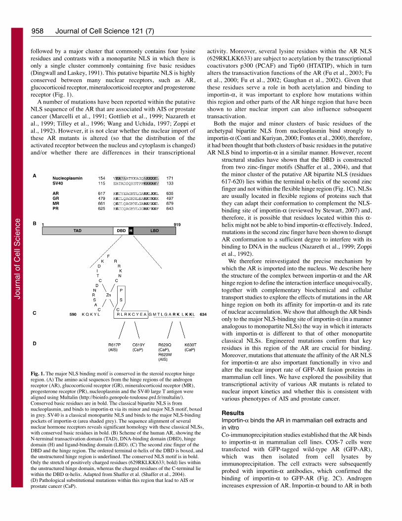
followed by a major cluster that commonly contains four lysineresidues and contrasts with a monopartite NLS in which there isonly a single cluster commonly containing five basic residues(Dingwall and Laskey, 1991). This putative bipartite NLS is highlyconserved between many nuclear receptors, such as AR,glucocorticoid receptor, mineralocorticoid receptor and progesteronereceptor (Fig. 1).
A number of mutations have been reported within the putativeNLS sequence of the AR that are associated with AIS or prostatecancer (Marcelli et al., 1991; Gottlieb et al., 1999; Nazareth etal., 1999; Tilley et al., 1996; Wang and Uchida, 1997; Zoppi etal., 1992). However, it is not clear whether the nuclear import ofthese AR mutants is altered (so that the distribution of theactivated receptor between the nucleus and cytoplasm is changed)and/or whether there are differences in their transcriptional
activity. Moreover, several lysine residues within the AR NLS(629RKLKK633) are subject to acetylation by the transcriptionalcoactivators p300 (PCAF) and Tip60 (HTATIP), which in turnalters the transactivation functions of the AR (Fu et al., 2003; Fuet al., 2000; Fu et al., 2002; Gaughan et al., 2002). Given thatthese residues serve a role in both acetylation and binding toimportin-α, it was important to explore how mutations withinthis region and other parts of the AR hinge region that have beenshown to alter nuclear import can also influence subsequenttransactivation.
Both the major and minor clusters of basic residues of thearchetypal bipartite NLS from nucleoplasmin bind strongly toimportin-α (Conti and Kuriyan, 2000; Fontes et al., 2000), therefore,it had been thought that both clusters of basic residues in the putativeAR NLS bind to importin-α in a similar manner. However, recent
structural studies have shown that the DBD is constructedfrom two zinc-finger motifs (Shaffer et al., 2004), and thatthe minor cluster of the putative AR bipartite NLS (residues617-620) lies within the terminal α-helix of the second zincfinger and not within the flexible hinge region (Fig. 1C). NLSsare usually located in flexible regions of proteins such thatthey can adapt their conformation to complement the NLS-binding site of importin-α (reviewed by Stewart, 2007) and,therefore, it is possible that residues located within this α-helix might not be able to bind importin-α effectively. Indeed,mutations in the second zinc finger have been shown to disruptAR conformation to a sufficient degree to interfere with itsbinding to DNA in the nucleus (Nazareth et al., 1999; Zoppiet al., 1992).
We therefore reinvestigated the precise mechanism bywhich the AR is imported into the nucleus. We describe herethe structure of the complex between importin-α and the ARhinge region to define the interaction interface unequivocally,together with complementary biochemical and cellulartransport studies to explore the effects of mutations in the ARhinge region on both its affinity for importin-α and its rateof nuclear accumulation. We show that although the AR bindsonly to the major NLS-binding site of importin-α (in a manneranalogous to monopartite NLSs) the way in which it interactswith importin-α is different to that of other monopartiteclassical NLSs. Engineered mutations confirm that keyresidues in this region of the AR are crucial for binding.Moreover, mutations that attenuate the affinity of the AR NLSfor importin-α are also important functionally in vivo andalter the nuclear import rate of GFP-AR fusion proteins inmammalian cell lines. We have explored the possibility thattranscriptional activity of various AR mutants is related tonuclear import kinetics and whether this is consistent withvarious phenotypes of AIS and prostate cancer.
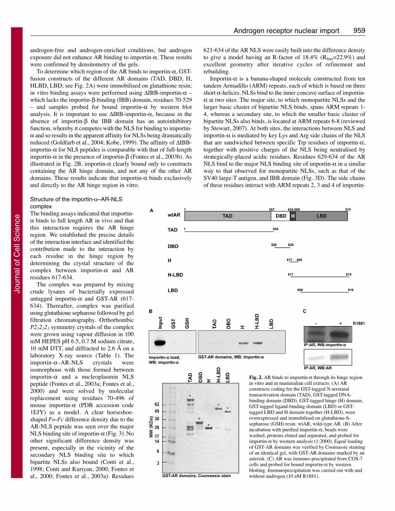
ResultsImportin-α binds the AR in mammalian cell extracts andin vitroCo-immunoprecipitation studies established that the AR bindsto importin-α in mammalian cell lines. COS-7 cells weretransfected with GFP-tagged wild-type AR (GFP-AR),which was then isolated from cell lysates byimmunoprecipitation. The cell extracts were subsequentlyprobed with importin-α antibodies, which confirmed thebinding of importin-α to GFP-AR (Fig. 2C). Androgenincreases expression of AR. Importin-α bound to AR in both
Journal of Cell Science 121 (7)
Fig. 1. The major NLS binding motif is conserved in the steroid receptor hingeregion. (A) The amino acid sequences from the hinge regions of the androgenreceptor (AR), glucocorticoid receptor (GR), mineralocorticoid receptor (MR),progesterone receptor (PR), nucleoplasmin and the SV40 large T antigen werealigned using Multalin (http://bioinfo.genopole-toulouse.prd.fr/multalin/).Conserved basic residues are in bold. The classical bipartite NLS is fromnucleoplasmin, and binds to importin-α via its minor and major NLS motif, boxedin grey. SV40 is a classical monopartite NLS and binds to the major NLS-bindingpockets of importin-α (area shaded grey). The sequence alignment of severalnuclear hormone receptors reveals significant homology with these classical NLSs,with conserved basic residues in bold. (B) Scheme of the human AR, showing theN-terminal transactivation domain (TAD), DNA-binding domain (DBD), hingedomain (H) and ligand-binding domain (LBD). (C) The second zinc finger of theDBD and the hinge region. The ordered terminal α-helix of the DBD is boxed, andthe unstructured hinge region is underlined. The conserved NLS motif is in bold.Only the stretch of positively charged residues (629RKLKK633; bold) lies withinthe unstructured hinge domain, whereas the charged residues of the C-terminal liewithin the DBD α-helix. Adapted from Shaffer et al. (Shaffer et al., 2004).(D) Pathological substitutional mutations within this region that lead to AIS orprostate cancer (CaP).
Jour
nal o
f Cel
l Sci
ence
959Androgen receptor nuclear import
androgen-free and androgen-enriched conditions, but androgenexposure did not enhance AR binding to importin-α. These resultswere confirmed by densitometry of the gels.
To determine which region of the AR binds to importin-α, GST-fusion constructs of the different AR domains (TAD, DBD, H,HLBD, LBD; see Fig. 2A) were immobilised on glutathione resin;in vitro binding assays were performed using ΔIBB-importin-α –which lacks the importin-β-binding (IBB) domain, residues 70-529– and samples probed for bound importin-α by western blotanalysis. It is important to use ΔIBB-importin-α, because in theabsence of importin-β the IBB domain has an autoinhibitoryfunction, whereby it competes with the NLS for binding to importin-α and so results in the apparent affinity for NLSs being dramaticallyreduced (Goldfarb et al., 2004; Kobe, 1999). The affinity of ΔIBB-importin-α for NLS peptides is comparable with that of full-lengthimportin-α in the presence of importin-β (Fontes et al., 2003b). Asillustrated in Fig. 2B, importin-α clearly bound only to constructscontaining the AR hinge domain, and not any of the other ARdomains. These results indicate that importin-α binds exclusivelyand directly to the AR hinge region in vitro.
Structure of the importin-α–AR-NLScomplexThe binding assays indicated that importin-α binds to full length AR in vivo and thatthis interaction requires the AR hingeregion. We established the precise detailsof the interaction interface and identified thecontribution made to the interaction byeach residue in the hinge region bydetermining the crystal structure of thecomplex between importin-α and ARresidues 617-634.
The complex was prepared by mixingcrude lysates of bacterially expresseduntagged importin-α and GST-AR (617-634). Thereafter, complex was purifiedusing glutathione sepharose followed by gelfiltration chromatography. OrthorhombicP212121 symmetry crystals of the complexwere grown using vapour diffusion in 100mM HEPES pH 6.5, 0.7 M sodium citrate,10 mM DTT, and diffracted to 2.6 Å on alaboratory X-ray source (Table 1). Theimportin-α–AR-NLS crystals wereisomorphous with those formed betweenimportin-α and a nucleoplasmin NLSpeptide (Fontes et al., 2003a; Fontes et al.,2000) and were solved by molecularreplacement using residues 70-496 ofmouse importin-α (PDB accession code1EJY) as a model. A clear horseshoe-shaped Fo-Fc difference density due to theAR-NLS peptide was seen over the majorNLS binding site of importin-α (Fig. 3). Noother significant difference density waspresent, especially in the vicinity of thesecondary NLS binding site to whichbipartite NLSs also bound (Conti et al.,1998; Conti and Kuriyan, 2000; Fontes etal., 2000; Fontes et al., 2003a). Residues
621-634 of the AR NLS were easily built into the difference densityto give a model having an R-factor of 18.4% (Rfree=22.9%) andexcellent geometry after iterative cycles of refinement andrebuilding.
Importin-α is a banana-shaped molecule constructed from tentandem Armadillo (ARM) repeats, each of which is based on threeshort α-helices. NLSs bind to the inner concave surface of importin-α at two sites. The major site, to which monopartite NLSs and thelarger basic cluster of bipartite NLS binds, spans ARM repeats 1-4, whereas a secondary site, to which the smaller basic cluster ofbipartite NLSs also binds, is located at ARM repeats 6-8 (reviewedby Stewart, 2007). At both sites, the interactions between NLS andimportin-α is mediated by key Lys and Arg side chains of the NLSthat are sandwiched between specific Trp residues of importin-α,together with positive charges of the NLS being neutralised bystrategically-placed acidic residues. Residues 629-634 of the ARNLS bind to the major NLS binding site of importin-α in a similarway to that observed for monopartite NLSs, such as that of theSV40 large-T antigen, and IBB domain (Fig. 3D). The side chainsof these residues interact with ARM repeats 2, 3 and 4 of importin-
Fig. 2. AR binds to importin-α through its hinge regionin vitro and in mammalian cell extracts. (A) ARconstructs coding for the GST-tagged N-terminaltransactivation domain (TAD), GST-tagged DNA-binding domain (DBD), GST-tagged hinge (H) domain,GST-tagged ligand-binding domain (LBD) or GST-tagged LBD and H domain together (H-LBD), wereoverexpressed and immobilised on glutathione-S-sepharose (GSH) resin. wtAR, wild-type AR. (B) Afterincubation with purified importin-α, beads werewashed, proteins eluted and separated, and probed forimportin-α by western analysis (1:2000). Equal loadingof GST-AR domains was verified by Coomassie stainingof an identical gel, with GST-AR domains marked by anasterisk. (C) AR was immuno-precipitated from COS-7cells and probed for bound importin-α by westernblotting. Immunoprecipitation was carried out with andwithout androgen (10 nM R1881).
Jour
nal o
f Cel
l Sci
ence

960
α to make contact with the P1-P5 binding pockets, through acombination of electrostatic and hydrogen bonds.
However, the way in which residues 621-628 of the AR NLSinteract with importin-α is significantly different to that seen withother NLSs, either bipartite or monopartite, or the IBB domain ofimportin-α. Although the AR NLS peptide made contact withimportin-α ARM repeat 5, at ARM repeat 6 the peptide curvedaway from importin-α and did not make contact with ARM repeats7 and 8, as is seen for bipartite NLSs such as that fromnucleoplasmin (Conti and Kuriyan, 2000; Fontes et al., 2000).
There was no difference density over the minor NLS binding siteof importin-α to indicate that the AR peptide might contact thissite.
Crystals of importin-α in complex with amino acid residues 617-634 of AR were also generated by co-crystallising purified importin-α with a synthetic AR peptide that corresponded to residues 617-634. These crystals were generated under the same conditions asbefore and the resultant Fo-Fc difference density map showed thesame horse-shoe shape observed with residues 621-628. Dissolutionof the crystals followed by reverse-phase HPLC and mass
Journal of Cell Science 121 (7)
Fig. 3. The structure of the AR hinge region bound toimportin-α. (A) 2.6 Å resolution Fo-Fc differenceelectron-density map (contoured at 3σ) showing thedensity resulting from the AR hinge region aftersubtraction of density due to importin-α in crystals of thecomplex. The structure of the underlying importin-α isshown in yellow. Electron density resulting from the ARhinge region was only seen over the primary NLS-binding site of importin-α. (B) Relationship between thebinding sites on importin-α for the AR hinge region(blue) and the bipartite NLS from nucleoplasmin (red).Whereas the nucleoplasmin NLS binds to both the majorand minor binding sites on importin-α, the AR hingeregion binds to the major site together with an adjacentregion of the importin-α surface that is not involved inthe interaction with other NLSs. (C) Schematicillustration of the positions of the Armadillo (ARM)repeats of importin-α corresponding to the modelsshown in A and B. (D) Schematic representation of theinteracting residues for different molecules (AR, SV40large-T antigen, nucleoplasmin and the importin-α IBBdomain) bound to importin-α. In all cases, binding at themajor site involves insertion of side chains between aseries of Trp residues (W142, W184, W273) onimportin-α, complemented by H-bonds between key Asn(N146, N188, N235) and the NLS main-chain peptides,and also neutralization of negative charges bystrategically placed acidic residues on importin-α.However, details of the interactions at the major site aredifferent and, significantly, a series of residues(621EAGMTLGA628) immediately upstream of thenegative cluster (629RKLKK634) in the AR hingeregion bind to importin-α in a manner not seen in otherNLSs. Only the bipartite nucleoplasmin NLS binds tothe secondary site.
Jour
nal o
f Cel
l Sci
ence
961Androgen receptor nuclear import
spectrometry (MALDI) confirmed that the peptide was intact andhad not been proteolysed.
Mutations in the AR hinge region influence binding affinity forimportin-αTo test our model for the structure of the importin-α–AR-NLSinterface in vitro, we used isothermal titration calorimetry (ITC) toquantify the effects of mutating key residues within the AR hinge
region on its affinity for importin-α (Table 2). Importin-α bindingaffinity was reduced for mutations of single Lys residues (Lys630,Lys632, Lys633), with the greatest binding contribution fromLys630 – consistent with its contact with the P2-binding pocket ofimportin-α (Conti and Kuriyan, 2000; Fontes et al., 2000; Hodelet al., 2001). However, the most striking effect was evident withthe triple Lys-to-Ala substitution KKK630/632/633AAA, whichcompletely abolished importin-α binding. Although it appearedfrom our structural model that AR Met624 might make ahydrophobic contact with importin-α, mutation of this residue didnot generate a significant change in importin-α binding affinity,suggesting that this interaction is relatively weak.
There have been several reports of point mutations within theAR sequence (617-634) that are associated with the clinicalphenoptypes of AIS and prostate cancer (Marcelli et al., 1991;Gottlieb et al., 1999; Nazareth et al., 1999; Tilley et al., 1996;Wang and Uchida, 1997; Zoppi et al., 1992). ITC was used todetermine the binding affinities of these known human mutationsfor importin-α (Table 2). These show that mutations within theAR that bind to the major NLS-binding pockets of importin-α(i.e. R629W, R629Q and K630T) had significantly reducedaffinity for importin-α, whereas other AR mutations in the N-terminus of the peptidethat do not form part of the NLS (R617P,C619Y) did not alter binding affinity.
These results indicated that the AR has an essentially monopartiteNLS (comprised of residues 629-634) that binds to the major NLS-binding pockets of importin-α and that mutation of AR residueswithin this region significantly weakened its affinity for importin-α. Consistent with this hypothesis, the Arg-to-Ala substitutionsR617A and K618A only reduced the ITC Kd of the AR peptidefor ΔIBB-importin-α by twofold (to 10±2 μM), whereas thecorresponding mutation in the nucleoplasmin peptide(nucleoplasmin KR155,156AA) reduced its Kd 12-fold to 2.5 μM,comparable with that of the wild-type AR peptide. We further
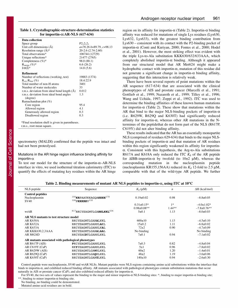
Table 1. Crystallographic-structure-determination statisticsfor importin-α:AR-NLS (617-634)
Data collectionSpace group P212121Unit cell dimensions (Å) a=78.26 b=89.79, c=98.13Resolution range (Å)* 20-2.6 (2.74-2.60)Total observations* 106744 (12729)Unique reflections* 21477 (2765)Completeness (%)* 98.0 (88.1)Rmerge (%)* 6.6 (26.2)I/σ(I)* 14.8 (5.6)
RefinementNumber of reflections (working, test) 19883 (1374)Rcry:Rfree (%) 18.4:22.9Total number of non-H atoms 3401Number of water molecules 33r.m.s. deviation from ideal bond length (Å) 0.012r.m.s. deviation from ideal bond angles 1.3
(degree) Ramachandran plot (%)
Core region 95.4Allowed region 4.1Generously allowed region 0.3Disallowed region 0.3
*Final resolution shell is given in parentheses.r.m.s., root mean square.
Table 2. Binding measurements of mutant AR NLS peptides to importin-α, using ITC at 10°C
NLS peptide Sequence Kd (μM) n ΔH (kcal/mol)
Control peptidesNucleoplasmin 155KRPAATKKAGQAKKKK170 0.19±0.02 0.98 –8.8±0.05SV40 126PKKKRKV132
0.31±0.15* 1* –4.0±1.02*0.98±0.08** 1.44** –7.8±0.76**
wtAR 617RKCYEAGMTLGARKLKKL634 5±0.1 1.13 –8.0±0.03
AR NLS mutants to test structure modelAR K630A RKCYEAGMTLGARALKKL 600±10 1.13 –4.5±0.10AR K632A RKCYEAGMTLGARKLAKL 15±0.2 1.11 –6.8±0.02AR K633A RKCYEAGMTLGARKLKAL 72±2 0.90 –4.7±0.09AR KKK630,2,3AAA RKCYEAGMTLGARALAAL No binding No bindingAR M624D RKCYEAGDTLGARKLKKL 7±0.1 0.94 –7.1±0.02
AR mutants associated with pathological phenotypesAR R617P (AIS) PKCYEAGMTLGARKLKKL 7±0.3 0.82 –4.8±0.04AR C619Y (CaP) RKYYEAGMTLGARKLKKL 5±1 0.96 –7.8±0.02AR R629W (AIS) RKCYEAGMTLGAWKLKKL 60±2 1.20 –1.4±0.01AR R629Q (CaP) RKCYEAGMTLGAQKLKKL 48±2 1.19 –3.6±0.06AR K630T (CaP) RKCYEAGMTLGARTLKKL 140±10 0.94 –2.6±0.30
Control peptide were nucleoplasmin, SV40 and wtAR NLSs. Mutant peptides were NLS regions containing amino acid substitutions within the interface thatbinds to importin-α, and exhibited reduced binding affinity. AR mutants associated with pathological phenotypes contain substitution mutations that occurnaturally in AIS or prostate cancer (CaP), and also exhibited reduced affinity for importin-α.
For SV40, the two sets of values represent the binding to the major and minor importin-α NLS-binding sites: *, binding to major importin-α binding site;**, binding to minor importin-α binding site.
No binding, no binding could be demonstrated.Mutated amino acid residues are in bold.
Jour
nal o
f Cel
l Sci
ence
962
validated this model physiologically for the binding interface ofimportin-α–AR (i.e. importin-α in complex with the whole of theAR) by carrying out nuclear import assays in living culturedmammalian cells.
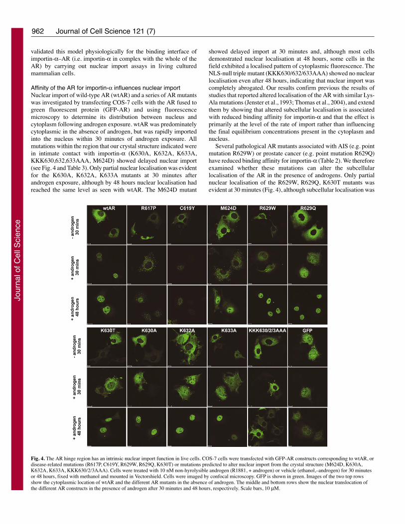
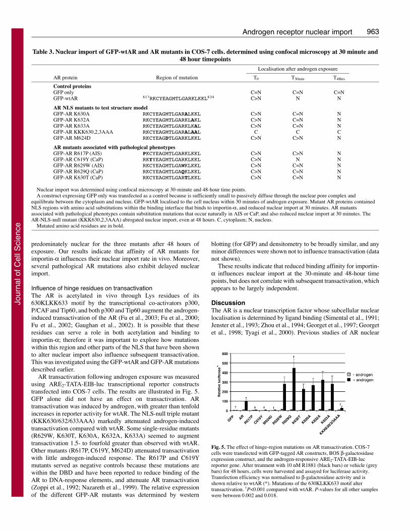
Affinity of the AR for importin-α influences nuclear importNuclear import of wild-type AR (wtAR) and a series of AR mutantswas investigated by transfecting COS-7 cells with the AR fused togreen fluorescent protein (GFP-AR) and using fluorescencemicroscopy to determine its distribution between nucleus andcytoplasm following androgen exposure. wtAR was predominatelycytoplasmic in the absence of androgen, but was rapidly importedinto the nucleus within 30 minutes of androgen exposure. Allmutations within the region that our crystal structure indicated werein intimate contact with importin-α (K630A, K632A, K633A,KKK630,632,633AAA, M624D) showed delayed nuclear import(see Fig. 4 and Table 3). Only partial nuclear localisation was evidentfor the K630A, K632A, K633A mutants at 30 minutes afterandrogen exposure, although by 48 hours nuclear localisation hadreached the same level as seen with wtAR. The M624D mutant
showed delayed import at 30 minutes and, although most cellsdemonstrated nuclear localisation at 48 hours, some cells in thefield exhibited a localised pattern of cytoplasmic fluorescence. TheNLS-null triple mutant (KKK630/632/633AAA) showed no nuclearlocalisation even after 48 hours, indicating that nuclear import wascompletely abrogated. Our results confirm previous the results ofstudies that reported altered localisation of the AR with similar Lys-Ala mutations (Jenster et al., 1993; Thomas et al., 2004), and extendthem by showing that altered subcellular localisation is associatedwith reduced binding affinity for importin-α and that the effect isprimarily at the level of the rate of import rather than influencingthe final equilibrium concentrations present in the cytoplasm andnucleus.
Several pathological AR mutants associated with AIS (e.g. pointmutation R629W) or prostate cancer (e.g. point mutation R629Q)have reduced binding affinity for importin-α (Table 2). We thereforeexamined whether these mutations can alter the subcellularlocalisation of the AR in the presence of androgens. Only partialnuclear localisation of the R629W, R629Q, K630T mutants wasevident at 30 minutes (Fig. 4), although subcellular localisation was
Journal of Cell Science 121 (7)
Fig. 4. The AR hinge region has an intrinsic nuclear import function in live cells. COS-7 cells were transfected with GFP-AR constructs corresponding to wtAR, ordisease-related mutations (R617P, C619Y, R629W, R629Q, K630T) or mutations predicted to alter nuclear import from the crystal structure (M624D, K630A,K632A, K633A, KKK630/2/3AAA). Cells were treated with 10 nM non-hyrolysible androgen (R1881, + androgen) or vehicle (ethanol,–androgen) for 30 minutesor 48 hours, fixed with methanol and mounted in Vectorshield. Cells were imaged by confocal microscopy. GFP is shown in green. Images of the two top rowsshow the cytoplasmic location of wtAR and the different AR mutants in the absence of androgen. The middle and bottom rows show the nuclear translocation ofthe different AR constructs in the presence of androgen after 30 minutes and 48 hours, respectively. Scale bars, 10 μM.
Jour
nal o
f Cel
l Sci
ence
963Androgen receptor nuclear import
predominately nuclear for the three mutants after 48 hours ofexposure. Our results indicate that affinity of AR mutants forimportin-α influences their nuclear import rate in vivo. Moreover,several pathological AR mutations also exhibit delayed nuclearimport.
Influence of hinge residues on transactivationThe AR is acetylated in vivo through Lys residues of its630KLKK633 motif by the transcriptional co-activators p300,P/CAF and Tip60, and both p300 and Tip60 augment the androgen-induced transactivation of the AR (Fu et al., 2003; Fu et al., 2000;Fu et al., 2002; Gaughan et al., 2002). It is possible that theseresidues can serve a role in both acetylation and binding toimportin-α; therefore it was important to explore how mutationswithin this region and other parts of the NLS that have been shownto alter nuclear import also influence subsequent transactivation.This was investigated using the GFP-wtAR and GFP-AR mutationsdescribed earlier.
AR transactivation following androgen exposure was measuredusing ARE2-TATA-EIB-luc transcriptional reporter constructstransfected into COS-7 cells. The results are illustrated in Fig. 5.GFP alone did not have an effect on transactivation. ARtransactivation was induced by androgen, with greater than tenfoldincreases in reporter activity for wtAR. The NLS-null triple mutant(KKK630/632/633AAA) markedly attenuated androgen-inducedtransactivation compared with wtAR. Some single-residue mutants(R629W, K630T, K630A, K632A, K633A) seemed to augmenttransactivation 1.5- to fourfold greater than observed with wtAR.Other mutants (R617P, C619Y, M624D) attenuated transactivationwith little androgen-induced response. The R617P and C619Ymutants served as negative controls because these mutations arewithin the DBD and have been reported to reduce binding of theAR to DNA-response elements, and attenuate AR transactivation(Zoppi et al., 1992; Nazareth et al., 1999). The relative expressionof the different GFP-AR mutants was determined by western
blotting (for GFP) and densitometry to be broadly similar, and anyminor differences were shown not to influence transactivation (datanot shown).
These results indicate that reduced binding affinity for importin-α influences nuclear import at the 30-minute and 48-hour timepoints, but does not correlate with subsequent transactivation, whichappears to be largely independent.
DiscussionThe AR is a nuclear transcription factor whose subcellular nuclearlocalisation is determined by ligand binding (Simental et al., 1991;Jenster et al., 1993; Zhou et al., 1994; Georget et al., 1997; Georgetet al., 1998; Tyagi et al., 2000). Previous studies of AR nuclear
Table 3. Nuclear import of GFP-wtAR and AR mutants in COS-7 cells. determined using confocal microscopy at 30 minute and48 hour timepoints
Localisation after androgen exposure
AR protein Region of mutation T0 T30min T48hrs
Control proteins GFP only C=N C=N C=NGFP-wtAR 617RKCYEAGMTLGARKLKKL634 C>N N N
AR NLS mutants to test structure model GFP-AR K630A RKCYEAGMTLGARALKKL C>N C=N NGFP-AR K632A RKCYEAGMTLGARKLAKL C>N C=N NGFP-AR K633A RKCYEAGMTLGARKLKAL C>N C=N NGFP-AR KKK630,2,3AAA RKCYEAGMTLGARALAAL C C CGFP-AR M624D RKCYEAGDTLGARKLKKL C>N C>N N
AR mutants associated with pathological phenotypesGFP-AR R617P (AIS) PKCYEAGMTLGARKLKKL C>N C>N NGFP-AR C619Y (CaP) RKYYEAGMTLGARKLKKL C>N N NGFP-AR R629W (AIS) RKCYEAGMTLGAWKLKKL C>N C=N NGFP-AR R629Q (CaP) RKCYEAGMTLGAQKLKKL C>N C=N NGFP-AR K630T (CaP) RKCYEAGMTLGARTLKKL C>N C=N N
Nuclear import was determined using confocal microscopy at 30-minute and 48-hour time points.A construct expressing GFP only was transfected as a control because is sufficiently small to passively diffuse through the nuclear pore complex and
equilibrate between the cytoplasm and nucleus. GFP-wtAR localised to the cell nucleus within 30 minutes of androgen exposure. Mutant AR proteins containedNLS regions with amino acid substitutions within the binding interface that binds to importin-α, and reduced nuclear import at 30 minutes. AR mutantsassociated with pathological phenotypes contain substitution mutations that occur naturally in AIS or CaP, and also reduced nuclear import at 30 minutes. TheAR-NLS-null mutant (KKK630,2,3AAA) abrogated nuclear import, even at 48 hours. C, cytoplasm; N, nucleus.
Mutated amino acid residues are in bold.
Fig. 5. The effect of hinge-region mutations on AR transactivation. COS-7cells were transfected with GFP-tagged AR constructs, BOS β-galactosidaseexpression construct, and the androgen-responsive ARE2-TATA-EIB-lucreporter gene. After treatment with 10 nM R1881 (black bars) or vehicle (greybars) for 48 hours, cells were harvested and assayed for luciferase activity.Transfection efficiency was normalised to β-galactosidase activity and isshown relative to wtAR (*). Mutations of the 630KLKK633 motif altertransactivation. †P<0.001 compared with wtAR. P-values for all other sampleswere between 0.002 and 0.018.
Jour
nal o
f Cel
l Sci
ence

964
import have highlighted the importance of key residues involvedin nuclear import (Jenster et al., 1993; Poukka et al., 2000; Thomaset al., 2004; Zhou et al., 1994) but did not establish the precisenuclear import pathway that was employed. In this study, we haveshown a direct interaction between the nuclear import receptorimportin-α and the AR, determined the crystal structure of thebinding interface of importin-α–AR-NLS, and defined itsrelationship to nuclear import and subsequent transactivation.
Importin-α binds to the AR in mammalian cell extracts and invitroStudies focussing on the nuclear import of other hormone receptorshave reported the binding of importins to the glucocorticoid receptor(GR). Importin-α has been shown to bind to a classical NLS site inthe GR (amino acid residues 479-497, termed GR NLS1 site) (Savoryet al., 1999) and, more recently, importins 7 and 8 have been reportedto interact with the GR, either through an unspecified site within theGR LBD (termed the GR NLS2 site) or indirectly through its NLS1site by binding with importin-α (Freedman and Yamamoto, 2004).Here, we show that importin-α binds to the AR in mammalian cellextracts, although this binding is not enhanced by androgen (Fig.2C). One would intuitively expect an enhancement of binding in thepresence of ligand, given that the AR translocates to the cell nucleusupon androgen exposure (Georget et al., 1997; Georget et al., 1998;Jenster et al., 1993; Poukka et al., 2000; Simental et al., 1991; Thomaset al., 2004; Tyagi et al., 2000; Zhou et al., 1994). Such ligand-enhanced binding has been reported for GR binding to importin-α,but we did not observe this with the AR, even when co-immunoprecipitating with anti-AR or anti-importin antibodies,which suggests that the nuclear import of liganded AR results morefrom its release from cytoplasmic components rather than fromenhanced exposure of its NLS.
Importin-α binds to the AR hinge region (Fig. 2B), confirmingprevious studies which indicated that residues within this regiondetermine AR nuclear localisation (Jenster et al., 1993; Poukka etal., 2000; Thomas et al., 2004; Zhou et al., 1994). The directinteraction between the AR NLS and importin-α is consistent withthe classical nuclear-protein-import machinery mediating ARnuclear import. Furthermore, heat shock proteins (such as Hsp90)bind to the AR in proximity to this region and dissociate followingconformational changes in the LBD induced by ligand binding(Georget et al., 2002; Kuil et al., 1995; Marivoet et al., 1992; Prattet al., 2004; Kallenberger et al., 2003; Schaufele et al., 2005; Wurtzet al., 1996; Savory et al., 1999). It has been proposed that thedissociation of Hsp90 facilitates greater exposure of the hingeregions of steroid receptors (Kuil et al., 1995; Savory et al., 1999),although our results suggest that such processes do not enhance thebinding of the AR to importin-α, at least in our cell-line assays.However, the AR was overexpressed in our cultured cell assaysand it is possible that binding of importin-α to the AR in the absenceof androgen was due to relatively low levels of Hsp90, becauseHsp90 normally masks the NLS site.
Structure of the binding interface in the importin-α–ARcomplexThe molecular details of the binding of NLSs to importin-α havebeen established from the crystal structures of nucleoplasmin, SV40and several other NLS-containing proteins bound to importin-α(Conti and Kuriyan, 2000; Conti et al., 1998; Fontes et al., 2003a;Fontes et al., 2000). Nucleoplasmin contains a classical bipartiteNLS (Fig. 1A), which consists of a minor and major component,
and binds to importin-α as illustrated in Fig. 3C. The SV40 largeT antigen contains a monopartite NLS and binds to importin-αmoststrongly at the major site. Although residues 617-634 of the ARshow sequence homology with the putative bipartite NLS sharedby many hormone receptors, our crystal structure of the importin-α–AR complex shows that the AR binds to importin-α primarilythrough residues 629-634 (629RKLKKL634) and that the more N-terminal residues (617RK618) do not bind. Thus, binding of theAR NLS to importin-α is different to that described fornucleoplasmin, and more in keeping with that of the SV40monopartite NLS (Fig. 3D). The failure of residues 617-618 to bindis consistent with their being an integral component of the secondzinc finger. In addition to the crystal structure, the ITC data confirmthat AR residues 629-634 (629RKLKKL634) provide thepredominant binding to importin-α and indicate that the strongestinteraction occurs through Lys630. This residue occupied the P2-binding pocket of importin-α that has been shown with other NLSsto be the most important contact for importin-α binding (Hodel etal., 2001; Makkerh et al., 1996; Robbins et al., 1991). Mutation ofthe N-terminal residues in the AR peptide (R617P, C619Y,analogous to the minor component of the nucleoplasmin NLS) havebeen reported to reduce AR binding to DNA (Nazareth et al., 1999;Zoppi et al., 1992) but did not reduce binding affinity for importin-α. Furthermore, the structure of the AR-DBD has been recentlyreported (Shaffer et al., 2004) and the location of AR residues 617and 618 lie within the terminal α-helix of the second zinc finger,whereas residues 629-634 (629RKLKKL634) lie within theunstructured and flexible hinge region (Fig. 1C). That importin-αonly binds to residues 629-634 (629RKLKKL634) is thereforecompletely consistent with the structure of the AR. We also showthat pathological mutations associated with AIS (i.e. point mutationR629W) or prostate cancer (i.e. point mutations R629Q, K630T)show a reduced binding affinity for importin-α that has not beenreported previously.
Binding affinity for importin-α and nuclear import are related,but the hinge region has a distinct function on transactivationThe functional significance of altered NLS-binding affinity forimportin-α and its effect on localisation of NLS peptides has beenreported in yeast cells (Hodel et al., 2006). Here, we show that thebinding affinity for importin-α also influences nuclear import ofwhole proteins in mammalian cells. The reduced binding affinityfor a range of AR mutants to importin-α (AR K630A, K632A,K633A, KKK630,632,633AAA) was determined by ITC, and theirsubsequent delay in nuclear import was illustrated by confocalmicroscopy. This observation was most striking for the NLS triplemutant KKK630,632,633AAA, which completely abrogatednuclear import at 48 hours, and suggests that the physiologicalsignificance of the AR NLS2 domain is different to that proposedpreviously (Poukka et al., 2000). The altered import of several ofthese mutants has been previously reported (Jenster et al., 1993;Thomas et al., 2004) but here we show that this is associated withreduced binding affinity for importin-α. We show this influenceof binding affinity on import kinetics is also evident in naturallyoccurring pathological mutations, such as mutations R629W orR629Q and K630T, that are associated with AIS or prostate cancer,respectively.
It is apparent that there are additional factors that influencenuclear import of the AR. We observed that the nuclear importof the R617P mutant was delayed at 30 minutes – perhaps owingto disruptions in AR DBD structure and sequestration in
Journal of Cell Science 121 (7)
Jour
nal o
f Cel
l Sci
ence
965Androgen receptor nuclear import
cytoplasmic aggregates. Several mutations within the AR zincfinger domains that do not involve the putative NLS (C579F,F582Y) have been reported to reduce nuclear localisation of theAR (Kawate et al., 2005). From inspection of the structure of thezinc fingers of the DBD domain, it is likely that such mutationsinterfere with the conformation of the protein in the zinc-fingerDBD region and thereby generate proteins in which the ARstructure was at least locally disrupted. Such a structural disruptioncould potentially mask the NLS and thus reduce nuclear importof the AR. Indeed, mutations of key Cys residues within the ARDBD zinc fingers have been reported to cause cytoplasmicaggregations after androgen exposure (Jenster et al., 1993; Kawateet al., 2005), perhaps as a direct result of such structure disruption.It is therefore possible that mutations of the minor basic clusterlocated in the second zinc-finger (R617, K618) can also disruptits structure, and so reduce nuclear accumulation – either throughindirectly preventing binding to importin-α in the cytoplasm orinterfering with the binding of the AR to DNA in the nucleus(Nazareth et al., 1999; Zoppi et al., 1992). The R617P mutationis likely to disrupt the AR DBD structure as it reduces the bindingaffinity of the AR to DNA response elements (Zoppi et al., 1992)and forms cytoplasmic aggregates after androgen exposure thatresemble those of other DBD mutants that abolish the DBD zincfinger structure (Jenster et al., 1993). Although speculative, theR617P mutant may form these aggregates as a result of disruptionin binding to Hsp90. Nuclear import of the AR is facilitated byHsp90 (Georget et al., 2002), perhaps owing to stabilisation ofthe AR in an active confirmation (Kuil et al., 1995; Georget etal., 2002). However, Hsp90 fails to colocalise with cytoplasmicaggregates of various AR hinge mutants (Thomas et al., 2004),and specific inhibition of Hsp90 with geldanamycin can precipitateformation of cytoplasmic aggregates and reduce nuclear import(Marcelli et al., 2006). It is therefore feasible that the R617Pmutant forms cytoplasmic aggregates and has delayed nuclearimport as a result of altered protein conformation and reducedbinding to Hsp90.
Since the AR is a nuclear transcription factor whose subcellularlocalisation is determined by its NLS, we sought to determinewhether the NLS also has a role on transactivation that was distinctfrom its role in nuclear import. The NLS triple mutant(KKK630,632,633AAA), which abrogated AR import, alsomarkedly attenuated androgen-induced transactivation comparedwith wtAR. Some single-residue mutations (R629W, K630T,K630A, K632A, K633A) seemed to augment transactivation,whereas others (M624D) attenuated transactivation. Overall theseresults imply that nuclear localisation of the AR is required for afull androgen-induced response, but the hinge has a separate,distinct role in transactivation that is apparent once nuclearlocalisation has been attained. Whereas the effect of the triple Lysmutation is to completely abrogate androgen-inducedtransactivation, the results obtained with the single-site mutantsshow either augmentation or attenuation of transactivation.Unravelling the cause of this is complicated by the large numberof coregulators which have been shown to interact with the AR atsites overlapping with the hinge, including coactivators such asp300, P/CAF (Fu et al., 2000; Fu et al., 2002), Tip60 (Brady etal., 1999; Gaughan et al., 2002), Ubc9 (Poukka et al., 1999),SNURF (Moilanen et al., 1998; Poukka et al., 2000), ARIP3(Moilanen et al., 1999), and co-repressors, such as HDAC1(Gaughan et al., 2002; Fu et al., 2003), NCoR (Cheng et al., 2002),and cyclinD1 (Reutens et al., 2001). Lys mutations within the AR
630KLKK633 motif have been reported to alter the binding of co-regulators p300, HDAC1 and NCoR (Fu et al., 2003; Fu et al.,2002), and also attenuate coactivator-enhanced transactivation (Fuet al., 2002). It is quite plausible that mutations within the NLSand DBD interfere with binding of some of the other co-regulatorslisted above, and this may therefore modify transactivation byaltering the composition of the AR transcription complex (Louieet al., 2003; Shang et al., 2002). A recent study on the glucocorticoidreceptor (GR) demonstrated that the selective substitutions of itsNLS with the SV40 NLS did not alter the nuclear localisation ofmutant GR or recruitment to the MMTV promoter. Thesesubstitutions did, however, attenuate both the recruitment of theGR-transcription complex and of mutant GR transactivation,implying a direct function of its hinge region on co-regulatorassembly (Carrigan et al., 2007). Haelens et al., have also recentlyshown that AR hinge residues have a function on transactivationthat is independent on other functions, such as DNA binding(Haelens et al., 2007).
As a consequence, single-residue mutations in the NLS arepredicted to have effects on AR transactivation that are highlydependent on cellular context, co-regulator expression patterns andthe promoter used in reporter assays. This highlights the hazardsof using reporter assays in an attempt to understand the mechanisticeffects of single-site mutations within this region in AIS and relatedconditions. The triple-Lys mutant, however, provides a potent toolto distinguish between genomic and non-genomic AR functionsthrough the clear-cut inhibition of both nuclear entry and ARtransactivation.
In summary, we have demonstrated that importin-α binds tothe AR hinge region, and have established the structure of thisbinding interface. The way in which AR-NLS binds to importin-α differs from the binding observed with classical monopartiteand bipartite NLSs, and biochemical analysis demonstrates that,in vivo, altered binding affinity influenced the nuclear import ofa range of AR mutants mediated by importin-α, including severalmutants associated with different clinical phenotypes. The widerange of transcriptional activities of hinge and DBD mutantsindicates the hinge region has a distinct role on transactivationthat is separate from its role on nuclear import. Further studiesare needed to clearly define the role of the AR hinge region inthis process.
Materials and MethodsUnless otherwise stated all chemicals were purchased from Sigma Aldrich.
Vectors and reagentsThe following plasmids have been described previously: pSV40-ARo containing thefull-length AR gene (kindly donated by Albert Brinkmann, Erasmus University,Rotterdam, The Netherlands) (Brinkmann et al., 1989) and pET30a (Novagen)containing the untagged importin-α construct [mouse importin-α 2 (Accession:NP_034785 XP_992959), residues 70-529, kindly donated by Yoshiyuki Matsuura,MRC LMB, Cambridge, UK] (Matsuura and Stewart, 2005).
The following AR domains were generated by PCR using pSV40-ARo as a templateand cloned into the pGEX 4T1 vector (Amersham): transactivation domain (TAD,residues 1-555), DNA-binding domain (DBD, residues 556-620), hinge domain (H,residues 617-668); hinge- and ligand-binding domain (HLBD, residues 617-918);LBD (residues 669-919) and AR NLS region (residues 617-635).
To generate the GFP-tagged AR, the full-length AR gene was amplified by PCRusing pSV40-ARo as a template and cloned into the pQBI 25/50 fc3 vector(Qbiogene). Site-directed mutagenesis was performed to generate the following GFP-AR mutants: R617P; C619Y; R629W; R629Q; K630T; M624D; K630A; K632A;K633A; KKK630,632,633AAA. To generate GST-tagged importin-α, the pET30aimportin-α ΔIBB construct (ΔIBB importin-α, residues 70-529) was used as atemplate, amplified by PCR and cloned into pGEX-4T1. Primers and restriction sitesused to generate these constructs are illustrated in supplementary material Tables S1and S2. All constructs were fully verified for integrity by sequencing.
Jour
nal o
f Cel
l Sci
ence
966
The following antibodies were used: AR441 (Dako Cytomation) for western blottingof the AR, N-20 (Santa Cruz Biotechnology) for immunoprecipitation of the AR,and ab22534 (Abcam) for western blotting of importin-α.
Mammalian cell culture and transfectionCOS-7 cells were purchased from the Cancer Research UK cell bank and wereroutinely cultured in DMEM supplemented with 10% foetal bovine serum (FBS;Labtech). For transfection cells were grown to 40% confluence and transfected usingFuGENE6 reagent (Roche) according to the manufacturer’s instructions. Prior toandrogen treatment cells were washed in PBS and grown in Phenol-Red-free RPMIsupplemented with charcoal-stripped FBS (Perbio) for 24 hours. Cells were androgentreated with either the DHT analogue R1881 (10 nM) or an equal volume of vehicle(ethanol).
Bacterial cell culture and transformationBL21-DE3 RIL (Stratagene) were transformed with the various pGEX-4T1 constructsunder manufacturer’s instructions and cultured in 2�TY medium at 37°C to an opticaldensity of 0.3 (A 600 nm), before reducing the growth temperature to 20°C andinducting cells with 1 mM isopropyl thio β-D-galactoside (IPTG). Cells were thengrown for a further 16 hours at 20°C prior to pelleting and resuspension on ice inbuffer A [50 mM Tris, 150 mM NaCl, 4 mM DTT, 1 mM phenyl-methyl-sulfonylfluoride (PMSF), enzyme inhibitors (Complete, Roche)]. All subsequent steps werecarried out a 4°C.
Western blotting COS-7 cells were washed in PBS, pelleted and protein lysates obtained byresuspending pellets in modified RIPA buffer [50 mM Tris pH 7.8, 150 mM NaCl,5 mM EDTA, 15 mM MgCl2, 0.5% sodium deoxycholate, 1 mM DTT, mammalianprotease inhibitor cocktail (Roche), 20 mM N-ethylmaleimide, 0.1% NP40] and passedfive times through a 26G needle. Lysates were centrifuged and the supernatantsretained as the soluble protein lysates. Equivalent concentrations of total protein,determined by Bradford assay, were compared using SDS-PAGE and westernanalysis. Proteins were detected using rat anti-importin-α antibody (1:2000) (Abcam)or mouse anti-AR antibody (1:2000) (AR441, Dako Cytomation). Mouse anti-actin(1:5000, Abcam) was used as a loading control. Proteins were visualised using anti-mouse or anti-sheep HRP-conjugated secondary antibodies (1:1000, DakoCytomation) and ECL-Plus (Amersham Biosciences). If the detected signal wasbeyond the dynamic range of film, diaminobenzidine (Vector Laboratories) was usedfor detection.
ImmunoprecipitationPrior to immunoprecipitation, COS-7 cells were plated into 10-cm2 dishes and grownto 60% confluence. Medium was changed to Phenol-Red-free DMEM supplementedwith 10% charcoal-stripped serum and cells were transfected with 2 μg of eachGFP-AR construct and Fugene6 reagent before growing for a further 24 hours. Cellswere starved in this androgen-free medium for a further 24 hours and then treatedwith either R1881 or vehicle for 30 minutes. Cells were lysed and immunoprecipitatedin the presence of R1881 or vehicle at 4°C. The AR was immunoprecipitated from750 μg of whole-cell lysate using 3 μl rabbit anti-AR antibody (N-20, Santa CruzBiotechnology) and protein G sepharose (Amersham). Control immunoprecipitationshad an equivalent concentration of sheep IgG added (Vector Laboratories). Sepharosewas washed five times in high-stringency RIPA buffer supplemented with 2% NP40,sample buffer was then added and protein analysed by SDS-PAGE and westernblotting.
GST-pull-down assaysBL21 DE3-RIL cell pellets expressing pGEX-4T1 protein constructs were resuspendedin buffer A on ice and lysed by sonication (Misonix Inc.). All subsequent steps werecarried out a 4°C. The supernatants were obtained by centrifuging cell lysates (40,000g for 45 minutes), filtered (0.4 μm) and applied to glutathione sepharose resin(Amersham) for 1 hour. The resins were then washed in buffer A to remove non-specific protein binding.
In vivo binding assaysCOS-7 cells were transfected with the GFP-wtAR vector (pQBI25/50fc3 wtAR) andgrown in androgen enriched or vehicle treated media. Cells were lysed andsupernatants analysed for AR and importin-α by immunoprecipitation and westernblotting.
In vitro binding assaysThe AR domains (TAD, DBD, H, HLBD, LBD) were expressed as GST-fusionproteins in BL21-DE3 RIL cells (Fig. 2A) and pulled down on glutathione resin asdescribed above. The resins were washed in buffer A, proteins separated on SDS-PAGE and visualised by Coomassie staining. Resin volumes were then adjusted forequalised protein levels and incubated with 2 mg untagged importin-α in buffer Afor 2 hours. Samples were washed with buffer A, and proteins separated by SDS-PAGE and visualised by Coomassie staining to detect the presence of bound importin-α. Because of similarities in molecular weight of importin-α and co-existing proteins
in these assays, subsequent western blotting for importin-α was carried out on thesamples. The samples were diluted 1:1000 and proteins separated on SDS-PAGE andtransferred onto nitrocellulose membrane. The samples were probed for importin-αusing Ab22534 (1:2000; Abcam). Blots were incubated using the anti-rat HRP-conjugated secondary antibody (Ab6734, 1:5000; Abcam) and visualised using ECL-Plus (Amersham Biosciences).
Crystallisation, data collection and structure determinationThe AR NLS and untagged importin-α were transformed and expressed in BL21-DE3 RIL cells. Cell pellets were resuspended in buffer A and all subsequent stepswere carried out at 4°C. Cells were lysed by passing through an Emulsiflex C5 high-pressure cell homogeniser (Glen Creston) in the presence of DNAse (Sigma). Thesupernatants were obtained by centrifugation (40,000 g for 45 minutes) and filtered(0.4 μm). Supernatant from 4L of AR NLS culture was incubated with glutathionesepharose resin (Amersham) for 1 hour and then washed in buffer B (50 mM Tris-HCl, 300 mM NaCl, 4 mM DTT), prior to incubation with supernatant from 6 Limportin-α culture for 4 hours. The complex was washed with buffer A, resuspendedin buffer C (20 mM Tris-HCl, 50 mM NaCl, 4 mM DTT) and incubated with thrombin(Sigma) for a further 16 hours. The importin-α–AR-NLS complex was eluted,incubated with p-aminobenzamidine (Sigma) for 1 hour and purified over an S-75gel filtration column. 10 litres of culture yielded 15 mg of pure protein complexvisualised as two bands on a Coomassie-stained SDS-PAGE gel. The proteincomplex was concentrated to 20 mg/ml, snap frozen in liquid nitrogen and stored at–80°C.
To determine conditions favourable for crystallisation of the importin-α–AR-NLScomplex, parameters were screened around commercial conditions (MolecularDimensions MD1-01) and those described for the crystallisation of the importin-α–nucleoplasmin complex (Fontes et al., 2000). 2 μl of protein solution and 2 μl ofreservoir solution were suspended from a coverslip by hanging-drop method over areservoir of 500 μl. The largest crystals were obtained using reservoir conditions of100 mM HEPES pH 6.5, 0.7 M sodium citrate, 10 mM DTT.
Crystals were picked up in a cryoloop and transiently exposed to cryoprecipitate[reservoir solution containing 23% glycerol (v/v)], after which the crystal was vitrifiedin a stream of anhydrous nitrogen at 100 K. A 2.6 Å resolution data set (Table 1)was collected at 100 K in house using a Rigaku X-ray generator equipped withOsmic mirrors and a Mar345dtb image plate detector. Data were processed usingMOSFLM (CCP4, 1994) and reduced using SCALA and TRUNCATE (CCP4, 1994).Free-R flags were set to be identical to those used for the structure of the importin-α–nucleoplasmin-NLS complex (Fontes et al., 2000). A structural model wasproduced using residues 70-496 of importin-α in the structure of the importin-α–nucleoplasmin-NLS complex (PDB accession number 1EJY) and, after rigid bodyrefinement using REFMAC5 (CCP4, 1994), a clear difference density was visibleinto which residues 621-634 of the AR NLS were built. After iterative cycles ofrefinement with REFMAC5 and model building using O (Jones et al., 1991) andthe addition of 33 water molecules, a final structure with an R-factor of 18.8%(FreeR=22.9%) and excellent geometry was produced (Table 1). Asn239 of importin-α is a Ramachandran outlier as in other mouse importin-α structures (Fontes et al.,2000; Kobe, 1999; Matsuura and Stewart, 2005). Coordinates and structure factorshave been deposited with the protein data bank (PDB) with accession code 3BTR.
To explore the possibility of peptide proteolysis within the crystals ofprotein complex, crystals were dissolved in 0.1% trifluoroacetic acid (TFA) andanalysed by reverse-phase chromatography (HPLC) and matrix-assisted laserdesorption/ ionization (MALDI) mass spectrometry. This confirmed the peptidewas intact.
ITC-binding studiesImportin-α (residues 70-529) inserted into the GST-fusion expression vector pGEX-4T1, was transformed and expressed in BL21 DE3-RIL cells. The supernatant wasincubated with glutathione sepharose resin for 4 hours and the resin then washed inbuffer B, prior to resuspension in buffer D (100 mM HEPES, pH 7.4, 50 mM NaCl,4 mM DTT) and incubated with thrombin (Sigma) for a further 16 hours. The importin-α protein was eluted, incubated with p-aminobenzamidine (Sigma) for 1 hour andpurified over an S-75 gel filatration column; 12 litres of culture yielded 100 mg ofpure protein visualised as a single band on a Coomassie-stained SDS-PAGE gel.
The peptides used in the isothermal titration calorimetry (ITC) experiments weresynthesised to greater than 95% purity and analysed by reverse phase chromatographyand laser desorption mass spectrometry (ADL Advanced Biomedical). ITC was usedto investigate the binding of AR NLS peptides to importin-α using a VP-ITCcalorimeter (MicroCal Inc., CA). Experiments were carried out in 100 mM HEPESpH 7.4, 50 mM NaCl, 4 mM DTT at 10°C. The cell contained 1.36 ml importin-αsolution (typically 100 μM), and peptide (typically 2 mM) was injected in 5 μl aliquotsevery 4 minutes, typically to a four- to fivefold molar excess. The concentrations ofthe peptides were typically 20 times greater than that of importin-α. If possible, proteinconcentration was selected to be at least five times greater than that of the estimateddissociation constant. The manufacturer’s ORIGIN software fitted titration curvesand yielded the stoichiometry (N), the binary equilibrium constant Ka (Kd
–1) and theenthalpy of binding (ΔH) from the data. The dissociation constant (Kd) wassubsequently calculated (Kd=1/Ka).
Journal of Cell Science 121 (7)
Jour
nal o
f Cel
l Sci
ence
967Androgen receptor nuclear import
Confocal nuclear-import assaysSubcellular localisation assays were performed as described (Whitaker et al., 2004).Briefly, COS cells were grown on glass coverslips and transfected with GFP-taggedAR constructs using Fugene6 (Roche). Cells were starved in Phenol-Red-freemedium, supplemented with 10% charcoal-stripped serum for 24 hours. This mediumwas replaced with pre-warmed medium containing either vehicle (ethanol) or R1881for either 30 minutes or 48 hours. Cells were fixed in 100% methanol for 10 minutesat –20°C before mounting with Vectashield containing DAPI (Vector Laboratories).GFP-AR and GFP-AR mutants were visualised using a Zeiss Meta 510 confocalmicroscope using a 63� objective.
Transcriptional activityCOS cells were grown in 24-well plates to 40% confluence. Prior to transfectioncells were washed and medium was changed to Phenol-Red-free DMEM withcharcoal-stripped serum as described. Using Fugene6, cells were transfected with 75ng per well GFP-tagged AR constructs, 50 ng per well ARE2-TATA-EIB-lucconstruct, and co-transfected with 25 ng/well BOS-β-galactosidase (Mizushima andNagata, 1990) as a control for transfection efficiency. GFP alone was used as a negativecontrol and had no effect on AR transactivation. Twelve hours after transfection,medium was replaced with medium containing either 10 nM R1881 or vehicle(ethanol) and cells were harvested 48 hours later. Luciferase activity was measuredusing the Luclite luciferase assay kit (Packard Biosciences). Results were normalisedusing β-galactosidase activity with a Galacton Galactolite assay kit (Tropix). All assayswere completed on at least three separate occasions. Error bars indicate standarddeviation from the mean. P-values were calculated using a two-tailed t-test.
We thank Bostjan Kobe (University of Queensland, Australia) forsupplying the Rfree flags used for the structure determination ofimporin-α–nucleoplasmin-NLS. We thank our colleagues inCambridge, especially Astrid Giesecke, Neil Marshall, Eva Schmidt,Marijn Ford, Oli Daumke and Harvey McMahon for their invaluableassistance, advice and comments. M.L.C. gratefully acknowledgesfinancial support given by a Cancer Research UK Gordon Hamilton-Fairley Clinical Research Training Fellowship, a Joint Prostate CancerFellowship from the Royal College of Surgeons of Edinburgh and theProMPT NCRI/MRC Prostate Cancer Collaborative, and a Universityof Cambridge Raymond and Beverly Sackler Fellowship. This workwas supported in part by the Wellcome Trust Programme Grant 080522(to M.S.), a Cancer Research UK Programme Grant (held by D.E.N.),and the ProMPT Northern Collaborative (lead investigator D.E.N.),funded by NCRI (MRC, CR UK, Department of Health).
ReferencesBrady, M. E., Ozanne, D. M., Gaughan, L., Waite, I., Cook, S., Neal, D. E. and Robson,
C. N. (1999). Tip60 is a nuclear hormone receptor coactivator. J. Biol. Chem. 274, 17599-17604.
Brinkmann, A. O. (2001). Molecular basis of androgen insensitivity. Mol. Cell. Endocrinol.179, 105-109.
Brinkmann, A. O., Faber, P. W., Van Rooij, H. C., Kuiper, G. G., Ris, C., Klaassen,P., Van Der Korput, J. A., Voorhorst, M. M., Van Laar, J. H., Mulder, E. et al.(1989). The human androgen receptor: domain structure, genomic organization andregulation of expression. J. Steroid Biochem. 34, 307-310.
Carrigan, A., Walther, R. F., Salem, H. A., Wu, D., Atlas, E., Lefebvre, Y. A. andHache, R. J. (2007). An active nuclear retention signal in the glucocorticoid receptorfunctions as a strong inducer of transcriptional activation. J. Biol. Chem. 282, 10963-10971.
Chen, C. D., Welsbie, D. S., Tran, C., Baek, S. H., Chen, R., Vessella, R., Rosenfeld,M. G. and Sawyers, C. L. (2004). Molecular determinants of resistance to antiandrogentherapy. Nat. Med. 10, 33-39.
Cheng, S., Brzostek, S., Lee, S. R., Hollenberg, A. N. and Balk, S. P. (2002). Inhibitionof the dihydrotestosterone-activated androgen receptor by nuclear receptor corepressor.Mol. Endocrinol. 16, 1492-1501.
Claessens, A., Tanner, T., Denayer, S., Callewaert, L. and Claessens, F. (2007). Thehinge region regulates DNA binding, nuclear translocation, and transactivation of theandrogen receptor. Cancer Res. 67, 4514-4523.
Conti, E. and Kuriyan, J. (2000). Crystallographic analysis of the specific yet versatilerecognition of distinct nuclear localization signals by karyopherin alpha. Structure 8,329-338.
Conti, E., Uy, M., Leighton, L., Blobel, G. and Kuriyan, J. (1998). Crystallographicanalysis of the recognition of a nuclear localization signal by the nuclear import factorkaryopherin alpha. Cell 94, 193-204.
Dingwall, C. and Laskey, R. A. (1991). Nuclear targeting sequences-a consensus? TrendsBiochem. Sci. 16, 478-481.
Fang, Y., Fliss, A. E., Robins, D. M. and Caplan, A. J. (1996). Hsp90 regulates androgenreceptor hormone binding affinity in vivo. J. Biol. Chem. 271, 28697-28702.
Fontes, M. R., Teh, T. and Kobe, B. (2000). Structural basis of recognition of monopartiteand bipartite nuclear localization sequences by mammalian importin-alpha. J. Mol. Biol.297, 1183-1194.
Fontes, M. R., Teh, T., Jans, D., Brinkworth, R. I. and Kobe, B. (2003a). Structuralbasis for the specificity of bipartite nuclear localization sequence binding by importin-alpha. J. Biol. Chem. 278, 27981-27987.
Fontes, M. R., Teh, T., Toth, G., John, A., Pavo, I., Jans, D. A. and Kobe, B. (2003b).Role of flanking sequences and phosphorylation in the recognition of the simian-virus-40 large T-antigen nuclear localization sequences by importin-alpha. Biochem. J. 375,339-349.
Freedman, N. D. and Yamamoto, K. R. (2004). Importin 7 and importin alpha/importinbeta are nuclear import receptors for the glucocorticoid receptor. Mol. Biol. Cell 15,2276-2286.
Fu, M., Wang, C., Reutens, A. T., Wang, J., Angeletti, R. H., Siconolfi-Baez, L.,Ogryzko, V., Avantaggiati, M. L. and Pestell, R. G. (2000). p300 and p300/cAMP-response element-binding protein-associated factor acetylate the androgen receptor atsites governing hormone-dependent transactivation. J. Biol. Chem. 275, 20853-20860.
Fu, M., Wang, C., Wang, J., Zhang, X., Sakamaki, T., Yeung, Y. G., Chang, C., Hopp,T., Fuqua, S. A., Jaffray, E. et al. (2002). Androgen receptor acetylation governs transactivation and MEKK1-induced apoptosis without affecting in vitro sumoylation andtrans-repression function. Mol. Cell. Biol. 22, 3373-3388.
Fu, M., Rao, M., Wang, C., Sakamaki, T., Wang, J., Di Vizio, D., Zhang, X., Albanese,C., Balk, S., Chang, C. et al. (2003). Acetylation of androgen receptor enhances coactivatorbinding and promotes prostate cancer cell growth. Mol. Cell. Biol. 23, 8563-8575.
Gaughan, L., Logan, I. R., Cook, S., Neal, D. E. and Robson, C. N. (2002). Tip60 andhistone deacetylase 1 regulate androgen receptor activity through changes to theacetylation status of the receptor. J. Biol. Chem. 277, 25904-25913.
Georget, V., Lobaccaro, J. M., Terouanne, B., Mangeat, P., Nicolas, J. C. and Sultan,C. (1997). Trafficking of the androgen receptor in living cells with fused green fluorescentprotein-androgen receptor. Mol. Cell. Endocrinol. 129, 17-26.
Georget, V., Terouanne, B., Lumbroso, S., Nicolas, J. C. and Sultan, C. (1998).Trafficking of androgen receptor mutants fused to green fluorescent protein: a newinvestigation of partial androgen insensitivity syndrome. J. Clin. Endocrinol. Metab. 83,3597-3603.
Georget, V., Terouanne, B., Nicolas, J. C. and Sultan, C. (2002). Mechanism ofantiandrogen action: key role of hsp90 in conformational change and transcriptionalactivity of the androgen receptor. Biochemistry 41, 11824-11831.
Giovannucci, E., Stampfer, M. J., Krithivas, K., Brown, M., Dahl, D., Brufsky, A.,Talcott, J., Hennekens, C. H. and Kantoff, P. W. (1997). The CAG repeat within theandrogen receptor gene and its relationship to prostate cancer. Proc. Natl. Acad. Sci.USA 94, 3320-3323.
Goldfarb, D. S., Corbett, A. H., Mason, D. A., Harreman, M. T. and Adam, S. A.(2004). Importin alpha: a multipurpose nuclear-transport receptor. Trends Cell Biol. 14,505-514.
Gorlich, D. and Kutay, U. (1999). Transport between the cell nucleus and the cytoplasm.Annu. Rev. Cell Dev. Biol. 15, 607-660.
Gottlieb, B., Beitel, L. K., Lumbroso, R., Pinsky, L. and Trifiro, M. (1999). Update ofthe androgen receptor gene mutations database. Hum. Mutat. 14, 103-114.
Haelens, A., Tanner, T., Denayer, S., Callewaert, L., Claessens, F. (2007). The hingeregion regulates DNA binding, nuclear translocation, and transactivation of the androgenreceptor. Cancer Res. 67, 4514-4523.
Heinlein, C. A. and Chang, C. (2004). Androgen receptor in prostate cancer. Endocr. Rev.25, 276-308.
Hodel, A. E., Harreman, M. T., Pulliam, K. F., Harben, M. E., Holmes, J. S., Hodel,M. R., Berland, K. M. and Corbett, A. H. (2006). Nuclear localization signal receptoraffinity correlates with in vivo localization in Saccharomyces cerevisiae. J. Biol. Chem.281, 23545-23556.
Hodel, M. R., Corbett, A. H. and Hodel, A. E. (2001). Dissection of a nuclear localizationsignal. J. Biol. Chem. 276, 1317-1325.
Jenster, G., Trapman, J. and Brinkmann, A. O. (1993). Nuclear import of the humanandrogen receptor. Biochem. J. 293, 761-768.
Kallenberger, B. C., Love, J. D., Chatterjee, V. K. and Schwabe, J. W. (2003). A dynamicmechanism of nuclear receptor activation and its perturbation in a human disease. Nat.Struct. Biol. 10, 136-140.
Kawate, H., Wu, Y., Ohnaka, K., Tao, R. H., Nakamura, K., Okabe, T., Yanase, T.,Nawata, H. and Takayanagi, R. (2005). Impaired nuclear translocation, nuclear matrixtargeting, and intranuclear mobility of mutant androgen receptors carrying amino acidsubstitutions in the deoxyribonucleic acid-binding domain derived from androgeninsensitivity syndrome patients. J. Clin. Endocrinol. Metab. 90, 6162-6169.
Kobe, B. (1999). Autoinhibition by an internal nuclear localization signal revealed by thecrystal structure of mammalian importin alpha. Nat. Struct. Biol. 6, 388-397.
Kuil, C. W., Berrevoets, C. A. and Mulder, E. (1995). Ligand-induced conformationalalterations of the androgen receptor analyzed by limited trypsinization. Studies on themechanism of antiandrogen action. J. Biol. Chem. 270, 27569-27576.
La Spada, A. R., Wilson, E. M., Lubahn, D. B., Harding, A. E. and Fischbeck, K. H.(1991). Androgen receptor gene mutations in X-linked spinal and bulbar muscularatrophy. Nature 352, 77-79.
Laudet, V. and Gronemeyer, H. (ed.) (2002). The General Organization of NuclearReceptors. The Nuclear Receptor Factsbook. London: Academic Press.
Linja, M. J. and Visakorpi, T. (2004). Alterations of androgen receptor in prostate cancer.J. Steroid Biochem. Mol. Biol. 92, 255-264.
Lobaccaro, J. M., Lumbroso, S., Belon, C., Galtier-Dereure, F., Bringer, J., Lesimple,T., Heron, J. F., Pujol, H. and Sultan, C. (1993). Male breast cancer and the androgenreceptor gene. Nat. Genet. 5, 109-110.
Jour
nal o
f Cel
l Sci
ence
968
Louie, M. C., Yang, H. Q., Ma, A. H., Xu, W., Zou, J. X., Kung, H. J. andChen, H. W. (2003). Androgen-induced recruitment of RNA polymerase II to anuclear receptor-p160 coactivator complex. Proc. Natl. Acad. Sci. USA 100, 2226-2230.
Macara, I. G. (2001). Transport into and out of the nucleus. Microbiol. Mol. Biol. Rev.65, 570-594, table of contents.
Makkerh, J. P., Dingwall, C. and Laskey, R. A. (1996). Comparative mutagenesis ofnuclear localization signals reveals the importance of neutral and acidic amino acids.Curr. Biol. 6, 1025-1027.
Marcelli, M., Zoppi, S., Grino, P. B., Griffin, J. E., Wilson, J. D. and Mcphaul, M. J.(1991). A mutation in the DNA-binding domain of the androgen receptor gene causescomplete testicular feminization in a patient with receptor-positive androgen resistance.J. Clin. Invest. 87, 1123-1126.
Marcelli, M., Stenoien, D. L., Szafran, A. T., Simeoni, S., Agoulnik, I. U., Weigel, N.L., Moran, T., Mikic, I., Price, J. H. and Mancini, M. A. (2006). Quantifying effectsof ligands on androgen receptor nuclear translocation, intranuclear dynamics, andsolubility. J. Cell. Biochem. 98, 770-788.
Marivoet, S., Van Dijck, P., Verhoeven, G. and Heyns, W. (1992). Interaction of the 90-kDa heat shock protein with native and in vitro translated androgen receptor and receptorfragments. Mol. Cell. Endocrinol. 88, 165-174.
Matsuura, Y. and Stewart, M. (2005). Nup50/Npap60 function in nuclear protein importcomplex disassembly and importin recycling. EMBO J. 24, 3681-3689.
Mizushima, S. and Nagata, S. (1990). pEF-BOS, a powerful mammalian expression vector.Nucleic Acids Res. 18, 5322.
Moilanen, A. M., Poukka, H., Karvonen, U., Hakli, M., Janne, O. A. and Palvimo, J.J. (1998). Identification of a novel RING finger protein as a coregulator in steroidreceptor-mediated gene transcription. Mol. Cell. Biol. 18, 5128-5139.
Moilanen, A. M., Karvonen, U., Poukka, H., Yan, W., Toppari, J., Janne, O. A. andPalvimo, J. J. (1999). A testis-specific androgen receptor coregulator that belongs to anovel family of nuclear proteins. J. Biol. Chem. 274, 3700-3704.
Nazareth, L. V., Stenoien, D. L., Bingman, W. E., 3rd, James, A. J., Wu, C., Zhang,Y., Edwards, D. P., Mancini, M., Marcelli, M., Lamb, D. J. et al. (1999). A C619Ymutation in the human androgen receptor causes inactivation and mislocalization of thereceptor with concomitant sequestration of SRC-1 (steroid receptor coactivator 1). Mol.Endocrinol. 13, 2065-2075.
Poukka, H., Aarnisalo, P., Karvonen, U., Palvimo, J. J. and Janne, O. A. (1999). Ubc9interacts with the androgen receptor and activates receptor-dependent transcription. J.Biol. Chem. 274, 19441-19446.
Poukka, H., Karvonen, U., Yoshikawa, N., Tanaka, H., Palvimo, J. J. and Janne, O.A. (2000). The RING finger protein SNURF modulates nuclear trafficking of the androgenreceptor. J. Cell Sci. 113, 2991-3001.
Pratt, W. B., Galigniana, M. D., Morishima, Y. and Murphy, P. J. (2004). Role ofmolecular chaperones in steroid receptor action. Essays Biochem. 40, 41-58.
Quigley, C. A., De Bellis, A., Marschke, K. B., El-Awady, M. K., Wilson, E. M. andFrench, F. S. (1995). Androgen receptor defects: historical, clinical, and molecularperspectives. Endocr. Rev. 16, 271-321.
Reutens, A. T., Fu, M., Wang, C., Albanese, C., Mcphaul, M. J., Sun, Z., Balk, S. P.,Janne, O. A., Palvimo, J. J. and Pestell, R. G. (2001). Cyclin D1 binds the androgenreceptor and regulates hormone-dependent signaling in a p300/CBP-associated factor(P/CAF)-dependent manner. Mol. Endocrinol. 15, 797-811.
Robbins, J., Dilworth, S. M., Laskey, R. A. and Dingwall, C. (1991). Two interdependentbasic domains in nucleoplasmin nuclear targeting sequence: identification of a class ofbipartite nuclear targeting sequence. Cell 64, 615-623.
Savory, J. G., Hsu, B., Laquian, I. R., Giffin, W., Reich, T., Hache, R. J. and Lefebvre,Y. A. (1999). Discrimination between NL1- and NL2-mediated nuclear localization ofthe glucocorticoid receptor. Mol. Cell. Biol. 19, 1025-1037.
Schaufele, F., Carbonell, X., Guerbadot, M., Borngraeber, S., Chapman, M. S., Ma,A. A., Miner, J. N. and Diamond, M. I. (2005). The structural basis of androgen receptoractivation: intramolecular and intermolecular amino-carboxy interactions. Proc. Natl.Acad. Sci. USA 102, 9802-9807.
Shaffer, P. L., Jivan, A., Dollins, D. E., Claessens, F. and Gewirth, D. T. (2004). Structuralbasis of androgen receptor binding to selective androgen response elements. Proc. Natl.Acad. Sci. USA 101, 4758-4763.
Shang, Y., Myers, M. and Brown, M. (2002). Formation of the androgen receptortranscription complex. Mol. Cell 9, 601-610.
Simental, J. A., Sar, M., Lane, M. V., French, F. S. and Wilson, E. M. (1991).Transcriptional activation and nuclear targeting signals of the human androgen receptor.J. Biol. Chem. 266, 510-518.
Stanford, J. L., Just, J. J., Gibbs, M., Wicklund, K. G., Neal, C. L., Blumenstein, B.A. and Ostrander, E. A. (1997). Polymorphic repeats in the androgen receptor gene:molecular markers of prostate cancer risk. Cancer Res. 57, 1194-1198.
Stewart, M. (2006). Structural basis for the nuclear protein import cycle. Biochem. Soc.Trans. 34, 701-704.
Stewart, M. (2007). Molecular mechanism of the nuclear protein import cycle. Nat. Rev.Mol. Cell Biol. 8, 195-208.
Thomas, M., Dadgar, N., Aphale, A., Harrell, J. M., Kunkel, R., Pratt, W. B. andLieberman, A. P. (2004). Androgen receptor acetylation site mutations cause traffickingdefects, misfolding, and aggregation similar to expanded glutamine tracts. J. Biol. Chem.279, 8389-8395.
Tilley, W. D., Buchanan, G., Hickey, T. E. and Bentel, J. M. (1996). Mutations in theandrogen receptor gene are associated with progression of human prostate cancer toandrogen independence. Clin. Cancer Res. 2, 277-285.
Tyagi, R. K., Lavrovsky, Y., Ahn, S. C., Song, C. S., Chatterjee, B. and Roy, A. K.(2000). Dynamics of intracellular movement and nucleocytoplasmic recycling of theligand-activated androgen receptor in living cells. Mol. Endocrinol. 14, 1162-1174.
Wang, C. and Uchida, T. (1997). [Androgen receptor gene mutations in prostate cancer].Nippon Hinyokika Gakkai Zasshi 88, 550-556.
Whitaker, H. C., Hanrahan, S., Totty, N., Gamble, S. C., Waxman, J., Cato, A. C.,Hurst, H. C. and Bevan, C. L. (2004). Androgen receptor is targeted to distinctsubcellular compartments in response to different therapeutic antiandrogens. Clin. CancerRes. 10, 7392-7401.
Wooster, R., Mangion, J., Eeles, R., Smith, S., Dowsett, M., Averill, D., Barrett-Lee,P., Easton, D. F., Ponder, B. A. and Stratton, M. R. (1992). A germline mutation inthe androgen receptor gene in two brothers with breast cancer and Reifenstein syndrome.Nat. Genet. 2, 132-134.
Wurtz, J. M., Bourguet, W., Renaud, J. P., Vivat, V., Chambon, P., Moras, D. andGronemeyer, H. (1996). A canonical structure for the ligand-binding domain of nuclearreceptors. Nat. Struct. Biol. 3, 87-94.
Zhou, Z. X., Sar, M., Simental, J. A., Lane, M. V. and Wilson, E. M. (1994). A ligand-dependent bipartite nuclear targeting signal in the human androgen receptor. Requirementfor the DNA-binding domain and modulation by NH2-terminal and carboxyl-terminalsequences. J. Biol. Chem. 269, 13115-13123.
Zoppi, S., Marcelli, M., Deslypere, J. P., Griffin, J. E., Wilson, J. D. and McPhaul, M.J. (1992). Amino acid substitutions in the DNA-binding domain of the human androgenreceptor are a frequent cause of receptor-binding positive androgen resistance. Mol.Endocrinol. 6, 409-415.
Journal of Cell Science 121 (7)
Jour
nal o
f Cel
l Sci
ence