structural heart disease interventions - summitmd.comsummitmd.com/pdf/pdf/1146_sructural...
TRANSCRIPT
Ron Waksman, MD, FACC, FSCAIRon Waksman, MD, FACC, FSCAIProfessor of Medicine, Georgetown University,Professor of Medicine, Georgetown University,
Associate Chief of Cardiology, Associate Chief of Cardiology, Washington Hospital Center, Washington DCWashington Hospital Center, Washington DC
Structural Heart Disease Structural Heart Disease InterventionsInterventions
“New Opportunities to the Intervetional “New Opportunities to the Intervetional Cardiologist with Structural Heart Disease”Cardiologist with Structural Heart Disease”
DISCLOSURES
Ron Waksman, MD••Consulting FeesConsulting Fees
-- Abbott Vascular, Biotronik, Medtronic CardioVascular, Abbott Vascular, Biotronik, Medtronic CardioVascular, Inc, Boston Scientific CorporationInc, Boston Scientific Corporation
••Grants/Contracted ResearchGrants/Contracted Research-- Abbott Vascular, Biotronik, Boston Scientific Abbott Vascular, Biotronik, Boston Scientific
Corporation, The Medicines Company, Corporation, The Medicines Company, GlaxoSmithKline, ScheringGlaxoSmithKline, Schering--Plough, sanofiPlough, sanofi--aventis aventis U.S. LLCU.S. LLC
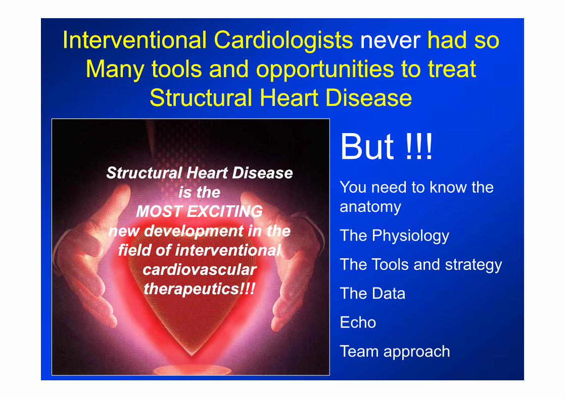
Interventional Cardiologists Interventional Cardiologists never never had so had so Many tools and opportunities to treat Many tools and opportunities to treat
Structural Heart DiseaseStructural Heart Disease
Structural Heart Disease Structural Heart Disease is the is the
MOST EXCITINGMOST EXCITINGnew development in the new development in the
field of interventional field of interventional cardiovascular cardiovascular therapeutics!!!therapeutics!!!
But !!! You need to know the anatomy
The Physiology
The Tools and strategy
The Data
Echo
Team approach

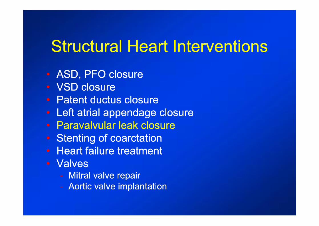
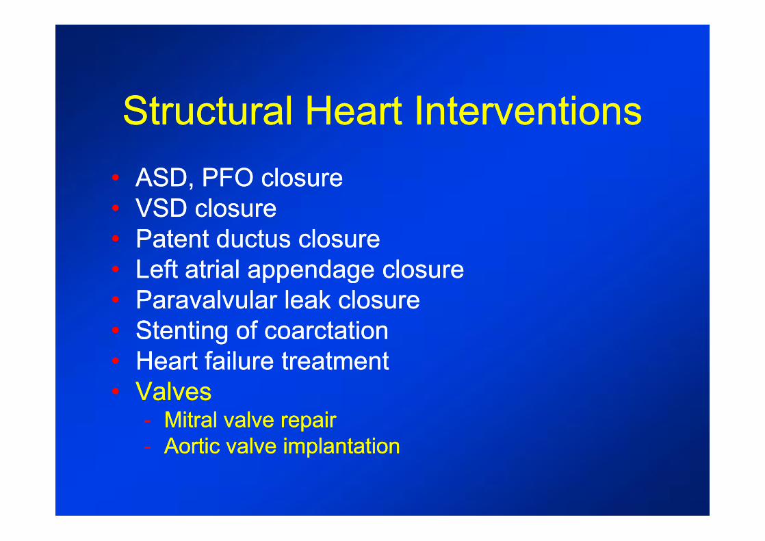
Structural Heart InterventionsStructural Heart Interventions•• ASD, PFO closureASD, PFO closure•• VSD closureVSD closure•• Patent ductus closurePatent ductus closure•• Left atrial appendage closureLeft atrial appendage closure•• Paravalvular leak closureParavalvular leak closure•• Stenting of coarctationStenting of coarctation•• LV apical aneurysm treatmentLV apical aneurysm treatment•• ValvesValves
-- Mitral valve repair Mitral valve repair -- Aortic valve implantationAortic valve implantation

The Atrial SeptumThe Atrial Septum
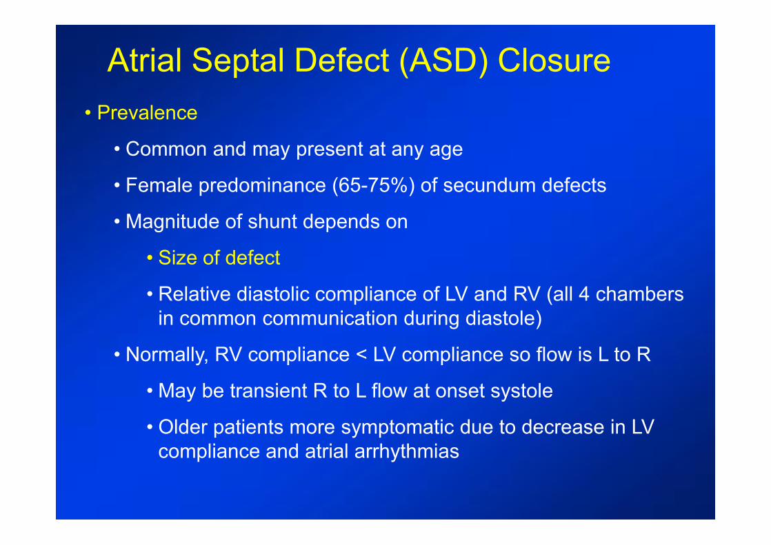
Atrial Septal Defect (ASD) Closure• Prevalence
• Common and may present at any age
• Female predominance (65-75%) of secundum defects
• Magnitude of shunt depends on
• Size of defect
• Relative diastolic compliance of LV and RV (all 4 chambers in common communication during diastole)
• Normally, RV compliance < LV compliance so flow is L to R
• May be transient R to L flow at onset systole
• Older patients more symptomatic due to decrease in LV compliance and atrial arrhythmias

Paradoxical Embolism via ASD in a 57 yo Man
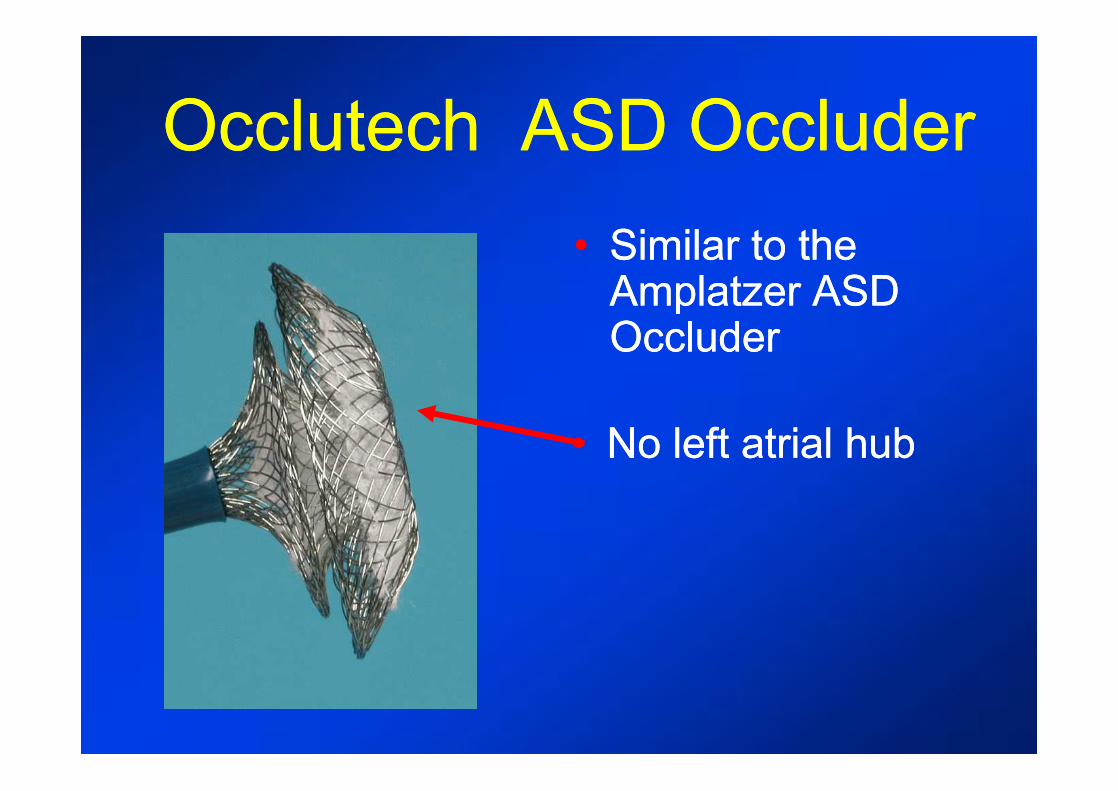
Occlutech ASD OccluderOcclutech ASD Occluder•• Similar to the Similar to the
Amplatzer ASD Amplatzer ASD OccluderOccluder
•• No left atrial hubNo left atrial hub
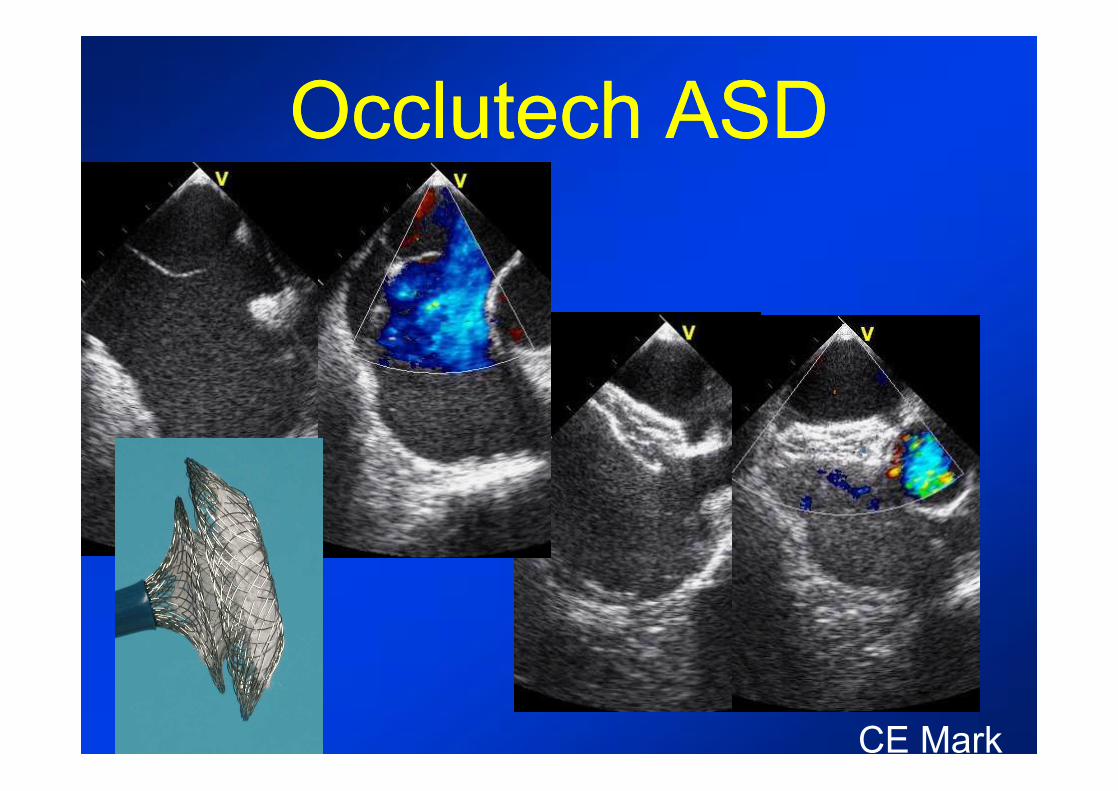
Occlutech ASDOcclutech ASD
CE Mark
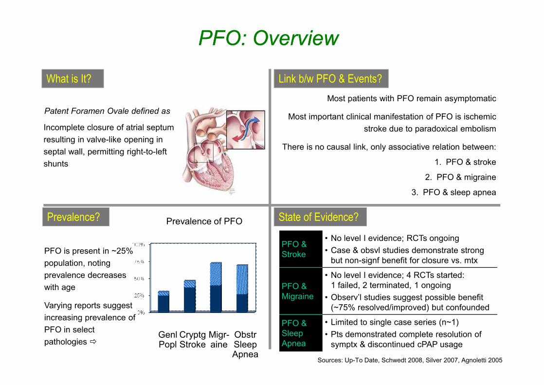
PFO: OverviewPFO: Overview
What is It? Link b/w PFO & Events?
State of Evidence?
PFO is present in ~25% population, noting prevalence decreases with age
Varying reports suggest increasing prevalence of PFO in select pathologies ð
Sources: Up-To Date, Schwedt 2008, Silver 2007, Agnoletti 2005
Prevalence of PFO
Genl Popl
Obstr Sleep Apnea
Patent Foramen Ovale defined as
Prevalence?
Migr-aine
Cryptg Stroke
Incomplete closure of atrial septum resulting in valve-like opening in septal wall, permitting right-to-left shunts
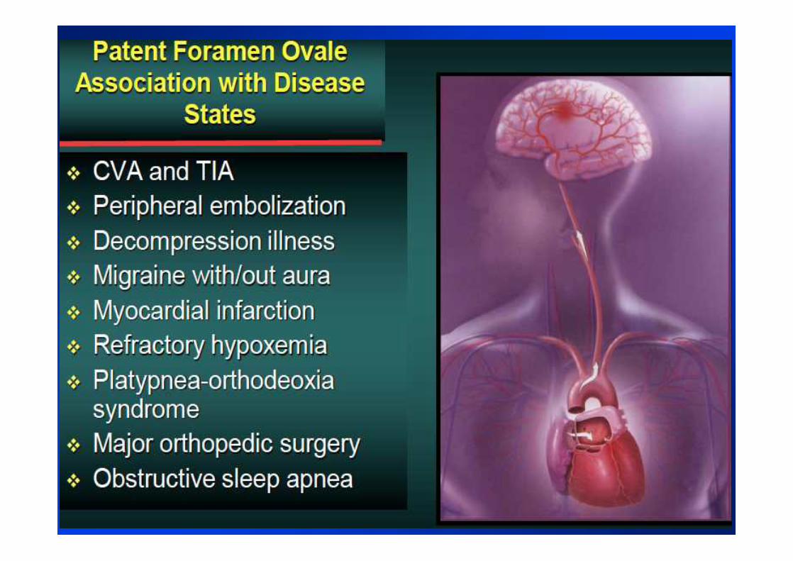
Most patients with PFO remain asymptomatic
Most important clinical manifestation of PFO is ischemic stroke due to paradoxical embolism
There is no causal link, only associative relation between:
1. PFO & stroke
2. PFO & migraine
3. PFO & sleep apnea
PFO & Stroke
• No level I evidence; RCTs ongoing• Case & obsvl studies demonstrate strong
but non-signf benefit for closure vs. mtx
PFO & Migraine
• No level I evidence; 4 RCTs started: 1 failed, 2 terminated, 1 ongoing
• Observ’l studies suggest possible benefit (~75% resolved/improved) but confounded
PFO & Sleep Apnea
• Limited to single case series (n~1)• Pts demonstrated complete resolution of
symptx & discontinued cPAP usage
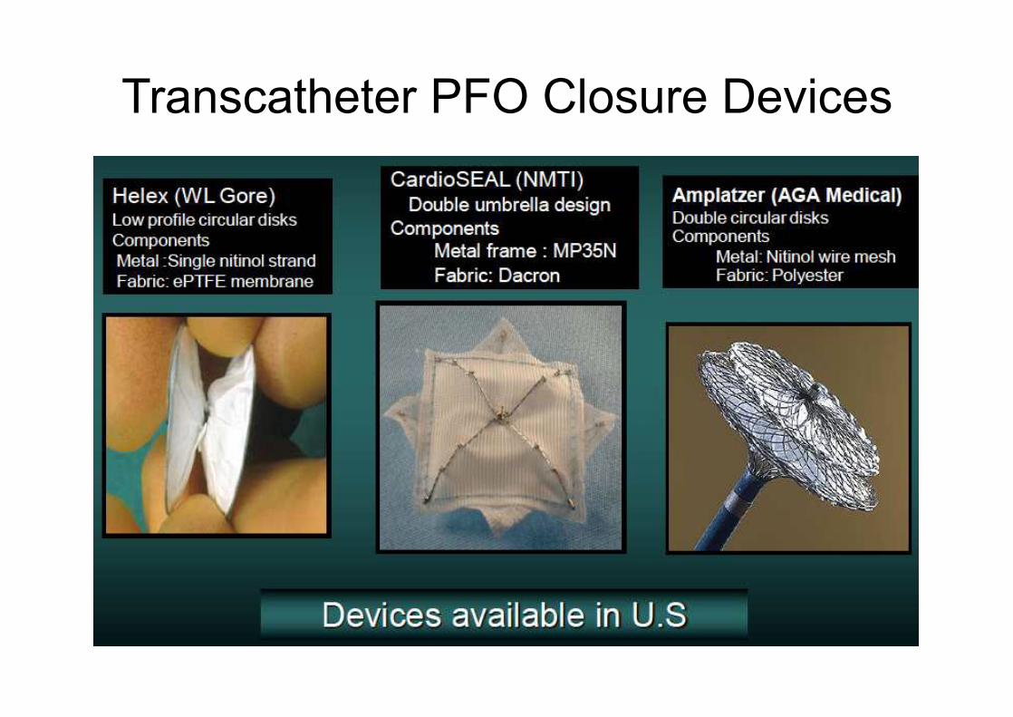
Transcatheter PFO Closure Devices
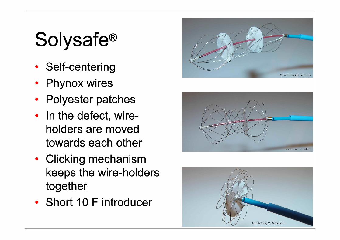
SolysafeSolysafe®®
•• SelfSelf--centeringcentering•• Phynox wires Phynox wires •• Polyester patches Polyester patches •• In the defect, wireIn the defect, wire--
holders are moved holders are moved towards each other towards each other
•• Clicking mechanism Clicking mechanism keeps the wirekeeps the wire--holders holders togethertogether
•• Short 10 F introducer Short 10 F introducer
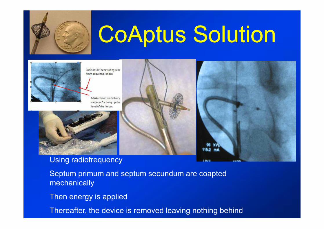
CoAptus SolutionCoAptus Solution
RALA
Using radiofrequency
Septum primum and septum secundum are coapted mechanically
Then energy is applied
Thereafter, the device is removed leaving nothing behind
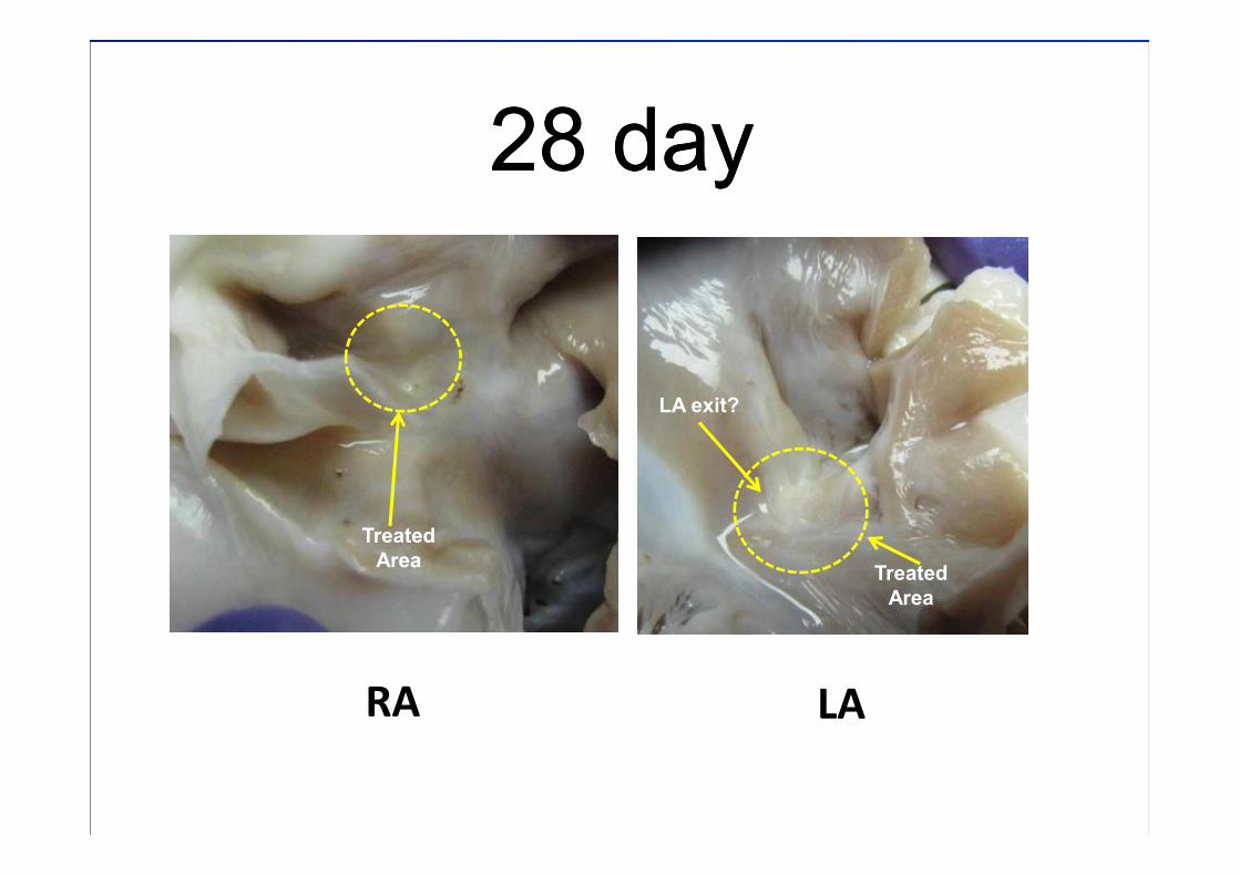
28 day28 day
LARA
Treated Area Treated
Area
LA exit?
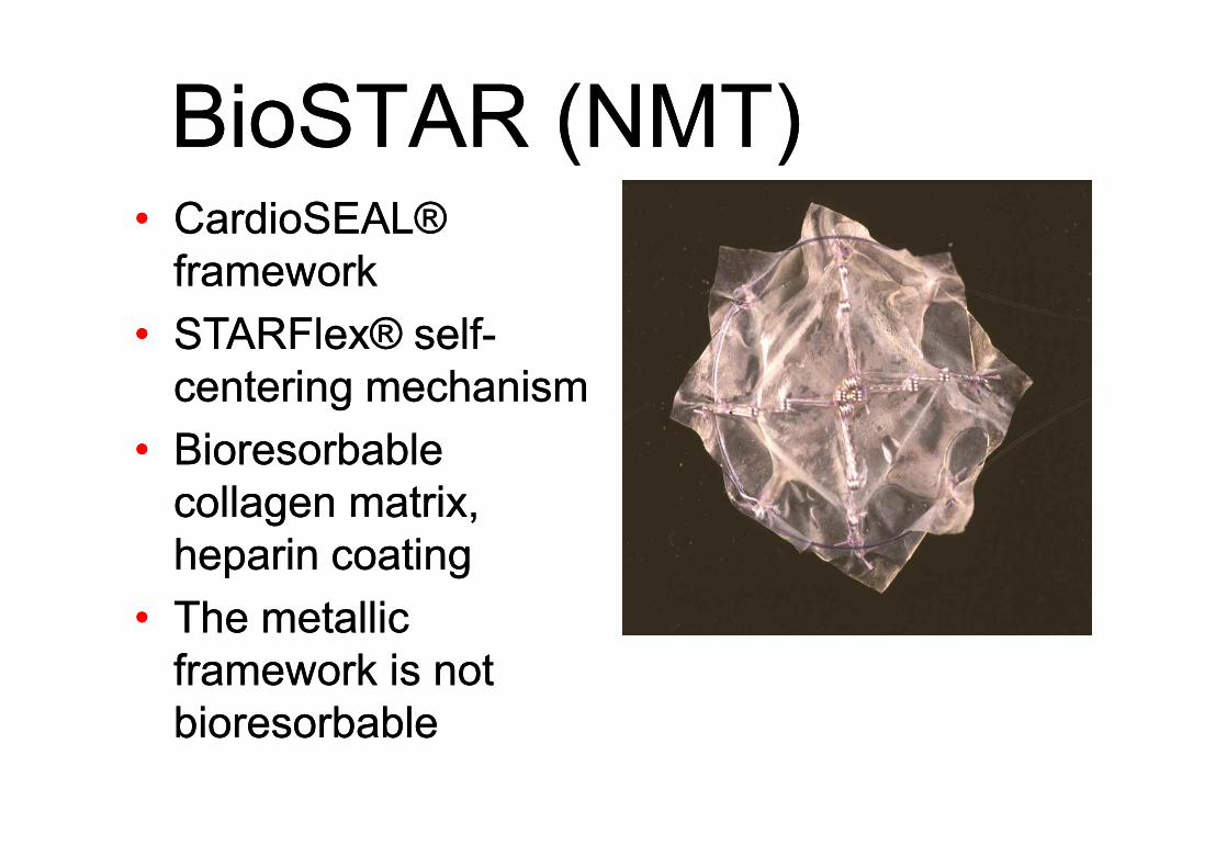
BioSTAR (NMT)BioSTAR (NMT)•• CardioSEAL® CardioSEAL®
frameworkframework•• STARFlex® selfSTARFlex® self--
centering mechanismcentering mechanism•• Bioresorbable Bioresorbable
collagen matrix, collagen matrix, heparin coatingheparin coating
•• The metallic The metallic framework is not framework is not bioresorbablebioresorbable
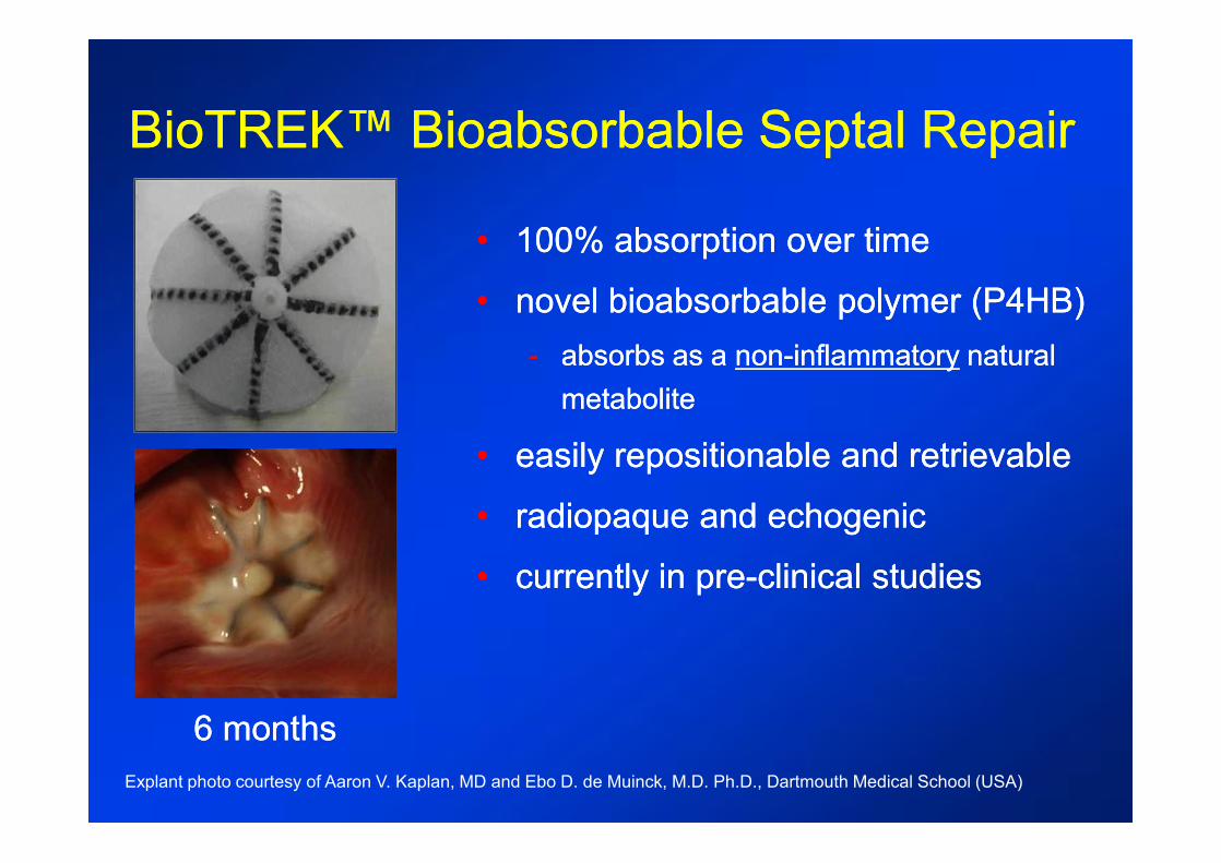
BioTREKBioTREK™ Bioabsorbable Septal Repair™ Bioabsorbable Septal Repair
6 months6 months
•• 100% absorption over time100% absorption over time
•• novel bioabsorbable polymer (P4HB)novel bioabsorbable polymer (P4HB)-- absorbs as a absorbs as a nonnon--inflammatoryinflammatory natural natural
metabolitemetabolite
•• easily repositionable and retrievableeasily repositionable and retrievable
•• radiopaque and echogenicradiopaque and echogenic
•• currently in precurrently in pre--clinical studiesclinical studies
Explant photo courtesy of Aaron V. Kaplan, MD and Ebo D. de Muinck, M.D. Ph.D., Dartmouth Medical School (USA)
20
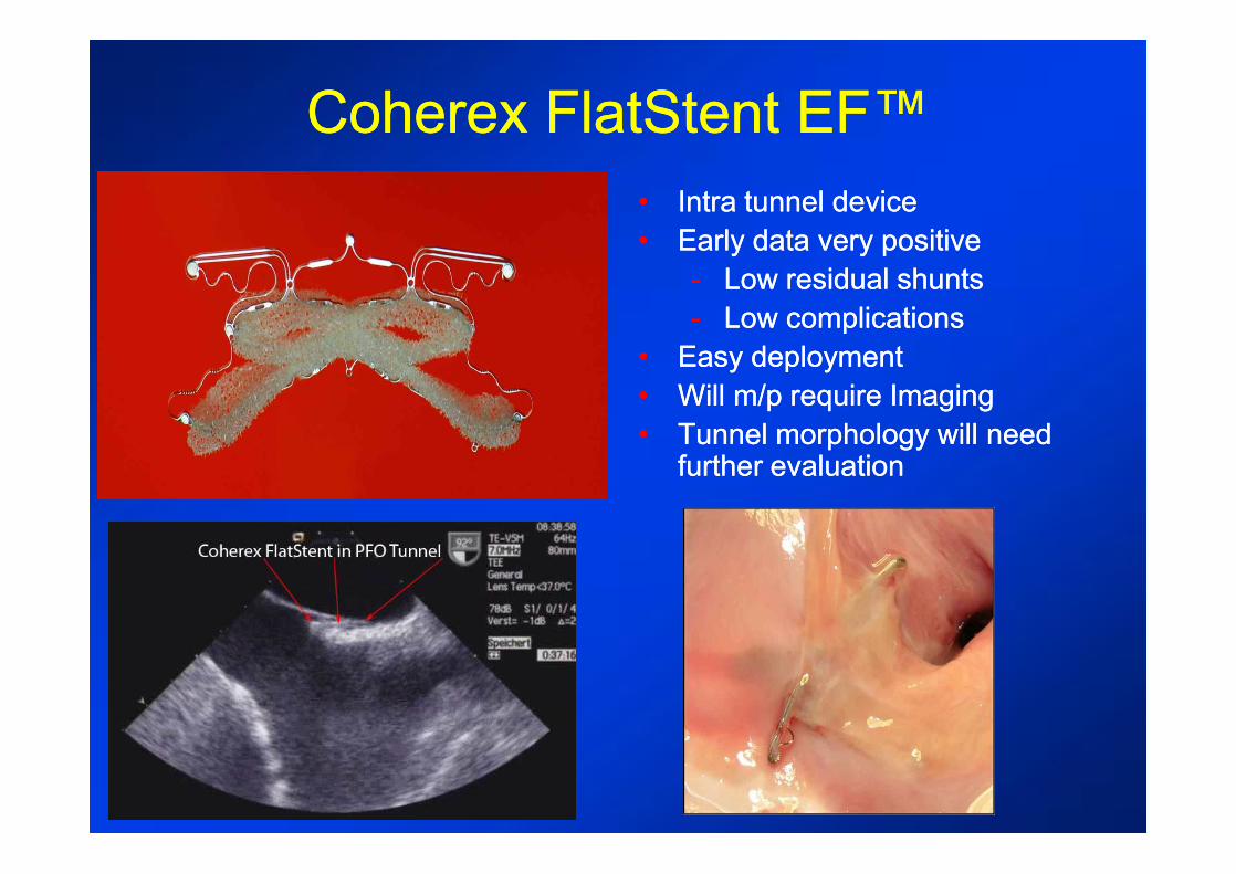
Coherex FlatStent EF™Coherex FlatStent EF™•• Intra tunnel deviceIntra tunnel device•• Early data very positive Early data very positive
-- Low residual shuntsLow residual shunts-- Low complicationsLow complications
•• Easy deploymentEasy deployment•• Will m/p require ImagingWill m/p require Imaging•• Tunnel morphology will need Tunnel morphology will need
further evaluationfurther evaluation

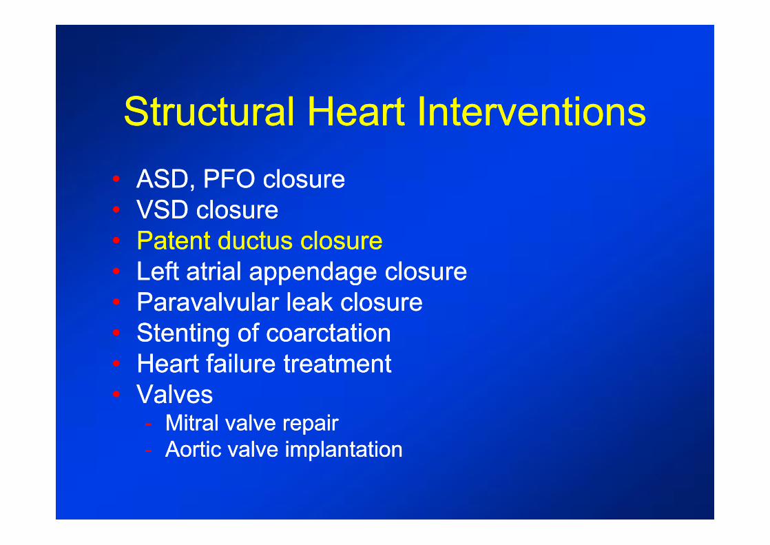
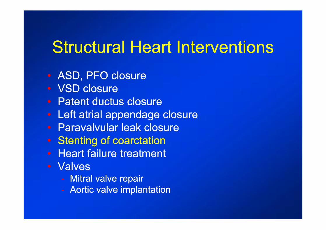
Structural Heart InterventionsStructural Heart Interventions•• ASD,ASD, PFO closurePFO closure•• VSD closureVSD closure•• Patent ductus closurePatent ductus closure•• Left atrial appendage closureLeft atrial appendage closure•• Paravalvular leak closureParavalvular leak closure•• Stenting of coarctationStenting of coarctation•• Heart failure treatmentHeart failure treatment•• ValvesValves
-- Mitral valve repair Mitral valve repair -- Aortic valve implantationAortic valve implantation

AIAITRTRPersistent Persistent AVAV--BlockBlock
Transient Transient AVAV--BlockBlock
Residual Residual ShuntShunt
(6 Mon FU)(6 Mon FU)SuccessSuccessnnStudyStudy
2.6%2.6%8%8%3%3%16.5%16.5%0%0%91.5%91.5%6868Li et al, 2005Li et al, 2005
3.5%3.5%12%12%8989Sun et al, Sun et al, 20052005
009.5%9.5%9.5%9.5%81%81%2626Anil et al, Anil et al, 20052005
2.6%2.6%2.62.6%%2.6%2.6%7.5%7.5%4%4%97.5%97.5%122122Carminati et Carminati et
al,2005al,2005
2.82.8%%2.8%2.8%91%91%3535Fu et al, Fu et al,
20062006
14%14%14%14%85.5%85.5%77Dajer et Dajer et al,2006al,2006
Transcatheter VSD Closure using the AGA Device
Courtesy TP Le

The PFM VSD CoilThe PFM VSD CoilThe PFM VSD CoilThe PFM VSD Coil
• Novel attachment mechanism • Stiff distal loops, covered with polyester filaments
5.5F delivery catheter; Distal Coil Diameter: 8,10,12,14 mm
ACT: 200 - 250 sec.
Courtesy TP Le

Occlusion of VSD using the PFM VSD Coil
Courtesy TP Le> 150 patients > 150 patients àà No AV BlockNo AV Block
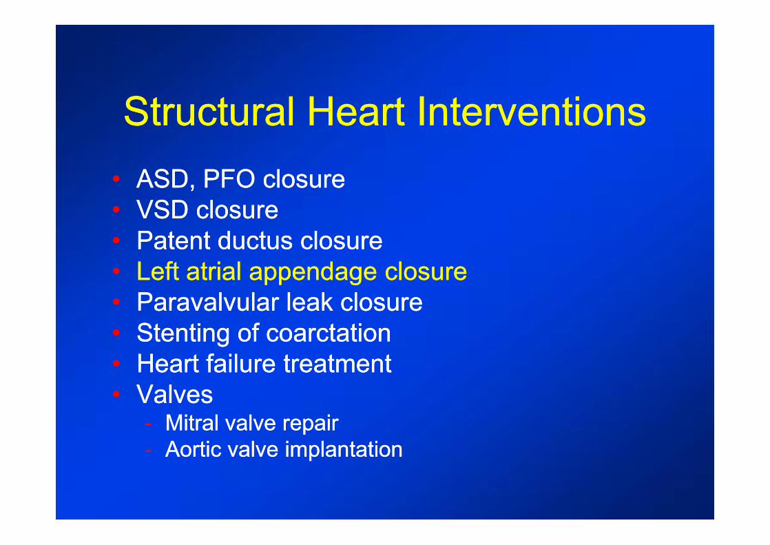
Structural Heart InterventionsStructural Heart Interventions•• ASD,ASD, PFO closurePFO closure•• VSD closureVSD closure•• Patent ductus closurePatent ductus closure•• Left atrial appendage closureLeft atrial appendage closure•• Paravalvular leak closureParavalvular leak closure•• Stenting of coarctationStenting of coarctation•• Heart failure treatmentHeart failure treatment•• ValvesValves
-- Mitral valve repair Mitral valve repair -- Aortic valve implantationAortic valve implantation

ADO-I
C: 4 to 14 mm
B: 5 to 8 mm
Sheath: 5 to 7 Fr
Amplatzer Duct Occluder ADOAmplatzer Duct Occluder ADO
ADO-II
A: 3 to 6 mm
Sheath: 4 to 5 Fr
ADO-II
ADO-I
CE Mark
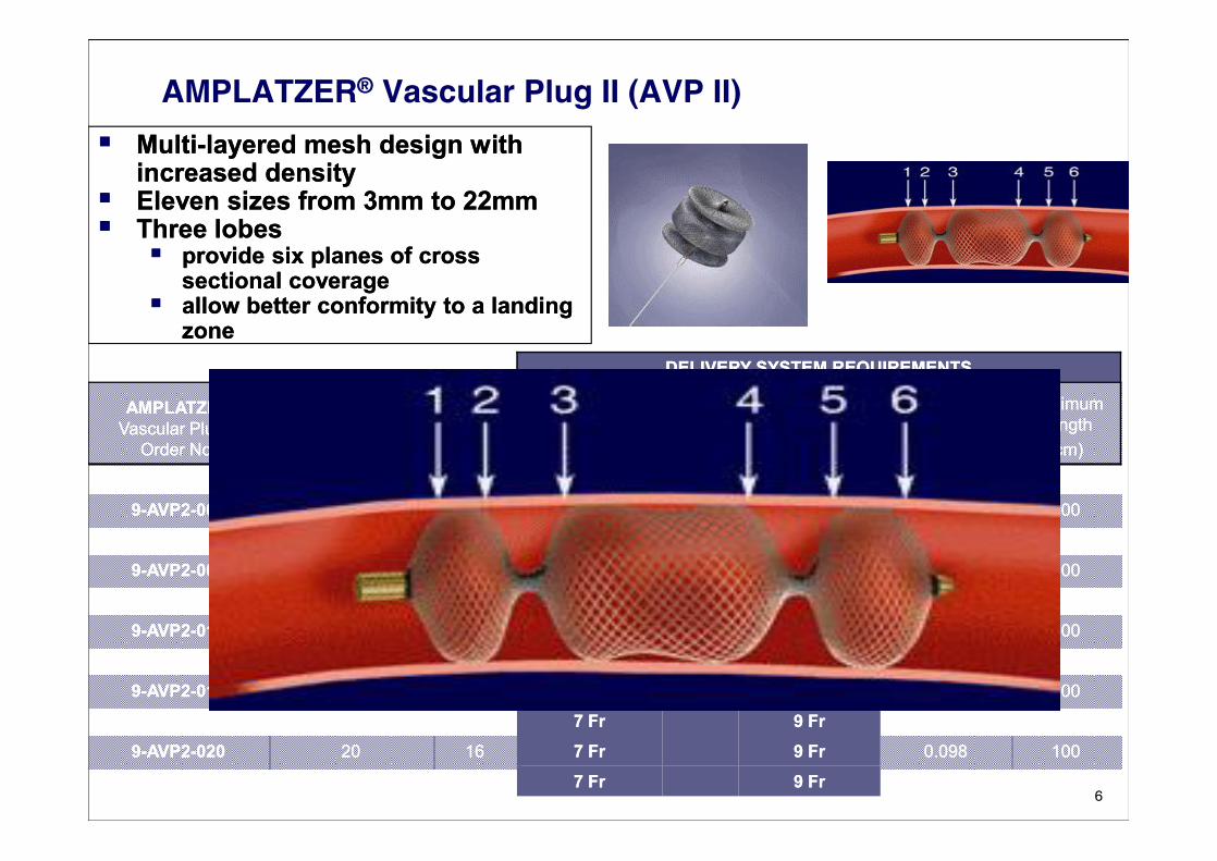
AMPLATZER® Vascular Plug II (AVP II)§§ MultiMulti--layered mesh design with layered mesh design with
increased densityincreased density§§ Eleven sizes from 3mm to 22mmEleven sizes from 3mm to 22mm§§ Three lobes Three lobes
§§ provide six planes of cross provide six planes of cross sectional coveragesectional coverage
§§ allow better conformity to a landing allow better conformity to a landing zonezone
6
DELIVERY SYSTEM REQUIREMENTSDELIVERY SYSTEM REQUIREMENTS
AMPLATZER AMPLATZER Vascular Plug II Vascular Plug II
Order No.Order No.
AMPLATZER AMPLATZER Vascular Plug II Vascular Plug II Diameter (mm)Diameter (mm)
Device Device Length Length (mm)(mm)
Sheath Sheath Minimum SizeMinimum Size OROR
Guide Guide CatheterCatheter
Minimum SizeMinimum Size
Minimum ID Minimum ID Required Required (inches)(inches)
Maximum Maximum LengthLength(cm)(cm)
99--AVP2AVP2--003003 33 66 4 Fr4 Fr 5 Fr5 Fr 0.0560.056 100100
99--AVP2AVP2--004004 44 66 4 Fr4 Fr 5 Fr5 Fr 0.0560.056 100100
99--AVP2AVP2--006006 66 66 4 Fr4 Fr 5 Fr5 Fr 0.0560.056 100100
99--AVP2AVP2--008008 88 77 4 Fr4 Fr 5 Fr5 Fr 0.0560.056 100100
99--AVP2AVP2--010010 1010 77 5 Fr5 Fr 6 Fr6 Fr 0.0700.070 100100
99--AVP2AVP2--012012 1212 99 5 Fr5 Fr 6 Fr6 Fr 0.0700.070 100100
99--AVP2AVP2--014014 1414 1010 6 Fr6 Fr 8 Fr8 Fr 0.0860.086 100100
99--AVP2AVP2--016016 1616 1212 6 Fr6 Fr 8 Fr8 Fr 0.0860.086 100100
99--AVP2AVP2--018018 1818 1414 7 Fr7 Fr 9 Fr9 Fr 0.0980.098 100100
99--AVP2AVP2--020020 2020 1616 7 Fr7 Fr 9 Fr9 Fr 0.0980.098 100100
99--AVP2AVP2--022022 2222 1818 7 Fr7 Fr 9 Fr9 Fr 0.0980.098 100100
Structural Heart InterventionsStructural Heart Interventions•• ASD,ASD, PFO closurePFO closure•• VSD closureVSD closure•• Patent ductus closurePatent ductus closure•• Left atrial appendage closureLeft atrial appendage closure•• Paravalvular leak closureParavalvular leak closure•• Stenting of coarctationStenting of coarctation•• Heart failure treatmentHeart failure treatment•• ValvesValves
-- Mitral valve repair Mitral valve repair -- Aortic valve implantationAortic valve implantation
Atrial FibrillationAtrial Fibrillation•• Is a major cause of strokeIs a major cause of stroke•• Thrombi develop in the left Thrombi develop in the left
atrial appendage (LAA)atrial appendage (LAA)•• LAA occlusion could be an LAA occlusion could be an
alternative to lifelong alternative to lifelong anticoagulationanticoagulation
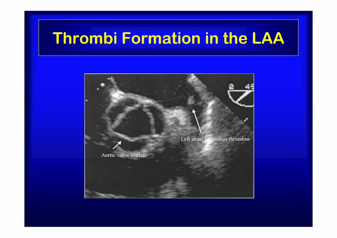
Thrombi Formation in the LAA
31
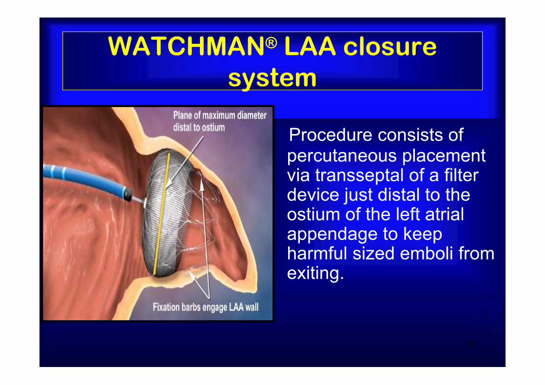
WATCHMAN® LAA closure system
Procedure consists of percutaneous placement via transseptal of a filter device just distal to the ostium of the left atrial appendage to keep harmful sized emboli from exiting.
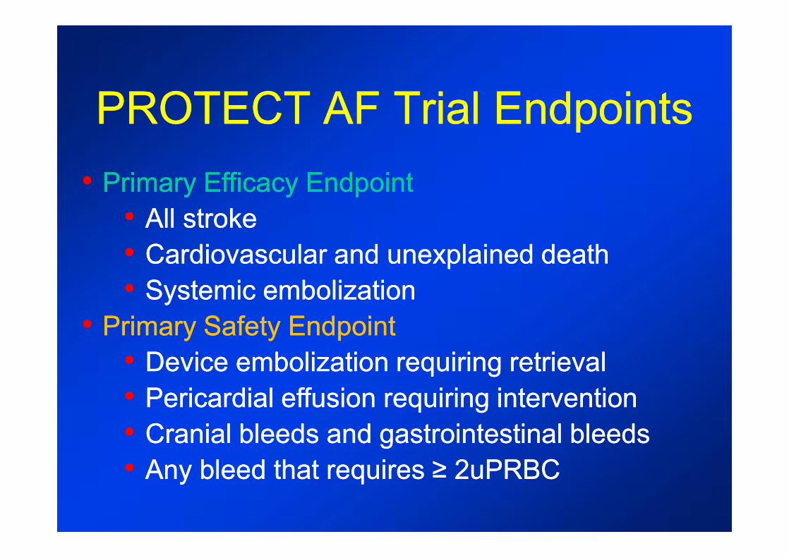
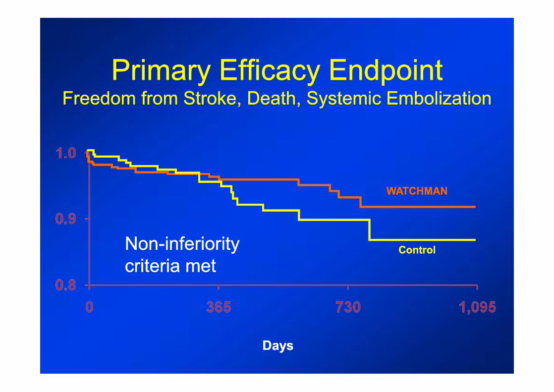
PROTECT AF Trial EndpointsPROTECT AF Trial Endpoints•• Primary Efficacy EndpointPrimary Efficacy Endpoint
•• All strokeAll stroke•• Cardiovascular and unexplained deathCardiovascular and unexplained death•• Systemic Systemic embolizationembolization
•• Primary Safety Endpoint Primary Safety Endpoint •• Device Device embolizationembolization requiring retrievalrequiring retrieval•• Pericardial effusion requiring interventionPericardial effusion requiring intervention•• Cranial bleeds and gastrointestinal bleedsCranial bleeds and gastrointestinal bleeds•• Any bleed that requires ≥ 2uPRBCAny bleed that requires ≥ 2uPRBC
WATCHMANWATCHMAN
ControlControl
Primary Efficacy EndpointPrimary Efficacy EndpointFreedom from Stroke, Death, Systemic EmbolizationFreedom from Stroke, Death, Systemic Embolization
DaysDays
Non-inferiority criteria metNon-inferiority criteria met
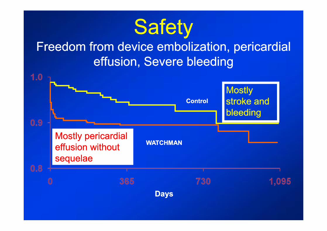
SafetySafetyFreedom from device embolization, pericardial Freedom from device embolization, pericardial
effusion, Severe bleedingeffusion, Severe bleeding
DaysDays
WATCHMANWATCHMAN
ControlControl
Mostly pericardial Mostly pericardial effusion without effusion without sequelaesequelae
Mostly Mostly stroke and stroke and bleedingbleeding

Other significant fndingsOther significant fndingsNoninferiorityNoninferiority for all strokes for all strokes
26% lower in device group26% lower in device groupSuperiority for hemorrhagic stroke Superiority for hemorrhagic stroke
91% lower in device group91% lower in device groupNoninferiorityNoninferiority for mortalityfor mortality
39% lower rate in device group39% lower rate in device groupMost events in the device group were Most events in the device group were
procedural effusions that decreased over procedural effusions that decreased over the course of the studythe course of the study
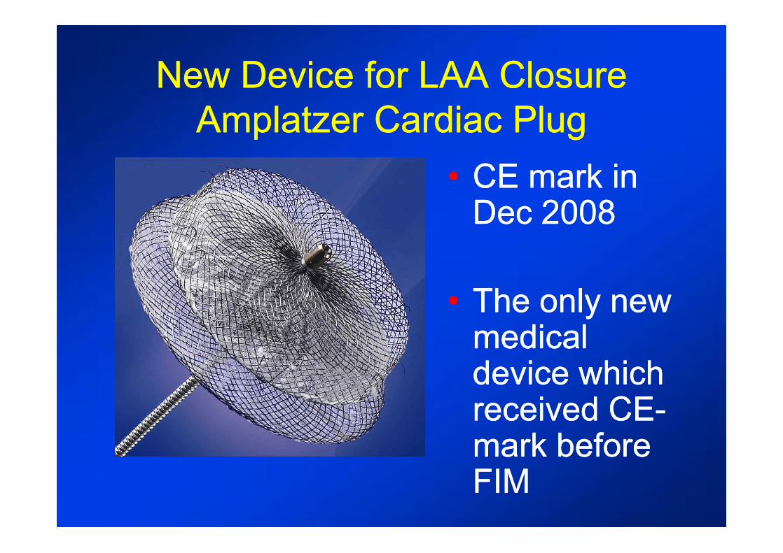
New Device for LAA ClosureNew Device for LAA ClosureAmplatzer Cardiac PlugAmplatzer Cardiac Plug
•• CE mark in CE mark in Dec 2008Dec 2008
•• The only new The only new medical medical device which device which received CEreceived CE--mark before mark before FIMFIM

Amplatzer Cardiac PlugAmplatzer Cardiac Plug
•Nitinol mesh and polyester patch•Lobe and a disc connected by a central waist•Disc is self-orienting•Available in 8 diameters sizes, 16, 18, 20, 22, 24, 26, 28, and 30 mm.
Structural Heart InterventionsStructural Heart Interventions•• ASD,ASD, PFO closurePFO closure•• VSD closureVSD closure•• Patent ductus closurePatent ductus closure•• Left atrial appendage closureLeft atrial appendage closure•• Paravalvular leak closureParavalvular leak closure•• Stenting of coarctationStenting of coarctation•• Heart failure treatmentHeart failure treatment•• ValvesValves
-- Mitral valve repair Mitral valve repair -- Aortic valve implantationAortic valve implantation
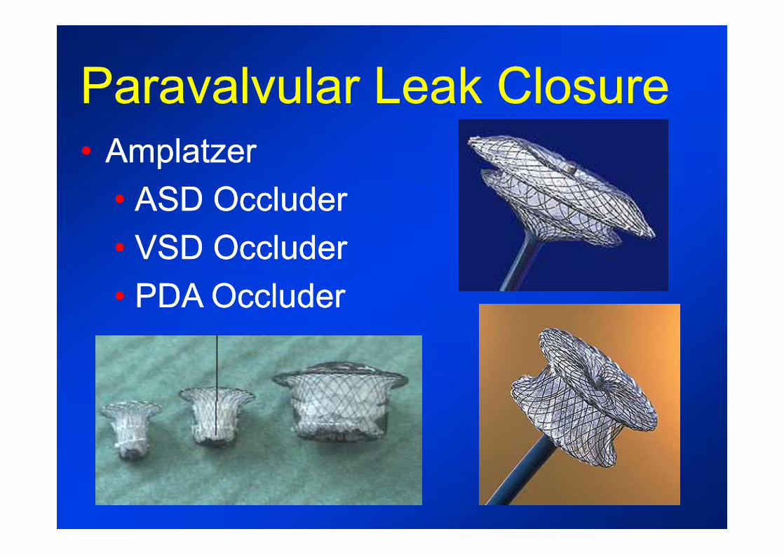
Paravalvular Leak ClosureParavalvular Leak Closure•• Amplatzer Amplatzer
•• ASD OccluderASD Occluder•• VSD OccluderVSD Occluder•• PDA OccluderPDA Occluder
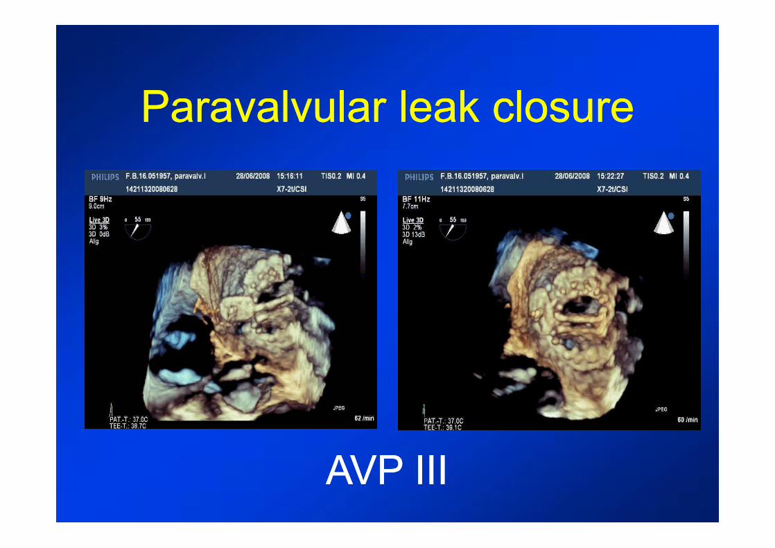
Paravalvular leak closureParavalvular leak closure
AVP IIIAVP III
Structural Heart InterventionsStructural Heart Interventions•• ASD,ASD, PFO closurePFO closure•• VSD closureVSD closure•• Patent ductus closurePatent ductus closure•• Left atrial appendage closureLeft atrial appendage closure•• Paravalvular leak closureParavalvular leak closure•• Stenting of coarctationStenting of coarctation•• Heart failure treatmentHeart failure treatment•• ValvesValves
-- Mitral valve repair Mitral valve repair -- Aortic valve implantationAortic valve implantation

AtriumTM Covered stent to prevent rupture and aneurysm
Multicenter Multicenter clinical trial has clinical trial has startedstarted
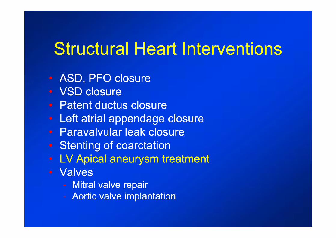
Structural Heart InterventionsStructural Heart Interventions•• ASD,ASD, PFO closurePFO closure•• VSD closureVSD closure•• Patent ductus closurePatent ductus closure•• Left atrial appendage closureLeft atrial appendage closure•• Paravalvular leak closureParavalvular leak closure•• Stenting of coarctationStenting of coarctation•• LV Apical aneurysm treatmentLV Apical aneurysm treatment•• ValvesValves
-- Mitral valve repair Mitral valve repair -- Aortic valve implantationAortic valve implantation
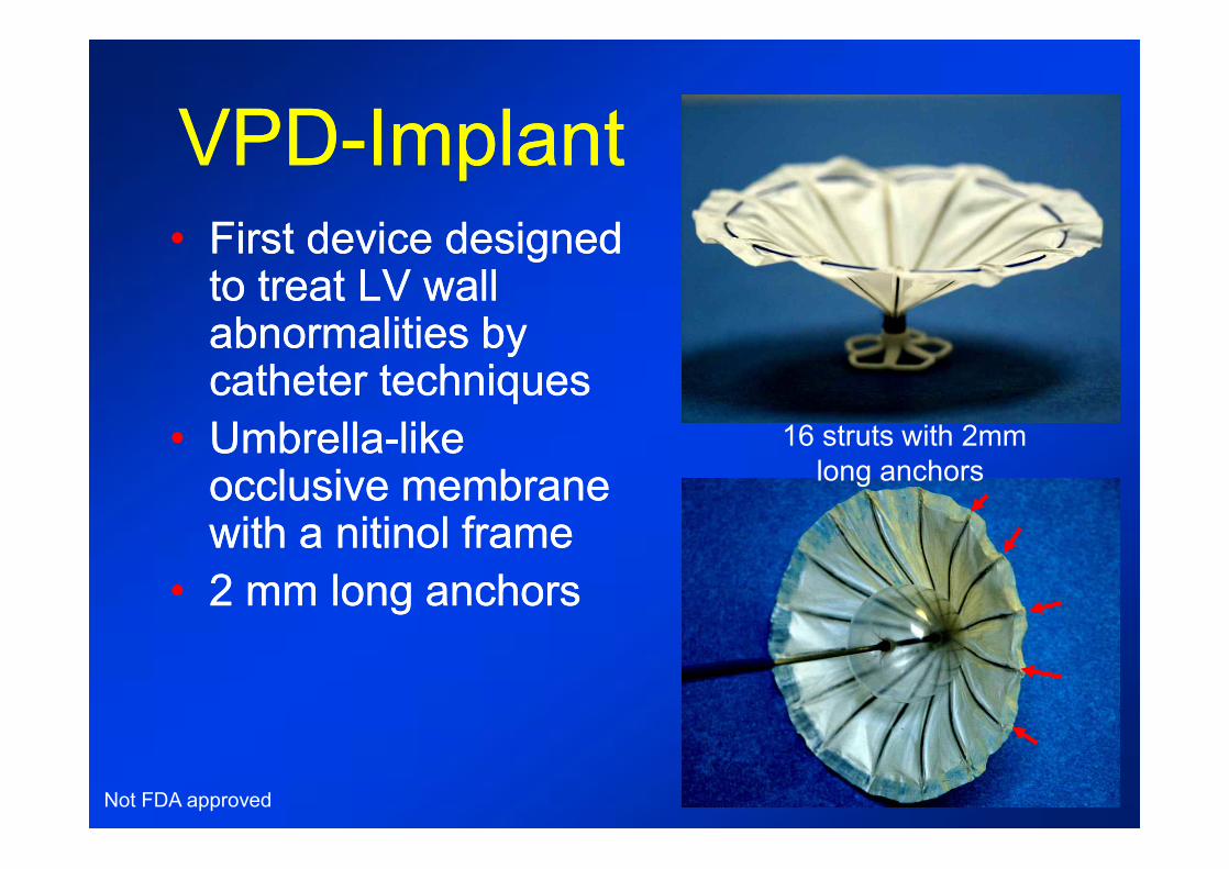
VPDVPD--ImplantImplant•• First device designed First device designed
to treat LV wall to treat LV wall abnormalities by abnormalities by catheter techniquescatheter techniques
•• UmbrellaUmbrella--like like occlusive membrane occlusive membrane with a nitinol framewith a nitinol frame
•• 2 mm long anchors2 mm long anchors
16 struts with 2mmlong anchors
Not FDA approved
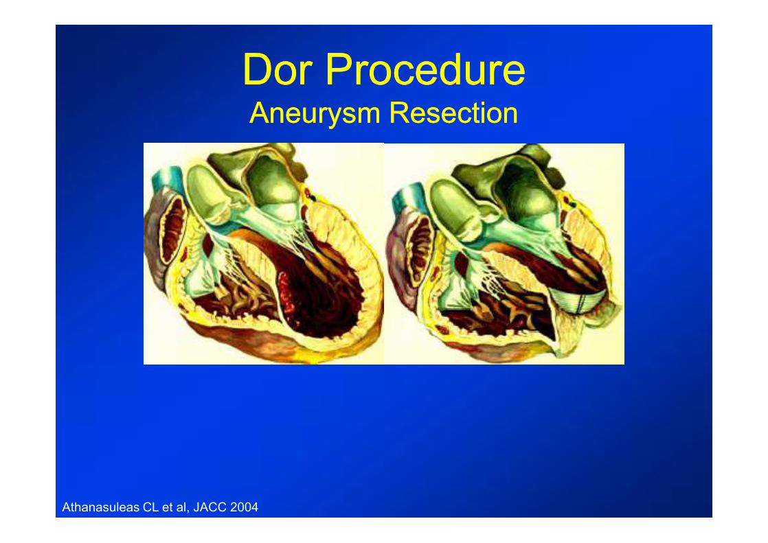
Dor ProcedureDor ProcedureAneurysm ResectionAneurysm Resection
Athanasuleas CL et al, JACC 2004

VPD ImplantVPD ImplantCT ScanCT Scan
beforebefore 6 months6 months
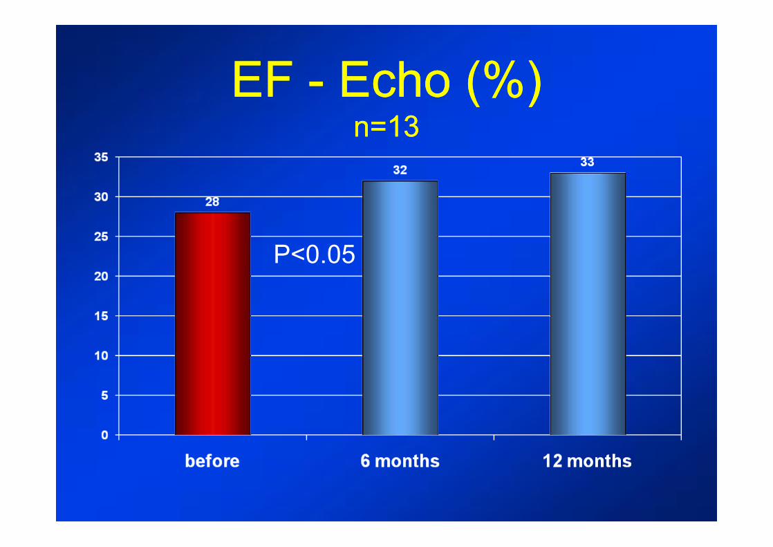
EF EF -- Echo (%)Echo (%)n=13n=13
P<0.05
Structural Heart InterventionsStructural Heart Interventions•• ASD,ASD, PFO closurePFO closure•• VSD closureVSD closure•• Patent ductus closurePatent ductus closure•• Left atrial appendage closureLeft atrial appendage closure•• Paravalvular leak closureParavalvular leak closure•• Stenting of coarctationStenting of coarctation•• Heart failure treatmentHeart failure treatment•• ValvesValves
-- Mitral valve repair Mitral valve repair -- Aortic valve implantationAortic valve implantation

4
Mitral RegurgitationMitral RegurgitationWhat is It?
What Causes It?
Risks?
How is it Treated?Two Distinct Etiologies
LV to LA Regurgitation
Primary
Functional
Muscle Damage/Loss
LV Remodeling/ Enlargement
Mitral Annulus Enlargement Causing Increased MR
Increased Load/Stress
Self perpetuating, MR begets MR
Intrinsic leaflet abnormalities
Incomplete coaptation caused by heart dilation
• Myxomatous degeneration• Rheumatic disease• Congenital
• Ischemic heart disease• Non-ischemic cardiomyopathy• Acute MI
1 million
2.6 million
May initiate or exacerbate heart failure
Incomplete leaflet coaptation or leaflet prolapse results in LV blood regurgitating into LA resulting in increased load and stress in the LV
LV
LA
Annuloplasty ring
Leaflet resection
Chordal transfer
Edge-to-edge
LV reshaping
Primary Functional
ü ü
ü
ü
ü
ü
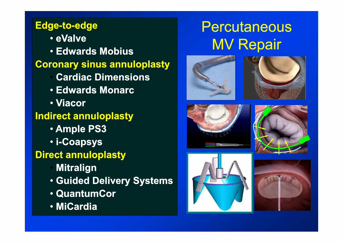
Percutaneous Percutaneous MV RepairMV Repair
EdgeEdge--toto--edgeedge•• eValveeValve•• Edwards Edwards MobiusMobius
Coronary sinus Coronary sinus annuloplastyannuloplasty•• Cardiac DimensionsCardiac Dimensions•• Edwards Edwards MonarcMonarc•• ViacorViacor
Indirect Indirect annuloplastyannuloplasty•• Ample PS3 Ample PS3 •• ii--CoapsysCoapsys
Direct Direct annuloplastyannuloplasty•• MitralignMitralign•• Guided Delivery SystemsGuided Delivery Systems•• QuantumCorQuantumCor•• MiCardiaMiCardia
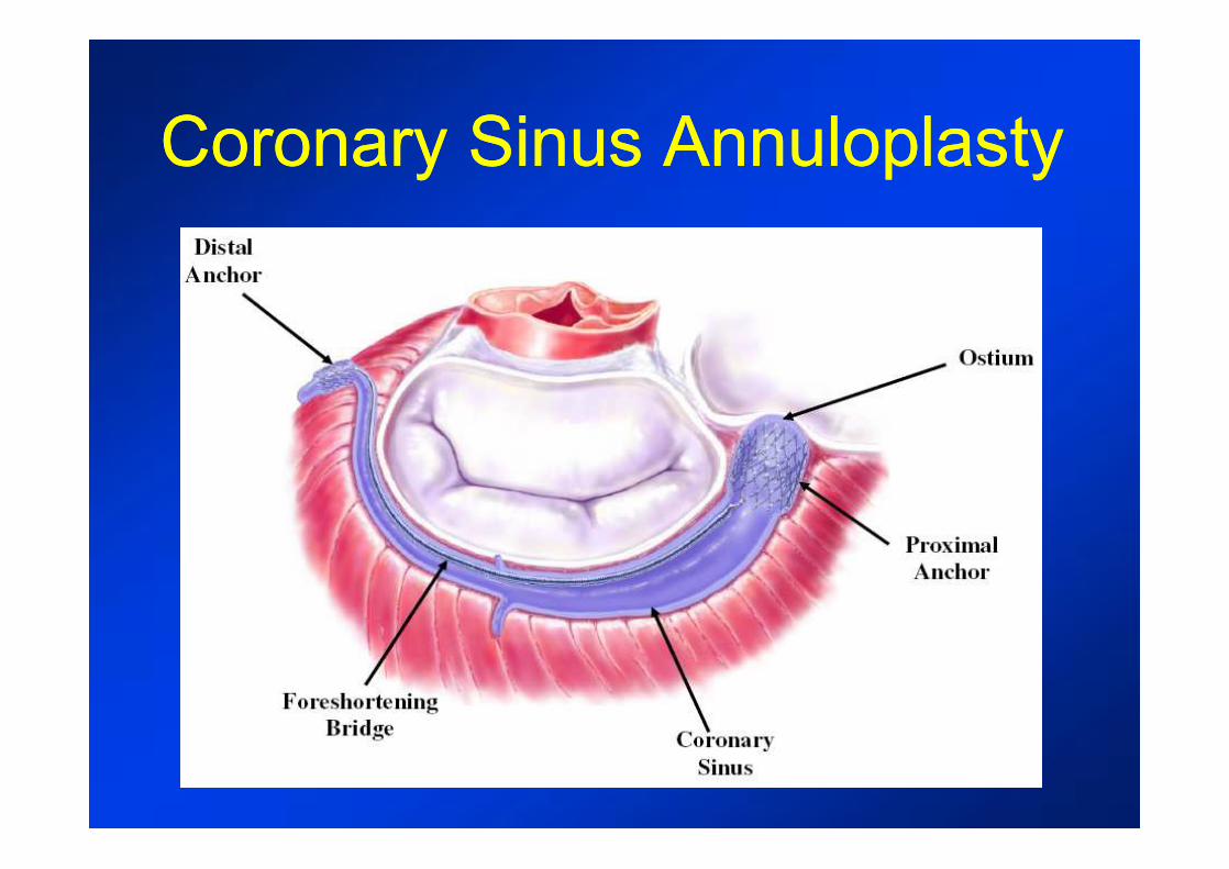
Coronary Sinus AnnuloplastyCoronary Sinus Annuloplasty
4Investigational Device only in the US; Not available for sale in the US
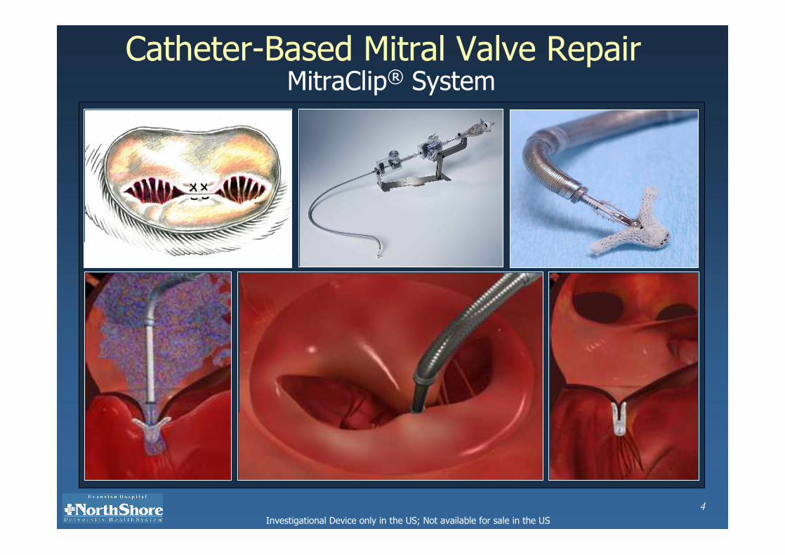
Catheter-Based Mitral Valve RepairMitraClip® System
5Investigational Device only in the US; Not available for sale in the US
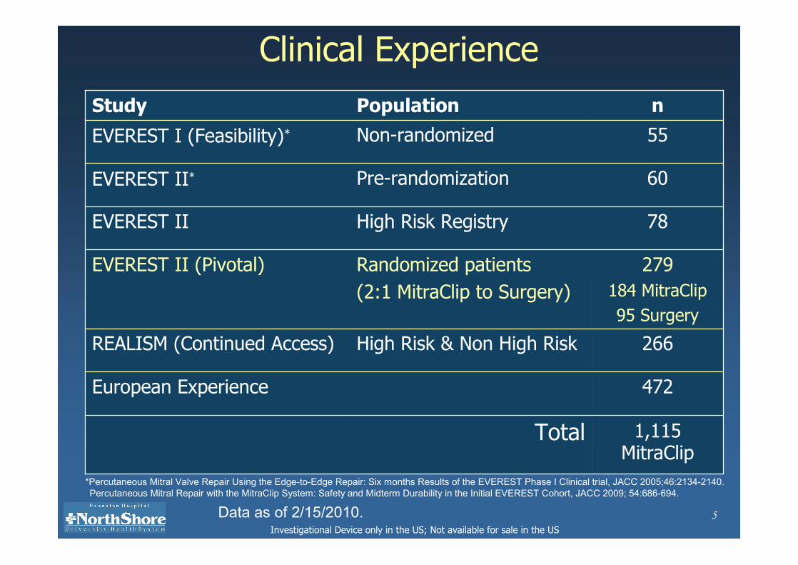
Clinical Experience
266High Risk & Non High RiskREALISM (Continued Access)
279184 MitraClip95 Surgery
Randomized patients(2:1 MitraClip to Surgery)
EVEREST II (Pivotal)
472European Experience
60Pre-randomizationEVEREST II*
78High Risk RegistryEVEREST II
nPopulationStudy
1,115 MitraClip
Total
55Non-randomized EVEREST I (Feasibility)*
Data as of 2/15/2010.
*Percutaneous Mitral Valve Repair Using the Edge-to-Edge Repair: Six months Results of the EVEREST Phase I Clinical trial, JACC 2005;46:2134-2140.Percutaneous Mitral Repair with the MitraClip System: Safety and Midterm Durability in the Initial EVEREST Cohort, JACC 2009; 54:686-694.
6Investigational Device only in the US; Not available for sale in the US
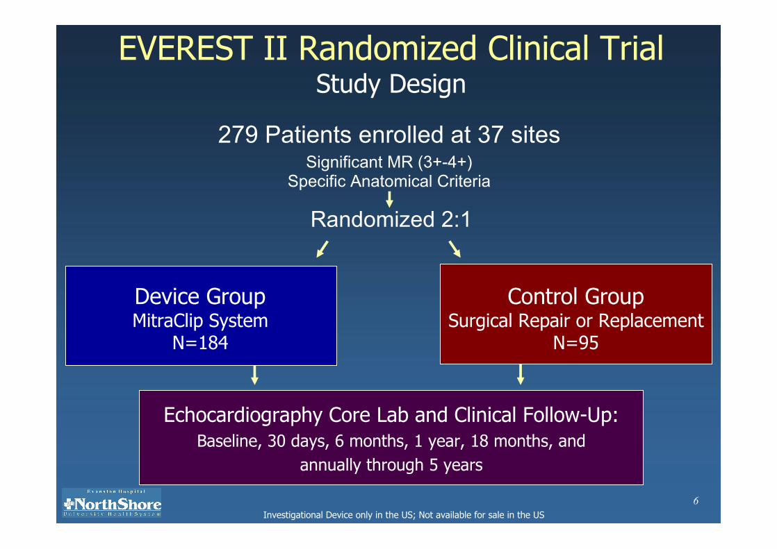
EVEREST II Randomized Clinical TrialStudy Design
279 Patients enrolled at 37 sites
Randomized 2:1
Echocardiography Core Lab and Clinical Follow-Up: Baseline, 30 days, 6 months, 1 year, 18 months, and
annually through 5 years
Control GroupSurgical Repair or Replacement
N=95
Significant MR (3+-4+)Specific Anatomical Criteria
Device GroupMitraClip System
N=184
17Investigational Device only in the US; Not available for sale in the US
0 20 40 60 80 100
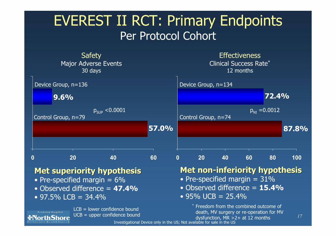
EVEREST II RCT: Primary EndpointsPer Protocol Cohort
0 20 40 60
9.6%
Device Group, n=136
Control Group, n=79
57.0%
Met superiority hypothesisMet superiority hypothesis• Pre-specified margin = 6% • Observed difference = 47.4%• 97.5% LCB = 34.4%
72.4%
87.8%
Control Group, n=74
Device Group, n=134
Met nonMet non--inferiority hypothesisinferiority hypothesis• Pre-specified margin = 31% • Observed difference = 15.4%• 95% UCB = 25.4%
SafetyMajor Adverse Events
30 days
EffectivenessClinical Success Rate*
12 months
LCB = lower confidence boundUCB = upper confidence bound
pSUP <0.0001 pNI =0.0012
* Freedom from the combined outcome of death, MV surgery or re-operation for MV dysfunction, MR >2+ at 12 months

Aortic Aortic StenosisStenosisWhat is It?
What Causes It?
Risk? Aortic stenosis is a slow-progressing disease with a long asymptomatic period.
Aortic stenosis causes obstruction of blood ejection from the left ventricle, is primarily caused by degeneration or calcification
40 50 60 70 80
20
40
60
80
100 Onset of symptoms
Survi
val (%
)
Years of Age
Asymptomatic latent period
40 50 60 70 80
20
40
60
80
100 Onset of symptoms
Survi
val (%
)
Years of Age
Asymptomatic latent period
MAJOR RISK FACTORS:
Male Elderly
LDL/HTN Diabetes
Smoking
symptom onset marks a critical point in the pathology.
• Degeneration and calcification of aortic leaflets prevents complete opening of valves
• Congenital condition (e.g., biscupid valve – earlier onset)
• Rheumatic fever (rare in industrialized countries, >AR)
Follow-Up (yrs)
100%
80%
60%
40%
20%
0 1 2 3 4 5
Survi
val (%
)
Valve replacementValve replacement
No surgeryNo surgeryp < 0.01
Follow-Up (yrs)
100%
80%
60%
40%
20%
0 1 2 3 4 5
Survi
val (%
)
Valve replacementValve replacement
No surgeryNo surgeryp < 0.01
How is it Treated Surgery is critical at the onset of symptoms

Approved TAVI Devices
CoreValve Revalving System™ : self-expandable
Edwards SAPIEN™ THV : balloon expandable
Nitinol frame
Porcine pericardial lealfet
26 and 29 mm inflow
Stainless steal frame
Bovine pericardial lealfet
23 and 26 mm
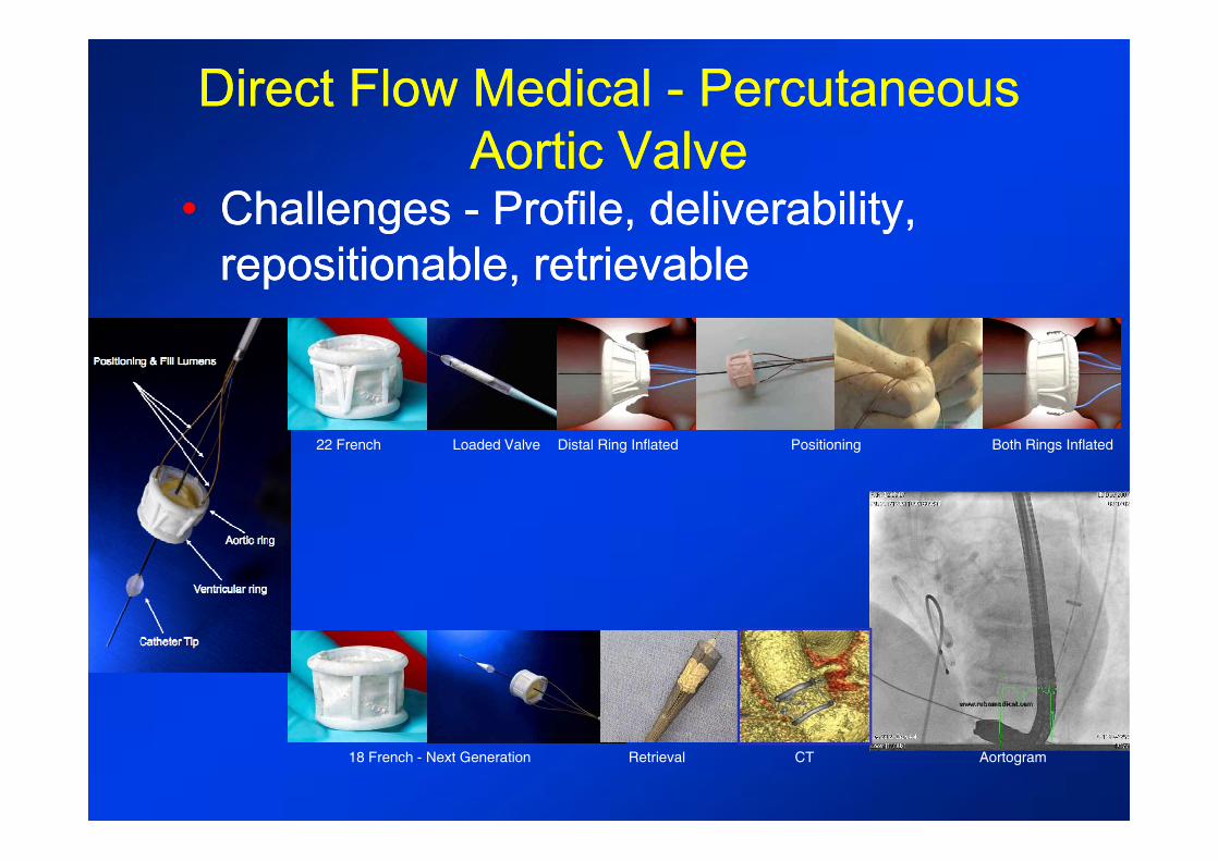
Direct Flow Medical Direct Flow Medical -- Percutaneous Percutaneous Aortic ValveAortic Valve
•• Challenges Challenges -- Profile, deliverability, Profile, deliverability, repositionable, retrievablerepositionable, retrievable
Distal Ring Inflated Both Rings Inflated
Retrieval
22 French
18 French - Next Generation
Loaded Valve Positioning
CT Aortogram
59
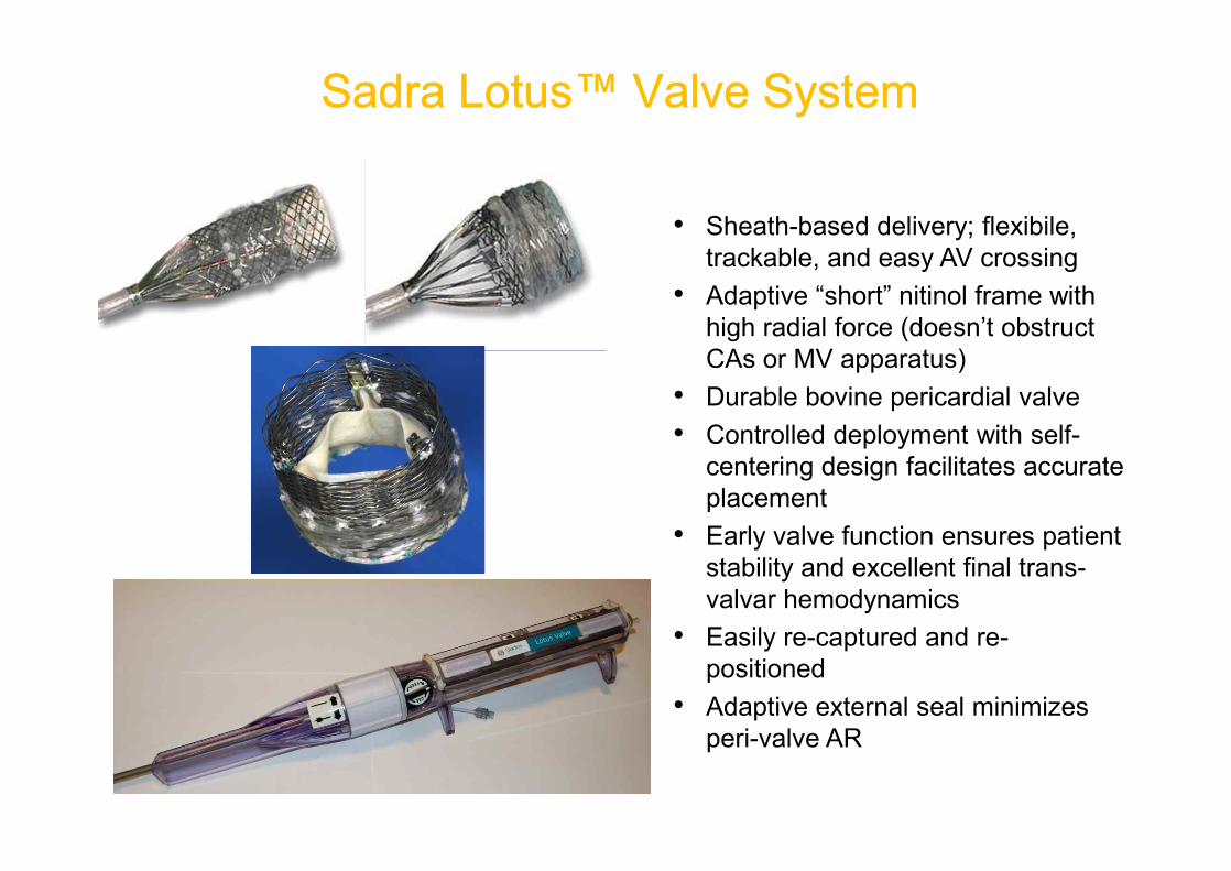
SadraSadra LotusLotus™™ Valve SystemValve System
• Sheath-based delivery; flexibile, trackable, and easy AV crossing
• Adaptive “short” nitinol frame with high radial force (doesn’t obstruct CAs or MV apparatus)
• Durable bovine pericardial valve• Controlled deployment with self-
centering design facilitates accurate placement
• Early valve function ensures patient stability and excellent final trans-valvar hemodynamics
• Easily re-captured and re-positioned
• Adaptive external seal minimizes peri-valve AR

New OpportunitiesNew OpportunitiesNew OpportunitiesNew Opportunities
Structural Heart Disease InterventionStructural Heart Disease Intervention
•• We are entering a new exciting era: lesserWe are entering a new exciting era: lesser--invasiveinvasivetranscatheter treatment of Structural heart diseasetranscatheter treatment of Structural heart disease..
•• There is a There is a clear unmet clinical needclear unmet clinical need –– many patients many patients with structural heart disease are poorly served with with structural heart disease are poorly served with either surgery or medical therapy either surgery or medical therapy
•• The explosion of The explosion of innovative devices and conceptsinnovative devices and conceptsenable us to provide a wide array of minimal invasive enable us to provide a wide array of minimal invasive solutions to structural diseasesolutions to structural disease
•• Multidisciplinary team approach Multidisciplinary team approach and innovative and innovative devices are key to the success of this programdevices are key to the success of this program
Hybrid Room
Thank you for your attention !Thank you for your attention !