structural insights into 1 frameshifting promoted by stem
TRANSCRIPT
Structural insights into +1 frameshifting promoted byexpanded or modification-deficient anticodonstem loopsTatsuya Maehigashi1, Jack A. Dunkle1, Stacey J. Miles, and Christine M. Dunham2
Department of Biochemistry, Emory University School of Medicine, Atlanta, GA 30322
Edited by Rachel Green, Johns Hopkins University, Baltimore, MD, and approved July 10, 2014 (received for review May 21, 2014)
Maintenance of the correct reading frame on the ribosomeis essential for accurate protein synthesis. Here, we reportstructures of the 70S ribosome bound to frameshift suppressortRNASufA6 and N1-methylguanosine at position 37 (m1G37) modi-fication-deficient anticodon stem loopPro, both of which cause theribosome to decode 4 rather than 3 nucleotides, resulting in a +1reading frame. Our results reveal that decoding at +1 suppressiblecodons causes suppressor tRNASufA6 to undergo a rearrangementof its 5′ stem that destabilizes U32, thereby disrupting the con-served U32–A38 base pair. Unexpectedly, the removal of them1G37 modification of tRNAPro also disrupts U32–A38 pairing ina structurally analogous manner. The lack of U32–A38 pairingprovides a structural correlation between the transition from ca-nonical translation and a +1 reading of the mRNA. Our structuresclarify the molecular mechanism behind suppressor tRNA-induced+1 frameshifting and advance our understanding of the roleplayed by the ribosome in maintaining the correct translationalreading frame.
near cognate | processivity
The three polymerase reactions of DNA replication, RNAtranscription, and protein translation are essential to life and
all involve a delicate balance between speed and fidelity. Thebacterial ribosome decodes three mRNA nucleotides into a sin-gle amino acid at a rate of ∼20 residues/s with high fidelity (103–104) (1, 2). High translational fidelity requires the strict main-tenance of the triplet mRNA reading frame with shifts in eitherthe 5′ or 3′ direction (positive or negative shifting, respectively)occurring infrequently (∼1 in 30,000) (3). Upon shifting of themRNA, the proteins expressed are nonsense polypeptides andtypically degraded (4). However, frameshifting rates may beunderestimated because of rapid halting of protein synthesis by thepostpeptidyl transfer quality control mechanism, which recognizesnear- and noncognate interactions in the P site (5). In this case, theidentification of such incorrect proteins would be difficult to detect.Although it was hypothesized almost 50 years ago that the
triplet genetic code was immutable, the identification of muta-gen-induced genomic insertions that still yielded the expressionof the correct protein implied otherwise (6–8). This alternativereading of mRNA indicated that the ribosome had the ability todecode a nontriplet reading frame under certain circumstances.It was reasoned that suppressors to these genomic insertions shouldbe located adjacent to the original mutation site. However, mostidentified frameshift suppressors were extragenic; genetic mappingshowed that the majority of these suppressors were nucleotideinsertions, deletions, and modifications in tRNA genes, typically atthe anticodon stem loop (ASL) (9, 10). Over the next several dec-ades, genetic characterization of these suppressors played a largerole in the fundamental understanding of tRNA structure, decod-ing, and other general features of ribosome function (reviewedin ref. 11).The first bacterial +1 suppressor sequenced was tRNASufD
(suppressor of frameshift D), a derivative of tRNAGlyCCC that
contained a cytosine insertion in its ASL and decoded an expandedglycine codon 5′-GGG-G-3′ (inserted guanosine follows theGGG glycine codon and is preceded by a hyphen; all codons aredenoted 5′ to 3′) (12). Given the Watson–Crick complementaritybetween the inserted nucleotide at the anticodon (C) and codon(G), a 4-bp interaction between the codon and anticodon wasproposed as the mechanistic basis for the +1 mRNA readingframe (13). Additional evidence to support this hypothesis in-cluded the isolation of suppressor tRNAs from both bacteria (7,12, 14–17) and yeast (18, 19), all containing additional nucleo-tides in their anticodon loops that had the ability to Watson–Crick pair with the corresponding expanded mRNA codon. This4-bp decoding or quadruplet model was further expanded uponto state that the size of the tRNA anticodon loop dictated themRNA length decoded by the ribosome rather than the numberof potential interactions that form between the tRNA–mRNApair (20). Subsequently, this elegant model, termed the “yard-stick” model, became dogma in textbooks.However, emerging evidence began to call both models into
question. The first indication that a 4-nt codon–anticodon in-teraction is not essential for +1 decoding was the identificationof suppressor tRNASufJ (a tRNAThr
GGU derivative), which lacksWatson–Crick complementarity between the inserted nucleotidein the anticodon stem and the additional residue in the mRNA(21, 22). The second indication was that mutational studies oftRNASufD and other +1 tRNA suppressors showed that Watson–
Significance
Biological fitness is dependent on the accurate flow of geneticinformation from DNA to mRNA to protein. Breakdown in ri-bosome translational fidelity is detrimental because of itscentral role in the production of proteins. Altering the 3-basegenetic code usually results in the expression of aberrant ornonsense proteins that are degraded. Here, we describe mo-lecular snapshots of the ribosome in the process of decodinga 4-base codon by a frameshift suppressor tRNA that results ina +1-nt shift of the mRNA reading frame. Conformational dy-namics of the anticodon stem loop seem to drive remodeling ofthe tRNA–mRNA interaction to promote the +1 movement,which we predict occurs after accommodation of the tRNAonto the ribosome.
Author contributions: T.M., J.A.D., and C.M.D. designed research; T.M., J.A.D., and S.J.M.performed research; T.M., J.A.D., and C.M.D. analyzed data; and T.M., J.A.D., and C.M.D.wrote the paper.
The authors declare no conflict of interest.
This article is a PNAS Direct Submission.
Data deposition: The crystallography, atomic coordinates, and structure factors have beendeposited in the Protein Data Bank, www.pdb.org (PDB ID codes 4L47, 1VVJ, 4L71, 4LEL,4LFZ, 4LNT, 4LSK, 4LT8, and 4P70).1T.M. and J.A.D. contributed equally to this work.2To whom correspondence should be addressed. Email: [email protected].
This article contains supporting information online at www.pnas.org/lookup/suppl/doi:10.1073/pnas.1409436111/-/DCSupplemental.
www.pnas.org/cgi/doi/10.1073/pnas.1409436111 PNAS Early Edition | 1 of 6
BIOCH
EMISTR
Y
Crick complementarity between the anticodon and codon wasnot essential for suppression of the +1 site (19, 23–26). More-over, the isolation of +1 suppressors with normal 7-nt anticodonloops illustrated that factors other than an increased loop sizemay influence frameshifting (27, 28).High-resolution X-ray crystal structures of the 70S ribosome
have provided molecular insights at multiple steps during proteinsynthesis (reviewed in ref. 29). Despite these advances, veryfew structural studies have been able to provide a molecular un-derstanding of mRNA frameshifting. Here, we investigated thestructural basis for +1 decoding by a well-studied suppressor,tRNASufA6 (17, 30). Frameshift suppressor tRNASufA6 is a derivativeof tRNAPro
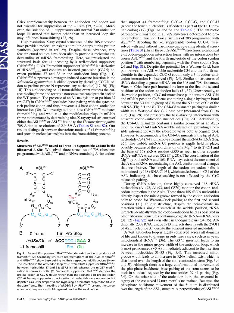
CGG and contains an inserted guanosine (G37.5) be-tween positions 37 and 38 in the anticodon loop (Fig. 1A).tRNASufA6 suppresses a mutagen-induced cytosine insertion in theSalmonella typhimurium histidine operon by decoding CCC-N co-don as proline (where N represents any nucleotide) (17, 30) (Fig.1B). This 4-nt decoding or +1 frameshifting event restores the cor-rect reading frame and reverts a nonsense truncated protein back tothe WT protein. The presence of an N1-methylation at position 37(m1G37) in tRNASufA6 precludes base pairing with the cytosine-rich proline codon and thus, prevents a 4-base codon–anticodoninteraction (30). We investigated both how tRNASufA6 mediatesframeshifting and what role the modification plays in mRNAframemaintenance by determining nineX-ray crystal structures ofeither the ASLSufA6 or ASLPro bound to the Thermus thermophilus70S A site at resolutions of 2.9–3.9 Å (Tables S1 and S2). Ourresults distinguish between the variousmodels of+1 frameshiftingand provide molecular insights into the frameshifting process.
ResultsStructures of ASLSufA6 Bound to Three +1 Suppressible Codons in theRibosomal A Site. We solved three structures of 70S ribosomesprogrammed with ASLSufA6 andmRNAs containing A-site codons
that support +1 frameshifting: CCC-A, CCC-G, and CCC-U(where the fourth nucleotide is decoded as part of the CCC pro-line codon) (17) (Figs. 1A and 2A and Table S1). The antibioticparomomycin was used in all 70S structures determined to pro-mote better diffraction. Two structures of 70S programmed withASLSufA6 decoding the +1 suppressible codon CCC-U weresolved with and without paromomycin, revealing identical struc-tures (Table S1). In all three 70S-ASLSufA6 structures, a canonical2-nt codon–anticodon interaction forms with no interactions be-tween ASLSufA6 and the fourth nucleotide of the codon (codonposition 7 with numbering beginning with the P-site codon) (Fig.2A and Fig. S1). Despite the potential for a Watson–Crick basepair between the ASL wobble nucleotide C34 and the fourth nu-cleotide in the expanded CCC-G codon, only a 3-nt codon–anti-codon interaction is observed (Fig. 2A). Similar to structures oftRNA decoding cognate mRNAs on the 30S and 70S ribosomes,Watson–Crick base pair interactions form at the first and secondpositions of the codon–anticodon helix (31, 32). Unexpectedly, atthe wobble position, a C•Cmismatch base pair between ASL C34and mRNA position C6 forms containing a single hydrogen bondbetween the N4 amino group of C34 and the N3 atom of C6 of themRNA (Fig. 2A andB). TheC34•C6mismatch pairing is a similarwidth as a Watson–Crick C-G base pair (measured from Cl′ toC1′) (Fig. 2B) and preserves the base-stacking interactions withadjacent codon–anticodon nucleotides (Fig. 2A). Additionally,the C34•C6 mismatch contains a similar geometry as an ASL-modified cmo5U•U mRNA wobble interaction, providing a pos-sible rationale for why the ribosome views both as cognate (33).However, to accommodate the C34•C6 mismatch, the tip of ASLnucleotide C34 (N4 atom)moves toward themRNAby 1.6Å (Fig.2C). The wobble mRNA C6 position is rigidly held in place,possibly because of the coordination of a Mg2+ to its 2′-OH andthe base of 16S rRNA residue G530 as seen in other cognatetRNA–mRNA structures (32) (Fig. 2D). The coordination of theMg2+ by bothmRNAand 16S rRNAmay restrict themovement ofthe A-site mRNA, necessitating the ASL conformational changesthat we observe. The length of the codon–anticodon helix ismaintained by 16S rRNA C1054, which stacks beneath C34 of theASL, indicating that base stacking is not affected by the C•Cmismatch pairing.During tRNA selection, three highly conserved 16S rRNA
nucleotides (A1492, A1493, and G530) monitor the codon–anti-codon interaction in the A site. These three 16S rRNA nucleotidesdirectly inspect the minor groove formed by the codon–anticodonhelix to probe for Watson–Crick pairing at the first and secondpositions (31). In our structure, despite the near-cognate in-teraction with a single mismatch at the wobble position, rRNAinteracts identically with the codon–anticodon helix as observed inother ribosome structures containing cognate tRNA–mRNA pairs(31, 32) (Fig. S2) and even other near-cognate pairs (34, 35). Ad-ditionally, 23S rRNA residue 1913 interacts directly with the 2′-OHof ASL nucleotide 37, despite the adjacent inserted nucleotide.A 7-nt anticodon loop is highly conserved across all domains
of life and known to diverge in only rare cases, such as in yeastmitochondrial tRNAThr (36). The G37.5 insertion leads to anincrease in the minor groove width of the anticodon loop, whichis most pronounced (∼5 Å) immediately adjacent to the insertionbetween nucleotides 31–33 (Fig. 3A). This increased minorgroove width leads to an increase in RNA helical twist, which isdistributed over the length of the entire anticodon stem (Fig. 3 Aand B). Although there is a large conformational movement ofthe phosphate backbone, base pairing of the stem seems to beback in standard register by the nucleotides 29–41 pairing (Fig.3A). On the other side of the anticodon loop, the structural in-tegrity of the conserved U-turn motif is maintained. Because thephosphate backbone movement of the 5′ stem is distributedover the length of the ASL, structural superpositioning of ASLSufA6
Fig. 1. Frameshift suppressor tRNASufA6 decodes a 4-nt codon to produce a +1frameshift. (A) Secondary structure representations of the ASLs of tRNAPro
and tRNASufA6 show base pairing to their respective mRNA codons (blue).The insertion in the anticodon loop of +1 frameshift suppressor tRNASufA6 isbetween nucleotides 37 and 38; G37.5 is red, whereas the m1G37 modifi-cation is shown in both. (B) Frameshift suppressor tRNASufA6 decodes theproline codon as CCC-U (blue) rather than the cognate 3-nt proline codonCCC (0 frame), suppressing the insertion N nucleotide (any nucleotide butdepicted as a U for simplicity) and bypassing a premature stop codon UGA inthe zero frame. The +1 reading of hisD3018 by tRNASufA6 restores the correctamino acid sequence with Glu (green) read as the next codon.
2 of 6 | www.pnas.org/cgi/doi/10.1073/pnas.1409436111 Maehigashi et al.

onto ASLPro reveals little variation in its global position (Fig. 3Aand Fig. S3).
ASLSufA6 Undergoes a Context-Dependent Conformational Change atU32. Important tertiary features govern ASL stability, includinga hydrogen bond between the O2 of a pyrimidine at position 32and the N6 of A38 (Fig. 3 B and C). The insertion of G37.5 inASLSufA6 impedes the formation of the U32–A38 interaction,because G37.5 occupies the equivalent physical position of A38in the ASL (Fig. 3A). Despite excellent electron density for mostof the defining features of the ASL in all three structures of 70S-ASLSufA6 decoding +1 suppressible codons, there is a distinctabsence of electron density for U32, which is located on theopposite side of the loop from the G37.5 insertion (Fig. 4A).To determine if this disordering ofU32 is an inherent property of
ASLSufA6, we solved a structure of 70S ribosome-ASLSufA6 de-coding a cognate proline codon (CCG) (Fig. 4B and Table S1).Surprisingly, in this mRNA–ASL context, the electron density forU32 is clearly visible (Fig. 4B). The presence of U32 electrondensity in this context indicates that disordering of U32 is not in-herently caused by the G37.5 insertion in tRNASufA6. Rather, it isthe combination of the suppressor tRNA and mRNA sequencecontext that seems to drive the observed flexibility of U32.We next examined whether the disordering of U32 is struc-
turally characteristic of the +1 suppressible or C•C near-cognate
base pair. We solved two structures of 70S bound to ASLPro,which contains a 7-nt anticodon loop and an m1G37 modifica-tion, decoding both +1 suppressible codons CCC-G and CCC-U(Fig. S4 and Table S2). An identical C34•C6 mismatch pair
Fig. 2. Details of the interaction of ASLSufA6 with mRNA in the ribosomal Asite. (A) Watson–Crick base pairs at the first two positions of the A-site codon(C4 and C5) form when ASLSufA6 decodes a CCC-N codon, whereas a mis-match interaction occurs between ASL C34•C6 codon at the wobble position(shown with codon G7; codon numbering starts from the P-site codon). Theinserted nucleotide G37.5 is 3′ of the anticodon (red), whereas the nucleo-tide that lacks any electron density (U32) is shown in blue. (B) A detailedview of the base pairing between C34 of ASLSufA6 and the C6 or wobbleposition of the mRNA. (Upper) ASLSufA6 forms the C•C mismatch pair in thecontext of a +1 frameshift CCC-G codon but (Lower) can also form a Watson–Crick C-G base pair with a cognate CCG codon. 2Fo-Fc electron densitymap contoured at 1.0 σ is shown in blue. The overall width of the C34•C6mismatch and C34–G6 base pair is similar at 10.7 and 11.0 Å, respectively, asmeasured between C1′ atoms as depicted by the bar. (C) A comparison ofthe position of ASLSufA6 C34 when decoding either a +1 suppressible codon(purple) or a cognate proline codon (green) shows an ∼1.6 Å movement ofC34 (N4 of the base) to allow the C34•C6 pairing. (D) Direct coordinationbetween the 2′-OH of C6 of the mRNA, a Mg2+ ion (green sphere), and 16SrRNA residue G530 (gray) fixes the position of C6 requiring C34 movement toaccommodate the C•C pair. Fo-Fc difference electron density maps are con-toured at 3 σ (green mesh), indicating the presence of the Mg2+ ion.
Fig. 3. Remodeling of ASLSufA6 structure induced by the G37.5 insertion. (A)Overview of the 8-nt anticodon loop of ASLSufA6 (green) compared with thecanonical 7-nt anticodon loop of ASLPro (gray). The G37.5 insertion (red)causes a widening of the ASL minor groove by ∼5 Å between nucleotides 30and 32 located on the opposite side of the insertion site. In ASLSufA6, G31 andU32 span the distance of 3 nt (G37.5, A38, and C39) on the 3′ side of the ASL;however, in canonical tRNAs, such as tRNAPro, G31 and U32 span only 2 nt(A38 and C39) on the 3′ side of the ASL. In both cases, the G31–C39 base pairforms the boundary of the base-paired stem region. The ribose and base ofASLSufA6 nucleotide U32 (blue) are not observed in electron density maps,although its phosphate and the following phosphate are visible, allowing itsposition to be approximated. (B) Secondary representations of both ASLPro
and ASLSufA6 show that expansion of the ASLSufA6 anticodon loop to 8 ntabrogates the conserved hydrogen bond between U32 and A38 (boxed) andalters the incline of base pairs in ASLSufA6. (C) The U32–A38 interaction inASLPro (gray) shown alongside U32 (blue) and G37.5 (red) of ASLSufA6, em-phasizing the lack of interaction.
Maehigashi et al. PNAS Early Edition | 3 of 6
BIOCH
EMISTR
Y

forms at the wobble position while electron density for U32 isobserved (Fig. S5). These results indicate that U32 disordering isthe consequence of a specific context: the noncanonical 8-ntanticodon loop of ASLSufA6 binding to a suppressible codon withthe pattern CCC-N within the ribosomal A site.
Modification State of G37 in ASLPro Also Controls U32 Dynamics.RNA modifications of tRNAs are widespread, and ASL mod-ifications are known to influence decoding (37). Both the anti-codon nucleotides 34 and 37 are highly modified, and the lackof modifications increases frameshifting (38, 39). Modificationsat the ASL wobble nucleotide 34 mainly facilitate unusual basepairing to expand the genetic code (33, 37, 40), whereas nucle-otide 37 modifications participate in stacking interactions withother anticodon nucleotides and may preorder anticodon nu-cleotide 36 for mRNA recognition (41, 42). Specifically, the lackof m1G37 modification in tRNAPro promotes +1 frameshiftingon near-cognate codons (43, 44). The potential mechanistic sim-ilarities of this phenomenon to ASLSufA6-mediated frameshifting ledus to investigate how the m1G37 modification of tRNAPro affects itsstructure at the decoding center. We first solved the structure ofASLProm1G37 decoding its cognate CCG codon bound to the70S ribosome (Fig. 4D). This complex adopts a conformationconsistent with the ribosome recognizing a cognate codon–anti-codon pair (31, 32). Next, we determined the structure of 70Sbound to ASLPro lacking the m1G37 modification with the sameCCGcodon (Fig. 4C). In this structure, nucleotides 30–32of theASLhave poor electron density compared with the 70S-ASLProm1G37structure (Fig. 4C andD). This lack of electron density suggests that
the loss of them1Gmodification at position 37 disrupts theU32–A38interaction in a manner similar to when ASLSufA6 decodes +1suppressible codons (Fig. 4A). The structural similarities inmodification-deficient ASLPro and suppressor ASLSufA6 suggestthat they share a common mechanism to induce +1 frameshifting.
DiscussionThe work here provides a structural basis for the roles of tRNAstructure, mRNA sequence, and RNA modifications in thenoncanonical reading of the genetic code. Although ASLSufA6
contains an additional nucleotide insertion within the anticodonloop, this extra nucleotide does not cause a rearrangement of theanticodon that allows for a 4-nt interaction between the codonand anticodon. Instead, an ASL C34•C6 mismatch forms atthe wobble position upon ASLSufA6 decoding one of the +1suppressible proline codons: CCC-A, CCC-G, or CCC-U (30).The overall geometry of the C34•C6 pairing is nearly superim-posable with modified wobble base interactions as seen in 30Sstructures containing an ASL cmo5U34 decoding either C or U(33). Both our results and the 30S-cmo5U34•U/C structuresshow movements of the anticodon nucleotides toward themRNA to accommodate these unusual pairings, suggestinga mechanism by which the wobble position achieves plasticity. Inthe case of ASLSufA6, the phosphate backbone on the oppositestrand to the G37.5 insertion undergoes a conformational rear-rangement that considerably widens the minor groove of theASL by a maximum of 5 Å (Fig. 3A). In contrast, with the 70S-cmo5U•U/C pairings, the anticodon only shifts closer to thecodon to facilitate this pairing, and no additional movements arenoted elsewhere in the ASL (33).Modifications 3′ to the anticodon at tRNA position 37 are
found on the same isoacceptor tRNAs throughout all threekingdoms, implying their evolutionary importance (45). Deletionof the methyltransferase that modifies nucleotide 37 in tRNAPro
GGGcauses substantial increases in +1 frameshifting (43, 44). Ourstructures of ASLPro decoding its cognate codon showed strikingdifferences depending on the presence of the m1G37 modifica-tion (Fig. 4 C and D). Without the modification, U32 is pre-sumed to be conformationally dynamic because of the lack ofelectron density that we observe (Fig. 4D), similar to the 70S-ASLSufA6 structures where the disorder of U32 correlates withthis suppressor decoding +1 suppressible codons CCC-A/G/U(disordered) or a cognate CCC codon (ordered) (Fig. 4 A andB). In support of the importance of U32 in maintaining the in-tegrity of the ASL are previous 30S structural studies of engi-neered ASLs containing an extra nucleotide 5′ of the anticodonthat also cause a +1 reading of specific mRNA codons (46).Structures of these ASLs revealed a similar destabilization of the5′ anticodon stem, also disrupting the U32–A38 pair (46).The structural changes that we observe in ASLSufA6 are dis-
tinct from another suppressor tRNA, the Hirsh suppressor. TheHirsh suppressor tRNA mediates UGA stop codon readthroughby decoding a C34•A6 mismatch pairing at the wobble positionfacilitated by a distortion in the tRNA body caused by a G24Amutation (34, 47). A comprehensive tRNA distortion seen withboth A9C and G24A mutations extends to both extreme ends ofthe tRNA (34): to the decoding center to promote stop codonreadthrough and to the GTPase center, where an increase in therate of GTP hydrolysis by EF-Tu occurs during tRNA selection(48). In contrast, although the ASLSufA6 G37.5 insertion in theanticodon loop affects the structure of the base-paired stem, wepredict that structural perturbations are not propagated alongthe entire tRNA body, because the ASL is almost entirely backinto register by the 28–41 bp (Fig. 3A).Instead, what seems to promote +1 decoding by ASLSufA6 is
the lack of a 32–38 interaction, which has previously been shownto be important for correct tRNA selection (49, 50). The nu-cleotide identity of the 32–38 pair is finely tuned to both the
Fig. 4. Dynamics of ASL nucleotide U32 are controlled by the ASL andmRNA context. The ASLSufA6 G37.5 insertion (red) interferes with the U32–A38 base pair, increasing the dynamics of U32 (blue) such that it undergoesan ordered to disordered conformational switch depending on whether it isbound to (A) a +1 frameshifting codon CCC-A/G/U or (B) a cognate CCGcodon. Anticodon nucleotides 34, 35, and 36 that interact with the mRNAcodon are indicated by an arc, whereas U32 is indicated with an arrow. (C)The absence or (D) presence of the m1G modification at G37 in ASLPro that isalso known to promote +1 frameshifting controls the dynamics of the 5′stem of the ASL when decoding a cognate CCG codon. 2Fo-Fc electrondensity contoured at 1.0 σ is shown in blue.
4 of 6 | www.pnas.org/cgi/doi/10.1073/pnas.1409436111 Maehigashi et al.

strength of the codon–anticodon interaction and the correspondingaminoacyl group to ensure uniform binding of tRNAs to the ri-bosome. Any changes to this pair increase near-cognate tRNAincorporation (49, 50). Our structural results, along with struc-tural studies of 30S-extended ASL complexes (46), imply thatinsertions that perturb the structural integrity of the anticodonloop of suppressor tRNAs may direct recoding by destabilizationof the 32–38 pairing.Many models, including both the quadruplet and yardstick
models, argued for shifting of the mRNA frame during decodingin the A site of the ribosome (25). However, structural studies ofthe ribosome showed close monitoring of the codon–anticodonhelix and the anticodon loop by rRNA, reflecting the importanceof mRNA frame maintenance in the A site (32). Conversely,there are no ribosomal interactions with the ASL stem. Based onour results and other structural studies of the ribosome (32, 51),we propose that, by virtue of the space and size restrictions in theA site, the ribosome exclusively decodes three nucleotides in the Asite (Fig. 5A). This three-nucleotide decoding occurs regardless ofwhether there is an insertion in the anticodon loop, the absence oftRNA modifications, or the propensity to near-cognate base pair.Most, if not all, frameshifting models that depict a larger than3-nt interaction between the codon and the anticodon in the Asite are incompatible with our structural understanding of thedecoding center.Upon tRNA–mRNA translocation to the P site, the inter-
actions between the tRNA and the ribosome entirely readjust toallow strict inspection of the anticodon stem with minimal con-tact with the codon–anticodon helix (Fig. 5C) (32). The P-sitetRNA is held rigidly in place by 16S rRNA residues A1338 andG1339 and ribosomal proteins S9 and S13 to properly orient thetRNA for peptide bond formation. Additionally, 16S rRNAG1338, A1339, and A790 are proposed to function as a gatebetween the P and E sites (32, 52), but they may also inspect thestructural integrity of the P-site tRNA. Upon superpositioning ofASLSufA6 into the P site, the 16S rRNA residues and the C-terminal tail of S9 interact with the ASL stem directly adjacent tothe U32–A38 pair; these interactions may alter the dynamics ofU32 (Fig. 5C). Indeed, mutation of the terminal residue of S9alone (Arg128 in Escherichia coli and Arg130 in T. thermophilus)has been shown to promote +1 frameshifting, implicating itsimportant role in frame maintenance (53). Although it is clearthat after the mRNA–tRNA pair is translocated to the P site, the
mRNA is in the +1 frame based on toeprint primer extensionassays of suppressor tRNAs (54, 55), it is unclear precisely howand where the +1 frameshift event occurred.Based on decades of genetic, biochemical, and structural
studies, two likely possibilities exist to explain the molecular basisof +1 frameshifting (11). The first possibility is an alteration intranslocation of the tRNA–mRNA pair on the 30S from the A toP site by translation factor EF-G (Fig. 5B). The second possibilityis that after canonical 3-nt translocation, the ribosome loses itsgrip on theP-site tRNA stemand slips in the+1 direction.Althoughour structures here are of +1 frameshifting-prone ASLs in the Asite, there are hints as to how +1 frameshifting occurs in this par-ticular context. The loss of the U32–A38 base pair in two +1 fra-meshifting contexts, modification-deficient ASLPro and ASLSufA6,implies that the integrity of this base pair plays a significant role in+1 frameshifting (Fig. 5A). In support of this concept, the U32–A38 base pair has been shown to dramatically affect tRNA dis-crimination of cognate vs. near-cognate codons both in vitro and invivo, demonstrating an important role in stability and function (49,50). Although the ribosome does not directly contact U32–A38,the destabilization of the stem caused by the removal of the U32–A38 interaction may be recognized by EF-G during translocation(Fig. 5B). Evidence for this comes from a recent cryo-EM pre-translocation structure of 70S bound to EF-G, where domain IVresidues appear to interact with the 5′ stem of the ASL proximal toU32 (56). The process of translocation involves the dynamicremodeling of intersubunit bridge interactions and the ∼20-Å re-location of the mRNA–tRNA pair. Therefore, it is tempting tohypothesize that EF-G causes a rearrangement of the anticodonloop by virtue of its interactionswith theASL stem, and then, directgripping with 16S rRNA and S9 residues in the P site facilitatesconformational rearrangements of the stem. The remodeling ofthe P-site tRNA results in changes that are propagated to thecodon–anticodon interaction, allowing a readjustment into the+1frame. These data are consistent with and provide a new molec-ular basis for previously proposed repairing models, wherebymRNA–tRNA repairing is initiated by the ribosome gripping theASL stem (30).Although it is unlikely that a single mechanism exists to ex-
plain all occurrences of mRNA frameshifting (11, 57), this studyrepresents a first step toward understanding the molecular detailsrequired for +1 frameshifting. We envisage that most +1 frame-shift suppressor tRNAs adopt noncanonical conformations in the5′ region of the ASL that mediate a rearrangement in the P site.
Materials and MethodsRibosome Purification and Crystallization. All mRNAs and unmodified ASLswere chemically synthesized and purchased from IDT (Table S3), m1G mod-ified ASLs were purchased from Dharmacon, and E. coli tRNAfMet was pur-chased from Chemical Block Ltd. Purification of T. thermophilus HB8 70Sribosomes, the formation of 70S complexes, crystallization trials, andcryoprotection are as previously described (32).
Structural Studies of 70S Ribosome Complexes. X-ray diffraction data werecollected at either the Southeast Regional Collaborative Access Team 22-IDbeamline or the Northeastern Collaborative Access Team ID24-C or ID24-Ebeamlines at the Advanced Photon Source. Data were integrated and scaledusing the program XDS (58), and solved by molecular replacement usingcoordinates from a 70S structure containing mRNA and tRNAs (Tables S1 andS2) (Protein Data Bank ID codes 3I9D and 3I9E) (59). Crystallographic re-finement was performed using the PHENIX software suite (60) followed byiterative rounds of manual building in Coot (61). Additional refinementdetails can be found in SI Materials and Methods. All figures were preparedin PyMOL (www.pymol.org).
Structural Comparisons. Comparisons of RNA helical parameters for ASLSufA6
bound to the CCC-U codon vs. ASLPro bound to the CCC-U codon were madeusing the program 3DNA (62). The modeling of ASLSufA6 in the ribosomalP site was done by superpositioning the A-site ASLSufA6 model onto the P-sitetRNAfMet using the least-squares fit in Coot (61).
Fig. 5. A model for +1 frameshifting for tRNASufA6 and m1G37-deficienttRNAPro. (A) In the A site, the ASL (gray) interacts with a 3-nt codon (yellow)with the additional nucleotide (7) that is read as the preceding codon shownin red. The conserved 32–38 are depicted as blue and green, respectively. Theanticodon nucleotides 34–36 and mRNA codons 4–6 are closely monitored by16S rRNA residues G530, C1054, A1492, and A1493 (light purple) and 23SrRNA A1913 (yellow). The architecture of the A site implies that a 3-ntcodon–anticodon interaction is exclusively read by the ribosome. (B) Domain IVof EF-G (teal) interacts with the ASL 5′ stem and nucleotide 32, potentiallyinspecting the integrity of the anticodon loop. (C) On movement into the Psite by EF-G, the 16S rRNA residues A1338, G1339 (light purple), S9 (purple),and S13 (pink) inspect the stem of the ASL, whereas very few interactions aremade with the codon–anticodon helix, which is now in a +1 frame.
Maehigashi et al. PNAS Early Edition | 5 of 6
BIOCH
EMISTR
Y

ACKNOWLEDGMENTS. We thank Frank M. Murphy IV and staff members ofthe NE-CAT beamlines for assistance during data collection and GraemeL. Conn, Crystal E. Fagan, and Marc A. Schureck for critical reading of themanuscript. Research reported in this publication was supported by NationalInstitute of General Medical Sciences of the National Institutes of HealthGrant R01GM093278 (to C.M.D.). This work is based on research conductedat the Advanced Photon Source on the Northeastern Collaborative Access
Teambeamlines,which is supportedbyNational Center for Research ResourcesNational Institutes of Health Grant RR-15301, and the Southeast RegionalCollaborative Access Team beamline. Use of the Advanced Photon Source, anOffice of ScienceUser Facility operated for theUSDepartment of EnergyOfficeof Science by the Argonne National Laboratory, was supported by USDepartment of Energy Contract DE-AC02-06CH11357. C.M.D. is a Pew Scholarin the Biomedical Sciences.
1. Bouadloun F, Donner D, Kurland CG (1983) Codon-specific missense errors in vivo.EMBO J 2(8):1351–1356.
2. Edelmann P, Gallant J (1977) Mistranslation in E. coli. Cell 10(1):131–137.3. Jørgensen F, Kurland CG (1990) Processivity errors of gene expression in Escherichia
coli. J Mol Biol 215(4):511–521.4. Kurland CG (1992) Translational accuracy and the fitness of bacteria. Annu Rev Genet
26:29–50.5. Zaher HS, Green R (2009) Quality control by the ribosome following peptide bond
formation. Nature 457(7226):161–166.6. Riddle DL, Roth JR (1970) Suppressors of frameshift mutations in Salmonella typhi-
murium. J Mol Biol 54(1):131–144.7. Riyasaty S, Atkins JF (1968) External suppression of a frameshift mutant in salmonella.
J Mol Biol 34(3):541–557.8. Yourno J, Tanemura S (1970) Restoration of in-phase translation by an unlinked
suppressor of a frameshift mutation in Salmonella typhimurium. Nature 225(5231):422–426.
9. Riddle DL, Roth JR (1972) Frameshift suppressors. II. Genetic mapping and dominancestudies. J Mol Biol 66(3):483–493.
10. Kohno T, Roth JR (1978) A Salmonella frameshift suppressor that acts at runs of Aresidues in the messenger RNA. J Mol Biol 126(1):37–52.
11. Atkins JF, Björk GR (2009) A gripping tale of ribosomal frameshifting: Extragenicsuppressors of frameshift mutations spotlight P-site realignment. Microbiol Mol BiolRev 73(1):178–210.
12. Riddle DL, Carbon J (1973) Frameshift suppression: A nucleotide addition in the an-ticodon of a glycine transfer RNA. Nat New Biol 242(121):230–234.
13. Roth JR (1981) Frameshift suppression. Cell 24(3):601–602.14. Magliery TJ, Anderson JC, Schultz PG (2001) Expanding the genetic code: Selection of
efficient suppressors of four-base codons and identification of “shifty” four-basecodons with a library approach in Escherichia coli. J Mol Biol 307(3):755–769.
15. Riddle DL, Roth JR (1972) Frameshift suppressors. 3. Effects of suppressor mutationson transfer RNA. J Mol Biol 66(3):495–506.
16. Yourno J (1972) Externally suppressible +1 “glycine” frameshift: Possible quadrupletisomers for glycine and proline. Nat New Biol 239(94):219–221.
17. Sroga GE, Nemoto F, Kuchino Y, Björk GR (1992) Insertion (sufB) in the anticodon loopor base substitution (sufC) in the anticodon stem of tRNA(Pro)2 from Salmonella ty-phimurium induces suppression of frameshift mutations. Nucleic Acids Res 20(13):3463–3469.
18. Cummins CM, Donahue TF, Culbertson MR (1982) Nucleotide sequence of the SUF2frameshift suppressor gene of Saccharomyces cerevisiae. Proc Natl Acad Sci USA79(11):3565–3569.
19. Gaber RF, Culbertson MR (1984) Codon recognition during frameshift suppression inSaccharomyces cerevisiae. Mol Cell Biol 4(10):2052–2061.
20. Spirin AS (1986) Ribosome Structure and Protein Biosynthesis (Benjamin/CummingsPublishing Company, Inc., Menlo Park, CA).
21. Bossi L, Roth JR (1981) Four-base codons ACCA, ACCU and ACCC are recognized byframeshift suppressor sufJ. Cell 25(2):489–496.
22. Bossi L, Smith DM (1984) Suppressor sufJ: A novel type of tRNA mutant that inducestranslational frameshifting. Proc Natl Acad Sci USA 81(19):6105–6109.
23. Moore B, Persson BC, Nelson CC, Gesteland RF, Atkins JF (2000) Quadruplet codons:Implications for code expansion and the specification of translation step size. J MolBiol 298(2):195–209.
24. Curran JF, Yarus M (1987) Reading frame selection and transfer RNA anticodon loopstacking. Science 238(4833):1545–1550.
25. Anderson JC, Magliery TJ, Schultz PG (2002) Exploring the limits of codon and anti-codon size. Chem Biol 9(2):237–244.
26. Gaber RF, Culbertson MR (1982) The yeast frameshift suppressor gene SUF16-1 enc-odes an altered glycine tRNA containing the four-base anticodon 3′-CCCG-5′. Gene19(2):163–172.
27. Atkins JF, Gesteland RF, Reid BR, Anderson CW (1979) Normal tRNAs promote ribo-somal frameshifting. Cell 18(4):1119–1131.
28. Tucker SD, Murgola EJ, Pagel FT (1989) Missense and nonsense suppressors can correctframeshift mutations. Biochimie 71(6):729–739.
29. Schmeing TM, Ramakrishnan V (2009) What recent ribosome structures have revealedabout the mechanism of translation. Nature 461(7268):1234–1242.
30. Qian Q, et al. (1998) A newmodel for phenotypic suppression of frameshift mutationsby mutant tRNAs. Mol Cell 1(4):471–482.
31. Ogle JM, et al. (2001) Recognition of cognate transfer RNA by the 30S ribosomalsubunit. Science 292(5518):897–902.
32. Selmer M, et al. (2006) Structure of the 70S ribosome complexed with mRNA andtRNA. Science 313(5795):1935–1942.
33. Weixlbaumer A, et al. (2007) Mechanism for expanding the decoding capacity oftransfer RNAs by modification of uridines. Nat Struct Mol Biol 14(6):498–502.
34. Schmeing TM, Voorhees RM, Kelley AC, Ramakrishnan V (2011) How mutations intRNA distant from the anticodon affect the fidelity of decoding. Nat Struct Mol Biol18(4):432–436.
35. Demeshkina N, Jenner L, Westhof E, Yusupov M, Yusupova G (2012) A new understandingof the decoding principle on the ribosome. Nature 484(7393):256–259.
36. Li M, Tzagoloff A (1979) Assembly of the mitochondrial membrane system: Sequencesof yeast mitochondrial valine and an unusual threonine tRNA gene. Cell 18(1):47–53.
37. Agris PF, Vendeix FA, Graham WD (2007) tRNA’s wobble decoding of the genome: 40years of modification. J Mol Biol 366(1):1–13.
38. Urbonavicius J, Qian Q, Durand JM, Hagervall TG, Björk GR (2001) Improvement ofreading frame maintenance is a common function for several tRNA modifications.EMBO J 20(17):4863–4873.
39. Bouadloun F, Srichaiyo T, Isaksson LA, Björk GR (1986) Influence of modification nextto the anticodon in tRNA on codon context sensitivity of translational suppressionand accuracy. J Bacteriol 166(3):1022–1027.
40. Nasvall SJ, Chen P, Bjork GR (2004) The modified wobble nucleoside uridine-5-oxyacetic acid in tRNAPro(cmo5UGG) promotes reading of all four proline codons invivo. RNA 10(10):1662–1673.
41. Grosjean H, Söll DG, Crothers DM (1976) Studies of the complex between transferRNAs with complementary anticodons. I. Origins of enhanced affinity betweencomplementary triplets. J Mol Biol 103(3):499–519.
42. Dao V, et al. (1994) Ribosome binding of DNA analogs of tRNA requires base mod-ifications and supports the “extended anticodon” Proc Natl Acad Sci USA 91(6):2125–2129.
43. Björk GR, Wikström PM, Byström AS (1989) Prevention of translational frameshiftingby the modified nucleoside 1-methylguanosine. Science 244(4907):986–989.
44. Hagervall TG, Tuohy TM, Atkins JF, Björk GR (1993) Deficiency of 1-methylguanosinein tRNA from Salmonella typhimurium induces frameshifting by quadruplet trans-location. J Mol Biol 232(3):756–765.
45. Björk GR (1995) Genetic dissection of synthesis and function of modified nucleosidesin bacterial transfer RNA. Prog Nucleic Acid Res Mol Biol 50:263–338.
46. Dunham CM, et al. (2007) Structures of tRNAs with an expanded anticodon loop inthe decoding center of the 30S ribosomal subunit. RNA 13(6):817–823.
47. Hirsh D (1970) Tryptophan tRNA of Escherichia coli. Nature 228(5266):57.48. Cochella L, Green R (2005) An active role for tRNA in decoding beyond codon:anti-
codon pairing. Science 308(5725):1178–1180.49. Ledoux S, Olejniczak M, Uhlenbeck OC (2009) A sequence element that tunes
Escherichia coli tRNA(Ala)(GGC) to ensure accurate decoding. Nat Struct Mol Biol16(4):359–364.
50. Olejniczak M, Uhlenbeck OC (2006) tRNA residues that have coevolved with their an-ticodon to ensure uniform and accurate codon recognition. Biochimie 88(8):943–950.
51. Jenner L, Demeshkina N, Yusupova G, Yusupov M (2010) Structural rearrangementsof the ribosome at the tRNA proofreading step. Nat Struct Mol Biol 17(9):1072–1078.
52. Schuwirth BS, et al. (2005) Structures of the bacterial ribosome at 3.5 A resolution.Science 310(5749):827–834.
53. Näsvall SJ, Nilsson K, Björk GR (2009) The ribosomal grip of the peptidyl-tRNA iscritical for reading frame maintenance. J Mol Biol 385(2):350–367.
54. Phelps SS, et al. (2006) Translocation of a tRNA with an extended anticodon throughthe ribosome. J Mol Biol 360(3):610–622.
55. Walker SE, Fredrick K (2006) Recognition and positioning of mRNA in the ribosome bytRNAs with expanded anticodons. J Mol Biol 360(3):599–609.
56. Brilot AF, Korostelev AA, Ermolenko DN, Grigorieff N (2013) Structure of the ribo-some with elongation factor G trapped in the pretranslocation state. Proc Natl AcadSci USA 110(52):20994–20999.
57. Dinman JD (2012) Mechanisms and implications of programmed translational fra-meshifting. Wiley Interdiscip Rev RNA 3(5):661–673.
58. Kabsch W (2010) Xds. Acta Crystallogr D Biol Crystallogr 66(Pt 2):125–132.59. Jenner LB, Demeshkina N, Yusupova G, Yusupov M (2010) Structural aspects of
messenger RNA reading frame maintenance by the ribosome. Nat Struct Mol Biol17(5):555–560.
60. Adams PD, et al. (2010) PHENIX: A comprehensive Python-based system for macro-molecular structure solution. Acta Crystallogr D Biol Crystallogr 66(Pt 2):213–221.
61. Emsley P, Lohkamp B, Scott WG, Cowtan K (2010) Features and development of Coot.Acta Crystallogr D Biol Crystallogr 66(Pt 4):486–501.
62. Lu XJ, Olson WK (2003) 3DNA: A software package for the analysis, rebuilding andvisualization of three-dimensional nucleic acid structures. Nucleic Acids Res 31(17):5108–5121.
6 of 6 | www.pnas.org/cgi/doi/10.1073/pnas.1409436111 Maehigashi et al.