studies on accessory cells, cytokines and their receptors
TRANSCRIPT

IMMUNOPATHOPHYSIOLOGY OF PSORIASIS
Studies on accessory cells, cytokines and their receptors

CIP-GEGEVENS KONINKLIJKE BIBLIOTHEEK, DEN HAAG
Prens, Errol Prospera
lmmunopathophysiology of psoriasis: studies on accessory cells, cytokines
and their receptors I Errol Prospera Prer.s. - [S.I. : s.n.]. - Ill.
Proefschrift Rotterdam.- Met lit. opg.- Met samenvatting in het Nederlands.
ISBN 90-73436-10-9
NUGI 743
Trefw.: psoriasis.
No part of this thesis may be reproduced or transmitted in any form by any
means, electronic or mechanical, including photocopying, recording or any
information storage and retrieval system, without permission in writing from
the publisher (Department of Immunology, Erasmus University, P.O. Box
1738, 3000 DR Rotterdam, The Netherlands).

IMMUNOPATHOPHYSIOLOGY OF PSORIASIS
Studies on accessory cells, cytokines and their receptors
lmmunopathofysiologie van psoriasis
Onderzoek naar accessoire cellen, cytokinen en hun receptoren
PROEFSCHRIFT
ter verkrijging van de graad van doctor
aan de Erasmus Universiteit Rotterdam
op gezag van de rector magnificus
Prof. dr. C.J. Rijnvos
en volgens besluit van het College van Dekanen.
De openbare verdediging zal plaatsvinden op
4 november 1992 om 13.45 uur
door
Errol Prospero Prens
geboren te Willemstad, Curac;:ao.

PROMOTIECOMMISSIE:
Promotoren: prof. dr. R. Benner en prof. dr. Th. van Joost
Overige Jeden: prof. dr. F.T. Bosman prof. dr. B. Lowenberg
Dit proefschrift werd bewerkt binnen de afdeling lmmunologie van de
Faculteit der Geneeskunde en Gezondheidswetenschappen, Erasmus
Universiteit Rotterdam.
The corresponding legend to the figure on the cover is found in Chapter 8,
page 154.
Publication of this thesis was supported by Glaxo B.V., Zeist, Sandoz B.V.,
Uden, Schering-Piough, Weesp and Bipharma B.V., Weesp.
Het proefschrift werd gedrukt door Haveka B.V. te Alblasserdam~

Aan mijn ouders
Aan Trudy, Bas, Erik en Lisette


CONTENTS
Chapter 1 General aspects of psoriasis
1.1 Brief definition
1.2 Historical background
1.3 Epidemiology and genetics
1.4 Clinical manifestations
1.5 Histopathology
1.6 Pathophysiology
1.7 References
Chapter 2 Skin inflammation in psoriasis
2.1 Psoriasis, an immunological disease
2.2 Cellular constituents of human skin
2.3. Cy1okines
2.3.1 General aspects
2.3.2 Skin-derived cytokines
2.4 Cutaneous signalling system
2.5 Cytokines and their specific receptors in psoriasis
2.6 Aim of the study
2.7 References
Chapter 3 The autologous mixed epidermal cell - T lymphocyte
reaction is elevated in psoriasis. A crucial role for
epidermal HLA-DR+ ;co1a· antigen-presenting cells
Chapter 4 The role of the lFA-family of adhesion molecules,
CD36(0KM5) and membrane-bound ll-1 (mll-1) in the
autologous MEClR in psoriasis
Chapter 5 lnterleukin-1 and interleukin-6 in psoriasis
Chapter 6 Increased expression of interleukin-4 receptors and
decreased expression of interleukin-1 receptors on
epidermal cells in psoriasis
9
11 11 11 12 12 13 17
21
23 23 25 25 31
33 35 39 40
47
71
93
103

Chapter 7 Effects of cyclosporin A on epidermal cytokine
receptors in psoriasis. In vivo and in vitro studies
Chapter 8 General discussion and conclusions
8.1 Interactions of T lymphocytes with accessory cells
125
145
in psoriasis 147
8.2 Cytokines and their specific receptors in psoriasis 148
8.3 Mode(s) of action of cyclosporin A in psoriasis 150
8.4 Overall scheme of events of the pathophysiology of
psoriasis
8.5 Conclusions
8.6 References
Chapter 9 Summary 1 Samenvatting
Abbreviations
Dankwoord
Curriculum vitae
list of publications
151
153
155
161
171
173
177
179

Chapter 1
GENERAL ASPECTS OF PSORIASIS


General aspects of psoriasis 11
1.1 Brief definition
Psoriasis is a world wide occurring common inflammatory skin disease characterized by epidermal hyperproliferation and clinically by chronic erythematous and scaling papules or plaques that preferentially occur on the elbows, knees, scalp and the flexural regions.
1.2 Historical background
Psoriasis is derived from the Greek word "psora" meaning itch [1]. The first
written description of psoriasis-like skin diseases is attributed to Celsus [2]. Galen (133-200 A.D.) introduced the term psoriasis, although inttially it also included several other diseases. Up until the eighteenth century, psoriasis was grouped with leprosy which resulted in the same adverse social consequences. To date social isolation remains the main hazard of this disease. In 1841 Hebra postulated psoriasis as a distinct clinical entity approximately 30 years after its first accurate clinical definition and its various manifestations described by Willan [3].
1.3 Epidemiology and genetics
The prevalence of psoriasis varies with race and geographical area. In the Western population this figure is approximately 2% whereas psoriasis is almost absent in certain West-African communities, among North and South American Indians and the Eskimos [4-6]. Psoriasis tends to affect males and females equally.
It has been known for long that psoriasis strongly prevails in certain families. Generally, the following incidences of psoriasis have been observed in siblings: when neither parent has psoriasis± 7.5%, when one parent has psoriasis± 15% and when both parents have psoriasis this figure was ± 50%. Autosomal dominant inheritance with incomplete penetration is occasionally observed, but generally inheritance of psoriasis appears to be polygenic [1,7]. HLA typing showed an increased frequency of the HLA class I antigens B13, 817, B27, B37, Cw2 and HLA-Cw6 (relative risk 9 to 22) and of the HLA class II antigens DR6 and DR? in psoriatic patients as compared with non-psoriatic individuals [1,4,7,8]. Genetic factors appear to be linked with specific clinical manifestations of psoriasis [1 ,5]. Although not generally applied, some groups discern two types of psoriasis, type I and type II psoriasis. In analogy with autoimmune diabetes, type I psoriasis has an early onset, generally a more severe course, shows frequently positive family history and linkage disequilibrium for HLA antigens Cw6 and DR?. Type II psoriasis has a late onset, less severe course and is associated with HLA Cw2 and B27 [8].
However, the association with several genes and the concordance rate of 65%

12 Chapter 1
in identical twins imply that environmental factors are also involved in psoriasis [7,9].
1.4 Clinical manifestations
The characteristic erythematous, scaling psoriatic lesions begin as small pinpoint papules which expand centrifugally and coalesce. Drop-like "guttate", coin-like "nummular", or even larger "plaque type" lesions may occur. The latter type is the most common form. Linear or zonal psoriasis are sporadically observed. Eventually the whole skin surface may be affected, yielding "erythrodermic psoriasis" which is characterized by prominent, generalized erythema. Finally, annular psoriasis, a ringshaped psoriatic lesion, is the result of involutions initiating at the center of the lesions [4].
The "pustular types" are manifestations which are thought to be selective forms of psoriasis. They usually occur as localized or as generalized forms. The localized form is a persistent pustular eruption in the hands and the feet and is known as "Andrews-Barber" disease (pustulosis palmoplantaris) [10]. When the lesions are confined to the distal phalanges of the fingers or the toes and are accompanied by tissue necrosis, nail deformation or even bone changes the term "acrodermatitis continua of Hallopeau" is used. The generalized form of pustular psoriasis and its acute variant "Von Zumbusch" disease, in the past, could even be fatal [11, 12].
Psoriasis is not always confined to the skin. Common extracutaneous manifestations are "nail psoriasis" and mutilating "psoriatic arthropathy". The involvement of mucous membranes, conjunctivae, alopecia and occasional internal disorders are less common in psoriasis. [13-16].
The clinical course of psoriasis is generally characterized by remissions and exacerbations. Stable lesions which persist for years at a specific site such as the elbow, knee or scalp also occur. The first clinical manifestations usually occur during the second decade but onset in early infancy and very old age have also been described. Observations suggest that the earlier the age of onset of psoriasis, the more severe the disease [3]. Taken together, it may be stated that "each patient has hisjher own psoriasis" [17].
1.5 Histopathology
In the established lesion, the most common histological findings are elongated rete ridges, epidermal hyperplasia, absence of the granular layer, a thickened parakeratotic stratum corneum and a dense inflammatory infiltrate in the dermis. Infiltration of neutrophils into the epidermis generally leads to the development of Munro's micro abcesses. Excessive invasion of neutrophils from the tip of dermal papillae into the epidermis and subsequent subcorneal accumulation leads to typical

General aspects of psoriasis 13
spongiform pustules of Kogoj observed in pustular psoriasis [5]. Both types of micro abcesses are highly diagnostic for psoriasis [18]. The upward expansion of the dermal papillae is clinically evident from the characteristic pinpoint bleedings when scales are removed {Auspitz sign).
The inflammatory infiltrate is largely confined to the dermis and consists of polymorphonuclear leukocytes, lymphocytes and monocytesjmacrophages. The inflammation is most prominent in the active forms of psoriasis [4]. Infiltrating lymphocytes comprise mainly T cells; the ratio of CD4 + and CDS+ immunoregulatory cells is normal in quiescent lesions but may increase during exacerbations [19].
1.6 Pathophysiology
Clinically uninvolved skin is not normal Uninvolved skin of psoriatic patients histologically forms an intermediate between
normal healthy skin and lesional skin. Epidermal keratinocytes of uninvolved psoriatic skin show enhanced proliferation, capillaries are dilated and there is a dermal lymphocytic infiltrate with an equilibrium between different immunoregulatory T cells subsets. The activity of the enzyme phospholipase A2 (PLA2), and the arachidonic acid metabolite leukotriene B
4 (L TB
4) are increased in uninvolved psoriatic skin
[20,21]. These findings indicate that the whole skin possesses the potential to develop psoriasis [1 ,20].
Trigger factors in psoriasis The main trigger responsible for the events resulting in local epidermal
hyperplasia and recruitment of leukocytes in psoriatic lesions remains as yet unidentified. Careful time-course studies showed that extravasation and the influx in the epidermis of activated CD4 + T lymphocytes and monocytes were the earliest immunohistological alterations in newly developing psoriatic lesions. In addition, electron microscopically intercellular widening and loss of desmosomes is observed at this stage in the epidermis. Other early signs in the dermis are dilated tortuous capillaries and degranulation of mast cells in the upper dermis. Additional subpopulations of inflammatory cells are observed at later stages [19,21]. The observation that the epidermal and dermal changes are preceded by extravasation and subsequent infiltration of CD4 + T lymphocytes into the epidermis suggests that, whatever the trigger factor, T lymphocytes and monocytes are involved in the induction of new lesions [22].
Two types of exogenous trigger factors are discerned, local and systemic ones [5]. The best known local trigger factor is the Koebner phenomenon or isomorphic response. The Koebner reaction occurs in about 25% of psoriatic patients and

14 Chapter 1
Figure 1. Koebner phenomenon induced in the cubital fossa by repeated venipunctures. An identical
lesion was also induced at the contralateral side.
Table 1. Comparison of healthy control and lesional psoriatic epidermis
Skin type Fgerm GF Tcy Tep PR (%) (%) (h) (h) (cells/h)
Lesional psoriatic so 100 40 3.5 5
Healthy control 30 20-40 37 21 0.25
Reference 23 4 23 23 23
Fgerm: Fraction of germinative cells.
GF: Growth fraction or proportion of germinative cells that are actively cycling.
Tcy: Cell-cycle time in hours. Tep: Turnover time of the epidermis (transit time in hours)
PR: Production-rate of epidermal cells (cells per hour).

General aspects of psoriasis 15
comprises the development of typical psoriatic lesions approximately two weeks after any type of skin injury (Fig. 1). Systemic trigger factors include bacterial infections, especially hemolytic streptococci, hormones (e.g. (withdrawal of) systemic corticosteroids and somatotrophic hormone), emotional stress and drugs such as chloroquine (but apparently not hydroxychloroquine), lithium, ,8-adrenergic blocking agents and indomethacin [1 ,3]. The mode of action operative in case of a specific trigger factor is largely hypothetical. Promising and attractive molecular evidence on the mechanism of stress as a trigger factor of psoriasis are emerging.
Alterations in the number of cycling keratinocytes in psoriatic lesions The etiology of psoriasis is still unclear. Crucial roles are played by keratinocytes
and the inflammatory infiltrate, since epidermal hyperplasia and a prominent inflammatory infiltrate are characteristic features of the disease. Epidermal hyperplasia in psoriatic lesions is considered to be the result of an increase in the fraction of cycling keratinocytes leading to an increased rate of production of epidermal cells [4,23]. The latter has been shown not to be due to a decreased cell cycle time [24]. The differences in epidermal cell kinetics between healthy control and lesional psoriatic skin are summarized in Table I.
In culture, psoriatic keratinocytes do not exhibit an enhanced proliferative activity as compared with normal keratinocytes. Active lesional keratinocytes even appear to grow at a slower rate than uninvolved psoriatic and normal keratinocytes in culture [25,26].
Altered keratinocyte differentiation The increased number of proliferating keratinocytes in psoriatic lesions exhibit
some ultrastructural alterations such as widened intercellular spaces, abnormal gapjunctions and a marked decrease in tonofilaments [27]. The decrease in keratohyalin granules forms the ultrastructural basis for the absence of the granular layer observed in lesional skin biopsies upon light-microscopic examination [4]. Additional alterations affecting the plasma-membrane of keratinocytes are mainly of a biochemical nature. These include decreased membrane fucopeptides, altered lectin binding properties and an increased membrane N-acetylglucosamine and sialic acid content [28]. These changes presumably reflect modification of the epidermal glycosylation system. Abnormal differentiation of keratinocytes in psoriatic lesions also appears from altered cornification. High molecular weight keratins (67 kD) are either decreased or absent, and low molecular weight keratins (50 kD) are increased as compared with healthy skin. These keratins in psoriatic lesions tend to aggregate and to form abnormal structures. Disturbance in the synthesis of the cornified cell envelope is evident since involucrine, a major component of this envelope, forms much earlier in the maturation pathway. In addition, the membrane-bound transglutaminase which catalyzes the

16 Chapter 1
cross-linking of involucrine, is reduced in active parakeratotic regions [4,27].
Alterations in the levels of epidermal eicosanoids An increased amount of arachidonic acid and its metabolites are produced in
lesional psoriatic epidermis by the increased enzymatic activity of P~ [29]. PLA2 is normally inhibtted by lipocortin, but in psoriatic lesions lipocortin activity is decreased, probably due to an abnormal hyperphosphorylated isoform. Thus increased amounts of LTB
4 (and to a lesser extent LTC
4 and LTD
4), 12- and 15-hydroxyeicosatetraenoic
acid (HETE) are formed in lesional skin [30]. l TB 4 and 12-HETE are potent chemotactic agents for neutrophils and l TB
4 is also able to induce keratinocyte
proliferation [31]. Prostaglandins do not seem to be involved in the pathophysiology of psoriasis. Platelet activating factor (PAF), a potent chemoattractant, is also increased in psoriatic lesions, again as a result of the increased PLA2 activity [32]. Increased levels of complement (C
30, C 4 , C5) and derived chemotactic substances
(C5a, C5a des arg) are also observed in psoriasis [33]. Alterations in several other important epidermal enzyme activ"1ties like decreased
cyclic AMP, PKC/1, markedly increased cyclic GMP, PLC, ODC, calmodulin, tyrosine kinase and several proteases, e.g. tissue plasminogen activator, skin-derived antileukopeptidase (SKALPs), have also been reported [29,34-40].
Alterations in non-keratinocytes in psoriatic lesions In psoriatic lesions, besides keratinocytes some other skin cells also display
abnormalities, implying that they may be involved in the d"1sease process. These include fibroblasts, endothelial cells, monocytes, sensory neurons, (epi-)dermal dendritic cells and T cells. Especially the relation with emotional stress in the disease process is getting clearer, since increased neuropeptide expression (e.g. Substance P) and increased contact sites between nerve fibers and dermal mast cells is observed in psoriatic lesions [ 41]. The alterations observed in antigen presenting cells (APC), T lymphocytes and in relevant dermal cells in psoriatic skin are discussed in more detail in the following Chapters.
Experiments using athymic nude mice showed that skin biopsies from psoriatic lesions, following transplantation onto these mice, retained the major histological characteristics of psoriasis [42]. However, later on, combined grafting studies showed that those classical characteristics of psoriasis only persisted when psoriatic epidermis was transplanted together with psoriatic dermis [43]. Psoriatic keratinocytes thus need the contigutty of fibroblasts andjor other dermal cells to maintain epidermal hyperplasia. Clear alterations observed in relevant dermal cells are described below. Fibroblasts Although data on the role of fibroblasts in the pathogenesis of psoriasis are controversial, it is theoretically possible that dermal fibroblasts may promote hyperproliferation of adjacent epidermal cells. Fibroblasts also display some

General aspects of psoriasis 17
membrane-associated enzymatic alterations [44]. Growth, however, of normal adult and neonatal foreskin keratinocytes could not be increased when cultured on a feeder layer of normal or psoriatic fibroblasts [26,45]. In contrast, in a dermal equivalent model, psoriatic fibroblasts were shown to induce hyperproliferation in normal epidermal cells [46]. Monocytes Increased numbers of monocytes, lying in close approximation to the basement mebrane, have been observed in psoriatic lesions. These cells exhibited increased adhesiveness to normal endothelial cells in vitro and activate neutrophils via soluble mediators [47,48]. Endothelial cells Psoriatic endothelial cells are clearly activated. They display structural abnormalities which together with increased adhesiveness for leukocytes facilitate trafficking of immune cells [49].
More specific alterations observed in non-keratinocytes from psoriatic lesions are briefly discussed in Chapter 2.
Thus psoriatic epidermal cells show considerable plasma membrane and intracellular biochemical abnormalities, probably as a response to transmembranous signals elicited by cytokines, which are discussed in Chapter 2. The main question remains: which alteration(s) represent(s) the basic defect(s) and which are epiphenomena?
1. 7 References
1. Fry L: Psoriasis. A centenary review. Br J Dermatol 119: 445461, 1988.
2. Beh~et PE: Psoriasis: A brief historical review. Arch Dermatol Syph 33: 327·334, 1936.
3. Willan R: On cutaneous diseases. Kimber and Conrad, London p: 115, 1808.
4. Christophers E, Schubert C: Psoriasis. In Thody AJ, Friedmann PS (eds): Scientific basis of
dermatology (a physiological approach). pp. 151·174. Churchill Livingstone Pub!, London, 1986. 5. Zachariae H: Epidemiology and genetics. In Mier PD. Van de Kerkhof PCM: (eds): Textbook of
psoriasis. pp. 4·12, Churchi!l Livingstone Pub!., Edinburgh, 1986. 6. Convit J: Investigation of the incidence of psoriasis among Latin American Indians. Proc. of the
12th international congress of dermatology, Washington 1962. lnt Congress Series No. 55. Excerpta Medica Foundation, 1963.
7. Gottlieb AB, Krueger JG: HLA region genes and immune activation in the pathogenesis of
psoriasis. Arch Dermatol 126: 1 040·1 043, 1990. 8. Christophers E, Henseler T: Patient subgroups and the inflammatory pattern in psoriasis. Acta
Dermatol Venereol 69 (Suppl151): 88-92. 1989.
9. Baker BS, Fry L: The immunology of psoriasis. Br J Dermatol 126: 1·9, 1992.
10. Barber HW, Eyre JWH: Acrodermatitis (Hal!opeau) vel dermatitis repens (Crocker). Br J Dermatol
Syph 39: 485-520. 1927.
11. Von Zumbusch LR: Psoriasis und pustuloses exanthem. Arch Dermatol Syph 99: 335--346, 1909· 1910.
12. Ryan TJ, Baker H: The prognosis of generalized pustular psoriasis. Br J Dermatol 85:407-411,

18 Chapter 1
1971. 13. Reed WB, Becker SW: Psoriasis and arthritis. Arch Dermatol 81: 577-585, 1960.
14. Kaldeck R: Ocular psoriasis: Clinical review of eleven cases and some comments on treatment.
Arch Dermatol Syph 68: 4449, 1953.
15. Van de Kerkhof PCM, Chang A: Scarring alopecia and psoriasis. Br J Dermatol 126: 524-525, 1992.
16. Zachariae H: Psoriasis and the liver. In Roenigk HH, Maibach HI (eds): Psoriasis. pp. 59-82.
Marcel Dekker, New York, 1991.
17. Mali JWH: Psoriasis: a dynamic disease. Br J Dermatol 101: 725-731, 1979.
18. Stadler R. Schaumberg-Lever G. Orfanos CE: Histology. In Mier PD. Van de Kerkhof PCM (eds):
Textbook of psoriasis. pp. 40-54. Churchill Livingstone Publ, Edinburgh, 1986.
19. Baker BS, Griffiths CE, Lambert S, Powles AV, Leonard JN, Valdimarsson H, Fry L: The effects
of cyclosporin A on T lymphocyte and dendritic cell sub~populations in psoriasis. Br J Dermatol
116: 503-510. 1987.
20. Forster S, llderton E, Summerly R, Yardley HJ: The level of phospholipase ~ activity is raised
in uninvolved epidermis of psoriasis. Br J Oermatol120: 210-210, 1983.
21. Bos JD. Hulsebosch HJ. Krieg SR. Bakker PM. Conmane RH: Immunocompetent cells in psoriasis:
in situ immunophenotyping by monoclonal antibodies. Arch Oermatol Res 275: 181~189, 1983.
22. Placek W. Haftek M, Thivolet J: Sequence of changes in psoriatic epidermis. Immunocompetent
cell redistribution precedes altered expression of keratlnocyte differentiation markers. Acta Derm.
Venereal. (Stock) 68: 369-372. 1988.
23. Bauer FVV: Cell kinetics. In Mier PO, Van de Kerkhof PCM (eds): Textbook of psoriasis. pp. 100~
112. Churchill Livingstone Publ, Edinburgh, 1986.
24. Weinstein GO. Frost P: Abnormal cell proliferation in psoriasis. J Invest Dermatol 50: 254~258,
1988. 25. Uu SC. Parsons CS: Serial cultivation of epidermal keratinocytes from psoriatic plaques. J Invest
Dermatol 81: 54-61, 1983.
26. Baden HP, Kubilus J, MacDonald MJ: Normal and psoriatic keratinocytes and fibroblasts
compared in culture. J Invest Oermatol 76: 53~55, 1981.
27. DiCicco LM, Fraki JE, Mansbridge JN: The plasma membrane in psoriasis. lnt J Oermatol 26: 631 ~
638. 1987.
28. Van Erp PEJ. Mier PD: Molecular biology. In Mier PD. Van de Kerkhof PCM (eds): Textbook of
psoriasis. pp. 125-149. Churchill Uvingstone Publ, Edinburgh, 1986.
29. Voorhees JJ: Leukotrienes and other lipoxygenase products in the pathogenesis and therapy of
psoriasis and other dermatoses. Arch Oermatol 119: 541~547, 1983.
30. Camp ROR, Mallet AI, Woollard PM, Brain SO, Kobza Black A, Greaves MW: The identification of
hydroxy fatty acids in psoriatlc skin. Prostaglandins 26: 631-642. 1983.
31. Kragballe K, Desjarlais L, Voorhees JJ: Leukotrienes B4
, C4
, and 04 stimulate DNA synthesis in
cultured human epidermal keratinocytes. Br J Dermatol 113: 43~52, 1985.
32. Tagami H, lwatsuki K, Takematsu H: Psoriasis and leukocyte chemotaxis. J Invest Dermatol 88
(Suppl): 18-23. 1987.
33. Bergstresser PR, Sedgevick JB. Hurd ER: Neutrophil function in psoriasis: increased adherence
and enhanced superoxide generation. In: Beutner EH (ed). Autoimmunity in psoriasis. pp. 233~
238. CRC Press Boca Raton, Florida, 1982.
34. Voorhees JJ. Stawisiki MA, Duerr EA: Increased cyclic GMP and decreased cyclic AMP levels in
in the hyperplastic, abnormally differentiated epidermis of psoriasis. Life Sci 13: 639-653, 1973.

General aspects of psoriasis 19
35. Horn F, Marks F, Fischer G, Marcelo CL, Voorhees JJ: Decreased protein kinase C activity in
psoriatic versus normal epidermis. J Invest Dermatol 88: 220-222, 1987.
36. Bartel RL, Marcelo CL, Voorhees JJ: Partial characterization of phospholipase C activity in normal,
psoriatic uninvolved and lesional epidermis. J Invest dermatol 88: 447-451, 1987.
37. Gentleman S, Martensen TM, Digiovanna JJ, Chader GJ: Protein tyrosine kinase and protein
phosphotyrosine phosphatase in normal and psoriatic skin. Biochim Biophys Acta 798: 53-59,
1984.
38. Van de Kerkhof PC, Van Erp PE: Calmodulin levels are grossly elevated in the psoriatic lesion.
Br J Dermatol 108: 217-218, 1983. 39. Fraki JE, Lazarus GS, Gilger RS, Marchase P, Singer KH: Correlation of epidermal plasminogen
activator activity with disease activity in psoriasis. Br J Dermatol 108: 39-44, 1983.
40. Schalkwijk J, Chang A, Janssen P, De Jongh GJ, Mier PO: Skin-derived antileucoproteases
(SKALPs): characterization of two new elastase inhibitors from psoriatic epidermis. Br J Dermatol
122: 631-641. 1990.
41. Naukkarinen A, Harvima IT, Aalto ML, Harvima RJ, Horsmanheimo M: Quantitative analysis of
contact sites between mast cells and sensory nerves in cutaneous psoriasis and lichen planus
based on a histochemical double staining technique. Arch Dermatol Res 283: 433-437, 1991.
42. Krueger CG: Psoriasis, current concepts of its etiology and pathogenesis. In Dobson RL, Thiers
BH (eds): Year book of dermatology. pp. 13-69. Year book Medical Publ, Chicago, 1981.
43. Fraki JE, Briggaman RA, Lazarus GS: Transplantation of psoriatic skin onto nude mouse. J Invest
Dermatol SO (suppl): 31-35. 1983. 44. Raynaud F, Bauvois 8, Gerbaud P, Evain-Brion D: Characterization of specific proteases
associated with with the surface of human skin fibroblasts, and their modulation in pathology. J
Cell Physiol 151: 378-385. 1992.
45. Priestley GC, Lord R: Fibroblast-keratinocyte interactions in psoriasis: failure of psoriatic
fibroblasts to stimulate keratinocyte proliferation in vitro. Br J Dermatol 123: 467-472, 1990.
46. Saiag P, Coulomb 8, Lebreton C. BellE, Dubertret L: Psoriatic fibroblasts induce hyperproliferati
on of normal keratinocytes in a skin equivalent model in vitro. Science 230: 669-672, 1985.
47. LeRoy F, Brown KA, Greaves MW, Vora AJ, Slavin 8, Robinson M, Ellis BA, Dowd PM, Dumonde
DC: Blood mononuclear cells from patients with psoriasis exhibit an enhanced adherence to
cultured vascular endothelium. J Invest Dermatol 97: 511-516, 1991.
48. Pigatto PO, Pigatto LB, Bigardi A, Altomare G, Finzi AF: Factors secreted by untreated psoriatic
monocytes enhance neutrophil functions. J Invest Dermatol 94: 372-376, 1990.
49. Ryan T J: Microcirculation in psoriasis. Blood vessels, lymphatics, and tissue fluid. Pharmacal Ther
10: 27-64. 1980.


Chapter 2
SKIN INFLAMMATION IN PSORIASIS


Skin inflammation in psoriasis 23
2.1 Psoriasis, an immunological disease
In addttion to all previously mentioned alterations in psoriatic skin, the possibility of an ongoing immune response must also be considered. It has been postulated that psoriasis is a disease of keratinocyte proliferation mediated by T lymphocytes andjor pro-inflammatory products [1]. Such an immunologically induced hyperproliferation may be induced by recognition of a putative "psoriasis-related antigen". The occurrence of such an antigen is indicated by the finding that T cells of peripheral blood of psoriatic patients show a clear proliferative response in vitro to autologous unpurified epidermal cells from lesional as well as uninvolved skin (Chapter 3). This proliferation ofT cells is probably antigen-driven. The putative psoriasis (auto)antigen may be a glycoprotein-product of lesional keratinocytes regarded as a non-self, possibly a MHC class 11-bound sialylated peptide [2-4]. Molecular mimicry may also occur, e.g. with streptococcal or mycobacterial antigens. Alternatively, the antigen may be a retroviral product or a response to drug hypersensitivity resulting in the membrane expression of neo-peptides [5,6]. Autoantibodies directed against psoriatic skin antigens, e.g. stratum corneum antibodies, have indeed been reported [7]. Another observation supporting the v·,ew of an immunological mechanism in psoriasis is the marked immunosuppressive activity of most effective antipsoriatic treatment modalities. Cyclosporin A, for example inhibits epidermal antigen presenting cell (APC) function, and T cell activation resulting in clear reduction of T cell growth factors (Chapter 7) [8,9].
Pro-inflammatory mediators released by activated T cells may recruit leukocytes and initiate skin inflammation. Interactive signalling between resident and non-resident skin cells (shown in Table I) ensues. We propose the term cutaneous signalling system for this dynamic process. The separate cellular components of this system are described in the next paragraph, while the physiological and pathophysiolog·,cal role(s) of their products are discussed in paragraphs 2.3 to 2.5.
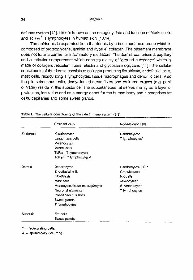
2.2 Cellular constituents of human skin
Human skin is the largest organ. It is a complex organ composed of the epidermis, the dermis and subcutaneous fat [1 0, 11]. The epidermis is the principal barrier between the host and the environment. The cellular constituents of the epidermis consist of keratinocytes, dendritic (Langerhans) cells (2-5% of total epidermal cells), melanocytes (± 5% of basal epidermal layer), sporadically Merkel cells and TcR-a.B+ and sporadically TcR--yo+ T lymphocytes. Keratinocytes make up about 95% of the cell mass of human epidermis and are responsible for the biochemical and physical integrity of the skin [11]. The antigen-presenting Langerhans cell in the epidermis plays a pivotal role in the immunological branch of the host
24 Chapter 2
defence system [12]. Little is known on the ontogeny, fate and function of Merkel cells and TCR')'o+ T lymphocytes in human skin [13, 14].
The epidermis is separated from the dermis by a basement membrane which is composed of proteoglycans, laminin and (type 4) collagen. The basement membrane does not form a barrier for inflammatory mediators. The dermis comprises a papillary and a reticular compartment which consists mainly of 'ground substance' which is made of collagen, reticulum fibers, elastin and glycosaminoglycans [11]. The cellular constituents of the dermis consists of collagen producing fibroblasts, endothelial cells, mast cells, recirculating T lymphocytes, tissue macrophages and dendritic cells. Also the pilo-sebaceous units, demyelinated nerve fibers and their end-organs (e.g. papil of Vater) reside in this substance. The subcutaneous fat serves mainly as a layer of protection, insulation and as a energy depot for the human body and it comprises fat cells, capillaries and some sweat glands.
Table I. The cellular constituents of the skin immune system (SIS)
Epidermis
Dermis
Subcutis
Resident cells
Keratinocytes Langerhans cells Melanocytes Merkel cells T cRa.B + T lymphocytes TcR')'s + T lymphocytes#
Dendrocytes Endothelial cells Fibroblasts Mast cells
Monocytesjtissue macrophages Neuronal elements Pilo-sebaceous units Sweat glands T lymphocytes
Fat cells
Sweat glands
* = recirculating cells. # = sporadically occurring.
Non-resident cells
Dendrocytes* T lymphocytes"'
Dendrocytesj(LC)* Granulocytes NK-cells Monocytes"" 8 lymphocytes
T lymphocytes

Skin inflammation in psoriasis 25
2.3 Cytokines
Cytokines are non-specifically inducible soluble hormone-like polypeptides with immunoregulatory functions. They are of clinical relevance, especially in infection and sepsis, auto-immunity, cancer and transplantation [15]. The term "cytokine" comprises monokines from monocytes, lymphokines from lymphocytes, hemopoietic, transforming and other mitogenic factors, the interferons and tumor necrosis factors.
2.3.1 General aspects
Cytokines were initially believed to be mainly inducible in cells of lymphohemopoietic origin, like regulatory T cells and macrophages [16]. It is now clear that many other cell types are also able to secrete cytokines under specific circumstances. They differ from endocrine hormones in that hormones are produced in specialized glands, are continuously present in the circulation and serve to maintain homeostasis. In contrast, cytokines are generally not constitutively produced, act over short distances as autocrine and paracrine intercellular signals in local tissues and serve to restore homeostasis. This group of heterogeneous glycoproteins has several more common characteristics. Cytokines are low molecular weight glycoproteins (usually < 25 kD in monomeric form). They are mostly synthesized as larger precursor molecules. They are extremely potent, generally acting in the picomolar range. They interact with high affinity (Kd = 0.01 to 1 nM) with specific cell surface receptors. Cytokine receptors on target cells are usually present in relatively low numbers (less than 10,000 receptors per cell). Cytokines interact in a network or a cascade system by inducing each other, transmodulating cytokine cell surface receptors and by crossregulation via synergistic, additive or antagonistic interactions on cell functions [17]. Their most characteristic feature is their pleiotropy, which means that cytokines have multiple effects on various cell types, and redundancy, which means that several cytokines can replace each other for specific target cell activities.
Three cytokines and their receptors are specifically addressed in this introductory chapter, because they display overlapping biological properties and fulfill key functions in cutaneous inflammation. These cytokines are interleukin(IL)-1, IL-6 and tumor necrosis factor alpha (TN F-a), which were initially believed to be produced mainly by activated monocytes [18]. Various other types of cells, including keratinocytes and fibroblasts, have now been shown also to produce these cytokines. The biochemical properties and the biological activities of these three cytokines are summarized in Table II.

26 Chapter 2
Table II. Cellular sources, biochemical and biological properties of ll-1a: and -/3, ll-6 and TNF--a#
Producers
Biological activities
* Adapted from 18.
lnter/eukin-1 (/L-1)
ll-1o: and -P
Macrophages, keratlnocytes, endothelial cells, T cells B cells, astrocytes, microglia, fibroblasts
Induction of: PGE2 synthesis
growth of fibroblasts bone resorption ICAM-1 expression fever
sleep anorexia synthesis of collagenase
growth and differentiation
of T and 8 cells
IL-6
Macrophages, T cells. 8 cells, keratinocytes, endothelial cells, fibroblasts, astrocytes, mesangial cells, bone
marrow stroma cells
Induction of: 8 cell differentiation plasmacytoma growth acute phase protein synthesis T cell activation and differentiation macrophage differentiation hematopoietic stem cell
growth
maturation of megakaryocytes neural ce!l different'1ation mesangial cell growth
myeloid leukemic cell differentiation
TN F-a
Macrophages, T cells, NK cells
Tumoricidal activity Inhibition of lipoprotein lipase Induction of:
bone resorption myeloid leukemic cell differentiation
procoagulant activity growth and differentiation
of B cells ICAM-1 expression
IL-1 is the term for two polypeptides, IL-1a (pi 5.3) and IL-1.8 (pi 7.2), that possess a broad spectrum of inflammatory, metabolic, physiological, hematopoietic and immunological activities (Table 1). IL-1a is predominantly produced by resting human keratinocytes as suggested by the IL-1a to IL-1.8 ratio of 2 [20,21]. IL-1.8, in contrast, is the main form produced by activated monocytes with an IL-1.B/IL-1a ratio of 25 to 50 [22]. ll-1a and IL-1.8 show approximately 46% homology at the DNA level, but the final amino acid homology is only 26'% [15]. The IL-1a and IL-1.8 genes are located on chromosome 2 (on band q13 and q13-q21, respectively, both comprising 7 exons), and are probably derived from a common ancestral gene via duplication.
Both molecules are synthesized as 31 kD precursors (IL-1a: 271 aminoacids (aa), IL-1.8: 269 aa) that are proteolytically cleaved to the 17.5 kD mature (153 aa for both molecules) proteins. However, IL-1a and IL-1.8 differ in the way they are processed and modified after translation. The precursor form of IL-1.8 is only partly biologically active, whereas the IL-1a precursor is fully biologically active [20]. Two different molecular domains on each IL-1 isoform mediate receptor binding. This implies differential biological activities of the two isoforms [23]. Keratinocytes, certain epithelial

Skin inflammation in psoriasis 27
cells and certain cells in the central nervous system are the only cells that constitutively produce ll-1. In keratinocytes, ll-1 is stored in an intracellular pool that is composed of pro-ll-1"' and pro-ll-1.8. Pro-IL-1.8, in contrast to pro-ll-1"', has minimal biological activity, and cannot be enzymatically cleaved efficiently by keratinocytes to the active mature form. Keratinocyte-derived pro-ll-1.8, therefore, is probably cleaved by proteases from other resident (epi)dermal cells or inflammatory cells [22]. The latter would imply that in the epidermis, under certain conditions, only ll-1<> activity may occur extracellularly [20,24]. Human epidermis thus contains an impressive reservoir of in total20 to 60 J.tg ll-1, which is equivalent to 4 to 12 nmoljlll-1 [25]. Since ll-1 can activate certain cells at concentrations lower than 10 pmoljl, this quantity forms a powerful weapon in the first line of defence of the host. IL-1, however, is safely stored in the keratinocyte and external stimuli are required for the release of this intracellular pool of IL-1.
Two types of IL-1R (IL-1Rtl and IL-1Rtll) have been identified [26]. The IL-1 receptor type I (IL-1 Rtl) is expressed on a wide variety of cells, which is in accordance with the pleiotropic functions of IL-1. IL-1Rtl is a single 80 kD glycoprotein (gp) with an extracellular domain of 319 amino acids (aa), a trans-membranous part of 21 aa and a cytoplasmic domain of 217 aa [27,28]. The extracellular part of the receptor possesses three immunoglobulin (lg-)like domains indicating that this receptor belongs to the lg-superfamily [19,29]. The different receptor families are briefly described in the legend of Fig. 1, which illustrates these cytokine binding proteins. ll-1 Rtl binds the mature forms of IL-1<> and IL 1-.8 and is also able to bind the precursor of IL-1<> but not of IL-1.8 [30]. The second receptor (IL-1Rtll), a single 68 kD gp, initially discovered on B cell lines, is also expressed on many cell types [26,31]. Soluble receptor forms are described [32]. Both IL-1 binding receptor-proteins are separate gene products [31]. At present, it is unclear whether the two types of receptors have common or different signalling pathways [33]. The IL-1 Rtl has the ability to rapidly internalize IL-1, whereas IL-1 bound to type II receptors remains at the cell surface for as long as 60 min and is poorly internalized [34]. Furthermore, the half-life of type II receptors is also shorter than that of type I receptors, 2 versus 5-12 hours [35]. Recently the same has shown that both types of IL-1R are accompanied by different IL-1 processing pathways [36]. It is noteworthy that IL-1 down-regulates the surface expression of its own type 1 receptor [37,38]. In addition, IL-1R expression is up-regulated by phorbol esters, UV irradiation and T cell cytokines [39]. Recently, a third ll-1 isoform is described, the IL-1-receptor antagonist (IL-1ra), which binds to the IL-1 Rand seems to play a regulatory role in cellular activation [40-42].
Signal transduction via IL-1 generally involves a sequence of four important elements. These are an IL-1 R linked GTP-binding protein, cAMP, protein kinase (PK) A and the DNA binding protein NF-KB [43]. ll-1 has been shown to activate a serine kinase that is distinct from PKA and PKC in fibroblasts [33].

28 Chapter 2
lnterteukin-6 (/L-6) IL-6 is a 21-28 kD gp wtth multifunctional properties. It is produced by both
lymphoid and non-lymphoid cells (Table II). The IL-6 gene is located on chromosome 7 (p15-21) and comprises 5 exons. The IL-6 promoter contains three functional domains that are involved in IL-6 induction [15]. These are 1) a 23 bp IL-6 multiresponsive element (MRE), 2) a nuclear factor (NF) IL-6 binding site, an element responsive to IL-6 itself and IL-1, TN Fa and lipopolysaccharide (LPS) and 3) a NF-~<B binding site, which mediates IL-6 induction by IL-1 and TNFa [19]. IL-6 synthesis involves different (cell-type-specific) proteolysis, glycosylation and post-translational NH
2-terminal processing and phosphorylation. This explains the differences in
molecular mass of the mature protein (44,45]. The functional significance of these distinct post-translationally modified forms of IL-6 remains elusive. Moreover, IL-6 complexes may be formed after secretion [ 46]. The amino acid sequence and the genomic structure of IL-6 show considerable homology with that of granulocyte-colony stimulating factor (G-CSF), which is indicative for similarities in the tertiary structure of these cytokines [18].
The IL-6 receptor (IL-6R) is expressed on lymphoid as well as non-lymphoid cells. IL-SR is composed of an external ligand-binding (340 aa), a transmembranous (29 aa) and an intracellular domain (80 aa). The receptor has one lg-like domain in the extracellular portion, which is not involved in ligand binding. The remainder of the extracellular part belongs to the cytokine receptor family (Fig. 1). The IL-6 receptor system consists of two functionally different polypeptide chains: a ligand-binding 80 kD IL-6R and a signal transducing 130 kD glycoprotein. Following binding of IL-6, the two proteins become associated at their extracellular segments [47]. The p80 IL-6R may occur in a soluble form, and complexes of IL-6 and IL-6R(p80) may mediate signal transduction and thus exert biological effects. Considering the pleiotropic functions of IL-6, it is conceivable that different gp130 variants or that a third associating molecule exists. At least two signalling pathways are involved in the induction of IL-6 by IL-1 or TN Fa. One requires PKC activation and the other involves adenylate cyclase. The latter is especially evident in fibroblasts [19].
Tumor Necrosis Factor Alpha (TNF-a)
TNF-a (cachectin), a 17 kD polypeptide, serves as an endogenous mediator of inflammatory, immune and host defence functions. The genes for TN F-a (and TNF-P) are closely located to the MHC class II region on chromosome 6 (p21.3) and consist of 4 exons [18]. TN F-a is mainly produced by macrophages, T lymphocytes and NK cells (Table II). It is produced as a 233 aa pro-TN F-a form, which is processed to a 157 aa mature non-glycosylated peptide. Secreted TNF-a occurs as a trimeric molecule with each subunit consisting of an anti-parallel p-sandwich. TN F-a and TNF-P display about 30% homology in amino acid sequence and have common biological

-"' .E .:!! 0 a m Q
1! m c
"' 0
>-Q
IL-2R~ IL-3R IL-4R Epo R
GM-CSFR
Skin inflammation in psoriasis 29
a; :2 Q;
" ~ x (I)
cell membrane
gp130
Figure 1. Structural relationship between cytokine receptors (adapted from 19). Most cytokine receptors
(TNF-R1 and TNF-R2 excluded) belong to either the lg-superfamlly or the cytokine receptor family. Only
the IL-6 receptor is composed of both elements. A third group of cytokine receptors may be formed
by the growth factor receptors (e.g. EGF-R) which are characterized by a large cytoplasmic domain with
tyrosine-kinase activity [15]. Little is known about signal transduction mechanisms. It is, however,
suggested that different target cells possess specific receptor-associating transducing proteins. These
may interact with structurally related cytokine receptors. In addition, each type of cytokine receptor may
also interact with multiple signal transducers. Such interactions could explain the pleiotropic and
redundant effects of cytokines.
activities. TNF receptors have been found on virtually all cells and tissues. At present, two
categories of TNF-binding proteins, a 55-60 kD (fNF-R1) and a 75-80 kD (fNF-R2) receptor have been reported reflecting the two types of TNF-R eDNA cloned. Both receptors bind TNF-<> as well as TNF-fi and can occur in soluble form [48-50]. Human TNF-R1 and TNF-R2 do neither belong to the cytokine receptor nor to the lg-receptorsuperfamily. The extracellular domains of TNF-R1 and TNF-R2 share 28% identity, which is equivalent to the homology that each of them share with a group of surface proteins including nerve growth factor, Fas antigen, Bp50, Ox40 and CD27. There is a complete absence of homology between the intracellular domains of the two TNF receptors, implying that they utilize distinct signalling pathways [51]. TNF receptors

30 Chapter 2
are continuously internalized and this process is accelerated by the binding of TNF. Cultured normal human keratinocytes, in contrast to the inflammatory infiltrate, express the 55 kO TNF-R only [52].
All three cytokines are generally not constitutively produced, but transiently induced by various stimuli. Inducers and suppressors of these cytokines are shown in Table Ill, while similar information for the receptors is shown in Table IV. In the cytokine network, IL-1 and TNF-" are considered as primary cytokines in inducing the local production of other cytokines such as IL-2, IL-6, IL-8, IFN-/l and CSF. This may explain the pleiotropic effects of these primary cytokines on a variety of cells. IL-1 and TNF-" are able to induce each other. Moreover, IL-1, IL-6 and TNF-" are able to induce their own production [19].
Table Ill. Inducers and suppressors of IL-1, IL-6 and TNF-a#
Cytokine
IL-1a/P
ll-6
TN F-a
Inducers
Lipopolysaccharide, staphylococcal exotoxins, Klebsiella glycoproteins, viruses, phorbol esters, calcium ionophores, leukotrienes, immune
complexes, C3a and C5a, phagocytosis, proteoglycans, collagens types 2 and 9, urate crystals, trauma, adherence. ll-1*, TNF ... IFN·")". GM-CSF
Upopolysaccharide, mitogens. phorbol esters, calcium ionophores, Staphylococcus aureus, viruses, JL-1, TN F-a:, IFN-p, PDGF, UV-irradiation, poly (I)(C),
cAMP and cAMP-generating agents
Upopolysaccharide, bacteria, PPD, viruses, immune complexes, phorbol esters, cGMP, JL-1, IL-2, PAF. TNF-<t, IFN--y, GM·CSF
Suppressors
Corticosteroids, retinoic acid, cAMP, PGE2• IFN--y, ll·2, TGF-p
Corticosteroids
Corticosteroids, CsA, Vit.-03 PGE2, PAF-Rantagonist, IL-6, TGF-p
• #
IL-1a may also down-regulate IL-1 via induction of PGE2 synthesis . Key references: 18,53-55.

Skin inflammation in psoriasis
Table IV. Inducers and suppressors of IL-1 R, IL-6R and TNF-R#
Cytokine receptor
IL-1R
IL-6R
TNF-R
Inducers
PMA, lectins, IL-2: T cells and B cells EBV, Dexamethasone: B cells, M0 PDGF, prostaglandins: FB IFN-'Y. UV-irradiation: KC
Lectins, !FNs, IL-2, IL-4
# Key references: 56-58.
2.3.2 Skin-derived cytokines
Suppressors
IL-1: T and B cells RA: KC
TGF-p: T cells, FB
IL-1, IL-4
PMA, IL-1
31
The resident and non-resident cell types in normal human skin provide a virtually unlimited local cytokine repertoire. Most cells need an inducing factor to produce and release cytokines. Epidermal cells such as Langerhans cells and keratinocytes are believed to initiate and orchestrate skin inflammation by the release of primary cytokines and via the induction of cytokine(s) and/or cytokine-receptors in adjacent cells. Cytokines derived from melanocytes, dermal T lymphocytes, fibroblasts, monocytes, mast cells and endothelial cells probably fulfill an accessory role in inflammatory processes in the skin [39]. The skin may thus be conceived as the most important peripheral limb of host defence [1 0]. The cytokines potentially produced by each cell type are listed in Table V.
Table V. Cytokine production capacity of relevant non-keratinocytes in normal human skin#
Cell type IL-1 IL-6 IL-6 TNF-. IFN-')' GM-CSF TGF-. TGF-p1 bFGF PDGF MCP-1
Dendrocytesj Langerhans cel!s + + Melanocytes + + + + ± + T cells + + + + + + + + Endothelial cells + + + + + + + + Fibroblasts + + + + + + + + + Mast cells + + ± + Tissue macrophages + + + + + + + + + +
# Key references: 53-55.59-66.

32 Chapter 2
Keratinocyte-derived cytokines Cytokines recently identified in cultured keratinocytes are listed in Table VI. The
'resting' keratinocyte is able to produce only one biologically active cytokine, namely IL-1<> [24,25]. External stimuli may result in the release of substantial amounts of e.g. active IL-1<> and pro-IL-1,!1 from keratinocytes. For example, skin trauma or UVexposure will result in the release of active IL-1 and TNF-et and subsequent autocrine and paracrine stimulation of contiguous keratinocytes, Langerhans cells and other cells initiating the "cytokine cascade". It has indeed been shown that cultured keratinocytes were responsive to exogenous IL-1 and TNF-cr in vitro [39]. The cytokines listed in Table VI are produced mainly by stimulated keratinocytes.
Table Vl. Keratinocyte derived cytokines#
Cytokines known to be produced
Cytokines assumed to be produced
Cytokines not produced by keratinocytes
# Key references: 39,67,68.
IL-1a and ·P: IL-3; IL-6; IL-8; TNF-a; IFN-a and-P1:
GM-. G- and M-CSF: TGF-a and -132: bFGF; PDGF;
MCP-1; suppressor-factors; neuropeptides and
thymopoietin
TNFP: IGF-1 and -2
IL-2; IL-4; IL-5; IFN-")'
Cytokines produced by Langerhans cells and other cells in the skin It is assumed that human Langerhans cells may direct and orchestrate the
immune response by the configuration of cytokines released at the moment T lymphocytes are activated [69]. For example, Langerhans cells might direct T lymphocytes towards specific differentiation stages such as the Th1 or Th2 subsets depending on the type of antigen recognized, the manner in which that antigen is processed and presented [70]. Skin mast cells and probably trafficking basophils have also been shown to produce cytokines. These ·,nclude IL-3, IL-4, IL-5, IL-6, IL-8, TNFcr(minu1e quantities) and GM-CSF [71]. Cytokines like IL-1, IL-2, IL-3, IL-6 and IL-8, released by surrounding cells, also have great influence on mast cells. Cytokines are able to lower the '1riggering threshold" or directly stimulate mediator release from mast cells and prime and induce them to synthesize arachidonic acid metabolites [72]. Also cross-linking of the Fc<R1 by antigen has been shown to induce the production of cytokines in mast cells [73].
In conclusion, the whole spectrum of cytokines is potentially available in human skin.

Skin inflammation in psoriasis 33
2.4 Cutaneous signalling system
In the last decade it has been shown that keratinocytes are not passive bystanders in the immune response of the skin. In contrast, keratinocytes appear to play an active and central role in the first line of defence of the host [74]. The observations that normal keratinocytes have the capacity to produce a broad spectrum of cytokines, compelled Kupper to introduce the activated keratinocyte model [67]. The concept of the activated keratinocyte is essential for the perception of cutaneous inflammation. Briefly, diverse stimuli like contact allergens and uv light may trigger cutaneous inflammation by direct activation of keratinocytes and subsequent production of pro-inflammatory cytokines and expression of adhesion molecules. Via paracrine stimulation in response to keratinocyte-derived cytokines, adjacent keratinocytes, dermal microvascular endothelial cells and fibroblasts are activated and induced to produce other cytokines and to up or down-regulate the expression of cytokine receptors andjor adhesion molecules. This results in selective recruitment of specific immunocompetent cells in the epidermis and the dermis. The ability of keratinocytes to produce an almost complete repertoire of cytokines and immunoregulatory peptides is compatible with the idea that they have an important pathophysiological role in cutaneous inflammation [74].
In further detail, the activated keratinocyte model proposes the following scheme of events. After activation of keratinocytes, several processes ensue. These are 1) as mentioned, de novo gene expression of various secondary cytokines such as IL-3, IL-6, IL-8, TNF-0< and G(M)-CSF with activating and chemotactic activities on leukocytes and mast cells; 2) (upregulation of) the expression of adhesion molecules on keratinocytes (ICAM-1) and endothelial cells (ICAM-1, ELAM-1, VCAM-1 and probably other as yet unidentified adhesion molecules); 3) keratinocyte proliferation and chemokinesis; 4) fibroblast proliferation, type 4 collagen and collagenase synthesis; and 5) chemotaxis [29,74]. The activation of the dermal vascular endothelium, as indicated by the up-regulation of membrane adhesion molecules, results in increased adhesiveness for different cell types and trapping of inflammatory cells in the capillaries. The composition of the inflammatory infiltrate, for example specific mononuclear cells, is believed to depend on the character and combination of chemotactic cytokines produced by keratinocytes, resident fibroblasts, monocytes, dendrocytes and tissue macrophages. These factors will dictate the net migratory behaviour of the inflammatory cells during the early phases of inflammation. For example, predominant release of IL-1 and IL-8 (cytokines with extreme chemotactic activity forT lymphocytes) by keratinocytes, will encourage directional migration ofT lymphocytes towards the epidermis. LFA-1 + migrated T lymphocytes will adhere to keratinocytes via interactions with the ligand ICAM-1 [74,75]. This type of adhesion may directly induce cytokine release from T lymphocytes and other inflammatory cells,

34 Chapter 2
resulting in further amplification of cutaneous inflammation. The keratinocyte-derived MCP-1, on the other hand, specifically recruits monocytes. The released proinfiammatory cytokines such as IFN-1 and TNF-a, produced by activated T cells and monocytes, respectively, are able to stimulate the keratinocyte and to maintain the cell in an activated state via the IL-1/IL-1R loop. Indeed, keratinocytes have been shown to possess the appropriate cytokine receptors necessary to respond to paracrine activation [67]. Receptors for IFN--y, for example, are constitutively expressed in high levels in normal epidermis [76,77]. Other receptors are also present on keratinocytes [52,78,79]. Routes by which keratinocytes can be activated are illustrated in Fig. 2.
IFN-r and TN F-a also induce ICAM-1 expression on keratinocytes, whereas the expression of HLA-DR on these cells can be induced by IFN-r only. These molecules
resting normal keratinocyte
---~-
IL-1a
T-cell
IL-1 R t
IL-1 t
monocyte
activated keratinocyte ------
t cytokine receptors t ICAM-1 (T-cell + M0) t HLA-DR (T-cell)
(growth, migration)
Figure 2. Routes of activation and biological responses of human keratinocytes (adapted from 39).
Keratinocytes may be activated in an autocrine or paracrine fashion. IL~ 1 may be involved in both
pathways. Keratinocytes activated in paracrine fashion by T cell cytokines differ from those activated by monocyte-derived cytokines. Conversely, different types of activated leukocytes are also generated upon paracrine activation by keratinocyte-derived cytokines [83].

Skin inflammation in psoriasis 35
are important for the evolution, full development and regulation of cutaneous inflammation. The induction of ICAM-1 and HlA-DR are obviously not confined to keratinocytes, but can also be induced on fibroblasts and endothelial cells. IFN-r is also able to induce the production of secondary chemotactic cytokines in keratinocytes, such as monocyte chemotactic protein-1, MCP-1, IFN-r-induced protein 10 (r-IP-10). TNF-odurthermore induces keratinocyte ll-8 [74,75]. MCP-1 and IL-8, both small peptides, are actually considered the most important skin derived chemotactic cytokines. The mentioned molecules belong to different subfamilies (C-X-C-family: IL-8, gro and C-C-family: MCP-1, MIP, RANTES) of a supergene family of chemotactic cytokines [75,80]. Both IFN-r and TNF-a are able to induce TGF-a production in keratinocytes. TGF-a, IL-1 and IL-6 are potent autocrine growth factors for keratinocytes, ontogenetically designed to fulfill a central role in re-epithelialization during wound healing [81,82].
In the amplification phase of cutaneous inflammation, the recruitment of leukocytes and the interaction between epidermal cells and inflammatory cells is intensified [74,75]. In contrast with initial, antigen-independent activation of keratinocytes, an antigen-dependent component may be involved in the later stages [74]. The dedine of activation signals causes the keratinocyte to down-regulate its release of ll-1, the expression of IL-1R and to become refractory to further stimulation. Finally, under normal conditions down-regulatory mechanisms switch off the state of activation, e.g. via loss of keratinocyte ICAM-1 expression. Consequently, cutaneous inflammation is resolved. Restoration of the intracellular pool of active IL-1 renders the keratinocyte responsive again to subsequent trigger factors of cutaneous inflammation [39]. Keratinocytes thus orchestrate skin inflammation by serving as cellular 'signal transducers', capable to transduce exogenous stimuli into production of cytokines,
cytokine-receptors, adhesion molecules and chemotactic factors [74]. The above discussed sequence of events represents carefully balanced
functional interactions between cellular constituents of the skin immune system and their soluble products. Dysbalances or genetically determined incapacities to produce (or similar abnormalities resulting in overproduction of) certain molecules of this interactive system, underly skin diseases such as psoriasis.
Our concept of the "cutaneous signalling system" emphasizes that not the mere physical or static presence of the cellular constituents of the skin immune system [1 OJ, but their functional interactions are of decisive importance for the prevention or development of skin disease.
2.5 Cy1okines and their specific receptors in psoriasis
The combination of keratinocyte hyperproliferation with epidermal leukocyte accumulation distinguishes psoriasis from other epidermal hyperplasias. The

36 Chapter 2
pathogenesis of psoriasis is thus linked, at least in part, to processes which regulate leukocyte trafficking and inflammation. As put forward in the previous section, this makes cytokines and their receptors suspects in the pathogenesis of psoriasis. In the last few years a~erations regarding the cytokine profile in psoriasis have indeed been reported. These alterations will be discussed briefly. One should keep in mind, however, that the cascade of keratinocyte derived pro-inflammatory cytokines operative in psoriasis, under normal conditions has beneficial effects for the host.
lnterleukin-1 Lesional psoriatic cytosolic extracts of keratome specimens contained less IL-1"'
but increased amounts of IL-1fi protein as compared with extracts of uninvolved psoriatic skin or normal skin [84]. IL-1fi mRNA is also increased in psoriatic skin extracts [85]. The considerable IL-1a predominance in normal skin is apparently lost in psoriasis due to the down-regulation of IL-1"' and up-regulation of IL-1fi [20]. Comparable results were obtained with 10,000 g supernatants of normal and psoriatic skin specimens, although the mature form was only present in psoriatic skin [86]. When aqueous extracts of the stratum corneum (scale extracts) were used, it was observed that preferentially mature IL-1a was decreased in psoriatic lesions as compared with normal skin. IL-1fi was almost undetectable in both types of scale extracts [87,88]. In addition, suction blister fluids from psoriatic lesions as well as from normal skin contained similarly low levels of IL-1a and no IL-1fi. However, IL-1fi was over-expressed on the plasma membrane and in the intracellular compartment of epidermal cells [88,89]. Slightly elevated levels of il-1"' as well as IL-1fi mRNA were detected in cultured keratinocytes from psoriatic lesions. Lesional fibroblasts, in contrast, expressed almost undetectable levels of IL-1"' and IL-1fi mRNA [89]. To make the picture still more complex, immunohistological studies by others showed that both normal and psoriatic epidermis expressed more IL-1c>than IL-1fi. In normal skin, the expression of IL-1"' was predominantly intercellular whereas in lesional psoriatic epidermis its distribution was predominantly within the cytoplasm [90].
About the IL-1 bioactivity there is more consensus in the literature. The totalll-1 bioactivity in psoriatic epidermis is considerably decreased as compared with normal skin [91]. IL-1a comprises all the bioactive material [85,87]. The IL-1fi in the psoriatic lesion may be of a novel form: it is a processed molecule with negligible bioactivity [20]. The presence of an IL-1 inhibitor in psoriatic skin may explain at least in part the reduced IL-1 bioactivity [85,92]. In psoriatic scale extracts, however, inhibitory activity could not be detected [93]. The decreased IL-1a levels in psoriatic lesions may be due to the elevated IL-6 levels in these lesions, since IL-6 is known to inhibit IL-1 production [94]. Since several enzymatic abnormalities have been reported in psoriatic skin, it may be that the abnormal isoforms of IL-1 are due to improper cleavage or even non-cleavage of IL-1 precursor molecules by defective serine-proteases [95].

Skin inflammation in psoriasis 37
/nterleukin-1 receptors It has been postulated that the primary defect in psonas1s may be the
dysregulation of keratinocyte IL-1 R in the absence of an increase of epidermal IL-1 [25]. Our own investigations revealed a decreased expression of IL-1 R on lesional EC. The results on other cytokine receptors on EC in psoriasis are described in detail in Chapter 6.
/nterleukin-6 Increased levels of IL-6 were observed in skin from active psoriatic plaques by
immunohistochemistry. Strong staining was confined to keratinocytes and infiammatory dermal cells [81 ,96-98]. Using in situ hybridization, abundant IL-6 mRNA was demonstrated in psoriatic epidermis. The increase in IL-6 mRNA was more pronounced in the transitional zone (the border between psoriatic plaques and uninvolved skin) of progressive psoriatic lesions [81 ,96,99]. In contrast, another investigator, using in situ hybridization, failed to detect increased IL-6 mRNA in lesional epidermis [100]. IL-6 protein expression was also detected in a higher percentage of keratinocytes cultured from lesional psoriatic skin relative to cultured keratinocytes from uninvolved psoriatic skin [81]. However, IL-6 mRNA was expressed at very low levels in cultured keratinocytes from both psoriatic lesions and normal skin, whereas cultured lesional fibroblasts showed a clear IL-6 mRNA expression [89]. Our own studies revealed that cultured psoriatic fibroblasts produce increased amounts of bio-active IL-6 as compared with fibroblasts from healthy controls [De bets et al., submitted]. Blister fluids from psoriatic lesions contained elevated levels of bioactive IL-6, while the levels in sera of these patients were undetectable [89]. Another study of patients with severe psoriasis did show elevated serum levels of IL-6, probably derived from activated peripheral blood mononuclear cells [81 ,96, 101]. However, neither IL-6 nor IL-6 activity could be detected in extracts of scales from psoriatic lesions and normal skin [87].
/nterleukin-8
On the one hand, immunoreactive IL-8 was detected in suprabasal keratinocytes in psoriatic lesions, whereas IL-8 expression was not found in normal skin [102]. We confirmed these observations [Prens et al., manuscript in press]. On the other hand, others reported a reciprocal relationship between the expression of IL-8 and the disease activity [103]. Biologically active IL-8 was unequivocally shown to be elevated in extracts of psoriatic lesions [87]. High levels of IL-8 mRNA were also detected in lesional epidermis, especially in active plaques [101 ,16]. Using in situ hybridization, IL-8 mRNA was confined to the upper epidermal layers [100]. Since this cytokine specifically attracts neutrophils and T cells it was considered a key cytokine in the maintenance of psoriasis.

38 Chapter 2
Tumor necrosis factor " and f, TNF-a and TNF-f, were detected in extracts of scales from psoriatic lesions in
contrast to extracts of normal skin. The TNF in these samples possessed no equivalent biological activities [87]. Using immunohistochemical localization TN F-a was identified especially in macrophages in the upper dermis in lesional skin, while there was no detectable TNF in normal skin [102]. Another group did not detect TNF-a, neither in psoriatic nor in normal skin [97]. In another study both TNF-a protein and TNF-a activity were undetectable in both extracts of scales and blister fiuids [104]. TNF-a mRNA was also undetectable in psoriatic lesions using in situ hybridization [100]. Psoriatic monocytes from untreated patients seemed to activate neutrophils more efficiently, probably via increased TNF-a production [101]. Finally, TNF-ct and TNF-f, immunoreactivity was not increased in supernatants of stimulated peripheral blood mononuclear cells from psoriatic patients [1 05].
Interferons Suction blister fluids from psoriatic plaques, but neither uninvolved skin nor sera
from psoriatic patients, contained IFN--y and some IFN-et activity [106]. Another group could not confirm this finding [107]. Expression of IFN correlated positively with disease activity. IFN-a and IFN--y were primarily confined to basal keratinocytes, the stratum corneum and dermal mononuclear cells, respectively. Interferons were undetectable in uninvolved skin or normal healthy skin [108]. The IFN-a and IFN--y proteins were also detected in extracts of psoriatic lesions in contrast to extracts of normal skin. However, no equivalent biological activities could be detected [87, 107]. In contrast to normal keratinocytes, psoriatic keratinocytes were indeed found to produce -y-IP-10, possibly as a result of elevated levels of IFN-r in psoriatic lesions [109].
Interferon receptors. Psoriatic keratinocytes and normal keratinocytes respond differently to IFN-r. Psoriatic keratinocytes showed a weaker HLA-OR expression and less inhibition of cell growth upon stimulation with IFN-r. It has been shown that psoriatic KC do not down-modulate their EGF/TGF-aR upon exposure to IFN--y. Thus possible alterations in the IFN-rR or receptor-signalling mechanism in psoriasis are indicated [75, 11 OJ. The IFN-rR was observed only in the basal layers in psoriatic epidermis [76].
Colony stimulating factors In one study, G-CSF and GM-CSF protein and bioactivity were undetectable in
scale extracts of psoriatic lesions and normal skin [87]. However, other investigators did detect increased levels of immunoreactive GM-CSF (but not G-CSF) in lesional scale extracts, while these cytokines were undetectable in blister fiuids [111,112]. Psoriatic monocytes appeared to produce increased amounts of GM-CSF [1 01].

Skin inflammation in psoriasis 39
Growth factors Transforming growth factor (TGF) "'· TGF-a is overexpressed in psoriatic
epidermis, with the expression primarily confined to the upper stratum spinosum. In uninvolved and normal healthy skin, in contrast, the protein is predominantly expressed in the stratum basale and the lower stratum spinosum [113]. In lesional epidermal extracts increased TGF-0! protein and TGF-a mRNA have been detected. TGF-a has also been detected in lesional dermis [102,114].
TGF-~. TGF-~ transcripts were not increased in psoriasis and EGF mRNA was even undetectable in both psoriatic and normal skin [114, 115]. Psoriatic endothelial cells are less responsive to the down-regulatory effects of TGF-~ on the expression of adhesion molecules [116]. Growth factor receptors
Transforming growth factor-CJ. receptors. The natural receptor lor TGF-0!, the EGF receptor, is overexpressed in psoriatic epidermis, with a distribution similar to that of TGF-CJ. [117]. However, no increase in EGF /TGFa-R mRNA was observed in psoriasis [114].
Insulin-like growth factor receptors. An increased expression oiiGF-1 R has been observed in lesional psoriatic epidermis [118]. Fibroblasts are a potential source of IGF-1 in this respect [119].
Platelet-derived growth factor receptors. Psoriatic dermal endothelial cells and fibroblasts express an increased number of PDGF-R-~ [120].
Other important immunoregulatory cytokines such as IL-2 and IL-4 were undetectable in scale extracts of psoriatic lesions and normal skin [87]. However, others did report elevated levels of IL-2 in lesional extracts but not in suction blister fluids of lesional skin. Increased levels of soluble IL-2R did occur in blister fiuids from psoriatic skin [107]. In addition, soluble IL-2R was increased in serum of psoriatic patients [121]. Our group did not detect immunoreactive IL-4 in culture supernatants of psoriatic and normal epidermal cells. Finally, over-expression of proto-oncogenes is probably not involved during the steady-state epidermal hyperplasia in psoriasis [115].
2.6 Aim of the study
The above data show that prodigious alterations occur in the expression of cytokines and their receptors in psoriasis. Although a crucial role lor cytokines in the pathogenesis is almost definite, the results do not unequivocally demonstrate a primary cytokine defect in psoriasis. Furthermore, the data described are often contradictory. This may be caused by the use of different skin specimens, the detection of cytokines and receptors at different levels (eg. transcription, protein and

40 Chapter 2
bioactivity) combined with the use of different detection techniques. Despite the fact that data on cytokines in normal and psoriatic skin is far from complete, several alterations in psoriasis are evident: 1) dysregulation of IL-1; 2) increased levels of IL-6, IFN--y and especially IL-8 and TGF-a; 3) altered responsiveness of keratinocytes to IFN--y; and finally; 4) clearly increased expression of the EGFjTGF-a receptor.
Taking these findings into account, psoriasis may be considered a multi-factorial immunological disease in genetically predisposed individuals with primary defect(s), probably residing in the cutaneous signalling system. A specifically altered cytokine profile in association with an altered response of the keratinocytes to these cytokines links inflammation with the hyperactive state in the epidermis in psoriasis. It is conceivable that this deviant cutaneous signalling system plays a central role in the pathogenesis of psoriasis.
The aims of our studies were to further delineate the basic abnormalities in skin APC and the cutaneous cytokine network in psoriasis. The experimental work on APC, cytokines and cytokine receptors in the psoriatic skin are described in Chapters 3 to 7. In Chapter 8, the implications of our findings are discussed in the context of the literature data on the altered cytokine network in psoriasis.
2.7 References
1. Valdimarsson H, Baker BS, Jonsdottir !, Fry L: Psoriasis: a disease of abnormal keratinocyte
proliferation induced by T lymphocytes. lmmunol Today 7: 256-259, 1986.
2. Easty DJ, Patel K, Otto WR, Dunn MJ, Kiil J, Evans OJ: A study of protein synthesis in cells cultured from involved psoriatic skin. Electrophoresis 12: 579-584, 1991.
3. Madsen P, Rasmussen HH, Leffers H, Honore B, Dejgaard K, Olsen E, Kii! J, Walburn E, Andersen AH, Basse 8: Molecular cloning, occurrence, and expression of a novel partially secreted protein
'psoriasin' that is highly up-regulated in psoriatic skin. J Invest Dermatol 97: 701-712, 1991.
4. Prens EP, Benne K, Van Joost T, Benner R: The autologous mixed epidermal cell - T lymphocyte
reaction is elevated in psoriasis: A crucial role for HLA-DR+ /CD1a· antigen presenting cells. J
Invest Dermatol 96: 880-887, 1991.
5. Iversen 0, Rodahl E. Dalen AB: Rabbit antibodies against the major internal protein of a
retrovirus-like particle bind to epidermal cells in psoriatic skin. Arch Viral 86: 341-346, 1985.
6. Numahara T, Nakshima K, Yamamoto S, Nishimoto M: Significance of squamous cell carcinoma
related antigen in psoriasis and generalized eczema. Dermatologica 178: 73-74, 1989.
7. Krogh HK, Tender 0: Antibodies in psoriatic scales. Scand J lmmunol 2: 45-51, 1973.
B. Dupuy P, Bagot M, MichelL. Descourt B, Dubertret L: Cyclosporin A inhibits the antigen-presen
ting functions of freshly isolated human Langerhans cells in vitro. J Invest Derrnatol 96: 408-413,
1991.
9. Herold KC, Lancki OW, Moldwin RL, Fitch FIN: Immunosuppressive effects of cyclosporin A on
cloned T cells. J lmmuno1136: 1315-1321, 1986.

Skin inflammation in psoriasis 41
10. Bas JD, Kapsenberg ML: The skin immune system, its cellular constituents and their interactions.
lmmunol Today 7: 235-240, 1986. 11. Uitto J, Oikarinen A, Thody AJ: Mechanical and physical functions of skin. In Thody AJ,
Friedmann PS (eels): Scientific basis of dermatology (a physiological approach). pp. 6-25 Churchill
Livingstone Publ, London, 1986. 12. Wolff K, Stingl G: The langerhans cell. J Invest Dermatol 80: 17$-21$, 1983. 13. Narisawa Y, Hashimoto K, Bayleff T, Nihei Y, Ishihara M, Lawrence 0, Eta H, Hori K: Cytokeratin
polypeptide profile of Merkel cells in human fetal and adult skin: difference of expression of
cytokeratins in epidermal and dermal Merkel cells, J Invest Dermatol 98: 171-180, 1992. 14. Tigelaar RE,lewis JM, Bergstresser PR: TCR -yjs+ dendritic epOermal T cells as constituents of
skin-associated lymphoid tissue. J Invest Dermatol 94: 58S-63S, 1990.
15. Arai K, Lee F, Miyajima A, Miyatake S, Arai N, Yokota T: Cytokines: coordinators of immune and
inflammatory responses. Ann Rev Biochem 59: 783-836, 1990. 16. Sauder ON: The role of epidermal cytoklnes in inflammatory skin diseases. J Invest Dermatol 95:
27$-28$, 1990.
17. Balkwill FR, Barke F: The cytokine network. lmmunol Today 10: 299-304, 1989.
18. Bendtzen K: lnterleukin 1, interleukin 6 and tumor necrosis factor in infection. inflammation and immunity. Immune! Letters 19: 183-192, 1988.
19. Akira S, Hirano T, Taga T, Kishimoto T: Biology of multifunctional cytokines: ll-6 and related
molecules (IL-1 and TNF). FASEB J 4: 2860-2867, 1990. 20. Cooper KD, Hammerberg C, Baadsgaard 0. Elder JT, Chan LS, Taylor RS, Voorhees JJ, Fisher
G: lnterleukin 1 in human skin: dysregulation in psoriasis. J Invest Dermatol 95: 24S-26S, 1990. 21. Gatto H, Vtac J, Charveron M: levels of immunoreactive interleukin 1a and 1b in basal,
suprabasal and cornified epidermal cell populations of normal human epidermis. Comparison with
keratinocytes cultured in defined medium. Eur J Dermatol 1: 55-59, 1991.
22. Mizutani H. Black R, Kupper TS: Human keratinocytes produce but do not process pro·
interleukin-1(1l-1) beta. Different strategies of ll-1 production and processing in monocytes and
keratinocytes. J Clin Invest 87: 1066-1071, 1989.
23. Radeke HH, Martin M, Tapley N, Keever V, Resh K: Differential biological activities of human
interleukin 1a and interleukin 1,8. Eur Cytokine Net 2: 51~59, 1991.
24. Blanton RA, Kupper TS, McDougall JK, Dower S: Regulation of interleukin 1 and its receptor in
human keratinocytes. Proc Natl Acad Sci USA 86: 1273·1277, 1989.
25. Kupper TS: Mechanisms of cutaneous inflammation (interactions between epidermal cytokines, adhesion molecules, and leucocytes). Arch Dermatol 125: 1406-1412, 1989.
26. Bomsztyk K, Sims JE, Stanton TH, Slaak J, McMahan CJ, Valentine MA: Evidence for different interleukin 1 receptors in murine 8 and T cell lines. Proc Nat! Acad Sci 86: 8034-8038, 1989.
zr. Sims JE, March CJ, Cosman D, Widmer MB, McDonald HR, McMahan CJ. Grubin CE, Wignall
JM, Jackson Jl, Call SM, Friend D, Alpert AR, Gillis S, Urdal Dl, Dower SK: eDNA expression
and cloning of the ll·1 receptor, a member of the immunoglobulin superfamily. Science 241: 585~ 589, 1988.
28. Urdal DL, Call SM, Jackson Jl, Dower SK: Affinity purification and chemical analysis of the interleukin 1 receptor. J Bioi Chern 263: 2870·2877, 1988.
29. Dinarello CA, Clark BD, Puren AJ, Savage N, Rosoff PM: The interleukin 1 receptor. lmmunol
Today 10: 49-51, 1989.
30. Mosley 8, Urdal DL, Prickett K, larsen A, Cosman D, Conlan PJ, Gillis S. Dower SK: The IL-1
receptor binds the human interleukin 1a precursor but not the interleukin 1.0 precursor. J Bioi

42 Chapter 2
Chern 262: 2941-2944, 1987.
31. Chizzonite R, Truitt T, Kilian PL. Stem AS, Nunes P, Parker KP: Two high affinity interleukin 1
receptors represent separate gene products. Proc Natl Acad Sci USA 86: 8029-8033, 1989. 32. Symons JA, Eastgate JA, Duff GW: Purification and characterization of a novel soluble receptor
for interleukin 1. J Exp Med 174: 1251-1254, 1991. 33. O'Neill LAJ, Bird TA, Saklatvala J: lnterleukin 1 signal transduction. Immune! Today 11: 392-394,
1990. 34. Horuk R, Huang JJ, Covington M, Newton RC: A biochemical and kinetic analysis of the
interleukin-1 receptor. Evidence for differences ln molecular properties of IL-1 receptors. J Biol Chern 262: 16275-16278, 1987.
35. Horuk R, McCubrey JA: The inter!eukin-1 receptor in Raji human B-lymphoma cells. Molecular
characterization and evidence for receptor-mediated activation of gene expression. Biochem J
260: 657-663, 1989. 36. Horuk R: Differences in internalization and intracellular processing of interleukin-1 associated with
the two forms of interleukin-1 receptor found in B-cells and T-eens. Biochem J 273: 79-83, 1991.
37. Ye K. Koch KC, Clark BD, Dinarello CA: lnterleukin-1 down-regulates gene and surface expression
of interleukin-1 receptor type I by destabilizing its mRNA whereas interleukin-2 increases its
expression. Immunology 75: 427434, 1992.
38. Matsushima K. Yodoi J, Tagaya Y, Oppenheim JJ: Down-regulation of interleukin 1 (IL 1) receptor
expression by IL 1 and fate of internaliZed 1251-labeled IL 1 beta in a human large granular
lymphocyte cell line. J lmmunol 137: 3183-3188, 1986.
39. KupperTS: The role of epidermal cytokines. In Shevach E. Oppenheim JJ (eds): lmmunophysiolo
gy: the role of cells and cytokines in immunity and inflammation. pp. 285-305. University Press,
New York, 1990.
40. Carter DB, Deibel MR. Dunn CJ. Tomich C-SC, Laborde Al, Slightom JL, Berger AE, Bienkowski
MJ, Sun FF, McEwan RN, Harris PKW, Yern AW, Waszak GA, Chosay JG, Sieu LC, Hardee MM,
Zurcher-Neely HA, Reardon IM, Heinrikson RL, Truesdell SE, Shelly JA, Eessalu TE, Taylor BM,
Tracey DE: Purification, cloning, expression and biological characterization of an interleukin-1
receptor antagonist protein. Nature 344: 633-638, 1990.
41. Hannum CH, Wilcox CJ, Arend WP, Joslin FG, Dripps DJ, Heimdal PL, Armes LG, Sommer A,
Eisenberg SP, Thompson AC: lnterleukin-1 receptor antagonist activity of a human interleukin-1
inhibitor. Nature 343: 336-340, 1990.
42. Mcintyre K::N, Stephan GJ, Kolinsky KD, Benjamin WR, Plocinski JM, Kaffka KL. Campen CA.
Chizzonite RA, Kilian Pl: Inhibition of interleukin 1 (IL-1) binding and bioactivity in vitro and
modulation of acute inflammation in vivo by IL-1 receptor antagonist and anti-ll-1 receptor
monoclonal antibody. J Exp Med 173: 931-939, 1991.
43. Mizer SB: Cyclic AMP and interleukin 1 signal transduction. lmmunol Today 11: 390-391, 1990.
44. Van Oamme J: Biochemical and biological properties of human HPGF /ll-6. Ann NY Acad Sci
5n: 104-112, 1989.
45. May LT. Shaw JE, Khanna AK, Zabriskie JB, Seghal PB: Marked cell-type-specific differences in
glycosylation of human interleukin-6. Cytokine 3: 204-214, 1991.
46. Segha! PB: lnterleukin 6: molecular pathophysiology. J Invest Dermatol 94: 2S-6S, 1990.
47. Taga T, Hibi M, Hirata Y, Yamasaki K, Yasukawa K, Matsuda T, Hirano T, Kishimoto T: Jnterleukin
6 triggers the association of its receptor with a possible signal transducer, gp 130. Cell sa: 573-
581, 1989.
48. loetscher H, Pam Y-C, Lahm H-W, Gentz R, Brockhaus M, Tabuchi H, Lesslauer W: Molecular

Skin inflammation in psoriasis 43
cloning and expression of the 55 kD tumor necrosis factor receptor. Cell 61: 351-359, 1990.
49. Schall TJ, Lewis M, Koller KJ, Lee A. Rice GJ, Wang GHW: Molecular cloning and expression of
a receptor for human tumor necrosis factor. Cell61: 361-370, 1990.
50. Smith CA. Davis T, Anderson D, Salam L, Beckmann MP, Jerzy R, Dower SK, Cosman D.
Goodwin RG: A receptor for tumor necrosis factor defines an unusual family of cellular and viral
proteins. Science 248: 1019-1023, 1990.
51. Tartaglia LA, Goedde! DV: Two types of TNF receptors. lmmunol Today 13: 151-153, 1992.
52. Trefzer U, Brockhaus M, Loetscher H, Parlow F, Kapp A, SchOpf E, Krutmann J: 55-kd tumor
necrosis factor receptor is expressed by human keratinocytes and plays a pivotal ro!e in
regulation of human keratinocyte ICAM-1 expression. J Invest Dermatol 97: 911-916, 1991.
53. Oppenheim JJ, Kovacs EJ, Matsushima K, Durum SK: There is more than one interleukine 1.
lmmunol Today 7: 45-56, 1986.
54. Kishimoto T: The biology of interleukin 6. Blood 74: 1-10, 1990.
55. Beutler B, Ceraml A: Cachectin (tumor necrosis factor): an endogenous mediator of shock and
inflammatory response. In Shevach E. Oppenheim JJ {eds): lmmunophysiology: the role of celts
and cytokines in immunity and inflammation. pp. 226-237. University Press, New York, 1990.
56. Dower SK, Phil D, Owarnstrom EE, Page RC, Blanton RA, Kupper TS. Raines E, Ross R, Sims
JE: Biology of the interleukin-1 receptor. J Invest Dermatol 94: 68S-73S 1990.
57. Taga T, Kawanishi K, Hardy RR, Hirano T, Kishimoto T: Receptors for B cen stimulatory factor 2
(BSF-2): quantitation, specificity, distribution and regulation of the expression. J Exp Med 166:
967-981, 1987.
58. Aggarwal BB, Aiyer RA, Pennica D. Gray PW, Goedde! DV: Human tumor necrosis factors:
structure and receptor interactions. In C!BA Foundation Symposium no. 131 on tumor necrosis
factor and related cytokines. pp. 39-51. John Wiley, New York, 1987.
59. Baggiolini M, Walz A, Kunkel SL: Neutrophil-activating peptide-1 jinterleukin 8, a novel cytokine
that activates neutrophils. J Clin Invest 84: 1045-1049, 1989.
60. Pestka S, Langer JA, Zoon KC, Samuel CE: Interferons and their actions. Ann Rev Biochem 56:
727-777, 1987.
61. Clark SC, Kamen R: The human hematopoietic colony-stimulating factors. Science 236: 1229-
1237, 1987.
62. Derynck R: Transforming growth factor a. Cel! 54: 593-595. 1988.
63. Roberts AB, Sporn MB: The transforming growth factors p. In Spom MB, Roberts AS (eds). The
handbook of experimental pharmacology. Vol 95(1): Peptide growth factors and their receptors.
pp. 419-472. Springer-Verlag, Heidelberg, 1990.
64. Gospodarowicz 0, Ferrara N, Schweigerer L, Neufeld G: Structural characterization and biological
functions of fibroblast growth factor. Endocrine RevS: 95-114, 1987.
65. Deuel TF, Kawahara RS, Mustoe TA, Pierce GF: Growth factors and wound healing: platelet
derived growth factor as a model cytokine. Ann Rev Med 42: 567-584, 1991.
66. Leonard EJ, Yoshimura T: Human monocyte chemoanractant protein-1 (MCP-1).1mmunol Today
11: 97-101, 1990.
67. Kupper TS: The activated keratinocyte: a model for inducible cytokine production by non-bone
marrow derived cells in cutaneous inflammatory and immune responses. J Invest Dermatol 94:
146S-150S, 1990.
68. Luger TA, Schwarz T: Epidermal cytokines. In Bos JD, Das PK, Kapsenberg ML (eels}: The skin
immune system. pp. 258-291. CRC Press Boca Raton, Rorida, 1990.
69. Mossman TR, Moore KW: The role of IL-10 in crossregu!ation ofT H 1 and T H2 responses. !mmunol

44 Chapter 2
Today 12: A49-A53, 1991.
70. Magilavy DB, Fitch PN, Gajewski TF: Murine hepatic accessory cells support the proliferation of T H 1 but not T H2 helper T lymphocyte clones. J Exp Med 170: 985-990, 1989.
71. Stevens RL Austen KF: Recent advances in the cellular and molecular biology of mast cells.
lmmunol Today 10: 381-386, 1989.
72. Subramanian N, Bray MA: lnterleukin-1 releases histamine from human basophi!s and mast cells
in vitro. J lmmunol 138: 271-275, 1987.
73. Gurish MF. Ghildyal N, Arm J, Austen KF, Avraham S, Reynolds 0, Stevens RL: Cytokine mRNA
are preferentially increased relative to secretory granule protein mRNA in mouse bone marrow
derived mast cells that have undergone lgE-mediated activation and degranulation. J lmmunol
146: 1527-1533, 1991.
74. Barker JN, Mitra RS, Griffiths CE, Dixit VM, Nickoloff BJ: Keratinocytes as initiators of inflammation. Lancet 337: 211-214, 1991.
75. Nickoloff BJ, Grifftths CEM, Barker JNWN: The role of adhesion molecules, chemotactic factors,
and cytokines in inflammatory and neoplastic skin disease-1990 update. J 1 nvest Dermatol 94: 151$-157$, 1990.
76. Nickoloff SJ: Characterization of 125
1-IFN-gamma binding to cultured human keratinocytes. J
Invest Dermatol 89: 513-516, 1987.
n. Scheynius A, Franssen J, Johansson C. Hammar H, Baker 8, Fry L, Valdimarsson H: Expression
of interferon-gamma receptors in normal and psoriatic skin. J Invest Dermatol98: 255-258, 1992.
78. Larsen CG, Anderson AO. Oppenheim JJ, Matsushima K: Production of interleukin 8 by human
dermal fibroblasts and keratinocytes in response to interleukin 1 or tumor necrosis factor.
Immunology 68: 31-36, 1989.
79. Yoshizaki K, Nishimoto N, Matsumoto K, Tagah H. Taga T, Deguchi Y, Kuritani T, Hirano T,
Hashimoto K, Okada N, Kishimoto T: lnterleukin 6 and expression of its receptor on epidermal
keratinocytes. Cytokine 2: 381-387, 1990.
80. Schr6der J-M: Chemotactic cytokines in the epidermis. Exp Dermatol 1: 12-19, 1992.
81. Grossman RM, Krueger J, Yourish 0, Granelli-Pipemo A, Murphy DP, May LT. KupperTS, Sehgal
PB, Gottlieb AB: lnterleukin 6 is expressed in high levels in psoriatic skin and stimulates
proliferation of cultured human keratinocytes. Proc Nat! Acad Sci USA 86: 6367-6371, 1989.
82. Hancock GE, Kaplan G, Cohn ZA: Keratlnocyte growth regulation by the products of immune
cells. J Exp Med 168: 1395-1402, 1988.
83. Sal V, Mcindoe A, Dentan G, Hudson D, Lombardi G, lamb J, Lechler R: Antigen presentation
by keratinocytes induces tolerance in human T cells. Eur J lmmunol 20: 1893-1897, 1990.
84. Hammerberg C, Fisher G, Voorhees JJ, Cooper KD: Human epidermis processes both IL-1alpha
and !L-1beta into novel molecular isoforms (Abstr.). J Invest Dermatol 95: 410, 1990.
85. Cooper KD, Hammerberg C, Baadsgaard 0, Elder JT, Chan LS, Sauder ON, Voorhees JJ, Fisher
G: IL-1 activity is reduced in psoriatic skin. Decreased ll-1 alpha and increased nonfunctionallL-1
beta. J lmmunol 144: 4593-4603, 1990.
86. Gruaz 0, Didierjean L, Grassi J, Frobert J, Dayer J-M, Saurat J-H: lnterleukin 1 alpha and beta
in psoriatic skin: enzymoimmunoassay, immunoblot studies an the effect of systemic retinoids.
Dermato!ogica 197: 202-206, 1989.
87. Gearing AJH, Fincham NJ, Bird CR, Wadhwa M, Meager A, cartwright JE, Gamp RDR: Cytokines
in skin lesions of psoriasis. Cytokine 2: 68· 75, 1990.
88. Takematsu H, Ohmoto Y, Tagami H: Decreased levels of ll-1 alpha and beta in psoriatic lesiona!
skin. Tohoku J Exp Med 161: 159-169, 1990.

Skin inflammation in psoriasis 45
89. Prens EP, Benne K, Damme J van, Bakkus M, Brake! K, Senner R, Van Joost T: lnterleukin 1 and
interleukin 6 in psoriasis. J Invest Dermatol 95: 121$-124$, 1990. 90. Romero U, lkejima T, Pincus SH: In situ localization of interleukin-1 in normal and psoriatic skin.
J Invest Dermatol 93: 518-522, 1989.
91. Takematsu H, Suzuki R, Tagami H, Kumagai K: lnterleukin-1-llke activity in horny layer extracts: decreased actlvity in scale extracts of psoriasis and sterile pustular dermatoses. Dermatologica
172: 236-240, 1986.
92. Hammerberg C, Fisher G, Baadsgaard 0, Voorhees JJ, Cooper KD: Characterization of ll-1 and
inhibitor in psoriatic skin and normal skin (Abstr.). J Invest Dermatol 92: 439, 1989. 93. Terui T, Suzuki R, Kumagai K. Takematsu H, Kato T, Tagaml H: Absence of IL-1 inhib"rtor in
psoriatic scale extracts. Tohoku J Exp Med 18: 149-156, 1988.
94. Schindler R, Mancilla J, Endres S, Ghorbani R, Clark SC, Dinarello CA: Correlations and
interactions in the production of interleukin-6(1L-6), ll-1, and tumor necrosis factor(TNF) in human blood mononuclear cells: IL-6 suppresses IL-1 and TNF. Blood 75: 40-47, 1990.
95. Auron PE, Warner SJ, Webb AC, Cannon JG, Bernheim HA, McAdam KJ, Rosenwasser W.
loPreste G. Mucci SF. Dinarello CA: Studies on the molecular nature of human interleukin 1. J
lmmunol138: 1447-1456, 1987. 96. Neuner P, Urbanski A, Trautinger F, MOller A, Kirnbauer R, KappA, SchOpf E, Schwarz T, luger
TA: Increased IL-6 production by monocytes and keratinocytes in patients with psoriasis. J Invest
Dermatol 97: 27-33, 1991.
97. Oxholm A: Epidermal expression of interleukin-6 and tumor necrosis factor-alpha in normal and immunoinflammatory skin states in humans. APMlS (suppl) 24: 1-23, 1992.
98. Castells-Rodellas A, Castell JV, Ramirez-Bosca A, Nicolas JF, Valcuende-Cavero F, Thivolet J: interleukin 6 in normal skin and psoriasis. Acta Derm Venereal (Stockh) 72: 165-168, 1992.
99. Ohta Y, Katayama !, Funato T, Yokozeki H. Nishiyama S. Hirano T, Kishimoto T, Nishioka K: In situ expression of messenger RNA of interleukin-1 and interleukin-6 involved in formation of
psoriatic lesions. Arch Dermatol Res 283: 351-356, 1991.
100. Gillitzer R, Berger R. Mielke V, Muller C, Wo!ff K, Sting! G: Upper keratinocytes of psoriatic lesions
express high levels of NAP-1/IL-8 mANA in situ. J Invest Dermatol 97: 73-79, 1991.
101. Pigatto PO, Pigatto LB. Bigardi A. Altomare G. Finzi AF: Factors secreted by untreated psortatlc
monocytes enhance neutrophil functions. J Invest Dermatot 94: 372-376, 1990.
102. Nickoloff BJ, Karabin GD, Barker JNWN, Griffiths CEM. Sarma V, Mitra RS, Elder JT, Kunkel SL,
Dixit VM: Cellular localization of interleukin-8 and its inducer, tumor necrosis factor-alpha in psoriasis. Am J Pathol138: 129-140,1991.
103. Sticherling M, Bornscheuer E, SchrOder J-M. Christophers E: localization of neutrophil-activating
peptide-1 jinterleukin-8-immunoreactivity in normal and psoriatic skin. J Invest Dermatol 96: 26-30, 1991.
104. Takematsu H, Otta H, Tagami H: Absence of tumor necrosis factor a in suction blisterflulds and
stratum corneum from patients with psoriasis. Arch Dermatol Res 281: 398400, 1989.
105. KappA, Textor A. Krutmann J. Moller A: lmmunomodulating cytokines in atopic dermatitis and
psoriasis: production of tumor necrosis factor and lymphotoxin by mononuclear cells in vitro. Br J Dermatol 122: 587-592, 1990.
106. Bjercke JR, Uvden JK, Degre M, Matre R: lnterteron in suction blister fluid from psoriatic lesions.
Br J Dermatol 1 OS: 295-299, 1983.
107. Takematsu H, Tagami H: lnterleukin 2, soluble interleukin 2 receptor. and interferon-gamma in the
suction blister fluids from psoriatic skin. Arch Dermator Res 282: 149-152, 1990.

46 Chapter 2
108. Uvden JK, Nilsen R, Bjerke BR, Matre, R: In situ localization of interferons in psoriatic lesions.
Arch Dermatol 281: 392-397, 1989.
109. Gottlieb AB, luster AD, Posnett ON, Carter OM: Detection of a (gamma) interferon induced
protein -yiP-10 in psoriatic plaques. J Exp Med 168: 941-948, 1988.
110. Baker BS. Powles AV, Valdimarsson H, Fry L: An altered response by psoriatic keratinocytes to gamma interferon. Scand J lmmunol 28: 735-740, 1988.
111. Takematsu H, Tagami H: Granulocyte-macrophage colony-stimulating factor in psoriasis. Dermatolgica 181: 16-20, 1990.
112. Takematsu H, Fukushima M, Tagami H: Lack of increase in granulocyte colony-stimulating factor
in psoriatic skin. Acta Dermato Vernereol (Stockh) 70: 500-502, 1990.
113. Gottlieb AB, Chang CK, Posnett ON, Fanelli B. Tam JP: Detection of transforming growth factor
a in normal, malignant, and hyperpro!iferative human keratinocytes. J Exp Med 101: 670-675,
1988.
114. Elder JT, Fisher GJ, Lindquist PD. Bennet GL, Pittelkow MR. Coffey RJ, Ellingsworth L, Derynck
R, Voorhees JJ: Overexpression of transforming growth factor a in psoriatic epidermis. Science
243: 81 Hl14, 1989.
115. Elder JT, Klein SB, Tavakoll A, Fisher GJ. Nickoloff BJ. Voorhees JJ: Growth factor and proto
oncogene expression in psoriasis. J Invest Oermatol 95: 78-98, 1990. 116. Cai JP, Falanga V, Taylor JR. Chin YH: Transforming growth factor-beta differentially regulates
the adhesiveness of normal and psoriatic dermal microvascular endothelial cells for peripheral
blood mononuclear cells. J Invest Dermatol 98: 405-409, 1992. 117. Nanney LB. Stoscheck CM, Magid M, King LE: Altered epidermal growth factor binding and
receptor distribution in psoriasis. J Invest Dermatol 86: 260-265, 1986.
118. Krane JF, Gotlieb AB, Carter DM, Krueger JG: The insulin-like growth factor I receptor is
overexpressed in psoriatic epidermis, but is differentially regulated from the epidermal growth
factor receptor. J Exp Med 175: 1081-1090, 1992.
119. Nickoloff BJ, Mitra P, Hintz Rl, Rosenfeld RG: Further characterization of the keratinocyte
somatomedinCjinsulin-Hke growth factor 1 (SM-Cj!GF-1) receptor and the biological responsiveness of cultured keratinocytes to SM-CjiGF-1. Dermatologica 177: 265-273, 1988.
120. Krane JF, Murphy DP, Gotlieb AB, Carter DM, Hart CE, Krueger JG: Increased dermal expression
of platelet-derived growth factor receptors in growth activated skin wounds and psoriasis. J Invest
Dermatol 96: 983-988, 1991
121. KappA, Piskorski A, Schopf E: Elevated levels of interleukin 2 receptor in sera of patients with atopic dermatitis and psoriasis. Br J Dermatol 119: 707-710, 1988.

Chapter 3
THE AUTOLOGOUS MIXED EPIDERMAL CELLT LYMPHOCYTE REACTION IS ELEVATED
IN PSORIASIS A crucial role for epidermal HLA-DR+ /CD1a
antigen presenting cells*
E.P. Prens, K. Benne, Th. van Joost and R. Benner
Departments of Dermatology and Immunology, University Hospital Rotterdam-Dijkzigt and Erasmus University Rotterdam, The Netherlands.
* This chapter is an adapted version of the paper published in the Journal of Investigative Dermatology 96: 880-887, 1991.


T cell reactivity in psoriasis 49
SUMMARY
The objective of this study was to determine whether epidermal cells (EG) from psoriatic lesions and uninvolved skin could stimulate autologous T lymphocytes in the in vitro autologous mixed epidermal cell- T lymphocyte reaction (autologous MEGLR). The functional role of antigen-presenting cell (APG) subsets was concurrently determined in this reaction. Mononuclear cells and purified T lymphocytes from peripheral blood of psoriatic patients showed a clear proliferative response to autologous unpurified epidermal cells from involved as well as uninvolved skin. The autologous mixed leukocyte reaction (MLR) was not elevated in psoriatic patients. In healthy controls and contact allergy patients, T lymphocyte proliferation was not observed in the autologous MEGLR and in the autologous MLR. The level of proliferation in the autologous MEGLR from psoriatic patients correlated to the number of epidermal cells that were added. To exclude the possibility that the observed proliferation in the autologous MEGLR in psoriasis was due to the presence of epidermal T lymphocytes that were being stimulated and expanded in vitro, the stimulator EG were gamma irradiated (30 Gy) in some experiments. Preincubation of EG with cyclosporin A (GsA) significantly inhibited the autologous MEGLR. The GsA-induced inhibition could be neutralized by the addition of fresh untreated EG to these cultures. This indicated that one of the modes of action of GsA in resolving psoriasis is, as some investigators have already shown, via inhibition of epidermal accessory cell function. In the autologous MEGLR, APG from psoriatic skin could initiate this reaction, whereas APG from peripheral blood could not. This occurred in an MHG class II restricted fashion. Depletion experiments showed that Langerhans cells (HLA-DR + /GD1 a+) were not the principal stimulators of autologous T lymphocytes in the MEGLR. These results indicated that mainly HLA-DR+ /GD1a- epidermal cells from psoriatic patients could stimulate autologous peripheral blood T lymphocytes in an MHG class II restricted fashion.
INTRODUCTION
The etiology of psoriasis is still unclear [1-5]. Some early studies led to the assumption that psoriasis was a genetically determined primary keratinocyte disease with the net result of increased proliferation [3]. Keratinocytes have indeed been observed to produce, in vivo and in vitro, a wide array of inflammatory mediators and cytokines including those capable of autocrine stimulation of keratinocyte growth [4-9]. However, the main trigger responsible for the cascade of events resulting in local epidermal hyperplasia, and recruitment of leukocytes in psoriatic lesions remains unidentified. It has been demonstrated that intra-epidermal infiltration of GD4 +

50 Chapter 3
lymphocytes precedes the epidermal changes in developing psoriatic lesions [10]. Native and recombinant lymphokines can induce, in vivo and in vitro, the expression on keratinocytes of aberrant molecules such as HLA-DR, gamma IP-10, and ICAM-1, which are normally expressed in psoriatic lesions [7-9]. Supernatants from unstimulated peripheral blood mononuclear cells (PBMC), Con-A stimulated PBMC and alia-stimulated purified T lymphocytes were shown to be able to induce keratinocyte proliferation in vitro [11]. An important observation, clearance of severe psoriasis alter allogeneic bone marrow transplantation, has recently been reported [12]. These studies indicate that the abnormalities in psoriatic keratinocytes may be triggered by, or are highly dependent on, bone marrow-derived leukocytes and their cytokines. They support the recent hypotheses on immune mechanisms in psoriasis [13-15].
Further evidence for the role of the cellular immune system in psoriasis has been obtained in recent years. lmmunophenotyping of the inflammatory infiltrate using cryostat sections showed a marked increase in the number of HLA-DR+ /CD1a· dendritic cells and a slight increase in HLA-DR+ /CD1a + Langerhans cells in the epidermis from early psoriatic skin lesions [16, 17]. The infiltrate in the dermis at this stage consisted mainly of activated (CD4 + /CD25 + /HLA-DR + jVLAa-4 +)helper-inducer or memory T lymphocytes [16-18]. Sera of psoriatic patients also showed signs of T cell activation. These sera were recently shown to contain elevated levels of soluble IL-2 receptor (TAC, [19]). Intra-epidermal CD4 + T lymphocytes were observed in close apposition to HLA-DR + dendritic cells, a picture normally seen in contact hypersensitivity (DTH-Iike) skin reactions [14]. CD4+ T lymphocytes appeared to be bound specifically to dermal capillary endothelia in psoriatic plaques, but not in uninvolved skin from either psoriatic patients or normal individuals [20]. The presence of gamma interferon (IFN--y) was demonstrated in suction blister fluid from psoriatic plaques, but not in blister fluid from uninvolved skin or in sera from psoriatic patients [21]. Gamma IFN-induced protein (gamma IP-10), HLA-DR and ICAM-1 were expressed on keratinocytes and in the dermal infiltrate in psoriatic plaques [22]. Since keratinocytes are not known to produce IFN--y, its most likely sources would be intra-epidermal or dermal T lymphocytes [23]. Although beneficial effects have also been reported, induction or exacerbation of psoriasis upon treatment with recombinant INF--y and INF" have been well documented [24,25]. Cyclosporin A (CsA), was observed to be effective in clearing psoriasis [26-29]. The therapeutic effect of CsA and other therapeutic regimens in psoriasis was correlated to a decrease or disappearance of HLA-DR+ /CD1a- cells in the epidermis, and to a decrease in CD4 + T lymphocytes in the epidermal and dermal inflammatory infiltrate [17,18,30]. Considered together, these data suggest an involvement of CD4+ T lymphocytes and HLA-DR+ /CD1a- dendritic cells in the pathogenesis of psoriasis.
These observations have been corroborated by functional in vitro data. An

T cell reactivity in psoriasis 51
increased in vitro autologous mixed epidermal cell - T lymphocyte reaction autologous (MECLR), induced by unpurified epidermal cells, has recently been described in psoriatic patients [31-33]. In addition to an elevated autologous MECLR, Steinmuller et al also reported on the release of a leukocyte migration-inhibition factor by psoriatic EC, whereas cell-mediated cytotoxicity was not observed in psoriatic patients [32]. We have also observed an increased in vitro autologous MECLR in psoriatic patients [34,35].
In the epidermis of normal skin, only one predominant type of APC, the MHC class II positive, Fe and C3 receptor bearing Langerhans cell(s), has been observed [36,37]. Epidermal cell suspensions depleted of Langerhans cells are unable to present antigen to antigen-specific T lymphocytes [38]. Langerhans cells are also the main allostimulatory cells in the allogeneic MECLR [39,40]. Epidermal cell suspensions depleted of HLA-DR+ /CD1a + Langerhans cells, therefore, do not stimulate allogeneic T cells [40,41]. The functional significance of the two dendritic cell subsets, namely HLA-DR+ /CD1a + and HLA-DR+ /CD1a- cells present in involved psoriatic skin, is not yet clear.
In this communication we report the results of in vitro studies on the kinetics of the autologous MECLR, the role of epidermal Langerhans cells and other APC · subsets, and the role of MHC class II in T cell - APC interaction(s) in psoriasis.
MATERIALS AND METHODS
Patients and controls
Twenty.four otherwise healthy patients (9 men, 15 women, median age 43) with active plaque· type, untreated psoriasis (needing clinical treatment) were tested. The autologous mixed leukocyte reaction (MLR) and the autologous MECLR were performed in all patients. Their PBMC were immune· phenotyped using a broad panel of monoclonal antibodies (MoAb). As controls, autologous MLR and autologous MECLR were performed in 24 age- and sex·matched healthy volunteers (individuals without history or signs of skin disease undergoing abdominal plastic surgery) and in patients with active allergic contact dermatitis, mainly to nickel.
Preparation of epidermal cell suspensions
Epidermal sheets from involved and uninvolved skin of psoriatic patients were obtained using the suction blister technique [42]. Raising blisters in psoriatic plaques appeared quite difficult The time necessary to obtain blisters was approximately 2.5-4 hours; even then, in some cases no blisters were obtained. Single cell suspensions of EC were prepared using standard methods [39]. Briefly, epidermal sheets were floated in a Ca2
+- and Mg2+ -free phosphate-buffered saline (PBS) solution containing
0.25% trypsin (ICN Biochemicals, Cleveland, OH) for 30 min, and gently agitated every 5 min, followed by a 15-min incubation in a 0.025% deoxyribonuclease (DNase; Sigma, St. Louis, MO) solution at room temperature. The epidermal cells were filtered through sterile 1 00-,u.m and 30~JLm mesh nylon gauzes and resuspended in RPM! 1640 medium supplemented with penicillin, streptomycin, fresh L·g!utamine, 20 mM Hepes (RPM! complete medium), 15% heat-inactivated human AB serum, and 0.01% ONase. Cells

52 Chapter 3
were counted using a hemocytometer and their viability was determined by trypan blue exclusion.
Removal of Jangerhans cells and other HLA-DR+ cells from epidermal cell suspensions Langerhans cells (HLA-DR+ jCD1a +) or HLA-DR+ cells were removed from the EC suspension
by an immunomagnetic resetting technique using anti-CD1a and anti-HLA-DR MoAb and goat anti
mouse-conjugated paramagnetic beads (Dynal, Oslo, Norway) [43]. Briefly, suspended EC were labeled for 30 min at 4°C with optimal dilutions of MoAb, washed three times, incubated with the conjugated
magnetic beads, spun down and the pellet incubated for 30 min. The pellet was carefully resuspended
and the rosettes were counted using a hemocytometer. The rosetted fraction was concentrated to one
side of the tube using a strong magnet. The non~rosetted cells were collected, I eaving the rosetted cells adhered to the side of the tube. The latter procedure was repeated twice. In all depletion experiments,
the numbers of CD1a + and HLA-DR+ EC were determined using a two~step immunofluorescence
technique. The number of rosetted cells was compared with the fluorescence data before depletion and
the efficiency checked again later by immunofluorescent staining and counting under a microscope.
Isolation of peripheral blood mononuclear cells
PBMC were isolated using density gradient centrifugation on Lymphoprep (density 1.077,
Nyegaard, Oslo, Norway) [44]. All rinsing, also when isolating other cells, was done in RPM! complete
medium containing 1% human AB serum.
Isolation of purified T lymphocytes T lymphocytes were isolated by a resetting technique using 2~aminoethylisothiouroniumbromide
(AEf)-treated sheep red blood cells (SRBC) [45]. The purity of this fraction, determined by immunofluo~
rescence after incubation with anti-CD3(Leu-4) was approximately 95%, whereas 99% was CD2 +
(OKT11 +). In this fraction, contamination with C014 + monocytes and CD20 + B lymphocytes averaged
0- 1% as determined by fluorescence microscopy or FACScan (Becton Dickinson, Sunnyvale, CA).
Isolation and purification of peripheral blood monocytes Monocytes were purified from the plastic adherent fraction of PBMC, or from the non-resetting
fraction after resetting the T lymphocytes. using AET-treated SRBC as described by Gaudernack and
Bjercke (46].
Monoclonal antibodies and immunophenotyping of cell suspensions The following MoAb were used: C01a (6611C7, Dr. M. van der Rijn, Amsterdam), CD2(0KT11,
Ortho Diagnostic Systems, Raritan, NJ), CD3(Leu-4), CD4(Leu-G), CDS(Leu-2), CD16(Leu-11 b), CD25(1L-2
receptor), HlA-DR (all from Becton Dickinson), CD20 (81, Coulter Clone, Hialeah, FL), CD11 b (OKM-1,
Ortho Diagnostic Systems), transferrin receptor (f9, 661G10, Dr. M. van der Rijn), C014 (My3, My4, Dr.
J.D. Griffin, Boston, MA), CD15 (VlM-05, Dr. W. Knapp, Vienna, Austria). The MoAb and antisera used
in blocking and panning experiments were dialyzed against complete RPM! medium without serum and
stored at -20°C until use. lmmunophenotyping was performed using a two-step immunofluorescence
technique as described by van Dongen et at [47]. Cells were counted using Standard Zeiss 16 micros
copes equipped with a IV FL epi-lllumination condenser (Carl Zeiss, Oberkochen, FRG). The percentage
of positive cells was calculated by counting the number of cells showing positive fluorescence from a
total of 200 - 400 cells, or by FACScan analysis (Becton Dickinson).
T cell reactivity in psoriasis 53
Autologous MECLR and MLR
Mixed cultures of unpurffied epidermal cells and PBMC or purified T cells were set up in
quadruplicate in round-bottomed microtiter plates (Costar, Cambridge, MA). To each well 4 x 10⁴
epidermal cells and 1 -2 x 10⁵ PBMC or T cells were added in a final volume of 0.2 ml complete RPM!
medium supplemented with 15% human AB serum. The autologous MLR comprised the spontaneous
proliferation of separately plated fractions of PBMC andjor of cocultured purified T and autologous non
T cells (monocytes, dendritic cells and B cells). In pilot experiments the stimulatory capacity of 30 Gy
irradiated EC and PBMC were compared with that of unirradiated EC and PBMC. As the autologous
response using unirradlated EC and PBMC did not differ significantly from that of 30 Gy-irradiated EC
and PBMC, only unirradiated EC and PBMC were used in subsequent experiments. On days 6, 7 and
8, the cultures were pulsed with 0.5 p.Ci = 18.5 kBq tritiated thymidine ([3H]TdR, 185 GBqjmmol, Amer
sham, Buckinghamshire, UK) and harvested 8 h later using a semi-automatic cell harvester (Skatron,
Oslo, Norway) onto glass fiber filters. The amount of [3H]TdR that was incorporated was determined
by liquid scintillation counting (Tri-Carb 1500, Packard Instrument Co., Downersgrove, ll). The results
were expressed as mean ± SEM disintegrations per min {dpm) of quadruplicate cultures.
Cell suspensions were foced with 0.01% glutaraldehyde (Grade 1, Sigma Chemica! Co.) for 30 sec
on ice, followed by five rinses with cold complete RPM! medium containing 15% human AB serum.
Blocking of the autologous MECLR with MoAb
In some experiments MoAb specific for monomorphic MHC class 11 (anti-HLA-DR, Becton
Dickinson) were added to the autologous MECLR cultures. In other studies at our laboratory this MoAb
effectively blocked MHC class II restricted responses.
Preincubation of EC with Cyclosporin A
The effect of Cyclosporin A (CsA) on the autologous MECLR was studied by incubating EC from
psoriatic lesions for 1 hat 37"C with 2.5 p.gfml CsA (pure substance, kindly provided by Sandoz, Basel,
Switzerland) in complete medium containing 1% human AB serum. After incubation, the EC were rinsed
fwe times with warm (37"C) complete medium containing 15% human AS serum. The last two rinses
did not contain detectable levels of CsA as measured using the standard Sandoz Cyclosporin A
radioimmunoassay (CsA-RIA) kit.
Statistical analysis The results were analyzed with STATA™ (computer program for statistical analysis. Computing
Resource Center, Los Angeles, CA). The test used to determine statistical significance is mentioned
together with the ·p" values in abbreviated form, e.g. the Wilcoxon test (WT), Wilcoxons signed rank
sum test (WSRT).
RESULTS
The Autologous MECLR in psoriatic patients and in controls The PBMC of all psoriatic patients showed a clear proliferative response to
autologous unpurified epidermal cells (Table 1). The level of the responses did not correlate with the clinical severity of their psoriasis and showed considerable inter-patient variability. Unpurified epidermal cells from psoriatic lesions induced higher proliferation

54 Chapter 3
rates in autologous T lymphocytes than unpurified epidermal cells from uninvolved skin (p<D.001, WT). The autologous MLR was normal in psoriatic patients (Table 1). Gradual increment in the number of epidermal cells from psoriatic lesions resulted in increased proliferative responses of PBMC, showing maximum T lymphocyte proliferation at 4-8 x 10⁴ epidermal cells per well (Fig. 1 ). To exclude the possibility that the observed proliferation in the autologous MECLR in psoriasis was due to the presence of epidermal T lymphocytes which were being stimulated and expanded in vitro, the EC were gamma irradiated (30 Gy) in some experiments. The results of these experiment are shown in Table II. It can be seen that the differences between irradiated and non-irradiated EC were not significant (WSRT).
In the age- and sex-matched control group of healthy volunteers and in patients with allergic contact dermatitis, no significant proliferation was observed in the autologous MECLR, using 4 x 10⁴ stimulator EC and 105 responder PBMC (Table 1).
The immunophenotypic pattern of PBMC samples from psoriatic patients did not differ significantly from those of healthy controls and patients with active allergic con-
Table I. Proliferation rates (mean ± SEM) in the autologous MECLR and in different cell subsets from psoriatic patients, healthy controls, and allergic contact dermatitis (ACD) patientsa
EC T cells PBMc" EC + PSMc"
PsoriasisPd 94 ± 6.4 789 ± 132 2.1n±455 42.688 ± 6, 730
(n=16) (n=16) (n=24) (n=24)
PsoriasisNd 81 ± 7.3 579 ± 140 1,987 ± 634 20,369 ± 4,320°
(n=8) (n=8) (n=8) (n=8)
Controls 89 ± 7.1 621 ± 118 1,907 ± 373 2.178 ± 320
(n=16) (n=16) (n=24) (n=24)
ACD patients 76 ± 6.9 797 ± 214 1,746 ± 420 1,957 ± 401
(n=16) (n=16) (n=16) (n=16)
a Unpurified epidermal cells (EC) (4 x 10⁴ ECjwell), purified T cells (105 cellsjwell) and PSMC (105
cellsjwell) were cultured separately. b The autologous MLR is represented here. PBMC were plated Jn two separate fractions (total
105 jweil), in this case using unirradiated stimulator PBMC. The results are shown asthe mean ± SEM dpm of the maximum day-fH]TdR incorporated.
c In the autologous MECLR 4 x 1 04 unirradiated EC were cocultured for 6-8 d with 1 05 autologous PBMC.
d Psoriasis P = EC from psoriatic plaque; Psoriasis N = EC from uninvolved skin from psoriatic patients.
e p < 0.001, WT, when compared with MECLR using EC from psoriatic plaques.

T cell reactivity in psoriasis 55
tact dermatitis (Table Ill). The only exception was that most PBMC samples from psoriatic patients contained significantly more transferrin receptor (4.3%; CD71 +) and ll-2 receptor (4.4%; CD25+) positive activated T lymphocy1es than PBMC samples from healthy controls (0.5% CD71 and 0.2% CD25+; p<0.001. WT; Table Ill). These results may be infiuenced by the fact that PBMC fractions from three patients contained high numbers (mean 17%) of activated T lymphocy1es. If these three patients were excluded, the mean percentage of CD25 + and CD71 + cells in psoriatic patients was 2.9%. Epidermal cells from psoriatic lesions contained more HlA-DR +
cells than uninvolved and control skin (P <0.0005, WT, Table IV).
The roles of skin APC and peripheral blood APC in the autologous MECLR When purified T lymphocy1es from psoriatic patients were cocultured with autolo
gous unpurified EC, lower proliferation rates were observed than after coculturing
120~---------------------------.
-;i" 100
' 0
X
E 80 a. :!2. c 0 e
60 0 2-0
" .<: a: "0 40 1-
I'
"'""' 20
0~----~----------------~ 10 20 40 so 100
epidermal cells ( x 1 Q-3)
Figure 1. Dose-response curves of the autologous MECLR.lncreasing numbers of unpurified epidermal ce!ls from psoriatic lesions were co-cultured with fixed numbers of autologous PBMC (105 cells/well). On days 6, 7, and 8 the cultures were pulsed with 0.5 JLCi (= 18.5 kBq) rH]TdR per well and harvested 8 h later. The results are the arithmetic mean ± SEM dpm of quadruplicate cultures on the day of maximum proliferation. Curve, the results of an individual patient.

56 Chapter 3
Table 11. The effect of irradiated stimulator EC on the autologous MECLR in psoriatic patientsa
Patient n=8
2
3
4
5 6 7
8
EC + PBMCb
27,801 ± 4,885
22,808 ± 5,000
18,145 ± 4,202
17,102 ± 2,366
43,041 ± 3,009
22.052 ± 3,061
20,524 ± 1 ,661
16,184 ± 3,286
EC/irr + PBMC autologous MLR0
24,183 ± 3,619 2,135 ± 1,068
20,858 ± 1 ,930 2,858 ± 667
20,338 ± 3,537 795 ± 317
22.856 ± 4,637 1,403 ± 216
38,626 ± 2,156 3,566 ± 691
22,175 ± 3,551 2,405 ± 1,251
20,554 ± 1 ,973 1,643 ± 341
15,469 ± 724 1,224 ± 356
a The results are shown as the mean ± SEM cpm of the maximum [3H]TdR incorporation.
b In the autologous MECLR, 4 x 10⁴
irradiated and unirradiated EC from psoriatic plaques were
cocultured for 6-8 days with 105 autologous PBMC. The results obtained using irradiated psoriatic
EC are not significantly different from those obtained using unirradiated EC (VVSRT).
c In the autologous MLR 105 PBMC were co-cultured with 105 irradiated autologous PBMC.
Table Ill. Jmmunophenotyping of PBMC samples from psoriatic patients, controls, and allergic contact
dermatitis (ACD) patientsa
MoAb Psoriasis Controls ACD
(n=24) (n=60) (n=24)
CD20 6.1 ± 2.5 7.0 ± 3.7 62 ± 3.6
CD3 57.7 ± 8.2 59.1 ± 13.1 61.1 ±9.6
CD4 38.2 ± 6.6 38.4 ± 10.9 38.1 ± 9.5
CDS 20.5 ± 6.3 20.8 ± 5.4 23.1 ± 5.4
CD4/CD8 2.0 ± 0.9 2.0 ± 0.8 1.7 ± 0.8
CD14 22.6 ± 8.6 18.9 ± 8.5 19.1 ± 7.7
HlA-DR 29.3 ± 8.3 27.5 ± 8.8 27.6 ± 8.1
CD25(1L-2R) 4.4 ± 9.7b 0.2 ± 0.2 1.1 ± 0.6
CD71(fFR)0 4.3 ± 4.8b 0.5 ± 0.5 1.7 ± 1.2
a The percentage mean± SO of monocyte and lymphocyte subsets in PBMC samples from psoriatic
patients, healthy controls and AGO patients. Clusters of differentiation (CD) are shown.
b p < 0.001, wr, when compared to the results in healthy controls.
c TFR = transferrin receptor.

T cell reactivity in psoriasis 57
unpurified PBMC together with EC (Table V). When the non-T cell fraction (after AET resetting, APC in Table V) was cocultured with autologous EC no significant prolifera tion occurred (Table V). This demonstrated that T lymphocytes were proliferating in the autologous MECLR. Proliferation ofT lymphocytes via back stimulation in the EC suspension was excluded, because irradiation of the EC suspension did not affect the proliferative response in the autologous MECLR (Table Ill).
The role of peripheral blood and skin APC was studied by eliminating their antigen-processing function by fixing with 0.01% glutaraldehyde for 30 sec. Addition of fiXed peripheral blood APC to EC and to purified T lymphocytes resulted in a significantly lower response than when using unfixed peripheral blood APC (p < 0.02, WSRT). However, when the unpurified fixed EC suspension was used, proliferation was almost completely inhibited, even in the presence of unfixed peripheral blood APC (p<0.02, WSRT) (Table V). These results suggest that peripheral blood APC have an accessory rather than an initiating function in the autologous MECLR.
The role of HlA-DR+ /CD1a + and HlA-DR+ /CD1a· epidermal APC in the autologous MECLR
To determine which epidermal APC subset was involved in the activation ofT lymphocytes in psoriasis, EC were depleted of HLA-DR+ /CD1a +cells only, or of both HLA-DR+ /CD1a + and HLA-DR+ /CD1a· cells. When the CD1a-depleted EC fraction from psoriastic patients was cocultured with autologous T lymphocytes, a minimal inhibition of the MECLR was observed. In Fig. 2, the paired results of four representative patients are shown. In the remaining patients, paired studies could not be performed owing to insufficient number of EC. However, individual depletion and HLA-DR blocking studies were performed. The results of these studies were in
Table IV. The mean± SO percentage of HLA-DR+ and CD1a +cells in EC suspensions from psoriatic patients and controls
Psoriasis Pa (n = 10) Psoriasis Na (n = 8) Controls (n = 10)
1.9 ± 0.6%
1.6 ± 0.4%
1.8 ± 0.7%
1.6 ± 0.4%
1.5 ± 0.4%
a Psoriasis P = EC from psoriatic plaques; Psoriasis N = EC from uninvolved skin of psoriatic patients. b Significance p < 0.0005, wr, when compared to the results in healthy controls.

58 Chapter 3
120
"' ' 100 0
X
E a. 80 :£ c: 0
~ 60 0 2-0
" -~ 40 0: "0 ..... I" 20 "-
0
untreated CD1 a depleted HLA-DR depleted HLA-DR blocked
Figure 2. The role of MHC class II positive skin APC subsets in the autologous MECLR. Purified T lymphocytes (105 cellsjwell) were co-cultured with autologous untreated EC, CD1a depleted EC, or C01a plus HlA-DR-depleted EC, all at a concentration of 4 x 10
4 EC/well. On days 6, 7, and 8 the
cultures were pulsed with 0.5 p;Ci (= 18.5 kBq) [3H]TdR per well and harvested 8 h later. Blocking was performed by addition of dialyzed anti-HLA-DR MoAb (Becton Dickinson, dilution 1:100) to the cultures. Filled, bold speckled, fine speckled, and blank bars each represents the arithmetic mean ± SEM dpm of quadriplicate cultures on the day of maximum proliferation of an individual patient. Paired results of four representative patients are shown.
agreement with those shown in Fig. 2. Removal of all HLA-DR + cells from the EC suspension from psoriatic patients resulted in a strong inhibition (mean 80 to 90%) of the autologous MECLR. The same inhibition was observed by the addition of antimonomorphic HLA-DR MoAb to these cultures (Fig. 2). These results indicate that epidermal HLA-DR+ jCD1a· APC mainly stimulate autologous T lymphocytes in the MECLR, whereas HLA-DR+ jCD1a + APC did weakly or not at all.
Our observation that EC from uninvolved psoriatic skin stimulated autologous T lymphocytes raised the question whether EC from normally appearing skin from psoriatic patients would be able to stimulate in the autologous MECLR. This was studied using PBMC and irradiated EC from an identical twin discordant lor psoriasis

T cell reactivity in psoriasis 59
Table V. The proliferative activity (mean ± SEM ) of in vitro (co)cultured peripheral blood cell subsets and EC from psoriatic patients
Co-cultured cell types
EC PBMC
T cells APC
EC + APC
T + APC
EC + PBMC
EC + T
EC+T+APC
EC + T + APCfix
ECfix + T + APC
Proliferation rate, mean ± SEM in dpma (n = 8)
basal proliferation rates 127 ± 35
2,371 ± 794 722 ± 102
559 ± 173
831 ± 171
1,107 ± 237
The role(s) of peripheral blood and epidermal APC in autologous MECLR0
54,024 ± 9,730
35,283 ± 13,508
61,610 ± 16,690
32,125 ± 10,819
1,869 ± 345
a Proliferation rates represent the mean ± SEM dpm [3H]TdR incorporation values of quadruplicate cultures of eight patients.
b Unpurified EC (4 x 10⁴ ECjwell) from psoriatic lesions were cocu!tured with autologous PBMC, purified peripheral blood T cells (1 05 cellsjwell). or with autologous T cells and non-T cens (APC) (4 x 10⁴ cellsjwell). Glutaraldehyde (0.01%)-fixed APC or EC were also cocultured with purified T cells and unfixed EC or APC. On days 6, 7, and 8 the cultures were pulsed with 0.5tLCi (= 18.5 kBq) ["H]TdR per well and harvested 8 h later.
(women, aged 67, the affected sibling having psoriasis for over 50 years). It appeared that EC from apparently normal skin from the unaffected sibling was able to stimulate autologous PBMC and PBMC from the affected sibling and vice versa (Fig. 3). Although these results were obtained from a single set of twins, they indicated that normal skin from a genetically predisposed individual without the disease had a similar capacity to stimulate proliferation in the MECLR as uninvolved psoriatic skin.
Inhibition of the autologous MECLR by CsA Preincubation of EC with 2.5 1'9 CsA per ml resulted in a clear inhibition of the
autologous MECLR (n =5, mean = 69 ± 7% SEM, p < 0.05, WSRT) (Fig. 4). Pretreatment with the solvent (ethanol/PBS 1:1000) alone had no effect on the
accessory capacity of EC or PBMC (data not shown). To check for the possible

60
,.. ' 0 x E a. ~ c 0
~ 0 e-0 (.)
.E a: ~ S: "-
55
50
45
40
10
5
0
PBp/PBs
Chapter 3
PBp+Ep/ PBs+Es
PBp+PBs/ PBs+PBp
PBp+Es/ PBs+Ep
Figure 3. Comparison of the abilities of EC and PBMC from an identical twin discordant for psoriasis to stimulate in a two-way autologous MECLR. Blank bars, the proliferation obtained with responder cells from the psoriatic patient (PBp); solid bars, the proliferation obtained with responder cells from the
unaffected twin sister (PBs). Irradiated unpurified EC (from uninvolved skin) and PBMC were cocultured with fiXed numbers of autologous responder PBMC (105 cellsjwell) from the psoriatic patient and unaffected sister and vice versa. On days 6, 7, and 8 the cultures were pulsed with 0.5 .~tCi ( = 18.5 kBq) rH]TdR per well and haNested 8 h later. The results are the arithmetic mean ± SEM dpm of quadruplicate cultures on the day of maximum proliferation.
release of GsA from the preincubated EG, the GsA levels in EG culture supernatants were measured. On days 7 and 8 of culture, low levels i.e., 25 ng GsA per ml, could be measured in three of five culture supernatants. These levels appeared not to interfere with the assay, in that addition of up to 75 ngjml GsA did not inhibit the autologous MEGLR (Table VI) or the GonA response (data not shown). Others have shown that when free GsA was added at the beginning of a MECLR, significant inhibition occurred at concentrations in the 100 ngjml range [48]. In addition, when EC preincubated with CsA and untreated EC were cocultured together in equal

T cell reactivity in psoriasis 61
" '
100 0
X
E 0.. :;:. c 0
~ 0 e-0
" .s; a: ~ I' "-
patient 1 patient 2 patient 3 patient 4
Figure 4. The effect of preincubation of lesional EC with 2.5 Jt9 CsA per ml on the autologous MECLR.
lesiona! EC were incubated with CsA in RPM! complete medium containing 1% human AB serum for
1 h at 37° C. The cells were washed 5 times with warm RPMI complete medium containing 15% human
AB serum. On days 6, 7, and 8 the cultures were pulsed with 0.5 uCi ( = 18.5 kBq) rH]TdR per well and
harvested 8 h later. The results are the arithmetic mean ± SEM dpm of quadruplicate cultures on the
day of maximum proliferation. The difference between the treated (speckled bars) and untreated (solid
bars) is statistically significant (p < 0.05, WSRT). Blank bars, the autologous MLR of each patient
Table VI. The effect of GsA-pretreated EC on the autologous MECLR and neutralization of the inhibitory
effects by the addition of fresh ECa
75 ng CsA
AMECLR
18,348 ± 3,632a
19,882 ± 3,214
AMECLR/CsA pulse
7,828 ± 4,885a
nt
AMECLR/CsA pulse + Fresh EC
21,628 ± 9,503b
nt
a Unpurified EC (4 x 10⁴ ECjwell) from psoriatic lesions were co-cultured with autologous PBMC (105
cellsjwell). On days 6, 7, and 8 the cultures were pulsed with 0.5 ,c; ( = 18.5 kBq) fH]TdR per well
and harvested 8 h later.
b Proliferation rates represent the mean ± SEM cpm ['H]TdR incorporation of quadruplicate cultures
in 5 patients.

62 Chapter3
numbers {4 x 10⁴ each), the GsA-induced inhibition of proliferation in the autologous MECLR was neutralized {Table VI). This suggests that the observed inhibitory effect was caused by the effect of CsA on the accessory function of EC and not by direct inhibition of the proliferation of the co-cultured lymphocytes.
DISCUSSION
The results indicate that in psoriatic patients autologous unpurified EC stimulate T lymphocyte proliferation. Such a reaction could not be demonstrated in the control group of healthy volunteers and in patients with allergic contact dermatitis. Elevated proliferation has been reported in the autologous MECLR in a limited number of healthy individuals [49]. The latter observation cannot be compared to ours, because the number of Langerhans cells {LC) used differed considerably_namely 25 x 103
purified LC per well versus approximately 500 - 2000 LC in our experiments. In the autologous MLR, generally high cell numbers are utilized {up to 5 x 10⁵ per well) [50,51]. In our study, the number of stimulator EC (2- 4 x 10⁴ per well) was equal to the number of EC used to present antigens such as nickel or tetanus. Using the latter system, low background and high antigen-specific proliferation values are obtained. In the present studies, proliferation in the autologous MECLR was a consistent finding in psoriatic patients, whereas, using identical in vitro conditions, it did not occur in a large number of healthy individuals (personal observations). In cutaneous T cell lymphoma, the autologous MECLR was increased when EC from involved skin were used. However, low proliferation values {background) were observed when EC from uninvolved skin were used as stimulators [52].
Recently, it was observed that also after 4 MED UVB irradiation autologous MECLR activity could be demonstrated in which suppressor-inducer and suppressor /cytotoxic T lymphocytes were preferentially activated by HLA-DR + /CD1 aepidermal APC [53]. In addition to an elevated autologous MECLR, EC from psoriatic lesions were found to produce leukocyte migration-inhibition factor, whereas these features were not observed in lichen planus [32]. The latter finding seems surprising at first, because lichen planus is known to exhibit some 0mmuno)histologic characteristics of psoriasis, i.e., a prominent mononuclear inflammatory infiltrate and HLA-DR+ /CD1a· epidermal cells. These diseases, however, also have fundamental clinical and (immuno)histologic differences. Thus, it is conceivable that both diseases evolve via different pathophysiologic pathways. Based on clinical and experimental observations, it was recently reported whether these two diseases were mutually exclusive [54].
The proliferative response in the autologous MECLR in psoriatic patients depends on the number of unpurified epidermal cells added to the cultures, but is inducible

T cell reactivity in psoriasis 63
even by relatively small numbers of autologous EC (104 EC, containing ± 500 HLADR+ cellsjwe\1). Although it has been shown that psoriatic epidermis contains a considerable number of T lymphocytes, the contribution of these cells to the proliferative response via backstimulation seems negligible (Tables II and V). We calculated an average of one T cell per 104 EC using a limiting dilution procedure. This would correspond to lour T cells per well (data not shown). Even under optimal culture conditions, such low numbers of T cells would not contribute significantly to the observed high incorporation of [lH]thymidine. The results obtained using gammairradiated EC exclude the possibility that the proliferation in the autologous MECLR was due to the presence of epidermal T lymphocytes that were being backstimulated and expanded in vitro. However, it may well be that this observation does not strictly reflect the in vivo situation. The method used to isolate EC (e.g., trypsin/DNase digestion, mechanical disruption, and sieving) may be responsible for the observed inability of lesional T lymphocytes to participate in vitro in the autologous MECLR. The stimulation observed using EC from uninvolved skin also argues against an important role for lesional T lymphocytes in the autologous MECLR in psoriasis, because activated T cells are hardly observed in uninvolved skin. Uninvolved skin of psoriatic patients has been shown to contain small numbers of HLA-DR + /CD1 a· APC (Table Ill and [48]). The autologous MECLR induced by EC from uninvolved skin could be blocked by addition of anti-HLA-DR MoAb or by depletion of HLA-DR + cells from the EC suspensions (data not shown). The presence of these HLA-DR+ /CD1a· epidermal APC, in combination with metabolically and biochemically altered keratinocytes, could explain the in vitro elicitation of an autologous MECLR by EC from uninvolved skin.
Unpurifted EC from psoriatic patients stimulate autologous T lymphocytes in the autologous MECLR and therefore contain the necessary APC to initiate this reaction. The results of the dose-response studies, and depletion and fixation experiments indicate that HLA-DR+ /CD1 a· APC are the principal stimulators in the EC suspensions from psoriatic lesions. The proliferation observed in this system was induced only by autologous unpurified EC and not by autologous peripheral blood APC. Although it is still unclear whether the autologous MLR is exclusively antigen driven, it is clear that MHC class II plays a central role [50]. It might be that epidermal HLA-DR+ APC present an antigen (putative psoriasis related) to these lymphocytes [13]. The putative antigen is possibly MHC class II associated with X, where X may represent a yetundefined (probably skin-specific) peptide residing in the peptide-binding groove of the MHC class II molecule [33,50,53,55].
The real specificity of in vitro cloned putative autoreactive T cells remains of concern [56]. Non-specific effects induced as a result of cross-reactivity especially with xenogeneic proteins (e.g., FCS components, SRBC components) in the culture medium should be minimized or prevented [56]. The chance of such effects in short term in vitro memory - T cell responses as performed by us is negligible, because we

64 Chapter3
used medium containing human AB serum for culturing and rinsing. The occurrence of such interference in our system is also highly unlikely, because whole PBMC in the autologous MLR, cocultured purified T and non - T cells from psoriatic patients, and cocultured PBMC and EC in controls, showed only background levels of proliferation (Tables 1 and V). The results of fiXation studies suggest that glutaraldehyde fixation inhibited the re-expression of such an antigen in association with MHC class II on the plasma membrane of EC. This possibility cannot be ruled out, because EC were fixed immediately after trypsinization. This means that the antigen is probably trypsin sensitive and that it is re-expressed on the membrane of EC in the initial phase of culturing. Induction of the autologous MECLR by sheer-increased numbers of {bare) HLA-DR molecules occurring in psoriatic lesions is unlikely, as HLA-DR molecules are not affected by trypsin andjor glutaraldehyde treatment. This was confirmed in immunofluorescence studies in which no alteration in HLA-DR expression on PBMC and EC was observed following trypsin andjor glutaraldehyde treatment. Removal of 40 to 90% of the total HLA-DR+ EC population by depleting HLA-DR+ /CD1a + LC from the EC resulted only in mild inhibition of the autologous MECLR (Fig. 3). Furthermore, fixation with glutaraldehyde did not inhibit the antigen-presenting function of nickel-pulsed EC (data not shown), indicating that at the concentrations of glutaraldehyde used, HLA-DR molecules remained functional. These results suggest that the stimulatory capacity of epidermal APC is altered in psoriatic lesions. Indeed, epidermal APC from psoriatic lesions have recently been shown to stimulate allogeneic T lymphocytes more strongly than EC from uninvolved psoriatic skin or from healthy individuals [33]. This increased allogeneic stimulation was attributed to an increased number of HLA-DR+ JCD1a- cells in lesional EC suspensions. In this study, we also observed higher numbers of HLA-DR + cells than CD1 a+ cells in EC suspensions from psoriatic lesions (p < 0.0005, 'NT; Table IV). We analyzed the role of these two (dendritic) cell subsets in the autologous MECLR by selective depletion of one or both of these APC from the EC suspensions. Our observation that HLA-DR + /CD1 a+ LC were poor stimulators of the autologous MECLR is in sharp contrast to the superior capacity of these cells to present native antigen to specific T lymphocytes or T cell hybridomas [33,57-60]. In the latter system, depletion of HLA-DR+ /CD1a + cells from fresh EC suspensions resulted in complete inhibition of proliferation. This discrepancy is attributed to the findings that in normal human epidermis the LC is the only population that expresses HLA-DR, whereas in psoriatic lesions also other cell types express HLA-DR [16-18,41]. The current results show that a HLA-DR+ /CD1a- APC subset indeed plays a crucial role in the autologous MECLR.
The inhibition of the autologous MECLR by CsA is not surprising, as it has been shown that CsA is able to inhibit the antigen-presenting function of APC in other systems [61 ,62]. It was recently demonstrated that the antigen-presenting capacity in the skin of psoriatic patients undergoing oral CsA therapy is significantly inhibited in

T cell reactivity in psoriasis 65
an allogeneic MECLR model [48]. Here we show that in vitro treatment of psoriatic EC with CsA also inhibited their capacity to stimulate autologous T lymphocytes. The latter results support the conclusions by Cooper et al that one of the modes of action of CsA in psoriasis occurs via the inhibition of antigen-presenting activity of lesional epidermal APC [48].
Recently, it was reported that cultured LC lost the expression of CD1 and cytoplasmic Birbeck granules, while concurrently the expression of MHC class II was upregulated and powerful accessory functions were acquired. Cultured LC became potent stimulators of both autologous and allogeneic MECLR [57-60,63]. The data on the capacity of fresh and cultured LC to present and process antigen remain controversial [60,62 vs 59]. Streilein et al attempted to clarify this matter by using murine cells, and showed that fresh LC processed and presented native antigen more efficiently than cultured LC, whereas cultured LC appeared equally efficient in · presenting preprocessed antigen and superior at stimulating allogeneic T lymphocytes [63]. In the same study [63], it was also shown that cultured LC exhibit a unique feature that was not shared by fresh LC namely an extraordinary capacity to stimulate syngeneic T lymphocytes. The authors considered "fresh and cultured LC as the in vitro representatives, respectively, of their in vivo counterparts: intraepidermal LC, and LC that have migrated to draining lymph nodes (nodal LC)".
Elevated levels of cytokines and of infiammatory mediators may alter the microenvironment in psoriatic lesions in such a way that in vivo lymph node or in vitro culture conditions are simulated. Because the phenotype and some functional characteristics of "cultured" LC and HLA-DR+ /CD1a- APC occurring in psoriatic lesions may be similar, it is tempting to speculate that this subset resembles the in vivo equivalent of in vitro "cultured" LC. It remains to be established whether the HLA-DR+ /CD1a·
APC subset in EC in other dermatoses exhibit similar functional characteristics.
We thank Drs. Henk Adriaansen and Jacques van Dongen_ and co-workers of the clinical immunology section for immunophenotyping the peripheral blood samples and Drs. Herbert Hooijkaas, Ina Klasen and Bhupendra Tank tor helpful discussions and for critically reviewing the manuscript. Diana Heinsius and Geertje de Korte are thanked for typing the manuscript and Tar van Os and Johan van der Stek for preparing the figures.
REFERENCES
1. Zachariae, H: Epidemiology and genetics. !n: Mier PD. Van de Kerkhof PCM (eels.). Textbook of psoriasis. Churchill Uvingstone, Edinburgh, 1986, pp. 4-12.
2. Eccles, JT, Wright V: Psoriatic arthritis: Clinical aspects and management. In: Weinstein GD, Voor~ hees JJ (eds.). Dermato!ogic clinics, Psoriasis, Vol 2:3. W.B. Saunders Co., Philadelphia, 1984, pp. 4n-491.

66 Chapter 3
3. Van Scott EJ, Ekel TM: 1963. Kinetics of hyperplasia in psoriasis. Arch Dermatol 88: 373-381,
1963. 4. Fry L: Psoriasis. Br J Oermatol 119: 445-461, 1988. 5. Bas JD: The pathomechanisms of psoriasis; the skin immune system and cyclosporin. Br J
Dermatol 118: 141-155, 1988.
6. Fincham NJ, Camp RDR, Gearing AJH, Bird CJ, Cunningham FM: Neutrophil chemoattractant and
IL-1-Iike activity in samples from psoriatic skin lesions. Further characterization. J lmmunol 140:
4294-4299, 1988.
7. Kupper TS: The activated keratinocyte: A model for inducible cytokine production by non-bone marrow-derived cells in cutaneous inflammatory and immune responses. J Invest Dermatol 94:
146$-150$, 1990.
8. Nickoloff BJ, Mitra RS, Elder JT, Fisher GJ, Voorhees JJ: Decreased growth inhibition by recombinant gamma interferon is associated with increased transforming growth factor-a production in keratinocytes cultured from psoriatic lesions. Br J Dermatol121: 161-174, 1989.
9. Krueger JG, Krane JF, Carter OM, Gottlieb AB: Role of growth factors, cytokines and their
receptors in the pathogenesis of psoriasis. J Invest Dermatol 94: 135S-140S, 1990.
10. Placek W, Haftek M, Thivolet J: Sequence of changes in psoriatic epidermis. Immunocompetent
cell redistribution precedes altered expression of keratinocyte differentiation markers. Dermator
Venereol (Stockholm) 68: 369-372. 1988.
11. Hancock GE, Kaplan G, Cohn ZA: Keratinocyte growth regulation by the products of immune
cells. J Exp Med 168: 1395-1402. 1988.
12. Eedy DJ, Burrows 0, Bridges JM, Jones FGC: Clearance of severe psoriasis after allogeneic bone
marrow transplantation. Br Med J 300: 908, 1990.
13. Valdimarsson H. Baker BS, Jonsdottir I, Fry L: Psoriasis: a disease of abnormal keratinocyte
proliferation induced by T lymphocytes. lmmunol Today 7: 256-259, 1986. 14. Gottlieb AB, Ufshitz B, Fu SM, Staiano-Coico L, Wang CY, Carter OM: Expression of HLA-DR
molecules by keratinocytes, and presence of Langerhans cells in the dermal infiltrate of active
psoriatic plaques. J Exp Med 164: 1013-1028, 1988.
15. Gottlieb AB: Immunologic mechanisms in psoriasis. JAm Acad Dermatol18: 1376-1380, 1988. 16. Baker 8$, Lambert S, Powles AV, Vafdimarsson H. Fry L: Epidermal DR+ dendritic cells in
inflammatory skin diseases. Acta Oermatol Venereal (Stockholm) 68: 209-217, 1988.
17. Baker 8$, Griffiths CE, lambert S, Powles AV, Leonard JN, Valdimarsson H, Fry L: The effects of cyclosporin A on T lymphocyte and dendritic cell sub-populations in psoriasis. Br J Dermatol
116: 503-510. 1987.
18. Gupta AK, Baadsgaard 0, Ellis CN, Voorhees JJ, Cooper KD: Lymphocytes and macrophages of the epidermis and dermis in lesional psoriatic skin, but not epidermal Langerhans cells are
depleted by treatment with cyclosporin A Arch Dermatof Res 281: 219-226, 1989. 19. KappA, Piskorski A, Schopf E: Elevated levels of interfeukin 2 receptor in sera of patients with
atopic dermatitis and psoriasis. Br J Dermatof 119: 707-710, 1988.
20. Sackstein R, Falanga V, Streilein JW, Chin Y: Lymphocyte adhesion to psoriatic dermal en
dothelium is mediated by a tissue-specific receptor /ligand interaction. J Invest Dermatol 91: 423-428, 1988.
21. Bjercke JR, Livde JK, Degre M, Matre R: Interferon in suction blister fluid from psoriatic lesions.
Br J Dermatol 1 OS: 295·299, 1983.
22. Gottlieb AB, Luster AD, Possnett ON, Carter OM: Detection of a gamma interferon-induced protein IP-10 in psoriatic plaques. J Exp Med 168: 941-948, 1989.

T cell reactivity in psoriasis 67
23. KupperTS: The role of epidermal cytokines. In: Shevach E. Oppenheim J (eds.). The role of cells
and cytokines in immunity and inflammation. Oxford University Press, New York, 1990, pp. 285-
305. 24. Fierlbeck G. Rassner G, Muller C: Psoriasis induced at the injection site of recombinant interferon
gamma. Results of immunohistologic investigations. Arch Dermatol 126: 351--355, 1990.
25. Hartmann F, Van Wussow P, Deicher H: Exacerbation of psoriasis during treatment with alpha
interferon. Dtsch Med Wochenschr 114: 96-98, 1989.
26. Van Joost Th, Heule F, StolzE, Beukers R: Short-term use of cyclosporin A in severe psoriasis.
Br J Dermatol 114: 615-620. 1985. 27. Van Joost Th, Bas JD, Heule F, Mainardi MMHM: Low-dose cyclosporin A in severe psoriasis.
A double blind study. Br J Dermatol118: 183-190, 1988.
28. Ellis CN, Gorsulowsky DC, Hamilton TA, Billings JK, Brown MD, Headington JT, Cooper KD, Duell
EA, Arnesly TM, Turcotte JG, Voorhees JJ: Cyclosporin improves psoriasis in a double-blind
study. JAMA 256: 1310-1316, 1986.
29. Griffiths CEM, Powles AV, Leonard JN, Fry L, Baker BS, Valdimarsson H: Clearance of psoriasis
with low dose cyclosporin. Br Med J 293: 731-732, 1986.
30. Baker BS, Swain AF, Griffiths CEM, Leonard JN, Fry L, Valdimarsson H: The effects of topical
treatment with steroids or dithranol on epidermal T lymphocytes and dendritic cells in psoriasis.
Scand J lmmunol 23: 471-477, 1985.
31. Schopf RE, Hoffman A, Jung M, Morsches 8, Bark K: Stimulation ofT cells by autologous mono
nuclear leukocytes and epidermal cells in psoriasis. Arch Oermatol Res 279: 89-94, 1986.
32. Steinmuller 0, Zinsmeister AR, Rogers RS: Cellular autoimmunity in psoriasis and lichen planus.
J Autoimm 1: 279-298, 1988.
33. Baadsgaard 0, Hansen E. Chan l, Tong P, Wantzin GL, Voorhees JJ, Fox DA. Cooper KD:
lesional psoriasis T cell cultures reflect the in situ subset distribution and demonstrate
autoreactiv'tty (abstr). J Invest Dermatol 90: 545, 1988.
34. Prens EP, Benne K van Joost Th, Benner R: T cell autoreactivity in psoriasis. (abstr). J Mol
Biochem 12E: 21. 1988.
35. Van Joost Th, Prens EP, Benne K, Benner R: Antigen presentation in the autologous mixed
epidermal cell - T lymphocyte reaction in psoriasis. (abstr). J Invest Dermatol 91: 406, 1988.
36. Sting! G. Wolff-5chreiner EC, Pichler EJ, Gschnaitt V, Knapp W: Epidermallangerhans cells bear
Fe and C3 receptors. Nature 268: 245~246, 1977.
37. Klareskog l, Malmnas-Tjernlund U, Forsum U, Peterson PA: Epidermal Langerhans cells express
Ia antigens. Nature 268: 248-250, 1977.
38. Sultivan S, Bergstresser PR, Tigelaar RE, Streilein JW: Induction and regulation of contact
hypersensitivity by resident. bone marrow derived, dendritic epidermal cells: Langerhans cells and
Thy-1+ epidermal cells. J lmmunol137: 2460-2467, 1986.
39. Sontheimer RD: The mixed epidermal cell-lymphocyte reaction. I. Human epidermal cells elicit a
greater allogeneic lymphocyte response than do autologous peripheral blood lymphoid cells. J lmmunol 130: 2612-2614, 1983.
40. Sontheimer RD: The mixed epidermal cell-lymphocyte reaction. IL Epidermal Langerhans cells are
responsible for the enhanced allogeneic lymphocyte-stimulating capacity of normal human
epidermal cell suspensions. J Invest Dermatol 85: 21s-25s, 1985.
41. Baadsgaard 0, Gupta AK, Taylor RS, Ellis CN, Voorhees JJ, Cooper KD: Psoriatic epidermal cells
demonstrate increased numbers and function of non-Langerhans antigen presenting cells. J Invest Dermatol 92: 190-196, 1989.

68 Chapter 3
42. KHstala U, Mustakallio KK: Dermoepidermal separation with suction. Electron microscopic and
histochemical study of initial events of blistering of human skin. J Invest Dermatol 48: 466-477,
1968. 43. Nilsson H, Johansson C, Scheynius A: Removal of langerhans cells from human epidermal cell
suspensions by immunomagnetic particles. J lmmunol Methods 105: 165-169, 1987.
44. Boyum A: Isolation of mononuclear cells and granulocytes from human· blood. Isolation of mononuclear cells by one centrifugation, and of granulocytes by combining centrifugation and
sedimentation at 1 g. Scand J Clin Lab Invest 21: 77-89, 1968.
45. Moore AL, Zusman J: Conditions enhancing the percentage and size of E rosettes formed by T
lymphocytes. J lmmunol Methods 23: 275-284, 1978.
46. Gaudemack G, Bjercke S: Dendritic cells and monocytes as accessory cells in T cell responses
in man. I. Phenotypic analysis of dendritic cells and monocytes. Scand J !mmunol 21: 493~500,
1985. 47. Van Dongen JJM, Adriaansen HJ, Hooijkaas H: Immunological marker analysis of cells in the
various hematopoietic differentiation stages and their malignant counterparts. In: Ruiter OJ.
Fleuren GJ, Warnaar SO (eds.). Application of monoclonal antibodies in tumor pathology. M.
Nijhoff, Oordrecht, 1987, pp. 87~116.
48. Cooper KD, Baadsgaard 0, Ellis CN, Duell E, Voorhees JJ: Mechanisms of Cyclosporin A
inhibition of antigen presenting activity in uninvolved and lesional psoriatic epidermis. J Invest
Dermatol 94: 649-656, 1990.
49. Rasanen l, Lehto M, Renuala T, Jansen C, Lehtiene M, Leinikki P: langerhans cell and T
lymphocyte functions in patients with atopic dermatitis with disseminated cutaneous herpes
simplex virus infection. J Invest Dermatol S9: 15~18, 1987.
50. Glimcher LH, Longo DL Green I, Schwartz RH: Murine syngeneic mixed lymphocyte response.
I. Target antigens are self Ia molecules. J Exp Med 154: 1652~1670, 1981.
51. Shen HH, Talle MA, Goldstein G. Chess l: Functional subsets of human monocytes defined by
monoclonal antibodies: A distinct subset of monocytes contains the cells capable of inducing the
autologous mixed lymphocyte culture. J lmmunol 130: 698~705, 1983.
52. Hansen ER, Baadsgaard 0, Lisby S, Cooper KD, Thomsen K, Vejlsgaard Gl: Cutaneous T-ee!!
lymphoma lesional epidermal cells activate autologous CD4 + T lymphocytes: Involvement of both
CD1 + OKMS + and CD1 + OKMS~ antigen-presenting cells. J Invest Dermatol 94: 485-491, 1990.
53. Baadsgaard 0, Fox DA, Cooper KO: Human epidermal cells from ultraviolet light~exposed skin
preferentially activate autoreactive C04+2H4+ suppressor-inducer lymphocytes and CDS+
suppressor/cytotoxic lymphocytes. J lmmunol140: 1738-1744, 1988.
54. Shiohara T, Hayakawa J, Nagashima M: Psoriasis and lichen planus: Coexistence in a single
patient. Are both diseases mutually exclusive? Dermatologica 179: 178-182. 1989.
55. Davis MM, Bjorkman P J: T -cell antigen receptor and T Mcell recognition. Nature 334:395-402, 1988.
56. Bretscher PA: How significant are ''autoreactive" T cells? lmmunol Today 7:301-302, 1986.
57. Schuler G, Steinman AM: Murine epidermal Langerhans ce11s mature into potent
immunostimulatory dendritic cells in vitro. J Exp Med 161: 526~533.
58. Witmer~Pack MD, Olivier W, Valinsky J, Schuler G, Steinman RM: Granulocyte/macrophage
colony stimulating factor is essential for the viability and function of cultured murine epidermal
Langerhans cells. J Exp Med 166: 1484~1498, 1987.
59. Shimada S, Caughman SW, Sharrow SO, Stephany 0, Katz S: Enhanced antigen~presenting
capacity of cultured langerhans cells is associated with markedly increased expression of Ia
antigen. J !mmunol 139: 2551~2555, 1987.

T celf reactivity in psoriasis 69
60. Romani NS, Koide S, Crowley M, Witmer-Pack MD, Livingstone AM, Fathman CG, lnaba K,
Steinman RM: Presentation of exogenous protein by dendritic cells toT cell clones. Intact protein
is presented best by immature, epidermal Langerhans cells. J Exp Med 169: 1169-1178, 1989.
61. Furue M, Katz S: The effect of cyclosporine on epidermal cells. I. Cyclosporine inhibits accessory
cefl functions of epidermal Langerhans cells in vitro. J lmmunol 140: 4139-4143, 1988.
62. Roberts MS. Knight SC: Low-dose immunosuppression by cyclosporine operating via antigen
presenting dendritic cells. Transplantation 50: 91-95, 1990.
63. Streilein JW, Grammer SF: In vitro evidence that Langerhans cells can adopt two functionally
distinct forms capable of antigen presentation to T lymphocytes. J lmmunol 143: 3925-3933. 1989.


Chapter 4
THE ROLE OF THE LFA-FAMILY OF ADHESION MOLECULES, CD36(0KM5) AND
MEMBRANE BOUND IL-1 (miL-1) IN THE AUTOLOGOUS MECLR IN PSORIASIS*
E.P. Prens, K. 't Hooft-Benne, J. Van Damme, L. Aarden, B. Tank, Th. van Joost and R. Benner
Departments of Dermatology and Immunology, University Hospital Rotterdam-Dijkzigt
and Erasmus University Rotterdam, Central Laboratory of the Netherlands Red Cross
Blood Transfusion Service, Amsterdam, The Netherlands and The Rega Institute, University of Leuven, Belgium
* Submitted for publication.


Adhesion molecules, OKMS and m/L-1 in psoriasis 73
SUMMARY
Increased levels of "aberrant" and certain constitutive membrane molecules such as CD36(0KM5), CD54(1CAM-1), 'Y-IP10, mll-1 and CD29(4B4) are expressed by epidermal cells (EC) from plaque-type psoriatic lesions. These molecules are involved in important accessory or signalling functions in the immune system. It is not yet clear whether these molecules are only passively expressed or whether they fulfill an important biological role in psoriasis. The first objective of this study was to corroborate and compare the expression of CD29, CD45RA, CD54(1CAM-1 ), CD36(0KM5) and mll-1 in cryostat skin sections and in epidermal cell suspensions (ECS) using immunoperoxidase and immunofluorescence techniques. Secondly, the functional relevance of lymphocyte function-associated antigens (and ligands), CD36(0KM5) and mll-1 was delineated in vitro in the autologous mixed epidermal cell - T lymphocyte reaction (MECLR). Their role was studied by blocking with specific MoAb and antisera or, in the case of CD36(0KM5), by depleting CD36(0KM5) + cells from the ECS prior to culturing. Increased numbers of CD36(0KM5) + (27 ± 12%), CD54(1CAM-1)+ (32 ± 16%) and mll-1 + (55± 23%) cells were observed in ECS from psoriatic lesions. In ECS from normal skin the numbers of CD36(0KM5) + and CD54 + cells ranged between 0 and 2% in all cases, while for mll-1 this figure was 10 ± 3%. MoAb to CD2, CD11a(LFA-1et), CD18(LFA-1.8), CD54(1CAM-1) and CD58(LFA-3), and an antiserum to IL-1, inhibited the autologous MECLR in all patients. This indicates that the adhesion molecules and mll-1 on EC from psoriatic lesions fulfill an accessory role in the autologous MECLR. Depletion of CD36(0KM5) + cells from psoriatic ECS did not affect the proliferative responses in the autologous MECLR demonstrating that, whereas the expression of CD36(0KM5) in ECS from psoriatic lesions was increased, this molecule was not involved in the stimulation of autologous T lymphocytes in psoriasis. This is in clear contrast to the pivotal role played by UV-induced CD11 b(OKM1))CD36(0KM5) + epidermal APC in the autologous MECLR and by CD11b(OKM1))CD36(0KM5)+ peripheral blood APC in the autologous MLR. The accessory function of mll-1 in the autologous MECLR observed in this study illustrates its role in psoriasis and emphasizes the relevance of its presumed dysregulation in this disease.
INTRODUCTION
The autologous MECLR is elevated in psoriasis patients. This is mainly due to the spontaneous stimulation of autologous T lymphocytes in the absence of antigen by HLA-DR+ /CD1a- epidermal APC [1-3]. Cytokines produced by activated lymphocytes have been shown to be mitogenic for keratinocytes and to induce the

74 Chapter 4
increased expression of "aberrant" molecules in psoriatic skin [4-9]. These observations are in line with the important role(s) played by immune-pathogenetic mechanisms in this disease [7, 1 0-15].
Epidermal cells from psoriatic lesions express increased levels of "aberrant" membrane molecules such as CD36(0KM5), CD54(1CAM-1) and r-IP10 [16-19]. Increased levels of constitutive molecules such as membrane bound IL-1 (mll-1) and CD29(484) have also been reported [20-22]. Recently, meticulous studies showed that normal human epidermis contains very few CD36(0KM5) + cells [17,23]. Native and recombinant lymphokines such as IFN-r, IL-1, IL-4, and/or TNF-", have been shown to induce or upregulate, in vivo and in vitro, the expression ofr-IP10, HLA-DR, ICAM-1 and/or CD36(0KM5) on keratinocytes [4,5,24]. Whether the "aberrant" molecules are only passively expressed by keratinocytes or fulfilled an important biological role in 0nflammatory) skin diseases is unclear and has led to several speculations [16, 18,25-28]. The functional significance of these molecules on epidermal cells has been questioned since these molecules are involved in important inter-leukocyte accessory or signalling functions in other in vivo and in vitro systems.
In the autologous mixed lymphOcyte reaction (MLR), CD11b(OKM1f/CD36 (OKM5) + monocytes are potent stimulators of autologous T lymphocytes [29]. Epidermal CD11b(OKM1f/CD36(0KM5)+ APC which are induced in human skin by UV-irradiation show the same distinctive antigen-presenting properties as their counterparts in the peripheral blood [25,26,30]. Apart from being the thrombospondin-receptor, CD36(0KM5) also functions as the receptor for Malaria falciparum [31 ,32] and as such it plays an important role in the sequestration of this parasite [33,34].
In psoriatic lesions, CD36(0KM5) is intensely expressed on keratinocytes, sporadically on intra-epidermal dendritic cells and consistently on dermal dendritic and endothelial cells. Although the number of intra-epidermal CD36(0KM5) + dendritic cells appeared to be rather low, it was considered possible that in co-operation with keratinocytes, these cells could stimulate autologous T lymphocytes in the autologous MECLR [35-39]. In addition, keratinocytes can produce different pro-inflammatory cytokines [40]. Therefore, keratinocytes may play a more important role in causing inflammation than previously suspected [7,24,40]. Furthermore, keratinocytes seem to regulate skin inflammation via their ability to modulate cutaneous immunological reactions, e.g. by inducing T cell unresponsiveness or by producing cytokine inhibitors [37, 40-42].
Anti-CD11 a (LFA-1 a) and anti-CD18(LFA-1 P) MoAb have been shown to inhibit (allo)antigen-specific and mitogen-induced T cell activation and proliferation [43-45]. The activation of antigen-specific peripheral blood T lymphocytes via epidermal APC is more dependent on adhesion molecules than activation via peripheral blood APC [46]. Epidermal cells from psoriatic lesions express an increased level of ICAM-1 [7,9,47-49]. Generally LFA-1/ICAM-1 are involved in T lymphocyte-endothelial cell

Adhesion molecules, OKMS and miL-1 in psoriasis 75
interactions which are necessary for their migration to the sites of inflammation. Such a mechanism has also been demonstrated in psoriasis [8,13,15,50]. Adhesion molecules such as CD11a(LFA-1a), CD18(LFA-1,8) and CD54(1CAM-1) thus play a role in the immune response by facilitating antigen-presentation or lymphocytic infiltration [45,51,52]. Both CD2 and LFA-3 are more specifically involved in the regulation ofT cell functions. With the exception of NK cell-mediated cytolysis, the patterns of inhibition caused by anti-CD2 and anti-CD58(LFA-3) MoAb were similar to those observed using anti-LFA-1 (<> and ,8) MoAb [45,53]. Thus far, the role of CD2, CD11a(LFA-1a), CD18(LFA-1.8), CD54(1CAM-1) and CD58(LFA-3) in the activation of autologous T lymphocytes in psoriasis has remained unclear.
Membrane bound IL-1 (mll-1) fulfills an important accessory role in the activation of autologous T lymphocytes in vitro in several (murine) models [54-56]. The function of mll-1 in humans has also been documented [57-61]. In a previous study, we reported on the increased expression of mll-1 on keratinocytes from psoriatic lesions [20]. A dysregulation of IL-1 in psoriasis has also been observed by others [62-64]. Blocking ofll-1 (receptor) with natural or recombinant IL-1 inhibitors, analogs and for antagonists seem promising and their application in clinical trials is planned [65-69]. This prompted us to investigate the possible functional role(s) of mll-1 in psoriasis.
In this communication, we report the results of our investigations into the expression of certain constitutive and "aberrant" molecules, the LFA-family of adhesion molecules, and especially mll-1, in cryostat sections and ECS of lesional psoriatic skin. The results were compared with the results of in vitro studies on their functional role in the autologous M ECLR.
MATERIALS AND METHODS
Patients and controls Otherwise healthy patients with stable plaque-type, untreated psoriasis were investigated after
informed consent had been obtained. The autologous mixed lymphocyte reaction (MLR) and the autologous MECLR were performed in all patients. The number of patients in each experiment is shown in the Tables and Rgures (n==x). The epidermal cell suspensions (ECS). cryostat skin sections and PBMC were immunophenotyped using a panel of MoAb. As controls, the MLR, autologous MECLR and in vitro blocking studies were performed using ECS and PBMC of patients with patch test-confirmed nickel contact dermatitis and also using nickel and tetanus toxoid as antigens. Cryostat skin sections and ECS from nickel dermatitis patients and healthy volunteers without history or signs of skin disease undergoing abdominal plastic surgery served as controls in the immunophenotyping investigations.
Preparation of ECS
Split-skin specimens from involved skin of psoriasis patients and from surgically excised abdominal skin of controls were obtained using a portable dermatome. In nickel dermatitis patients, blister roofs were obtained using the suction blister technique [70]. Single cell suspensions of EC were

76 Chapter 4
prepared using standard methods [71]. Briefly, the split-skin specimens were rinsed thoroughly with
phosphate-buffered saline (PBS), cut in pieces of ± 0.5 cm2 and floated in Ca2+ and Mg2
+ -free PBS
containing 0.0125% trypsin (ICN Biochemicals, Cleveland, OH) and 0.02% EDTA for 45 min at room
temperature and in the presence of 0.025% deoxyribonuclease solution (ONase; Sigma, St. Louis, MO)
during the last 15 min of the incubation. At this stage, the epidermis was separated from the dermis
using fine forceps. The epidermal sheets were cut with scissors and the EC were filtered through sterile
i 00-.u.m and 30-"'m mesh nylon gauzes and resuspended in RPM I 1640 medium supplemented with
penicillin, streptomycin, fresh L-glutamine, 20 mM Hepes (RPM I complete medium), 20% heat-inactivated
human AB serum and 0.01% DNase. Cells were counted using a hemocytometer and their viability was
determined by trypan blue exclusion.
Removal of CD36(0KM5) + cells from ECS
CD36(0KMS) + cells were removed from ECS with an immunomagnetic resetting technique using
anti-CD36(0KMS) MoAb and goat-anti-mouse tg conjugated paramagnetic beads (Dynal, Oslo, Norway}
[72]. Briefly, suspended EC were labeled for 30 min at 4 oc with optimal dilutions of MoAb, washed
three times, incubated with the conjugated magnetic beads, spun down and the pellet incubated for 30
min. The pellet was carefully resuspended and the rosettes were counted using a hemocytometer. The rosetted fraction was concentrated to one side of the tube using a strong magnet. The non-rosetted
cells were collected. leaving the rosetted cells adhered to the side of the tube. The latter procedure was
repeated twice. In all depletion experiments, the number of OKMS(CD36) + EC was assessed using an
indirect immunofluorescence technique. The number of rosetted cells was compared with the fluores
cence data before depletion and the efficiency of the depletion was checked again later by immunofluo
rescent staining and counting under a microscope.
Isolation of peripheral blood mononuclear cells
PBMC were isolated using density gradient centrifugation on Lymphoprep (density LOn, Nyegaard, Oslo, Norway) [73]. All rinsing, also when isolating other cells, was done in RPMl complete
medium containing 1% human AB serum; FCS was not used in this study.
Isolation of purified T lymphocytes
T lymphocytes were isolated with a resetting technique using 2-aminoethylisothiouronium bromide
(AET)-treated sheep red blood cells [74]. The purity of this fraction as determined by immunofluorescen
ce after incubation with anti-CD3(Leu4) and FITC-conjugated goat-anti-mouse lg secondary antibody
was found to be approximately 95o/o, whereas 99% was CD2(0KT11)+ In this fraction, contamination
with CD14 + monocytes and CD20 + B lymphocytes ranged from 0 to 1% as determined by fluorescence
microscopy or FACscan analysis (Seeton Dickinson, Sunnyvale, CA).
Monoclonal antibodies and immunophenotyping of cell suspensions The following MoAb were used for immunophenotyping the peripheral blood samples of all
patients and controls: CD2 (OKT11, Ortho Diagnostic Systems, Raritan, NJ). CD11a(LFA-1a:)
(CLB-LFA1/2, Dr. R. van Uer, Central Laboratory of the Red Cross Blood Transfusion Service (CLB),
Amsterdam), CD18(LFA-1p) (CLB-LFA!/1, Dr. R. van Uer), CD54(1CAM-1) (RR1/1.1.1, Dr. R. Rothlein,
Boehringer lngelheim, Ridgefield, CC), CDSS(LFA-3) (TS2/9, Dr. T.S. Springer, Dana Farber lnst~ute,
Boston, MA), CD3(Leu-4), CD4(Leu-3), CDS( Leu-1), CDS(Leu-2), CD16(Leu-11b). CD25(1L-2 receptor),
HI.A-DR (all from Becton Dickinson), CD20 (B1, Coulter Immunology, Hialeah, FL), CD11b(CR3,0KMt)
and CD36 (OKMS) (Ortho Diagnostic Systems), CD71 (transferrin receptor, T9. 661G10, Dr. M. van der

Adhesion molecules, OKM5 and m/L-1 in psoriasis 77
Rijn, Amsterdam), CD14 (My3, My4, Dr. J.D. Griffin, Boston, MA), CD15 (VIM-D5, Dr. W. Knapp, Vienna,
Austria), CD29 (464, Coulter Immunology), CD45RA (2H4, Coulter Immunology), IL-1p (Cistron Biotech·
no!ogy, Pine Brook, NJ) and TcR--yo (11F2, Dr. J. Borst, The Netherlands Cancer Institute, Amsterdam).
The following MoAb were used for immunophenotyping the ECS and/or cryostat skin sections:
CD1a (6611C7, Dr. M. van der Rijn), CD54(1CAM-1) (RR1/1.1.1, Dr. R. Rothlein), HLA-DR, CD45(HLe-1·
FITC-conjugated, Becton Dickinson), CD36(0KM5, Ortho), TcR-rs (11F2, Dr. J. Borst), CD29(464,
Coulter), CD45RA(2H4, Coulter), anti-IL·1P (Cistron).
lmmunophenotyping of EC, density gradient isolated PBMC or whole blood samples were
performed using indirect immunofluorescence and double-labeling (SimuiSETF) immunofluorescence
techniques, followed by FACscan analysis. The indirect immunofluorescence technique was performed
as described by Van Dongen et al. [75]. CD36(0KM5) /CD45 double-labeling comprised addttion of
rhodamine labeled goat-anti-mouse lgG to detect OKMS, incubation for 20 min with 10% normal mouse
serum followed by incubation with the optimal concentration of HLe1-FITC. Double-labeling was also
performed to detect CD3/CD4, CD3/CD8, CD3/CD25 and CD3jHLA-DR double positive cells according
to prescribed standard procedures (SimulSET™). The FACscan data were analyzed using the
SimuiSETC and lysisc software programs (Becton Dickinson). Some peripheral blood and all EC
samples were counted using Standard Zeiss 16 microscopes equipped with a IV FL epi-illumination
condenser (Carl Zeiss, Oberkochen, Germany). The percentage of positive cells was calculated by
counting the number of cells showing positive fluorescence from a total of 200 _- 400 cells. The results
were expressed as the mean percentage ± SO.
Indirect immunoperoxidase staining of cryostat skin sections
Immunohistochemistry was performed on 5 JLm thick cryostat sections of OCT embedded biopsy
specimens from lesional skin. Indirect immunoperoxidase staining was done with 3 amino~9-ethyl-carba
zole (AEC) as the substrate. Briefly, the thoroughly air-dried sections were fixed in acetone for 5 min
at room temperature, dried again and rinsed in PBS (pH 7.8) containing 0.1% bovine serum albumin,
incubated for 30 min at 37"C with an optimal dilution of the primary MoAb and subsequently with the
peroxidase-conjugated secondary rabbit anti-mouse lg antiserum in the presence of 10% normal human
and 10% normal rabbit serum. Finally, the sections were rinsed with distiHed water and counter-stained
slightly with Mayer's hematoxylin and mounted in Aquamount (BDH. Pool. UK). The controls comprised
the use of isotype-control MoAb to an irrelevant epitope and omission of the first and second steps
(PBS), and omission of the addition of normal human and rabbit sera.
Autologous MECLR and MLR
Mixed cultures of unpurified EC and PBMC were set-up in quadruplicate in round-bottomed
microtiter plates (Costar, Cambridge, MA). To each well, 4 x 10⁴ EC and 1-2 x 10⁵ PBMC were added
in a final volume of 0.2 ml complete RPM! medium supplemented with 15% human AB serum. The
autologous MLR comprised the spontaneous proliferation of separately plated fractions of PBMC. In a
previous study, the stimulatory capacity of 30 Gy irradiated EC and PBMC were compared with that of
unirradiated EC and PBMC. Since the autologous response using unirradiated EC and PBMC did not
differ significantly, only unirradiated EC and PBMC were used [3]. On days 6, 7 and 8 the cultures were
pulsed wtth 0.5 .ci ~ 18.5 kBqtritiated thymidine ([3H]TdR, 185 GBqjmmol, Amersham, Buckingham
shire, UK) and harvested 8 hours later using a semi-automatic eel! harvester (Skatron, Oslo, Norway)
onto glass fiber filters. The amount of fH]TdR that was incorporated was determined by liquid scintil
lation counting (.8-plate, PharmaciajlKB, Uppsala, Sweden). The results were expressed as mean ± SEM
counts per minute (cpm) of quadruplicate cultures.

78 Chapter4
Table l. Source and specificity of antibodies used in depletion and blocking experiments
MoAb
NKI l-Q?
CLB LFA1/1 OKT11
TS 2/9
RR1J1.1.1
HLA-DR
OKMS
Source
Nether!. cancer lnst.
Centr. lab. Blood Trans
Ortho Co.
T.S. Springer R.R. Rothlein
Becton Dickinson
Ortho Co.
Polyclonal antibodies
Rabb~ a-IL-1
Goat a-ll-6 J. van Damme*
L Aarden#
Specificity
LFA-1a/CD11a
LFA-1P/CD18
LFA-2/CD2
LFA-3/CD58
ICAM-1 /CD54
CD36
human IL-1.8
human ll-6
lsotype
I9G1
I9G1
I9G2a lgG1
I9G1
lgG2a
I9G1
Normal goat, mouse and rabbit sera, at a dilution of 1/100, were used as controls.
Final concentration
10 •9/ml
10 •9/ml
1 •9/ml
1 •9/ml
1 ."/f111 0.25 •9/ml
1 •9/ml
1/1000
1/1000
* Ref:. Van Damme J, De ley M, Van Snick J, Oinarello CA, Billau A. J lmmunol 139:1867-1872, 1987.
# Ref: Helie M, Boeije L. de Groot E, de Vas A, Aarden LA. J Jmunol Meth 138:47-56. 1991.
Blocking of the autologous MECLR with antibodies The MoAb used in blocking experiments are summarized in Table I. Sodium azide (NaN~. a toxic
preservative was removed from the MoAb by overnight dialysis at 4"C against an excess of RPMI
complete medium. They were then stored at -20"C until use. In some experiments MoAb specific for
monomorphic MHC class I! (anti-HLA-DR, Becton Dickinson) were added to the mixed epidermal cell
- T lymphocyte cultures. In other studies at our laboratory this MoAb effectively blocked MHC class II
restricted responses.
The antiserum to ll-1 was raised in a rabbit by immunization with highly purified natural human IL-1.8.
It contained no preservatives and was shown to neutralize natural as well as recombinant human IL-1p
(Dr. J. Van Damme, Leuven, Belgium). The antiserum to ll-6 was raised in a goat by immunization with
human recombinant IL-6. It too contained no preservatives and was shown to neutralize natural as well
as recombinant human lL--6 (Dr. L Aarden, CLB, Amsterdam). At our laboratory complete neutralization
was observed with these antisera at dilutions of 1/16,000, 2.51U/ml rll-1.8 and 5 IU rlL-6/ml in the D10N
and the 89 bio~assays, respectively.
Positive control antigens in the in vitro T lymphocyte proliferation assay A stock solution containing pure tetanus toxin {exotoxin from Clostridium tetani), without
preservatives or aluminum hydroxide, was purchased from CalbiochemjBehring Diagnostics (San Diego,
CA) and stored at 4 "C. it was used at a final concentration of 3 .u.ljml in cultures. Concanavalin A
(Con-A) type IV~S was purchased from Sigma Chemical Co. and used at a final concentration of 10
Jdjml in cultures. A stock solution containing 10 mg nickel-sulphate (NiSO 4.6H20 analytical grade, purity
>99%, Merck, Darmstadt, Germany) per ml was prepared in distilled water and stored at -20"C. It was

Adhesion molecules, OKMS and miL-1 in psoriasis 79
used in culture at final concentrations of 5, 7.5 and 10 Jtgfml. The culture and labeling conditions and other procedures paralle!led those used in the autologous MECLR.
Statistical analysis The results were analyzed with STAT A ™ (computer program for statistical analysis, Computing
Resource Center. Los Angeles, CA). The test used to determine statistical significance is mentioned in the abbreviated form e.g Wilcoxon test (WT} and Witcoxons signed rank sum test (VVSRT), together with the "p~ values.
RESULTS
lmmunophenotyping of peripheral blood cells In all psoriasis patients the absolute number of peripheral blood leukocytes, lym
phocytes and monocytes fell within the normal range. The peripheral blood of most patients, however, contained an increased fraction of activated (CD3 + /CD25 +, CD3+ /HLA-DR+) T cells as determined by double immunofluorescence staining (p < 0.01, WT, not shown). The number of TcR-y.S+ peripheral blood T lymphocytes, cos+ B cells and of CD11a+, CD18+, ICAM-1+, LFA-3+, OKM1+, OKMS+, mll-1~+ peripheral blood leukocytes did not differ significantly from those of controls (not shown).
lmmunophenotyping of ECS and immunohistology of cryostat skin sections In the ECS from psoriatic lesions no TcR-y.S+ cells, but 3.2 ± 0.5% CD45+ cells were
observed. In psoriatic ECS, the number of CD36(0KM5) +, CD54(1CAM-1) + and mll-1~+ cells were clearly increased. Immunofluorescence microscopy showed that lesional psoriatic ECS contained 23 ± 12% CD36(0KM5) +, 31 ± 14% ICAM-1 + and 57 ± 21% mll-1 + cells (Table II). In psoriatic ECS, the number of cells that were positive for both CD45 and CD36(0KM5) was less than 1% in all experiments. Uninvolved ECS
Table II. Mean± SD percentage of CD36(0KMS)+. CD58(1CAM-1)+ and mll-1+ EC in lesional and
uninvolved psoriatic skin, and in healthy control skin
CD36(0KMS) +
CD58(1CAM-1) + mll-1 +
pp·
23 ± 12%
31 ± 14%
57± 21%
0.3 ± 0.1% 0.6 ± 0.1% 12 ± 3%
0.5 ± 0.1% 0.5 ± 0.1%
4 ± 0.5%
* PP= lesional psoriatic skin (n=15); PN= uninvolved psoriatic skin (n=S); NN= healthy control skin (n=10).

80 Chapter4
Figure 1. Expression of CD29 in uninvolved (A) and involved (8) psoriatic skin (indirect immunoperoxi
dase technique, magnification x250 and x400 respectively). Note CD29 expression only on basal cells
in A, while in lesional psoriatic epidermis (8) expression on suprabasal cells is also seen.

Adhesion molecules, OKMS and m/L-1 in psoriasis 81
from psoriasis patients contained 0-2% CD36(0KM5) + and ICAM-1 + cells, but 12 ± 3% mll-1 + cells (p < 0.05, WT, if compared to normal control skin). In ECS from healthy controls, the number of CD36(0KM5) + and CD54(1CAM-1) + EC was negligible (means 0.5 and 0.5, range: 0 to 2%). The number of cells that were positive for mll-1 was 4 ± 0.5%, which could be upregulated to a maximum of approximately 30% mll-1 + cells 6 hours after in vitro UVB-irradiation (not shown). The results of immunofluorescence and immunohistochemical investigations into the expression of mll-1 in psoriasis are not shown here since they have already been published separately [20]. The results of microscopical examination of the immunohistochemically stained cryostat sections were as follows. CD29(484). The constitutive expression of CD29 on basal epidermal cells was clearly increased in cryostat sections of psoriatic lesions. It was frequently observed that both single cells and clusters of cells located in the spinous layer were also positive (Figs. 1aand 1b). CD36(0KM5). There was a characteristic strong intercellular expression of CD36(0KM5) in the upper epidermal layers similar to that reported by others [16]. Cells with a clear dendritic morphology which were CD36(0KM5) + were sporadically observed intra-epidermally, but in contrast, these cells were frequently observed in the upper papillary dermis. Endothelial cells were also strongly positive. CD45RA(2H4). Epidermal cells from psoriatic lesions expressed significantly decreased levels of CD45RA as compared with uninvolved psoriatic skin and healthy control skin. In cryostat sections from the latter, the strongest staining was normally observed in the supra-basal layers. Lesional cryostat sections in most cases showed a faint diffuse staining indicating that the characteristic staining pattern in the upper epidermal layers was lost (Figs. 2a and 2b).
CD54(/CAM-1). ICAM-1 was expressed on the keratinocytes overlying the clusters of inflammatory cells in the dermis. Keratinocytes overlying and flanking the rete ridges showed the most pronounced staining. Dermal capillaries and some dermal mononuclear cells were also positive (not shown).
Blocking of the autologous MECLR with MoAb to adhesion molecules The functional role of adhesion molecules was investigated by adding blocking
antibodies to the autologous MECLR. The proliferative responses that were obtained were compared with the proliferative responses of untreated epidermal cells co-cultured with autologous peripheral blood lymphocytes. The results of blocking experiments using MoAb specific for the adhesion molecules CD2(LFA-2/T11), CD11a(LFA-10<), CD18(LFA-1.8), CD54(1CAM-1) and CD58(LFA-3) are shown in Fig. 3. It can be seen that all MoAb significantly inhibited the autologous MECLR (p < 0.0025, WSRT).

82 Chapter 4
Figure 2. Expression of C045RA in uninvolved (A) and involved (B) psoriatic skin (indirect
immunoperoxidase technique, magnification x400). A diffuse (nuclear, cytoplasmic and weak membrane)
staining is obseuved in A, whereas the expression of CD45RA is reduced in B.

Adhesion molecules, OKM5 and miL -1 in psoriasis 83
30
~
' 25 0
X
E 20 0.. .2-c 0
~ 15 0 2-0
" I .s 10
a: T js: I' 5 "-
T T T
0
~ I I PBMC AMECLR anti-LFA-1a anti-CD18 anti-ICAM-1 anti-CD2 anti-LFA-3
Figure 3. The effect of blocking with anti-LFA MoAb on the autologous MECLR. Each bar represents
the mean± SEM fHJTdR incorporated in the autologous MLR, MECLR and blocked MECLR from twelve
patients. The culture and fH]TdR labeling procedures are described in the Materials and Methods
section. The source and specificity of the MoAb used are shown in Table I. All MoAb sign"rficantly
inhibtted the autologous MECLR (p < 0.0025. WSRT).
Blocking of the autologous MEClR with an ll-1-specific antiserum Since there was an increased expression of mll-1 on psoriatic EC and since an
accessory role of mll-1 had already been demonstrated in earlier studies, the function of this molecule was investigated by adding an IL-1-specific rabbit antiserum to the autologous MECLR. Prior to adding autologous PBMC, the EC were pre-incubated for 30 min at room temperature with the anti-IL-1 antiserum. As controls 1% normal rabbit serum, a goat IL-6-specific antiserum and anti- HLA-DR MoAb were also added to the autologous MECLR. The IL-1-specific antiserum caused a varied but consistent inhibitio~ (mean 35 ± 14%, p < 0.002, WSRT) of the autologous MECLR (Fig. 4). Normal rabbit serum (1%, not shown) or anti-IL-6 antiserum did not affect the outcome of the autologous MECLR, while anti-HLA-DR MoAb significantly inhibited this reaction (91 ± 8%, p < 0.002, WSRT) (Fig. 4). Furthermore, as a control, the anti-IL-1 antiserum was added to AET-SRBC-purified peripheral blood T lymphocytes from 8 different patients with proven contact dermatitis to nickel and autologous EC were

84 Chapter 4
30
" ' 25 0
X
E 20 "-.£_
c 0
~ 15 I 0 "-0 " £; 10
0:
~ I' 5 ~
T 0 I I I
PBMC AMECLR anti-IL-1 anti-IL-6 anti-HLA-DR
Figure 4. The effect of blocking with anti-IL-1, anti-IL-6 and anti-HLA-DR antibodies on the autologous
MECLR. Each bar represents the mean ± SEM [3H]TdR incorporated in the autologous MLR, MECLR
and blocked MECLR from fifteen patients. The culture and [3H]TdR labeling procedures are described in the Materials and Methods section. The source and specificity of the MoAb and antisera used are shown in Table L The anti-HLA-DR MoAb and anti-IL-1 polyclonal antiserum significantly inhibited the
autologous MECLR (p < 0.002, WSRT).
used as APC to present nickel. In these experiments addition of anti-IL-1 antiserum caused varying effects. In 3 cases a slight inhibition and in 5 cases a slight increase in the proliferative response was observed (not shown). The results in these cases were in clear contrast to the consistent inhibitory effects obtained in the autologous MECLR in psoriasis.
The investigations on depletion or blocking with the MoAb 484 and 2H4 were not conducted, since it was not technically possible to prepare a sufficient quantity (one mg) of purified NaN3-free MoAb, without high financial costs.
Depletion of CD36(0KM5) + cells from psoriatic ECS Depletion of CD36(0KM5) + cells using an immunomagnetic resetting technique
was highly reproducible with an almost complete removal of CD36(0KM5) + cells from ECS from psoriatic lesions. Alter depletion, the ECS contained 0-0.5% CD36(0KM5) +

Adhesion molecules, OKM5 and miL~ 1 in psoriasis 85
cells as determined by immunofluorescence microscopy. This meant a maximum of 200 CD36(0KM5) + cells per well with 99% being keratinocytes in the autologous MECLR. As shown in Table Ill, the depletion of these cells did not significantly affect the autologous MECLR. In 3 cases a slight decrease and in 6 cases various increases in the proliferative responses were observed.
Table m. The effect of depletion of CD36{0KM5) + epidermal cells on the autologous MECLR {AMECLR) in psoriasis patients
Proliferation {cpm)"' Patient Background AMECLR AMECLR-CD36(0KM5) depleted
1. 944 ± 175 24,636 ± 2536 27,351 ± 1605
2. 484 ± 180 16,196 ± 1628 19,029 ± 1254
3. 1773 ± 862 15,369 ± 4759 21,843 ± 6630
4. 613 ± 206 20,940 ± 2132 24.911 ± 2037
5. 1093 ± 223 16,428 ± 1452 14,052 ± 1175
6. 951 ± 317 15,324 ± 1596 18,773 ± 2567
7. 2537 ± 763 37,442 ± 7059 39,190 ± 4872
8. 1319 ± 341 26,635 ± 1755 24,860 ± 2199
9. 1161 ± 128 15,496 ± 1235 12,864 ± 1307
* Figures represent the mean ± SEM rH]TdR incorporation of quadruplicate cultures. The specificity and the concentrations of MoAb used are shown in Table I.
DISCUSSION
The increased number of activated T lymphocytes in the peripheral blood of psoriasis patients observed in this study is in accordance with that observed in our previous study in which single staining procedures were used [3]. The normal number of TcRro+ cells in the peripheral blood and the absence of these cells in ECS from lesional psoriatic skin exclude an important role of these cells in the pathogenesis of psoriasis.
The results of immunohistochemical studies on cryostat sections confirmed the presence of "aberrant" molecules in psoriatic lesions. An increased expression of CD36(0KM5) and CD54(1CAM-1) has been reported in a number of inflammatory dermatoses [16,18,19,26,76]. The reduced expression of CD45RA on keratinocytes in lesional psoriatic skin to our knowledge has not been previously reported.
In this study an increased number of CD36(0KM5) + EC was also observed upon immunofluorescence microscopy of EC suspensions. The microscopic appearance of

86 Chapter4
CD36(0KM5) + EC was that of medium to large-sized keratinocytes. The low number of CD45+ jCD36(0KM5) + EC as determined by double-labeling, and its distribution in cryostat sections indicated that the majority of the CD36(0KM5) + cells in lesional skin were not bone marrow-derived and thus were most likely keratinocytes. Although a considerable proportion of psoriatic keratinocytes showed intense expression of CD36(0KM5), the results of depletion experiments indicated that these cells had no accessory function in the autologous MECLR. These results and the results of our previous study on the autologous MECLR in psoriasis suggest that keratinocytes do not fulfill an accessory function in this system. This may be explained by the fact that a limited number of keratinocytes in psoriatic lesions express MHC class II antigens which seem necessary to perform such an accessory function [38,39].
The results of the blocking studies using MoAb to the LFA-family of adhesion molecules indicate a clear functional accessory role of these molecules in the autologous MECLR in psoriasis. This observation was not unexpected since the accessory function of the LFA-family of adhesion molecules has been documented in other in vitro systems in which skin APC were used [46,77]. Increased LFA-mediated adherence of peripheral blood lymphocytes on cryostat sections of psoriasis lesions has also been reported [50,78,79]. In this study the MECL cultures to which anti-LFA MoAb were added showed a reduced cell-cell aggregation (few clustered cells). Taken together, the results of this study and our previous study lead to the conclusion that in the autologous MECLR, the LFA-mediated accessory function is probably mediated via HLA-DR+ jCD1a)CD58(LFA-3)+ /ICAM-1+ APC which have not yet been characterized further [3]. The role of CD54(1CAM-1) + keratinocytes in the autologous MECLR seems restricted to co-immobilization of T lymphocytes resulting in cluster formation, facilitating the activation ofT lymphocytes by other APC in the epidermis. Simon et al reported on LFA-mediated vigorous stimulation of allogeneic and autologous PBMC by PMA-treated cultured keratinocytes [80]. MHC class I and II antigens appeared not to be involved, while IFN-r pretreatment abrogated the stimulatory capacity of PMA-treated keratinocytes in this functional in vitro system [80]. On the one hand, the in vitro induction of ICAM-1 on keratinocytes may have in vivo relevance in the antigen-independent initiation phase of inflammation as described by Barker et al [24]. On the other hand, IFN-r which abrogates keratinocyte stimulation, is generally produced in the effector phase of inflammatory skin conditions and has been shown to induce CD54(1CAM-1) and HLA-DR on keratinocytes in vivo. In addition CD54(1CAM-1)+ keratinocytes were co-located with LFA-1+ T cells in inflammatory skin [8,50]. The full implications of this attractive model for in vivo skin inflammation, therefore, remain difficult to predict. In any case, it does not apparently operate in the autologous MECLR.
The observed inhibition by the polyclonal antibodies to IL-1, indicates that mll-1 on psoriatic keratinocytes play an accessory role in autologous MECLR. Since

Adhesion molecules, OKM5 and miL-1 in psoriasis 87
freejsoluble ll-1 was either totally absent or present in very low amounts in the supernatants of psoriatic ECS, it is conceivable that the observed accessory function of ll-1 must have originated from a membrane bound form. There is no definite proof of this as yet, since fixing EC with glutaraldehyde strongly inhibited the autologous MECLR [3]. Membrane bound IL-1 has been demonstrated mainly on monocytes. It has also been demonstrated on keratinocytes and B lymphocytes [54-57,59,81]. This form of ll-1 differs from the free soluble form in its molecular weight (31 kD vs. 17 kD) and is expressed as an intrinsic membrane protein or via lectin-like binding [60]. In human skin, a 52 kD isoform has also been reported [82,83]. The functional relevance of miL-1 has been well documented in several in vitro systems, but its role in chronic inflammatory skin disease is not yet fully understood. Cyclosporin-A, an efficacious anti-psoriatic drug, has been shown to reduce the expression and accessory function of miL-1 in vitro [84]. This observation in conjunction with our results suggest an additional alternative mode of action of CsA in psoriasis.
The accessory function of miL-1 in the autologous MECLR and its elevated expression on EC from uninvolved psoriatic lesions observed in this study, emphasizes the relevance of (m)IL-1-dysregulation in this disease. Further studies are currently in progress to investigate its precise role in psoriasis.
We thank J. van der Stek, T.M. van Os, R. de Jager, R. Debets, Or. R. Rothlein, Or. T. Springer,
Dr. C. Figdor and Dr. R. van Lier tor their contribution.
REFERENCES
1. Schopf RE, Hoffmann A. Jung M, Morsches 8, Bark K: Stimulation of T ceHs by autologous
mononuclear leukocytes and epidermal cells in psoriasis. Arch Derm Res 279: 89-94, 1986.
2. Steinmuller D, Zinsmeister AR, Rogers RS: Cellular autoimmunity in psoriasis and lichen planus.
J Autoimmun 1: 279-298. 1988.
3. Prens EP, Benne K, Van Joost T, Benner R: The autologous mixed epidermal eel!- T lymphocyte
reaction is elevated in psoriasis: A crucial role for epidermal HLA-DR+ /CD1a- antigen presenting
cells. J Invest Dermato! 96: 880-887, 1991.
4. Hunyadi J, Simon MJ: Expression of OKM5 antigen on human keratinocytes in vitro upon
stimulation with gamma-interferon. Acta Derm Venereal (Stockh) 66: 527-530, 1985.
5. Smaller BR, McNutt NS, Gray MH, Krueger J, Hsu A, Gottlieb AS: Detection of the interferon-gam
ma-induced protein 10 in psoriasiform dermatitis of acquired immunodeficiency syndrome. Arch
Dermato[ 126: 1457-1461, 1990.
6. KappA, Textor A, Krutmann J, Moiler A: lmmunomodulating cytokines in atopic dermatitis and
psoriasis: production of tumor necrosis factor and lymphotoxin by mononuclear cells in vitro. Sr
J Dermatol 122: 587·592. 1990. 7. Krueger JG, Krane JF, Carter OM , Gottlieb AS: Role of growth factors, cytokines, and their
receptors in the pathogenesis of psoriasis. J Invest Dermatol 94: 135$-140$, 1990.

88 Chapter 4
a. Nickoloff BJ, Griffiths CE: lntraepidermal but not dermal T lymphocytes are positive for a cell-cycle--associated antigen (Ki-67) in mycosis fungoides. Am J Pathol 136: 261-266, 1990.
9. Pigatto PD, Pigatto LB, Bigardi A. Altomare G, Finzi AF: Factors secreted by untreated psoriatic
monocytes enhance neutrophil functions. J Invest Dermatol 94: 372-376, 1990.
10. Valdimarsson H, Baker 8$, Jonsdottir I, Fry L: Psoriasis: a disease of abnormal keratinocyte proliferation induced by T lymphocytes. lmmunol Today 7: 256-259. 1986.
11. Cooper KD: Psoriasis. leukocytes and cytokines. Dermatol Clin 8: 737-745, 1990.
12. Gottlieb AB: Immunologic mechanisms in psoriasis. JAm Acad Dermatol 95: 1376-1380, 1990.
13. Nickoloff BJ, Griffiths CE, Barker JN: The role of adhesion molecules, chemotactic factors, and
cytokines in inflammatory and neoplastic skin disease-1990 update. J Invest Dermatol 94: 151S-
157S, 1990. 14. Nickoloff BJ: The cytokine network in psoriasis [editorial; comment]. Arch Dermatol 127: 871..s84,
1991.
15. Streilein JW: Speculations on the immunopathogenesis of psoriasis: T -cell violation of a
keratinocyte sphere of influence. J Invest Dermatol 95: 20$-21$, 1990.
16. Barker JN, Markey AC, Allen MH, MacDonald OM: Keratinocyte expression of OKMS antigen in
inflammatory cutaneous disease. Br J Dermatol 120: 613-818, 1989.
17. Sayer HP, Smolle J, Kerr H: Distribution patterns of the OKMS antigen in normal and diseased
human epidermis. J Cutan Pathol 16: 60-65, 1989.
18. Juhlin L: Expression of CD36 (OKMS) antigen on epidermal cells in normal and diseased skin.
Acta Derm Venereal (Stockh) 69: 403-406, 1990.
19. Usby S, Ralfkiaer E. Hansen ER, Vejlsgaard GL: Keratinocyte and epidermal leukocyte expression
of CD36 (OKMS) in benign and malignant skin diseases. Acta Derm Venereal (Stockh) 70: 18-22,
1989. 20. Prens EP, Benne K, Van Damme J, Bakkus M, Brake! K, Benner R, Van Joost T: lnterleukin-1 and
interleukin-6 in psoriasis. J Invest Dermatol 95: 121$-124$, 1990.
21. Rowden G, Davis 0, Luckett D, Poulter L: Identification of CD4 +, 2H4 + (T8y +) suppressor-inducer
cells in normal human epidermis. Br J Dermatol 119: 147-154, 1988.
22. Staquet MJ, Dezutter-Dambuyant C. Zambruno G, Schmitt D: Human epidermal basal
keratinocytes express CDW29 antigens. Br J Oermatol 121: 577-585, 1989.
23. Weber-Mathiesen K, Sterry W: Organization of the monocytejmacrophage system of normal
human skin. J Invest Dermatol 95: 83-89, 1990.
24. Barker JN, Mitra RS, Griffiths CE. Dixit VM, Nickoloff BJ: Keratinocytes as initiators of
inflammation. Lancet 337: 211-214, 1991.
25. Cooper KD, Neises GR, Katz Sl: Antigen-presenting OKMS + melanophages appear in human
epidermis after ultraviolet radiation. J Invest Dermatol 86: 363-370, 1986.
26. Baadsgaard 0. Fox DA, Cooper KD: Human epidermal cells from ultraviolet light-exposed skin
preferentially activate autoreactive CD4 + 2H4 + suppressor-inducer lymphocytes and CDS+
suppressorjcytotoxic lymphocytes. J lmmunol 140: 1738-1744, 1988.
27. Smolle J, Kerl H: Organization of the monocyte/macrophage system of normal human skin. J. Invest. Dermatol 97: 161, 1991.
28. De Panfilis G, Lonati A, Pasolini G, Manara GC, Torresani C, Ferrari C, Rowden G: A new cell
type in normal human epidermis? J Invest Dermatol 98: 121-122, 1992.
29. Shen HH, Talle MA, Goldstein G, Chess L: Functional subsets of human monocytes defined by
monoclonal antibodies: a distinct subset of monocytes contains the cells capable of inducing the
autologous mixed lymphocyte culture. J lmmunol 130: 698-705, 1983.

Adhesion molecules, OKMS and miL-1 in psoriasis 89
30. Usby S. Baadsgaard 0. Cooper KD. Hansen ER. Mehregan D. Thomsen K. Allen E. l.ange
Vejlsgaard G: Phenotype, ultrastructure, and function of C01 +DR+ epidermal cells that express
CD36 (OKMS) in cutaneous T-celllymphoma. Scand J lmmunol32: 111-119. 1991.
31. Barnwell JW, Ockenhouse CF, Knowles D: Monoclonal antibody OKMS inhibits the in vitro binding
of Plasmodium falciparum-infected erythrocytes to monocytes, endothelial, and C32 melanoma
cells. J lmmunol 135: 3494-3497. 1984. 32. Asch AS, Barnwell J, Silverstein RL. Nachman RL: Isolation of the thrombospondin membrane
receptor. J Clin Invest 79: 1054-1061, 1987.
33. Oquendo P, Hundt E, Lawler J, Seed B: CD36 directly mediates cytoadherence of Plasmodium falciparum parasitized erythrocytes. Cell 58: 95-101, 1988.
34. Ockenhouse CF, Tandon NN, Magowan C, Jamieson GA. Chulay JD: Identification of a platelet
membrane glycoprotein as a falciparum malaria sequestration receptor. Science 243: 1469-1471,
1989. 35. Jalkanen $, Saari S, Kalimo H, Lammintausta K Vainio E. Leino R, Ouijvestijn AM, Kalimo K:
Lymphocyte migration into the skin: the role of lymphocyte homing receptor (C044) and
endothelial cell antigen (HECA-452). J Invest Dermatol 94: 786-792, 1990. 36. Kaiserlian D. Nicolas JF, Revillard JP: Constitutive expression of la molecules by murine epithelial
cells: a comparison between keratinocytes and enterocytes [letter}. J Invest Dermatol94: 385-386,
1990.
37. Bat V, Mcindoe A, Denton G. Hudson 0, Lombardi G. Lamb J, Lechler R: Antigen presentation
by keratinocytes induces tolerance in human T cells. Eur J Immune! 20: 1893-1897, 1990. 38. Cunningham AL. Noble JR: Role of keratinocytes in human recurrent herpetic lesions. Ability to
present Herpes simplex virus antigen and act as targets forT lymphocyte cytotoxicity in vitro. J
Clin Invest 83: 490-496, 1989.
39. Udey MC, Peck RD. Pentland AP, Schreiner GF, Lefkowith JB: Antigen-presenting cells in
essential fatty acid-deficient murine epidermis: keratinocytes bearing class II (Ia) antigens may potentiate the accessory cell function of Langerhans cells. J Invest Oermatol. 96:950-958, 1991.
40. Kupper TS: The activated keratinocyte: a model for inducible cytokine production by non-bone
marrow-derived cells in cutaneous inflammatory and immune responses. J Invest Oermatol 94: 146$-150$. 1990.
41. Phipps RP, Roper RL, Stein SH: Alternative antigen presentation pathways: accessory cells which
down-regulate immune responses. Reg lmmunol 2: 326-339, 1989.
42. Krutmann J, Schwarz T, Kirnbauer R, Urbanski A Luger TA: Epidermal cel!-contra-interleukin 1
inhibits human accessory cell function by specifically blocking interleukin 1 activity. Photochem
Photobiol 52: 783-788. 1990.
43. Krensky AM, Sanchez-Madrid F, Robbins E. Nagy JA Springer TS. Burakoff SJ: The functional
significance, distribution and structure of LFA-1, LFA~2 and LFA-3: cell surface antigens associated
with CTL-target interactions. J 1mmunol131: 611-616, 1983.
44. Shaw S, Luce GG: The lymphocyte function-associated antigen (LFA)-1 and CD2(LFA-3 pathways
of antigen-independent human T cell adhesion. J lmmunol 139: 1037-1045, 1987.
45. Makgoba MW, Sanders ME, Shaw S: The CD2-LFA-3 and LFA-1-ICAM pathways: relevance toT cell recognition. I mmuno! Today 10: 417-422, 1989.
46. Prens EP, Benne K, Van Joost T, Benner R: Differentia! role of lymphocyte function-associated
antigens in the activation of nickel-specific peripheral blood T lymphocytes. J Invest Dermatol 97: 885-891. 1991.
47. Baadsgaard 0, Tong P, Elder JT, Hansen ER, Ho V, Hammerberg C. Lange-Vejlsgaard G, Fox

90 Chapter 4
DA. Fisher G, Chan LS: UM404+ (CDw60) T cells are compartmentalized into psoriatic skin and
release lymphokines that induce a keratinocyte phenotype expressed in psoriatic lesions. J Invest
Dermatol 95: 275-282, 1990.
48. Edwards 8, Ballardie F, AndrewS, Freemon! A, O'Driscoll J, Chalmers R: Epidermal ICAM~1 in
psoriasis after long-term cyclosporin [letter]. lancet 337: 1420, 1991.
49. Petzelbauer ?, Sting! G, Wolff K, Volc-Piatzer 8: Cyclosporin A suppresses JCAM-1 expression by
papHJary endothelium in healing psoriatic plaques. J Invest Dermatol 96: 362-369, 1991.
SO. Griffiths CE, Voorhees JJ, Nickoloff BJ: Characterization of intercellular adhesion mo!ecule-1 and
HLA-DR expression in normal and inflamed skin: modulation by recombinant gamma interferon
and tumor necrosis factor. JAm Acad Dermatol 20: 617-629, 1989. 51. Lange-Wantzin G. Ralfkiaer E. Usby S, Rothlein R: The role of intercellular adhesion molecules
in inflammatory skin reactions. Br J Dermatol 119: 141·145, 1988.
52. Lewis RE. Buchsbaum M, Whitaker D, Murphy GF: Intercellular adhesion molecule expression in the evolVing human cutaneous delayed type hypersensitivity reaction. J Invest Dermatol 93:
672-on. 1989.
53. Larson R. SpringerTS: Structure and function of leukocyte integrins. lmmunol Rev 114: 182·217,
1990.
54. Unanue ER, Allen PM: Biochemistry and biology of antigen presentation by macrophages. Cell
lmmunol 99: 3-6, 1986.
55. Kurt.Jones EA. Beller Dl, Mizel SB, Unanue ER: Identification of a membrane-associated
interleukin 1 in macrophages. Proc Natl Acad Sci USA. 82: 1204·1208, 1985. 56. Kurt .Jones EA. Fiers W, Pober JS: Membrane interleukin 1 induction on human endothelial cells
and dermal fibroblasts. J lmmunol 139: 2317-2324, 1986.
57. Bonnefoy JY, Denoroy MC, Guillot 0, Martens CL. Banchereau J: Activation of normal human 8
cells through their antigen receptor induces membrane expression of JL-1 alpha and secretion of IL·1 beta. J lmmunol 143: 864-869, 1989.
58. Bailly S, Ferrua B, Fay M, Gougerot-Pocidalo MA: Paraformaldehyde fixation of LPS-stimulated
human monocytes: technical parameters permitting the study of membrane ll·1 activity. Eur Cytokine Netw 1: 47-51, 1990.
59. Blanton RA, Kupper TS, McDougall JK. Dower S: Regulation of interleukin 1 and its receptor in
human keratinocytes. Proc Nat! Acad Sci USA 86: 1273-12n. 1989.
60. Brody OT, Durum SK: Membrane IL-1: ll-1 alpha precursor binds to the plasma membrane via
a lectin-like interaction. J lmmunol143: 1183-1187, 1989.
61. Huleihel M, Douvdevani A, Segal S, Apte RN: Regulation of interleukin 1 generation in
immune-activated fibroblasts. Eur J lmmunol 20: 731-738, 1990.
62. Cooper KD, Hammerberg C, Baadsgaard 0, Elder JT, Chan LS, Sauder ON, Voorhees JJ, Fisher
G: ll-1 activity is reduced in psoriatic skin. Decreased IL-1 alpha and increased nonfunctionalll·1
beta. J lmmunol 144: 4593-4603, 1990.
63. Cooper KD, Hammerberg C. Baadsgaard 0, Elder JT, Chan LS. Taylor RS, Voorhees JJ, Fisher
G: lnterleukin·1 in human skin: dysregulation in psoriasis. J Invest Dermatol 95: 24S-26S, 1990. 64. Corbeil J, Evans LA. Vasak E. Cooper DA, Penny R: Culture and properties of cells derived from
Kaposi sarcoma. J Jmmunol146: 2972-2976, 1991.
65. Arend WP, Dayer JM: Cytokines and cytokine inhibitors or antagonists in rheumatoid arthritis. Arthrttis Rheum 33: 305-315, 1990.
66. Rosenthal GJ, Corsini E, Craig WA, Comment CE, luster Ml: Pentamidine: an inhibitor of interleukin-1 that acts via a post-translational event Toxicol Appl Pharmacol107: 555-561, 1990.

Adhesion molecules, OKM5 and miL-1 in psoriasis 91
67. MacDonald HR, Wingfield P, Schmeissner U, Shaw A, Clore GM, Gronenborn AM: Point mutations of human interleukin-1 with decreased receptor binding affinity. FEBS Lett 209: 295-298, 1986.
68. Seckinger P, Kaufmann MT, Dayer JM: An interleukin 1 inhibitor affects both cell-associated
interleukin 1-induced T cell proliferation and PGE2jcollagenase production by human dermal
fibroblasts and synovial cells. lmmunobio!ogy 180: 316-.327, 1990.
69. Pelletier JP, Roughley PJ, DiBattista JA, McCollum R, Martei-Pelletier J: Are cytokines involved
in osteoarthritic pathophysiology? Semin Arthritis Rheum 20: 12-25, 1991.
70. Kiistala U, Mustaka!Jio KK: Dermoepidermal separation with suction. Electron microscopic and
histochemical study of initial events of blistering of human skin. J Invest Dermatol 48: 466477,
1968. 71. Sontheimer RD: The mixed epidermal cell-lymphocyte reaction. II. Epidermallangerhans cells are
responsible for the enhanced allogeneic lymphocyte-stimulating capacity of normal human
epidermal cell suspensions. J Invest Dermatol 85: 21S-25S, 1985.
72. Nilsson H, Johansson C, Scheynius A: Removal of Langerhans cells from human epidermal cell
suspensions by immunomagnetic particles. J lmmunol Methods 105: 165-169, 1987.
73. Boyum A: Isolation of mononuclear cells and granulocytes from human blood. Isolation of
mononuclear cells by one centrifugation, and of granulocytes by combining centrifugation and
sedimentation at 1g. Scand. J Clin Lab Invest 21: 77-89, 1968.
74. Moore AL, Zusman J: Conditions enhancing the percentage and size of E rosettes formed by T
lymphocytes. J lmmunol Methods 23: 275-284, 1978.
75. Van Dongen JJM, Adriaansen HJ, Hooijkaas H: Immunological marker analysis of cells in the
various hematopoietic differentiation stages and their malignant counterparts. In: Application of
monoclona[ antibodies in tumor pathology. Ruiter OJ, Fleuren J, Warnaar SO (eds.). M. Nijhoff.
Dordrecht, 1987, pp 87-116. 76. Baadsgaard 0, Fisher GJ, Voorhees JJ, Cooper KD: Interactions of epidermal cells and T cells
in inflammatory skin diseases. JAm Acad Dermatol 23: 1312-1316, 1989.
n. Teunissen MBM, Wormmeester J, Rongen HAH, Kapsenberg ML, 8os JD: Conversion of human
epidermal Langerhans cells into interdigitating cells in vitro is not associated with functional
maturation. Eur J Dermatol 1: 45-54, 1991.
78. Chin YH. Falanga V. Taylor JR. Cai JP. Bax J: Adherence of human helper jmemoryT -cell subsets to psoriatic derma! endothelium. J Invest Dermatol 94: 413-418, 1990.
79. Nickoloff BJ, Griffrths GEM: Lymphocyte trafficking in psoriasis: a new perspective emphasizing
the dermal dendrocyte with active dermal recruitment mediated via endothelial cells followed by intraepidermal T -cell activation. J Invest Dermatol 95: 355-.375, 1990.
80. Simon JC, Cruz PJ, Bergstresser PR, Davis LS, ngelaar RE: Phorbol myristate acetate-activated
keratinocytes stimulate proliferation of resting peripheral blood mononuclear lymphocytes via a
MHC-independent, but protein kinase C- and intercellular adhesion molecule-1-dependent, mechanism. J lmmunol146: 476-484, 1991.
81. Beuscher HU, Colten HR: Structure and function of membrane lL-1. Mol lmmunol 25: 1189-1199,
1987. 82. Didierjean L, Salomon D, Merot Y, Siegenthaler G, Shaw A, Dayer JM, Saurat JH: Localization
and characterization of the interleukin 1 immunoreactive pool (tL-1 alpha and beta forms) in
norma! human epidermis. J Invest Dermatol 92: 809-816, 1989.
83. Anttila HS, Reitamo S, Erkko P, Miettinen A, Didierjean L, Saurat JH: Membrane and cytosolic
interleukin-1 alpha and beta in normal human epidermal cells: variability of epitope exposure in immunohistochemistry. J Invest Dermato! 95: 31--38, 1990.

92 Chapter 4
84. Wasik MA, Beller Dl: Induction of macrophage membrane interfeukln-1 expression by T-cell dependent and T -cell independent pathways is inhibited by cyclosporin A. Clin lmmunol lmmunopathol 52: 331-340, 1989.

Chapter 5
INTERLEUKIN-1 AND INTERLEUKIN-6 IN PSORIASIS*
E.P. Prens, K. Benne, J. van Damme, M.H.C. Bakkus, C.M.J. Brakel, R. Benner and Th. van Joost
Departments of Dermatology and Immunology, University Hospital Rotterdam-Dijkzigt and Erasmus University Rotterdam, The Netherlands and
Rega Institute, University of Leuven, Belgium.
• J. Invest. Derma to!. 95: 121 S-124S, 7990.


Chapter 5 95
SUMMARY
We report on the levels of expression of IL-1 and IL-6 in skin from psoriatic patients. Different approaches were pursued. Initially, the levels of IL-1.8 and IL-6 were measured in suction blister fluid from lesional and uninvolved skin from psoriatic patients, using a sensitive enzyme-linked immunosorbent assay (ELISA) and bioassay. Skin sections were also examined for the presence of IL-1 and IL-6 using IL-1.8-and IL-6-specific antibodies. Finally, the expression of IL-1 and IL-6 mRNA was determined in cultured keratinocytes (KC) and fibroblasts from psoriatic skin.
Suction blister fluid from lesional and uninvolved psoriatic skin and from skin of healthy individuals did not contain detectable levels(> 100 pgjml) of IL-1,8. Blister fluid from psoriatic lesions contained low but significant levels of IL-6, whereas the serum levels of IL-6 in these patients was undetectable. Using cryostat skin sections and an IL-1,8-specific monoclonal antibody (MoAb) in an indirect immunoperoxidase technique, a diffuse staining in the entire epidermis was observed in sections of uninvolved skin from psoriatic patients. In cryostat sections of psoriatic lesions, a faint diffuse staining of the epidermis and a pronounced "dot-like" intracellular staining pattern was observed. On the other hand, the same IL-1,8-specific MoAb showed, in an indirect immunofluorescence technique using un!xed epidermal cells (EC), bright membrane staining in EC suspensions from psoriatic lesions. Slightly elevated levels of IL-1.8 and IL-1" mRNA were observed in cultured KC from psoriatic lesions as compared to normal KC and in the HEp-2 cell line. Very low levels of IL-6 mRNA were expressed in KC from psoriatic lesions and healthy individuals. Fibroblasts from psoriatic lesions expressed extremely low levels of IL-1" and IL-1,8, but high levels of IL-6 mRNA.
The results point to a paradoxical situation in psoriatic skin: blister fluid from psoriatic lesions contains no IL-1,8, while IL-1.8 is overexpressed on the plasma membrane and in the intracellular compartment of EC. This finding may help in explaining the observed absence of IL-1 in aqueous extracts of psoriatic scales. Since cultured KC from psoriatic lesions express minimal levels of IL-6 mRNA, dermal fibroblasts, probably together with the inflammatory infiltrate, may represent a major source of IL-6 in psoriatic lesions in vivo.
INTRODUCTION
Locally produced pro-inflammatory cytokines are considered to play an important role in the initiation andjor maintenance of inflammatory diseases, e.g. psoriasis [1-3]. In psoriatic lesions there are increased levels of some cytokines and inflammatory mediators, whereas the levels of others are decreased [2-7]. Cytokine-induced products are also observed in psoriatic lesions [8]. lnterleukine-1 (IL-1) and, to a

96 IL-1 and IL-6 in psoriasis
much lesser extent, IL-6 are constitutively produced by EC [9,10]. Certain biologic activities of IL-1, such as T cell and polymorphonuclear leukocyte chemoattraction and stimulation of keratinocyte proliferation, support the involvement of IL-1 in the pathogenesis of psoriasis [1,11-14]. Until recently, however, only limited data on the role of IL-1 and IL-6 in psoriasis were available. This was probably due to the fact that measuring the activity of IL-1 or ETAF (EC-derived thymocyte-activating factor) in skin extracts using bio-assays was rather unreliable and troublesome [2,3]. The widely used IL-1 thymocyte co-mitogenesis (LAF) assay has recently been shown to be unsuitable for distinguishing IL-1 and IL-6 activities [15]. IL-1-Iike activities were therefore erroneously attributed to IL-6.
Recently, newer methods for the detection of IL-1 and IL-6 have become available [15-18]. In this study we measured IL-1.8 and IL-6 in suction blister fiuid from lesional and uninvolved psoriatic skin using an ELISA technique and a bio-assay. The use of an ELISA technique to measure IL-1.8 circumvents the possible false negative results that can readily occur in a bio-assay, e.g., due to the presence of IL-1 inhibitors in psoriatic lesions as reported by Fincham et al. [2]. To determine the possible source(s) of IL-1 and IL-6 in vivo, cryostat skin sections and EC suspensions from psoriatic lesions were examined for IL-1 and IL-6 using indirect immunoperoxidase and indirect immunofluorescence techniques. Furthermore, the expression of IL-1 and IL-6 mRNA was also determined in vitro using cultured KC and cultured fibroblasts (FB) from psoriatic lesions, uninvolved skin, and skin from healthy individuals.
MATERIALS AND METHODS
IL-1,8 and IL-6 in suction blister fluid
Suction blisters were raised as described by Kiistala and Mustakallio [19]. The contents of the
blisters was aspirated using a needle and syringe and stored at -20"'C in small aliquots until use. Sera from the patients were stored and assayed in the same way as the control sera. /L-1/3 specific ELISA: IL-1 was measured using an IL-1,8-specific EUSA kit (Cistron Biotechnology Corp., Pine Brook, NJ).
Using this EUSA system the detection limit. in our hands. was 100 pg IL-1.8 per mL /L-6 Bio-Assay: ll-6 was measured in a functional bio-assay using ll-6-dependent hybridoma cells as described previously [17,18]. Briefly, .2x103 ll-6-dependent hybridoma cells (C8106H8 or 7T01) were cultured with serial dilutions of blister fluid andjor serum. After 3 to 4 d, proliferation was measured calorimetrically. A selected spleen cell Con-A supernatant containing 1000 Ujml senred as the internal standard.
lmmunostaining of skin sections and cell suspensions An IL-1.8-specific MoAb (lgM isotype) was purchased from Cistron Biotechnology Corp. This MoAb
was generated by immunizing mice with rHull-1.8 and, according to the specifications of the manufacturer, it should not cross-react with IL-1a, TNF, IL-3, ll-4, IL-6 and none of the interferons. IL-1.8 specificity and possible cross-reactivity with ll-6 were rechecked in our laboratory by pre-adsorption

Chapter 5 97
tests with recombinant human \L-1.8, IL-6, and purified natural human ll-1. One monoclonal and one
polyclonal antibody specific to human IL-6 and with IL-6-neutralizing capacity were kindly provided by
Or. M. Helle (CLB, Amsterdam). Standard indirect immunoperoxidase and indirect immunofluorescent
staining techniques were used on skin cryostat sections of 10 psoriatic patients and 10 healthy age- and
sex-matched control indMduals.
Keratinocyte and fibroblast cultures
Keratinocytes from psoriatic patients and controls were cultured in a serum-free medium (KGM,
Clonetics, Corp., San Diego, CA). Fibroblasts were cultured in Oulbecco's Modified Eagles Medium
(DMEM) supplemented w~h antibiotics and 5% FCS.
ll-1et:, ll-1,8 and ll-6 eDNA probes and northern blot hybridization
ll-i<r, IL-1.8 and ll-6 mRNA were detected using Northern blot analysis and hybridization with
human IL-1a, IL-1$ and IL.Q DNA probes, as described by Jeffreys and Flavell [20]. IL-1 (• and P) probes
were generously donated by Genetics Institute (Cambridge, MA). The IL-6 probe was kindly provided
by Dr. J. Brakenhoff and Dr. LA Aarden (CLB, Amsterdam, The Netherlands). Briefly, autoradiograms
of Northern blots were prepared by hybridization of 25 JL9 total RNA on nylon filters with linearized 32?
labeled DNA probes specific for IL-1. (1.7 kb), IL-1P (1.3 kb), and IL.Q (0.3 kb). Total RNA from cultured
KC and FB from psoriatic patients, healthy contro!s, and control cell lines were used. As a control, a
glyceraldehyde-3-phosphate dehydrogenase (GAPDH) probe was used to detect the 1.2 kb GAPDH
band in Northern blots.
RESULTS
ll-1 and ll-6 in suction blister fluid Suction blister fluid from lesional and uninvolved psoriatic skin and from skin of
healthy individuals did not contain detectable levels (i.e., > 100 pgjml) of IL-1fi. The
Table I. Levels of IL-6 in Suction Blister fluida
Patient
(n=S)
1
2 3 4
5
Psoriasis P
(U/ml)
50
20
80 40
200
Psoriasis N Control skin
(Ujml) (Ufml)
4 < 3b
5
< 3
nt
12
a Psoriasis P, blister fluid from psoriatic plaques; Psoriasis N, blister fluid from uninvolved skin from
psoriatic patients. b Mean value obtained in suction blister fluid from five healthy controls. IL-6 was measured in a bio-
assay using IL-6 dependent hybridoma cells.

98 IL-1 and IL-6 in psoriasis
-,
,, ·- .. ,. ,. 1 ·/"c
,.'1 ':,
')
Figure 1. Detection of IL-1.8 in cryostat sections of uninvolved skin (A) and psoriatic lesions {8) Qndirect immunoperoxidase technique; magnification X32 and X320, respectively). Different staining
patterns are observed: diffuse staining of the epidermis in uninvolved skin and an intracellular "dot-like~
staining pattern in resional skin.
Figure 2. Detection of !L-1.8 in EC suspensions from uninvolved skin (A) and psoriatic lesions (B)
Qndirect immunofluorescence technique; magnification X630). Variable basal expression of membrane
bound JL-1.8 (A) in approximately 8 to 12% of EC, bright expression of membrane bound IL-1.8 (B) in
approximately 60 to 80% of EC.

Chapter 5 99
supplied recombinant human IL-1~ standard and a supernatant of Con-A stimulated PBMC served as positive controls. Blister fluid from psoriatic lesions contained low but significant levels of IL-6, whereas no significant levels were detected in blister fiuid from uninvolved psoriatic skin and skin from healthy controls (Table 1). The sera of healthy controls and psoriatic patients had no detectable IL-6 activity.
IL-1 and IL-6 in cryostat skin sections and EC suspensions Using an IL-1~-specific MoAb in an indirect immunoperoxidase technique on
cryostat skin sections, a moderate diffuse staining of the whole epidermis was observed in sections of uninvolved skin from psoriatic patients and, to a lesser extent, in skin sections of healthy controls (Fig. 1A). In cryostat sections of psoriatic lesions, a faint diffuse staining of the epidermis and a pronounced "dot-like" intracellular staining pattern were observed (Fig. 1 B). Basal cells were more prominently stained than cells in the other epidermal layers. On the other hand, upon immunofiuorescence staining of freshly prepared unfixed EC in suspension, using the same IL-1~-specific MoAb, showed an intense membrane staining of EC from psoriatic lesions (Fig. 2B). In the latter case, approximately 60 - 80% of all EC showed this bright membranebound IL-1~ expression. In EC suspensions from uninvolved psoriatic and healthy control skin, a variable expression was observed in approximately 10% of the total EC population (Fig. 2A). IL-6 activity, however, was neither detected in skin sections nor in the freshly prepared "suspension-labeled" EC with the monoclonal and polyclonal antibodies used.
IL-1", IL-1~ and IL-6 mRNA expression in cultured KC, FB, and control cell lines The levels of IL-1~ and IL-1" mRNA were slightly increased in cultured KC from
psoriatic lesions, as compared to normal KC and the HEp-2 cell line. No significant difference in the constitutive production of IL-1a and IL-1~ mRNA was observed (Fig. 3). Keratinocytes from psoriatic lesions and healthy individuals expressed very low levels of IL-6 mRNA. Fibroblasts from psoriasis expressed undetectable levels of IL-1" an IL-1~, but slightly increased levels ofll-6 mRNA (Fig. 3). Addition of recombinant human IFN--y and IL-1~ to some cell lines hardly influenced the expression of IL-1", ll-1~ or ll-6 mRNA.
DISCUSSION
The data presented here show that IL-6 levels are increased in suction blister fluid from lesional psoriatic skin. A similar increase in the level of IL-1 was not observed. IL-6 was undetectable in the sera from psoriatic patients, suggesting that IL-6 is locally produced in psoriatic lesions. A recent study using blister fluid from

100 /L-1 and /L-6 in psoriasis
f ,_ ~
z :i: ~ "' ~
+ E' + g + g ;j "' '"' "' "' ;j
" 0 ;j " ;j " 0 ;j ;j ;j ;j 0 ;j ~ ~ ~ c 9- ~ 9- ~ ~ ~
~ 9- ~ ~ ~
~ '" '" '" "' ~ "' '" "' '" "' '" "' '" ~ ~ ~ ~ ~ ~ ~ ~ ~ ~ ~ ~
-28S
• "18$ ... .
A B c
Figure 3. Autoradiogram of Northern blot. using 25 llQ total RNA from cultured KC and fibroblasts from psoriatic patients, healthy controls and control cell lines. KG-derived RNA, probed for human ll-1 a, IL-1.8 and IL-6 mRNA. shows increased expression of IL-1a (2.1 kb} (A) and IL-1.8 (1.7 kb) (B) and very low levels of ll-6 mRNA {1.3 kb) (C). Significant expression of !L-6 but almost undetectable levels of IL-1 (a and P) message is observed in fibroblast-derived RNA
pemphigus vulgaris and pemphigoid bullae showed that detection of locally produced IL-1 in blister fluids was possible [20]. It is unclear whether this discrepancy is due to
different sensitivities of the assays used or represent different levels of IL-1. Our attempts to detect IL-6 in fixed or unfixed skin sections and EC suspensions using IL·
6-specific MoAb were unsuccessful. The finding that minimal levels of IL-6 mRNA were expressed in cultured keratinocytes from psoriatic lesions, whereas dermal fibroblasts
expressed increased levels of IL-6, indicate that dermal fibroblasts, probably together

Chapter 5 101
with the inflammatory infiltrate, are the major source of IL-6 in psoriatic lesions in vivo. The expression of IL-1 in psoriatic skin appears to be rather paradoxical: IL-1.8
is not released at a detectable level in blister fluid from psoriatic lesions, whereas IL-1.8 is overexpressed on plasma membranes and in the intracellular compartment of EC. The significance of this obseJVation, concerning e.g., the possible biological activity and role, is not yet clear. It was recently shown that aqueous extracts of psoriatic scales contained decreased or undetectable levels of IL-1 activity [2,3]. The obseJVed peculiar expression-site of IL-1.8 in psoriatic lesions may explain the decreased levels of IL-1 in aqueous extracts of psoriatic scales [2,3]. Our immunohistochemical and fluorescence results on IL-1 and IL-6 in psoriatic skin partially confirm the data of some recent studies [5,7,21 ,22]. Oxholm et al [5] found increased expression of IL-6, but failed to demonstrate IL-1 in lesional psoriatic skin. Romero et al [7] found increased inter- as well as intracellular expression of IL-1a and IL-1.8 in psoriatic skin. Increased membrane bound IL-1.8 [23] and preferential cy1oplasmic accumulation of IL-1.8 [22] have been described in other systems. The differences may be explained by the fact that different antibodies were used by the various investigators. The use of a panel of IL-1 (a and .B) and IL-6-specific MoAb or polyclonal antisera for 'in situ' detection of these cy1okines, should resolve these discrepancies.
We thank Dr. B. Tank for correcting the English text, and Messrs. T.M. van Os and J. van der
Stek for preparing the figures.
REFERENCES
1. Bendtzen K: lnterleukin 1, interleukin 6 and tumor necrosis factor in infection, inflammation and
immunity. lmmunol Lett 19:183~192, 1988.
2. Fincham NJ, Camp RDR, Gearing AJH. Bird CJ, Cunningham FM: Neutrophil chemoattractant and
IL~1~like activity in samples from psoriatic skin lesions. Further characterization. J lmmunol
140:4294-4299, 1988.
3. Takematsu H, Suzuki R, Tagami H, Kumagai K: lnterleukin~1~1ike activity in horny layer extracts:
decreased activity in scate extracts of psoriasis and sterile pustular dermatoses. Dermatologica 172:236-237, 1988.
4. Elder JT, Fischer GJ, Lindhuist PB, Bennet GL, Pittelkow MR. Coffey RJ. Ellingsworth, Derynck
R, Voorhees JJ: Overexpression of TGF~o: in psoriatic epidermis. Science 243:811-814, 1989.
5. Oxholm A. Oxholm P, Staberg B, Bendtzen K: lnterleukin-6 in the epidermis of patients with
psoriasis before and during PUVA treatment. Acta Derm Venereol (Stockh) 69:195~199, 1989.
6. Bjerke JR, Uvden JK, Oegre M. Matre R: Interferon in suction blister fluid from psoriatic lesions. Br J Dermato! 108:295~299, 1983.
7. Romero U, lkejima T, Pincus SH: In situ localization of interleukin~ 1 in normal and psoriatic skin.
J Invest Dermatol 93:518-522, 1989.
8. Gottlieb AB, luster AD, Possnett DN, Carter DM: Detection of a gamma interferon~induced protein

102 /L-1 and /L-6 in psoriasis
IP-10 in psoriatic plaques. J Exp Med 168:941-948, 1989.
9. Hauser C, Saurat JH, Scmitt A, Jaunin F, Dayer JM: lnterleukin 1 is present in normal human
epidermis. J lmmunol136:3317-3323, 1986.
10. Kimbauer R, Kock A, Schwarz T, Urbanski A, Krutmann J, Borth W, Damm 0, Shipley G, Ansel
JC, LugerTA: IFN-.82. B cell differentiation factor 2, or hybridoma growth factor (IL-6) is expressed
and released by human epidermal cells and epidermoid carcinoma cell lines. J lmmunol
142:1922-1928. 1989. 11. Valdimarsson H, Baker BS, Jonsdittir I, Fry L: Psoriasis: a disease of abnormal keratinocyte
proliferation induced by T lymphocytes. lmmunol Today 7:256-259. 1986.
12. Gottlieb AB, Lifshitz B, Fu SM. Staiano-Coico L, Wang CY, Carter OM: Expression of HLA-DR
molecules by keratinocytes, and presence of Langerhans cells in the dermal infiltrate of active
psoriatic plaques. J Exp Med 164:1013-1028, 1986.
13. Kupper TS: The role of epidermal cytokines. In: Shevach E. Oppenheim J (eels.). The lmmuno
physiology of cells and cytokines. Oxford University Press, New York (in press).
14. Ghilcrest SA, Sauder ON: Autocrine growth stimulation of human keratinocytes by epidermal cell
derived thymocyte activating factor (ETAF): Implications for cellular aging (abstr). Clin Res
32:585A. 1984.
15. Helle M, Boeije L, Aarden LA: Functional discrimination between interleukin 6 and interleukin 1.
Eur J lmmunol 18:1535-1540, 1988.
16. Gaffney EV, Koch GA. Tsai SC, Ungenfelter SE, Piscitelli J: Quantitation of human interleukin-1.8
with polyclonal antibodies. J Jmmunot Meth 101:271-2n, 1987.
17. Van Damme J, Opdenakker G. Simpson RJ, Rubira MR, Cayphas S, Vink A. Billiau A, Van Snick
J: Identification of the human 26-kD protein, interferon .B2 (IFN-.82), as a 8 cell
hybridomajplasmacytoma growth factor induced by interleukin 1 and tumor necrosis factor. J Exp
Med 155:914-919, 1987.
18. Van Damme J, Cayphas S, Opdenakker G, Billiau A, Van Snick J: lnterleukin 1 and
poly(rl)'poly(rC) induce production of a hybridoma growth factor by human fibroblasts. Eur J lmmunol17:1-7, 1987.
19. Kiistala U, Mustakallio KK: Dermoepidermal separation with suction. Electron microscopic and his
tochemical study of initial events of blistering of human skin. J Invest Dermato148:466-477, 1968.
20. Jeffreys AJ, Flavell RA: A physical map of the DNA regions flanking the rabbit .e-globin gene. Cell
12:429-439, 1977.
21. Grande SA, Glukhenky BT, Drannik GN, Epshtein EV, Kostromin AP, Korostash TA: Mediators of
inflammation in blister fluids from patients with pemphigus vulgaris and bullous pemphigoid. Arch
Dermatol 125:925-930. 1989.
22. Singer II, Scott S, Hall GL, Umjuco G, Chin J, Schmidt JA: lnterleukin 1 beta is localized in the
cytoplasmic ground substance but is largely absent from the Golgi apparatus and plasma
membranes of stimulated human monocytes. J Exp Med 167:389-407, 1988.
23. Kurt-Jones EA. Beller or, Mizet SB, Unanue ER: Identification of a membrane-associated
interleukin 1 in macrophages. Proc Natl Acad Sci USA 82:1204-1208, 1985.

Chapter 6
INCREASED EXPRESSION OF INTERLEUKIN-4 RECEPTORS AND DECREASED EXPRESSION OF
INTERLEUKIN-1 RECEPTORS ON EPIDERMAL CELLS IN PSORIASIS
E.P. Prens, J.P.J.J. Hegmans, R.A.M. Chin A Lien, R.J.J. Troost, J.E.M.A. Debets, Th. van Joost
and R. Benner
Departments of Dermatology and Immunology, University Hospital Rotterdam-Dijkzigt and Erasmus University, Rotterdam, The Netherlands


/Lw 1 and /L-4 receptors in psoriasis 105
SUMMARY
Increased levels of expression of cytokines such as IL-6, ll-8, IFN--y, TGF-a and TNF-a have been observed in psoriatic lesions. These cytokines may trigger keratinocyte hyperprolileration either directly by serving as growth factors e.g. IL-6, IL-8, or by activation of keratinocytes and the subsequent induction of autocrine growth loops. Proliferation of the psoriatic keratinocytes may be maintained by stimulation of autocrine and paracrine growth by cytokines (such as IL-6, IL-8, TGF-a, Insulin-like Growth Factors and Fibroblast Growth Factors) produced by activated keratinocytes, fibroblasts, endothelial cells and/or the recruited inflammatory cells. The activation and putative autocrine growth of keratinocytes are regulated by a complex process involving interactions of cytokines with their appropriate receptors. Since dysregulation of keratinocyte proliferation is one of the main abnormalities observed in psoriasis, cytokine receptors may play an important role in the pathophysiology of psoriasis. To date, little is known about the expression of cytokine receptors in healthy and diseased skin. Therefore we investigated the expression of the following interleukin-receptors (IL-nR): IL-1R, IL-4R, IL-6R and TNF-R in freshly isolated epidermal cell suspensions (ECS) from healthy skin and from untreated lesional psoriatic skin. The results showed that the IL-1 R expression was significantly decreased in psoriatic ECS compared with normal control ECS. We propose the provocative hypothesis that, the decreased expression of IL-1R may be-the result of increased bio-activity of IL-1 in psoriatic lesions. No differences were observed in the expression of IL-6R and TNF-R in psoriatic and in healthy control ECS. Viable psoriatic ECS, however, expressed significantly increased levels of IL-4R as compared with healthy control ECS. Over-expression of IL-4R occurred while neither unstimulated nor LPS-PMA-stimulated psoriatic ECS, however, from the same patients produced any detectable immunoreactive IL-4. Our results correspond with the observed increased expression of IL-4R on epithelial tumors, and thus imply that over-expression of IL-4R may be a marker of keratinocyte proliferation and activation. The significance of these findings in the context of the pathophysiology of psoriasis is discussed.
INTRODUCTION
Although new data on the expression and regulation of cytokines in psoriatic skin are rapidly accumulating, their exact role in the pathogenesis of psoriasis still remains elusive. In psoriatic lesions increased levels of cytokines such as IL-1fi, IL-6, IL-8, IFN--y, TNF-a and TGF-a have been observed [1-10]. Cytokines may trigge~
hyperproliferation of keratinocytes either directly by serving as a growth factor (e.g. IL-6 and IL-8), or via activation (e.g. TN F-a and IFN--y) [11, 12]. It has been shown that

106
Table I. Keratinocyte derived cytokines
Cytokines known to be produced
Cytokines assumed to be produced
Cytokines not produced by keratinocytes
Chapter 6
IL-1a and -P; !L-3; ll-6; IL-8; TNF-a; IFN"'<l: and-,81; GM-, G- and M-CSF; TGF-. and -P2; bFGF; PDGF;
MCP-1; suppressor-factors; neuropeptides and
thymopoietin
TNFp; IGF1 and 2
IL-2; IL-4; ll-5; IFN-y
activated normal human keratinocytes are able to produce several cytokines which have well-defined immunological functions (Table 1). Proliferation of psoriatic keratinocytes may be maintained by stimulation of autocrine and paracrine growth by cytokines or growth factors (IL-6, IL-8, transforming growth factor a (TGF-a), insulin-like growth factors (IGF) and/or fibroblast growth factors (FGF) produced by activated keratinocytes, the inflammatory infiltrate, fibroblasts and/or endothelial cells [13-15]. In vivo regulation of keratinocyte activation and autocrine and paracrine growth mechanisms are complex processes involving interactions between cytokines from different cellular sources and their receptors. The question remains whether the characteristic pathophysiological epidermal alterations observed in the epidermis in psoriasis arise spontaneously due to an intrinsic defect in the keratinocytes or whether they result from an immunological event initiated by bone marrow-derived cells [16-20]. Cytokines from activated lymphocytes can activate EC to proliferate and to produce cytokines and also to express "aberrant" molecules [15, 19,21-25]. Cytokines produced by activated EC may also affect the function of the epidermal Langerhans cells (LC) and moreover, recruit other types of inflammatory cells to the site of inflammation [24,26,27].
Psoriatic EC were shown to stimulate autologous T lymphocytes in vitro. This was mediated via HlA-DR+ /CD1a· epidermal antigen-presenting cells (APC) resulting in an elevated autologous mixed epidermal cell T-lymphocyte reaction (AMECLR) [20,28-30]. This illustrates the interaction between keratinocytes and bone marrow-derived cells in psoriasis. It may be that bone marrow-derived cells are essential in initiating psoriasis, because severe recalcitrant psoriasis cleared after bone marrow transplantation [17]. In contrast, psoriasis was induced in a recipient of allogeneic (HlA-matched) bone marrow from an individual with psoriasis [31]. In any' case, these data emphasize the importance of immune-pathogenic mechanisms in psoriasis [16,24,32,33].
Investigations on the expression of cytokine receptors in healthy human skin and in psoriasis are scarce [1 0,34,35]. These investigations in psoriasis mainly concern the

/L-1 and /L-4 receptors in psoriasis 107
overexpression of the EGFjTGF-a receptor [10,12,19,34]. The ligand lor the EGF-R TGF-aR is also over-expressed in psoriatic lesions [9]. Using an immunoperoxidase technique and frozen skin sections, Scheynius et al., observed an altered pattern of the expression of the IFN-rR in psoriatic epidermis as compared with normal human skin [36]. Over-expression of IL-1R on psoriatic keratinocytes has also been reported [37]. The latter and other reports seem to confirm the dysregulation of IL-1 in psoriasis proposed by Cooper et al. (fable II) [1,3,8,38-42]. In studies on cytokines and their receptors in other skin diseases, elevated IL-6R mRNA and increased production of immunoreactive and bioactive IL-6 have been described in Kaposi sarcoma skin lesions and in two Kaposi sarcoma cell-lines [43].
In the present study, the levels of expression of various interleukin-receptors were investigated on freshly isolated EC from involved psoriatic and normal human skin. The results clearly indicate that the interleukin receptor-profile is altered in psoriatic lesions.
Table II. Expression of IL-1 in control skin and in psoriatic epidermis
Type of skin sample
Extracts of keratome Specimens
Stratum corneum Scale extracts
Suction blister fluid
Cryostat skin sections QIP /II F)
Epidermal cell suspensions
Cultured keratinocytes (KGM)
ll-1 lsoform
ll-1c:
IL-1p
IL-1a IL1-P
ll-1a IL-1p
ll-1o:
ll-1P
IL-1a IL-1p
Healthy control skin mRNA IR BA
++ ++ ±
+ +
±
± -/+ + +
+ +
+
+
+ TT
+
+
+
±
Psoriatic lesions mRNA IR BA
-/± H ±/1# TT TT u·;-
++
+
TT
t t
±
+ t
+ t
+ ±
Reference
[1.39.40.70.71]
[1.39.40.70.71]
[8.72]
[8.72]
[72] [3.72]
(1.3.4.41.70] (1.3.41.70]
(71.73] [3.73]
(1.3.49.71]
[1.3.71]
= increased production and release of non-functional mature or novel inactive pro-IL-1,8 isoforms. # = conflicting data from different investigators. Decreased bio-activity is not due to the occurrence of an ll-1 inhibitor in psoriatic lesions [1,42]. IR = immuno-reactive; BA = bioactive; IIPjllF = indirect immunoperoxidase. indirect immunofluorescence; KGM = keratinocyte growth medium; - = absent (undetectable); ± = detected at low levels; + = detectable; + + = clearly detectable; t jii = 1
(markedly) increased; I 1 H = (markedly) decreased.

108 Chapter 6
MATERIAlS AND METHODS
Patients and controls Thirty other.vise healthy patients with stable plaque-type, untreated psoriasis were studied after
informed consent. The disease activity, severity and extent were determined for each patient and expressed as PASI scores. Epidermal cell suspensions (ECS) were stained using a panel of biotinylated
recombinant human (rHu) cytokines and monoclonal antibodies. ECS from 15 healthy volunteers Qndividuals without history or signs of skin disease undergoing abdominal or breast plastic surgery)
served as controls.
Preparation of epidermal cell suspensions Split-skin specimens from involved skin of psoriasis patients and from surgically excised
abdominal or breast skin from controls were obtained using a portable dermatome. Single cell suspen
sions of epidermal cells were prepared from the split skin specimens using standard methods [44].
Briefly, the split~skin specimens were rinsed thoroughly with Ca2+ and Mg
2+ free phosphate-buffered
saline (PBS) and cut in pieces of ± 0.5 square em. Subsequently the pieces were floated in PBS
containing 0.0625% trypsin (ICN Biochemicals. Cleveland. OH. U.S.A.). 0.32% glucose and 0.1% EDTA for 45 min (at 37 .. C; 5°/oCOJ and during the last 15 min of the incubation in the presence of 0.025%
deoxyribonuclease (DNase; Sigma, St. Louis, Missouri, U.S.A.). At this stage the epidermis could be
separated from the dermis by fine forceps. The sheets were cut with scissors and the epidermal cells
were filtered through sterile 100 p;m and 30 p;m mesh nylon gauzes, washed twice in RPMI1640 medium
supplemented with 100 Ujml penicillin, 100 llg/ml streptomycin, 6 mM fresh L~glutamine (RPM! complete
medium), 250 p;gjml trypsin inhibitor (derived from egg~white, Sigma). Cells were counted using a hemocytometer. Their viability was determined, directly after isolation by trypan blue exclusion. The
viability was checked again during FACScan analysis, using propidium iodide and fluorescein-diacetate.
Previous studies showed that cells with positivity for FITC after uptake and hydrolysis of FITC-diacetate
are viable cells [45]. Using this priciple, Hansson et aL described an adapted method to study cell
proliferation, using carboxy~fluoresceindiacetate, which seemed to have the same level of sensitivity as the conventional [3H]TdR incorporation method [46}. The number of patients studied in a specific
experiment is mentioned in the figures and tables (n=x).
Monoclonal antibodies, immunofluorescence staining CD1a (661JC7, Dr. M. van der Rijn, Amsterdam; and for specific applications: 6611C7-FITC
conjugated, Dr. T. Godthelp/A.Kieinjan, Rotterdam, The Netherlands), anti-HLA-DR (biotinylated, Becton
Dickinson, San Jose, CA. USA), CD45(HLe-1-FITC-conjugated, Becton Dickinson), anti-TNF-R1 (p55, htr-'
9) and TNF-R2 (p75, utr-1) MoAb (Dr. M. Brockhaus, Hoffmann-LaRoche, Basle, Switzerland), anti-IL-4R MoAb (MrS, M. Larche, London), and a polyclonal rabbit antiserum to IL-6R (Dr. P.C. Heinrich, Aachen,
Germany) were used to examine the composition of the freshly prepared ECS of patients and controls.
ECS were immunophenotyped using direct and indirect immunofluorescence techniques. The
immunofluorescence techniques were performed as described by Van Dongen et at. [48]. Some EC
samples were counted using Standard Zeiss 16 microscopes equipped with an IV FL epi-illumination
condenser (Carl Zeiss, Oberkochen, FRG). The percentage of positive cells was calculated by counting
the number of cells showing positive fluorescence from a total of 200 - 400 cells.
Labeling of ECS using biotinylated rHu cytokines
ECS were stained using a panel of biotinylated rHu cytokines. The expression of the following

/L-1 and /L-4 receptors in psoriasis 109
interleukin-receptors {ll-nR) were studied: ll-1R (using rll-1o:}, IL4R, ll-6R and TNF-R. The cells were labeled using biotinylated recombinant human cytokines and avidin-FtTC (all purchased from R&D Systems, Minneapolis, MN) or streptavidin-phycoerythrin (Becton Dickinson) as the second step,
fo!lowed by FACScan analysis. Unbiotinylated rHull-1.8 was generously provided by GlaxojBiogen (Geneva. Switzerland). ll-4 was obtained from DNAX (Palo Alto, CA. USA) via Dr. H.F.J. Savelkoul (Rotterdam) and ll-6 was obtained from Drs. LA Aarden/J. Braakenhof, CLB (Amsterdam). Recombinant HuTNF-o: and ll-1o: were purchased from Amersham (Buckinghamshire, UK) and Genzyme (Cambridge. MA. USA) respectively.
ECS were labeled with biotinylated rHu cytokines using the protocol supplied by the manufacturer. Briefly, 105 EC were incubated for 1 hr at 4" C with specific rHu cytokine solution containing 65 to 300 ng rHu cytokine protein. Unbound cytokine was removed by two rinses. This was followed by incubation with avidin-FITC or strepavidin-phycoerythrin for 30 min at 4 "C in the dark. Finally, the cells were rinsed twice and resuspended in FACScan-buffer. and kept on ice till FACScan analysis was performed. The whole procedure of preparation, labeling and analysis of ECS normally required about 4 to 5 hours. The FACScan analysis was conducted using the FACScanc, Paint a Gatec and lysisc software programs (Becton Dickinson). The results are presented either as histograms (y-axis representing the
number of cells, x-axis the fluorescence intensity), or as the mean ± SEM percentage of positive cells. The lever of expression of cytokine receptors was calculated and expressed as the difference between EC stained with avidin-FITC or strepavidin-PE alone and EC stained with biotinylated rHu cytokine and (strept)avidin-FITC/PE.
To determine the relative fluorescence intensities of different EC samples, and express them as an indirect va!ue for the number of binding sites per eel!, the following formula was used: FSN (fluorescence signal-to-noise ratio) "" 10 [(Ch#exp.- Ch#contr.J/Ch#D], where Ch#exp. = measured mode
channel number of experimental sample, Ch#controt = measured mode channel number of negative centro! sampte. and Ch#D"" number of channels per decade (46].
Controls
The controls comprised the omission of the first step (PBS). The specificity, in case of labeling with rHu cytokines, was checked by pre-incubating ECS with equivalent amounts of unbiotinylated
("cold") rHu-cytokines followed by the above mentioned complete labeling procedure. The latter procedure almost completely reduced the binding of biotinylated rHu cytokines. The sensitivity and specificity were also checked during sometime-course studies using stimulated and unstimulated human PBMC from several donors, 010(N4)M, B9, El-4, U937. KG1a and THP-1 cell lines. ln these studies reproducible labeling and fluorescence intensities were obtained. Two stripping-buffers were used consecutively to obtain efficient stripping of receptor-bound cytokines in order to check for in vivo binding of cytokines to their receptors [50,51]. Briefly, EC were suspended and kept on ice for 1 min in buffer l (pH 4.0) [50}, neutralized by adding Hepes containing medium and rinsed. Next the EC were suspended and kept on ice for 30 sec in buffer H (pH 3.0) [51], neutralized again by adding medium containing Hepes and rinsed twice, followed by the complete labeling procedure.
Measurement of immunoreactive Jl-4 in ECS supernatants
lesional psoriatic and healthy control EC samples were cultured overnight at a concentratrion of
106 ECjml, in complete RPMI+ with 20% human AB-serum and/or in serum-free low calcium medium (KBM, Clonetics, San Diego, CA, USA). In some cases the EC were stimulated with 1 ngjm! LPS and 50 ngjml PMA After a culture period of 16 hr, the ECS were spun down at 1000g, the supernatants
collected and stored at -80"C until used. When serum-free culture medium was used. 0.5% BSA was

110 Chapter 6
added to the supernatants prior to freezing. All culture media used were also stored and assayed in parallel as blank controls. Immunoreactive IL-4 was measured using a sensitive ELISA (R&D systems).
The detection limit, using human recombinant IL-4 as a reference standard. was 4 picograms.
Statistical analysis The results were analyzed with STAT A™ (computer program for statistical analysis, Computing
Resource Center, los Angeles, CA). The test used to determine statistical significance is mentioned in
the abbreviated form, e.g Wilcoxon test (WT) and Wilcoxons' signed rank sum test (WSRT), together
with the "p" values.
RESULTS
The viability of cells in the total ECS directly after preparation from healthy and psoriatic skin ranged from 70 to 85% as assessed by trypan-blue exclusion. The viability of cells in the total ECS at FACScan analysis ranged from 55% to 75% as assessed by propidium iodide {PI) uptake and FITC-diacetate labeling. Sub populations
Figure 1. Characteristic forv.rard scatter versus side scatter (FSCJSSC) dot plots of ECS of healthy
control and psoriatic skin. Gate A comprises total EC excluding cellular debris, gate 8 the viable,
F!TC+ JPr cells, after uptake and processing of FITC-diacetate (fluorescence signal shown in D), gate C the non-viable, PI+ JFITC(diacetatef cells, showing uptake of PI (fluorescence signal shown in E). As
can be seen these are clearly mutually exclusive populations. Identification of these populations was
reproducible in ECS isolated from all patients and controls.

/L-1 and /L-4 receptors in psoriasis 111
of EC showing uptake of PI and those that were positive lor FITC, after uptake and processing of FITC-diacetate, were clearly mutually exclusive populations (Figs. 1 b and 1d) compared with (Figs. 1c and 1e). On the basis of the above mentioned findings 3 gates could be established in the forward scatter versus side scatter plots. The first gate comprised total EC excluding the cellular debris (Fig. 1 a). The second gate comprised the viable EC (PI)FITC-diacetate +) and the third gate the non-viable EC (PI+ /FITC-diacetate- subpopulation (Fig. 1b and c). Identification of these subpopulations was reproducible in ECS isolated from all patients and controls. The expression of IL-nR was determined separately in each EC subpopulation.
Expression of ll-nR on freshly isolated ECS
An example of the expression of the fluorescence signal, in this case after incubation with biotinylated rHuiL-4, on FACScan analysis is given in Fig. 2. The expression of most cytokine receptors varied considerably between the various ECS isolated from different psoriatic patients, whereas this variability was less pronounced in EC preparations isolated from healthy controls. To compensate for the variability in psoriatic patients, a relatively large group (n =30) was investigated. The expression of IL-nR receptors on EC from psoriatic skin and from healthy controls were compared.
FL-2
Figure 2. lllustratio~ of interleukin receptor expression on an EC sub-population. It shows the
background fluorescence and the expression of IL-4R in viable ECS. This population correspond with
the FSS/SSC plot of fraction 8 shown in Fig 1.

112 Chapter 6
Expression of interleukin-1 receptor As shown in Fig. 3, the number of IL-1R+ cells in ECS was highest in the
non-viable EC fraction. This was found in both the psoriatic patients and controls. In ECS from psoriatic patients the number of IL-1R+ cells in the viable fraction was almost half of that in the non-viable fraction, while in healthy control ECS this difference was less clear (a non-viable to viable ratio of 1.9, versus 1.3 in EC from healthy controls). The mean number of IL-1R+ cells in viable psoriatic ECS was 37 ± 7%, the mean number in viable EC from healthy controls was 58 ± 9%, p < 0.002, WT. Moreover, the number o!IL-1R+ cells in the complete ECS from psoriatic patients was lower than that in the complete ECS from healthy controls, although this difference was not significant. Comparison of the relative fluorescence intensities (relative signal-to-noise values) showed that the number of IL-1 binding sites per cell in the viable ECS from psoriatic patients did not differ significantly from that in the viable ECS from healthy controls (Fig. 5).
Ill total EC 0 viable EC D non-viable EC 90
A B PPEC
so
70
~ c 60
2l 50 a; 0
~ 40
05 0 "- 30
20
10
0
Figure 3. Expression of interleukin-1 receptor in normal and psoriatic EC. In normal and psoriatic ECS the highest number of IL-1R+ cells are seen in the non-viable EC fraction. Viable psoriatic EC showed a markedly decreased number of ll-1R+ cells compared to that in the non-viable fraction. In healthy
control ECS this difference was less clear (non-viable to viable ratio of 1.90, versus 1.31 respectively).

IL~ 1 and /L-4 receptors in psoriasis 113
Expression of interleukin-4 receptor A significantly increased number of ll-4R + cells was observed in the viable
fraction of ECS from psoriatic patients as compared with healthy controls (Fig. 4). The number of ll-4R + cells in the viable fraction of ECS from psoriatic patients was twice the number of that in the viable fraction of ECS from healthy controls (mean value psoriatic ECS is 62% versus 28% in healthy control ECS. The expression of ll-4R+ cells in the total and non-viable fraction of ECS from psoriatic patients did not differ significantly from that of controls. Comparison of the relative signal-to-noise values showed that the number of ll-4 binding sites per cell in the viable fraction of ECS from psoriatic patients was nearly twice that in the viable fraction of ECS from healthy controls (p < 0.002, WT, Fig. 5).
Expression of interleukin-6 and TNF receptors No differences were observed between the number of IL-6R+ cells in the EC
fractions from psoriatic patients and healthy controls. The numbers of TNF-O!R- cells
1lii total EC [J viable EC 0 non-viable EC
A NN EC 70
60
50
;? "--$. 40 (5 0
~ 30 "" u;
0 a. 20
10
0
IL-4R
Figure 4. Expression of interleukin-4 receptor in normal and psoriatic ECS. The number of tl-4R+ cells is significantly increased in the viable fraction of psoriatic ECS as compared with viable EC from healthy controls (p< 0.002. WT).

114 Chapter 6
in ECS from psoriatics was slightly higher than in ECS from healthy controls. This difference, however, was not statistically significant. The number of IL-6 and TNF binding sites per cell assessed by the relative fluorescence in all fractions also did not differ significantly.
Immunoreactive IL-4 in supernatants of overnight cultured ECS Supernatants of psoriatic, healthy control and LPS-PMA-stimulated ECS did not
contain detectable levels of (immunoreactive) IL-4. In total 10 psoriatic patients and 5 healthy control ECS supernatants were tested. IL-4 was also undetectable in the supernatants of ECS that were cultured in basal medium (not shown).
Correlation between disease activity and IL-R expression No correlation could be established between the PASI score and the expression
of interleukin receptors on psoriatic EC. This is valid for all EC fractions and all interleukin receptors studied.
11!1 NN EC OPPEC
3.5 A B
2: 3.0
!£. ~ zs
'" c: 2 .s zo "' " c: "' " 1.5
"' ~ 0
" 0: 1.0 <D > ~ ~ 0.5
0
ll-1 R IL-4R 1 signal to noise ratio
FigureS. Relative fluorescence intensities (signal to noise ratios = S/N ratio) of IL-1 and JL-4 receptors on freshly isolated viable normal (NN) and psoriatic (PP) EC. The difference between the fluorescence intensities of ll-4R+ psoriatic and healthy control cells is statistically significant (p < 0.03, \NT; NN n=10, PP n=16).

/L-1 and IL-4 receptors in psoriasis 115
DiSCUSSION
Results of this study indicate that cycling EC from psoriatic lesions contain a significantly decreased number of IL-1R+ and an increased number of IL-4R+ cells as compared with cycling EC from healthy controls. These differences became evident quite early in the study, in spite of the considerable variation in the number of IL-nR+ cells in the ECS from psoriatic patients. The pattern of IL-nR expression in ECS from healthy controls was more constant indicating a low inter -individual variation. Major fluctuations in the basal IL-1R expression were also observed in different lots of cultured normal neonatal keratinocytes [52]. The fluctations were observed, however, only in different lots but not from experiment to experiment with keratinocytes from the, same individual [52]. The latter caused the investigators to suggest that the fluctuations were donor specific, which is also our impression. For proper interpretation of the results and because data on the expression of IL-nR in healthy and diseased human skin are very limited, the number of individuals that were investigated in this study were increased to 30 psoriatic patients and 15 healthy controls. The results presented here portray authentic epidermal expression of IL-R, since we have shown that psoriatic ECS normally only contain in mean 3% CD45 + bone marrow-derived cells (Prens et al., submitted).
Table Ill. Characteristics of the cytokine receptors studied
Cytokine Nature and Glyco- Number Affinity Expression Soluble receptor MW (kD) sylation per cell form
IL-1 Rtl p80 yes 0-2x10⁴ Kd= 1 nM Broad ?
IL-1Rtll p68 yes 0-2x10⁴ Kd= 1 nM B cel!s
neutrophils yes
IL-4R p130(p75?) yes 102-3.103 Kd~ 40-120 pM Broad yes
IL-6R p80 yes 102-12.103 Kd~10 pM/1 nM Broad yes
TNF-R1 pSS yes 102-104 Kd~ 0.5 nM Broad yes
TNF-R2 p75 yes 102-104 Kd~0.1 nM Broad yes
kD= kilo Dalton; Kd =dissociation constant; p = protein; ll-1Rtl/ll = receptor type 1/2.

116 Chapter 6
The original ECS, in initial experiments, was divided into two samples which were processed in parallel in order to check for interference of receptor-bound ligand on EC, which might mask the genuine number of IL-nR. Ugand possibly bound to EC in vivo was dissociated from the receptors using two different stripping buffers (pH4 and pH3) consecutively prior to incubation with biotinylated rHu-interleukins. The IL-nR expression of acid-stripped EC was compared with that of the untreated EC sample. These experiments showed that acid-stripping resulted in only minor shifts in the expression of the different IL-nR (results not shown). In contrast, the fluorescence signal of in vitro labeled control EC was almost completely removed (not shown).
Table IV. Classification and biological effects of ligand binding on different cytokine receptors
Cytokine receptor
ll-1 R. lg superfamily
ll-4R,
haematopoietin/ cytokine-receptor super-family
ll.SR, lg-like and haematopoeitinj cytokine recept or superfamily
TNF-R1/2
t
" increased. decreased.
Internalization
yes, rapidly, nuclear localization
yes, rapidly
yes??
yes
Signal transduction
via !L-1R-Iinked
G-protein,
phospholipase C, Diacyl-glycerol, protein kinases, NF-x;B
Phopho-lnositol
mediated/adenylate
cyclase
via surface gp130,
PKC. adenylate cyclase, and nuclear factor (NF}-Il-6
phospholipase A2, serine phosphorylation of intracellular 26 kD protein, PKC
Effects on receptors*
ll·1Rtl "· ll.SR-mRNAI
"· TNF·R", LH-R "· TGF-pR t. EGF-R
affinity !
ll-4R t, Fc<RII t. Fc-yR1·3". blocks
generation of high affinity IL-2R in T lymphocytes
ll·2R t. IL.SR t
TNF-R "· ll-2Ra t
• Only biological effects on other (cytokine) receptors are shown. Every cytokine additionally has
a broad spectrum of other relevant biological activities. LH-R luteinizing hormone receptor.

/L-1 and /L-4 receptors in psoriasis 117
These observations suggest that in vivo, in psoriatic skin lesions and in healthy control skin, native IL-1, IL-4, IL-6 and TNF-a bind to their respective receptors on the EC and are rapidly internalized. The theoretical possibility that the receptors in psoriatic lesions are not occupied due to absence of the ligand is not very likely since e.g. IL-6 and TN F-a are abundantly present in psoriatic lesions [3,38,39]. It thus appears that most interleukin receptors on EC are occupied only for a short period. Since no interference occurred due to occupied IL-nR on EC, the results presented here reflect the actual number of IL-nR+ EC of a given patient with a certain level of disease activity. Relevant information about the cytokine receptors studied is given in Table Ill and IV.
The observed dissimilarity in the expression of IL-R in different EC fractions was most apparent and significant for IL-1R and IL-4R. Previous studies have shown that certain receptors may be expressed preferentially by cells of a specific epidermal layer [34,36,53,54]. For example, IFN--y receptors are expressed throughout the entire epidermis in normal skin, whereas in psoriatic lesions the expression of IFN--yR was confined only to the lower part of the epidermis [36] (Table V). EGF jTGF-aR expression was shown to be predominantly expressed in the basal layer in healthy control skin, whereas in psoriatic skin upper layers showed increased expression of EGFfTGF-aR [34] (Table V). The meaning and implications of these variations in IL-nR expression in different EC fractions are still unclear and can only be speculated.
Table V. Membrane expression of different cytokine receptors in control skin and in psoriatic epidermis
Cytokine Normal Epidermal Psoriatic Epidermal receptor skin fraction/layer skin fraction/layer
IL-1R ++ non-viable > viable(cycling) " non-viable » viable(cycling)
IL-4R + all it viable(cycling) )) non-viable
IL-6R + non-viable > viable + all
TNF-R ± all + all
EGFjTGF.,R + basaljlower spinous it a!ljupper spinous
IFN--yR + all " basal and spinous
ifit increased j markedly increased.
" decreased. +/++ detectable respectively clearly detectable. ± weakly expressed.

118 Chapter 6
,Blanton et al. observed a significantly (9 to 20-fold) increased expression of IL-1 R in cultured postconfluent normal neonatal keratinocytes, in which squamous differentation was induced by high Ca2+ concentrations (2mM) and PMA stimulation (10 ngjml) [52]. Extrapolation of these findings to in vivo conditions would imply that the high expression of IL-1 R in the non-viable EC fraction of healthy and psoriatic skin represents normal increased expression due to squamous differentiation.IL-1R, IL-4R, IL-6R and TNF-R were determined in the present study using biotinylated rHu cytokines. EGF jTGF-R was determined using radioactively labeled EGF, FN--yR was detected using mouse MoAb and indirect immunoperoxidase technique and are data from references number [34, 36].
The decreased number of IL-1R+ cells in the viable fraction of psoriatic ECS could be the result of intrinsicly altered IL-1 R turn-over, or down-regulation due to chronic in vivo over-exposure to IL-1 in psoriatic lesions. The latter provocative hypothesis is proposed since quite some characteristics of psoriatic lesions coincide with increased IL-1 bio-activity in vivo. It has been shown that in psoriatic lesions, like in proliferating cultured keratinocytes, IL-1.8 mRNA and protein are over-represented, whereas IL-1<> is relatively decreased [1,39]. Although, on the one hand, IL-1.8 seems to lack bio-activity in psoriatic lesions, it may be secreted and converted in vivo into a biological active form by aberrant proteases form psoriatic keratinocytes 'in the activated state' or by proteases from contiguous intra-epidermal inflammatory cells [40]. IL-1a, on the other hand, may be decreased due to rapid consumption by psoriatic EC. The proposed hypothesis is conceivable provided regulatory mechanisms operative in EC are identical to that in other cell types where exposure to IL-1 has been shown to result in down-regulation of both types of IL-1R [55]. In addition, manifestations such as the nuclear localization of IL-1 in lesional psoriatic epidermis, overexpression of IL-6 and IL-8 (both IL-1 inducible cytokines), significant neutrophil chemo-attraction (via IL-1-induced IL-8) and increased production of arachidonic acid metabolites by IL-1-primed neutrophils could be regarded as evidence of in vivo bio-availability, binding and internalization of IL-1 [1,3,41,56]. Finally, increased EGF-R/TGF-R expression in psoriatic lesions may also be attributed to IL-1, since IL-1 has been shown to decrease the affinity of EGF-R/TGF-R for its ligand [9,57]. However, rapid and probably altered metabolism of IL-1 mRNA, IL-1 protein synthesis and IL-1 R expression and internalization in psoriatic lesions does not permit detection of its activity by classical means such as simple tissue/cell extraction procedures followed by Northern or Western blotting, bio-assay or ELISA.
The clinical significance of more insight into regulatory interactions between cytokines and their receptors may be illustrated by the observation that IL-1R antagonists can prevent disease in the animal model. lnhibiton of IL-1 R with natural or recombinant IL-1 inhibitors, analogues or antagonists, is currently being investigated in clinical trials e.g. in the field of rheumatoid arthritis [58-62].

IL-1 and /L-4 receptors in psoriasis 119
The relevance of the elevated IL-4R expression observed in psoriatic EC in thi~ study is unclear. To our knowledge studies on the levels of IL-4 in supernatants of psoriatic ECS have not yet been reported. The presence of this cytokine in psoriatic lesions was theorethically possible since IL-4 is almost exclusively produced by T lymphocytes and activated IL-2R+ T lymphocytes are present in the epidermal layers and in the dermal inflammatory infiltrate in these lesions [63.64]. It was unclear whether they belonged to the T h 1 or T h2 T lymphocyte sub-class, especially since little is known about the profile of cytokines produced by T lymphocytes in situ in psoriatic lesions. However, elevated IL-2, IFN--y and gamma interferon induced protein-10 (-y-IP-10) levels have been reported in psoriatic lesions [5,6,8,63]. Immunoreactive IL-4 was undetectable in scale extracts from healthy control skin and from psoriatic lesions [8]. The latter and the observation that in vitro cloned T lymphocytes from psoriatic lesions produced IL-2 and IFN--y (data on IL-4, IL-5 or IL-10 were, however, not reported), indicate that they may belong to the Th 1 subtype [65]. Thus, our results that supernatants of stimulated and unstimulated ex vivo psoriatic ECS, containing T lymphocytes, do not contain IL-4, favor a role for T h 1 T lymphocytes in the pathophysiology of psoriasis.
The biological effect(s) of IL-4 on normal and psoriatic keratinocytes is unknown. It has been reported that IL-4 has the capacity to inhibit the growth of malignant cells and, moreover, that the expression of IL-4R is increased in certain epithelial tumors [66,67]. IL-4 administered subcutaneously in hydrophilic gels around the cutaneous lesions induced by Leishmania major in Balb/c mice caused resolution of the skin lesions [68]. It is noteworthy that chronic cutaneous Leishmaniasis lesions in man are also characterized by epidermal hyperplasia and conspicuous dermal granulomatous inflammation.
Autocrine growth mechanisms are now believed to play an important role in AIDS related Kaposi sarcoma, because a significantly increased expression of immunoreactive and bioactive IL-6 and of IL-6R mRNA were shown in Kaposi sarcoma cell-lines and skin lesions [43]. These observations tempt speculations on a possible relationship between altered proliferation and differentiation involving autocrine growth stimulation as observed in carcinomas, Kaposi sarcoma and psoriasis and the production and expression of cytokines and cytokine receptors. The increased expression of IL-4R may represent a physiological attempt towards negative feedback signalling, since it is known that IL-4 is able to counteract the effects of IL-1, IL-6, IL-8 and TNF-a via down-regulation of their genes at the level of transcription [69] . .._evels of the latter cytokines are indeed increased in psoriatic lesions.
We thank Dr. B. Naafs of the Depanment of Dermatology for his cooperation and Dr. J.M.
Ramsefaar of the Depanment of Plastic Surgery for providing skin samples of healthy controls. Dr. B.

120 Chapter 6
Tank is thanked tor critically reading the manuscript and correcting the English, Messrs. T. van Os and
J. van der Stek, R. de Jager for their contribution in the preparation of the illustrations and manuscript.
REFERENCES
1. Cooper KD, Hammerberg C. Baadsgaard 0, Elder JT, Chan LS, Sauder ON, Voorhees JJ , Fisher
G: IL-1 activity is reduced in psoriatic skin. Decreased IL-1 alpha and increased nonfunctionallL-1
beta. J lmmunol 144: 4593-603. 1990. 2. Grossman RM, Krueger J, Yourish D, Granetri-Pipemo A, Murphy DP, May LT. KupperTS, Sehgal
PB, Gottlieb A: lnterteukin 6 is expressed in high levels in psoriatic skin and stimulates proliferation of cultured human keratinocytes. Proc Natl Acad Sci USA 86: 6367-6371, 1989.
3. Prens EP, Benne K, Van Dam me J, Bakkus M, Brake! K. Benner R, Van Joost T: lnterleukin~ 1 and
interleukin-6 in psoriasis. J Invest Dermatol 95: 121$~1248, 1990.
4. Ohta Y, Katayama I, Funato T, Yokozeki H, Nishiyama S, Hirano T, Kishimoto T, Nishioka K: In
situ expression of messenger RNA of interleukin~ 1 and interleukin-6 in psoriasis: interleukin-6
involved in formation of psoriatic lesions. Arch Dermatol Res 283: 351-356, 1991. 5. Bjerke JR. Uvden JK. Degre M, Matre R: Interferon in suction blister fluid from psoriatic lesions.
Br J Dermatol 1 OS: 295-299, 1983.
6. Uvden JK. Nilsen R, Bjerke JR , Matre R: In situ focalization of interferons in psoriatic lesions.
Arch Dermatoi Res 281: 392~7, 1989. 7. Anttila H$1, Reitamo S, Erkko P, Ceska M, Moser 8, Baggiolini M: lnterleukin-8 immunoreactivity
in the skin of healthy subjects and patients with pafmoplantar pustulosis and psoriasis. J Invest
Derrnatol 98: 96~ 1 01, 1992. 8. Gearing AJ, Fincham NJ, Bird CR. Wadhwa M, Meager A, Cartwright JE, Camp RD: Cytokines
in skin lesions of psoriasis. Cytokine 2: 68-75, 1990.
9. Elder JT, Fisher GJ, Lindquist PB. Bennet GL, Pittelkow GL, Coffey RJ, et al.: Overexpression of
transforming growth factor alpha in psoriatic epidermis. Science 243: 811-814, 1989.
10. Gottlieb AS, Chang CK, Posnen ON, Fanelli 8, Tam JP: Detection of transforming growth factor
alpha in normal, malignant, and hyperproliferative human keratinocytes. J Exp Med 167: 670~5,
1988.
11. Barker JN, Mitra RS, Griffiths CE, Dixit VM, Nickoloff BJ: Keratinocytes as initiators of
inflammation. Lancet 337: 211-4, 1991.
12. Nickoloff BJ, Mitra RS, Elder JT. Fisher GJ, Voorhees JJ: Decreased growth inhibition by
recombinant gamma intelieron is associated with increased transforming growth factor-a
production in keratinocytes cultured from psoriatic lesions. Br J Dermato! 121: 161~174, 1989.
13. Kupper TS: The activated keratinocyte: a model for inducible cytokine production by non~bone
marrow--derived cells in cutaneous inflammatory and immune responses. J Invest Dermatol 94:
146S-149S, 1990.
14. Coffey RJ, Derynck R, Wilcox JN, Bringman TS, Goustin AS, Moses HL, Pittelkow MR: Production
and auto~induction of transforming growth factor-a in human keratinocytes. Nature 328: 817-820, 1987.
15. Hancock GE, Kaplan G, Cohn ZA: Keratinocyte growth regulation by the products of immune
cells. J Exp Med 168: 1395-1403, 1988.
16. Valdimarsson H, Baker BS, Jonsdottir I, Fry L: Psoriasis: a disease of abnormal keratinocyte
proliferation induced by T lymphocytes. Immunology Today 7: 256·259, 1986.

IL-1 and IL-4 receptors in psoriasis 121
17. Eedy OJ, Burrows D. Bridges JM, Jones FGC: Clearance of severe psoriasis after allogenic bone
marrow transplantation. Br Med J. 300: 908, 1990. 18. Gottlieb AB: Immunologic mechanisms in psoriasis. J Invest Derrnatol. 95: 18S-19S, 1990.
19. Krueger JG, Krane JF, Caner OM, Gottlieb AS: Role of growth factors, cytokines, and their receptors in the pathogenesis of psoriasis. J Invest Dermatol 94: 135S-140S, 1990.
20. Steinmuller D. Zinsmeister AR, Rogers RS: Cellular autoimmunity in psoriasis and lichen planus.
J Autoimmun 1: 279-298, 1988.
21. Hunyadi J, Simon MJ: Expression of OKM5 antigen on human keratinocytes in vitro upon
stimulation with gamma-interferon. Acta Derrn Venereal (Stockh) 66: 527-30, 1985. 22. Smaller BR, McNutt NS, Gray MH, Krueger J, Hsu A, Gottlieb AB: Detectlon of the
interferon-gamma-induced protein 10 in psoriasiform dermatitis of acquired immunodeficiency
syndrome. Arch Dermatol 126: 1457-61, 1990.
23. KappA, Textor A, Krutmann J, Moller A: lmmunomodulating cytokines in atopic dermatitis and psoriasis: production of tumour necrosis factor and lymphotoxin by mononuclear ce!ls in vitro. Br J Dermato! 122: 587-92, 1990.
24. Nickoloff BJ, Griffiths CE. Barker JN: The role of adhesion molecules, chemotactic factors. and
cytokines in inflammatory and neoplastic skin disease-1990 update. J Invest Dermatol 94: 151$-157$, 1990.
25. Pigatto PD, Pigatto LB, Bigardi A, Altomare G, Finzi AF: Factors secreted by untreated psoriatic monocytes enhance neutrophil functions. J Invest Dermatol 94: 372-6, 1990
26. Demidem A, Taylor JR. Grammer SF, Streilein JW: T-lymphocyte-activating properties of epidermal antigen-presenting cells from normal and psoriatic skin: Evidence that psoriatic epidermal antigen-presenting cells resemble cultured norma! Langerhans cells. J Invest Dermato! 97: 454-460, 1991.
27. Larsen CG, Anderson AO, Appella E, Oppenheim JJ, Matsushima K: Neutrophil activating protein
(NAP-1) is also chemotactic forT lymphocytes. Science 243: 1464-1468, 1989.
28. Schopf RE, Hoffmann A, Jung M, Morsches 6, Bark K: Stimulation of T cells by autologous
mononuclear leukocytes and epidermal cells in psoriasis. Arch Dermatot Res 279: 89-94, 1986.
29. Streilein JW, Grammer SF, Yoshikawa T, Demidem A, Vermeer M: Functional dichotomy between
Langerhans cells that present antigen to naive and to memory /effector T lymphocytes. lmmunol Rev 117: 159-183, 1990.
30. Prens EP, Benne K, Van Joost T, Benner R: The autologous mixed epidermal cell - T lymphocyte
reaction is elevated in psoriasis: A crucial role for HLA-DR+ jCD1a- antigen presenting cells. J Invest Dermatol 96: 880-887, 1991.
31. Gardembas-Pain M, lfrah N, Foussard C, St.Andre J, Verret J: Psoriasis after allogeneic bone marrow transplantation. Arch Dermatol 126: 1523-155, 1990.
32. Cooper KD: Psoriasis. leukocytes and cytokines. Dermatol Clin 8: 737-45, 1990. 33. Streilein JW: Speculations on the immunopathogenesis of psoriasis: T-cell violation of a
keratinocyte sphere of influence. J Invest Dermatol 95: 20S-21S, 1990.
34. Nanney LB, Stoscheck CM, Magid M, King W: Altered 1251-epidermal growth factor binding and
receptor distribution in psoriasis. J Invest Dermatol 86: 260-5, 1986.
35. King W. Gates RE, Stoscheck CM, Nanney LB: Epidermal growth factor/transforming growth
factor alpha receptors and psoriasis. J Invest Dermatol 95: 1990.
36. Scheynius A, Franssen J, Johansson C, Hammar H, Baker 6, Fry l, Valdimarsson H: Expression
of interferon-gamma receptors in normal and psoriatic skin. J Invest Dermatol 98: 255-8, 1992.
37. Sherman l, Gisem R, Kupper TS: Expression of interleukin 1 receptors in human epidermis:

122 Chapter 6
increased expression in psoriatic epidermis. (Abstr.) J Invest Dermatol 94: 578, 1990.
38. Cooper KD, Hammerberg C, Baadsgaard 0, Elder JT, Chan LS, Taylor RS, Voorhees JJ, Flsher
G: lnterleukin-1 in human skin: dysregulation in psoriasis. J Invest Dermatol 95: 24$-26$, 1990.
39. Gruaz D. Didierjean L, Grassi J, Frobert Y, Dayer JM, Saurat JH: lnterleukin 1 alpha and beta in
psoriatic skin: enzymoimmunoassay, immunoblot studies and effect of systemic retinoids. Dermatologica 179: 202-6, 1989.
40. Hammerberg C, Fisher G, Voorhees JJ, Cooper KD: Human epidermis processes both IL-1 alpha
and ll-1 beta into novel molecular isoforms (Abstr.). J Invest Dermatol 95: 410, 1990.
41. Romero U, lkejima T, Pincus SH: In situ localization of interleukin-1 in normal and psoriatic skin.
J Invest Dermatol 93: 518-22, 1989.
42. Terui T, Suzuki R, Kumagai K. Takematsu H, Kato T, Tagami H: Absence of !L-1 inhibitor in
psoriatic scale extracts. T ohoku J Exp Med 156: 149-56. 1988.
43. Miles SA. Rezai AR. Salazar-Gonzalez JF, Vander Meyden M, Stevens RH, logan OM, Mitsuyasu
RT, Taga T, Hirano T, Kishimoto T: AIDS Kaposi sarcoma-derived cells produce and respond to
interleukin 6. ?roc Nat! Acad Sci USA 87: 4068-72, 1990.
44. Sontheimer RO: The mixed epidermal cell-lymphocyte reaction. 11. Epdermal langerhans cells are
responsible for the enhanced allogeneic lymnphocyte-stimulating capacity of nonnal human
epidermal cell suspensions. J Invest Oermatol 85: 21S-25S, 1985.
45. Rotman 8, Papermaster BW: Membrane properties of living mammalian cells as studied by
enzymatic hydrolysis of fluorogenic esters. Proc Natl Acad Sci USA 55: 134-139, 1966.
46. Hansson Y, Jacobson E, Ortlund J, Paulie S, Perlmann P: A rapid method for detection of cellular
proHferation using carboxyfluorescein. Assay of growth factors (IL-2, IL-1) and growth inhibiting
antibodies. J lmmunol Methods 100: 261-7, 1987.
47. Boyum A: Isolation of mononuclear cells and granulocytes from human blood. Isolation of
mononuclear cells by one centrifugation, and of granulocytes by combining centrifugation an
sedimentation at 1g. Scand. J Clin Lab Invest 21: n-ss. 1968.
48. Van Dongen JJM, Adriaansen HJ, Hooijkaas H: Immunological marker analysis of cells in the
various hematopoietic differentiation stages and their malignant counterparts. In Ruiter OJ, Fleuren
J, Wamaar SO {eds): Application of monoclonal antibodies in tumor pathology. M. Nijhoff
Dordrecht, 1987.
49. Sharrow SO: Immunofluorescence and cell sorting. In Coligan JE, Kruisbeek AM, Margulies DH,
Shevach E. Strober W (eds): Unit 5.2. Current protocols in Immunology. Greene Publishing and
Wiley lnterscience, New York, 1991.
50. Kumagai KT, Abo T, Sekizawa T, Sasaki M: Dissociation of cell-bound immunoglobulins with acid
pH or at 37 C. J lmmunol 115: 982-986, 1975.
51. Muegge K, Durum SK Cytokines and their cellular receptors. In Coligan JE, Kruisbeek AM,
Margulies OH, Shevach EM. Strober W (eds): Current protocols in Immunology. Greene
Publishing and Wiley lnterscience, New York, 1991.
52. Blanton RA, Kupper TS, McDougall JK, Dower S: Regulation of interleukin 1 and its receptor in
human keratinocytes. Proc Natl Acad Sci USA 86: 1273-7, 1989.
53. ltami S, Kino J, Halprin KM, Adachi K: Immunohistochemical study of 8-adrenergic receptors in
the psoriatic epidermis using an anti-alprenolol anti-idiotypic antibody. Arch Dermatol Res 279:
439-443, 1987.
54. Mommaas-Kienhuis AM, Grayson S, Wijsman MC, Vermeer BJ, Elias PM: low density lipoprotein
receptor expression on keratinocytes in normal and psoriatic epidermis. J Invest Dermatol S9:
513-517, 1987.

IL-1 and IL-4 receptors in psoriasis 123
55. Ye K, Koch KC, Clark BD, Dinarello CA: !nterleukin-1 down-regulates gene and surface expression
of interteukin-1 receptor type I by destabilizing its mRNA whereas interleukin-2 increases its
expression. Immunology 75: 427-34, 1992.
56. Barish L Rosenbaum R, McDonald B, Rosenwasser W: Recombinant interleukin-1p interacts with
high affinity receptors to activate neutrophil leukotriene B4 synthesis. Inflammation 14: 15'1-162,
1990. 57. Bird T A, Sak!atvala J: ll-1 and TNF transmodulate epidennal growth factor receptors by a protein
kinase C independent mechanism. J lmmunol 142: 126-133, 1989.
58. Arend WP, Dayer JM: Cytokines and cytokine inhibitors or antagonists in rheumatoid arthritis.
Arthritis Rheum 33: 305-15, 1990.
59. Rosenthal GJ, Corsini E, Craig WA, Comment CE, Luster Ml: Pentamidine: an inhibitor of
interleukin-1 that acts via a post-translational event. Toxicol Appl Pharmacol107: 555-61, 1990.
60. MacDonald HR, Wingfield P, Schmeissner U, Shaw A, Clore GM, Gronenbom AM: Point mutations
of human interleukin-1 with decreased receptor binding affinity. FEBS lett 209: 295-8, 1986.
61. Seckinger P, Kaufmann MT, Dayer JM: An interleukin 1 inhibitor affects both cell-associated
interleukin '1-induced T cell proliferation and PGE2jcotlagenase production by human dermal
fibroblasts and synovial cells. lmmunobiology 180: 316-27. 1990.
62. Pelletier JP, Roughley PJ, DiBattista JA, McCollum R, Martel-Pelletier J: Are cytokines involved
in osteoarthritic pathophysiology? Semin Arthritis Rheum 1991.
63. Gottlieb AS, Luster AD, Posnett ON, Carter DM: Detection of a gamma interferon-induced protein
IP-'10 in psoriatic plaques. J Exp Med 168: 941-948, 1989.
64. Baker BS, Griffiths CE. Lambert S, Powles AV, leonard JN, Valdimarsson H. Fry l: The effects
of cyclosporin A on T lymphocyte and dendritic cell sub-populations in psoriasis. Br J Derrnatol
116: 503-510, 1987.
65. Baadsgaard 0, Tong P, Elder JT, Hansen ER, Ho V. Hammerberg C, Lange-Vejlsgaard G, Fox
DA, Fisher G. Chan LS, et al.: UM404+ (CDw60) T cells are compartmentalized into psoriatic skin
and release lymphokines that induce a keratinocyte phenotype expressed in psoriatic lesions. J
Invest Dermatol 95: 275-82, 1990.
66. Tepper Rl, Pattengale PK, Leder P: Murine interleukin-4 displays potent anti-tumor activity. CeU
57: 503-512, 1989.
67. Mat I, Larche M, Melcher D, Ritter MA: Tumour-associated upregulation of the ll-4 receptor
complex. Br J Cancer (Suppl) 10: 96-98, 1990.
68. Carter KC, Gallagher G. Baillie AJ, Alexander J: The induction of protective immunity to
leishmania major in the Balbjc mouse by interleukin-4 treatment. Eur J lmmunol 19: 779-782,
1989.
69. Hart PH, VItti GF, Burgess DS, Whitty GA, Piccoli OS, Hamilton JA: Potential antiinflammatory
effects of interleukin 4: Suppression of human monocyte tumor necrosis factor a:, interleukin 1,
and prostaglandin E2' Proc Nat! Acad Sci USA 86: 3803-3807, 1989.
70. Didie~ean l, Salomon D, Merot Y, Siegenthaler G, Shaw A, Dayer JM, Saurat JH: localization
and characterization of the interleukin 1 immunoreactive pool (ll-1 alpha and beta forms) in
normal human epidermis. J Invest Dermatol 92: 809-16, 1989.
71. Gatto H, VLac J, Charveron M: Levels of immunoreactive interleukin 1a and 1b in basal,
suprabasal and cornified epidermal cell populations of normal human epidermis. Comparison with
keratinocytes cultured in defined medium. Eur J Dermatol1: 55-59, 1991.
72. Takematsu H, Ohmoto Y, Tagami H: Decreased levels of ll-1 alpha and beta in psoriatic lesional skin. Tohoku J Exp Med 161: 159-69, 1990.

124 Chapter 6
73. Anttila HS, Reitamo S, Erkko P, Miettinen A. Didierjean L, Saurat JH: Membrane and cytosolic interleukin-1 alpha and beta in normal human epidermal cells: variability of epitope exposure in
immunohistochemistry. J Invest Dermatol 95: 31--8, 1990.

Chapter 7
EFFECTS OF CYCLOSPORIN A ON EPIDERMAL CYTOKINE RECEPTORS IN PSORIASIS.
IN VIVO AND IN VITRO STUDIES
E.P. Prens, Th. van Joost, J.P.J.J. Hegmans,
K. 't Hooft-Benne and R. Benner
Departments of Dermatology and Immunology, University Hospital Rotterdam Dijkzigt and Erasmus University, Rotterdam, The Netherlands


CsA and epidermal IL-nR 127
SUMMARY
In the last several years, oral Cyclosporin A (CsA) has become a more frequently utilized treatment modality lor severe recalcitrant psoriasis. Although considerable progress has been made in unraveling the mode of action of CsA in psoriasis, certain aspects still remain elusive. Since increased levels of cy1okines such as IL-6, IL-8, IFN-r, TGF-a and TNF-a have been observed in psoriatic lesions, cytokines and their respective receptors are believed to play a crucial role in the pathophysiology of psoriasis. Cytokines are able to exert their biological functions only if their respective receptors are expressed on the target cells. To date, little is known about the influence of CsA on cytokine receptors in healthy and psoriatic skin. Therefore, we investigated the expression of interleukin-receptors (IL-nR) such as IL-1R, IL-4R, IL-6R and TNF-R in freshly prepared epidermal cell suspensions (ECS) of psoriatic lesions before and two weeks later during oral CsA treatment. Freshly prepared ECS from healthy control skin were also initially investigated for the expression of interleukin-receptors and investigated again, simultaneously with ECS from psoriatic patients after in vitro stimulation with LPS and PMA. During 2 weeks of oral treatment with CsA, a significant increase in the number of TNF-R+ cells was observed, while the slight increase in the number of IL-1 R+ and IL-6R+ cells during treatment was not statistically significant. The significantly increased number of IL-4R+ viable EC from psoriatic lesions remained unaltered during oral CsA treatment.
ECS prepared from healthy skin which were stimulated in vitro with LPS and PMA showed a shift towards the 'psoriatic phenotype'. CsA added to ECS from psoriatic lesions and control skin in vitro did not appear to have clear effect(s) on the expression of epidermal cytokine receptors. The increase in the number of TNF-R+ cells during oral treatment with CsA could be due to an undirected inhibition of cy1okines from e.g. the inflammatory infiltrate by CsA because this effect could not be reproduced by addition of CsA to ECS in vitro. Thus, clinical improvement in psoriasis obtained by oral treatment with CsA is accompanied by minor alterations in the expression of cytokine receptors on EC prepared from psoriatic lesions.
INTRODUCTION
The new anti-psoriatic drug Cyclosporin A (CsA) is gaining more acceptance as an effective treatment modality for severe recalcitrant psoriasis. Recently, this drug has been officially approved and registered for treatment of psoriasis in the Netherlands. CsA has the potential to clear psoriasis via two distinct pathways. The first pathway comprises its immunosuppressive effects on the inflammatory infiltrate. CsA inhibits the transcription of IL-2, IFN-r, IL-3, IL-4 and TN F-a in T lymphocytes and at higher

128 Chapter 7
concentrations it also inhibits the expression of IL-2 receptor [1-3]. Although initially debated, GsA may inhib~ the capacity of Langerhans cells to present antigen and to stimulate T lymphocytes [4-7].
The second pathway comprises a direct effect of GsA on keratinocytes which leads to an improvement in psoriasis [7]. However, the observed inhibition in growth of cultured keratinocytes caused by GsA [8] led to queries on its clinical relevance because the inhibitory effect of GsA was observed only under serum-free culture conditions or at doses exceeding the presently recommended therapeutic dose [7,8]. The inhibitory effect of GsA on the growth does not seem to depend on the inhibition of autocrine growth factors such as TGF-a and IL-6 [9]. Other basic effects of GsA observed were down-regulating the expression of IGAM-1 on keratinocytes and decreased r-IP10 immunoreactivity in psoriatic plaques [9,10]. Reduced expression of IGAM-1 on papillary endothelial cells caused by GsA is an important mechanism lor hindering the recruitment of inflammatory cells into the skin and the subsequent decrease in an ongoing immune response. AlthOugh these details have been elucidated, considerable part of the mode of action of GsA in psoriasis remains unclear. The latter obviously coincides with the fact that the understanding of the pathogenesis of psoriasis is still incomplete.
Data on the expression and the regulation of cytokines in psoriatic skin are accumulating rapidly. Their exact role in the pathogenesis of psoriasis, however, remains elusive. In psoriatic lesions, increased levels of expression of immunoreactive cytokines such as ll-1,8, IL-6, IL-8, IFN-r, TN F-a and TGF-a have been observed [11-20]. Cytokines may trigger hyperproliferation of keratinocytes either directly by serving as a growth factor (e.g. IL-6, IL-8), indirectly via activation by TNF-a and IFN--y or both, (e.g. IL-1) [21, 22]. It has been shown that activated normal human keratinocytes were able to produce several cytokines which have well defined immunological functions [23]. Proliferation of the psoriatic keratinocyte may be maintained via persistent stimulation of autocrine and paracrine growth by cytokines or growth factors such as TGF-a, insulin-like growth factors {IGFs) andjor fibroblast growth factors (FGFs) produced by activated keratinocytes, the cells of the recruited inflammatory infiltrate, fibroblasts andjor endothelial cells [24, 25].
In vivo regulation of keratinocyte activation by autocrine and paracrine growth mechanisms are complex processes involving interactions between cytokines from different cellular sources and their respective receptors. Gytokines from activated lymphocytes can activate epidermal cells (EG) to proliferate and to produce cytokines and also to express "aberrant" molecules [25-31]. Gytokines produced by activated EG may also affect the function of the epidermal Langerhans cell and recruit other types of inflammatory cells to the site of inflammation [32-34]. Available data from the literature on the expression of cytokine mRNA, protein and bioactivity in vivo are summarized in Table I.

GsA and epidermai/L-nR 129
Table I. Summary on literature data on the in vivo expression of cytokines in normal and lesional
psoriatic epidermis
Cytokine Normal Epidermis Psoriatic Epidermis Reference mRNA IR BA mRNA IR BA Number
IL-1a: ++ ++ u u ± [11.14.53,54]
IL-1p ± tt tt Uf-.
[11 '14,53,54]
IL-2 + [55]
IL-3 + + [24]
GM-CSF + tt [56. 57]
MCAF/MCP-1 + + + + [58]
IL-4 [Prens, submitted]
IL-6 + ± t tt tt [12,14]
IL-B + + + tt tt tt [18.59]
IFN-'Y + + tt [18,55.60]
IFN-a + + [18]
TN F-a + + [59]
TGF-. + + tt tt [19]
TGF-p + + + + [61]
* = increased production, but non-functional mature and novel (pro-)IL-1.8 isoforms. ±/ + = detectable. + + = easily detectable. t ;t t = increased/clearly increased expression. ! f!! = decreased/clearly decreased expression.
Investigations on the expression of cytokine receptors in psoriasis have shown that the EGF/TGF-a receptor was over-expressed [22,29,35,36]. TGF-<>, the ligand for
the EGF-R, was also over-expressed in psoriatic lesions [19]. Scheynius et al observed an altered pattern in the expression of the IFN--yR in psoriatic epidermis as compared with normal human skin [37]. Over-expression of IL-1R on psoriatic keratinocytes has also been reported [38]. Recently, we observed an increased number of IL-4R+ cells and decreased number of IL-1R+ cells in lesional psoriatic EC suspensions (ECS) [Prens et al, submitted].
We reasoned that clinical improvement of psoriasis due to oral treatment with CSA may be accompanied by alterations in the expression of IL-nR in psoriatic epidermis. Therefore, in the present study, in vivo and in vitro effects of CsA treatment on the expression of interleukin receptors on lesional EC from psoriatic patients and in vitro effect(s) of CsA on EC from healthy controls were investigated.

130 Chapter 7
MATERIALS AND METHODS
Patients and controls Eight patients with recalcitrant psoriasis of long duration participated in a clinical study to
investigate the effect(s) of oral CsA treatment The patients were otherwise healthy and provided informed consent as required by the Medical Ethical Committee of the University Hospital Rotterdam-Dijkzigt. Ail patients had stable plaque-type psoriasis which had not been treated for at least 4
weeks prior to receiving CsA The disease activity, severity and extent were calculated for each patient
before treatment and every two weeks thereafter and expressed as PASI scores. If the appropriate routine laboratory investigations were normalities, CsA was administered at a dose of 3 to 5 mgjkg body weight/day as reported previously [39,40].
Preparation of epid erma! cell suspensions Split-skin specimens from involved skin of untreated psoriatic patients, psoriatic patients who were
treated with CsA and from surgically excised abdominal or breast skin from healthy controls without
history or signs of skin disease, were obtained using a portable dermatome. Single cell suspensions of
EC were prepared from the split skin specimens using standard methods [41]. Briefly, the split~skin
specimens were rinsed thoroughly with Ca2+ and Mg2+ -free phosphate-buffered saline (PBS), cut in
pieces of ± 0.5 cm2 and floated in PBS containing 0.0625% trypsin (JCN Biochemicals, Cleveland, 01,
U.S.A.), 0.32% glucose and 0.1% EDTA for 45 min at 37°C and in the presence of 0.025% deoxyribonu
clease (DNase; Sigma, St. Louis, MO. U.SA) during the last 15 min of the incubation. At this stage, the
epidermis was separated from the dermis using fine forceps. The epidermal sheets were cut with
scissors and the EC were filtered through sterile 1 00-.~tm and 30-.~tm mesh nylon gauzes and rinsed twice
with RPMI 1640 medium supplemented with 100 Ujml penicillin, 100 .~tQ/ml streptomycin, 6 mM fresh
L-glutamine, 20 mM Hepes (RPM! complete medium} and 250 .~tQ/ml trypsin inhibitor (derived from
egg-white, Sigma). Cells were counted using a hemocytometer and their viability was determined by
trypan blue exclusion. The viability was checked again during FACScan analysis using propidium iodide
and FITC-diacetate. Previous studies showed that cells which were positive for FITC after uptake and
hydrolysis of FITC-diacetate or carboxyfluorescein diacetate were viable, proliferating cells [42,43]. The
number of patients studied in a specific experiment is mentioned in the figures and tables (n=x).
lmmunophenotyping
CD1a (6611C7, Dr. M. van der Rijn, Amsterdam) and CD45 (HLe-1-FITC-conjugated, Becton
Dickinson, San Diego, CA) MoAb were used to immunophenotype and to examine the composition of
the freshly prepared ECS of patients and controls. ECS were immunophenotyped using direct and
indirect immunofluorescence techniques. The indirect immunofluorescence technique was performed
as described by Van Dongen et al. [45]. Some EC samples were counted using Standard Zeiss 16
microscopes equipped with a IV FL epi-illumination condenser (Carl Zeiss, Oberkochen, FRG). The per
centage of positive cells was calculated by counting the number of cells showing positive fluorescence
from a total of 200 - 400 cells.
labeling of ECS using biotinylated rHu cytokines ECS were stained using a panel of biotinylated recombinant human (rHu) cytokines. The
expression of the following interleukin-receptors (IL-nR) were studied: IL-1 R (using riL-1o:), IL-4R, IL-6R
and TNF-R. The cells were labeled using biotinylated recombinant human cytokines and avidln-FJTC (all
purchased from R&D Systems, Minneapolis, MN) or streptavidin-phycoerythrin (Becton Dickinson) as

CsA and epidermal IL-nR 131
the second step, followed by FACScan analysis. Unbiotinylated rHulL-1,8 was generously provided by
Glaxo(Biogen (Geneva, Switzerland). IL-4 was obtained from DNAX (Palo Alto, CA) and IL-6 was
obtained from Drs. LA. AardenjJ. Braakenhof CLB (Amsterdam). Recombinant HuTNF-a and IL-1a:were
purchased from Amersham (Buckinghamshire, UK) and Genzyme (Cambridge, MA) respectively.
ECS were labeled with biotinylated rHu cytokines using the protocol supplied by the manufacturer.
Briefly, 105 EC were incubated with specific rHu cytokine solution containing, depending on the specific
cytokine used. 65 to 300 ng rHu cytokine protein for 1 hr at 4"C. Unbound cytokine was removed by
two rinses. This was followed by incubation with avidin-FtTC or streptavidin-phycoerythrin for 30 min at
4"C in the dark. FinatJy, the cells were rinsed twice and resuspended in FACScan-buffer and kept on
ice till FACScan analysis was performed. The whole procedure of preparation, labeling and analysis of
ECS normally required about 4 to 5 hours. The FACScan analysis was conducted using the FACScanc,
Paint a Gatec and Lysisc software programs (Becton Dickinson). The results are presented either as
histograms (y·axis representing the number of cells, x-axis the fluorescence intensity), or as the mean
± SEM percentage of positive cells. The level of expression of cytokine receptors was calculated and expressed as the difference between EC stained with avidin-F!TC or streptavidin-PE alone and EC
stained with biotinylated rHu cytokine and (strept)avidin-FlTCfPE.
To determine the relative fluorescence intensities of different EC samples. and express them as
an indirect value for the number of binding sites per cell, the fotJowing formula was used: FSN (fluorescence signal-to-noise ratio) = 10 [(Ch#exp.- Ch#contr.)/Ch#D], where Ch#exp. = measured mode
channel number of experimental sample, Ch#control = measured mode channel number of negative
control sample, and Ch#D= number of channels per decade [46].
Controls
The controls comprised the use of isotype-control MoAb to an irrelevant epitope and omission
of the second step (PBS). The specificity was checked by pre-incubating ECS with equivalent amounts
of unbiotinylated ("cold") rHu cytokines followed by the above mentioned complete labeling procedure.
The latter procedure almost completely reduced the binding of biotinylated rHu cytokines. The sensitivity
and specificity were also checked during the course of the studies using stimulated and unstimulated
human PBMC, 010(N4)M, 89, EL-4, U937, KG1a and THP-1 celt tines. In these studies reproducible
labeling and fluorescence intensities were obtained. Equally diluted quantities of ethanol. the solvent for
CsA, were used as controls for the in vitro studies with CsA The solvent did not exert any adverse
effect on EC.
Two stripping-buffers were used consecutively to obtain efficient stripping of receptor-bound
cytokines in order to check for in vivo binding of cytokines to their receptors [47, 48]. Briefly, EC were
suspended and kept on ice for 1 min in buffer I (pH 4.0) [47]. neutralized by adding medium containing
Hepes and washed. Next the EC were suspended and kept on ice for 30 sec in buffer II (pH 3.0) [48],
neutralized again by adding medium containing Hepes, rinsed twice and followed by the complete
labeling procedure.
Short-term culture of EC and time-course studies following stimulation with LPS and PMA
Lesional psoriatic and healthy control EC samples were cultured at a concentration of 106 ECjm!,
in a total volume of 1 ml complete RPMI medium containing 20% human AB serum. Both types of EC
samples were either stimulated with 1 ngjml LPS and 50 ngjml PMA. In initial experiments, unstimulated
EC were also cultured simultaneously. To each culture, 2.5 .~~-gjml CsA (generously supplied by Sandoz,
Basel) was added, whereas cultures to which CsA was not added served as controls. After culturing for
1, 2. 4 and 16 h, the EC were harvested and assayed for the expression of receptors as described

132 Chapter 7
earlier. CsA was always added at least 30 min prior to stimulation with LPS and PMA.
Statistical analysis
The results were analyzed with STATA™ (computer program for statistical analysis, Computing Resource Center, Los Angeles, CA). The test used to determine statistical significance is mentioned in the abbreviated form, e.g twowsample Wilcoxon test ('NT) and Wilcoxons signed rank sum test (WSRT), together with the "p" values.
RESULTS
The viability of EC was determined directly after isolation and again during FACScan analysis. Directly after isolation, the viability ranged from 70 to 80% as assessed by trypan blue exclusion. The viability of cells in the total ECS at FACScan analysis ranged from 50% to 75% as assessed by propidium iodide (PI) uptake and FITC·diacetate labeling. As described previously, subpopulations of EC showing uptake of PI (PI+ /FITC} and those which were positive for FITC after uptake and processing of FITC-diacetate (PC/FITC +), were clearly mutually exclusive populations [Prens et al, submitted]. When fresh EC were used, the total ECS (excluding cellular debris), the viable and non-viable EC fractions could be easily gated and separately analyzed. However, culturing clearly changed the physical properties of most viable
() F"--~==7.: (/) (/)
. · ..
. :.-··,
FSC
Figure 1. An example of the altered physical properties of cultured EC. In Fig 1a a dot plot of a freshly prepared ECS from a psoriatic lesion with distinct EC fractions. Fig 1 b shows that after a 16 h culture period the original distribution of EC in the dot plot is significantly altered.

CsA and epidermai/L-nR 133
EC which resulted in a more dispersed distribution of viable EC in the FSC/SSC dot plots. Thus, it was no longer possible to set a gate for these scatter plots which contained a homogenous population of viable EC (more than 95% viable cells). The latter was also attributable to the fact that the overall viability within the ECS declined to approximately 30% viable cells after culturing for 16 h (Fig. 1 ).
Expression of interleukin receptors on freshly isolated EC before and after 2 weeks of oral treatment with CsA
Similar to that reported in our previous study (Prens et al., submitted), in this study also no correlation could be established between the expression of cytokine receptors and the disease activity expressed as the PASI score (results not shown). Although skin lesions of all patients showed considerable regression during two weeks of CsA treatment (the improvement ranged from 30% to 60% as compared with the pre-treatment PASI scores), no correlation could be established between the improvement rate and the expression of cytokine receptors.
II pre-GsA 0 post-GsA 90
80
70 ~
~ 60
"' 'ijj
" ., so ~ ·u; 40 0 c. c::
"" <::: ~
"' 10
0
IL-1R IL-4R IL-6R TNF-R IL-1 R IL-4R IL-6R TNF-R
Figure 2. Effects of oral treatment with CsA on the expression of cytokine receptors on freshly prepared ECS from psoriatic lesions. Fig 2a shows the mean ± SEM number of !L-nR+ cells in the viable EC fraction before and after 2 weeks of oral treatment with CsA. Fig 2b shows the mean number ± SEM of IL-nR+ cells in the non-viable EC fraction before and after 2 weeks of treatment. The increase in the number of TNF-R was statistically significant (p < 0.05, WSRT, n = 8).

134 Chapter 7
/nterleukin-1 receptors. The number of IL-1 R + cells in the non-viable EC fraction of ECS from psoriatic patients was as high as the controls, as shown in Figs. 2a and 2b. We recently showed that the number of IL-1R+ cells was significantly decreased in viable fraction of ECS from psoriatic lesions. Before CsA treatment, the mean number of IL-1 R + cells in viable psoriatic ECS was 30 ± 10%. The mean number after treatment was 44 ± 14% (Fig. 2a). In the non-viable EC fraction these figures were 66 ± 9% and 81 ± 2%, respectively (Fig. 2b). The number of IL-1 binding sites per EC did not change significantly in all EC fractions during CsA treatment when the relative fluorescence intensities (fiuorescence signal-to-noise (S/N) values) were compared (Fig. 3).
/nterleukin-4 receptors. As shown in a recent study [Prens et al, submitted], the viable fractions of psoriatic EC clearly contained more IL-4R+ cells than those of controls. Before CsA treatment, the mean number of IL-4R+ cells in viable psoriatic
~ <?:·;; c 2 .s ~ c "' " "'
3 r-------~-~N~N~-E~C~~O~,=PP=-=Ec=-p-re--Os=-A---=D~P~P~-E=c=-p-os-t--C-sA--------~
*
2
"' , 0 " ""
0
IL-6R TNF-R
Figure 3. Effects of oral treatment with CsA on the fluorescence intensities (signal to noise~S/N ratio) of cytokine receptors on freshly prepared ECS from psoriatic lesions. Closed bars represent, as a
reference, the mean ± SEM SjN ratio of IL-nR+ cells from control ECS and of ECS from psoriatic
lesions. The ratios before and after 2 weeks of CsA treatment are represented by the speckled and open bars, respectively.

CsA and epidermai/L-nR 135
EC was 65 ± 13%. The mean number after treatment was 61 ± 13% (Fig. 2a). In the non-viable EC fraction, these figures were 56 ± 8% and 64 ± 12% respectively (Fig. 2b). These differences were not statistically significant in any EC fraction. The number of IL-4 binding sites per EC did not change significantly in any EC fraction during CsA
treatment (Fig. 3). lnterleukin-6 receptors. During treatment with CsA, the number of IL-6R+ EC
increased, especially in the non-viable EC fraction. The differences, however, were not statistically significant (Figs. 2a and b). The number of IL-6 binding sites per cell did not differ significantly in any EC fraction (Fig. 3).
TNF receptors. After 2 weeks of CsA treatment, a significant increase in the number of TNF-R+ EC was observed {p<0.05, WSRT). This increase was most evident in the non-viable EC fraction (Fig. 2b). Although the number of TNF binding sites per cell had increased after two weeks of treatment with CsA, this increase was not statistically significant. The results on the expression of cytokine receptors in the total ECS were similar to those shown in Figs. 2a and 2b and are, therefore, not shown.
1111 TNF-R 0 IL-6R + stim 0 TNF-R + stim ~r--------------------------------=-----------,
Gl IL-6R
30
~ !!!. Q; (.)
~ 20
·a; 0 a. a: c: ~ 10
4h cult 16h cult
Figure 4. The spontaneous decline in the number of ll-6R+ (circle) cells and TNF-R+ cells (square). Cells were labeled after culturing control EC for 1, 4 and 16 h (closed symbols). Stimulation with 1 ngjm! LPS and 50 ngjml PMA prevents this decline (open symbols).

136 Chapter 7
In vitro effects of CsA on the expression of interleukin receptors on psoriatic and healthy control EC
Initial time-course experiments showed that the expression of IL-6R and TNF-R on unstimulated EC of psoriatic patients and of controls rapidly declined on in vitro culturing (Fig. 4). CsA added in vitro to the EC had no effect on this spontaneous decline. The expression of IL-1R and IL-4R remained quite stable under unstimulated conditions. EC stimulated with LPS and PMA showed little or no decrease in the expression of IL-6R and TNF-R. The rapid decline in the number of IL-6R+ and TNF-R + cells could be due to the transfer of EC from the in vivo integument to the in vitro milieu. This in vitro conditioning was accompanied by a progressive decline in cell viability. Stimulation with LPS of PMA was imperative in making feasible the in vitro investigations into the effect of CSA on the expression IL-6- and TNF-receptors on EC. Moreover, in psoriatic lesions EC are in a more or less constant state of activation. Stimulation of EC with LPS and PMA in vitro was, thus, regard.ed as a
Ill fresh EC 13 o/n stim. EC D o/n stim. + CsA EC 70
A PP EC B 60
~ 50
.!!2 a;
40 " Q)
~ ·;;; 30 0
c. c: ~
_!J
"' 10
0
Figure 5. The effect of culturing in the presence of 2.5 .~~-gjml CsA on the number of IL-1R+ in the total ECS. Fig 5a shows the effects of CsA on in vitro cultured EC from healthy controls. The decrease in
the number of IL-1R+ cells is statistically significant (p < 0.02, WSRT, n == 12). Fig Sb shows that in vitro stimulation with LPS and PMA and culturing of psoriatic EC in the presence of CsA has only minor effects (n = 12).

CsA and epidermal IL-nR 137
means to keep psoriatic EC activated and as a means to activate control EC in vitro. The time-course studies showed a merely stabilizing effect of LPS and PMA stimulation on the expression of all IL-nR during the first 4 h. Therefore we allowed for the conditioning period in vitro, and further measurements were all conducted after culturing for 16 h.
lnterleukin-1 receptors. In LPS and PMA stimulated ECS of healthy controls, the number of IL-1 R + EC significantly decreased during the 16 h culture period, as compared with freshly isolated EC (p < 0.02, WSRT, Fig. 5), whereas the relative number of IL-1 binding sites per EC did not change significantly. ECS of healthy controls which were stimulated with LPS and PMA and cultured in presence of CsA for 16 h showed a less pronounced decrease in the number of IL-1R+ cells (Fig. 5).
II fresh EC Iilli o/n stim. EC D o/n stim. + GsA EC
8 PP EC 50
~
~ 40
"' 'iji
" g; 30
~ 0 a. a: " 20 ~
10
Figure 6. The effect of culturing in the presence of 2.5 p.gjmt CsA on the number of IL-4R + cells in the total ECS. Fig 6a shows the effects of CsA on cultured EC from healthy controls. The increase in the number of ll-4R+ cells in ECS cultured in the presence of CsA is statistically significant as compared with the number of ll-4R+ cells in freshly isolated EC (p < 0.05, WSRT, n=12). Fig Sb shows that in
vitro stimulation with LPS and PMA and culturing of psoriatic EC in the presence of CsA resulted in a slight increase in the number of ll-4R+ cells (n=12).

138 Chapter 7
The mean number of IL-1R+ EC and the number of ll-1 binding sites per EC of psoriatic patients did not alter significantly after stimulation with LPS and PMA or in the presence of CsA. The mean percentage of IL-1R+ EC was 42 ± 4% in fresh ECS, 36 ± 6"/o in stimulated, cultured ECS and 39 ± 7% in ECS stimulated in the presence of CsA.
/nterleukin-4 receptors. ECS of healthy controls, stimulated with LPA and PMA showed some increase in the number of ll-4R + cells as compared with freshly isolated control EC (Fig. 6), whereas the number of IL-4 binding sites per cell did not change significantly during 16 h of culturing (not shown). The number of IL-4R+ EC of healthy controls stimulated with LPS and PMA in the presence of CsA was higher than in untreated stimulated EC (Fig. 6). This increase was statistically significant as compared with the number of ll-4R + cells in freshly isolated control EC (p < 0.05,
WSRT, Fig. 6). In ECS from psoriatic lesions the mean number of IL-4R+ EC slightly increased
from 32 ± 3% (fresh EC) to 38 ± 6% (stimulated, cultured EC) to 40 ± 3% (stimulated, cultured EC + CsA). The number of IL-4 binding sites per EC did not alter significantly during culturing with LPS/PMA and after addition of CsA (not shOwn).
lnterleukin-6 receptors. In ECS of healthy controls, the mean number of IL-6R+ EC remained unchanged after LPS and PMA stimulation and culturing for 16 h (21 ±
4%). The number of IL-6 binding sites per cell did not alter during the 16 h culture period. When stimulated with LPS and PMA and cultured in presence of CsA a slight increase in the number of IL-6R+ cells was observed (32 ± 6"/o).
The mean number of IL-6R+ EC of psoriatic patients did not alter significantly after stimulation with LPS and PMA and after addition of CsA in vitro (not shown). The number of IL-6 binding sites per EC slightiy decreased when stimulated and cultured in the presence of CsA.
Table II. Summary of the effects of in vivo and in vitro treatment with CsA on epidermal cytokine
receptors
lnterleukin In vivo Oral CsA 2.5 p.gjml CsA in vitro During 16 h Receptor PP EC NN EC PP EC
LPS/PMA GsA LPSjPMA GsA
ll-1R j H ! ! IL-4R ~ j ii ~
IL-6R j T ~
TNF-R iT ~ ~;• j
... = unaltered. t ;tr = increased 1 significantly increased . .!.j.l.! =decreased; significantly decreased. PP = lesionaJ psoratic EC. NN = healthy control EC.

CsA and epiderma!IL-nR 139
TNF receptors. In ECS of controls and psoriatic patients, no significant changes in the number of TNF-R+ EC and in the number of TNF binding sites per cell were observed during in vitro stimulation and after addition of CsA. Although, the number of TNF-R+ cells in ECS of psoriatic patients almost doubled (from 10% to 18%) after the addition of CsA to EC in vitro, the difference was not statistically significant. All results are summarized in Table II.
DISCUSSION
We believe that the results obtained in this study favour an authentic expression of IL-nR on epidermai cells without any interference by contaminating inflammatory cells, because psoriatic ECS normally contain a negligible number (approximately 3%) of CD45+ bone marrow-derived cells (Prens et al., submitted). In addition, control experiments, using a stripping technique involving two different stripping-buffers consecutively prior to incubation with biotinylated rHu-interleukins showed that there were minor changes in the expression of the different IL-nR (results not shown). In contrast, the same stripping technique almost completely eliminated the fluorescence signal of in vitro labeled control EC. These results indicate that the actual number of IL-R+ EC were determined because the likely interference caused by IL-R that were occupied on EC was minimal.
Although a clearly increased expression of IL-1R and IL-6R on EC of some patients was observed it failed to reach statistical significance. This could have been due to the varying number of IL-R + EC. Similar to the observations in our study, major fluctuations in the expression of IL-1R were also observed in different batches of cultured neonatal keratinocytes [49]. These fluctuations were believed to be donor specific [49]. Moreover, in the current study the clinical response to oral CsA treatment also varied considerably from patient to patient. Therefore, we decided to evaluate the effect of CsA treatment modalities on the biological properties of EC, for other further clinical trials only at points of predefined improvement in the PASI score.
The significant increase in the number of TNF-R+ EC was unexpected because the role of TNF in the pathophysiology of psoriasis is poorly documented. TN F-a, a pro-inflammatory cytokine, is active in the earliest stages of inflammatory processes and is able to induce the production of a broad range of other cytokines and inflammatory mediators. Gearing et al demonstrated significant quantities of TN F-a in extracts of scales from psoriatic lesions [18]. The effects of CsA treatment in vivo on the expression of TNF-R were only partially reproducible in vitro, but failed to reach statistical significance. This suggests that the increase was secondary to the inhibition in the production of inflammatory cytokines by CsA. With the technique that was used in our study, it was not possible to distinguish between the p55 TNF-R1 and p75 TNFRII.
We showed that freshly prepared EC from psoriatic lesions contained a significantly decreased number of IL-1 R + and an significantly increased number of IL-4R+ cells as compared with viable EC from healthy controls. In this study, it

140 Chapter 7
appeared that EC from healthy controls stimulated with LPS and PMA showed a shift towards the 'psoriatic phenotype' which was characterized by a decreased number of IL-1R+ and an increased number of JL-4R+ cells (Prens et al, submitted). This shift is illustrated in Fig. 7 in which the number of IL-1R+ and IL-4R+ cells in fresh ECS and simulated ECS of controls are compared with those of psoriatic patients. The decreased expression of IL-1 R may also be the result of increased JL-1 production and release by EC due to stimulation with LPS and PMA. In other cell types, exposure to IL-1 has been shown to result in down-regulation of the expression of the IL-1 R [51, 52]. ECS from untreated psoriatic lesions appeared to release IL-1.8 because supernatants of the cultured ECS used in this study contained increased levels of immunoreactive JL-1.8 and bioactive IL-1 when compared with supernatants of ECS from skin of healthy controls (Debets et al, in press). Cooper et al also showed that immunoreacting IL-1 B was realsed from biopsy samples of psoriatic lesions [11]. In contrast to our observations, Blanton et al. observed a significantly (9 to 20-fold) increased expression of JL-1 R in post-confluent cultured neonatal keratinocytes which had been stimulated with 10 ngjml PMA [49]. Stimulation with PMA also caused the
1111 fresh NN EC liiil o/n stim. NN EC 0 fresh PP EC 0 o/n stim. PP EC 70
B 60
50 ~ c J!l 40 a; " ~ 30 ·a; 0 a.
20
10
Figure 7. Illustration of the shift towards the 'psoriatic phenotype' of healthy control EC during in vitro stimulation with LPS and PMA. Fig 7a shows the number of IL-1R+ cells in freshly prepared ECS of healthy controls and ECS from psoriatic lesions after stimulation and culturing for 16 h. Fig 7b shows the number of IL-4R on freshly isolated viable EC of healthy controls and EC from psoriatic lesions and after culturing total ECS for 16 h (n=12).

CsA and epidermai/L-nR 141
release of Il-1 in the culture supernatant (60-fold) and a dramatic increase (>4000-fold) in cellular Il-1<>. These discrepancies in the results may be explained by the fact that we used total ECS which also contained different cell types such as Langerhans cells and melanocytes.
With regard to the increase in the expression of Il-4R, time-course experiments showed that the increase in the number of Il-4R + EG was already evident after 2 to 4 h. This increase was most clear in freshly prepared psoriatic EGS and in the viable EG fraction. Despite the fact that gating of a homogeneous viable population {>95% viable cells) in a flow cytometer was not possible after 16 h of culture, the increase was still observed in the total EG population. In an earlier study we proposed that over-expression of Il-4R could be a manifestation or a marker lor keratinocyte proliferation and activation.
In previous studies into the effects of oral GsA in psoriasis patients, striking reductions were observed in the number of activated T lymphocytes in psoriatic lesions. In addition, in a recent study the clear effects of GSA on the immune infiltrate were corroborated [9]. In contrast GsA had no significant effect on markers of keratinocyte growth and activation such as IL-6, and TGF-a. The expression of keratin K16 was also demonstrated [9]. Therefore, it was proposed that the principal effect of GsA treatment in psoriasis patients was in decreasing the activation of immune cells in the inflamatory infiltrate [9].
The results presented here indicate that clinical improvement of psoriasis obtained by oral treatment with GsA was accompanied by, with the exception of TNFR, minor alterations in the profile of the interleukin receptors in psoriatic lesions during the first two weeks of treatment. These findings indicate that although cytokines and their respective receptors are involved in the pathophysiology of psoriasis, treatment of psoriasis with GsA does not have a prominent effect on the expression of the epidermal cytokine receptors that were investigated. Furthermore, the lack of effect of GsA in vitro on the expression of epidermal IL-4R, a marker of epithelial activation and proliferation, seems to confirm the view that clinical improvement of psoriasis obtained by GsA is not primarily achieved via its effect(s) on epidermal cells but rather via its effect(s) on the inflammatory infiltrate.
We thank Drs. RAM. Chin A Lien_ B. Naafs and F. Heule of the Department of Dermatology for their cooperation and Dr. J.M. Ramselaar of the Department of Plastic and Reconstructive SurgeJY for providing skin samples of healthy controls. Dr. B. Tank is thanked for critically reading the manuscript and correcting the English. Messrs. T. van Os and J. van der Stek, and Ms. G. de Korte are thanked for their contributions to the preparation of the manuscript and the illustrations.
REFERENCES
1. Elliott JF, lin Y, Mize! SB, Bleackly RC, Hamish DG, Paetkau V: Induction of interfeukin 2 messenger RNA inhibited by cyclosporin A Science 226: 1439-1441, 1984.
2. Reem GH, Cook LA, Vilcek J: Gamma interferon synthesis by human thymocytes and T

142 Chapter 7
lymphocytes inhibited by cyclopsorin A. Science 221: 63-65, 1983. 3. Herold KC. Lancki OW, Moldwin RL, Fitch FIN: Immunosuppressive effects of cyclosporin A on
cloned T cells. J lmmunol136: 1315-1321, 1986. 4. Dupuy P, Bagot M, Michel l, Descourt 8, Dubertret L: Cyclosporin A inhibits the antigen-pre
senting functions of freshly isolated human langerhans cells in vitro. J Invest Dermatol 96: 408-413, 1991.
5. Teunissen MBM, Wormmeester J, Rongen HAH, Kapsenberg ML, Bas JD: Conversion of human epidermal Langerhans cells into interdigitating cells in vitro is not associated with functional maturation. Eur J Dermatol1: 45-54, 1991.
6. Prens EP, Benne K, Van Joost T, Benner R: The autologous mixed epidermal cell - T lymphocyte reaction is elevated in psoriasis: a crucial role for epidermal HLA-DR+jCD1aantigen presenting cells. J Invest Dermatol 96: 880..SS7, 1991.
7. Cooper KD, Baadsgaard 0, Duell E. Fisher G. Ellis CN, Voorhees JJ: Langerhans cell sensitivity to in vitro versus in vivo loading with cyclosporine A J Invest Dermatol 98: 259-261, 1992.
8. Fisher GJ. Duel EA. Nickoloff BJ, Annesley TM, Kowalke JK, Ellis CN, Voorhees JJ: Levels of cyclosporin in epidermis of treated psoriasis patients differentially inhibit growth of keratinocytes cultured in serum free versus serum containing media. J Invest Oermatol 91: 142-146, 1988.
9. Gottlieb AB, Grossman RM, Khandke L, Cater OM, Seghal PB, Man Fu S. Granelll-Piperno A, Rivas M, Barazani L Krueger J: Studies of the effect of cyclosporlne in psoriasis in vivo: Combined effects on activated T lymphocytes and epidermal regenerative maturation. J. Invest. Dermatol 98: 302·309. 1992.
10. Horrocks C. Duncan Jl, Oliver AM, Thomson AW: Adhesion molecule expression in psoriatic skin lesions and the influence of cydosporin A Clin Exp lmmunol 84: 157-62, 1991.
11. Cooper KD, Hammersberg C, Baadsgaard 0. Elder JT, Chan LS, Sauder DN, Voorhees JJ, Fisher G: ll-1 activity is reduced in psoriatic skin. Decreased !l-1 alpha and increased nonfunctional ll-1 beta. J Jmmunol 144: 4593-603, 1990.
12. Grossman RM, Krueger J, Yourish D. Granelli-Piperno A, Murphy OP, May LT, Kupper TS, Sehgal PB, Gottlieb A: Jnterleukin 6 is expressed in high levels in psoriatic skin and stimulates proHferatlon of cultured human keratinocytes. Proc Nat! Acad Sci USA 86: 6367-6371, 1989.
13. Prens EP. Benne K, van Oamme J, Bakkus M, Brake! K, Benner R. van Joost T: lnterleukin-1 and interleukin-6 in psoriasis. J Invest Oermator 1990.
14. Ohta Y, Katayama I, Funato T, Yokozeki H, Nishiyama S, Hirano T, Kishlmoto T, Nishioka K: In situ expression of messenger RNA of interleukin-1 and interleukin-6 in psoriasis: interleukin-6 involved in formation of psoriatic lesions. Arch Dermatol Res 283: 351-356, 1991.
15. Sjercke JR. Uvde JK. Degre M, Matre R: Interferon in suction blister fluid from psoriatic lesions. Br J Dermatol 1 os: 295-299, 1983.
16. Uvden JK. Nilsen R, Bjercke JR, Matre R: ln situ localization of interferons in psoriatic lesions. Arch Dermatoi Res 281: 392-7, 1989.
17. Anttila HSI, Reitamo S, Erkko ?, Ceska M, Moser B, Baggiolini M: lnterleukin-8 immunoreactivity in the skin of healthy subjects and patients with palmoplantar pustulosis and psoriasis. J Invest Dermatol 98: 9EH01, 1992.
18. Gearing AJ, Fincham NJ, Bird CR, Wadhwa M, Meager A, Cartwright JE, Camp RD: Cytokines in skin lesions of psoriasis. Cytokine 2: 68-75, 1990.
19. Elder JT, Fisher GJ, lindquist PB. Bennet GL. Pinelkow Gl, Coffey RJ, et al.: Overexpression of transforming growth factor alpha in psoriatic epidermis. Science 243: 811-814, 1989.
20. Gottlieb AB, Chang CK. Posnett ON, Fanelli 8, Tam JP: Detection of transforming growth factor alpha in normal, malignant, and hyperproltferative human keratinocytes. J Exp Med 167: 670-5, 1988.
21. Barker JN, Mitra RS, Griffiths CE, Dixit VM, Nickoloff BJ: Keratinocytes as initiators of inflammation rsee comments]. Lancet 337: 211-4, 1991.
22. Nickoloff BJ, Mitra RS, Elder JT, Fisher GJ, Voorhees JJ: Decreased growth inhibition by

CsA and epiderma/IL-nR 143
recombinant gamma interferon is associated with increased transforming growth factor-·a" production in keratinocytes cultured from psoriatic lesions. Br J Dermato1121: 161-174, 1989.
23. Kupper TS: Production of cytokines by epithelial tissues. A new mode! for cutaneous inflammation. Am J Derrnatopathol 11: 69-73, 1989.
24. Kupper TS: The activated keratinocyte: a model for inducible cytokine production by non-bone marrow~erived celts in cutaneous inflammatory and immune responses. J Invest Dermatol 94: 146S-149S. 1990.
25. Hancock GE, Kaplan G. Cohn ZA: Keratinocyte growth regulation by the products of immune cells. J Exp Med 168: 1395-1403, 1968.
26. Hunyadi J, Simon MJ: Expression of OKMS antigen on human keratinocytes in vitro upon stimulation with gamma-interferon. Acta Derm Venereal (Stockh) 66: 527-30, 1985.
27. Smeller BR, McNutt NS, Gray MH, Krueger J, Hsu A Gottlieb AB: Detection of the interferon-gamma-induced protein 10 in psoriasiform dermatitis of acquired immunodeficiency syndrome. Arch Dermatol126: 1457-61, 1990.
28. KappA. Textor A Krutmann J, Meller A: lmmunomodu!ating cytokines in atopic dermatitis and psoriasis: production of tumor necrosis factor and lymphotoxin by mononuclear cells in vitro. Br J Dermatol 122: 587-92, 1990.
29. Krueger JG, Krane JF, Carter DM, Gottlieb AB: Role of growth factors, cytokines, and their ·receptors in the pathogenesis of psoriasis. J Invest Dermatol 94: 135$-1408, 1990.
30. Pigatto PD. Pigatto LB. Bigardi A, Altomare G, Finzi AF: Factors secreted by untreated psoriatic monocytes enhance neutrophil functions. J Invest Dermatol 94: 372-6. 1990.
31. Baadsgaard 0, Tong P, Elder JT, Hansen ER, Ho V, Hammerberg C, lange-Vejlsgaard G, Fox DA, Fisher G. Chan l8 , eta!.: UM404+ (COw60) T cells are compartmentalized into psoriatic skin and release !ymphokines that induce a keratinocyte phenotype expressed in psoriatic lesions. J Invest Dermatol 95: 275-82, 1990.
32. Demidem A, Taylor JR. Grammer SF, Strei!ein JW: T -lymphocyte..activating properties of epidermal antigen-presenting cells from normal and psoriatic skin: Evidence that psoriatic epidermal antigen-presenting cells resemble cultured normal langerhans cells. J. tnvest Dermatol 97: 454-460, 1991.
33. Sica A. Wang JM, Coletta F, Dejana E, Mantovani A, Oppenheim JJ, Larsen CG. Zachariae CO, Matsushima K: Monocyte chemotactic and activating factor gene expression induced in endothelial cells by lL-1 and tumor necrosis factor. J lmmunol 144: 3034-8, 1990.
34. Nickoloff BJ. Griffiths CE, Barker JN: The role of adhesion molecules, chemotactic factors, and cytokines in inflammatory and neoplastic skin disease-1990 update. J Invest Dermatol. 1990.
35. Nanney LB. Stoscheck CM. Magid M, King LJ: Altered [125l]epidermal growth factor binding and receptor distribution in psoriasis. J Invest Dermato! 86: 260·5, 1986.
36. King W, Gates RE, Stoscheck CM, Nanney LB: Epidermal growth factor /transforming growth factor alpha receptors and psoriasis. J Invest Derrnato! 95: 1990.
37. Scheynius A. Franssen J, Johansson C, Hammar H, Baker 8, Fry L , Valdimarsson H: Expression of interferon·gamma receptors in normal and psoriatic skin. J Invest Dermatol 98: 255-8, 1992.
38. Sherman L, Gisel!i R. Kupper TS: Expression of interleukin 1 receptors in human epidermis: increased expression in psoriatic epidermis. J. Invest. Dermatol. 94: 578. 1990.
39. Van Joost T, Heule F, Stolz E, Beukers R: Short·term use of cyclosporin A in severe psoriasis. Br J Dermatol 114: 615-620, 1985.
40. Van Joost T, Bas JD, Heule F, Meinardi MMHM: Low-dose cyclosporin A in severe psoriasis. Br J Dermatol 118: 183-190, 1988.
41. Sontheimer RD: The mixed epidermal cell-lymphocyte reaction. 11. Epiderrnallangerhans cells are responsible for the enhanced atlogeneic !ymphocyte·stimulating capacity of normal human epidermal cell suspensions. J Invest Dermatol 85: 218·258, 1985.
42. Rotman 8, Papermaster BW: Membrane properties of living mammalian cells as studied by

144 Chapter 7
enzymatic hydrolysis of fluorogenic esters. Proc Natl Acad Sci USA 55: 134-139, 1966. 43. Hansson Y, Jacobson E, Ortlund J, Paulie S, Perlmann P: A rapid method for detection of
cellular proliferation using carboxyfluorescein. Assay of growth factors 0L-2, IL-1) and growth inhibiting antibodies. J lmmunol Methods 100: 261-7, 1987.
44. Boyum A: Isolation of mononuclear cells and granulocytes from human blood. Isolation of mononuclear cells by one centrifugation. and of granulocytes by combining centrtfugation an sedimentation at 1g. Scand J Clin Lab Invest 21: n..ss, 1968.
45. Van Dongen JJM. Adriaansen HJ, Hooijkaas H. "Immunological marker analysis of cells in the various hematopoietic differentiation stages and their malignant counterparts .. "Application of monoclonal antibodies in tumor pathology. Ruiter DJ, Fleuren J, Warnaar SO eeL 1987 M. Nijhoff. Dordrecht
46. Sharrow SO: ~Immunofluorescence and cell sorting." Unit 5.2. Current protocols in Immunology. Coligan JE, Kruisbeek AM, Margulies DH, Shevach E, Strober W ed. 1991 Greene Publishing and Wiley lnterscience. New York.
47. Kumagai KT, Abo T, SekiZawa T, Sasaki M: Dissociation of cell-bound immunoglobulins with acid pH or at 37 C. J lmmunol115: 982-986, 1975.
48. Muegge K, Durum SK. bCytokines and their cellular receptors." Current protocols in Immunology. Coligan JE, Kruisbeek AM, Margulies DH, Shevach EM , Strober W eeL 1991 Greene Publishing and Wiley lnterscience. New York.
49. Blanton RA, Kupper TS, McDougall JK, Dower S: Regulation of interleukin 1 and its receptor in human keratinocytes. Proc Nat! Acad Sci USA S6: 1273-7, 1989.
50. Smith MR. Munger WE, Kung HF, Takacs l, Durum SK: Direct evidence for an intracellular role for tumor necrosis factor-alpha 1. Microinjection of tumor necrosis factor kills target cells. J lmmunol 144: 162-9, 1990.
51. Matsushima K, Yodoi J, Tagaya Y, Oppenheim JJ: Down-regulation of interleukin 1 (ll 1) receptor expression by ll 1 and fate of internalized 1251-labeled ll1 beta in a human large granular lymphocyte cell line. J lmmunol 137: 3183-8, 1986.
52. Ye K, Koch KC, Clark BD, Dinarello CA: lnterleukin-1 down-regulates gene and surface expression of interleukin-1 receptor type I by destabilizing its mRNA whereas interleukin-2 increases its expression. Immunology 75: 427-34, 1992.
53. Gruaz D, Didierjean L, Grassi J, Frobert Y, Dayer JM, Saurat JH: lnterleukin 1 alpha and beta in psoriatic skin: enzymeimmunoassay, immunoblot studies and effect of systemic retinoids. Dermatologica 179: 202-6, 1989.
54. Gatto H, Viae J. Charveron M: levels of immunoreactive interleukin ~a and 1b in basal, suprabasal and cornified epidermal cell populations of normal human epidermis. Comparison with keratinocytes cultured in defined medium. Eur. J. Derrnatol 1: 55-59, 1991.
55. Takematsu H, Tagami H: lnterleukin 2, soluble interleukin 2 receptor, and interferon-gamma in the suction blister fluids from psoriatic skin. Arch Dermatol Res 282: 149-52, 1990.
56. Takematsu H, Tagami H: Granulocyte-macrophage colony-stimulating factor in psoriasis. Dermatologica 181: 16-20, 1990.
57. Kupper TS, lee F, Birchall N, Clark S, Dower S: lnterleukin-1 binds to specific receptors on human keratinocytes and induces granulocyte-macrophage colony-stimulating factor mRNA and protein. J Clin Invest 82: 1787-1792, 1988.
58. SchrOder J-M: Chemotactic cytokines in the epidermis. Exp Dermatol 1: 12-19. 1992. 59. Gillitzer R, Berger R. Mielke V, Muller C, Wolff K, Sting! G: Upper keratinocytes of psoriatic skin
lesions express high levels of NAP-1/ll-8 mRNA in situ. J Invest Derrnatol 97: 73-9, 1991. 60. Barker JN, Karabin GD, StoofTJ, Sarma VJ, DixitVM, Nickoloff BJ: Detection of interferon-gam
ma mANA in psoriatic epidermis by polymerase chain reaction. J Derrnatol Sci 2: 106-11, 1991. 61. Elder JT, Ellingsworth lR, Fisher GJ, Voorhees JJ: Transforming growth factor-beta in psoriasis.
Pathogenesis and therapy. Ann NY Acad Scl1990.

Chapter 8
GENERAL DISCUSSION AND CONCLUSIONS


General discussion and conclusions 147
8.1 Interactions of T lymphocytes with accessory cells in psoriasis
Our observations that epidermal cells (EC) from psoriatic lesions and uninvolved skin stimulated autologous T lymphocytes in the in vitro autologous mixed epidermal cell - T lymphocyte reaction (autologous MECLR) (Chapter 3) coincided with similar observations reported by three other groups [1-3]. Such an elevated autologous MECLR has also been reported in cutaneous T cell lymphoma and after 4 MED UVB irradiation of normal skin [4,5]. Our subsequent goal was to delineate the functional role of antigen-presenting cell (APC) subsets in this reaction. It was observed that "classical" Langerhans cells (HLA-DR+ /CD1a +)were not the principal stimulators of T lymphocytes in the MECLR, but HLA-DR + /CD1 a- APC. Our studies further indicated that keratinocytes did not fulfill an accessory function in this reaction. This may be explained by the fact that a limited number of keratinocytes in psoriatic lesions expressed MHC class II antigens which apparently seem necessary to perform an accessory function (Chapter 3) [6, 7].
Several studies indicated that increased levels of "aberrant" and certain constitutive membrane molecules such as CD29(4B4), CD36(0KM5), CD54(1CAM-1), -y-IP10 and mll-1 on epidermal cells from plaque-type psoriatic lesions [8-15]. The important accessory or signalling functions of these molecules in the immune system spurred us to examine the expression of CD29, CD45RA, ICAM-1, OKM5 and mll-1 in psoriatic lesions and in healthy skin (Chapter 4) [16-18]. The functional relevance of lymphocyte function-associated antigens (LFA) and ligands, OKM5 and mll-1 were investigated in detail in vitro using the autologous MECLR (Chapter 4). Simultaneous immunohistochemical and immunofiuorescence studies confirmed the presence of "aberrant" molecules in psoriatic lesions (Table 1). It was shown that adhesion mole-
Table I. Summary of alterations in the mean percentage of CD36(0KM5) +, CD54(1CAM-1) +, mll-1 +,
HLA-DR+ and CD1a + in epidermal cell suspensions (ECS) from lesional (PP), uninvolved psoriatic (PN)
and healthy control skin (NN)
Marker pp PN NN
CD36(0KM5) + 23 ± 12%t* 0.3 ± 0.1% 0.5 ± 0.1%
CD54(1CAM·1) + 31 ± 14%* 0.6 ± 0.1% 0.5 ± 0.1% miL-1 + 57± 21%* 12 ± 3%* 4 ± 0.5% HLA-DR+ 5.1 ± 1.6%* 1.9 ± 0.6% 1.6 ± 0.4% CD1a+ 1.8 ± 0.7% 1.6 ± 0.4% 1.5 ± 0.4%
t = Figures represent the mean percentage of positive cells ± SO. (Chapters 3 and 4.)
* = These figures are significantly higher than in uninvolved psoriatic and/or in control ECS.

148 Chapter 8
cules such as CD2(T11), CD11a(LFA-1<>), CD18(LFA-1.8), ICAM-1, CD58(LFA-3) and mll-1 on epidermal cells from psoriatic lesions fulfilled an accessory role in the autologous MECLR, whereas OKM5 was not involved. The latter probably refiects passive expression of this molecule on epidermal cells. Taken together, our studies led to the conclusion that in the autologous MECLR the accessory function is probably mediated via HLA-DR+ /CD1a"jLFA-3+ /ICAM-1 + APC which have not yet been characterized further. The accessory function of mll-1 in the autologous MECLR illustrated its role in psoriasis and emphasized the relevance of its presumed dysregulation in this disease.
8.2 Cytoklnes and their specific receptors in psoriasis
Data on the expression and the regulation of cytokines in psoriatic skin have accumulated rapidly. However, the exact role of cytokines in the pathogenesis of psoriasis still remains elusive. In psoriatic lesions, increased expression of cytokines such as IL-1.8 (mainly immunoreactive), IL-6, IL-8, IFN--y, TNF-<> and TGF-<> have been observed [19-28]. Since it was shown that activated normal human keratinocytes were able to produce several pro-inflammatory cytokines, the question as to whether epidermal cells are able to trigger, activate and maintain psoriasis independently of immune cells emerged once again [29-30]. Cytokines may trigger hyperproliferation of keratinocytes either directly by serving as growth factors (e.g. IL-6, IL-8), indirectly via activation (e.g. TNF-a and IFN--y) or both (e.g. IL-1) [29-32]. Thus, it is conceivable that proliferation of keratinocytes in psoriasis may be maintained by autocrine and paracrine growth mechanisms.
The main goal of investigators examining the role of cytokines in inflammatory skin diseases has been to identify the most important cytokine(s), their cellular source and to suggest the likely sequence of events occurring in vivo in the "cytokine cascade". The potential cellular sources of such cytokines in psoriatic lesions are numerous.
We pursued different approaches to investigate cytokines in psoriatic lesions, because it had become clear that transcription, translation and secretion of cytokines were complex processes which were not strictly associated with each other in vivo.
It was observed that, with the techniques used, IL-1.8 was undetectable in suction blister fluid from lesional and uninvolved skin from psoriatic patients and from skin of healthy individuals. In contrast, we measured significant levels of IL-6 in suction blister fluid from lesional psoriatic skin, but not in the sera of the corresponding patients (Chapter 5). In skin sections, IL-1.8 was expressed in a distinctive dot-like pattern. In contrast, immunofluorescence staining of cytocentrifuge preparations of epidermal cell suspensions (ECS) from psoriatic lesions revealed an increased membrane staining. Slightly elevated levels of IL-1 mRNA, preferentially IL-1.8 mRNA, were also observed

General discussion and conclusions 149
in cultured keratinocytes (KC) from psoriatic lesions, whereas very low levels of ll-6 mRNA were found in KC from psoriatic lesions and healthy skin. Fibroblasts from psoriatic lesions expressed extremely low levels of 1L-1a and 1L-1fi, but high levels of ll-6 mRNA. Therefore, we consider dermal fibroblasts together with the inflammatory infiltrate as the major sources of local JL-6 production in lesional psoriatic skin (Chapter 5).
The paradoxical situation concerning ll-1 in psoriatic skin was also reported by Cooper eta!. [33]. Our findings that ll-1fi is stored intracellularly and in the plasma membrane of EC, could explain the observed absence of ll-1 in aqueous extracts of psoriatic scales [20,21]. Cooper et a!. reported further evidence for defects in the regulation of ll-1 in psoriasis, by demonstrating decreased levels of IL-1 in psoriatic lesions based on a decreased synthesis of 1L-1a and an increased production of non
functional 1L-1fi [33]. Additional studies at our laboratory showed that viable EC from psoriatic lesions
contained a statistically significant decreased number of IL-1 R + cells as compared with viable EC from healthy controls (Chapter 6). Based on this observation and the data mentioned earlier we propose the provocative hypothesis that the decreased expression of IL-1 R may be the result of increased bio-activity of IL-1 in psoriatic lesions. This proposal is supported by the observations that 1) the expression of the IL-1R was down-regulated upon exposure to IL-1 in vitro [34,35], and 2) cultured EC from untreated psoriatic lesions released IL-1fi in culture supernatants. The supernatants contained increased levels of immunoreactive IL-1fi and increased IL-1 bioactivity as compared with supernatants of cultured ECS from healthy skin [Debets eta!., in press].
We further showed that freshly prepared viable EC from psoriatic lesions contained a significantly increased number olll-4R+ cells as compared with viable EC from healthy controls. Moreover, EC from healthy controls stimulated with LPS and PMA showed a shift towards the "psoriatic phenotype", thus characterized by a decreased number of IL-1R+ cells and an increased number of IL-4R+ cells (Chapter 7). Time-course experiments showed that the increase in the number of IL-4R+ EC was already evident after 2 to 4 h of in vitro culturing. Overexpression of IL-4R occurred while neither unstimulated nor LPS/PMA-stimulated psoriatic EC from the same patients or healthy control EC produced any detectable levels of immunoreactive IL-4. These results parallel the observed increased expression of 1L-4R on epithelial tumors and indicate that overexpression of IL-4R could be a manifestation of keratinocyte proliferation and activation [36]. It has to be determined if EC upregulate their IL-4R in response to cytokines derived from the inflammatory infiltrate.
The role of TNF in the pathophysiology of psoriasis is not extensively documented. TN F-a, a pro-inflammatory cytokine, is active in the earliest stages of inflammatory processes and is able to induce the production of a broad range of other cytokines

150 Chapter 8
and inflammatory mediators [13,37]. Gearing et al. demonstrated significant quantities of immunoreactive TNF-a in scale extracts from psoriatic lesions [25]. However, several other groups failed to detect TN F-a protein or mRNA in lesional skin (Chapter 2, paragraph 5) [38,39]. In addition, locally and systemically administered TN F-a has been shown to improve psoriasis [40,41]. Normal and psoriatic EC express a similar number of TNF-R, whereas an increased expression of TNF-R on EC from psoriatic lesions were observed during treatment with cyclosporin A (CsA).
No abnormalities in the expression of IL-6R were observed in psoriatic ECS as compared with healthy control ECS (Chapter 6). This was rather unexpected because expression ofiL-6 was unequivocally demonstrated to be increased in psoriatic lesions [23,24,32,42]. This again illustrates the complexity of the process of transcription, translation and secretion of cytokines and their specific receptors, and that these steps are not always synchronized in vivo [43].
8.3 Mode(s) of action of cyclosporin A in psoriasis
CsA may exert its beneficial effects in clearing psonasrs via two distinct pathways. The first pathway comprises its immunosuppressive effects on the immune cells in the inflammatory infiltrate [44-46]. CsA inhibits the transcription of mRNA for cytokines such as IL-2, JFN--y, IL-3, JL-4 and TN F-a in T lymphocytes [47-49]. At high concentrations, CsA inhibits the expression of JL-2R and IL-4R as well. The second pathway comprises a direct growth inhibitory effect of CsA on keratinocytes [50]. Other basic effects of CsA observed were down-regulating the expression of ICAM-1 on keratinocytes and on papillary endothelial cells, and decreasing -y-IP10 immunoreactivity in psoriatic plaques [51-53]. Since certain aspects of the mode of action of CsA in psoriasis still remained elusive, we investigated the in vivo and in vitro effects of treatment with CsA on different immunobiological properties of epidermal cells (Chapters 3 and 7).
Immunohistological studies by others showed striking reductions in the number of activated T lymphocytes in biopsies of psoriatic lesions of patients who had received oral CsA treatment [44-46]. These results were reproducible in our laboratory. Further immunohistological investigations showed that CsA given orally to psoriatic patients had little or no influence on the levels of expression of most cytokines in their lesions. However, a partial but clear reduction in the levels of expression of IL-1.8, IL-6 and ll-8 was demonstrated [Prens et al., in press].
Our in vitro studies showed that GsA-pulsed EC significantly inhibited the autologous MECLR (Chapter 3). This was not unexpected because it had already been shown that CsA inhibited the capacity of Langerhans cells to present antigen and to stimulate T lymphocytes [46,54]. The antigen presenting capacity in the skin of psoriatic patients under oral CsA therapy was observed to be inhibited significantly

General discussion and conclusions 151
in an allogeneic MECLR model. The results of our investigations supported the conclusions by Cooper et al. that one of the modes of action of CsA in psoriasis occurs via the inhibition of antigen-presenting capacity of lesional epidermal APC [46]. In addition CsA decreased the expression and the accessory function of membranebound IL-1 (miL-1) in the MECLR (Chapter 4) [55].
Further studies showed that clinical improvement of psoriasis after therapy with CsA was accompanied by a significant increase in the number of TNF-R+ cells only (Chapter 7). This increase was considered secondary to the inhibition of the production of cytokines in vivo from the infiammatory infiltrate by CsA, because such an increase was not observed after addition of CsA to ECS in vitro.
The expression of the IL-4R, a marker for keratinocyte proliferation and activation, on EC also remained unaltered during in vitro culturing in the presence of CsA. These findings corroborate those of a recent study in which the suppressive effects of oral CsA treatment on the immune infiltrate were clearly observed, but there was little or no effect on markers of keratinocyte growth and activation such as IL-6, TGF-a and keratin K16 [51]. Therefore, it is conceivable that the primary effect of CsA in psoriatic patients is to reduce the inflammatory infiltrate and its immunobiological function [51]. This seems to be confirmed by the observation that the potent immunosuppressant FK506, which was recently shown to improve psoriasis, did not have a direct antiproliferative effect on keratinocytes [56].
From the combined results of our clinical and in vitro investigations it can be concluded that evaluation of the effect of clinical treatment modalities on the biological functions of EC should preferentially be performed at points of predefined clinical improvement as expressed in the PASI score rather than at fixed time-points.
8.4 Overall scheme of events of the pathophysiology of psoriasis
In uninvolved skin of psoriatic patients basic abnormalities such as slightly enhanced proliferation of keratinocytes and dilated capillaries with a slightly increased ICAM-1 expression predispose to facilitated activation of keratinocytes and of trafficking of peripheral blood leukocytes towards the epidermis [13,57]. These basic abnormalities in clinically normal skin emphasize that the whole skin has the potential to develop psoriasis. Disturbance of the existing, skin disease preventing, equilibrium between CD4 + and CDS+ immunoregulatory T lymphocytes in the dermal lymphocytic infiltrate of uninvolved skin probably accounts for the development of overt skin disease (Fig. 1a) [44].
Psoriasis an immunological disease. The following observations strongly support the view of an initial and an ongoing immune response in psoriasis: 1) the extravasation of CD4 + T lymphocytes and monocytes [58], 2) the subsequent infiltration of CD4 + T lymphocytes into the epidermis preceding the epidermal and dermal changes

152 Chapter 8
in newly developing lesions [58], 3) the predominance of activated (CD4/CD25/CD29/ CD45RO/CDw60/HLA-DR) + T lymphocytes, (CD1ajHLA-DR/factor XlllajRFD1) + dendrocytes and monocytes in fully developed psoriatic lesions [59-61], 4) the stimulation of T lymphocytes in the absence of antigen by autologous epidermal CD1a- APC [1-3], 5) the association between certain HLA antigens and psoriasis, 6) the usually life-long course of the disease, reflecting immunological memory, 7) the occurrence of spontaneous remissions and exacerbations suggesting imbalances between inducing and suppressive immunoregulatory mechanisms, 8) the acquisition and clearance of psoriasis following bone marrow transplantation [62,63], 9) the induction of psoriasis by local and systemic treatment with IFN-" and IFN-r [64,65] and 1 0) the beneficial effects of immunosuppressive agents such as corticosteroids, methotrexate, CsA, FK506 and PUVA [66-68]. Such an immune response may act as a trigger factor and as a maintenance factor for persistent epidermal hyperplasia in psoriasis. Thus, in this view, triggering of psoriasis occurs by exaggerated, genetically determined antigen presentation and T cell activation by epidermal APC, whereas epidermal hyperplasia in psoriasis reflects either an abnormal response of epidermal cells to or an abnormal production of growth promoting cytokines by the inflammatory infiltrate and/or resident dermal cells. Streptococcal, viral or undefined autoantigens (stratum corneum proteins) have been suggested as the putative sialylated "psoriasis antigen" responsible for the primary activation of CD4 + T cells and consequent initiation of psoriasis [67-70]. Cytokines produced following activation ofT cells and indirectly by activated EC recruit other types of inflammatory cells to the site of inflammation and so amplify the inflammatory response [19,25,71-73]. Additionally, the microenvironment of epidermal APC in psoriatic lesions may be affected in such a way that in vivo lymph node cond'rtions or in vitro culture conditions are simulated resulting in the phenotype and function of the so-called "cultured" Langerhans cells [74]. In vitro "cultured" or "mature" Langerhans cells share striking similarities such as the phenotype and functional characteristics with HLA-DR + /CD1 a- APC occurring in psoriatic lesions [75]. The latter subset is thus considered as the in vivo equivalent of in vitro "cultured" Langerhans cells. It has been shown that cytokines such as TNF-", IL-3 and GM-CSF have great influence on the functional capacities of in vitro cultured Langerhans cells, in that TNF-" sustains the Langerhans cells in their naive state, whereas IL-3 and GM-CSF facilitate the "mature or cultured" state of Langerhans cells [76-79].
Cytokines such as IL-6, IL-8, IFN-r, TGF-", GM-CSF and TN F-a, which occur in increased levels in psoriatic lesions, are able to activate and stimulate autocrine and paracrine growth of keratinocytes and so to maintain the psoriatic process. In addition, these cytokines from activated lymphocytes not only activate EC to proliferate and to produce cytokines, but also to express "aberrant" membrane molecules. The cytokines with growth promoting properties, such as IL-1, ll-6, IL-8, TGF-", and the growth

General discussion and conclusions 153
factors insulin-like growth factors (IGFs) andjor fibroblast growth factors (FGFs) are derived form activated keratinocytes, the inflammatory infiltrate, fibroblasts andjor endothelial cells in psoriatic lesions.
Cytokines are able to exert their biological functions only if appropriate receptors are expressed on the target cells. The implication(s) of the observed overexpression of the EGF jTGF-R, IL-4R and decreased IL-1 R in psoriatic lesions remain unclear. Psoriatic KC do not down-regulate their EGF fTGF-aR upon exposure to IFN--y in contrast to normal KC. We propose that the increased EGF fTGF-R represents, apart from IFN--y resistance, a feedback mechanism induced by decreased affinity for its ligand due to the increased presence of IL-1 in psoriatic lesions (Chapter 6) [80]. Quite some characteristics of psoriatic lesions indeed correspond with increased IL-1 bio-activity in vivo: 1) the nuclear localization of IL-1 in lesional psoriatic epidermis, 2)
overexpression of IL-6 and IL-8 (both IL-1 inducible cytokines), 3) significant neutrophil chemo-attraction (via IL-1-induced IL-8, LTB
4) and 4) increased production of
arachidonic acid metabolites by IL-1-primed neutrophils could be regarded as evidence of in vivo bio-availability, binding and internalization of IL-1.
The decreased number of IL-1 R+ cells is in agreement with this and represents an intrinsically altered IL-1R turn-over. Although, on the one hand, IL-1~ seems to lack bio-activity in psoriatic lesions, it may be secreted and converted in vivo into a biologically active form by aberrant proteases from psoriatic keratinocytes "in the activated state" or by proteases from contiguous intra-epidermal inflammatory cells [81]. IL-1a, on the other hand, may be decreased due to rapid consumption by psoriatic EC. This provocative hypothesis is conceivable provided that regulatory mechanisms operative in EC are identical to that in other cell types where exposure to IL-1 has been shown to result in down-regulation of both types of IL-1R [34,35].
The expression of IL-4R is increased in certain epithelial tumors [36,82]. This increased expression of IL-4R may represent a physiological attempt towards negative feedback signalling, since it is known that IL-4 is able to counter-act the effects of IL-1, IL-6, IL-8 and TNF-a via down-regulation of their genes at the level of transcription [83].
8.5 Conclusions
The fundamental aspects of the pathogenesis of psoriasis concern the primary factors which initiate the cascade of events such as (epi)dermal cell activation, the recruitment of the inflammatory infiltrate and the maintenance of persistent skin inflammation and epidermal hyperplasia. Although the pathogenesis of psoriasis clearly involves multiple factors, the generally almost uniform abnormalities which are observed in psoriatic lesions indicate that inflammation in psoriasis is achieved via a final common pathway. Unravelling this common inflammatory response is not only

154
a
Chapter 8
0 8 ~e
8 b.m.
Figure 1. Schematic illustration of events of inflammation in psoriasis. The basal distribution of skin
immune cells in uninvolved skin of psoriatic patients is shown in (A). low levels of the putative psoriasis 1
related antigen may be expressed by epidermal cells. Activation does not occur, probably due to the
natural barrier formed by the basement membrane (b.m.) which prevents massive antigen presentation
due to the maintenance of (the existing) equilibrium by immunoregulatory T lymphocytes. (B): Disclosure
of the putative psoriasis related antigen to lymphocytes, e.g. due to skin wounds or inflammatory
processes, leads to antigen presentation and activation ofT lymphocytes. The cytokines released in this
process may separately or synergistically induce proliferation of keratinocytes in a paracrine fashion.
Autocrine proliferation ensues due to cytokines released by the activated KC. The activated KC, T cells,
M0 and FB release cytokines which amplify the inflammatory reaction by recruitment of additional cell
types. DC = dendritic cell; LC = Langerhans cell; CD4+ = C04+ T lymphocyte; CDS+ = CDS+ T
lymphocyte; M0 = monocyte/macrophage; KC = keratinocyte; FB = fibroblast; .a = the putative
psoriasis related antigen. a = HLA-DR membrane molecule; A = LFA-1 membrane molecule.

General discussion and conclusions 155
essential for a better understanding of this process, but is also imperative for further investigations into the multiple etiological factors of psoriasis. It should be reminded that the initial sequence of events in psoriasis concerning cellular activation and release of cytokines represents a normal physiological mechanism with also beneficial effects for the host as in (epi)dermal hyperproliferation during wound healing [84]. In psoriasis, however, an non-beneficial outcome is achieved due to altered production andjor cellular responsiveness to the soluble products of inflammation. The clinical significance of more insight into regulatory interactions between cytokines and their receptors may be illustrated by the observation that e.g. ll-1 R antagonists can prevent disease in an animal model [85].
In this thesis additional evidence is provided on the altered antigen-presenting characteristics of lesional psoriatic epidermal Langerhans cells, the membrane molecules involved, the increased expression of mll-1, IL-6 and IL-4R and the decreased expression of ll-1 R in psoriasis. Additional modes of action for the therapeutic effect of CsA in psoriasis have also been suggested. Based on the evidence summarized in this thesis it is proposed that the characteristic abnormalities observed in psoriasis arise mainly due to an abnormal immune response of bone marrow-derived cells to an as yet unknown stimulus in combination with an abnormal production or response of keratinocytes to cytokines and other mediators of inflammation.
8.6 References
1. SchOpf RE, Hoffman A, Jung M, Morsches 8, Bork K: Stimulation ofT cells by autologous mononuclear leukocytes and epidermal cefls in psoriasis. Arch Dermatol Res 279: 89-94, 1986.
2. Steinmuner D, Zinsmeister AR, Rogers RS: Ceflular autoimmunity in psoriasis and lichen planus; J Autoimm 1: 279-298, 1988.
3. Demidem A. Taylor JR, Grammer SF, Streilein JW: T-lymphocyte-activating properties of epidennal antigen-presenting cells from normal and psoriatic skin: Evidence that psoriatic epide~l antigen-presenting cells resemble cultured normal Langerhans cells. J Invest Dermatol 97: 454-460, 1991.
4. Hansen ER, Baadsgaard 0, Lisby S, Cooper KD, Thomsen K, Vej!sgaard GL: Cutaneous T-cell lymphoma lesional epidennal cens activate autologous CD4 + T lymphocytes: Involvement of both CD1 +OKMS+ and CD1 +OKMs· antigen-presenting cells. J Invest Dermatol 94: 485491, 1990.
5. Baadsgaard 0, Fox DA, Cooper KD: Human epidennal cells from ultraviolet tight-exposed skin preferentially activate autoreactive CD4+2H4+ suppressor-inducer lymphocytes and CDS+
suppressor/cytotoxic lymphocytes. J lmmunol 140: 1738-1744, 1988.
6. Sal V, Mcindoe A, Denton G, Hudson D, Lombardi G. Lamb J, Lechler R: Antigen presentation by keratinocytes induces tolerance in human T cells. Eur J Immune! 20: 1893-1897, 1990.
7. Cunningham Al., Noble JR: Role of keratinocytes in human recurrent herpetic lesions. Ability to present Herpes simplex virus antigen and act as targets forT lymphocyte cytotoxicity in vitro. J
Clin Invest 83: 490-496, 1989.

156 Chapter 8
8. Staquet MJ, Dezutter-Dambuyant C, Zambruno G, Schmitt 0: Human epidermal basal keratinocytes express CDw29 antigens. Br J Dermatol 121: 577-585, 1989.
9. Rowden G. Davis D, Luckett 0, Poulter L: Identification of CD4 +, 2H4 + (T81+) suppressor-inducer
cells in normal human epidermis. Br J Dermatol 119: 147-154, 1988.
10. Barker JN, Markey AC, Allen MH, MacDonald OM: Keratinocyte expression of OKMS antigen in inflammatory cutaneous disease. Br J Oermatol 120: 613-818, 1989.
11. Sayer HP, Smolle J. Kerl H: Distribution patterns of the OKMS antigen in normal and diseased human epidermis. J Cutan Pathol 16: 60-65, 1989.
12. Juhlin L: Expression of CD36 (OKMS) antigen on epidermal cells in normal and diseased skin. Acta Derm Venerecl (Stockh) 69: 403-406, 1990.
13. Griffiths CE, Voorhees JJ, Nickoloff BJ: Characterization of intercellular adhesion molecule-1 and
HlA-DR expression in normal and inflamed skin: modulation by recombinant gamma interferon and tumor necrosis factor. J Am Acad Dermatol 20: 617-629, 1989.
14. Gottlieb AS, luster AD, Possnett DN, Carter DM: Detection of a gamma interferon-induced protein
IP-10 in psoriatic plaques. J Exp Med 168: 941-948, 1989.
15. Prens EP. Benne K, VanDamme J, Bakkus M, Brake! K, Benner R, Van JoostT: lnterleukin-1 and
interleukin-6 in psoriasis. J Invest Dermatol 95: 121S-124S, 1990.
16. Shaw S, luce GG: The lymphocyte function-associated antigen (LFA}-1 and CD2/LFA-3 pathways of antigen-independent human T cell adhesion. J !mmunol 139: 1037-1045, 1987.
17. Nickoloff BJ, Risen BL. Miltra RS, Dixit VH, Varani J: Inhibitory effect of gamma intelieron on
cultured human keratinocyte thrombospondin production, distribution and biologic activities. J Invest Dermatol 91: 213-218, 1988.
18. Makgoba MW, Sanders ME, Shaw S: The CD2-LFA-3 and LFA-1-ICAM pathways: relevance toT
cell recognition. lmmunol Today 10: 417-422. 1989.
19. Fincham NJ, Camp RDR, Gearing AJH, Bird CJ, Cunningham FM: Neutrophil chemoattractant and IL-1-Jike activity in samples from psoriatic skin lesions. Further characterization. J lmmuno1140: 4294-4299, 1988.
20. Takematsu H, Suzuki R, Tagami H, Kumagai K: lnterleukin-1-like activity in homy layer extracts:
decreased activity in scale extracts of psoriasis and sterile pustular dermatoses. Dermatologica
172: 236-237, 1988.
21. Romero U, lkejima T, Pincus SH: In situ localization of interleukin-1 in normal and psoriatic skin.
J Invest Dermatol 93: 518-522, 1989.
22. Oxholm A. Oxholm P, Staberg B, Bendtzen K: Jnterleukin-6 in the epidermis of patients with
psoriasis before and during PUVA treatment Acta Derm Venereal (Stockh) 69: 195-199, 1989.
23. Kimbauer R, Kock A. Schwarz T, Urbanski A, Krutmann J, Borth W, Damm D. Shipley G. Ansel
JC,lugerTA: IFN-_e2, B cell differentiation factor2, or hybridoma growth factor (ll-6) is expressed
and released by human epidermal cells and epidermoid carcinoma cell lines. J lmmunol 142: 1922-1928, 1989.
24. Anttila HS, Reitamo S, Erkko P, Ceska M, Moser B, Baggiolini M: lnterleukin-8 immunoreactivity
in the skin of healthy subjects and patients with palmoplantar pustulosis and psoriasis. J Invest Dermatol 98: 96-101, 1992.
25. Gearing AJ, Fincham NJ, Bird CR, Wadhwa M, Meager A. Cartwright JE, Camp RD: Cytokines in skin lesions of psoriasis. Cytokine 2: 68-75, 1990.
26. Bjercke JR, Uvden JK. Degre M, Matre R: lntelieron in suction blister fluid from psoriatic lesions. Br J Dermatol 1 os: 295-299, 1983.
27. Uvden JK. Nilsen R. Bjercke JR. Matre R: In situ localization of interferons in psoriatic lesions.

General discussion and conclusions 157
Arch Dermatol Res 281: 392-397, 1989.
28. Elder JT, Fischer GJ, Undhuist PB, Bennet GL, Pittelkow MR, Coffey RJ, Ellingsworth, Derynck
R, Voorhees JJ: Over expression of TGF-(1; in psoriatic epidermis. Science 243: 81 '1-814, 1989.
29. Kupper TS: The activated keratinocyte: a model for inducible cytokine production by non·bone
marrow-<:lerived cells in cutaneous inflammatory and immune responses. J Invest Dermatol 94:
146$-1505, 1990.
30. Barker JN. Mitra RS, Griffiths CE, Dixit VM, Nickoloff BJ: Keratinocytes as initiators of inflammation. Lancet 337:211-214,1991.
31. Ghilcrest BA. Sauder DN: Autocrine growth stimulation of human keratinocytes by epidermal cell·
derived thymocyte activating factor (ETAF): Implications for cellular aging (Abstr.). Clinical
Research 32: 585, 1984.
32. Grossman RM, Krueger J, Yourish 0, Granelli·Piperno A, Murphy DP, May LT, KupperTS, Sehgal
PB, Gottlieb A: lnterleukin 6 is expressed in high levels in psoriatic skin and stimulates
proliferation of cultured human keratinocytes. Proc Nat! Acad Sci USA 86: 6367 .s371, 1989.
33. Cooper KD, Hammerberg C. Baadsgaard 0, Elder JT, Chan LS, Sauder ON, Voorhees JJ, Fisher
G: \L-1 activity is reduced in psoriatic skin. Decreased ll·1 alpha and increased nonfunctionaliL·1
beta J lmmunol 144: 4593.S03, 1990.
34. Matsushima K, Yodoi J. Tagaya Y, Oppenheim JJ: Down·regulation of interleukin 1 (IL 1) recePtor
expression by IL 1 and fate of internalized 1251-labeled ll 1 beta in a human large granular
lymphocyte cell line. J lmmunol 137: 3183-8, 1986.
35. Ye K, Koch KC, Clark BD, Dinarello CA: lnterleukin-1 down·regu!ates gene and surface expression
of interleukin-1 receptor type I by destabilizing its mANA whereas interleukin-2 increases its
expression. Immunology 75: 427-434, 1992.
36. Mat l, Larche M, Melcher D. Ritter MA: Tumour·associated upregulation of the IL4 receptor
complex. Br J Cancer (Suppl) 10: 96-98, 1990.
37. Sica A, Wang JM. Colotta F, Dejana E. Mantovani A, Oppenheim JJ, Larsen CG, Zachariae CO,
Matsushirna K: Monocyte chemotactic and activating factor gene expression induced in
endothelial cells by IL~1 and tumor necrosis factor. J lmmunol 144: 3034-3038, 1990.
38. Takematsu H, Otta H, Tagami H: Absence of tumor necrosis factor-a in suction blister fluids and
stratum corneum from patients with psoriasis. Arch Oermatol Res 281: 398-400, 1989.
39. Gillitzer R, Berger R, Mielke V. Muller C, Wolff K, Sting! G: Upper keratinocytes of psoriatic lesions
express high levels of NAP·1/ll-8 mRNA in situ. J Invest Dermatol 97: 73·79, 1991.
40. Takematsu H. Ozawa H, Yoshimura T, Hara M, Sakakibara A, Oyama J, Tagami H: Systemic TNF
administration in psoriatic patients: a promising therapeutic modality for severe psoriasis. Br J
Dermatol124: 209·210, 1991.
41. Nagano K, Hori K, Nagane T, Sugawara T, Oh·ishi J, Hayashi H, Niitsu Y: Effect of tumour
necrosis factor in the mouse·tail model of psoriasis. Arch Dermatol Res 282: 459-462, 1990.
42. Ohta Y, Katayama I, Funato T, Yokozeki H, Nishiyama S, Hirano T, Kishimoto T, Nishioka K: In
situ expression of messenger RNA of interleukin·1 and interleukin.S in psoriasis: interleukin.S
involved in formation of psoriatic lesions. Arch Dermatol Res 283: 351--356, 1991.
43. Schindler R, Clark BD, Dinarello CA: Dissociation between interleukin·1 beta mRNA and protein
synthesis in human peripheral blood mononuclear cells. J Bioi Chern 265: 10232~10237, 1990.
44. Baker BS, Grifftths CE, Lambert S, Powles AV, Leonard JN, Valdimarsson H, Fry L: The effects
of cyclosporin A on T lymphocyte and dendritic cell sub·populations in psoriasis. Br J Oermatol
116: 503-510, 1987.
45. Gupta AK, Baadsgaard 0, Ellis CN, Voorhees JJ, Cooper KD: lymphocytes and macrophages

158 Chapter 8
of the epidermis and dermis in lesional psoriatic skin, but not epidermal langerhans cells are
depleted by treatment w~h cyclosporin A. Arch Dermatol Res 281: 219-226, 1989.
4$. Cooper KD, Baadsgaard 0, Ellis CN. Duell E, Voorhees JJ: Mechanisms of Cyclosporin A
inhibition of antigen presenting activity in uninvolved and lesional psoriatic epidermis. J Invest
Dermatol 94: 649-656, 1890. 47. Elliott JF, Un Y, Mizer SB, Bleackly RC, Hamish DG, Paetkau V: Induction of interleukin 2
messenger RNA inhibited by cyclosporin A Science 226: 1439-1441. 1984.
48. Reem GH, Cook LA. Vilcek J: Gamma interferon synthesis by human thymocytes and T lymphocytes inhibited by cyclosporin A. Science 221: 63-65, 1983.
49. Herold KC, lancki OW, Moldwin RL, Fitch FIN: Immunosuppressive effects of cyclosporin A on cloned T cells. J lmmunol 136: 1315-1321, 1986.
50. Fisher GJ, Duel EA. Nickoloff BJ, Annes!ey TM. Kowalke JK, Ellis CN, Voorhees JJ: Levels of
cyclosporin in epidermis of treated psoriasis patients differentially inhibit growth of keratinocytes
cultured in serum free versus serum containing media. J Invest Dermatol 91: 142-146, 1988.
51. Gottlieb AB, Grossman RM, Khandke L, Cater OM, Seghal PB, Man Fu S, Granelli-Piperno A,
Rivas M, Barazani L, Krueger J: Studies of the effect of cyclosporine in psoriasis in vivo:
Combined effects on activated T lymphocytes and epidermal regenerative maturation. J Invest
Dermatol 98: 302-309, 1992.
52. Horrocks C. Duncan Jl, Oliver AM, Thomson AW: Adhesion molecule expression in psoriatic skin
lesions and the influence of cyclosporin A. Clin Exp lmmunor 84: 157-162, 1991.
53. Petzel bauer P, Stingl G, Wolff K, Vo!c-Platzer 8: Cyclosporin A suppresses JCAM-1 expression by
papillary endothelium in healing psoriatic plaques. J Invest Dermatol 96: 362-369, 1991.
54. Dupuy P, Bagot M, MichelL, Descourt B, Dubertret L: Cyctosporin A inhibits the antigen-presen
ting functions of freshly isolated human Langerhans cells in vitro. J Invest Oermatol 96: 408-413,
1991.
55. Wasik MA, Beller 01: Induction of macrophage membrane interleukin-1 expression by T -ceH
dependent and T-cerr independent pathways is inhibited by cyclosporin A. Clin lmmunol
lmmunopathol 52: 331-340, 1989.
56. Somach S, Hebda P. Warty V, Jeyasothy 8: The effects of FK506 on keratinocytes. J Invest
Dermatol 96: 580-584, 1991.
57. Nickoloff BJ, Griffiths CE, Barker JN: The role of adhesion molecules, chemotactic factors, and
cytokines in inflammatory and neoplastic skin disease-1990 update. J Invest Dermatol 94: 151S~
157S, 1990.
58. Placek W, Haftek M, Thivolet J: Sequence of changes in psoriatic epidermis. Immunocompetent
cell redistribution precedes altered expression of keratinocyte differentiation markers. Acta Derm
Venereal (Stock) 68: 369-372, 1988.
59. Gottlieb AB, Lifshitz B, Fu SM, Staiano-Coico L. Wang CY, Carter OM: Expression of HLA-DR
molecules by keratinocytes, and presence of Langerhans cells in the dermal infiltrate of active
psoriatic plaques. J Exp Med 164: 1013-1028, 1986.
60. Baker BS, lambert S, Powles AV, Valdimarsson H, Fry L: Epidermal DR+ dendritic cells in
inflammatory skin diseases. Acta Derm. Venereol (Stockh) 68: 209-217, 1988.
61. Ceria R, Griffrths CE, Cooper KD, Nickoloff BJ, Headington JT: Characterization of factor Xllla
positive dermal dendritic cells in normal and inflamed skin. Br J Dermatol 121: 421-431, 1989.
62. Eedy OJ, Burrows D, Bridges JM, Jones FGC: Clearance of severe psoriasis after allogeneic bone
marrow transplantation. Br Med J 300: 908, 1990.
63. Gardembas-Pain M, lfrah N, Foussard C, St.Andre J, Verret J: Psoriasis after allogeneic bone

General discussion and conclusions 159
marrow transplantation. Arch Oermato112S: 153~155, 1990. 64. Fierlbeck G, Rassner G, Muller C: Psoriasis induced at the injection site of recombinant interferon
gamma. Results of immunohistologic investigations. Arch Dermatol 126: 351--355, 1990. 65. Hartmann F. Van Wussow P, Deicher H: Exacerbation of psoriasis during treatment with alpha
interferon. Dtsch Med Wochenschr 114: 96~98, 1989. 66. Va!dimarsson H, Baker BS, Jonsdittir I, Fry L: Psoriasis: a disease of abnormal keratinocyte
proliferation induced by T lymphocytes. !mmunol Today 7: 256-259, 1986.
67. Gottlieb AS: Immunologic mechanisms in psoriasis. JAm Acad Dermatol 18: 1376-1380, 1988.
68. Krueger JG, Krane JF, Carter DM, Gottlieb AS: Role of growth factors, cytokines, and their receptors in the pathogenesis of psoriasis. J Invest Dermatol 94: 135S-140S, 1990.
69. Baker BS, Powles AV, Malkani AK, lewis H, Valdimarsson H, Fry l: Altered cell-mediated
immunity to group A haemolytic streptococcal antigens in chronic plaque psoriasis. Br J Dermatol
125: 18-<32, 1991.
70. Iversen 0, Rodahl E. Dalen AS: Rabbit antibodies against the major internal protein of a retrovirus-like particle bind to epidermal cells in psoriatic skin. Arch Viral 86: 341-346, 1985.
71. Larsen CG, Anderson AO, Appella E. Oppenheim JJ, Matsushima K: Neutrophil activating protein (NAP-1) is also chemotactic for T lymphocytes. Science 243: 1464-1468, 1989.
72. Chin YH, Falanga V, Taylor JR. Gai JP, Sax J: Adherence of human helper ;memory T-cell subsets to psoriatic dermal endothelium. J Invest Dermatol 94: 413--418, 1990.
73. Nickoloff BJ, Griffiths CEM: lymphocyte trafficking in psoriasis: a new perspective emphasizing
the dermal dendrocyte with active dermal recruitment mediated via endothelial cells fo!lowed by intraepidermal T-cell activation. J Invest Dermatol 95: 355-375, 1990.
74. Streilein JW: Speculations on the immunopathogenesis of psoriasis: T-celt violation of a keratinocyte sphere of influence. J Invest Dermato! 95: 20S-21S, 1990.
75. Streilein JW, Grammer SF: In vitro evidence that Langerhans cells can adopt two functionally
distinct forms capable of antigen presentation toT lymphocytes. J Immune! 143:3925-3933, 1989.
76. Schuler G. Steinman RM: Murine epidermal Langerhans cells mature into potent immunostimulato
ry dendritic cells in vitro. J Ex:p Med 161: 526-533, 1985.
77. Wttmer-Pack MD, Olivier W, Valinsky J, Schuler G. Steinman RM: Granulocyte/macrophage
colony stimulating factor is essential for the viability and funct[on of cultured murine epidermal
Langerhans cells. J Exp Med 166: 1484-1498, 1987.
78. Shimada S, Caughman SW, Sharrow SO, Stephany 0, Katz S: Enhanced antigen-presenting capacity of cultured Langerhans cells is associated with markedly increased expression of Ia antigen. J lmmunol139: 2551-2555, 1987.
79. Romani NS, Koide S. Crowley M, Witmer-Pack MD. Livingstone AM, Fathman CG, lnaba K,
Steinman RM: Presentation of exogenous protein by dendritic cells toT cell clones. Intact protein
is presented best by immature, epidermal Langerhans cells. J Exp Med 169: 1169-1178, 1989. 80. Nickoloff BJ: Role of gamma interferon in cutaneous trafficking of lymphocytes with emphasis on
molecular and cellular adhesion events. Arch Dermato! 124: 1835-1843. 1988.
81. Hammerberg C, Fisher G. Voorhees JJ, Cooper KD: Human epidermis processes both ll-1 alpha
and !l-1 beta into novel molecular isoforms (Abstr.). J Invest Dermato! 95: 410, 1990. 82. Tepper Rl, Pattengale PK, Leder P: Murine interleukin--4 displays potent anti-tumor activity. Cell
57: 503-512, 1989.
83. Hart PH, Vitti GF, Burgess OS, Whitty GA. Piccoli DS, Hamilton JA: Potential anti-inflammatory effects of interleukin 4: Suppression of human monocyte tumor necrosis factor c:, interleukin 1,
and prostaglandin E2. Proc Natl Acad Sci USA 86: 3803-3807, 1989.

160 Chapter 8
84. Morhenn VB: Keratinocyte proliferation in wound healing and skin diseases. Immune! Today 4: 104-107, 1988.
85. Seckinger P, Kaufmann MT, Dayer JM: An interleukin 1 inhibitor affects both cell-associated interteukin 1-induced T cell proltferation and PGE2jcollagenase production by human dennal fibroblasts and synovial cells. lmmunobiology 180: 316--327, 1990.

Chapter 9
SUMMARY/ SAMENVATTING


Summary 163
Psoriasis is a chronic, non-infectious inflammatory skin disease characterized by well defined red scaly skin lesions. About 2% of the Western population suffers with varying severity from this disease. Strictly in medical terms, this disease does not follow a severe course in the majority of the patients. In about 10% of the patients, an inflammation of the joints (psoriasis arthropathica) is obseNed which strongly resembles rheumatoid arthritis. About 10% of the patients with this particular type of inflammation of the joints are seriously disabled. Appearance of skin, scalp and nails in psoriasis clearly disagrees with the "normal" Western idea of beauty. This may result in psychosocial problems such as social isolation and relational problems. Often, the financial costs of life-long therapy and hospital admissions for clinical treatment with the accompanying loss in productivity are considerable.
Historically, psoriasis has attracted considerable attention from investigators in the field of dermatological research. This led to very detailed knowledge on various aspects of the disease. The red scaly lesions obseNed in psoriasis appear to develop via hyperproliferation of keratinocytes, accompanied by an inflammatory infiltrate in the dermis. What triggers and maintains this process is unknown. Meanwhile, it is obvious that the inflammatory infiltrate is essential in inducing this disease. An example of a patient being cured of his persistent psoriasis after having received bone marrow transplantation has been described in the literature. A case of a patient without any previous history of psoriasis who developed psoriasis after being transplanted with bone marrow from a psoriatic patient has also been described. However, several major aspects of the pathogenesis of psoriasis still remain elusive. Therefore, it may take many years before a complete insight into the pathogenesis of this disease is to be obtained.
In this thesis, the results of clinical and laboratory investigations are presented on the role of the local immune system in psoriasis. This so-called "skin immune system" comprises bone marrow-derived cells and epidermal cells. In particular, the function of epidermal antigen-presenting cells, the contribution of cytokines and the influence of Cyclosporin A (CsA) were investigated.
A short review on the general aspects of psoriasis, historical background, epidemiology, genetic predisposition, clinical presentation, pathophysiology and pathogenesis is presented in Chapter 1.
The cellular elements which make up the skin and those which play a role in the process of inflammation are discussed in Chapter 2. The cytokines which fulfill a signalling role in inflammation and which induce their production via interactions with cells of the surrounding tissues are summarized and discussed with particular emphasis on psoriasis.
The results of experimental studies demonstrating that antigen-presenting cells from psoriatic lesions were of a different phenotype and were also functionally

164 Chapter 9
different from those of patients without psoriasis are described in Chapter 3. Antigen-presenting cells from psoriatic lesions were also observed to possess an exceptional ability. In the absence of antigen, these cells were able to stimulate peripheral blood lymphocytes of psoriatic patients. It is speculated that this "spontaneous" activation is caused by naturally occurring (auto)antigens present in psoriatic lesions.
In Chapter 4, several molecules are discussed which are involved in the induction, execution and maintenance of inflammatory reactions. In psoriatic patients, several of these molecules occur on antigen-presenting cells in the epidermis and appeared to be involved in the "spontaneous" activation of peripheral blood lymphocytes. Membrane bound interleukin-1, an expression form of which the role in chronic inflammatory diseases has not yet been reported, appeared to fulfill an accessory role in the activation of T lymphOcytes in psoriasis.
Investigations on the expression of interleukin-1 and interleukin-6 in psoriatic skin are dealt with in Chapter 5. In earlier publications, the presence of interleukin-1 could not be demonstrated in aqueous extracts of scales from psoriatic lesions. In contrast, from our immunohistochemical and immunofluorescence studies it appeared that interleukin-1 was substantially increased in psoriatic lesions and occurred mainly as membrane bound form on the epidermal cells of these lesions. lnterleukin-6 was clearly increased in the suction blister fluid from psoriatic plaques, whereas its level in peripheral blood of psoriatic patients was comparable with that in the peripheral blood of controls.
The results of the investigations into the expression of interleukin receptors on epidermal cells of psoriatic patients are presented in Chapter 6. There was an increased expression of interleukin-4 receptors on the epidermal cells of psoriatic lesions, whereas the expression of interleukin-1 receptors on these cells was decreased.
In Chapter 7, the results are presented of studies on the effects of CsA treatment on the expression of interleukin receptors in the lesions of psoriatic patients. Although clinically a rapid improvement of psoriatic lesions was observed, it appeared that two weeks of treatment with CsA had little effect on the expression of interleukin receptors on EC, except for the TNF receptor.
In conclusion, the investigations described in this thesis show that a specific sub-type of antigen-presenting cell(s) plays a crucial role in the "spontaneous" activation of autologous T cells in psoriasis. This process may not only be an important factor in the development of psoriatic lesions, but also in the maintenance of the disease. The production of certain cytokines was clearly increased in psoriatic plaques. lnterleukin-6 and interleukin-8 and interleukin-4 receptor expression were prominently increased. Furthermore, in psoriasis the location, production and regulation of the naturally occurring "and fi forms of interleukin-1 were abnor-

Summary 165
mal, while the ll-1 R expression was significantly decreased. GsA had no clear effect on most of the interleukin receptors within two weeks of treatment These observations confirm the view that the clinical improvement of psoriasis obtained with GsA does not primarily occur via its effects on epidermal cells, but rather via its effect on the inflammatory infiltrate.


Samenvatting 167
Psoriasis is een chronische niet-besmettelijke huidziekte gekenmerkt door rode schillerende huidalwijkingen waaraan, in meerdere of mindere mate, ongeveer 2% van de Westerse bevolking lijdt. Deze ziekte heel! in het merendeel van de gevallen in strikt medische zin een "niet ernstig" beloop. Ongeveer 10% van de psoriasis patienten vertoont een sterk op reuma gelijkende vorm van gewrichtsontsteking (psoriasis arthropathica). Bij ongeveer 10% van de psoriasis patienten met deze specifieke vorm van gewrichtsontsteking ontstaat vrij ernstige invaliditeit. De verborgen kant van deze huidziekte bevindt zich vooral op het psycho-sociale en economische vlak. De huid-, haar- en nagelverschijnselen van psoriasis zijn duidelijk niet in overeenstemming met die van het "normale" Westerse schoonheidsbeeld. Dit kan resulteren in het ontstaan van psycho-sociale problemen, zeals sociaal isolement en relationele problematiek. De kosten van vaak levenslange therapie en ziekenhuis opnames voor klinische behandeling, met werkverzuim als gevolg, zijn aanzienlijk.
Psoriasis heel! van oudsher veel aandacht getrokken van onderzoekers op dermatologisch gebied. Dit heel! tot vrij veel detaillistische kennis omtrent deze huidziekte geleid. De rode schillerende huidalwijkingen bij psoriasis blijken tot stand te komen door hyperproliferatie van keratinocyten in samenhang met een ontstekingsinfiltraat in de dermis. Wat dit proces losmaakt en onderhoudt, is nog onbekend. Het lijkt inmiddels duidelijk dat het ontstekingsinfiltraat essentieel is voor de inductie van de ziekte. Uit de literatuur is namelijk een voorbeeld bekend van een patient die beenmerg transplantatie onderging en daarna genas van zijn hardnekkige psoriasis. Oak is een patient beschreven waarbij door transplantatie van beenmerg van een psoriasis patient psoriasis werd ge·induceerd, terwijl die persoon voordien nooit aan psoriasis had geleden. Veel is nog onbekend van de pathogenese van psoriasis. Het zal daarom nag wei jaren duren voordat een volledig inzicht is verkregen in de pathogenese van deze huidziekte.
In dit proefschrift worden de resultaten beschreven van patientgebonden en laboratorium onderzoek naar het functioneren van het lokale immuunsysteem van de huid bij psoriasis. Dit bestaat uit beenmerg-afkomstige en epidermale cellen. Met name werden epidermale antigeen presenterende cellen, de bijdrage van cytokinen en cytokine-receptoren en de invloe_d van cyclosporine A (CsA) op het lokale immuunsysteem bij psoriasis bestudeerd.
In Hoofdstuk 1 word! een kart overzicht gegeven over psoriasis in het algemeen, zeals historische achtergrond, epidemiologie, genetische predispositie, klinische verschijnselen, patholysiologie en pathogenese.
In Hoofdstuk 2 worden de cellulaire elementen waaruit de huid is opgebouwd en die een belangrijke rol bij ontsteking spelen, besproken. De stollen die een signaallunctie veNullen bij ontsteking (cytokinen), en een ontsteking kunnen induceren in samenspel met cellen uit de omliggende weefsels, worden veNolgens

168 Hoofdstuk 9
besproken in het algemeen en bij psoriasis in het bijzonder. In Hoofdstuk 3 worden de resultaten beschreven van experimenten die
aantonen dat antigeenpresenterende cellen in de aangedane huid van psoriasis patienten een alwijkend fenotype hebben. Oak qua functie bleken antigeenpresenterende cellen uit aangedane psoriasis huid met die van personen zonder huidziekte te verschillen. Antigeenpresenterende cellen uit psoriasis plekken bezitten het uitzonderlijke vermogen om lymfocyten uit het perifere bloed van patienten "spontaan", zonder hulp van antigeen, te stimuleren. Het is mogelijk dat een in psoriasis huid voorkomend natuurlijk (auto)antigeen deze stimulatie veroorzaakt.
In Hoofdstuk 4 worden enkele eiwitten belicht die een belangrijke functie vervullen bij het opwekken, uitvoeren en in stand houden van ontstekingsreacties. Sommige van deze eiwitten, cytokinen genoemd, zijn aanwezig in of op antigeenpresenterende epidermale cellen. Zij blijken oak betrokken te zijn bij de spontane ac.tivatie van lymfocyten uit het perifere bloed door epidermale antigeenpresenterende huidcellen van psoriasis patienten. Membraan gebonden interleukine-1, een expressievorm waarvan de functie bij ziekten gekenmerkt door chronische ontsteking nag niet eerder beschreven is, blijkt bij de activatie van T lymfocyten een ondersteunende rol te vervullen.
In Hoofdstuk 5 wordt de expressie van interleukine-1 en interleukine-6 in psoriasis huid behandeld. In vroegere publikaties kon geen interleukine-1 aangetoond worden in "eelt"-extracten afkomstig van psoriasis plekken. Uit ons immunohistochemisch en immunofluorescentie onderzoek bleek dat interleukine-1 wei degelijk verhoogd aanwezig is in psoriasis laesies. Het interleukine-1 blijkt echter vooral in membraan-gebonden vorm voor te komen op epidermale cellen van psoriasis laesies. Het interleukine-6 bleek duidelijk verhoogd in het blaarvocht van psoriasis plaques, terwijl het voorkomen in het bloed van psoriasis patienten niet alweek van dat van controle personen zonder deze huidziekte.
In Hoofdstuk 6 worden de resultaten beschreven van onderzoek naar receptoren voor interleukinen op epidermale cellen van psoriasis patienten en gezonde controle-personen. Epidermale cellen van psoriasis laesies hadden een verhoogde receptor-expressie voor interleukine-4 en een verlaagde expressie van de receptor voor interleukine-1.
In Hoofdstuk 7 worden de resultaten beschreven van onderzoek naar de effecten van CsA op de expressie van interleukine receptoren in de huid van psoriasis patienten die behandeld werden met dit middel. Hoewel vrij snel klinische verbetering wordt waargenomen, blijkt dat twee weken behandeling met CsA nog weinig verandering veroorzaakt in de expressie van de meeste interleukine receptoren, met uitzondering van de TNF receptor. Deze resultaten bevestigen het vermoeden dat de klinische verbetering van psoriasis door CsA niet via een direct effect op epidermale cellen verloopt, doch voornamelijk via het remmende effect op

Samenvatting 169
het onstekingsinfiltraat. Concluderend kan gesteld worden dat het onderzoek, dat beschreven wordt
in dit proe!schrift, heeft aangetoond dat antigeenpresenterende cellen een belangrijke rol vervullen bij de "spontane" activatie van eigen T lymfocyten bij psoriasis. Dit mechanisms kan zowel in een zeer vroege f.ase bij het ontstaan van psoriasis plekken als bij het instandhouden van psoriasis een belangrijke factor zijn. In psoriasis plekken wordt een aantal cytokinen duidelijk in verhoogde mate geproduceerd. Met name de interleukine-6, interleukine-8 en interleukine-4 receptorexpressie zijn sterk verhoogd. Bij het interleukine-1 is van beide natuurlijke "- en {3-
vormen de lokalisatie, produktie en regulatie abnormaal, terwijl de interleukine-1 receptor expressie significant verlaagd is. Een geneesmiddel als CsA heeft na twee weken therapie geen duidelijke effecten op de meeste interleukine receptoren op epidermale cellen. Verbetering van psoriasis door CsA verloopt dus blijkbaar niet via een direct effect op epidermale cellen, maar voornamelijk via het remmende effect op het onstekingsinflltraat.


171
ABBREVIATIONS
aa : amino acids LPS : lipopolysaccharide
ACD : allergic contact dermatitis LT : leukotriene
AET : (2}~amino-ethylisothiouronium M0 : monocyte(s) (macrophage(s)
bromide MCP-1 : monocyte chemotactic protein-1
APC :antigen presenting cell(s) MHC : major histocompatibility complex
b.m. : basement membrane MLR : mixed lymphocyte reaction
BSA : bovine serum albumin MECLR : mixed epidermal cei!-T lymphocyte
CD : cluster of differentiation reaction cpm : counts per minute mlL-1 : membrane-bound ll-1
CsA : cyc!osporin A MoAb : monoclonal antibody ;antibodies DC : dendritic cell(s) MRE : multiresponsive element
DNA : deoxyribonucleic acid MW : molecular weight
EC : epidermal cell(s) NAP : neutrophil activating peptide
ECS : epidermal cell suspension(s) NF : nuclear factor ELAM : endothelial leukocyte adhesion p : protein
molecule PAF : platelet activating factor ELISA : enzyme-linked immunosorbent PBMC : peripheral blood mononuclear
assay cell(s) FAGS : fluorescence activated cell scanner PBS : phosphate buffered saline FB : fibroblast(s) PDGF : platelet derived growth factor FCS : fetal calf serum PE : phycoerythrin (b)FGF : (basic) fibroblast growth factor PG : prostaglandin FITC : fluorescein isothiocyanate PK : protein kinase FSC : forward scaner pi : isoelectric point GAPDH : glyceraldehyde-3-phosphate dehy- PI : propidium iodide
drogenase PLA2 : phospholipase A2 G(M)-CSF: granulocyte (macrophage) colony PMA : phorbol myristate acetate
stimulating factor PUVA : psoralen ultraviolet A therapy
gp : glycoprotein R : receptor h :hour rHu : recombinant human HETE : hydroxyeicosatetraenoic acid rll : recombinant interleukin HLA : human leucocyte antigen RNA : ribonucleic acid ICAM-1 : intercellular adhesion molecule-1 SKALP : skin-<ierived antileukopeptidase IFN : interferon SD : standard deviation lg : immunoglobulin SIS : skin immune system IGF : insulin-like growth factor SRBC : sheep red blood cell(s) IL : interleukin sse : side scatter IL-nR : interleukin receptor TcR : T cell receptor 'Y-IP-10 :'"(-interferon-induced protein 10 [3H]TdR : tritiated thymidine KC : keratinocyte(s) TGF : transforming growth factor
Kd : dissociation constant TNF : tumor necrosis factor kD(a) : kilo Dalton uv : ultraviolet LC : langerhans cell(s) VCAM : vascular cell adhesion molecule LFA : lymphocyte function-associated VLA : very late antigen
antigen(s)


173
DANKWOORD
Dit proefschrift is tot stand gekomen dankzij de inzet en steun van velen, vooral van aile psoriasis patienten die bleed en een stukje van hun huid hebben afgestaan voor dit onderzoek. Hierbij wil ik hen allen hartelijk danken voor hun bijdrage. Enkele andere personen wil ik in dit dankwoord graag met name noemen.
In de eerste pleats gaat mijn dank uit naar mijn promotoren prof. dr. R. Benner en prof. dr. Th. van Joost. Beste Rob, jij hebt mij, letterlijk en frguurlijk, de ruimte geboden om op jouw afdeling onderzoek te verrichten. lk hoop ook in de toekomst jouw stimulerende houding, inzichten en vertrouwen te mogen blijven ondervinden.
Beste Thee, jou wil ik bedanken voor het mogen verrichten van het patientgebonden onderdeel van dit onderzoek op de afdeling Dermatovenereologie. Je belangstelling en onze discussies, met name over het werkingsmechanisme van cyclosporine A in het huid-immuun systeem, heb ik zeer gewaardeerd.
De overige leden van de promotiecommissie, prof. dr. L.A. Aarden, prof. dr. F. Bosman, prof. dr. P.C.M van de Kerkhof, prof. dr. B. Lowenberg, prof. dr. E. Stolz, prof. dr. W. van Vloten en prof. dr. W. van Vloten ben ik erkentelijk voor hun bereidheid om het manuscript binnen een kort tijdsbestek te beoordelen. Prof. dr. E. Stolz wil ik bovendien danken voor het vertrouwen dat hij mij geschonken heeft door mij in opleiding te nemen, en voor zijn vele praktijkgerichte adviezen tijdens mijn opleiding tot dermatoloog.
Vrijwel aile resultaten in dit proefschrift zijn tot stand gekomen door technische ondersteuning van Sinka 't Hooft-Benne en, meer recent, Joost Hegmans. Beste Sinka, vanaf het eerste uur ben jij bij mijn onderzoek betrokken. geweest. Van jou heb ik de eerste laboratoriumtechnieken geleerd. Je technische kwaliteiten, hulp en inzet vormden een essentiele bijdrage aan dit proefschrift. Een woord van dank aan Cornel is hierbij zeker op zijn pleats in verband met de vele lange werkdagen van Sinka. Beste Joost, jouw rustige, technisch gedegen aanpak en enorme inzet hebben in beslissende mate ertoe bijgedragen dat dit proefschrift op dit moment kon worden afgerond. lk kijk uit naar nog meer jaren van vruchtbare en humorvolle samenwerking. Carine Meeuwsen-van Heiden wil ik danken voor haar hulp bij "het nikkel project".
Ook mijn andere directe medewerkers, Reno Debets en Roger Troost, wil ik danken voor hun enorme inzet en medewerking. Beste Reno, op voor jouw typische energieke en fanatieke wijze, heb jij ervoor gezorgd dat de bio-assays en ELISA's voor detectie van cytokinen nu goed functioneren in onze groep. Begonnen als stagiair uit Maastricht, en nu werkzaam als AIO, heb je een centrale plek in de groep verworven. Jouw enthousiaste hulp en meedenken hebben ervoor gezorgd dat het manuscript voor dit proefschrift op tijd en in een complete vorm

174
afgerond kon worden. lk waardeer het zeer dat jij mijn paranimf bent. Beste Roger, mijn dank voor je waardevolle hulp tijdens de afgelopen jaren. Je hebt het indertijd door mij geinitieerde patientgebonden "nikkelonderzoek" overgenomen en inmiddels bijna afgerond. lk hoop dat jij op afzienbare termijn ook aan het schrijven van een boekje toekomt. lk verheug me op de verdere hechte en vriendschappelijke samenwerking met jullie belden in de toekomst.
I would like to express my gratitude to prof. dr. K. Thestrup-Pedersen and dr. C. Gronh0j-larsen of the Department of Dermatology of the University of Arhus and the Marselisborg University Hospital, for familiarizing me with the suction blister technique, the thymocyte proliferation assay for the detection of IL-1 and for constructive discussions on skin-derived cytokines.
lk ben Jo van Damme, Rega lnstituut Katholieke Universiteit Leuven en Lucien Aarden, CLB, Amsterdam, zeer erkentelijk voor het verstrekken van gezuiverde humane cytokinen en anticytokine (monoclonale) antilichamen.
Bob Tank wil ik danken voor de vele levendige discussies en het veelvuldig corrigeren van het Engels gedurende de laatste jaren.
De (ex-)studenten Henk Bremer, Maura Swinkels, Edwin Uem en Martina Kozel hebben in de afgelopen jaren hun bijdrage aan de immunohistochemie van dit onderzoek geleverd. Bedankt!
Tom Godthelp, Alex Kleinjan, Marjolein Gerrits-Boeye, Andre Knulst, Huub Savelkoul, Ina Klasen, Herbert Hooijkaas, Marjan Versnel, Jacques van Dongen, Henk Adriaansen, Willem van Ewijk en vele andere collega's wil ik danken voor de prettige samenwerking en nuttig overleg.
De medewerkers van het diagnostisch lab, vooral Rene van den Beemd, Petra Adriaansen en Annet Wierenga, wil ik danken voor het "meenemen" van een, in de loop der tijd, aanzienlijk aantal bloedmonsters van allergische en psoriasis patienten voor immunolenotypering. Allen hartelijk dank. Het secretariaat van de afdeling lmmunologie, en met name Geertje de Korte en Rieke Elsenbroek en Marjo Quartel, wil ik hierbij danken voor hun perfecte en snelle secretariele ondersteuning.
Tar van Os en Johan van der Stek ben ik veel dank verschuldigd voor de vele flguren en grafieken die zij in een vaak kart tijdsbestek hebben geproduceerd. Ondanks de tijdsdruk bleef de kwaliteit altijd gehandhaafd. Mijn complimenten!
Tom Vermetten, Rein Smit, Jopie Solman, Elly Hoffman en Joke Solman verrichten veel werk achter de schermen dat essentieel is voor het goed functioneren van de afdeling. Ook hen wil ik hartelijk bedanken.
Mijn collega's van de afdeling Dermatologie van het AZR-Dijkzigt, met name Ben Naals, Ed Heule, Roell Chin A Lien en Otto IJsselmuiden, wil ik danken voor hun medewerking. Ook de verpleging van de afdeling, de Administratie en de Polikliniek Dermatologie wil ik hierbij bedanken voor hun hulp en voor de gezellige werksfeer. Op deze plaats dank ik ook collega dr. Ramselaar, plastisch chirurg, en medewerkers van OK 7 van het St.-Franciscus Gasthuis te Rotterdam voor hun

175
bijdrage. Mijn collega's Ida Vermeiden en Werner Habets hebben door hun positie
ve houding jegens mijn onderzoek ertoe bijgedragen dat dit ondanks de drukte van de praktijk doorgang kon vinden. Beste Werner, ik stel het bovendien zeer op prijs dat je mijn paranimf bent. De poli-assistenten te Goes, Middelburg en Vlissingen wil ik danken voor hun hulp, interesse en de zeer prettige werksfeer.
Mijn ouders ben ik zeer dankbaar voor een opvoeding gebaseerd op een menselijke, positieve levensvisie. Jullie belangstelling tijdens de studie, en jullie morele steun en warmte op afstand tijdens het onderzoek waren voor mij erg belangrijk. Estimada Ma y Pa, masha danki pa tur kos. Mijn schoonouders wil ik danken voor hun voortdurende belangstelling en de geboden gastvrijheid tijdens de vele weekeinden waarin het gestadig groeiende "gezin Prens" in "de polder" neerstreek. Jullie verfrissende en reele Katholieke levensstijl bewonder ik zeer.
Lieve Trudy, het is zover; het proefschrift is af. Jij hebt mij de ruimte geboden om naast ons beider praktijkwerkzaamheden aan mijn proefschrift te werken. Dit terwijl thuis onze fantastische kinderschare gestadig toenam en je slechts een beperkt beroep op mij kon doen. Daarom waardeer ik jouw buitengewone energie, inzet en steun des te meer. lk verheug me erop om meer tijd met jou en onze kinderen Bas, Erik en Lisette door te brengen.


177
CURRICUlUM VITAE
De schrijver van dit proefschrift werd op 25 juni 1956 te Willemstad, Curagao (NA) geboren. In 1974 werd het diploma Atheneum B behaald aan het Maria Immaculata Lyceum te Curagao. In datzelfde jaar werd begonnen met de studie Geneeskunde aan de Rijksuniversiteit Groningen. Het doctoraal examen Geneeskunde werd in 1979 afgelegd. Van medic 1979 tot en met november 1980 werden de co-assistentschappen doorlopen in het St. Jozef Ziekenhuis en het Geertruiden Ziekenhuis te Deventer (thans Stichting Deventer Ziekenhuizen). Het artsexamen werd in mei 1981 afgelegd aan de Rijksuniversiteit Groningen.
Van eind 1980 tot de aanvang van de opleiding tot dermatoloog werd patientgebonden onderzoek verricht en vend de eerste dermatologische scholing plaats in de praktijk van dr. G. Smeenk te Deventer. De opleiding tot Dermatovenereoloog werd genoten van december 1981 tot december 1985 op de afdeling Dermatologie en Venereologie, Academisch Ziekenhuis Dijkzigt te Rotterdam (opleiders: prof. dr. E. Stolz en prof. dr. Th. van Joost). In 1983 werd gedurende 3 maanden onder Ieiding van prof. dr. F. Camacho-Martinez, te Sevilla, Spanje, voltijds gewerkt aan de verfijning van dermato-chirurgische technieken. In december 1985 vend de inschrijving plaats in het specialisten register als Dermatovenereoloog.
Van januari 1986 tot heden werd, op de afdeling lmmunologie, Academisch Ziekenhuis Dijkzigt en Erasmus Universiteit Rotterdam (hoofd: prof. dr. R. Benner), onderzoek verricht naar immunoregulatie van allergische huidziekten en psoriasis. Dit onderzoek vend plaats in samenwerking met prof. dr. Th. van Joost en medewerkers van de afdeling Dermatologie van het Academisch Ziekenhuis Dijkzigt. Vanaf 1991 is hij part-time als Universitair Docent verbonden aan de afdeling lmmunologie van de Erasmus Universiteit en het Academisch Ziekenhuis Dijkzigt. Gedurende de onderzoeksperiode werd practicum-onderwijs in de lmmunologie gegeven aan derdejaars studenten Geneeskunde en een bijdrage geleverd aan de blokcursus Dermatologie veer co-assistenten. Tevens wordt als gastdocent meegewerkt aan post-graduate onderwijs in de lmmunologie aan de Haagse Hogeschool, Hogeschool Brabant en de Erasmus Summer School on Immunology.
Sedert 1986 is hij, eveneens part-time, werkzaam als dermatoloog; aanvankelijk in de praktijk van dr. J.D.R. Peereboom-Wynia, Stichting BronovojNebo te 's-Gravenhage en vervolgens in de Maatschap Huidartsen Midden-Zeeland (collegae dr. J.M.W. Habets en Mw. I. Vermeiden), Ziekenhuis Walcheren te MiddelburgjVIissingen en het Oosterschelde Ziekenhuis te Goes.


179
LIST OF PUBLICATIONS
1. Prens EP. Smeenk G: Effect of photochemotherapy on the cardiovascular system. Dermatologica 167: 208-211, 1983.
2. Prens EP, Peereboom-Wynia JDR, De Bruyn W, Van Joost Th, Stolz E: Clinical and scanning electronmicroscopic findings in a solitary case of Trichorhinophalangeal syndrome Type I. Acta Derm Venereal (Stockh) 64: 249-253, 1984.
3. Van Joost Th, Stolz E, Van der Hoek JC, Prens EP: Sensitivity to woodtar. Contact Dermatitis 11: 248, 1984.
4. Tjiam KH, de Roo JC, van Heijst BY, Den Boer MS, Dikland WJ, Prens EP, Michel MF, Stolz E: Comparison of two deoxycycline regimens in the treatment of non-gonococcal urethritis in the human male. Eur J Sex Transm Dis 3: 29-30, 1985.
5. Prens EP, Smeenk G: Effect of photochemotherapy on the cardiovascular system. In: Yearbook of Dermatology, pp. 356-358. Yearbook medical publishers, Inc., Chicago, ISBN 0-8151-2671-9, 1985.
6. Prens EP, Stolz E, Tank B, Vuzevski VD, van Joost Th: A rare clinical manifestation of localized cutaneous mastocytosis. J Am Acad Dermatol 15: 291-293, 1986.
7. Van Joost Th, Dikland W, Stolz E, Prens EP: Sensitization to chloramphenicol; a persistent problem. Contact Dermatitis 14: 176-178, 1986.
8. Prens EP, De Jong G, Van Joost Th: Sensitization to epichlorohydrin and epoxy system components. Contact Dermatitis 15: 85-90, 1986.
9. Prens EP, Benne K, Van Joost T, Benner R: In vitro nickel-specific T-lymphocyte proliferation: methodological aspects. In: Frosch PJ, Dooms-Goossens A, Lachappelle JM, Rycroft RJG, Scheper RJ (eds.): Current topics in contact Dermatitis, pp. 578-583. Springer-Verlag, Berlin Heidelberg, 1989.
10. Prens EP, Van Joost Th, Steketee J: Quantification of patch test reactions by transcutaneous P02 measurement. Contact Dermatitis 16: 142-146, 1987.
11. Prens EP, Benne K, Van Joost Th, Benner R: UVB treatment induces decreased patch test responses and in vitro lymphocyte proliferation to nickel sulphate in nickel allergic patients. In: Passchier WF, Bosnjakovic BFM (eds): Human exposure to ultraviolet radiation. Risks and regulations, pp. 117-120. Elsevier, Amsterdam, 1987.
12. Prens EP, Benne K, Geursen-Reitsma AM, Van Dijk G, Benner R, Van Joost Th: Effects of topically applied glucocorf1costeroids on patch test responses and recruitment of inflammatory cells in allergic contact dermatitis. Agents Actions 26: 125-127, 1989.
13. Prens EP, Stolz E: Sexueel overdraagbare aandoeningen op Curac;ao.

180
Rapport ten behoeve van de GGD en het Bestuurscollege van het Eilandgebied Curagao, Nederlandse Antillen, 1986.
14. Prens EP, Benne K, Van Damme J, Bakkus M, Brake! K, Benner R, Van Joost Th: lnterleukin-1 and interleukin-6 in psoriasis. J Invest Dermatol 95: 121S-124S, 1990.
15. Mouton JW, Horrevorts AM, Mulder PGH, Prens EP, Michel MF: Pharmacokinetics of Ceftazidime in serum and suction blister fluid during continuous end intermittent infusions in healthy volunteers. Antimicrob. Agents Chemother 34: 2307-2311, 1990.
16. Prens EP, Benne K, Van Joost Th, Benner R: The autologous mixed epidermal cell - T lymphocy1e reaction is elevated in psoriasis: a crucial role for epidermal HlA-DR+ /CD1a- antigen-presenting cells. J Invest Dermatol 96: 880-887, 1991.
17. Prens EP, Benne K, Van Joost Th, Benner R: Differential role of lymphocy1e function-associated antigens in the activation of nickel-specific peripheral blood T lymphocy1es. J Invest Dermatol 97: 885-891, 1991.
18. Van Joost Th, Kozel MMA, Tank B, Troost R, Prens EP: Cyclosporin A in atopic dermatitis. Modulation in the expression of immunological markers in lesional skin. J. Am. Acad. Dermatol., in press.
19. Prens EP, 't Hooft-Benne K, Van Damme J, Tank B, Van Joost Th, Benner R: The role of the LFA-family of adhesion molecules, CD36(0KM5) and membrane IL-1 (mll-1) in the autologous mixed epidermal cell - T lymphocy1e reaction in psoriasis. Submitted for publication.
20. Prens EP, Hegmans JPJJ, Chin A Lien RAM, Troost RJJ, Debets JEMA, Van Joost Th, Benner R: Increased expression of interleukin-4 receptors and decreased expression of interleukin-1 receptors on epidermal cells in psoriasis. Submitted for publication.
21. Prens EP, Van Joost Th, Hegmans JPJJ, 't Hooft-Benne K, Benner R: Effects of cyclosporin A on epidermal cy1okine receptors in psoriasis. In vivo and in vitro studies. Submitted for publication.
22. Prens EP, Liem E, IJsselmuiden OM, Kozel MMA, Van Joost Th, Benner R: Effects of oral cyclosporin A on epidermal cy1okines in psoriasis. Submitted for publication.