subcutaneous tissue: to suture or not to suture at...
TRANSCRIPT

Infectious Diseases in Obstetrics and Gynecology 1:259-264 (1994)(C) 1994 Wiley-Liss, Inc.
Subcutaneous Tissue:To Suture or Not to Suture at Cesarean Section
Van R. Bohman, Larry C. Gilstrap III, Susan M. Ramin,Bertis B. Little, Rigoberto Santos-Ramos, Kenneth G. Goldaber,
Jody Dax, and Kenneth J. LevenoDepartment of Obstetrics and Gynecology, University of Texas Southwestern Medical School,
Dallas, TX
ABSTRACT
Objective: The null hypothesis for this investigation was that there was no difference in the fre-quency ofwound disruption between women who had their subcutaneous tissues approximated withsuture and those who did not during cesarean section.
Methods: During alternating months, consecutive women delivered by cesarean section either did(N 716) or did not (N 693) have their subcutaneous tissues closed with suture. All data wereanalyzed using chi square, Student’s t-test, Fisher’s exact probability test, analysis of variance, orlogistic regression.
Results: A 32% decrease in the frequency ofwound disruption was observed when subcutaneoustissues were brought into apposition with suture at cesarean section (P 0.03).
Conclusions: Closure of Scarpa’s and Camper’s fascia with suture during cesarean section signif-icantly decreased the frequency ofwound disruption in this population. (C) 1994 Wiley-Liss, Inc.
KEy woPsWound disruption, infection, wound closure
pproximately in 4 women in the United Statesare delivered by cesarean section, and it is well
established that operative abdominal delivery is as-
sociated with a significant risk of infection com-pared with vaginal delivery. Another major com-
plication is wound disruption, which may beassociated with infections, hematomas, seromas,pulmonic complications, host immunocompromise,and poor nutritional status. 1-12 These risks are in-creased with preexisting operative site infection,breaks in sterile technique, 7 prolonged preopera-tive admissions1 that may result in colonizationwith resistant microbes, prolonged operative dura-tion, 2’8 use of electrocautery, 8 obesity, 3’9 advancedage, inadequate host immunocompetence, 10 increasedabdominal pressure, 9 liver disease, malnutrition, ste-
roid use, malignancies, and introduction of foreignmaterial or devitalized tissues to the healing site.7’
Wound breakdown leads to a prolonged hospital staywith added discomfort and may add significantly tothe already overburdened health care dollar.
There is no consensus of opinion regardingwhether or not it is advantageous to approximateScarpa’s and Camper’s fascia with suture ligatures.Approximating these tissue layers with suture oblit-erates dead space and helps approximate the skinedges, but also adds foreign material to the healingsite. The purpose ofthe present investigation was to
compare the frequency of wound disruption be-tween cesarean section wounds that did and did nothave Scarpa’s and Camper’s fascia approximatedwith suture.
Address correspondence/reprint requests to Dr. Larry C. Gilstrap III, Department of Obstetrics and Gynecology, UniversityofTexas Southwestern Medical School, 5323 Harry Hines Boulevard, Dallas, TX 75235-9032.
Received December 21, 1993Clinical Study Accepted April 25, 1994
SUBCUTANEOUS WOUND CLOSURE BOHMAN ET AL.
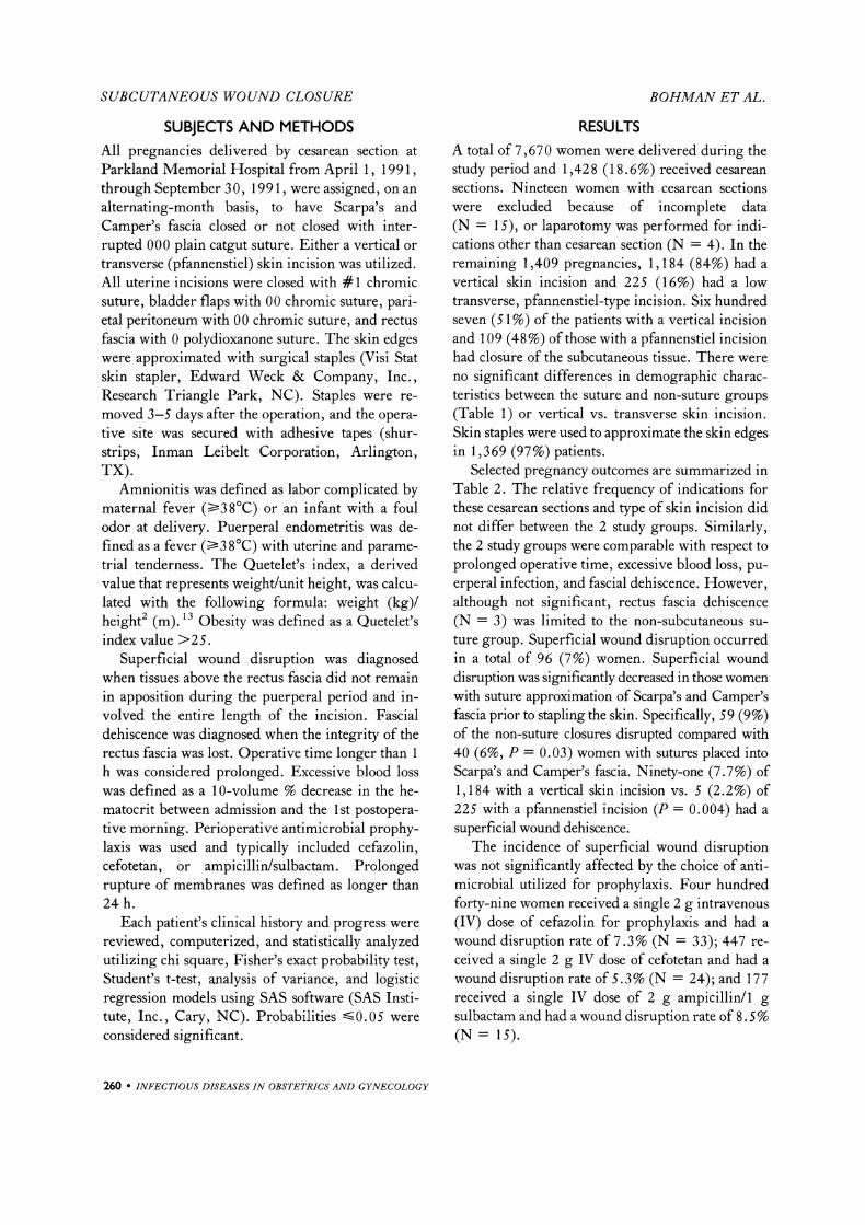
SUBJECTS AND METHODSAll pregnancies delivered by cesarean section at
Parkland Memorial Hospital from April 1, 1991,through September 30, 1991, were assigned, on an
alternating-month basis, to have Scarpa’s andCamper’s fascia closed or not closed with inter-rupted 000 plain catgut suture. Either a vertical ortransverse (pfannenstiel) skin incision was utilized.All uterine incisions were closed with #1 chromicsuture, bladder flaps with 00 chromic suture, pari-etal peritoneum with 00 chromic suture, and rectus
fascia with 0 polydioxanone suture. The skin edgeswere approximated with surgical staples (Visi Statskin stapler, Edward Weck & Company, Inc.,Research Triangle Park, NC). Staples were re-moved 3-5 days after the operation, and the opera-tive site was secured with adhesive tapes (shur-strips, Inman Leibelt Corporation, Arlington,TX).
Amnionitis was defined as labor complicated bymaternal fever (>38C) or an infant with a foulodor at delivery. Puerperal endometritis was de-fined as a fever (>3 gC) with uterine and parame-trial tenderness. The Quetelet’s index, a derivedvalue that represents weight/unit height, was calcu-lated with the following formula: weight (kg)/height (m). 1 Obesity was defined as a Quetelet’sindex value >25.
Superficial wound disruption was diagnosedwhen tissues above the rectus fascia did not remainin apposition during the puerperal period and in-volved the entire length of the incision. Fascialdehiscence was diagnosed when the integrity of therectus fascia was lost. Operative time longer thanh was considered prolonged. Excessive blood losswas defined as a 10-volume % decrease in the he-matocrit between admission and the 1st postopera-tive morning. Perioperative antimicrobial prophy-laxis was used and typically included cefazolin,cefotetan, or ampicillin/sulbactam. Prolongedrupture of membranes was defined as longer than24h.
Each patient’s clinical history and progress werereviewed, computerized, and statistically analyzedutilizing chi square, Fisher’s exact probability test,Student’s t-test, analysis of variance, and logisticregression models using SAS software (SAS Insti-tute, Inc., Cary, NC). Probabilities <0.05 wereconsidered significant.
RESULTSA total of 7,670 women were delivered during thestudy period and 1,428 (18.6%) received cesareansections. Nineteen women with cesarean sectionswere excluded because of incomplete data(N 15), or laparotomy was performed for indi-cations other than cesarean section (N 4). In theremaining 1,409 pregnancies, 1,184 (84%) had a
vertical skin incision and 225 (16%) had a lowtransverse, pfannenstiel-type incision. Six hundredseven (51%) of the patients with a vertical incisionand 109 (48%) of those with a pfannenstiel incisionhad closure of the subcutaneous tissue. There wereno significant differences in demographic charac-teristics between the suture and non-suture groups(Table 1) or vertical vs. transverse skin incision.Skin staples were used to approximate the skin edgesin 1,369 (97%) patients.
Selected pregnancy outcomes are summarized inTable 2. The relative frequency of indications forthese cesarean sections and type of skin incision didnot differ between the 2 study groups. Similarly,the 2 study groups were comparable with respect to
prolonged operative time, excessive blood loss, pu-erperal infection, and fascial dehiscence. However,although not significant, rectus fascia dehiscence(N 3) was limited to the non-subcutaneous su-ture group. Superficial wound disruption occurredin a total of 96 (7%) women. Superficial wounddisruption was significantly decreased in those womenwith suture approximation of Scarpa’s and Camper’sfascia prior to stapling the skin. Specifically, 59 (9%)of the non-suture closures disrupted compared with40 (6%, P 0.03) women with sutures placed intoScarpa’s and Camper’s fascia. Ninety-one (7.7%) of1,184 with a vertical skin incision vs. 5 (2.2%) of225 with a pfannenstiel incision (P 0.004) had a
superficial wound dehiscence.The incidence of superficial wound disruption
was not significantly affected by the choice of anti-microbial utilized for prophylaxis. Four hundredforty-nine women received a single 2 g intravenous(IV) dose of cefazolin for prophylaxis and had a
wound disruption rate of 7.3% (N 33); 447 re-ceived a single 2 g IV dose of cefotetan and had a
wound disruption rate of 5.3% (N 24); and 177received a single IV dose of 2 g ampicillin/1 gsulbactam and had a wound disruption rate of 8.5%(N 15).
260 INFECTIOUS DISEASES IN OBSTETRICS AND GYNECOLOGY

SUBCUTANEOUS WOUND CLOSURE BOHMAN ET AL.
TABLE I. Pregnancy demographics in 1,409 women with and without subcutaneous suturesat cesarean section
Subcutaneous suture
Demographic Yes Nofactor (N 716) (N 693) Comparison
Age (years) 25 6 25 -+ 6 NSGravidity 3 2 3 -+ 2 NSParity 2 - 2 -+ NSObesity 29 7 29 6 NSOperative time (min) 48 -+ 18 49 -+ 19 NSRupture of membranes (h) 5 +- 14 6 28 NSRace
Hispanic 331 (46%) 301 (43%) NSBlack 215 (30%) 235 (34%) NSWhite 153 (21%) 141 (20%) NSOther 17 (2%) 16 (2%) NS
Labor 416 (58%) 403 (58%) NSProlonged rupture of membranes 93 (I 3%) 100 (14%) NSDiabetes 34 (4%) 21 (3%) NSAmnionitis 52 (7%) 58 (8%) NS
aDifferences in the mean compared with Student’s t-test; differences in the frequency compared with chi square; obesity Quetelet’s index value >25[weight (kg)/height (m)]; prolonged rupture of membranes >24 h; amnionitis fever >38C in labor and/or foul-smelling infant at delivery; NS notsignificant.bMean - SD.CNumber with percentage in parentheses.
TABLE 2. Pregnancy outcomes in 1,409 women with and without subcutaneous sutures at cesarean sectiona
Subcutaneous suture
Yes NoOutcome (N 716) (N 693) Comparison
Indication for cesarean sectionRepeat 326 (46%) 300 (43%) NSDystocia 166 (23%) 177 (26%) NSAbnormal presentation 115 (16%) 96 (14%) NSFetal distress 95 (I 3%) 105 (I 5%) NSOther 14 (2%) 15 (2%) NS
Skin incision typePfannenstiel 109 (15%) 116 (17%) NSVertical 607 (85%) 577 (83%) NS
Prolonged operative time 109 (15%) 118 (17%) NSExcessive blood loss 32 (5%) 34 (5%) NSEndometritis 166 (23%) 169 (24%) NSFascial dehiscence 0 (0%) 3 (0.4%) NSSuperficial wound disruption 40 (6%) 59 (9%) 0.03
aFrequencies compared with chi square; prolonged operative time >1 h; excessive blood loss 10-volume % decrease in hematocrit; NS notsignificant.bNumber with percentage in parentheses.CFisher’s exact probability test.
Thirty-three (33%) of the 99 open wounds hadbacterial colonization, and 12 other wounds wereclinically infected because of cellulitis or purulentdrainage but no bacteria were isolated. Staphylococ-
cus epidermidis was present in 19 (58%) of openwounds, Staphylococcus aureus in 7 (21%), withStreptococcus pyogenes, Streptococcus agalactiae, En-terococci, gamma hemolytic streptococci, Coryne-
INFECTIOUS DISEASES IN OBSTETRICS AND GYNECOLOGY 261
SUBCUTANEOUS WOUND CLOSURE BOHMAN ET AL.
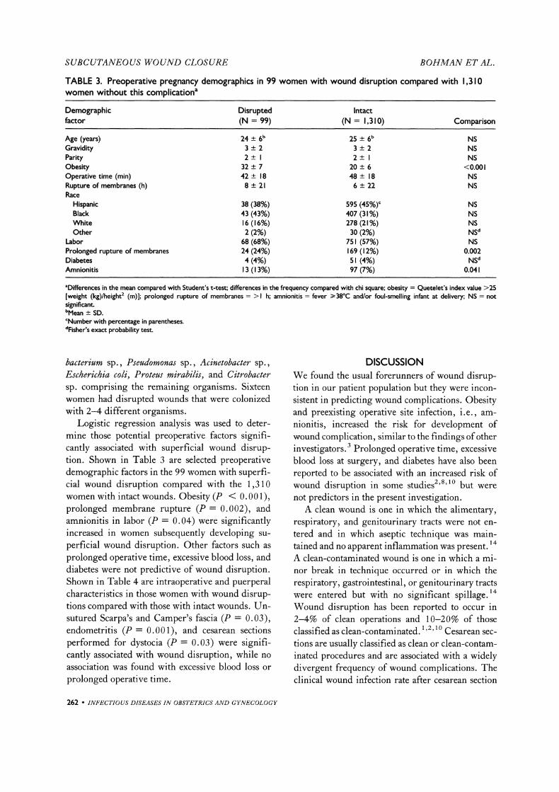
TABLE 3. Preoperative pregnancy demographics in 99 women with wound disruption compared with 1,310women without this complicationa
Demographic Disrupted Intactfactor (N 99) (N 1,310) Comparison
Age (years) 24 -+ 6 25 -+ 6 NSGravidity 3 -+ 2 3 -+ 2 NSParity 2 _+ 2 -+ NSObesity 32 _+ 7 20 -+ 6 <0.001Operative time (min) 42 -+ 18 48 -+ 18 NSRupture of membranes (h) 8 +- 21 6 +- 22 NSRace
Hispanic 38 (38%) 595 (45%) NSBlack 43 (43%) 407 (31%) NSWhite 16 (16%) 278 (21%) NSOther 2 (2%) 30 (2%) NS
Labor 68 (68%) 751 (57%) NSProlonged rupture of membranes 24 (24%) 169 (I 2%) 0.002Diabetes 4 (4%) S (4%) NSAmnionitis 13 (I 3%) 97 (7%) 0.041
aDifferences in the mean compared with Student’s t-test; differences in the frequency compared with chi square; obesity Quetelet’s index value >25[weight (kg)/height (m)]; prolonged rupture of membranes >1 h; amnionitis fever >38C and/or foul-smelling infant at delivery; NS not
significant.bMean +- SD.CNumber with percentage in parentheses.dFisher’s exact probability test.
bacterium sp., Pseudomonas sp., Acinetobacter sp.,Escherichia coli, Proteus mirabilis, and C#robactersp. comprising the remaining organisms. Sixteenwomen had disrupted wounds that were colonizedwith 2-4 different organisms.
Logistic regression analysis was used to deter-mine those potential preoperative factors signifi-cantly associated with superficial wound disrup-tion. Shown in Table 3 are selected preoperativedemographic factors in the 99 women with superfi-cial wound disruption compared with the 1,310women with intact wounds. Obesity (P < 0.001),prolonged membrane rupture (P 0.002), andamnionitis in labor (P 0.04) were significantlyincreased in women subsequently developing su-
perficial wound disruption. Other factors such as
prolonged operative time, excessive blood loss, anddiabetes were not predictive of wound disruption.Shown in Table 4 are intraoperative and puerperalcharacteristics in those women with wound disrup-tions compared with those with intact wounds. Un-sutured Scarpa’s and Camper’s fascia (P 0.03),endometritis (P 0.001), and cesarean sectionsperformed for dystocia (P 0.03) were signifi-cantly associated with wound disruption, while no
association was found with excessive blood loss or
prolonged operative time.
DISCUSSIONWe found the usual forerunners of wound disrup-tion in our patient population but they were incon-sistent in predicting wound complications. Obesityand preexisting operative site infection, i.e., am-
nionitis, increased the risk for development ofwound complication, similar to the findings ofotherinvestigators. 3 Prolonged operative time, excessiveblood loss at surgery, and diabetes have also beenreported to be associated with an increased risk ofwound disruption in some studies2’8’1 but werenot predictors in the present investigation.A clean wound is one in which the alimentary,
respiratory, and genitourinary tracts were not en-tered and in which aseptic technique was main-
14tained and no apparent inflammation was present.A clean-contaminated wound is one in which a mi-nor break in technique occurred or in which therespiratory, gastrointestinal, or genitourinary tractswere entered but with no significant spillage. 14
Wound disruption has been reported to occur in2-4% of clean operations and 10-20% of thoseclassified as clean-contaminated. 1,2,10 Cesarean sec-tions are usually classified as clean or clean-contam-inated procedures and are associated with a widelydivergent frequency of wound complications. Theclinical wound infection rate after cesarean section
262 INFECTIOUS DISEASES IN OBSTETRICS AND GYNECOLOGY
SUBCUTANEOUS WOUND CIOSURE BOHMAN ET AL.
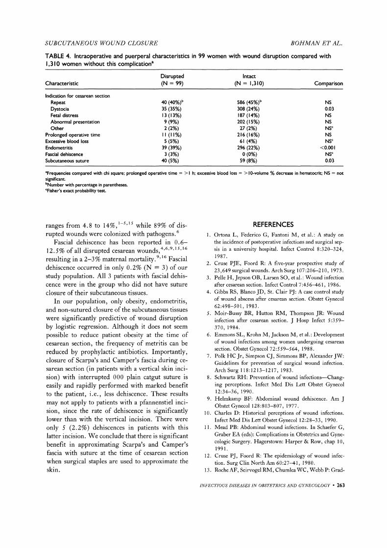
TABLE 4. Intraoperative and puerperal characteristics in 99 women with wound disruption compared with1,310 women without this complicationa
Disrupted IntactCharacteristic (N 99) (N 1,310) Comparison
Indication for cesarean section
Repeat 40 (40%) 586 (45%) NSDystocia 35 (35%) 308 (24%) 0.03Fetal distress 13 (I 3%) 187 (14%) NSAbnormal presentation 9 (9%) 202 (I 5%) NSOther 2 (2%) 27 (2%) NS
Prolonged operative time II (I I%) 216 (16%) NSExcessive blood loss 5 (5%) 61 (4%) NSEndometritis 39 (39%) 296 (22%) <0.001Fascial dehiscence 3 (3%) 0 (0%) NSSubcutaneous suture 40 (5%) 59 (8%) 0.03
aFrequencies compared with chi square; prolonged operative time >1 h; excessive blood loss > 10-volume % decrease in hematocrit; NS not
significant.bNumber with percentage in parentheses.CFisher’s exact probability test.
ranges from 4.8 to 14%, 1-5’1s while 89% of dis-rupted wounds were colonized with pathogens. 6
Fascial dehiscence has been reported in 0.6-12.5% of all disrupted cesarean wotlrlds, 4’6’9’ 15,16
resulting in a 2-3% maternal mortality. 9’ 16 Fascialdehiscence occurred in only 0.2% (N 3) of our
study population. All 3 patients with fascial dehis-cence were in the group who did not have suture
closure of their subcutaneous tissues.In our population, only obesity, endometritis,
and non-sutured closure of the subcutaneous tissueswere significantly predictive of wound disruptionby logistic regression. Although it does not seem
possible to reduce patient obesity at the time ofcesarean section, the frequency of metritis can bereduced by prophylactic antibiotics. Importantly,closure of Scarpa’s and Camper’s fascia during ce-sarean section (in patients with a vertical skin inci-
sion) with interrupted 000 plain catgut suture iseasily and rapidly performed with marked benefitto the patient, i.e., less dehiscence. These resultsmay not apply to patients with a pfannenstiel inci-sion, since the rate of dehiscence is significantlylower than with the vertical incision. There were
only 5 (2.2%) dehiscences in patients with thislatter incision. We conclude that there is significantbenefit in approximating Scarpa’s and Camper’sfascia with suture at the time of cesarean sectionwhen surgical staples are used to approximate theskin.
REFERENCES1. Ortona L, Federico G, Fantoni M, et al.: A study on
the incidence of postoperative infections and surgical sep-sis in a university hospital. Infect Control 8:320-324,1987.
2. Cruse PJE, Foord R: A five-year prospective study of23,649 surgical wounds. Arch Surg 107:206-210, 1973.
3. Pelle H, Jepson OB, Larsen SO, et al.: Wound infectionafter cesarean section. Infect Control 7:456-461, 1986.
4. Gibbs RS, Blanco JD, St. Clair PJ: A case control studyof wound abscess after cesarean section. Obstet Gynecol62:498-501, 1983.
5. Moir-Bussy BR, Hutton RM, Thompson JR: Woundinfection after cesarean section. J Hosp Infect 5:359-370, 1984.
6. Emmons SL, Krohn M, Jackson M, et al.: Developmentof wound infections among women undergoing cesarean
section. Obstet Gynecol 72:559-564, 1988.7. Polk HC Jr, Simpson CJ, Simmons BP, Alexander JW:
Guidelines for prevention of surgical wound infection.Arch Surg 118:1213-1217, 1983.
8. Schwartz RI--I: Prevention of wound infections--Chang-ing perceptions. Infect Med Dis Lett Obstet Gynecol12:34--36, 1990.
9. Helmkamp BF: Abdominal wound dehiscence. Am JObstet Gynecol 128:803-807, 1977.
10. Charles D: Historical perceptions of wound infections.Infect Med Dis Lett Obstet Gynecol 12:28-33, 1990.
11. Mead PB: Abdominal wound infections. In Schaefer G,Graber EA (eds): Complications in Obstetrics and Gyne-cologic Surgery. Hagerstown: Harper & Row, chap 10,1991.
12. Cruse PJ, Foord R: The epidemiology of wound infec-tion. Surg Clin North Am 60:27-41, 1980.
13. Roche AF, Seirvogel RM, Chumlea WC, Webb P: Grad-
INFECTIOUS DISEASES IN OBSTETRICS AND GYNECOLOGY 263
SUBCUTANEOUS WOUND CLOSURE BOHMAN ET AL.
ing body fatness from limited anthropometric data. Am JClin Nutr 34:2831-2838, 1981.
14. Olson M, O’Connor M, Schwartz ML: Surgical woundinfections. A 5-year prospective study of 20,193 woundsat the Minneapolis VA Medical Center. Ann Surg 199:253-259, 1984.
15. Poole GV Jr: Mechanical factors in the abdominal woundclosure: The prevention of fascial dehiscence. Surgery97:631-639, 1985.
16. Mowat J, Bonnar J: Abdominal wound dehiscence aftercaesarean section. Br Med J 2:256-257, 1971.
2ga4 INFECTIOUS DISEASES IN OBSVI’ETRICS AND GYNECOLOGY

Submit your manuscripts athttp://www.hindawi.com
Stem CellsInternational
Hindawi Publishing Corporationhttp://www.hindawi.com Volume 2014
Hindawi Publishing Corporationhttp://www.hindawi.com Volume 2014
MEDIATORSINFLAMMATION
of
Hindawi Publishing Corporationhttp://www.hindawi.com Volume 2014
Behavioural Neurology
EndocrinologyInternational Journal of
Hindawi Publishing Corporationhttp://www.hindawi.com Volume 2014
Hindawi Publishing Corporationhttp://www.hindawi.com Volume 2014
Disease Markers
Hindawi Publishing Corporationhttp://www.hindawi.com Volume 2014
BioMed Research International
OncologyJournal of
Hindawi Publishing Corporationhttp://www.hindawi.com Volume 2014
Hindawi Publishing Corporationhttp://www.hindawi.com Volume 2014
Oxidative Medicine and Cellular Longevity
Hindawi Publishing Corporationhttp://www.hindawi.com Volume 2014
PPAR Research
The Scientific World JournalHindawi Publishing Corporation http://www.hindawi.com Volume 2014
Immunology ResearchHindawi Publishing Corporationhttp://www.hindawi.com Volume 2014
Journal of
ObesityJournal of
Hindawi Publishing Corporationhttp://www.hindawi.com Volume 2014
Hindawi Publishing Corporationhttp://www.hindawi.com Volume 2014
Computational and Mathematical Methods in Medicine
OphthalmologyJournal of
Hindawi Publishing Corporationhttp://www.hindawi.com Volume 2014
Diabetes ResearchJournal of
Hindawi Publishing Corporationhttp://www.hindawi.com Volume 2014
Hindawi Publishing Corporationhttp://www.hindawi.com Volume 2014
Research and TreatmentAIDS
Hindawi Publishing Corporationhttp://www.hindawi.com Volume 2014
Gastroenterology Research and Practice
Hindawi Publishing Corporationhttp://www.hindawi.com Volume 2014
Parkinson’s Disease
Evidence-Based Complementary and Alternative Medicine
Volume 2014Hindawi Publishing Corporationhttp://www.hindawi.com