surface distribution and x-ray emission from …€¦ · surface distribution and x-ray emission....
TRANSCRIPT

Surface Distribution and X-Ray Emission From Scotch Tape
by
Kelly McGuire
A senior thesis submitted to the Department of Physics in partial fulfillment of the requirements for the degree of
Bachelor of Science
Department of Physics Brigham Young University – Idaho July –
2012
BRIGHAM YOUNG UNIVERSITY – IDAHO DEPARTMENT APPROVAL
of a senior thesis submitted by Kelly McGuire
This thesis has been reviewed by the research committee and senior thesis coordinator/advisor and has been found to be satisfactory.
Date David Oliphant, Senior Thesis Coordinator/Advisor
Date Kevin Kelley, Committee Member
Date Ryan Nielson, Committee Member
Date Stephen Turcotte, Department Chair


1
ABSTRACT
SURFACE DISTRIBUTION AND
X-RAY EMISSION FROM SCOTCH TAPE
Kelly McGuire
Department of Physics
Bachelor of Physics
Triboluminescence is an optical phenomenon in which light is generated when
certain materials are pulled apart, ripped or rubbed, and through the breaking of chemical
bonds. This observable effect is not fully understood; however, a few
strongly supported hypotheses are being developed to model the
triboluminescent event. It is believed that the separation and
ionization of electrical charges is the foundation for the creation of the
observed light and x-rays (figure1).
Time dependence of x-ray production and physical surface distribution was the
primary focus of my research. This research will help in the development and support of
current hypotheses, and may become the foundation for other theories in the future.
Other research teams such as Putterman’s UCLA group believe that finding a
definite mechanism for the x-ray emission of this type will allow them to harness the
energy more efficiently, which in turn will be used in applications such as medical devices
FIG. 1 Triboluminescent Light

2
to destroy tumors with bursts of x-rays. The Putterman's UCLA group believes there may be a
potential application to detect x-ray emissions from triboluminescent materials as they start to
fatigue.
ACKNOWLEDGMENTS
I would like to thank the Brigham Young University- Idaho Department of Physics for
the opportunity to perform research that has helped me to develop my scientific skills as
well as prepare me for future endeavors. I would also like to thank David Oliphant, Kevin
Kelley, and Ryan Nielson for the guidance and advice that was given to help in the
completion of this thesis. As well, I would like to acknowledge Karl Decker for his
collaboration and insights on this project, and Jon Wilson for his help in designing the
new chamber.

3
Table of Contents
Chapter 1
History…………………………………………………………………………………………………………..4 1.1 Discovery of Triboluminescence with Adhesives…………………………………4
1.2 Previous/Current Experiments……………………………………………………………5
1.3 Bremsstrahlung Radiation………………………………………………………………….7
1.4 Overview of My Research………………………………………………………………….8
Chapter 2 Methods and Equipment………………………………………………9
2.1 Previous Chamber………………………………………………………………………………9
2.2 Design of New Chamber……………………………………………………………………10
2.3 Experimental Setup……………………………………………………………………………12
2.4 Physical Structure of Tape Surface before Experiment………………………13
2.5 Time Dependent Parameters…………………………………………………………….18
2.6 Velocity Dependent Methods…………………………………………………………….19
Chapter 3 Results…………………………………………………………………………………..20
3.1 Physical Structure of Tape Surface After Experiment………………………..20
3.2 Time Dependence of X-Ray Count……………………………………………………..25
3.3 Velocity Dependence…………………………………………………………………………27
Conclusion…………………………………………………………………………………………………30
Bibliography……………………………………………………………………………………………..32
Appendix……………………………………………………………………………………………………33

4
Chapter 1 History
1.1 Discovery of Triboluminescence with Adhesives
The first hint of x-rays in triboluminescence appeared in a paper by J.W.
Obreimoff on, “The splitting strength of mica.” In addition to studying the ability of
polished glass plates to re-adhere to one another after splitting, Obreimoff describes
some electrical phenomena which appear when the mica is split in a high vacuum. In
anticipation of the results relating to Scotch tape, Obreimoff notes,
“If split in darkness, mica becomes slightly luminescent (triboluminescence). This is due
to electric discharges between the mica surfaces through the air. If we split them under
an air pressure of 1.0-0.1 mm. mercury the glow spreads to all the air in the vessel and
is similar to the glow of a Geissler tube. In a high vacuum ( mm. mercury)
the glass of the vessel fluoresces like an X-ray bulb. The light is feeble and can be
observed only after the eye has rested about 3 minutes in darkness.” 1
This fluorescing of the glass of the vessel suggested the presence of x-rays, and it
only appears when a vacuum is present, just like the tape experiments to be described.
I did not include mica in my experiments, but I believe there might be value in
studying triboluminescence further by future researchers.

5
1.2 Previous and Current Experiments Triboluminescence without x-rays in the peeling of tape has been studied by
many researchers. Seminal work by Dickenson2 included bursts of light having
nanosecond duration in 1988 and measurement of current flow from tape peeled from
metal surfaces in 1995. Also Miura in 1997 correlated the electrical discharge with the
fracture of adhesive filaments in tape.3
In these seminal works any x-rays that accompanied the observed
triboluminescence from peeling tape were not reported. In this regard, it is worthwhile
to note that x-rays were discovered by Röntgen in 1895 while experimenting with the
cathode ray tube (CRT). CRTs are devices which the positive anode attracts electrons
from the negative cathode in a vacuum. The electrons upon collision with the anode
emit x-rays by bremsstrahlung (further description can be found in section 1.3).
Recently, x-rays were reported by Camara4 at UCLA by peeling 3M brand Scotch tape at
steady 3 cm/s in a vacuum. The tape did not emit x-rays continuously, but in short
nanosecond bursts – accumulating
enough energy to produce an x-ray
image of a finger in a second (see
Figure 2). Most brands of clear
adhesive tape also give off x-rays, FIG. 2 X-ray image of finger

6
albeit with a different spectrum of energies, although why duct tape does not emit X-
rays is not explained.
Researchers at the University of Illinois at Urbana-Champaign also began conducting
experiments that exploited the effect to shed light -- literally -- on how materials fracture. They
published their findings in the 2007 edition of the Journal of the American Chemical Society.5
The Illinois researchers had to figure out some way to amplify the triboluminescent effect in
order to glean useful information from the fracture point. Suslick and his collaborators filled a
test tube with a semiliquid mixture of small sugar crystals and liquid paraffin and then immersed
a vibrating titanium rod into it. This generated ultrasound waves, creating acoustic cavitation
(lots of tiny bubbles constantly growing and collapsing in the paraffin). The shock waves caused
the sugar crystals to collide, nitrogen and oxygen bubbled through the semiliquid mixture, and
the result was bursts of light 100 to 1000 times brighter than the usual triboluminescence. So far
they've found the presence of carbon monoxide, CO2 ions, and other products of combustion,
and are now working on determining the chemical reactions taking place during
triboluminescence
Concerning the research that has been done at Brigham Young University –
Idaho, Jarom Decker has provided me with a good foundation to continue his research.
The design of his chamber (described in section 2.1) is the foundation for the design of
the current chamber design (described in section 2.2). Jarom’s experiment was aimed to
understand the fundamental processes that produce the x-rays, to determine angular
dependence between the tape and the spool, and to observe the time dependence of
total x-ray count.

7
Just to be brief, the results for the time dependence experiment showed a
decrease in x-ray count and a conglomeration of glue after 30 minutes. For the angular
distribution experiment, results did not show a definitive conclusion for angular
dependence. Jarom accredited the inability of showing an angular distribution of x-rays
to certain flaws in the apparatus he was using at the time.
1.3 Bremsstrahlung Radiation
Electromagnetic radiation is produced by the deceleration of a charged particle
when deflected by another charged particle, typically an electron by an atomic nucleus.
The moving particle loses kinetic energy, which is converted into a photon because
energy is conserved. The term is also used to refer to the process of producing the
radiation. "Bremsstrahlung" means "braking radiation" and is retained from the original
German to describe the radiation which is emitted when electrons are decelerated or
"braked" when they are fired at a metal target.
Accelerated charges give off electromagnetic radiation, and when the
energy of the bombarding electrons is high enough,
that radiation is in the x-ray region of the
electromagnetic spectrum. It is characterized by a
continuous distribution of radiation which becomes
more intense and shifts toward higher frequencies
FIG. 3 Bremsstrahlung Curves

8
when the energy of the bombarding electrons is increased. Figure 3 shows
Bremsstrahlung radiation curves that were produced when electrons of four different
energies bombarded tungsten targets. The Bremsstrahlung radiation curves are easily
reproducible when working with radiation in the x-ray region, and are very distinct and
recognizable from other types of curves that are produced by radiation.
1.4 Overview of My Research Time dependence of x-ray production and physical surface distribution was the
primary focus of this research. The goal was to determine if the change in surface
characteristics has any direct influence of charge build-up and discharge, and if so to
what extent.
Most of the research discussed in this paper will concentrate on the dynamics of
the tape’s surface and charge distribution, and the effects on x-ray emission by changing
the parameters of the experiment such as the surface area of the tape.
Another part of my research was to determine if the x-ray count was dependent
on changes in velocity, and if there was a lower limit and upper limit on rotational
velocity that would inhibit the emission of x-rays from the Scotch Tape.
Jarom Decker was running experiments towards the end of his research on this
project, in which he was able to produce results that suggested a velocity dependent x-
ray emission. My research picked up where he left off, and my results will be shown in
the discussion that follows.

9
Chapter 2 Methods and Equipment
2.1 Previous Chamber
The previous chamber was made from PVC pipe that consisted of a 6” diameter
chamber reduced to 1.5” at the bottom allowing for connection to a roughing pump. A
seal made from polyurethane and an acrylic plate sealed the chamber on top. Attached
to the acrylic plate was the apparatus used to dispense tape.
The acrylic plate was modified to mount an electric stepper motor and later a
mounting for multiple detectors, for measuring the angle distribution of the x-rays.
Aware Electronics RM-60 Geiger counters were used to detect x-rays for time
dependence and in angle distribution experiments. An AMPtek XR-100 CdTe x-ray
detector was used to measure the energy of the x-rays. Figure 4a below shows the
chamber in complete experimental setup. Figure 4b shows the step motor system, and
Figure 4c shows the spindles used for holding and peeling the tape.
Jarom made note in his thesis of some of the difficulties that he faced with the
old chamber design and equipment materials. The previous chamber varied greatly in
its ability to reach sufficient vacuum conditions. The seal was created by using an acrylic
top which was placed on top of an irregularly shaped rubber-like material. This

10
provided a strong and consistent seal in the vacuum system. The vacuum chamber was
directly connected to the pipe of the roughing pump.6
Outgassing was a constant challenge due to the difficulty in creating a closed
system and sealing the interior of the chamber from the exterior environment. The
aluminum foil lining inhibited our ability to detect an even distribution of x-ray emission.
The curvature of the spindles did not provide us with a dependable design where we
could guarantee that the tape would remain on the spindles. At times the tape would
fall off of the spindles and the experiment would be suspended, and the data had to be
thrown out. Other times, the tape would break because the two spools were not of the
same type, shape, or size.
FIG. 4a Complete Experimental Setup FIG. 4b Step Motor System

11
2.2 Design of New Chamber
Jon Wilson, a former mechanical engineer student, was the main developer of
the new chamber. It was decided between Jon, Karl Decker, and myself to use an acrylic
chamber to maximize the detection of x-ray emissions.
The previous design did not allow for continuous x-ray production, which created
challenges in consistency for experiments that required consistency such as time
dependence and velocity dependence.
Figures 5a and 5b show the complete setup of the new chamber. Notice that the
bulk of the operating components such as the stepper motor and vacuum pressure
gauge have been placed on the bottom of the chamber. This design allows the user to
access the interior of the chamber easily, which decreases the effort to set up the
experiment and change out the samples.
FIG. 5a Complete Experiment Setup

12
2.3 Experimental Setup
When the electrons are suddenly decelerated upon collision with the target
material, x-rays are produce; these x-rays produce the Bremsstrahlung radiation
previously discussed. If the bombarding electrons have sufficient energy, they can knock
an electron out of an inner shell of the target material atoms. Then, electrons from
higher states drop down to fill the vacancy, which emit x-ray photons with precise
energies determined by the electron energy levels.
If the electrons were to be accelerated at normal atmospheric conditions, there
would be tremendous amounts of matter in the air creating a barrier, which in turn
would not permit the electrons to reach the target material, nor reach sufficient
energies to emit x-rays. Thus, a considerable vacuum is required so that the amount of
matter is reduced to virtually nothing.
FIG. 5b Operating Equipment Below Chamber

13
The process of preparing the chamber is perhaps the most important and crucial
step in the setup of the experiment. In order to obtain significant x-ray emission, the
chamber must reach a substantial vacuum.
The process of preparing the chamber begins with cleaning all of the parts and
surfaces to be exposed to vacuum conditions with an alcohol (isopropyl seems to work
the best). For a vacuum chamber to reach its optimum pressure capacity, all sources of
out-gassing must be reduced. Evaporation and sublimation into a vacuum is called
outgassing. All materials, solid or liquid, have a small vapor pressure, and their
outgassing becomes important when the vacuum pressure falls below this vapor
pressure. In vacuum systems, outgassing has the same effect as a leak and can limit the
achievable vacuum.
The greatest barrier for this chamber to reach sufficient vacuum levels is the oil
from the researcher’s hands. The best method to reduce the transfer of oils to the tape
and the interior parts of the chamber is to wear latex gloves while preparing the
chamber.
Once the internal components of the chamber have been optimally cleaned and
prepped, we then added the tape to the spools. This procedure7 is quite delicate, and
can pose many problems while the experiment is running if not done correctly. One of
our biggest challenges during our experiments was to keep the tape from either falling
off the spools or breaking from too much tension. We corrected this problem by cutting
the width of the tape in half.

14
Once the tape has been placed on the spools correctly, we pump down the
chamber but do not start the stepper motor until the pressure in the chamber has
reached 8 milli-torr.
The x-ray detector we used is the XR-T100 X-RAY Detector and was used with the
MCA (Multi Channel Analyzer). Karl Decker’s thesis gives additional description of the
experimental setup and x-ray detector.7
2.4 Physical Structure of Tape Surface Before For our experiments, we used original matte finish Magic Scotch Tape ¾ “x 650”
from 3M. The adhesive structure of Scotch tape is fascinating to study with the use of
high powered microscopes. We were not able to obtain the physical structure
composition from 3M due to company policies and trade secrets.
Therefore, we used microscopy techniques to create a 3-dimensional height map
in order to study the physical features of the tape surface. Figures 6a and 6b are
topographies obtained by researchers from the University of Basel in Switzerland using
white-light interferometry and 3D optical metrology.7
FIG. 6a Scotch Tape Topography FIG. 6b Scotch Tape Topography Low magnification (5x) High magnification (50x)

15
The topography gives us a clear picture of variation in heights of the surface
components. The heights of the surface components before experiments were
performed ranged from 0 micrometers to about 10 micrometers. The red sections show
the highest points on the surface, while the blue indicates the lowest points. As
indicated in the figures above, there are clumps of hills and valleys. From this
microscopic observation, a hypothesis for time dependence was proposed: could it be
that 30 minutes is the maximum time for optimum x-ray count because these clumps
flatten and decrease, thus inhibiting discharge? As well, is the x-ray emission dependent
on the initial height and distribution of the clumps of valleys and hills? If so, is it more
dependent on the hills or the valleys? In other words, where is charge distribution
strongest on the surface of the tape?
A control sample was needed for comparison between the before and after
samples of the scotch tape pieces. In order to accomplish the task of mapping the
physical features of the adhesive surface, the z-stack tool in the PhotoSuite software
that came with the Olympus BX61 high powered microscope was used. The microscope
uses the z-stack by taking multiple pictures at different heights, and the change in
distance between each picture is specified by the user. For example, I set the
microscope in the PhotoSuite software to take 200 grayscale pictures from a minimum
height of 1 micrometer to a maximum height of 10 micrometers at 0.1 micrometer
distances.

16
The process included the creation of height maps by converting the 200
grayscale images obtained from using the z-stack tool, and then superimposing them to
form a three dimensional object.
The procedure was very sensitive, requiring the user to have a working
knowledge of the Olympus BX61 (figure 7a, 7b, and 7c) microscope, and a perspective
that allowed the user to visualize a 2-dimensional surface in 3-dimensions.
FIG. 7a Olympus BX61 Setup FIG. 7b SD70 Camera Attachment

17
FIG. 7c Automated Objective and Stage
System

18
The microscope shown above was used not only to help us visualize the surface
structure in three dimensions, but was also used to obtain 2-dimensional images as
shown in figure 8a and 8b. This allowed me to see more detailed changes in the surface
of the adhesive side of the tape such as patches of missing glue as shown in figure 8b
with the red circles.
FIG. 8a 2-Dimensional Bright
Field Image of Adhesive Side
FIG. 8b Wider View of 2-
Dimensional Bright Field Image
of Adhesive Side

19
2.5 Time Dependent Parameters
As we studied the data collected from previous experiments run by Jarom, we
noticed a cut-off point for x-ray counts. Consistently, at approximately 30 minutes of
runtime, the x-ray count would drop of dramatically. My goal was to test this
consistency by changing the parameters of the experiment.
The first parameter that I decided change was the width of the tape, which
would decrease the surface area, thus decreasing the total number of “hills” that could
store charge as the tape unraveled from the spools. Other articles had previously
reported testing the proportionality of surface area to x-ray emission, but their goal was
to determine if the total x-ray count would diminish or if the mechanism for
triboluminescence was dependent on the amount of tape used.
Their results showed relatively no decrease in x-ray count with decreasing
surface area. Having studied their results, I did not expect to see a change in the total
time to reach the critical x-ray emission or rather the point where x-ray emission began
to drop off dramatically.
The second parameter that I changed was the pressure in the vacuum chamber.
I ran the tape at different pressures ranging from 10 milli-torr to 3 milli-torr. The idea
behind changing the pressure was that a certain amount of stress needed to be applied
to create conditions suitable for triboluminescence.

20
2.6 Velocity Dependent Methods
I observed that changes in the velocity of the tape unraveling from the spools
caused change in the x-ray count. Tests were done to determine if there was a
significant difference between x-ray count and different velocities. The previous
chamber was not designed to allow for consistency in experimental methods, meaning it
was very difficult to repeat the same experiment twice. The new chamber, however,
did provide better dependability so that we could repeat the same experiment without
too much uncertainty between runs.
The current hypothesis is that as the tape unravels at higher velocities around
the spools, the glue flows more quickly and does not allow for charge to build up on the
tape’s surface. In other words, glue flow seems to be dependent on velocity. Figures 9a
and 9b demonstrate what this would look like at an instantaneous point in the peeling
process.8
FIG. 9b High peeling velocity. Red hatched
area shows where energy dissipation is the
greatest.
FIG. 9a Low peeling velocity

21
The method for determining the upper and lower limits on x-ray production with
velocity was quite simple. We performed tests at different velocities. I did not,
however, perform this experiment multiple times. I peeled the tape at five different
speeds (30 cm/s, 40 cm/s, 42 cm/s, 51 cm/s, and 59 cm/s), and did not repeat the
experiment again.
As mentioned in the experimental set up section, cutting the width of the tape in
half provided us with a solution to the tape moving up and down the spools and
breaking off of the spools. Being able to keep the tape in relatively the same position on
the spool for approximately the same amount of time for the five different speeds
helped to reduce the uncertainty in our experiment.
Chapter 3 Results 3.1 Physical Structure of Tape Surface After
After the samples had been placed on the spools and allowed to peel for
different times (5, 10, 15, and 30 minutes), the same process described in section 2.4
was implemented to create a three-dimensional object of the tape’s surface to observe
any physical changes.
It was observed that certain areas of the tape’s surface showed an absence of
glue which created greater spacing between the “hills”, and increased the amount of

22
valleys in the height map. Figures 10a, 10b, 10c, and 10d show the 2-Demsional images
of the adhesive side after 5, 10, 15, and 30 minutes respectively along with their
corresponding 3-dimensional height maps.
The red circles are placed around areas where there is significant loss of glue. It
seems that the glue is being relocated to different regions of the tape or is being
deposited onto the spool as the tape comes in contact with it during the peeling
process. The height maps show an increasing area of valleys (green) as the tape peels
for longer periods of time.
FIG. 10a Adhesive Side After 5
Minutes of Peeling
FIG. 10a-2 Height Map After 5
Minutes

23
FIG. 10b Adhesive Side After 10
Minutes of Peeling
FIG. 10b-2 Height Map After 10
Minutes

24
FIG. 10c Adhesive Side After 15
Minutes of Peeling
FIG. 10c-2 Height Map After 15
Minutes

25
FIG. 10d Adhesive Side After 30
Minutes of Peeling
FIG. 10d-2 Height Map After 30
Minutes

26
3.2 Time Dependence of X-ray Count
A strong correlation between time and x-ray emission was observed. For the
majority of the experiments, x-ray counts decreased as the time increased for the tape
peeling. However, it was also observed that approximately 30 minutes of runtime, the
x-ray emission would drop off to virtually zero. This phenomenon occurred consistently
for every sample.
In order to confirm this correlation, we ran the experiment for different time
periods. The time intervals for this portion of the experiment were 5, 10, 15, and 30
minutes. For the 5, 10, and 15 trials, the x-ray count did decrease, but not as rapidly as
the decrease at 30 minutes of runtime.
The graph below (Figure 11) was plotted in Logger Pro, and the data points were
plotted to show time dependence. A best fit was added to the graph, and we were able
to obtain an equation for the time dependence of the experiments for these different
time intervals:
( )
I need to note that the LoggerPro graph has not been calibrated for real-time,
meaning that the scale of the graph does not represent the time correctly. With that
being said, the graph still indicates a time dependent x-ray emission.

27
Both the graph and the data points in the table (Appendix 1, A1.) confirm a time
dependent system for x-ray emission from the Scotch Tape. One observation to note,
looking at the x-ray counts for the different channels, at times there are bursts of
FIG. 11 LoggerPro Graph of
Counts Per Bin vs. Bin Number
(Bin Number)
Counts Per
Bin

28
increased x-ray counts. For example, channels 128 through 132 there is a relatively
steady decrease, then the counts jump up to 9 and 10 in channels 133 and 134. I
believe that the surface of the tape still has some significantly high hills that allow for
electrical charge separation, ionization, and discharge.
Karl and I discussed the results concerning pressure dependency and x-ray count,
which he has reported in his thesis, but I will just say that x-ray counts increased as the
pressure decreased, and optimum x-ray emission occurred below approximately 8 milli-
torr.7
3.3 Velocity Dependence
As displayed in Figure 12, we notice a decrease in the amplitude of the x-ray
curve, while the speed of the tape increases.
The changes were quite significant, and the results seemed consistent with a
proportional change in x-ray counts to a change in velocity. As shown in the data, the x-
ray counts are inversely related to the speed of the tape. However, there seems to be
one exception that we observed, and that is at 42 cm/s the x-rays had the highest
energy and produced the highest x-ray count.
Even though Figure 12 suggests that there may be velocity dependence for x-ray
emission, the data is inconclusive. I performed this experiment only once for each of
the five speeds. I used the first vacuum chamber design, and challenges existed in being
consistent with the pressure. There exists a large uncertainty in the pressures during
this experiment.

29
Also, the stepper motor being used at the time was not functioning properly and
it was not very reliable in being consistent with the five different speeds, meaning there
is a large uncertainty in the speeds reported.
The last reason for this data being inconclusive is that we have an uncertainty in
the position of the XR-T100 X-Ray detector. We did not account for the position of the
detector for each of the five different speeds.
0
20
40
60
80
100
120
12
14
16
18
11
01
12
11
41
16
11
81
20
12
21
24
12
61
28
13
01
32
13
41
36
13
81
40
14
21
44
14
61
48
15
01
52
15
41
56
15
81
60
1
X-R
ay C
ou
nts
X-Ray Energy (keV)
X-Ray Emission at Varying Tape Speeds
<42 cm/s
<42 cm/s
42 cm/s
51 cm/s
59 cm/s
FIG. 12 X-ray Emission With 5
Different Speeds

30
FIG. 13 Speed Dependence With Error Bars
31
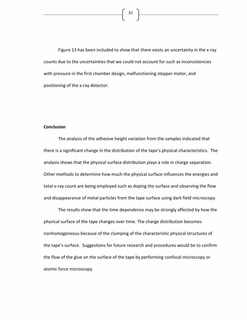
Figure 13 has been included to show that there exists an uncertainty in the x-ray
counts due to the uncertainties that we could not account for such as inconsistencies
with pressure in the first chamber design, malfunctioning stepper motor, and
positioning of the x-ray detector.
Conclusion
The analysis of the adhesive height variation from the samples indicated that
there is a significant change in the distribution of the tape’s physical characteristics. The
analysis shows that the physical surface distribution plays a role in charge separation.
Other methods to determine how much the physical surface influences the energies and
total x-ray count are being employed such as doping the surface and observing the flow
and disappearance of metal particles from the tape surface using dark field microscopy.
The results show that the time dependence may be strongly affected by how the
physical surface of the tape changes over time. The charge distribution becomes
nonhomogeneous because of the clumping of the characteristic physical structures of
the tape’s surface. Suggestions for future research and procedures would be to confirm
the flow of the glue on the surface of the tape by performing confocal microscopy or
atomic force microscopy.

32
There remain myriad avenues to explore in the pursuit of determining the
definite mechanism for this triboluminescent phenomenon. Some of those include
determining an angular distribution with less uncertainty. If one were to continue
observing the physical surface distribution using microscopy, a better method would be
to use white-light interferometry, and to create a logarithmic topographical map using
atomic force microscopy.
There exist multiple hypotheses that provide good approximations and a strong
foundation for future researchers to follow. The goal will be to narrow the gap between
the current hypotheses and to reduce them to one very strong hypothesis that can
provide much better predictions.

33
Bibliography
[1] Obreimoff, J. W. Proceedings of the Royal Society of London. Series A,
Containing Papers of a Mathematical and Physical Character Vol. 127, No. 805
(May 7, 1930), pp. 290-297
[2] Prevenslik, T., 2008, A Unified Theory of Natural Electrification, Proc. of 6th
Int. Conf. App. Electrostatics, Shanghai Maritime University, November 3-7, pp.
60-63.
[3] Miura, T., Chini, M., and Bennewitz, R., 2007, “Forces, charges, and light
emission during the rupture of adhesive contacts,” J. App. Phys., 103, pp.103509.
[4] C. G. Camara et al, Correlation Between Nanosecond X-ray Flashes and
Stick-Slip Friction in Peeling Tape. Nature 455, 1089 (2008).
[5] Suslick, Kenneth S., Evidence for a Plasma Core during Multibubble
Sonoluminescence in Sulfuric Acid. J. Am. Chem. Soc., 2007, 129 (13), pp 3838–
3839
[6] Decker, J.(2010). The study of x-rays from tape. In The Study Of X-rays From
Tape (pp. 1-33). Brigham Young University – Idaho, Rexburg, 2011
[7] Decker, K. (2012). The Time Dependence of the X-ray Triboluminescence of
Adhesive Tape (pp. 1-36). Brigham Young University – Idaho, Rexburg, 2012
[8] Gorb, S. N. (2010). Surface roughness of peeled adhesive tape: A mystery?. In
Surface roughness of peeled adhesive tape: A mystery? (pp. 1-5). Julich, Germany

34
Appendix 1
A1. LoggerPro data for Figure 10
Bin Number
Counts Per Bin
1 327 69 30
2 180 70 27
3 176 71 14
4 163 72 20
5 167 73 14
6 157 74 27
7 139 75 16
8 135 76 24
9 112 77 13
10 132 78 17
11 121 79 12
12 120 80 18
13 107 81 17
14 100 82 16
15 105 83 19
16 100 84 18
17 82 85 5
18 69 86 17
19 85 87 14
20 75 88 12
21 78 89 10
22 69 90 8

35
23 72 91 12
24 81 92 8
25 61 93 5
26 47 94 13
27 65 95 8
28 62 96 10
29 65 97 6
30 58 98 5
31 54 99 11
32 66 100 9
33 55 101 13
34 48 102 11
35 55 103 8
36 40 104 6
37 33 105 7
38 39 106 12
39 37 107 10
40 37 108 12
41 44 109 11
42 51 110 9
43 37 111 8
44 40 112 6
45 28 113 13
46 25 114 10
47 45 115 14
48 27 116 10
49 38 117 5
50 31 118 5
51 30 119 9
52 31 120 4
53 37 121 5
54 37 122 4
55 31 123 7
56 35 124 3
57 31 125 6
58 28 126 7
59 18 127 5
60 28 128 4
61 31 129 6
62 22 130 4
63 14 131 7
64 31 132 2
65 22 133 9

36
66 26 134 10
67 18 135 6
68 19 136 1
137 9
138 6
139 5
140 3
141 1