technology in palm of hand ms32017-2018
TRANSCRIPT


Technology in the Palm of your Hand MS3 August 2017
Goals:
1. Pull together MS 1 & 2 data and bring it to the bedside
2. Appreciate the Power of the History and Physical
3. Learn the concept of correlating clinically
4. Become familiar with the multitude of ancillary cardiac testing that are available
Suggestions before our session
Review lectures in Year 1 and 2 from Drs.' Robia, Samarel, McKiernan and Kristopaitis
while enjoying your favorite beverage or my choice Ice Cream
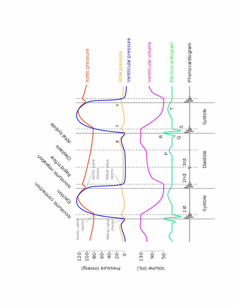
Spend 20 minutes starring at a Wigger's Diagram
Go to your learning resource center and listen to the heart sounds tutorial or visit the
Harvey manikin in the Med School
Bring to class a patients history or exam that is memorable or was challenging
Bring to class any weird ECG's you have run across
References:
Your personal choice for Internal Medicine textbook: Cecil's, Harrison's or other
Chapter on the Cardiac history and physical
www.blaufuss.org
Go thru their free heart sounds tutorial
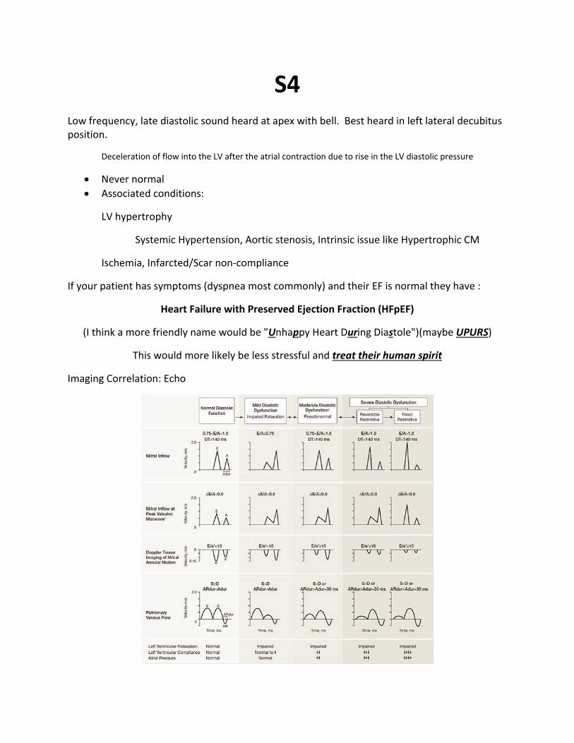
S4 Low frequency, late diastolic sound heard at apex with bell. Best heard in left lateral decubitus position.
Deceleration of flow into the LV after the atrial contraction due to rise in the LV diastolic pressure
Never normal
Associated conditions:
LV hypertrophy
Systemic Hypertension, Aortic stenosis, Intrinsic issue like Hypertrophic CM
Ischemia, Infarcted/Scar non‐compliance
If your patient has symptoms (dyspnea most commonly) and their EF is normal they have :
Heart Failure with Preserved Ejection Fraction (HFpEF)
(I think a more friendly name would be "Unhappy Heart During Diastole")(maybe UPURS)
This would more likely be less stressful and treat their human spirit
Imaging Correlation: Echo
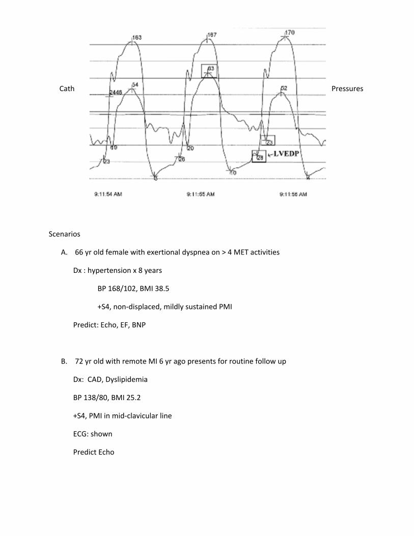
Cath Pressures
Scenarios
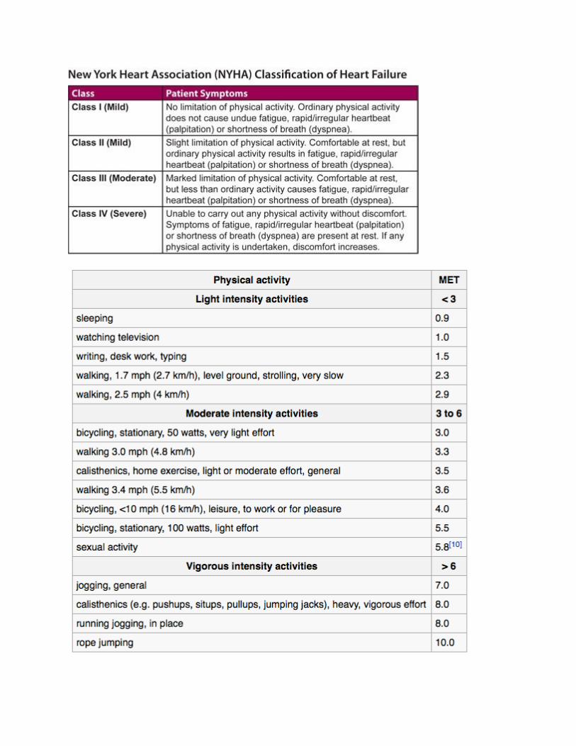
A. 66 yr old female with exertional dyspnea on > 4 MET activities
Dx : hypertension x 8 years
BP 168/102, BMI 38.5
+S4, non‐displaced, mildly sustained PMI
Predict: Echo, EF, BNP
B. 72 yr old with remote MI 6 yr ago presents for routine follow up
Dx: CAD, Dyslipidemia
BP 138/80, BMI 25.2
+S4, PMI in mid‐clavicular line
ECG: shown
Predict Echo

S3 Low frequency, early diastolic sound heard at apex with bell, best in the LL decubitus position
Deceleration of flow early in diastole due to finding the LV still full most commonly
due to impaired ejection.
Can be normal in youth/health due to the extremely high volume, rapid early filling phase
"Physiologic S3"
Associated with LV and reduced ejection fraction
If your patient has symptoms then it is called:
Heart Failure with Reduced Ejection Fraction (HFrEF)
Associated with either ischemic or non‐ischemic dilated cardiomyopathy
Assessment of EF:
PMI, precordial activity
Echo, CT, MRI, LV angiogram, Nuclear Medicine (SPECT, MUGA)
Stage and functional class:


Scenario:
48 yr old with 4 months of progressive SOB now having symptoms with ADL's and waking up
with a "smothering" sensation.
Denies chest pain
Exam: 108/88, 106, 22
JVP 8 cm at 45 degrees, Bibasilar rales, PMI in Anterior Axillary Line with "Gallop"
16 cm liver and 3 + edema
Diagnosis
Prognosis
Treatment
"I want to rule out CAD" What is your goal?
To detect "any" then do an autopsy ( A bit aggressive, but very accurate)
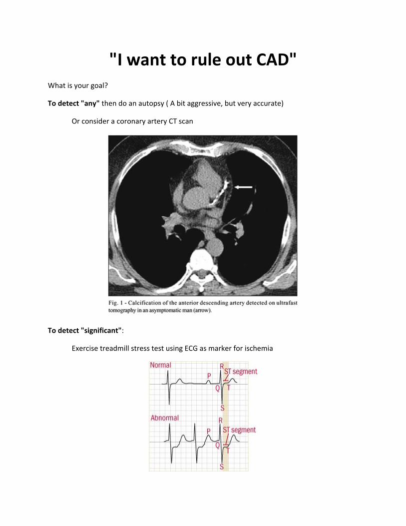
Or consider a coronary artery CT scan
To detect "significant":
Exercise treadmill stress test using ECG as marker for ischemia
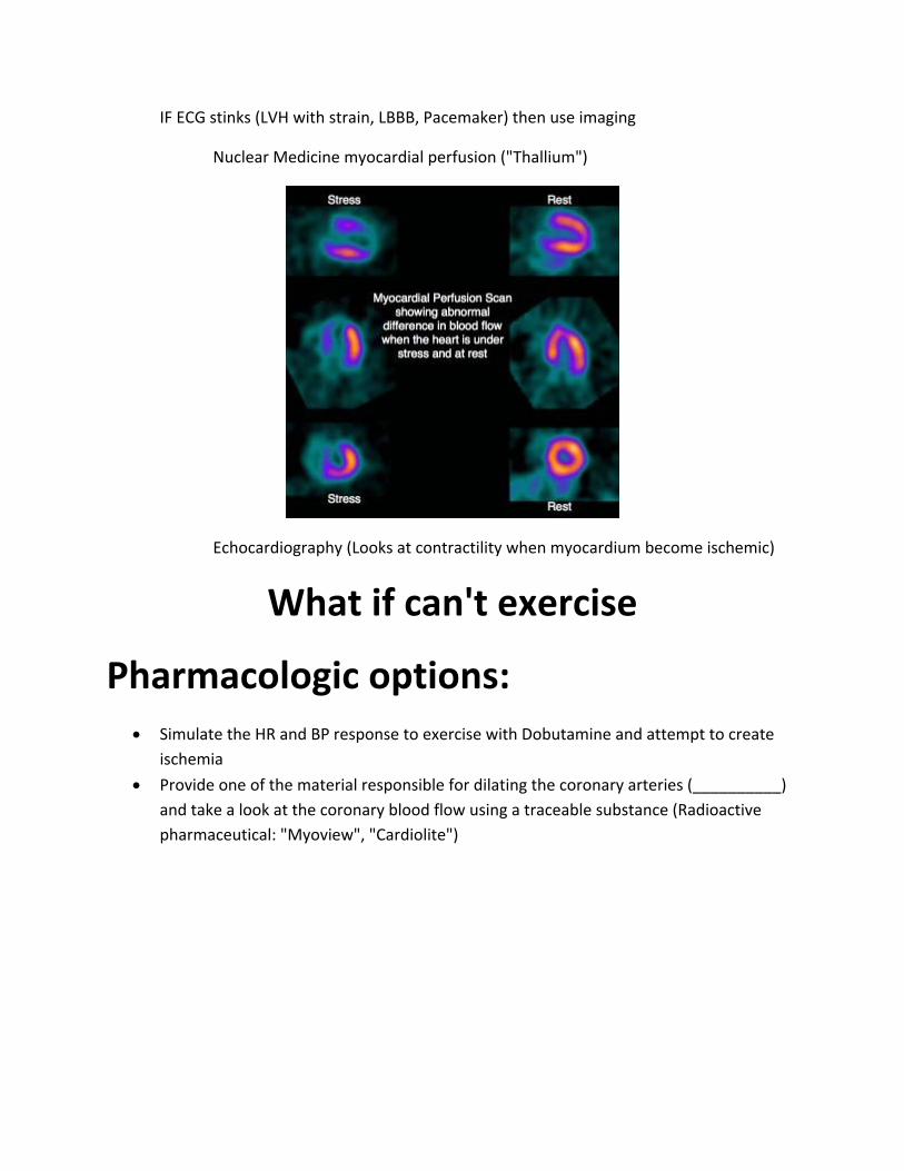
IF ECG stinks (LVH with strain, LBBB, Pacemaker) then use imaging
Nuclear Medicine myocardial perfusion ("Thallium")
Echocardiography (Looks at contractility when myocardium become ischemic)
What if can't exercise
Pharmacologic options: Simulate the HR and BP response to exercise with Dobutamine and attempt to create
ischemia
Provide one of the material responsible for dilating the coronary arteries (__________)
and take a look at the coronary blood flow using a traceable substance (Radioactive
pharmaceutical: "Myoview", "Cardiolite")

CT

MRI

Coronary Arteriography

Systolic Murmur: AS versus MR Aortic Stenosis Mitral Regurgitation
Overload Pressure Volume
PMI Sustained, not displaced Enlarged, displaced laterally
Pulse Potentially tardus and brevis Normal
Extra Heart S4 S3, diastolic rumble
Sounds Potentially Ejection Click


Aortic Insufficiency Blowing quality, heard with diaphragm along sternal border. Heard better when vertical and
leaning forward
Never normal
Associated with lots of variable conditions
Aortic dissection
Aortic root dilation
Endocarditis
Bicuspid aortic valve
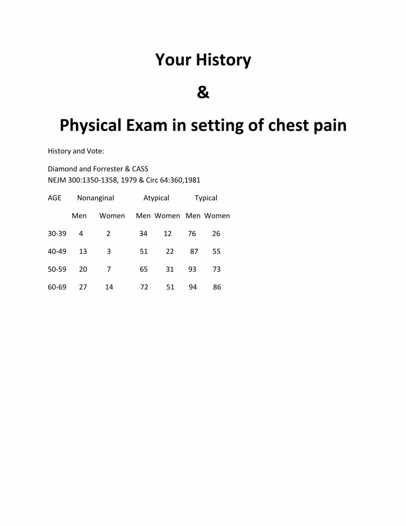
Your History
&
Physical Exam in setting of chest pain History and Vote:
Diamond and Forrester & CASS
NEJM 300:1350‐1358, 1979 & Circ 64:360,1981
AGE Nonanginal Atypical Typical
Men Women Men Women Men Women
30‐39 4 2 34 12 76 26
40‐49 13 3 51 22 87 55
50‐59 20 7 65 31 93 73
60‐69 27 14 72 51 94 86