the aortopathy of bicuspid aortic valve disease has
TRANSCRIPT
DOI: 10.1016/j.jtcvs.2008.01.022 2008;135:901-907 J Thorac Cardiovasc Surg
Dominik Fleischman, Robert Herfkens, R. Scott Mitchell and D. Craig Miller Shafie S. Fazel, Hari R. Mallidi, Richard S. Lee, Michael P. Sheehan, David Liang,
usually involves the transverse aortic archThe aortopathy of bicuspid aortic valve disease has distinctive patterns and
http://jtcs.ctsnetjournals.org/cgi/content/full/135/4/901located on the World Wide Web at:
The online version of this article, along with updated information and services, is
2008 American Association for Thoracic Surgery Association for Thoracic Surgery and the Western Thoracic Surgical Association. Copyright ©
is the official publication of the AmericanThe Journal of Thoracic and Cardiovascular Surgery
on September 2, 2009 jtcs.ctsnetjournals.orgDownloaded from
Fazel et al Surgery for Acquired Cardiovascular Disease
The aortopathy of bicuspid aortic valve disease hasdistinctive patterns and usually involves the transverseaortic archShafie S. Fazel, MD, PhD,a Hari R. Mallidi, MD,a Richard S. Lee, MD,a Michael P. Sheehan, MSN, RN, FNP,a
David Liang, MD, PhD,c Dominik Fleischman, MD,b Robert Herfkens, MD,b R. Scott Mitchell, MD,a and D. Craig Miller, MDa
ACD
Supplemental material isavailable online.
Objectives: Bicuspid aortic valves are associated with a poorly characterized connec-
tive tissue disorder that predisposes to aortic catastrophes. Because no criterion exists
dictating the appropriate extent of aortic resection in aneurysmal disease of the bicus-
pid aortic valve, we studied the patterns of aortic dilation in this population.
Methods: Sixty-four patients with bicuspid aortic valves who underwent computed
tomographic or magnetic resonance angiography and echocardiography were retro-
spectively identified between January 2002 and March 2006. Orthonormal 2–dimen-
sional or 3-dimensional aortic diameters were measured at 10 levels. Agglomerative
hierarchic clustering with centered correlation distance measurements and complete
linkage analysis was used to detect distinct patterns of aortic dilatation.
Results: Mean aortic diameter was 28.1 6 0.7 mm at the annulus and 21.7 6 0.4 mm
at the diaphragmatic hiatus. The aorta was largest in the tubular ascending aorta (45.9
6 1.0 mm). Compared with the descending aorta, the transverse aortic arch was also
dilated (P , .01). Cluster analysis showed 4 patterns of aortic dilatation: cluster I, aor-
tic root alone (n 5 8, 13%); cluster II, tubular ascending aorta alone (n 5 9, 14%);
cluster III, tubular portion and transverse arch (n 5 18, 28%); and, cluster IV, aortic
root and tubular portion with tapering across the transverse arch (n 5 29, 45%).
Conclusion: Distinct patterns of aortic dilatation in patients with bicuspid aortic
valves call for an individualized degree of aortic replacement to minimize late aortic
complications and reoperation. Patients in clusters III and IV should have transverse
arch replacement (plus concomitant root replacement in cluster IV). Patients in cluster
I should undergo complete aortic root replacement, whereas in patients in cluster II
supracommissural ascending aortic grafting is adequate.
Bicuspid aortic valves (BAVs) are the most common congenital cardiovascular
malformation, occurring in 0.9% to 2% of the population and affecting approx-
imately 4 million persons in the United States.1 BAVs are also the most common
cause of aortic valve disease in patients younger than 70 years in North America.2 Echo-
cardiographic examination of the aortic root and ascending aorta in patients with BAVs
has demonstrated more aortic dilatation than in subjects with tricuspid aortic valves.3-6
The aortic enlargement in patients with BAVs is typically asymmetric3 and might involve
the aortic arch.7 Even in patients with BAV disease without gross dilation of the ascend-
ing aorta, distances between the aortic valve and point of maximum diameter of the as-
cending aorta at the outer and inner curve are longer, suggesting that the aorta is also
elongated.8 Aortic aneurysmal disease in the BAV is due to an aortopathy9 that is present
independently of valvular abnormalities or hemodynamic factors.2,10 Even in the absence
of aortic dilation, this aortopathy causes abnormal aortic mechanical properties.11
The most important clinical consequence of an enlarged ascending aorta and the
underlying aortopathy is the higher incidence of aortic rupture and aortic dissection.
The presence of a BAV increases the risk of aortic dissection 9-fold and puts the
From the Departments of Cardiothoracic
Surgerya and Radiologyb and the Cardiovas-
cular Medicine Division,c Stanford Univer-
sity Medical School, Stanford, Calif.
Read in part at the Thirty-third Annual
Meeting of the Western Thoracic Surgical
Association, Santa Ana Pueblo, NM, June
27–30, 2007.
Received for publication Aug 2, 2007;
revisions received Dec 22, 2007; accepted
for publication Jan 23, 2008.
Address for reprints: D. Craig Miller, MD,
Department of Cardiothoracic Surgery,
Falk Cardiovascular Research Center, Stan-
ford University School of Medicine, Stan-
ford, California 94305-5247 (E-mail:
J Thorac Cardiovasc Surg 2008;135:901-7
0022-5223/$34.00
Copyright � 2008 by The American Asso-
ciation for Thoracic Surgery
doi:10.1016/j.jtcvs.2008.01.022
The Journal of Thoracic and Cardiovascular Surgery c Volume 135, Number 4 901 on September 2, 2009 jtcs.ctsnetjournals.orgDownloaded from
Surgery for Acquired Cardiovascular Disease Fazel et al
ACD
Abbreviations and AcronymsAI 5 aortic insufficiency
BAV 5 bicuspid aortic valve
CTA 5 computed tomographic angiography
CVG 5 composite valve graft
2-D 5 2-dimensional
3-D 5 3-dimensional
ICC 5 interclass correlation
MRA 5 magnetic resonance angiography
patient at risk of aortic dissection at a younger age.12,13
Notably, aortic dissection usually occurs in the presence of
a normally functioning BAV.12,14
Although it is clear that BAV disease is associated with
proximal aortic dilation, controversy exists concerning the
extent of thoracic aortic involvement, mainly because the ma-
jority of studies to date have only used echocardiography to
detect aortic dilatation.15 Herein we document thoracic aortic
morphology in patients with BAVs using computed tomo-
graphic angiography (CTA) or magnetic resonance angiogra-
phy (MRA). We then used hierarchic clustering, a tool used
to automatically detect data clusters in gene-array studies,16
to identify 4 distinct patterns of aortic involvement. These
4 distinct clusters argue that a custom-tailored surgical ap-
proach is most appropriate in dealing with the dilated aorta
in the setting of a BAV.
Materials and MethodsPatients and MethodsThe time interval of this retrospective review extended from January
1, 2002, to March 1, 2006. Only patients with thoracic aortic imag-
ing with either CTA or MRA were included. The patients were iden-
tified by means of electronic query of the radiologic (initially
identified 55 patients) and echocardiographic (initially identified 285
patients) databases for the key term ‘‘BAV’’ in either the requisition
or in the text field of the report. The surgical database was reviewed
manually to identify patients with BAVs who had undergone cardio-
vascular surgical intervention. A total of 102 patients with BAVs had
complete radiologic examination of the thoracic aorta. Patients with
only postoperative images (n 5 18), repaired aortic coarctation (n
5 10), and aortic dissection (n 5 10) were excluded from further anal-
ysis. The final analyzed cohort comprised 64 patients, of whom 62
also had a Stanford echocardiogram. The echocardiographic pattern
of cusp fusion was recorded in 44 patients on echocardiographic re-
review by one of the investigators. Average patient age was 45 6 1
years (range, 18–75 years). Of the 64 patients, 49 were men.
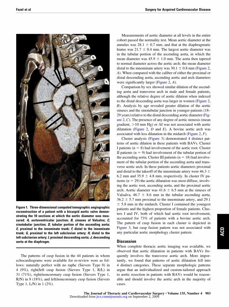
Thoracic aortic diameter was measured at 10 levels: (1) aortic an-
nulus, (2) sinuses of Valsalva, (3) sinotubular junction, (4) tubular
portion of the ascending aorta, (5) proximal to the innominate artery
(or common trunk in case of a bovine arch), (6) distal to the innom-
inate artery (or common trunk), (7) proximal to the left subclavian
artery, (8) distal to the left subclavian artery, (9) proximal descend-
ing aorta, and (10) distal descending thoracic aorta at the level of the
diaphragmatic hiatus (Figure 1). Aortic measurements were made in
902 The Journal of Thoracic and Cardiovascular Surgery c Apjtcs.ctsnetjourDownloaded from
the orthonormal plane to the aorta by using 3-dimensional (3-D)
reconstructions of the thoracic aorta or 2-dimensional (2-D) axial
images when 3-D images were not available. Measurements from
the 3-D images were obtained in arbitrary pixels that were converted
to metric measurements by normalizing to the aortic diameter mea-
sured at the diaphragmatic hiatus on both the 3-D and 2-D axial
images. This aortic level was chosen for determining the correction
factor because the 2-D axial cuts are most likely to be orthonormal to
the longitudinal axis of the descending thoracic aorta here in these
young patients. Correlation between the 3-D and 2-D axial measure-
ments was determined in 10 randomly selected patients who had
both sets of images available for analysis. Interobserver variability
was determined by comparing measurements made by 2 indepen-
dent blinded observers in another 10 randomly chosen patients.
Statistical AnalysisData are presented as means 6 1 standard error of the mean. The
aortic diameter at various levels was compared with the diameter
of the aorta at the diaphragmatic hiatus to detect patterns of aortic
dilation by using analysis of variance followed by the Dunnett
post hoc test. The diameter of the aorta was compared at various
levels to detect differences between groups by using the unpaired
nonequal variance Student t test. The interclass correlation (ICC)
technique was used to calculate interobserver variability from
both the 2-D axial and 3-D reconstructed images. Interobserver var-
iability at distinct aortic locations was measured with the Bland–Alt-
man confidence intervals method. Hierarchic cluster analysis was
performed with the Hierarchical Clustering Explorer version 3.5
(University of Maryland, www.cs.umd.edu/hcil/hce). The data
were first plotted after normalization by using the within-patient
z score. Complete linkage analysis with Pearson correlation coeffi-
cient similarity measurement was used to generate the clusterogram.
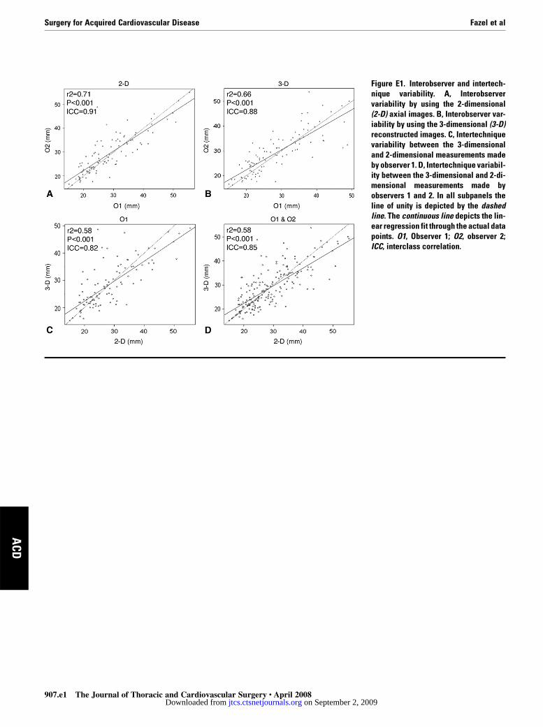
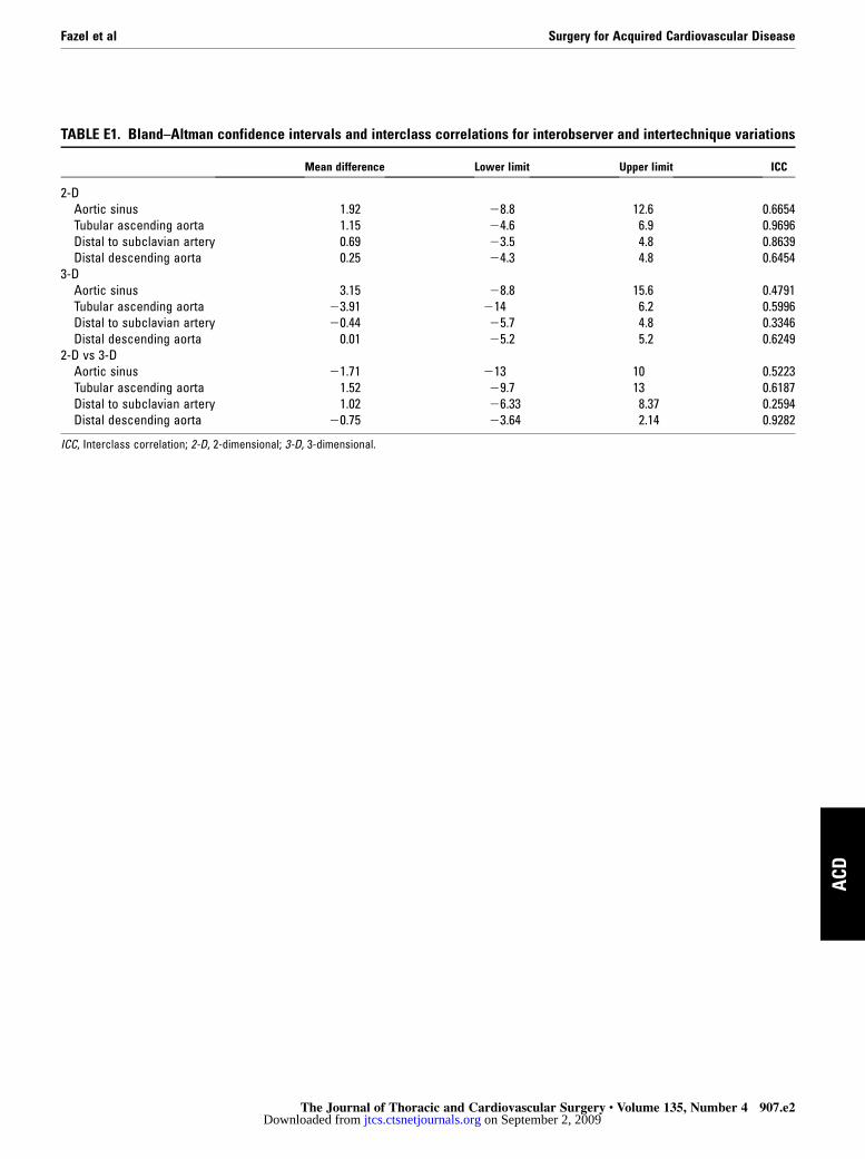
ResultsOverall interobserver variability was minimal: ICCs of 0.91
for the 2-D measurements and 0.88 for the 3-D measure-
ments. The overall correlation between 2-D and 3-D measure-
ments was excellent, with an ICC of 0.85 (see Figure E1). The
interobserver variability at various aortic locations by both
2-D and 3-D measurements assessed by using the Bland-
Altman method is summarized in Table E1. Overall, the reli-
ability of the various 2-D and 3-D measurements was very
good.
A mean aortic valve gradient of greater than 10 mm Hg (aor-
tic stenosis) was present in 39% (24/62) of patients for whom
echocardiographic reports were available. The distribution of
aortic gradients was as follows: 10 to 25 mm Hg, n 5 11; 26
to 50 mm Hg, n 5 10; and greater than 50 mm Hg, n 5 3.
The mean aortic valve gradient in patients who had any flow
acceleration across the valve (n 5 42) was 21.6 6 3.2 mm
Hg. Thirty-nine patients had aortic insufficiency (AI), which
was distributed as follows: 11 AI, n 5 13; 21 AI, n 5 15;
31 AI, n 5 9; and 41 AI, n 5 2. In patients with AI, the direc-
tion of the regurgitant jet was in a commissure in 2 patients,
central in 6 patients, and eccentric and directed along the ante-
rior leaflet of the mitral valve in 19 patients.
ril 2008 on September 2, 2009 nals.org
Fazel et al Surgery for Acquired Cardiovascular Disease
ACD
The patterns of cusp fusion in the 44 patients in whom
echocardiograms were available for re-review were as fol-
lows: naturally perfect with no raphe (Sievers Type 0) in
4 (9%), right/left cusp fusion (Sievers Type 1, R/L) in
31 (71%), right/noncoronary cusp fusion (Sievers Type 1,
R/N) in 8 (18%), and left/noncoronary cusp fusion (Sievers
Type 1, L/N) in 1 (2%).
Figure 1. Three-dimensional computed tomographic angiographicreconstruction of a patient with a bicuspid aortic valve demon-strating the 10 sections at which the aortic diameter was mea-sured: A, aortoventricular junction; B, sinuses of Valsalva; C,sinotubular junction; D, tubular portion of the ascending aorta;E, proximal to the innominate trunk; F, distal to the innominatetrunk; G, proximal to the left subclavian artery; H, distal to theleft subclavian artery; I, proximal descending aorta; J, descendingaorta at the diaphragm.
The Journal of Thojtcs.ctsnetjoDownloaded from
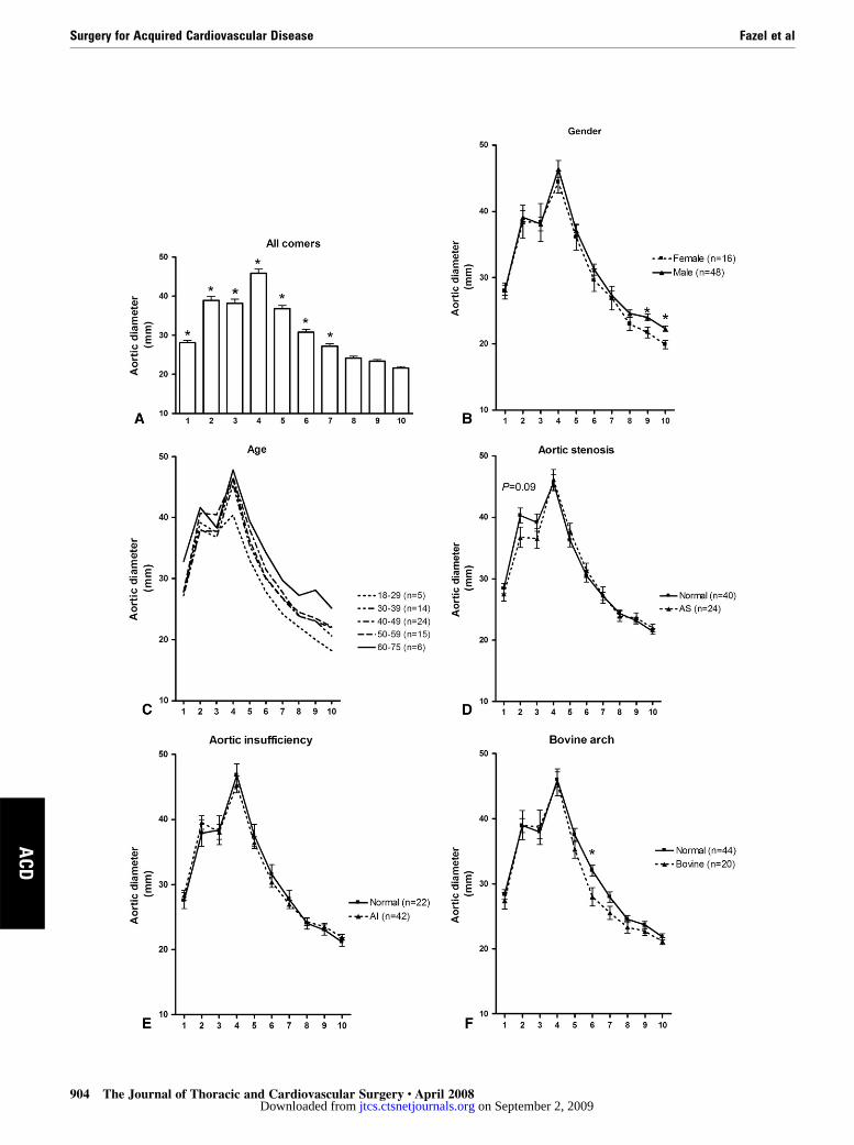
Measurements of aortic diameter at all levels in the entire
cohort passed the normality test. Mean aortic diameter at the
annulus was 28.1 6 0.7 mm, and that at the diaphragmatic
hiatus was 21.7 6 0.4 mm. The largest aortic diameter was
in the tubular portion of the ascending aorta, in which the
mean diameter was 45.9 6 1.0 mm. The aorta then tapered
to normal diameter across the aortic arch; the mean diameter
distal to the innominate artery was 30.1 6 0.8 mm (Figure 2,
A). When compared with the caliber of either the proximal or
distal descending aorta, ascending aortic and arch diameters
were significantly larger (Figure 2, A).
Comparison by sex showed similar dilation of the ascend-
ing aorta and transverse arch in male and female patients,
although the relative degree of aortic dilation when indexed
to the distal descending aorta was larger in women (Figure 2,
B). Analysis by age revealed greater dilation of the aortic
sinuses and the sinotubular junction in younger patients (18–
29 years) relative to the distal descending aortic diameter (Fig-
ure 2, C). The presence of any degree of aortic stenosis (mean
gradient, .10 mm Hg) or AI was not associated with aortic
dilatation (Figure 2, D and E). A bovine aortic arch was
associated with less dilatation in the midarch (Figure 2, F).
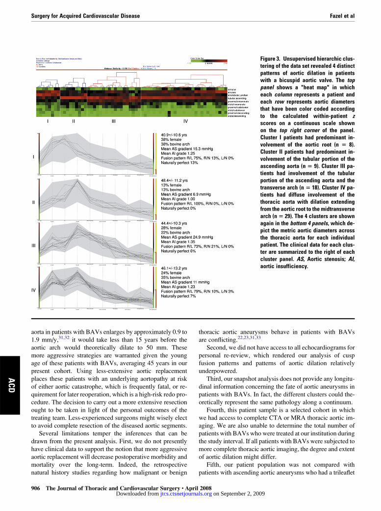
Cluster analysis (Figure 3) demonstrated 4 distinct pat-
terns of aortic dilation in these patients with BAVs. Cluster
I patients (n 5 8) had involvement of the aortic root. Cluster
II patients (n 5 9) had involvement of the tubular portion of
the ascending aorta. Cluster III patients (n 5 18) had involve-
ment of the tubular portion of the ascending aorta and trans-
verse aortic arch. In these patients aortic diameters proximal
and distal to the takeoff of the innominate artery were 46.3 6
6.2 mm and 35.9 6 4.8 mm, respectively. In cluster IV pa-
tients (n 5 29) the aortic dilatation was most diffuse, involv-
ing the aortic root, ascending aorta, and the proximal aortic
arch. Aortic diameter was 41.6 6 6.5 mm at the sinuses of
Valsalva, 46.7 6 8.6 mm in the tubular ascending aorta,
36.2 6 5.7 mm proximal to the innominate artery, and 29.1
6 5.8 mm in the midarch. Cluster I contained the youngest
patients and the highest proportion of female patients. Clus-
ters I and IV, both of which had aortic root involvement,
accounted for 73% of patients with a bovine aortic arch.
The pattern of cusp fusion in each cluster is depicted in
Figure 3, but cusp fusion pattern was not associated with
any particular aortic morphology cluster pattern.
DiscussionWhen complete thoracic aortic imaging was available, we
observed that aortic dilatation in patients with BAVs fre-
quently involves the transverse aortic arch. More impor-
tantly, we found that patterns of aortic dilatation fell into
4 distinct categories. These separate morphologic patterns
argue that an individualized and custom-tailored approach
to aortic resection in patients with BAVs would be reason-
able and should involve the aortic arch in the majority of
cases.
racic and Cardiovascular Surgery c Volume 135, Number 4 903 on September 2, 2009 urnals.org
Surgery for Acquired Cardiovascular Disease Fazel et al
ACD
904 The Journal of Thoracic and Cardiovascular Surgery c April 2008 on September 2, 2009 jtcs.ctsnetjournals.orgDownloaded from
Fazel et al Surgery for Acquired Cardiovascular Disease
ACD
Natural history observational studies have suggested that
an ascending aortic diameter of 6 cm (irrespective of whether
the valve is bicuspid or tricuspid) is an indication for surgical
graft replacement, with 6 cm being the hinge point beyond
which there is a 30% increase in the probability of rupture.17
Patients who underwent isolated aortic valve replacement
when the aortic diameter exceeded 5.1 cm experienced
a risk of aortic dissection in excess of 20% compared with
0.6% in patients with no ascending aortic dilatation.18,19 E.
Stanley Crawford first brought to our attention this grave sur-
gical error of omission more than 20 years ago. Patients with
BAVs undergoing aortic valve replacement commonly have
substantial dilation of the ascending aorta,20 which continues
to enlarge after aortic valve replacement.21 Furthermore,
patients with BAVs with moderate aortic dilatation who
undergo only aortic valve replacement sustain aortic dissec-
tion, rupture, and sudden death more frequently than similar
patients with a tricuspid aortic valve.22-24 Based on these
findings, the American College of Cardiology and American
Heart Association practice guidelines have now adopted
a more aggressive posture, favoring aortic replacement
when the aorta is larger than 45 mm in patients with BAVs
undergoing aortic valve replacement and 50 mm in patients
with BAVs with isolated thoracic aortic aneurysms.25
The extent of the aorta that should be replaced, however,
presently is unknown. In the present study 58% of patients
had aortic root involvement (clusters I and IV), and 73% of
patients had involvement of the proximal arch (clusters III
and IV). In the latter groups of patients, the aortic dilatation
extended into the midarch in 35%. This question of how
much aorta to replace highlights the importance of relative
aortic size, as introduced by Davies and colleagues,26 or of
observed aortic size indexed to predicted aortic size, as first
advocated by Legget and associates.27 Although the latter
is likely to be more reliable in predicting adverse clinical
events, it depends on accurate reference values that presently
do not exist for the aortic arch. Furthermore, the impetus to
replace all diseased aortic segments must be balanced against
any incremental operative risk inherent in a more aggressive
surgical approach.
Two main surgical options exist for the treatment of aortic
root aneurysms: composite valve graft (CVG) or valve-spar-
ing aortic root replacement. Both approaches can be carried
with low operative mortality and excellent long-term
outcomes.28,29 Compilation of our own results in the 5-year
The Journal of Thojtcs.ctsnetjoDownloaded from
period covered by this report has shown that 1 of 126 patients
undergoing elective mechanical CVG has died (0.79% mor-
tality rate). Similarly, none of the 106 patients undergoing
elective reimplantation-type valve-sparing root replacement
has died. Arguably, in young patients the more attractive op-
tion is valve-sparing aortic root replacement if the BAV
cusps are not fibrotic or calcified, so that the patient avoids
the thromboembolic and anticoagulant-related hemorrhagic
complications of a CVG with a mechanical prosthesis. This
approach, however, exposes the patient to a higher risk of
reoperation when the valvular pathology progresses or the
valve-sparing operation does not prove to be durable, but
this increased risk is not clearly defined in the current litera-
ture. Another approach, which we do not recommend in this
young patient population, is the separate valve graft, whereby
the aortic valve and the supracoronary aorta are replaced sep-
arately. This procedure leaves diseased aortic sinuses behind
and should only be undertaken when the patient is elderly and
has significant comorbidity and is deemed to be unfit for a full
aortic root replacement.
Total arch replacement is performed today with low mor-
bidity and mortality in centers with experienced personnel.30
The decision for aortic arch replacement is made preopera-
tively on review of aortic imaging. The extent of resection
then informs our choice of arterial cannulation. When aortic
arch replacement is planned, patients either undergo cannula-
tion of the axillary artery or the innominate artery. In all cases
we use a combination of selective antegrade cerebral perfu-
sion and hypothermia. Again, during the time interval of
the present study, none of the 62 patients undergoing CVG
and arch replacement and none of the 31 patients undergoing
valve-sparing root replacement and arch replacement have
died. However, it is realistic to expect some small increase
in risk if the arch is replaced concomitantly at centers with
less-experienced personnel. Despite this theoretic increased
risk, we believe that total aortic arch replacement (frequently
using the ‘‘peninsula technique,’’ leaving a small tongue of
greater curvature of the arch along the origins of the great
vessels, which is more aggressive than the traditional beveled
graft hemiarch replacement) should be carried out in the
cluster III patients with BAVs whose aortas measure 46
mm just at the takeoff of the innominate artery. In patients
in cluster IV, even an open distal anastomosis when replacing
the ascending aorta will leave behind the proximal aortic
arch, on average measuring 36 mm. Given that the ascending
Figure 2. Aortic dilation in patients with bicuspid aortic valves involves the aortic arch. A, Aortic diameters in allpatients. *P < .05, Dunnett posttest compared with the aortic diameter at the diaphragmatic hiatus. B, Aortic diam-eters by sex. C, Aortic diameters by age. Younger patients had greater relative dilation of the aortic root relative tothe distal descending aorta. D, Aortic diameters by presence of aortic stenosis. AS, Aortic stenosis. E, Aortic diam-eters by presence of aortic insufficiency. AI, Aortic insufficiency. F, Aortic diameters by presence of a bovine aorticarch. 1, Aortoventricular junction; 2, sinuses of Valsalva; 3, sinotubular junction; 4, tubular portion of the ascendingaorta; 5, proximal to the innominate trunk; 6, distal to the innominate trunk; 7, proximal to the left subclavian artery; 8,distal to the left subclavian artery; 9, proximal descending aorta; 10, descending aorta at the diaphragm.
racic and Cardiovascular Surgery c Volume 135, Number 4 905 on September 2, 2009 urnals.org
Surgery for Acquired Cardiovascular Disease Fazel et al
ACD
Figure 3. Unsupervised hierarchic clus-tering of the data set revealed 4 distinctpatterns of aortic dilation in patientswith a bicuspid aortic valve. The toppanel shows a ''heat map'' in whicheach column represents a patient andeach row represents aortic diametersthat have been color coded accordingto the calculated within-patient zscores on a continuous scale shownon the top right corner of the panel.Cluster I patients had predominant in-volvement of the aortic root (n 5 8).Cluster II patients had predominant in-volvement of the tubular portion of theascending aorta (n 5 9). Cluster III pa-tients had involvement of the tubularportion of the ascending aorta and thetransverse arch (n 5 18). Cluster IV pa-tients had diffuse involvement of thethoracic aorta with dilation extendingfrom the aortic root to the midtransversearch (n 5 29). The 4 clusters are shownagain in the bottom 4 panels, which de-pict the metric aortic diameters acrossthe thoracic aorta for each individualpatient. The clinical data for each clus-ter are summarized to the right of eachcluster panel. AS, Aortic stenosis; AI,aortic insufficiency.
aorta in patients with BAVs enlarges by approximately 0.9 to
1.9 mm/y,31,32 it would take less than 15 years before the
aortic arch would theoretically dilate to 50 mm. These
more aggressive strategies are warranted given the young
age of these patients with BAVs, averaging 45 years in our
present cohort. Using less-extensive aortic replacement
places these patients with an underlying aortopathy at risk
of either aortic catastrophe, which is frequently fatal, or re-
quirement for later reoperation, which is a high-risk redo pro-
cedure. The decision to carry out a more extensive resection
ought to be taken in light of the personal outcomes of the
treating team. Less-experienced surgeons might wisely elect
to avoid complete resection of the diseased aortic segments.
Several limitations temper the inferences that can be
drawn from the present analysis. First, we do not presently
have clinical data to support the notion that more aggressive
aortic replacement will decrease postoperative morbidity and
mortality over the long-term. Indeed, the retrospective
natural history studies regarding how malignant or benign
906 The Journal of Thoracic and Cardiovascular Surgery c Apjtcs.ctsnetjourDownloaded from
thoracic aortic aneurysms behave in patients with BAVs
are conflicting.22,23,31,33
Second, we did not have access to all echocardiograms for
personal re-review, which rendered our analysis of cusp
fusion patterns and patterns of aortic dilation relatively
underpowered.
Third, our snapshot analysis does not provide any longitu-
dinal information concerning the fate of aortic aneurysms in
patients with BAVs. In fact, the different clusters could the-
oretically represent the same pathology along a continuum.
Fourth, this patient sample is a selected cohort in which
we had access to complete CTA or MRA thoracic aortic im-
aging. We are also unable to determine the total number of
patients with BAVs who were treated at our institution during
the study interval. If all patients with BAVs were subjected to
more complete thoracic aortic imaging, the degree and extent
of aortic dilation might differ.
Fifth, our patient population was not compared with
patients with ascending aortic aneurysms who had a trileaflet
ril 2008 on September 2, 2009 nals.org
Fazel et al Surgery for Acquired Cardiovascular Disease
ACD
aortic valve; based on current information, it is unclear how
aneurysm extent and morphology differ between these pa-
tient subgroups. Nevertheless, we must take to heart Dr
John S. Child’s plea that we must now abandon the terms
‘‘poststenotic dilation’’ and ‘‘postregurgitant dilation’’ in de-
scribing the dilated aorta that accompanies a BAV because
these patients have an underlying connective tissue disorder
and the aneurysmal dilation is not a consequence of the coex-
istent valvular hemodynamic abnormality.15
We conclude that aortic dilatation in patients with BAVs
follows 4 distinctive patterns that militate for an individual-
ized, custom-tailored degree of ascending aortic and arch re-
placement. Patients in clusters III and IV requiring operations
should have the transverse arch replaced and not just the as-
cending aorta (along with concomitant root replacement in
cluster IV). In cluster I patients complete aortic root replace-
ment (reimplantation type valve-sparing or CVG) is neces-
sary, whereas in cluster II patients a supracommissural
ascending aortic graft is adequate. This individualized ap-
proach is our practice and should minimize the incidence of
late aortic complications and need for reoperation.
We thank Ms Sharmi Shafi for her contribution in collating a list
of bibliographic references.
References
1. Ratib O, Perloff JK, Child JS. Images in cardiovascular medicine. Bicus-pid aortic valve aneurysm. Circulation. 2004;109:671.
2. de Sa M, Moshkovitz Y, Butany J, David TE. Histologic abnormalitiesof the ascending aorta and pulmonary trunk in patients with bicuspidaortic valve disease: clinical relevance to the Ross procedure. J ThoracCardiovasc Surg. 1999;118:588-94.
3. Alegret JM, Duran I, Palazon O, Vernis JM, Ameijide A, Rabassa A,et al. Prevalence of and predictors of bicuspid aortic valves in patientswith dilated aortic roots. Am J Cardiol. 2003;91:619-22.
4. Nkomo VT, Enriquez-Sarano M, Ammash NM, Melton LJ III, Bailey KR,Desjardins V, et al. Bicuspid aortic valve associated with aortic dilatation:a community-based study. Arterioscler Thromb Vasc Biol. 2003;23:351-6.
5. Novaro GM, Tiong IY, Pearce GL, Grimm RA, Smedira N, Griffin BP.Features and predictors of ascending aortic dilatation in association witha congenital bicuspid aortic valve. Am J Cardiol. 2003;92:99-101.
6. Pachulski RT, Chan KL. Progression of aortic valve dysfunction in 51adult patients with congenital bicuspid aortic valve: assessment and fol-low up by Doppler echocardiography. Br Heart J. 1993;69:237-40.
7. Cecconi M, Nistri S, Quarti A, Manfrin M, Colonna PL, Molini E, et al.Aortic dilatation in patients with bicuspid aortic valve. J CardiovascMed (Hagerstown). 2006;7:11-20.
8. Bauer M, Gliech V, Siniawski H, Hetzer R. Configuration of the ascend-ing aorta in patients with bicuspid and tricuspid aortic valve disease un-dergoing aortic valve replacement with or without reduction aortoplasty.J Heart Valve Dis. 2006;15:594-600.
9. Fedak PW, Verma S, David TE, Leask RL, Weisel RD, Butany J.Clinical and pathophysiological implications of a bicuspid aortic valve.Circulation. 2002;106:900-4.
10. Niwa K, Perloff JK, Bhuta SM, Laks H, Drinkwater DC, Child JS, et al.Structural abnormalities of great arterial walls in congenital heart disease:light and electron microscopic analyses. Circulation. 2001;103:393-400.
11. Grotenhuis HB, Ottenkamp J, Westenberg JJ, Bax JJ, Kroft LJ, deRoos A. Reduced aortic elasticity and dilatation are associated with aor-tic regurgitation and left ventricular hypertrophy in nonstenotic bicuspidaortic valve patients. J Am Coll Cardiol. 2007;49:1660-5.
12. Larson EW, Edwards WD. Risk factors for aortic dissection: a necropsystudy of 161 cases. Am J Cardiol. 1984;53:849-55.
The Journal of Thorjtcs.ctsnetjouDownloaded from
13. Ward C. Clinical significance of the bicuspid aortic valve. Heart. 2000;83:81-5.
14. Edwards WD, Leaf DS, Edwards JE. Dissecting aortic aneurysmassociated with congenital bicuspid aortic valve. Circulation. 1978;57:1022-5.
15. Braverman AC, Guven H, Beardslee MA, Makan M, Kates AM, Moon MR.The bicuspid aortic valve. Curr Probl Cardiol. 2005;30:470-522.
16. D’Haeseleer P. How does gene expression clustering work? Nat Bio-technol. 2005;23:1499-501.
17. Elefteriades JA. Natural history of thoracic aortic aneurysms: indicationsfor surgery, and surgical versus nonsurgical risks. Ann Thorac Surg.2002;74(suppl):S1877-80.
18. Pieters FA, Widdershoven JW, Gerardy AC, Geskes G, Cheriex EC,Wellens HJ. Risk of aortic dissection after aortic valve replacement.Am J Cardiol. 1993;72:1043-7.
19. Prenger K, Pieters F, Cheriex E. Aortic dissection after aortic valvereplacement: incidence and consequences for strategy. J Card Surg.1994;9:495-8.
20. Morgan-Hughes GJ, Roobottom CA, Owens PE, Marshall AJ. Dilata-tion of the aorta in pure, severe, bicuspid aortic valve stenosis. Am HeartJ. 2004;147:736-40.
21. Yasuda H, Nakatani S, Stugaard M, Tsujita-Kuroda Y, Bando K,Kobayashi J, et al. Failure to prevent progressive dilation of ascending aortaby aortic valve replacement in patients with bicuspid aortic valve: compar-ison with tricuspid aortic valve. Circulation. 2003;108(suppl 1):II291-4.
22. Borger MA, Preston M, Ivanov J, Fedak PW, Davierwala P,Armstrong S, et al. Should the ascending aorta be replaced more fre-quently in patients with bicuspid aortic valve disease? J Thorac Cardi-ovasc Surg. 2004;128:677-83.
23. Russo CF, Mazzetti S, Garatti A, Ribera E, Milazzo A, Bruschi G, et al.Aortic complications after bicuspid aortic valve replacement: long-termresults. Ann Thorac Surg. 2002;74(suppl):S1773-6.
24. Matsuyama K, Usui A, Akita T, Yoshikawa M, Murayama M, Yano T,et al. Natural history of a dilated ascending aorta after aortic valvereplacement. Circ J. 2005;69:392-6.
25. Bonow RO, Carabello BA, Kanu C, de Leon AC Jr, Faxon DP,Freed MD, et al. ACC/AHA 2006 guidelines for the management of pa-tients with valvular heart disease: a report of the American College ofCardiology/American Heart Association Task Force on Practice Guide-lines (writing committee to revise the 1998 Guidelines for the Manage-ment of Patients With Valvular Heart Disease): developed incollaboration with the Society of Cardiovascular Anesthesiologists: en-dorsed by the Society for Cardiovascular Angiography and Interventionsand the Society of Thoracic Surgeons. Circulation. 2006;114:e84-231.
26. Davies RR, Gallo A, Coady MA, Tellides G, Botta DM, Burke B, et al.Novel measurement of relative aortic size predicts rupture of thoracicaortic aneurysms. Ann Thorac Surg. 2006;81:169-77.
27. Legget ME, Unger TA, O’Sullivan CK, Zwink TR, Bennett RL,Byers PH, et al. Aortic root complications in Marfan’s syndrome: iden-tification of a lower risk group. Heart. 1996;75:389-95.
28. David TE, Feindel CM, Webb GD, Colman JM, Armstrong S,Maganti M. Long-term results of aortic valve-sparing operations for aor-tic root aneurysm. J Thorac Cardiovasc Surg. 2006;132:347-54.
29. Gott VL, Greene PS, Alejo DE, Cameron DE, Naftel DC, Miller DC,et al. Replacement of the aortic root in patients with Marfan’s syndrome.N Engl J Med. 1999;340:1307-13.
30. Elefteriades JA, Dobrilovic N, Gega A. A new surgical paradigm: hybridopen and endovascular repair of the ascending aorta and aortic arch foracute type A dissection [letter]. J Thorac Cardiovasc Surg. 2007;132:735.
31. Davies RR, Kaple RK, Mandapati D, Gallo A, Botta DM Jr,Elefteriades JA, et al. Natural history of ascending aortic aneurysms inthe setting of an unreplaced bicuspid aortic valve. Ann Thorac Surg.2007;83:1338-44.
32. Ferencik M, Pape LA. Changes in size of ascending aorta and aorticvalve function with time in patients with congenitally bicuspid aorticvalves. Am J Cardiol. 2003;92:43-6.
33. Goland S, Czer LS, De Robertis MA, Mirocha J, Kass RM, Fontana GP,et al. Risk factors associated with reoperation and mortality in 252 pa-tients after aortic valve replacement for congenitally bicuspid aorticvalve disease. Ann Thorac Surg. 2007;83:931-7.
acic and Cardiovascular Surgery c Volume 135, Number 4 907 on September 2, 2009 rnals.org
Surgery for Acquired Cardiovascular Disease Fazel et al
ACD
Figure E1. Interobserver and intertech-nique variability. A, Interobservervariability by using the 2-dimensional(2-D) axial images. B, Interobserver var-iability by using the 3-dimensional (3-D)reconstructed images. C, Intertechniquevariability between the 3-dimensionaland 2-dimensional measurements madeby observer 1. D, Intertechnique variabil-ity between the 3-dimensional and 2-di-mensional measurements made byobservers 1 and 2. In all subpanels theline of unity is depicted by the dashedline. The continuous line depicts the lin-ear regression fit through the actual datapoints. O1, Observer 1; O2, observer 2;ICC, interclass correlation.
907.e1 The Journal of Thoracic and Cardiovascular Surgery c April 2008 on September 2, 2009 jtcs.ctsnetjournals.orgDownloaded from
Fazel et al Surgery for Acquired Cardiovascular Disease
TABLE E1. Bland–Altman confidence intervals and interclass correlations for interobserver and intertechnique variations
Mean difference Lower limit Upper limit ICC
2-DAortic sinus 1.92 28.8 12.6 0.6654Tubular ascending aorta 1.15 24.6 6.9 0.9696Distal to subclavian artery 0.69 23.5 4.8 0.8639Distal descending aorta 0.25 24.3 4.8 0.6454
3-DAortic sinus 3.15 28.8 15.6 0.4791Tubular ascending aorta 23.91 214 6.2 0.5996Distal to subclavian artery 20.44 25.7 4.8 0.3346Distal descending aorta 0.01 25.2 5.2 0.6249
2-D vs 3-DAortic sinus 21.71 213 10 0.5223Tubular ascending aorta 1.52 29.7 13 0.6187Distal to subclavian artery 1.02 26.33 8.37 0.2594Distal descending aorta 20.75 23.64 2.14 0.9282
ICC, Interclass correlation; 2-D, 2-dimensional; 3-D, 3-dimensional.
The Journal of Thoracic and Cardiovascular Surgery c Volume 135, Number 4 907.e2
ACD
on September 2, 2009 jtcs.ctsnetjournals.orgDownloaded from
DOI: 10.1016/j.jtcvs.2008.01.022 2008;135:901-907 J Thorac Cardiovasc Surg
Dominik Fleischman, Robert Herfkens, R. Scott Mitchell and D. Craig Miller Shafie S. Fazel, Hari R. Mallidi, Richard S. Lee, Michael P. Sheehan, David Liang,
usually involves the transverse aortic archThe aortopathy of bicuspid aortic valve disease has distinctive patterns and
Continuing Medical Education Activities
http://cme.ctsnetjournals.org/cgi/hierarchy/ctsnetcme_node;JTCSSubscribers to the Journal can earn continuing medical education credits via the Web at
Subscription Information
http://jtcs.ctsnetjournals.org/cgi/content/full/135/4/901#BIBLThis article cites 33 articles, 19 of which you can access for free at:
Citations
http://jtcs.ctsnetjournals.org/cgi/content/full/135/4/901#otherarticlesThis article has been cited by 2 HighWire-hosted articles:
Subspecialty Collections
http://jtcs.ctsnetjournals.org/cgi/collection/great_vessels Great vessels
This article, along with others on similar topics, appears in the following collection(s):
Permissions and Licensing
http://www.elsevier.com/wps/find/obtainpermissionform.cws_home/obtainpermissionformreceipt, is available at: An on-line permission request form, which should be fulfilled within 10 working days of
. http://www.elsevier.com/wps/find/supportfaq.cws_home/permissionusematerialcan be found online at: General information about reproducing this article in parts (figures, tables) or in its entirety
on September 2, 2009 jtcs.ctsnetjournals.orgDownloaded from