the clinician and the mycobacteriology...
TRANSCRIPT

The Clinician, the Program, and the Mycobacteriology
Laboratory
John Bernardo, M.D.Boston University School of Medicine
Massachusetts Department of Public Health

Effective TB Control depends on an integrated system that includes clinicians, laboratories and TB Controllers
APHL Task Force: The Future of TB Laboratory Services, 2003

Objectives
• To review the role of the mycobacteriologylaboratory in the diagnosis and management of tuberculosis– How the lab works– What the lab does– How to interpret results
• Discuss the potential of new tools

Will not discuss: Molecular EpidemiologyInterferon-gamma Release Assays

TB Among the Hobos
• 52 y/o gentleman, traveling (recently to WI) street person
• History of ROH abuse, heavy smoking• Presents in 4/09 with 2 mos increasing
cough, purulent sputum, wt loss• Questions???• CXR??


TB – or NOT TB?• Admitted to MGH
– Sputum smears AFB-positive– TST: 16mm induration– Started 4 drugs: TB suspect– Reported to 1-888-MASS MTB
• Contacts???• NAAT (MTD™) negative for M.tb complex
– Cultures subsequently grew M. avium• Negative for M.tb at 8 wk (final)
• Treatment changed to Clarithromycin + Ethambutol• Patient’s symptoms resolved rapidly
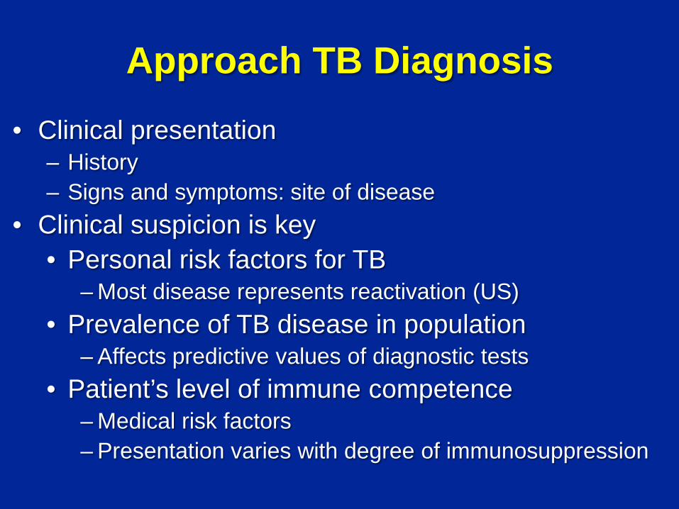
Approach TB Diagnosis
• Clinical presentation– History– Signs and symptoms: site of disease
• Clinical suspicion is key• Personal risk factors for TB
– Most disease represents reactivation (US)• Prevalence of TB disease in population
– Affects predictive values of diagnostic tests• Patient’s level of immune competence
– Medical risk factors– Presentation varies with degree of immunosuppression

TB is a Clinical Diagnosis most of the time
• Most clinicians will initiate multi-drug therapy if the disease is suspected on clinical grounds– But many cases go undiagnosed until a laboratory
reports a positive culture• How is that diagnosis confirmed?
– In the Laboratory

Role of Mycobacteriology Lab• Target: Mycobacterium tuberculosis Complex (MtbC)
– Use rapid methods to detect, identify (ID), and perform drug susceptibility testing (DST)
- TB vs. not TB• Non-tuberculous mycobacteria (NTM)
– Provide accurate / clinically relevant information (accurate ID IF clinically relevant; appropriate DST IF clinically relevant)
• Issue rapid, clinically useful, and reliable reports • Evaluate testing and reporting algorithms as necessary• Develop and maintain 2-way communication -
clinicians, care-givers, TB program, referring laboratories, etc.
B. Metchock, CDC, 9/2010

The TB Laboratory• Types
– Hospital-Clinical Laboratories• Process samples from within an institution and its affiliates
– Private Laboratories• Process samples on contract basis (e.g. Quest, LabCorp,
ARUP)– Network Laboratories
• Process samples for organization (e.g. VA)– Public Health Laboratories
• State/federally supported facilities: Your state lab– Reference Laboratories
• Provide specific services – culture confirmation, molec DST, drug level monitoring, … (e.g. CDC, National Jewish)
• Overall, n >1,932 (+ state labs)

Accommodating Escalating Complexity• Varying Levels of Service Offered
– Not all laboratories perform all tests• Most perform basic tests: smears, primary cultures
– Ability to perform appropriate tests• Equipment, personnel
– Secondary and Reference Laboratories• Receive/process samples for more complex tests
• Communication Challenges– Laboratory-to-laboratory– Provider-to-laboratory(ies)-to-provider-to- …
• Laboratory Competence– Determined locally – Centers for Medicare & Medicaid Services’ Clinical
Laboratory Improvement Amendments (CLIA) program• Proficiency testing

Diagnosis of TB:Demonstration of M. tuberculosis
• The Gold Standard• Secretions or tissue
– Subjected to laboratory techniques to identify the organism
• Ability to isolate organism varies with – Location of disease– Density of organisms at disease site

Standard MycobacteriologyLaboratory Tests
• Smear/stain for acid-fast organisms– Sputum, sterile fluids, tissue
• Culture for identification of organism– Includes speciation– Drug susceptibility studies (DST)
• Nucleic Acid Amplification (NAA)
• Therapeutic Drug Monitoring

Step-By-Step “Typical” TB smear and culture (1)
• Specimen received in lab• Specimen accessioned (assigned lab number; entered
into lab computer/worklog, etc.)• Specimen stored appropriately (refrigerated) until
processed – usually 1x/workday• Specimen processed (digested/ decontaminated) usually
by NALC/NaOH method in batch with other specimens• Smear prepared• Culture media inoculated (usually 1 broth and 1 solid) and
put into incubator/instrument
B. Metchock, CDC, 9/2010
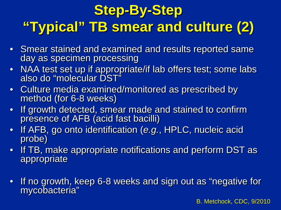
Step-By-Step “Typical” TB smear and culture (2)
• Smear stained and examined and results reported same day as specimen processing
• NAA test set up if appropriate/if lab offers test; some labs also do “molecular DST”
• Culture media examined/monitored as prescribed by method (for 6-8 weeks)
• If growth detected, smear made and stained to confirm presence of AFB (acid fast bacilli)
• If AFB, go onto identification (e.g., HPLC, nucleic acid probe)
• If TB, make appropriate notifications and perform DST as appropriate
• If no growth, keep 6-8 weeks and sign out as “negative for mycobacteria”
B. Metchock, CDC, 9/2010

Specimen Collection• Sputum: Spontaneous or Induced
• Initial: 3 good samples, 8-24hr apart (MMWR, 2005)• Monthly while on treatment until culture-negative
• Collect aseptically, avoid contamination • Sterile, leak-proof, disposable, non-breakable,
appropriately-labeled lab-approved containers• No fixatives or preservatives
• Avoid contamination with tap water • NTM may be in water
• Collect initial samples prior to therapy if possible
• Transport immediately or refrigerate

Sputum Smears: Definitions• Direct smear: stain performed on the submitted sample
– Decontaminated-liquified (NaOH and NALC) and concentrated (centrifuge at 3,000xg)
• Improves yield• Procedure kills >30% of mycobacteria
• Indirect smear: performed on growth from culture– Isolate from primary lab sent to second lab
• For further identification (confirmation) and drug susceptibility studies
• Kinyoun or Ziehl-Neelsen (heat) stain: Light microscopy (1000x mag/oil) – “Acid-fast”: Organisms retain red color following decolorization with
acid-alcohol (the Red Snapper)
• Fluorochrome stain: Fluorescence microscopy (450x mag)– Auramine O– Recommended initial staining procedure (incr sensitivity, decr time)

AFB Smear Microscopy• Variable sensitivity
– 40-70% for pulmonary TB (less in miliary TB, late HIV, children)– LOD >104 AFB/ml by Ziehl-Neelsen; >103/ml fluorochrome– Correlates with disease severity and infectiousness
• Not specific for M.tb Complex – Red snappers
• Inexpensive and quick– Turnaround time (TAT) <24hr
• Value– Usually provides the 1st evidence of TB
• Direct smear light microscopy is the primary diagnostic method in developing world
– Used to guide therapy (AFB in smear are quantified)– May guide additional testing (e.g., NAA)

International Guidelines for Examining and Reporting Acid-Fast Smears:
Organism Count at Specific Magnifications
* number of acid-fast bacilli observed per microscopic field
Number of AFB Observed
Report 200x, 250x 400x, 450x
No AFB seen 0 0Doubtful; repeat 1-2/30F* 1-2/70F
1+ 1-9/10F 2-18/50F2+ 1-9/F 4-36/10F3+ 10-90/F 4-36/F4+ >90/F >36/F
CDC

Fluorochrome stainIncreases sensitivityDecreases performance time


AFB Smears: Rule Out TB?
• A positive smear does not establish diagnosis
• A negative smear does not exclude TB
red snappers

Culture Isolation of M. tuberculosis: The Gold Standard
• Requires appropriate laboratory equipment and trained staff: Competence
• Allows for identification and speciation, drug susceptibility testing
• Performed on secretions or tissue
• Sensitive– Limits of Detection (LOD) 10 to 100 AFB/ml
• 10,000 AFB/ml for smear (Z-N) - more specimen goes into culture

Culture Methods• Solid media
– Agar (Middlebrooks) and egg-based (Lowenstein-Jensen) platforms
– Require up to 6 - 8 weeks– Advantage: Can identify colonies (pigmentation,
morphology)
• Broth – some are highly automated– BACTEC 460; MGIT; TREK; MB/BacT– More rapid recovery than solid media: 7-21 days
• Current recommendations are to use at least one type solid media and broth (mixed culture detection; increased sensitivity)


Colony Morphology

Broth (Liquid Media)BACTEC 460 Instrument
– Semi-automated; needles– Laboratory work-horse– 12B media– Radiometric– Detects CO2 production
by mycobacteria– DST for INH, RMP, EMB,
STR, PZA
B Metchock, CDC 9/2010

Mycobacteria Growth Indicator Tube(MGIT; Broth)
• Fluorescence quenched by O2 in O2-rich liquid media
• If mycobacteria present, O2 used up, no quench, fluoresces under UV light
• DST for INH, RMP, EMB, STR, PZA
B Metchock, CDC 9/2010

M.tb Culture Isolation• Negative cultures do not exclude infectious TB
– Sampling error, contamination, dead organisms, etc.
• False positive: cross-contamination?– Interpretation contextual – Depends on clinical suspicion of disease
• e.g. smear negative, low probability patient
• Cultures guide management– Declining # colonies correlate with response to therapy– Monitor sputum monthly until culture conversion– If culture-pos at 3 mos, look for reason (malabsorption, drug
resistance, etc)
• Rule Out TB?– A positive culture can establish diagnosis– A negative culture does not exclude TB

Identification of Mycobacteria• M.tb vs NTM: Treatment and Public Health implications
• Preliminary ID based on growth characteristics solid media– Colony morphology, pigment, rate of growth (REQUIRES GROWTH)
• Conventional biochemical tests (all mycobacteria) – 2-21 d (may not necessarily be accurate for NTMs)
• HPLC of cell wall mycolic acids (“all” mycobacteria) – 2 h – usually by reference labs
• Commercially available genetic probes – ACCUPROBE, GenProbe, San Diego, CA (www.genprobe.com)
probes for Mtb Complex, MAC, M. kansasii, M. gordonae– 2-4 h – many clinical labs
• “In-house” PCR/genetic sequencing/etc. – 1-2 d – reference labs/clinical labs
B Metchock, CDC 9/2010

Direct Detection of MTB Complex: Nucleic Acid Amplification Testing (NAAT)
• NAA assays– Amplicor
®-Roche: DNA
– MTD®-GenProbe: r-RNA
• Advantages– Excellent sensitivity (10-100 organisms/ml) & specificity for M.tb– TAT generally ≤ 48hr– Can affect treatment decisions, including isolation and other public
health interventions, invasive procedures• Disadvantages
– $$ Costly $$– No indication of viability of organism or of susceptibility
• Still requires culture for confirmation, DST
• FDA-approved ONLY for respiratory secretions (sputum, bronchial)– smear +/- patients, ≤ 7 days therapy (Amplicor
®: smear + only)
• “Off-label” use– Physician education is important

Nucleic Acid Amplification Testing for Respiratory Specimens
• Becoming standard of care … but do not test everyone– Base testing on suspicion and communication with laboratory
• Do not test smear positives when classic TB symptoms and history are present
• Do test smear negatives when clinical suspicion of TB is high
• CDC* – “Test at least one respiratory specimen from each patient with signs and symptoms of pulmonary TB for whom a diagnosis of TB is being considered (i.e. Real suspects) but has not yet been established, and for whom the test result would alter case management and TB control activities”
* MMWR (58:7-10), 2009

NAAT: Application & Interpretation
Sputum, Respiratory Secretions
NAA Positive
NAA Negative
Dx of TB established; cult. still required
Consider clinical picture and repeat testing
Consider clinical picture and repeat testing
Unlikely that M.tbwill be grown from sample (if controlled for inhibitor)
Cannot replace clinical judgment

Drug Susceptibility Testing (DST)• Mandatory on all new patients
– 1st isolate from each site of disease; at 3mos if still cult pos.– Guides treatment decisions
• Initially: for case and contacts• During treatment: determine reason for failure (emergence of resistance, absorption)
• Accurate and timely reporting of results is essential– Direct test; TAT 1 – 3 weeks
• Smear positive cases; primary sample is tested– Indirect test; TAT 7+ weeks
• Requires growth• Liquid media: 3 – 4 wk (can also do MIC for 1st line drugs)
• Direct agar proportion method: Gold Standard• Can test for multiple drugs, cheaply• Resolve agar/liquid media discrepancies

Susceptibility Testing of M. tuberculosis Complex
• Use rapid method (Broth-based)– Perform on all initial patient isolates – Test isolates from relapse or re-treatment cases; also if drug
resistance suspected• Test first-line drugs:
– INH, Rif, EMB, PZA, SM*• Test second-line drugs and higher conc. INH, EMB, SM:
– if resistant to rifampin or any 2 primary drugs
• Second-Line DST– Technically difficult (e.g., CS not recommended); not widely available– Cross-resistance– Methodologies not standardized – especially broth methods– Poor correlation with clinical response
NCCLS Standard M24-A, 2003* M24A2 will drop SM, add AK and LQN

Discordance in DST• Occurs between different labs, different methods,
and within the same method– What do they mean?– Which is right?
• Many possible reasons …– Human
• labeling, cross-contamination, …– Bacteria-specific
• direct vs subculture, clumps, … – Methodology related
• inoculation method, drug conc, media components, …

Laboratory Consortium (4 public health labs and CDC)
• Discordance INH (low level)– Within lab (BT vs. AP) – 2.4%– Interlab (BT) – 6.0%– Interlab (AP) – 12%
• Discordance EMB– Within lab (BT vs. AP) – 6.1%– Interlab (BT) – 20.2%– Interlab (AP) – 8.7%
• Discordance PZA– Interlab (BT) – 4%
B Metchock, CDC, 9/2010

Molecular DST Molecular assays for INH, Rif most common Detect polymorphisms associated with drug resistance Performed on clinical specimens or culture isolates Results available within 1-2 d
In-house assays Molecular beacons, pyrosequencing, RT-PCR
Commercial assays HAIN and INNO-LIPA line probe assays;
Cepheid GeneXpert® MTB/Rif™
Some Issues Multiple mutations may confer resistance – not identified Silent mutations – flagged but not really resistant None is FDA-approved (4/2011)

Molecular Beacons
PHRI

GeneXpert® MTB/RIF™Cepheid
• Closed, self-contained and automated platform• rt-PCR-based amplification of M.tb DNA
– 131 CFU/mL clinical LOD• Boehme NEJM 9/9/10: 1730 pulmonary TB suspects
– Sensitivity: 551/561 sm-pos (98.2%); 124/171 sm-neg(72.5%)*
– Specificity: 604/609 (99.2%)– Simultaneous detection of Rif resistance
• 200/205 Rif-R (97%)
• Costly
*increased to 90% with repeat testing

Molecular Detection of Drug Resistance (MDDR) Service at CDC: Rationale Clinical/Program: available to providers Make rapid confirmation of MDR TB available Make laboratory testing data available to clinicians
about second-line drug resistance in cases of Rif-resistant or MDR TB
Development Continuous correlation of molecular (genotyping)
results and DST (phenotypic) results Addition of new drugs and alleles
Research Determination of mechanisms of resistance
B. Metchock, CDC, 9/2010

MDDR Service:Drugs and Genes for Panel
Drug Gene(s)
RMP rpoB
INH inhA, katG
KAN rrs, eis
AMK rrs
CPM rrs, tlyA
FQ gyrA
PZA pncA
EMB embB B. Metchock, CDC, 9/2010

False-negative and False-positive results• False-negative cultures
– Improper collection/transport; overheating during transport/centrifugation; over-decontamination; media not inoculated correctly; clerical (labeling, transcribing, etc.)
• False-positive results – Another patient’s specimen or isolate; splashes; transfer on tools
or aerosols during processing; contaminated reagents; AFB in water; clerical
• Clues– Increased number of sm +/ cult – detected by lab– Single positive cult among many submitted on patient– Delayed, scanty growth; multiple pos cultures on rack – Clinician: … No Way this is TB…
• Resolution– Lab must have process in-place– Molecular testing often helpful

Serum Drug Level Monitoring
• Useful in selected circumstances• Helps determine therapeutic concentrations
– Allows adjustments for variable drug absorptions• Documents adherence to treatment• May reduce toxicities

Serum Drug Level Monitoring• Aminoglycosides
– To reduce toxicity, achieve therapeutic levels– In-house (Amikacin) vs send-out (Kanamycin)
• Ethambutol– Useful in renal insufficiency to reduce toxicity?
• Rifampin– To determine malabsorption (e.g. in severe HIV)
• Cycloserine– To determine therapeutic levels

The TB Laboratory: Challenges• Declining case rates
– Reduced competencies in low-incidence areas– Level of service: small labs “farming out” tests
• Shifting public health priorities– Reduced categorical funding for TB labs– Increased support for “crisis” responses (Anthrax, BT)
• Increasingly complex technologies– Capital investments– Training/educational needs of staff, users of services
• Demand for high-quality services– Budget issues– Public vs. private
• Erosion of public health laboratory’s key roles2011
