the cytoarchitecture of domain-specific regions in human high-level visual...
TRANSCRIPT

Cerebral Cortex, 2016; 1–16
doi: 10.1093/cercor/bhw361Original Article
O R I G I NA L ART I C L E
The Cytoarchitecture of Domain-specific Regionsin Human High-level Visual CortexKevin S. Weiner1,†, Michael A. Barnett1,†, Simon Lorenz2, Julian Caspers2,3,Anthony Stigliani1, Katrin Amunts2,4,5, Karl Zilles2,5,6, Bruce Fischl7,8
and Kalanit Grill-Spector1,9
1Department of Psychology, Stanford University, Stanford, CA 94305, USA, 2Institute of Neurosciences andMedicine (INM-1), Research Centre Jülich, 52428 Jülich, Germany, 3Department of Diagnostic andInterventional Radiology, Medical Faculty, University of Düsseldorf, 40225 Düsseldorf, Germany, 4Cécile andOskar Vogt Institute for Brain Research, Heinrich-Heine University of Düsseldorf, 40225 Düsseldorf, Germany,5JARA-BRAIN, Jülich-Aachen Research Alliance, 52428 Jülich, Germany, 6Department of Psychiatry,Psychotherapy and Psychosomatics, RWTH Aachen University, 52062 Aachen, Germany, 7Martinos Center forBiomedical Imaging and Department of Radiology, Harvard Medical School, Massachusetts General Hospital,Boston, MA 02114, USA, 8Computer Science and Artificial Intelligence Laboratory, MIT EECS/HST,Massachusetts Institute of Technology, Cambridge, MA 02139, USA and 9Stanford Neurosciences Institute,Stanford University, Stanford, CA 94305, USA
Address correspondence to Kevin S. Weiner, Department of Psychology, Stanford University, Stanford, CA 94305, USA. Email: [email protected];Kalanit Grill-Spector, Department of Psychology, Stanford University, Stanford, CA 94305, USA. Email: [email protected]
†Co first authors.
AbstractA fundamental hypothesis in neuroscience proposes that underlying cellular architecture (cytoarchitecture) contributes tothe functionality of a brain area. However, this hypothesis has not been tested in human ventral temporal cortex (VTC)that contains domain-specific regions causally involved in perception. To fill this gap in knowledge, we used cortex-basedalignment to register functional regions from living participants to cytoarchitectonic areas in ex vivo brains. Thisnovel approach reveals 3 findings. First, there is a consistent relationship between domain-specific regions andcytoarchitectonic areas: each functional region is largely restricted to 1 cytoarchitectonic area. Second, extractingcytoarchitectonic profiles from face- and place-selective regions after back-projecting each region to 20-μm thickhistological sections indicates that cytoarchitectonic properties distinguish these regions from each other. Third, somecytoarchitectonic areas contain more than 1 domain-specific region. For example, face-, body-, and character-selectiveregions are located within the same cytoarchitectonic area. We summarize these findings with a parsimonious hypothesisincorporating how cellular properties may contribute to functional specialization in human VTC. Specifically, we linkcomputational principles to correlated axes of functional and cytoarchitectonic segregation in human VTC, in whichparallel processing across domains occurs along a lateral–medial axis while transformations of information withindomain occur along an anterior–posterior axis.
© The Author 2016. Published by Oxford University Press.This is an Open Access article distributed under the terms of the Creative Commons Attribution Non-Commercial License (http://creativecommons.org/licenses/by-nc/4.0/), which permits non-commercial re-use, distribution, and reproduction in any medium, provided the original work is properly cited.For commercial re-use, please contact [email protected]
Cerebral Cortex Advance Access published November 30, 2016 at Stanford U
niversity on Decem
ber 1, 2016http://cercor.oxfordjournals.org/
Dow
nloaded from

Key words: brain map, cytoarchitecture, fusiform body area (FBA), fusiform face area (FFA), parahippocampal place area(PPA), visual word form area (VWFA)
IntroductionHuman ventral temporal cortex (VTC) contains functionalregions that are selective for specific domains such as faces(Puce et al. 1995; Kanwisher et al. 1997; Grill-Spector et al. 2004;Weiner and Grill-Spector 2010), places (Epstein and Kanwisher1998), words (Cohen et al. 2000), and bodies (Peelen andDowning 2005a). These regions are also causally involved in theperception of domain-specific information (Gaillard et al. 2006;Parvizi et al. 2012; Megevand et al. 2014; Rangarajan et al. 2014).Interestingly, the anatomical location of these regions is repro-ducible across people (Peelen and Downing 2005a, 2005b;Spiridon et al. 2006; Weiner and Grill-Spector 2010; Nasr et al.2011; Julian et al. 2012; Grill-Spector and Weiner 2014) and evenpredictable from cortical folding alone (Glezer and Riesenhuber2013; Grill-Spector and Weiner 2014). Recent evidence indicatesthat anatomical features such as white matter (Saygin et al.2012; Yeatman et al. 2012; Gomez et al. 2015; Osher et al. 2016;Wandell 2016; Weiner et al. 2016) and myelination (Glasserand Van Essen 2011) contribute to the consistent cortical loca-tion of functional regions especially for the large-scale posi-tioning of face-selective regions on the lateral fusiform gyrus (FG)and of place-selective regions in the collateral sulcus (CoS; Glasserand Van Essen 2011; Saygin et al. 2012; Gomez et al. 2015).
However, a long-standing and fundamental hypothesis inneuroscience is that differences in the microarchitecture ofcell bodies across cortical layers (referred to as cytoarchitec-ture) also contribute to the cortical location and functionalityof brain regions (Brodmann 1909; von Economo and Koskinas1925; Hubel and Wiesel 1977; Van Essen et al. 1992; Zilles andAmunts 2010; Amunts and Zilles 2015). This hypothesis pre-dicts that cortical regions in human VTC that process informa-tion related to different domains would be located withindifferent cytoarchitectonic areas. This hypothesis has not beentested for 2 main reasons. First, while the correspondencebetween cytoarchitecture and functional regions has beenexamined in non-human primates (typically macaques; Grosset al. 1972; Van Essen and Zeki 1978; Boussaoud et al. 1991;Zangenehpour and Chaudhuri 2005; Borra et al. 2010), the FG,which is a large macroanatomical component of VTC (Weinerand Zilles 2016), is a hominoid-specific gyrus that macaques
do not have. Thus, understanding if and how underlying cyto-architectonics contribute to the functional organization of theFG—and human VTC more broadly—necessitates measure-ments in humans. Second, only recently, observer-independentcytoarchitectonic techniques have revealed a microstructuralheterogeneity of human VTC consisting of 4 cytoarchitectonicareas spanning the FG and nearby sulci (Caspers et al. 2013;Lorenz et al. 2015). The identification of these cytoarchitectonicareas generates a new opportunity to examine the correspond-ence between the functional and cytoarchitectonic heterogen-eity of human VTC for the first time.
Understanding and quantifying the relationship between cyto-architectonic areas and functional regions of human VTC in indi-vidual brains requires relating functional magnetic resonanceimaging (fMRI) data at a macroscale (millimeters to centimeters)in living participants to cytoarchitectonic data at a microscale(microns) in postmortem (PM) brains. Prior studies successfullyachieved this goal in early visual cortex by leveraging the tightrelationship between both types of data and cortical folding usingcortex-based alignment (CBA) (Fischl et al. 2008; Hinds et al. 2009).Since there is a tight relationship between both types of data andcortical folding in VTC (Weiner et al. 2014; Lorenz et al. 2015), weapplied similar techniques in this study. In brief (Fig. 1; Materialsand Methods), we registered cytoarchitectonic areas defined inhistological slices of PM brains to a common brain using CBA togenerate probabilistic maps of cytoarchitectonic regions of inter-est (cROIs). Then, we used CBA to register probabilistic cROIs tofunctional regions of interest (fROIs) from individual living partici-pants and quantified the correspondence between cytoarchitec-tonic areas and functional regions. Crucially, the implementationof this analysis pipeline in the reverse order also enables theextraction of cytoarchitectonic profiles from fROIs by registeringand projecting probabilistic fROIs to individual histological sec-tions of PM brains.
Using this novel approach, we examined the relationshipbetween domain-specific regions and cytoarchitectonic areas ofhuman VTC. Our data indicate that there is a consistent rela-tionship between domain-specific regions and cytoarchitectonicareas: each functional region is largely restricted to 1 cytoarchi-tectonic area. However, this relationship is not bidirectional, as
Figure 1. Relating the cytoarchitectonic and functional organization of VTC. (A) Observer-independent methods identify cROIs in stained histological sections from
PM brains. (B) cROIs are projected from histological slices to anatomical T1-weighted images (T1s). (C) cROIs are projected from anatomical T1s to the cortical surface
reconstruction of each individual and then to the FreeSurfer average brain using CBA. (D) Maximum probability maps (MPMs) of cROIs on the FreeSurfer average brain
are determined from 10 PM brains. (E) The MPM of each cROI is projected to the cortical surface of each in vivo participant and compared with fROIs (outlines) in indi-
vidual brains.
2 | Cerebral Cortex
at Stanford University on D
ecember 1, 2016
http://cercor.oxfordjournals.org/D
ownloaded from

some cytoarchitectonic areas contain more than 1 domain-specific region. These findings support a new hypothesis sug-gesting what computational principles may explain the orderlybut complex relationship between functional regions and cyto-architectonic areas of human VTC.
Materials and MethodsDefinitions of functional regions and cytoarchitectonic areaswere done by independent researchers—researchers definingfunctional regions were blind to cytoarchitectonic definitionsand vice versa. Data collection, analyses, and definitions of fROIsof in vivo data were done by K.W., A.S., M.B., and K.G-.S. atStanford University. Data collection, analysis of PM data, anddefinition of cROIs were done at the Institute of Neuroscienceand Medicine (INM-1), Research Centre Jülich, by J.C., S.L., K.A.,and K.Z.
Participants of the In Vivo Study
Twelve right-handed participants (5 female, ages 19–44 years)with normal or corrected-to-normal vision were recruited fromStanford University for the functional mapping experiment,which was used to identify high-level visual regions in humanVTC. Procedures were approved by the Stanford Internal ReviewBoard on human subjects research.
In Vivo Data Acquisition and Analyses
Anatomical Scans and AnalysesScanning. A high-resolution anatomical volume of the wholebrain was acquired using a 3 T GE Discovery MR750 scanner (T1-weighted BRAVO pulse sequence; resolution: 1mm × 1mm ×1mm, TI = 450ms, flip angle = 12°, 1 NEX, FOV = 240mm).
Cortical surface reconstruction. Anatomical volumes were aligned tothe AC-PC plane. Using a combination of automated (FreeSurfer:http://surfer.nmr.mgh.harvard.edu) and manual segmentationtools (ITK-SNAP: http://www.itksnap.org/pmwiki/pmwiki.php),each anatomical volume was segmented to separate gray fromwhite matter, from which we reconstructed the cortical surface foreach participant (Wandell et al. 2000). These cortical surface recon-structions were used in bothmrVista and FreeSurfer.
Functional Scans and AnalysesData acquisition. Participants were scanned using a custom-builtphase-array, 32-channel head coil. We acquired 34 slices coveringoccipitotemporal cortex (resolution: 2.4mm × 2.4mm × 2.4mm;one-shot T2*-sensitive gradient echo acquisition sequence: FOV =192mm, TE = 30ms, TR = 2000ms, and flip angle = 77°).
Functional localizer. Two sessions of fMRI data were collectedfrom each participant on different days. On the first day, 10runs of an fMRI functional localizer experiment from Stiglianiet al. (2015) were collected. Each run contained blocks of differ-ent images from 5 different categories: faces (adult and childfaces), places (corridors and houses), bodies (limbs and head-less bodies), characters (numbers, pseudowords), and objects(cars and guitars). Each run contained images presented at dif-ferent rates (either 1, 2, 4, or 8 Hz). Stimulus trials were counter-balanced with blank baseline trials. In each run, stimuli wereshown at one rate. The order of runs at different rates wascounterbalanced across participants. The same set of 1440images was shown across all rates, but images did not repeat
within a rate. Participants conducted an oddball task duringwhich they fixated on a central dot and detected an oddballphase scrambled image that occurred randomly in a block. Theincidence of the oddball image was 0, 1, or 2 times per block.We chose this task as it keeps participants engaged during theexperiment and is frequently used in studies of human VTC(Kanwisher et al. 1997; Gauthier et al. 2000; Glezer et al. 2009;Weiner and Grill-Spector 2010, 2011; Stigliani et al. 2015.Additionally, responses in VTC in the oddball task are similarto other tasks that require explicit processing of the visualimages such as categorization tasks (Weiner and Grill-Spector2010; Weiner et al. 2010), 1 or 2-back tasks (Weiner and Grill-Spector 2010; Weiner et al. 2010; Bugatus et al. 2015), or select-ive attention (Bugatus et al. 2015, 2016).
On the second day, participants participated in 2 runs total-ing 11minutes of the 1 Hz localizer experiment with the samestimuli and task. The purpose of the second experiment was toextract response amplitudes from fROIs using independentdata (Fig. 2).
Data analysis. Data were analyzed with MATLAB using mrVistasoftware (http://github.com/vistalab) as in our prior publica-tions (Weiner and Grill-Spector 2010; Stigliani et al. 2015).
Time series processing. Functional data of each session weremotion corrected using an affine transformation (Nestares andHeeger 2000). Time series data were processed with a temporalhigh-pass filter using a 1/20 Hz cutoff and then converted topercentage signal change by dividing the time series of eachvoxel by its mean intensity. We estimated blood oxygen level–dependent (BOLD) response amplitudes for each condition witha general linear model (GLM) applied to the time series of eachvoxel using as predictors the experimental conditions con-volved with the hemodynamic impulse response function inSPM (http://www.fil.ion.ucl.ac.uk/spm/). Data were not spatiallysmoothed.
Functional regions of interest. FROIs were defined in individualparticipants from the functional localizer using anatomical andfunctional criteria (Weiner and Grill-Spector 2010, 2012;Stigliani et al. 2015). A common threshold (t > 3, voxel level)was used to define all regions across all participants. Since thecytoarchitectonic ROIs (described in the next section) arelocated in the occipitotemporal sulcus (OTS), FG, and CoS, werestricted our definition of fROIs to these anatomical regions.
As in our prior publications (Weiner and Grill-Spector 2010,2012; Stigliani et al. 2015), we identified 2 face-selective ROIs(faces > others) bilaterally on the posterior (pFus-faces, N = 12)and mid (mFus-faces, N = 12) FG; pFus-faces is often referred toas FFA-1 and mFus-faces as FFA-2, where FFA refers to the fusi-form face area (Kanwisher et al. 1997).
Between mFus-faces and pFus-faces, we identified a body-selective region (bodies and limbs > others) in the OTS (N = 12),which is also referred to as the fusiform body area (FBA; Peelenand Downing 2005a, 2005b; Schwarzlose et al. 2005).
As in our prior publications, we also identified 2 character-selective ROIs (characters > others; Stigliani et al. 2015) withinthe left hemisphere on the posterior (pOTS-chars, N = 12) andmid (mOTS-chars, N = 10) OTS. These ROIs are also referred to asVWFA-1 and VWFA-2, respectively (Stigliani et al. 2015), whereVWFA refers to the visual word form area (Cohen et al. 2000;Ben-Shachar et al. 2011; Dehaene and Cohen 2011). In the righthemisphere, we also report data from pOTS-chars/VWFA-1
Cytoarchitecture of Human High-level Visual Cortex Weiner et al. | 3
at Stanford University on D
ecember 1, 2016
http://cercor.oxfordjournals.org/D
ownloaded from

(N = 12), but not mOTS-chars/VWFA-2, as we were able to iden-tify VWFA-2 in only 2 participants within the right hemisphere.
Finally, we identify a place-selective ROI (places > others)within the CoS (N = 12), corresponding to the parahippocampalplace area (PPA; Epstein and Kanwisher, 1998). CoS-places/PPAhas been recently parcellated into separate posterior and anter-ior components based on functional connectivity and weakerselectivity in the posterior compared with the anterior portion(Baldassano et al. 2013; Silson et al. 2015). Our analyses focuson the anterior CoS-places/PPA because this cortical locationis the most consistent across participants and is predictiveof clusters exhibiting the highest place-selectivity in over
500 participants (Weiner et al. Under review). Nevertheless,additional analyses show weakly place-selective voxels in FG1(Supplementary Fig. 2).
Independent analysis of functional responses. Individual datafrom day 2 were aligned to each participant’s high-resolutionanatomical brain volume. Using fROIs defined in day 1, weextracted response amplitudes to faces, objects, bodies,places, and characters from day 2. Response amplitudes fromeach fROI were averaged across the 12 participants (Fig. 2A,bottom).
Figure 2. The definition and characteristics of functional regions and cytoarchitectonic areas in human VTC. (A) Functional organization of category-selective fROIs.
Top: fROIs in an example subject (S1). Bottom: independent analysis of mean ± standard error of the mean of the amplitude of response to adult and child faces (f),
bodies and limbs (b), cars and guitars (objects, o), corridors and buildings (places, p), and pseudowords and numbers (characters, c) in each fROI. Data are averaged
across 12 subjects bilaterally for all fROIs, except mOTS-chars/VWFA-2, which includes data from the left hemisphere of 10 subjects. (B) Cytoarchitectonic areas in
human VTC in an example PM subject. Top: cROIs in the FG and adjacent sulci in an example PM subject. Note that the medial cytoarchitectonic areas FG1 and FG3
encompass both the medial aspect of the FG and the CoS and the lateral cytoarchitectonic areas FG2 and FG4 encompass both the lateral FG and OTS. Bottom: example
histological slices illustrating the representative cytoarchitectonic structure of FG1–FG4. Acronyms: pFus: posterior fusiform; mFus: mid-fusiform; pOTS: posterior
occipitotemporal sulcus. mOTS: mid occipitotemporal sulcus.
4 | Cerebral Cortex
at Stanford University on D
ecember 1, 2016
http://cercor.oxfordjournals.org/D
ownloaded from
PM Data Acquisition and Analysis
Histological analyses were performed on eleven PM brains(ages 37–85, 6 females), which had no history of neurological orpsychiatric disease (with exception of 1 individual with transi-tory motor disease). These were the same brains used in priorcytoarchitectonic studies (Caspers et al. 2013; Lorenz et al.2015). Nine of the PM brains were shared across the Caspersand Lorenz studies (see table 1 in each of these studies), andeach cytoarchitectonic area was defined in 10 PM brains(5 females). All PM brains came from the body donor programof the Institute of Anatomy, University of Dusseldorf; all proce-dures were approved and in alignment with the program guide-lines (Amunts et al. 2000).
ScanningPM brains were scanned on a Siemens 1.5 T Scanner (Erlangen,Germany). Brains were removed from the subject’s skull 8–24 hafter death and then fixated in 4% formalin or Bodian’s fixativefor at least 6 months. A high-resolution anatomical volumeof the whole fixed brain was acquired using a T1-weighted3D-FLASH sequence (TR = 40ms, flip angle = 40°, TE = 5ms).
Histological ProcessingDetailed methods of histological processing have been pub-lished previously (Amunts et al. 2000; Caspers et al. 2013;Lorenz et al. 2015). Briefly, the fixated brains were embedded inparaffin, serially sectioned in coronal sections (20 μm thick),and cell bodies were stained using the Merker-method (Amuntset al. 2000). This method yields a high contrast between cellbodies (black) and the neuropil (unstained). 3D reconstructionsof the histological sections were computed using 1) the 3D-MRIvolume of each brain, 2) images of the paraffin block face forprecise alignment of the histological sections, and 3) digitizedimages of the cell body–stained sections.
Definition of Cytoarchitectonic RegionsFG1, FG2, FG3, and FG4. FG1 and FG2 were defined by Casperset al. (2013), and FG3 and FG4 were defined by Lorenz et al. (2015)from 20-μm thick sections of 10 PM brains using a statisticallytestable, quantitative, and observer-independent cortical parcel-lation technique (Amunts et al. 2000; Schleicher et al. 2000;Caspers et al. 2013). Procedures are described in detail in priorpublications (Caspers et al. 2013; Lorenz et al. 2015). In brief,gray-level indices (GLIs) were determined in digitized histo-logical sections as a measure of the volume proportion betweencell bodies and the neuropil. GLI profiles were calculated alongcurvilinear trajectories oriented perpendicular to the corticallayers, thus measuring the GLI from Layer I/Layer II border tothe innermost layer in cortical regions of interest. The shape ofGLI profiles was determined based on 10 features: the mean GLIvalue, the position of the center of gravity on the profile curve(cortical depth), the standard deviation of the mean GLI (indicat-ing the variability of the GLI throughout all layers), skewnessand kurtosis of the profile curve, and the respective featuresfrom the profile’s first derivative (Amunts et al. 2000; Schleicheret al. 2000). The borders between areas were determined basedon the cortical position of the greatest difference between neigh-boring GLI profiles quantified by the Mahalanobis distance andtested for significance (Hotelling’s T2 test; Bonferroni-corrected).Areal borders are expected at positions along the cortical ribbonshowing a significant dissimilarity in laminar patterns betweenadjacent blocks of profiles. To assure that the areal boundarywas not dependent on the block size, the procedure was titrated
for block sizes ranging from 8 to 24 profiles per block. Corticalborders were confirmed if they were consistently positioned inadjacent histological sections and across several block sizes.
As illustrated in Figure 2B, these 4 FG cytoarchitectonicareas macroanatomically include both the FG and adjacent sul-ci: the medial cytoarchitectonic areas, FG1 and FG3, include themedial aspect of the FG and the CoS, while the lateral cyto-architectonic areas, FG2 and FG4, include the lateral aspect ofthe FG and the OTS (Caspers et al. 2013; Lorenz et al. 2015).
Registering cROIs to Individual Cortical SurfacesAfter cROIs were defined in the histological sections (Fig. 1A),they were aligned to the anatomical MRI of each PM’s nativebrain volume (Fig. 1B) as described previously (Caspers et al. 2013;Lorenz et al. 2015). We also generated cortical surface reconstruc-tions of each PM brain by manually segmenting the anatomicalMRI to differentiate gray and white matter using ITK-SNAP(http://www.itksnap.org/pmwiki/pmwiki.php). From the bound-ary between white and gray matter, we reconstructed the corticalsurfaces in both mrVista and FreeSurfer. Each individual’s cROIswere then projected to their cortical surface (Fig. 1C).
Registration of Functional and Cytoarchictectonic ROIs to theFreesurfer Average BrainTo examine the correspondence between cROIs and fROIs in acommon anatomical space, we used CBA implemented inFreeSurfer (http://surfer.nmr.mgh.harvard.edu) to align each sub-ject’s cortical surface and ROIs to the FreeSurfer average brain,which is an average of the cortical surface of 39 independent par-ticipants (Fischl et al. 1999). As both living and PM subjects hadregions of interest defined in the mrVista software package, thealignment process for fROIs and cROIs was the same. Both cROIsand fROIs were saved as NIfTI files, imported into FreeSurfer,and projected to the respective cortical mesh of each subject inFreeSurfer. Using CBA, each subject’s ROIs were aligned to theFreeSurfer average brain (Fischl et al. 1999). This CBA uses ahigh-dimensional nonlinear registration algorithm, which alignsthe cortical folding patterns of each subject to the cortical foldingpatterns of the FreeSurfer average cortical surface. A mapping ofthe cortical folding patterns of each subject and the template toa sphere generate a point-to-point correspondence between sub-jects on the FreeSurfer average cortical surface.
Maximum Probability Maps of Cytoarchitectonic ROIsOn the FreeSurfer average cortical surface, we generated a max-imum probability map (MPM) of each cROI using data from 10PM brains (Fig. 3). We transformed each binary cROI (i.e., 1where the cROI is present and 0 elsewhere) to the common cor-tical surface and then averaged them on the common surfaceto generate a probabilistic map of each ROI. The value at eachnode of the map indicates the proportion of subjects that over-lapped at that node for a given cROI (Fig. 3A). We then used anexhaustive, leave-one-out cross-validation procedure to deter-mine the threshold that produced the most predictive probabil-istic cROIs (Fig. 3B). We estimated the predictability of theprobabilistic cROIs by calculating the dice coefficient betweenthe left-out and predicted cROIs: = | ∩
| + |Dice Coefficient P A
P A2 ,
where P are the nodes of the probabilistic cROI and A are thenodes of the actual cROI of the left-out subject. Since our cROIsare neighboring, probabilistic cROIs can have shared nodes onthe FreeSurfer average cortical surface. Thus, to ensure that eachnode in the common cortical surface is assigned to a single cROI,we generated an MPM of each probabilistic cROI. The MPMs were
Cytoarchitecture of Human High-level Visual Cortex Weiner et al. | 5
at Stanford University on D
ecember 1, 2016
http://cercor.oxfordjournals.org/D
ownloaded from

generated by first finding all overlapping nodes on the corticalsurface among the probabilistic cROIs and then assigning thosenodes to the cROI with the highest overlap across subjects. Forexample, if a given node on the cortical surface had a probabilis-tic value of 0.3 for FG1 and a probabilistic value of 0.6 for FG3,then this node was assigned to the MPM of FG3 and excludedfrom the MPM of FG1. We make these MPM ROIs freely availablefor download with FreeSurfer and also directly from this link:vpnl.stanford.edu/FGcROIAtlas.
Additionally, we also aligned the MPMs of our cROIs to the FSaverage_sym template (Greve et al. 2013), which is a cortical sur-face that enables between-hemisphere comparisons. Aligningthe MPMs of our cROIs to this surface illustrates that the topo-logical layout of each area relative to cortical folding is largelysimilar between the right and left hemispheres. Nevertheless,there are also slight differences. For example, FG3 and FG4extend more anteriorly in the left compared with the righthemispheres. Additionally, FG2 extends more laterally in theright compared with the left hemisphere (Supplementary Fig.1). Importantly, these slight topological deviations betweenhemispheres are also clearly evident in Figure 3.
Measuring the Correspondence between ROIs and cROIsWe quantified the correspondence between fROIs and cROIs bycalculating the percentage overlap between each individual
participant’s fROIs and the MPM of each cROI (Figs. 4, 7).Importantly, to assess if these overlap values are meaningful, wecomputed the chance proportion overlap between fROIs andcROIs using permutation testing and then determined if theoverlap values were significantly different than the estimatedchance level. We estimated 2 kinds of chance overlap propor-tions: 1) the chance proportion overlap between a given fROI andany of the cROIs and 2) the chance proportion overlap betweenany of the fROIs and any of the cROIs. The former was done byrandomly choosing cROIs and calculating the overlap between agiven fROI and a randomly chosen cROI, and the latter was doneby randomly choosing both fROIs and cROIs. Permutations weredone with 400 iterations per fROI per hemisphere and 400 itera-tions per cROI per hemisphere. Figures 4 and 7 show the latterestimate of chance overlap. While there were numerical differ-ences across methods, the range of chance overlap from bothmethods was similar ranging between 0.2 ± 0.03 and 0.25 ± 0.03.
Finally, we examined the spatial relationship between fROIsand cROIs in each participant. Using CBA, we projected the MPMof each cROI to the individual surface of each individual living par-ticipant (Fig. 1E). This enabled us to 1) visualize the correspond-ence between the MPM of the cROI and each participant’s fROIsand 2) examine the pattern of errors when fROIs were not per-fectly aligned to cROIs (4 example participants are shown in Fig. 8).
In addition to our single-subject analyses, we also generatedMPMs of fROIs on the FreeSurfer average cortical surface using
Figure 3. Cross-validated probabilistic maps of cytoarchitectonic areas. (A) Probabilistic maps of cytoarchitectonic areas on the Freesurfer (FS) average cortical surface.
Colors indicate the proportion of overlapping subjects at each point on the cortical surface. Black outline: mid-fusiform sulcus (MFS). The arrangement of the probabil-
istic cROIs on the FS average cortical surface maintains the relation to the cortical folding as seen in individual subjects. Specifically, the MFS serves as the boundary
between the probabilistic map of FG1 and FG2 (Weiner et al. 2014) and the boundary between probabilistic map of FG3 and FG4 (Lorenz et al. 2015). Top: FG4 and FG3.
Bottom: FG2 and FG1. (B) Exhaustive, leave-one-out cross-validation of group probability maps for each cROI. Each map was generated with 9 subjects. Then, we evalu-
ated how well this map predicted the left-out subject using the dice coefficient. This process was done 10 times for each left-out subject. Across FG1–FG4, the best
dice coefficient was obtained at thresholds of 0.3–0.4. Thus, for subsequent analyses, we used the 0.3 threshold. Line: mean dice coefficient. Shaded area: standard error
of the mean. Top: FG4 and FG3. Bottom: FG2 and FG1. Direct comparison of the MPMs of these cROIs across hemispheres is illustrated in Supplementary Figure 1.
6 | Cerebral Cortex
at Stanford University on D
ecember 1, 2016
http://cercor.oxfordjournals.org/D
ownloaded from
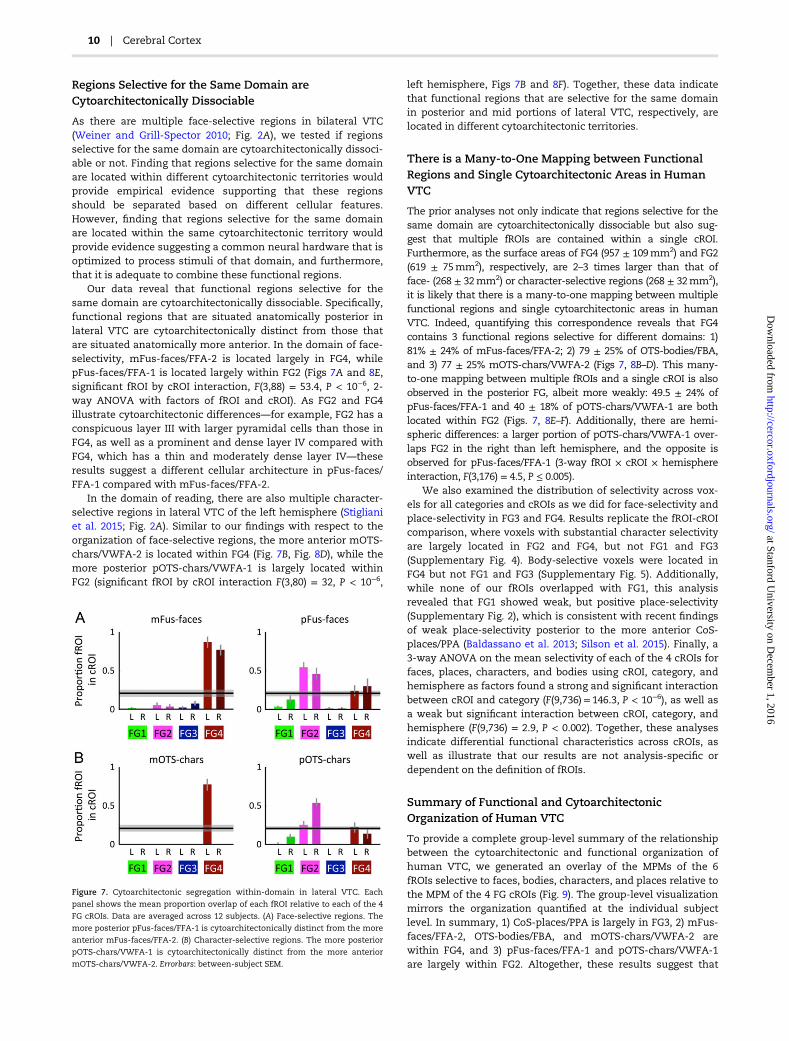
data from all living participants using the same method asdescribed above for cROIs. We then registered MPMs of all fu-nctional regions and cytoarchitectonic areas to the FreeSurferaverage cortical surface to generate a group-level summary offunctional–cytoarchitectonic relationships in human VTC (Fig. 9).
It should also be stated that we cannot rule out the possibilitythat there are slight imprecisions in our approach because ofindividual subject variability in fROIs, patterns of cortical folding,and their relationship. However, we are confident in the accur-acy of our approach as the resulting group MPMs of fROIs andcROIs accurately preserve the topology of each type of ROI andcortical folding in individual subjects, which indicates the preci-sion of these alignment techniques as previously shown (Fischlet al. 2008; Hinds et al. 2009). Errors exist in any study directlycomparing living and PM brains and are unavoidable until there
will be new methods that will enable measuring cytoarchitec-tonic areas in vivo, which will, in turn, allow the direct compari-son between cROIs and fROIs within the same individuals.
Examining the Effect of Contrast on the Distribution of CategorySelectivity Values within Each cROITo complement the analyses described above examining the coup-ling between fROIs and cROIs, we also assessed the functional–cytoarchitectonic relationship within VTC by comparing thedistribution of category selectivity values across all voxels withineach cROI. This analysis is hypothesis-free as it 1) does notnecessitate localizing any fROIs and 2) is not dependent on thethreshold or contrast used to define fROIs. To perform this ana-lysis, each probabilistic cROI was transformed to each living
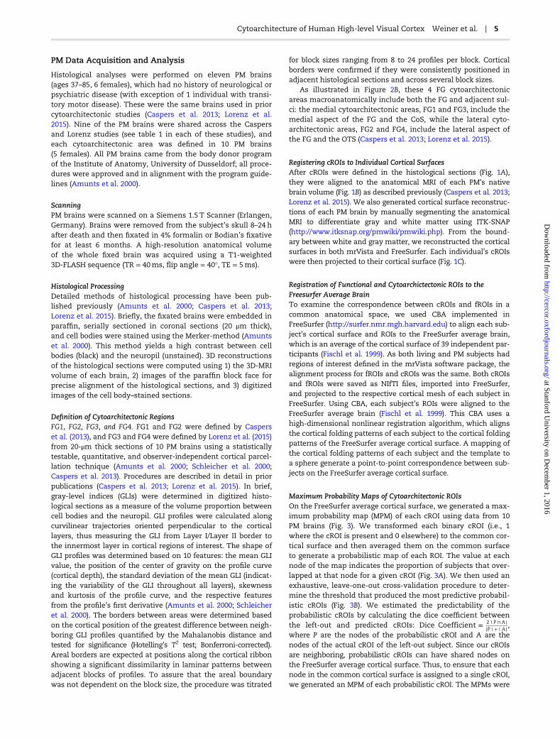
Figure 4. Face- and place-selective regions are located in distinct cytoarchitectonic areas. (A) Dark red outline of the MPM for mFus-faces/FFA-2 from 12 living indivi-
duals superimposed on the MPM for FG4 from 10 PM brains. (B) Four example left hemispheres illustrating mFus-faces/FFA-2 in individual subjects relative to the
MPM of FG4. (C) Mean proportion overlap of mFus-faces/FFA-2 relative to each cROI. Errorbars: Standard error of the mean across 12 subjects. Horizontal bar: chance
overlap probability. (D and E) Same as A and B, but for CoS-places/PPA (green outline) and FG3. (F) Same as (C), but for CoS-places/PPA.
Cytoarchitecture of Human High-level Visual Cortex Weiner et al. | 7
at Stanford University on D
ecember 1, 2016
http://cercor.oxfordjournals.org/D
ownloaded from

participant’s brain. Selectivity in each voxel within each cROIwas then determined by the t-value of the GLM contrast. InFigure 5, we illustrate the distribution of face- and place-selectivity across all voxels of FG3 and FG4. We assessed select-ivity to faces in each voxel using 2 contrasts: 1) preferred versusother categories and 2) preferred versus places. Selectivity toplaces in each voxel was assessed with 2 contrasts: 1) places ver-sus other categories and 2) places versus faces. We conducted anon-parametric Kolmogorov–Smirnov test (K-S test) to deter-mine if the distributions of selectivity values in FG3 and FG4were significantly different from each other in each subject.Additionally, we include distributions of selectivity values for allcategories and all FG cROIs in Supplementary Figures 2–5.Finally, to test if category selectivity significantly differed acrosscROIs, we ran a 3-way analysis of variance (ANOVA) on themean selectivity using category, hemisphere, and cROI asfactors.
Transforming MPMs of Face- and Place-Selective fROIsto Histological SectionsUsing CBA, we registered the MPM of face-selective and place-selective fROIs to the cortical surface of each PM brain. From thePM’s cortical surface, we registered both of these MPMs to theoriginal PM brain volume obtained with an MRI scan. Then, weregistered these MPM fROIs to the reconstructed 3D histologicalvolume using a nonlinear transformation implemented in ANTS(https://sourceforge.net/projects/advants/). Code used for thistransformation can be found here: github.com/VPNL/cyto-func-tional. This procedure localizes the MPM fROIs to the originalhistological slices within which the FG cROIs were defined, con-sequently enabling the extraction of GLI profiles from the prob-abilistic location of each fROI in histological sections (Fig. 6).
In these analyses, we focused on face- and place-selectiveregions because they are located centimeters away from eachother across the cortical ribbon and are identifiable on singlehistological sections with distinct macroanatomical landmarks.As such, we were able to validate the accuracy of the localiza-tion of fROIs in histological sections using 2 distinct domain-specific macroanatomical landmarks: 1) the MFS as a landmarkfor mFus-faces/FFA-2 (Weiner et al. 2014) and 2) the branchingof the CoS and intersection with the nearby anterior lingualsulcus as landmarks for the anterior portion of CoS-places/PPA(Weiner et al. Under review).
ResultsAre Face- and Place-Selective RegionsCytoarchitectonically Dissociable?
We first assessed the correspondence between the cortical loca-tion of cytoarchitectonic areas within VTC relative to face-selective (mFus-faces/FFA-2) and place-selective (CoS-places/PPA) regions as these are 2 of the most widely studied domain-specific regions in VTC, and their arrangement relative to thecortical folding is consistent across individuals. Qualitativeassessments of the cortical location of mFus-faces/FFA-2 andCoS-places/PPA relative to cytoarchitectonic areas indicate thateach of these regions is located within distinct cytoarchitec-tonic areas. At the group level, mFus-faces/FFA-2 is locatedwithin FG4 (Fig. 4A) and CoS-places/PPA is located within FG3(Fig. 4D). Quantitative evaluation of the overlap between fROIsin individual participants and the probabilistic maps of each ofthe 4 FG cROIs revealed that 81 ± 24% (mean ± standard devi-ation) of mFus-faces/FFA-2 is located within FG4 (Fig. 4C) and
75 ± 11% of CoS-places/PPA is located within FG3 (Fig. 4F).These levels of overlap are significantly above chance (ts > 8.5,Ps < 10−8, Fig. 4C,F-horizontal line) and show a significant fROIby cROI interaction (F(3,88) = 380, P < 10−6, 2-way ANOVA withfactors of fROI and cROI). Visualizing the location of fROIs rela-tive to cROIs in each participant (Fig. 4B,E) shows that the rela-tionship among fROIs and cROIs is visible at the individualsubject level: mFus-faces/FFA-2 is located within FG4 and rarelyextends medially from FG4 into FG3 (Fig. 4B,C), while CoS-places/PPA is largely located within FG3. Note that portions ofCoS-places/PPA not in FG3 extend medially into the lip of theCoS and the parahippocampal gyrus and rarely deviate intoFG1, FG2, or FG4 (Fig. 4E,F). Thus, mFus-faces/FFA-2 and CoS-places/PPA are located within different cytoarchitectonic areas.
We complemented these analyses by comparing the distri-bution of face- and place-selectivity values in FG3 and FG4.This analysis is hypothesis-free because it does not require thedefinition of fROIs. Results show that while the contrast quanti-tatively affects selectivity values, it does not qualitativelychange the pattern of results. Specifically, independent of con-trast, FG3 and FG4 are differentially selective for places andfaces, respectively (Fig. 5; 2-sample K-S tests comparing the dis-tributions of selectivity in FG3 and FG4 for all 4 contrasts: allK-S stats > 0.3, all Ps < 10−6). Likewise, a 4-way ANOVA of meanselectivity using as factors cROI (FG3/FG4), preferred category(faces/places), contrast (preferred versus the other category/preferred versus all categories), and hemisphere, reveals a sig-nificant cROI x preferred category interaction (F(3,352) = 348.51,P < 10−6), as well as significant cROI × preferred category × con-trast interaction (F(3,352) = 5.12, P < 0.002). The former inter-action indicates that changing the contrast does notqualitatively affect the result that FG3 and FG4 exhibit differentcategory preferences, while the latter (weaker) interactionreflects that changing the contrast quantitatively affects themagnitude of the t-values. These results complement our prioranalyses and provide empirical support demonstrating that the
Figure 5. Place-selective voxels are localized to FG3 and face-selective voxels
are localized to FG4. (A) Distribution of place-selectivity across all voxels of
cytoarchitectonic areas FG3 (left) and FG4 (right) for 2 contrasts: places versus
all other categories (black) and places versus faces (red). For both contrasts, FG3
illustrates a distribution with an average positive selectivity for places, while
FG4 illustrates a distribution with an average negative selectivity for places.
(B) Distribution of face-selectivity across all voxels of cytoarchitectonic areas
FG3 (left) and FG4 (right) for 2 contrasts: faces versus all other categories (black),
and faces versus places (red). For both contrasts, FG3 illustrates a distribution
with an average negative selectivity for faces, while FG4 illustrates a distribu-
tion with an average positive selectivity for faces. Shaded areas: standard devi-
ation across 12 subjects. Arrows: average selectivity. Dashed vertical line:
threshold used to define fROIs (t = 3). Solid vertical line: t= 0.
8 | Cerebral Cortex
at Stanford University on D
ecember 1, 2016
http://cercor.oxfordjournals.org/D
ownloaded from

coupling between face-selectivity and FG4, as well as place-selectivity and FG3, generalizes across analyses and is notdependent on the definition of fROIs.
What Are the Cytoarchitectonic Features of Face- andPlace-Selective Regions?
We probed the underlying cytoarchitecture of mFus-faces/FFA-2and CoS-places/PPA directly by back-projecting the MPMs of thesefunctional regions to cell body–stained sections and then extract-ing histological profiles of these regions to examine the distribu-tion of cells across layers (Fig. 6). This is akin to functional ROIanalyses in which an ROI is localized with one set of data, andfunctional properties are determined from independent data. Forthe first time, instead of probing functional properties of a region,
we extract cytoarchitectonic properties to reveal the cellularstructure of regions selective for faces and places.
Results show that mFus-faces/FFA-2 and CoS-places/PPA havedistinct cytoarchitectonic features as represented by GLI profiles ofcell body density across layers. The GLI profile of mFus-faces/FFA-2 is characterized by a prominent sublayer IIIc with medium- tolarge-sized pyramidal cells, a moderately dense layer IV, and aslight overall increase of GLI from layer II to layer VI with severallocal maxima and minima, but an absolute maximum in layer VI(Fig. 6C–E). By contrast, CoS-places/PPA is characterized by a rela-tively compact, dense layer II, a layer IV with little clusters ofgranular cells, and a GLI curve that does not show a trend toincrease towards layer VI as found in mFus-faces/FFA-2 (Fig. 6C–E).These results reveal the cytoarchitectonic features of face- andplace-selective regions for the first time and show that theseregions have distinct cytoarchitectonic characteristics.
Figure 6. Cytoarchitectonic profiles of face- and place-selective regions. (A) MPM of mFus-faces/FFA-2 (red) and CoS-places/PPA (green) projected onto a reconstruction
of a histological slice from an example PM brain. (B) Zoomed view of the cell body–stained histological section within the rectangular region shown in (A). Arrows:
boundaries of cROIs; Dashed lines: boundaries of CoS-places/PPA (green) and mFus-faces/FFA-2 (red). (C) Example histological slices and corresponding GLI profiles
within CoS-places/PPA and mFus-faces/FFA-2 from the locations indicated by the green and red asterisks in (B), respectively. Green and red curves: GLI of cell density
across layers. Note differences in cell density, laminar structure, and cell body size between the 2 sections. White arrowheads: different cell density as estimated by
the GLI in V/VI, illustrating heterogeneously packed layers V/VI of FG4 compared with the more homogeneous layers V/VI of FG3. Diamonds: broad and cell-sparse
layers IIIa/b of FG3 versus prominent and broad layer IIIc of FG4. (A–C) Data from PM1. (D and E) Example cytoarchitectonic profiles within CoS-places/PPA and mFus-
faces/FFA-2 from 2 additional PM brains. (D) PM4. (E) PM9.
Cytoarchitecture of Human High-level Visual Cortex Weiner et al. | 9
at Stanford University on D
ecember 1, 2016
http://cercor.oxfordjournals.org/D
ownloaded from
Regions Selective for the Same Domain areCytoarchitectonically Dissociable
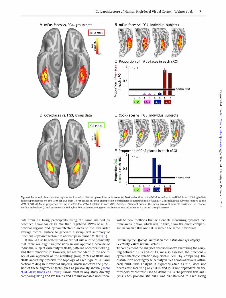
As there are multiple face-selective regions in bilateral VTC(Weiner and Grill-Spector 2010; Fig. 2A), we tested if regionsselective for the same domain are cytoarchitectonically dissoci-able or not. Finding that regions selective for the same domainare located within different cytoarchitectonic territories wouldprovide empirical evidence supporting that these regionsshould be separated based on different cellular features.However, finding that regions selective for the same domainare located within the same cytoarchitectonic territory wouldprovide evidence suggesting a common neural hardware that isoptimized to process stimuli of that domain, and furthermore,that it is adequate to combine these functional regions.
Our data reveal that functional regions selective for thesame domain are cytoarchitectonically dissociable. Specifically,functional regions that are situated anatomically posterior inlateral VTC are cytoarchitectonically distinct from those thatare situated anatomically more anterior. In the domain of face-selectivity, mFus-faces/FFA-2 is located largely in FG4, whilepFus-faces/FFA-1 is located largely within FG2 (Figs 7A and 8E,significant fROI by cROI interaction, F(3,88) = 53.4, P < 10−6, 2-way ANOVA with factors of fROI and cROI). As FG2 and FG4illustrate cytoarchitectonic differences—for example, FG2 has aconspicuous layer III with larger pyramidal cells than those inFG4, as well as a prominent and dense layer IV compared withFG4, which has a thin and moderately dense layer IV—theseresults suggest a different cellular architecture in pFus-faces/FFA-1 compared with mFus-faces/FFA-2.
In the domain of reading, there are also multiple character-selective regions in lateral VTC of the left hemisphere (Stiglianiet al. 2015; Fig. 2A). Similar to our findings with respect to theorganization of face-selective regions, the more anterior mOTS-chars/VWFA-2 is located within FG4 (Fig. 7B, Fig. 8D), while themore posterior pOTS-chars/VWFA-1 is largely located withinFG2 (significant fROI by cROI interaction F(3,80) = 32, P < 10−6,
left hemisphere, Figs 7B and 8F). Together, these data indicatethat functional regions that are selective for the same domainin posterior and mid portions of lateral VTC, respectively, arelocated in different cytoarchitectonic territories.
There is a Many-to-One Mapping between FunctionalRegions and Single Cytoarchitectonic Areas in HumanVTC
The prior analyses not only indicate that regions selective for thesame domain are cytoarchitectonically dissociable but also sug-gest that multiple fROIs are contained within a single cROI.Furthermore, as the surface areas of FG4 (957 ± 109mm2) and FG2(619 ± 75mm2), respectively, are 2–3 times larger than that offace- (268 ± 32mm2) or character-selective regions (268 ± 32mm2),it is likely that there is a many-to-one mapping between multiplefunctional regions and single cytoarchitectonic areas in humanVTC. Indeed, quantifying this correspondence reveals that FG4contains 3 functional regions selective for different domains: 1)81% ± 24% of mFus-faces/FFA-2; 2) 79 ± 25% of OTS-bodies/FBA,and 3) 77 ± 25% mOTS-chars/VWFA-2 (Figs 7, 8B–D). This many-to-one mapping between multiple fROIs and a single cROI is alsoobserved in the posterior FG, albeit more weakly: 49.5 ± 24% ofpFus-faces/FFA-1 and 40 ± 18% of pOTS-chars/VWFA-1 are bothlocated within FG2 (Figs. 7, 8E–F). Additionally, there are hemi-spheric differences: a larger portion of pOTS-chars/VWFA-1 over-laps FG2 in the right than left hemisphere, and the opposite isobserved for pFus-faces/FFA-1 (3-way fROI × cROI × hemisphereinteraction, F(3,176) = 4.5, P ≤ 0.005).
We also examined the distribution of selectivity across vox-els for all categories and cROIs as we did for face-selectivity andplace-selectivity in FG3 and FG4. Results replicate the fROI-cROIcomparison, where voxels with substantial character selectivityare largely located in FG2 and FG4, but not FG1 and FG3(Supplementary Fig. 4). Body-selective voxels were located inFG4 but not FG1 and FG3 (Supplementary Fig. 5). Additionally,while none of our fROIs overlapped with FG1, this analysisrevealed that FG1 showed weak, but positive place-selectivity(Supplementary Fig. 2), which is consistent with recent findingsof weak place-selectivity posterior to the more anterior CoS-places/PPA (Baldassano et al. 2013; Silson et al. 2015). Finally, a3-way ANOVA on the mean selectivity of each of the 4 cROIs forfaces, places, characters, and bodies using cROI, category, andhemisphere as factors found a strong and significant interactionbetween cROI and category (F(9,736)= 146.3, P < 10−6), as well asa weak but significant interaction between cROI, category, andhemisphere (F(9,736) = 2.9, P < 0.002). Together, these analysesindicate differential functional characteristics across cROIs, aswell as illustrate that our results are not analysis-specific ordependent on the definition of fROIs.
Summary of Functional and CytoarchitectonicOrganization of Human VTC
To provide a complete group-level summary of the relationshipbetween the cytoarchitectonic and functional organization ofhuman VTC, we generated an overlay of the MPMs of the 6fROIs selective to faces, bodies, characters, and places relative tothe MPM of the 4 FG cROIs (Fig. 9). The group-level visualizationmirrors the organization quantified at the individual subjectlevel. In summary, 1) CoS-places/PPA is largely in FG3, 2) mFus-faces/FFA-2, OTS-bodies/FBA, and mOTS-chars/VWFA-2 arewithin FG4, and 3) pFus-faces/FFA-1 and pOTS-chars/VWFA-1are largely within FG2. Altogether, these results suggest that
Figure 7. Cytoarchitectonic segregation within-domain in lateral VTC. Each
panel shows the mean proportion overlap of each fROI relative to each of the 4
FG cROIs. Data are averaged across 12 subjects. (A) Face-selective regions. The
more posterior pFus-faces/FFA-1 is cytoarchitectonically distinct from the more
anterior mFus-faces/FFA-2. (B) Character-selective regions. The more posterior
pOTS-chars/VWFA-1 is cytoarchitectonically distinct from the more anterior
mOTS-chars/VWFA-2. Errorbars: between-subject SEM.
10 | Cerebral Cortex
at Stanford University on D
ecember 1, 2016
http://cercor.oxfordjournals.org/D
ownloaded from

cytoarchitecture contributes to functional segregation bothwithin and across domains, but there are also cytoarchitectonicsimilarities among regions selective for different domains espe-cially within area FG4.
DiscussionBy leveraging recent advancements in understanding the func-tional and cytoarchitectonic organization of human VTC, we wereable to link functional regions in living participants to cytoarchi-tectonic areas in PM histological slices within VTC for the firsttime. Our findings provide empirical evidence supporting thehypothesis that cellular features contribute to functional organiza-tion in human VTC. The finding of a many-to-one mappingbetween domain-specific regions and a single cytoarchitectonic
area in human VTC challenges classic theories in the field ofneuroscience proposing that there is a one-to-one mappingbetween functional and cytoarchitectonic areas (Campbell 1905;Smith 1907; Brodmann 1909). We discuss the implications and the-oretical ramifications of our findings in the subsequent sections.
Beyond BOLD: Linking Cellular Architecture to theFunctional Organization of Domain-specific Regions
These findings provide the first insight into the cellular architec-ture of domain-specific regions in human VTC. This is animportant advancement for 3 reasons. First, previous fMRI studieshave shown that the functional organization of high-level visualregions in the FG and surrounding cortex is reproducible acrossindividuals (Levy et al. 2001; Nasr et al. 2011; Julian et al. 2012;
Figure 8. The functional and cytoarchitectonic organization of human VTC in individual subjects. Each panel shows the spatial relation of an fROI (contour) and a max-
imum probability cROI (indicated by the color map on the right) in an example hemisphere. Data are shown on the native inflated cortical surface of each subject. The
same 4 example subjects are shown in all panels. 4 left panels: right hemisphere; 4 right panels: left hemisphere. (A) CoS-places/PPA and probability map of FG3; Left hemi-
spheres are same as Figure 4E. (B) mFus-faces/FFA-2 and probability map of FG4. Left hemispheres are same as Figure 4B. (C) OTS-bodies/FBA and probability map of FG4.
(D) mOTS-chars/VWFA-2 and probability map of FG4. (E) pFus-faces/FFA-1 and probability map of FG2. (F) pOTS-chars/VWFA-1 and probability map of FG2.
Cytoarchitecture of Human High-level Visual Cortex Weiner et al. | 11
at Stanford University on D
ecember 1, 2016
http://cercor.oxfordjournals.org/D
ownloaded from
Weiner et al. 2014). The findings from the present study show thatthese measurements from a blood oxygen–dependent signal,which is an indirect measure of neural activity, have cellularunderpinnings. Second, fMRI measurements are at a larger spatialscale than the cortical microstructure. As such, we have improvedour scale of understanding regarding the functional–structuralorganization of human VTC from centimeters to microns. Third,we provide new insights regarding the functional–cytoarchitec-tonic organization of the FG, which cannot be inferred from priorresearch in non-human primate inferotemporal cortex (Grosset al. 1972; Van Essen and Zeki 1978; Boussaoud et al. 1991;Zangenehpour and Chaudhuri 2005; Borra et al. 2010) because theFG is a hominoid-specific structure absent in the macaque brain(Nasr et al. 2011; Weiner and Zilles 2016).
This study provides the first evidence that cellular differ-ences contribute to functional segregation of domain-specificregions in human high-level visual cortex. This segregation onthe cortical sheet occurs for regions selective for differentdomains as well as for distinct regions processing informationfrom the same domain. Specifically, we find that differences incellular architecture distinguish 1) human face- and place-selective regions and 2) regions selective for the same domaineven if they are located on the same macroanatomical territory,such as face-selective regions on the FG. Regarding the latterfinding, our results show that defining one large face-selectiveregion on the FG—perhaps due to a less stringent contrast (e.g.,faces > places) and/or a weak statistical threshold (e.g., t > 2)—as a single area (e.g., the FFA) would result in a functionalregion consisting of 2 distinct cytoarchitectonic components.As cytoarchitectonic differences are used to parcellate brainareas (Campbell 1905; Smith 1907; Brodmann 1909; Hubel andWiesel 1977; Van Essen 2003), these findings provide strong,microanatomical evidence to separate these functional regionsin human VTC.
Notably, we also find evidence for cytoarchitectonic similar-ities among regions processing information from differentdomains. Specifically, 1) cytoarchitectonically defined area FG4contains 3 different domain-specific regions selective for faces,bodies, and characters and 2) cytoarchitectonically defined FG2contains 2 different domain-specific regions selective for facesand characters. This finding of a common cellular architectureamong regions processing stimuli from different domains sug-gests the possibility that these functional regions share a com-mon computational need that is implemented by a commoncytoarchitecture (we discuss this hypothesis in detail in thenext section). Alternatively, while the cytoarchitecture of differ-ent domain-specific regions (e.g., for faces and characters) maybe common, there may be differences in other micron-level fea-tures, such as connectivity among neurons (Reckfort et al. 2015)and/or receptor architectonics (Caspers et al. 2015), which areundetected by the present method and may give rise to specia-lized neural circuitry for processing stimuli of particulardomains. These possibilities can be tested in future research.
We recognize that a limitation of the present study is thatindividual fROIs are compared to probabilistic locations of cROIsand probabilistic fROIs are compared to individual cROIs.Nevertheless, currently there is no method that enables cyto-architectonic analyses in vivo. Further, prior studies relatingcROIs and fROIs in human VTC were conducted at the group level(Van Essen et al. 2012; Abdollahi et al. 2014; Caspers et al. 2014).Thus, our approach quantifying the correspondence betweenMPM cROIs and fROIs in individual participants, and back-projecting MPM fROIs to individual 3D-reconstructed histologicalsections provides the most accurate method to date relating thefunctional and cytoarchitectonic organization of human VTC.
A New Hypothesis: Two Computational Axes inHuman VTC
These findings illustrate that cytoarchitectonic and functionalorganizational features of human VTC correspond to 2 dimen-sions on the cortical sheet (Fig. 10). The first is a lateral–medialaxis that differentiates regions across domains. The second isan anterior–posterior axis that distinguishes regions within adomain.
We propose that the lateral–medial axis is an axis of special-ization (Grill-Spector and Malach 2004). The physical segregationof regions from different domains may enable efficient parallelprocessing of domain-specific information either 1) within separ-ate cytoarchitectonic areas (e.g., face information in FG4 andplace information in FG3) or 2) within the same cytoarchitectonicarea (e.g., character, body, and face information on a lateral–medial gradient within FG4). The former supports the theorythat domain-specific regions that are cytoarchitectonically dis-tinct perform different computations. The latter suggests a newtheory that domain-specific regions can be cytoarchitectonicallysimilar and consequently computationally related. For example,throughout the visual system, anatomical properties of cells cod-ing foveal stimuli differ from those coding peripheral stimuli(Kennedy et al. 1986; Curcio and Allen 1990; Dacey and Petersen1992). Evidence indicates that this segregation perpetuates intohigh-level regions: CoS-places/PPA associated with FG3 shows aperipheral bias (Levy et al. 2001), while face- (Kay et al. 2015),word- (Hasson et al. 2002), and body-selective (Weiner and Grill-Spector 2011) regions associated with FG4 show a foveal bias.Thus, the cytoarchitectonic differences separating face- fromplace-selective regions may reflect different anatomical substratessupporting foveal and peripheral computations, respectively,
Figure 9. Cytoarchitectonic segregation within-domain and cytoarchitectonic
integration across domains. A summary of the relationship between cROIs
(contours) and fROIs (solid) in human VTC. MPMs of the cROIs and fROIs are
displayed on the FreeSurfer average cortical surface.
12 | Cerebral Cortex
at Stanford University on D
ecember 1, 2016
http://cercor.oxfordjournals.org/D
ownloaded from

while the cytoarchitectonic similarities shared between face-,body-, and character-selective regions in FG4 may reflectsimilar anatomical substrates supporting foveal computations.Cytoarchitectonic differences between lateral and medial VTCmay underlie additional computational differences that havebeen observed along a lateral–medial axis in VTC (Grill-Spectorand Weiner 2014) including (but not limited to) differences intemporal dynamics (Gilaie-Dotan et al. 2008) adaptation proper-ties (Weiner et al. 2010) as well as preferences to rectilinear ver-sus curvilinear visual features (Nasr et al. 2014).
We also propose that the anterior–posterior axis is asso-ciated with computational transformations (Grill-Spector andMalach 2004), which enable a behaviorally relevant representa-tion to be achieved through a series of sequential computationsacross a cortical hierarchy (DiCarlo and Cox 2007). A hallmarkof hierarchical processing is a progressive increase in popula-tion receptive field size (pRF; Wandell and Winawer 2015).Accumulating evidence suggests that face-selective regionsconstitute a functional hierarchy (Kravitz et al. 2013; Kay et al.2015; Silson et al. 2015), whereby pRFs become progressivelylarger and more foveal (Kay et al. 2015). Therefore, we postulatethat differences in cytoarchitectonics along the anterior–pos-terior axis reflect computational transformations linked topRFs: regions within the same cytoarchitectonic area may havesimilar pRF properties irrespective of domain (e.g., mFus-faces/FFA-2 and mOTS-chars/VWFA-2), while regions located in sep-arate cytoarchitectonic areas may have different pRF propertieswithin-domain (e.g., pFus-faces/FFA-1 and mFus-faces/FFA-2).In this model, the different histological properties reflectedalong the posterior–anterior hierarchical axis reflect dedicatedmicroanatomical hardware optimized for particular computa-tional transformations such as position and size, which can betested in future research.
ConclusionThese discoveries link the organization of domain-specificfunctional regions to the cellular heterogeneity of human VTC.Our findings integrate macroscopic functional properties and
microscopic anatomical properties and show that functionaland cytoarchitectonic organizational features vary along 2 cor-tical axes, which may reflect 2 computational axes within high-level visual cortex.
Supplementary MaterialSupplementary data are available at Cerebral Cortex online.
FundingSupport for this research was provided by grants: 1RO1EY02231801A1 and 1R01EY02391501A1 to K.G.S.; the European UnionSeventh Framework Programme (FP7/2007–2013) under grantagreement no. 604102 (Human Brain Project) to K.A. and K.Z.; andthe following grants to B.F.: the National Institute for BiomedicalImaging and Bioengineering (P41EB015896, R01EB006758, R21EB018907, R01EB019956), the National Institute on Aging (5R01AG008122, R01AG016495), the National Institute for Neurological Disor-ders and Stroke (R01NS0525851, R21NS072652, R01NS070963, R01NS083534, 5U01NS086625), and was made possible by theresources provided by Shared Instrumentation Grants 1S10RR023401, 1S10RR019307, and 1S10RR023043. Additional support wasprovided by the NIH Blueprint for Neuroscience Research (5U01-MH093765), part of the multi-institutional Human ConnectomeProject.
Author ContributionsK.S.W.: Designed the experiment, developed the pipeline andcode to analyze the relationship between cytoarchitectonic andfunctional data, analyzed the relationship between cytoarchi-tectonic and functional data, and wrote the manuscript.
M.B.: Developed the pipeline and code to analyze the rela-tionship between cytoarchitectonic and functional data, wrotecode that implements the transformations between data types,analyzed the data, and wrote the manuscript.
S.L.: Defined cytoarchitectonic areas FG3 and FG4 in PMbrains.
J.C.: Defined cytoarchitectonic areas FG1 and FG2 in PMbrains.
A.S.: Ran fMRI experiments in living participants, analyzedthe data, and defined fROIs in living participants.
K.Z.: Collected and developed methods for observer-independent analysis of cytoarchitecture, and analyzed cyto-architectonic data in PM brains, and contributed to themanuscript.
K.A.: Collected and developed methods for observer-independent analysis of cytoarchitecture, analyzed cytoarchi-tectonic data in PM brains, and contributed to the manuscript.
B.F.: Developed methods for cortex-based alignment andprovided design and code support for developing the pipelinerelating functional and cytoarchitectonic data.
K.G.S.: Designed the experiment, developed the pipeline andcode to quantify the relationship between functional and cyto-architectonic regions, analyzed the relationship between cyto-architectonic and functional data, wrote the manuscript, andsupported this research.
NotesWe thank Miguel R. Chuapoco, Yotam de la Zerda, and DavidLoftus for help with manually segmenting anatomical brainvolumes of PM brains. We thank Swaroop Guntupalli for help withthe analyses that incorporated functions from AFNI. In addition,
Figure 10. Two computational axes in VTC. A schematic illustration of the 2
proposed computational axes in VTC. (A) The lateral–medial axis is an axis of
specialization both within and across cytoarchitectonic areas. FG3 and FG4 are
depicted relative to domain-specific regions to illustrate the proposed lateral–
medial axis of specialization. (B) The posterior–anterior axis is an axis of trans-
formation. FG2 and FG4 are depicted relative to face- and character-selective
regions to illustrate the proposed anterior–posterior axis of hierarchical trans-
formation within regions selective for the same domain.
Cytoarchitecture of Human High-level Visual Cortex Weiner et al. | 13
at Stanford University on D
ecember 1, 2016
http://cercor.oxfordjournals.org/D
ownloaded from
B.F. has a financial interest in CorticoMetrics, an organizationwhose medical pursuits focus on brain imaging and measurementtechnologies. Conflict of Interest: B.F.’s interests were reviewed andare managed by Massachusetts General Hospital and PartnersHealthCare in accordance with their conflict of interest policies.
ReferencesAbdollahi RO, Kolster H, Glasser MF, Robinson EC, Coalson TS,
Dierker D, Jenkinson M, Van Essen DC, Orban GA. 2014.Correspondences between retinotopic areas and myelinmaps in human visual cortex. Neuroimage. 99:509–524.
Amunts K, Malikovic A, Mohlberg H, Schormann T, Zilles K.2000. Brodmann’s areas 17 and 18 brought into stereotaxicspace-where and how variable? Neuroimage. 11:66–84.
Amunts K, Zilles K. 2015. Architectonic mapping of the humanbrain beyond Brodmann. Neuron. 88:1086–1107.
Baldassano C, Beck DM, Fei-Fei L. 2013. Differential connectivitywithin the parahippocampal place area. Neuroimage. 75ŒC:236–245.
Ben-Shachar M, Dougherty RF, Deutsch GK, Wandell BA. 2011.The development of cortical sensitivity to visual wordforms. J Cogn Neurosci. 23:2387–2399.
Borra E, Ichinohe N, Sato T, Tanifuji M, Rockland KS. 2010.Cortical connections to area TE in monkey: hybrid modularand distributed organization. Cereb Cortex. 20:257–270.
Boussaoud D, Desimone R, Ungerleider LG. 1991. Visual topog-raphy of area TEO in the macaque. J Comp Neurol. 306:554–575.
Brodmann K. 1909. The principles of comparative localisationin the cerebral. In: Cortex based on cytoarchitectonics.Lausanne, Switzerland: Springer.
Bugatus L, Weiner KS, Grill-Spector K. 2015. Task differentiallymodulates the spatial extent of category-selective regionsacross anatomical locations. J Vis. 15:1170.
Bugatus L, Weiner KS, Grill-Spector K. 2016. Differential represen-tation of category and task information across high level vis-ual cortex and ventro-lateral prefrontal cortex. J Vis. 16:256.
Campbell AW. 1905. Histological studies on the localisation ofcerebral function. Cambridge: Cambridge University Press.
Caspers J, Palomero-Gallagher N, Caspers S, Schleicher A,Amunts K, Zilles K. 2015. Receptor architecture of visualareas in the face and word-form recognition region of theposterior fusiform gyrus. Brain Struct Funct. 220:205–219.
Caspers J, Zilles K, Amunts K, Laird AR, Fox PT, Eickhoff SB.2014. Functional characterization and differential coactiva-tion patterns of two cytoarchitectonic visual areas on thehuman posterior fusiform gyrus. Hum Brain Mapp. 35:2754–2767.
Caspers J, Zilles K, Eickhoff SB, Schleicher A, Mohlberg H,Amunts K. 2013. Cytoarchitectonical analysis and probabil-istic mapping of two extrastriate areas of the human poster-ior fusiform gyrus. Brain Struct Funct. 218:511–526.
Cohen L, Dehaene S, Naccache L, Lehericy S, Dehaene-Lambertz G, Henaff MA, Michel F. 2000. The visual wordform area: spatial and temporal characterization of an ini-tial stage of reading in normal subjects and posterior split-brain patients. Brain. 123(Pt 2):291–307.
Curcio CA, Allen KA. 1990. Topography of ganglion cells inhuman retina. J Comp Neurol. 300:5–25.
Dacey DM, Petersen MR. 1992. Dendritic field size and morph-ology of midget and parasol ganglion cells of the human ret-ina. Proc Natl Acad Sci USA. 89:9666–9670.
Dehaene S, Cohen L. 2011. The unique role of the visual wordform area in reading. Trends Cogn Sci. 15:254–262.
DiCarlo JJ, Cox DD. 2007. Untangling invariant object recogni-tion. Trends Cogn Sci. 11:333–341.
Epstein R, Kanwisher N. 1998. A cortical representation of thelocal visual environment. Nature. 392:598–601.
Fischl B, Rajendran N, Busa E, Augustinack J, Hinds O, Yeo BT,Mohlberg H, Amunts K, Zilles K. 2008. Cortical folding pat-terns and predicting cytoarchitecture. Cereb Cortex. 18:1973–1980.
Fischl B, Sereno MI, Tootell RB, Dale AM. 1999. High-resolutionintersubject averaging and a coordinate system for the cor-tical surface. Hum Brain Mapp. 8:272–284.
Gaillard R, Naccache L, Pinel P, Clemenceau S, Volle E, Hasboun D,Dupont S, Baulac M, Dehaene S, Adam C, et al. 2006. Directintracranial, FMRI, and lesion evidence for the causal role ofleft inferotemporal cortex in reading. Neuron. 50:191–204.
Gauthier I, Skudlarski P, Gore JC, Anderson AW. 2000. Expertisefor cars and birds recruits brain areas involved in face recog-nition. Nat Neurosci. 3:191–197.
Gilaie-Dotan S, Nir Y, Malach R. 2008. Regionally-specific adap-tation dynamics in human object areas. Neuroimage. 39:1926–1937.
Glasser MF, Van Essen DC. 2011. Mapping human cortical areasin vivo based on myelin content as revealed by T1- and T2-weighted MRI. J Neurosci. 31:11597–11616.
Glezer LS, Jiang X, Riesenhuber M. 2009. Evidence for highlyselective neuronal tuning to whole words in the “visualword form area”. Neuron. 62:199–204.
Glezer LS, Riesenhuber M. 2013. Individual variability in loca-tion impacts orthographic selectivity in the “visual wordform area”. J Neurosci. 33:11221–11226.
Gomez J, Pestilli F, Witthoft N, Golarai G, Liberman A,Poltoratski S, Yoon J, Grill-Spector K. 2015. Functionallydefined white matter reveals segregated pathways inhuman ventral temporal cortex associated with category-specific processing. Neuron. 85:216–227.
Greve DN, Van der Haegen L, Cai Q, Stufflebeam S, Sabuncu MR,Fischl B, Brysbaert M. 2013. A surface-based analysis of lan-guage lateralization and cortical asymmetry. J CognNeurosci. 25:1477–1492.
Grill-Spector K, Knouf N, Kanwisher N. 2004. The fusiform facearea subserves face perception, not generic within-categoryidentification. Nat Neurosci. 7:555–562.
Grill-Spector K, Malach R. 2004. The human visual cortex. AnnuRev Neurosci. 27:649–677.
Grill-Spector K, Weiner KS. 2014. The functional architecture ofthe ventral temporal cortex and its role in categorization.Nat Rev Neurosci. 15:536–548.
Gross CG, Rocha-Miranda CE, Bender DB. 1972. Visual propertiesof neurons in inferotemporal cortex of the Macaque. J Neu-rophysiol. 35:96–111.
Hasson U, Levy I, Behrmann M, Hendler T, Malach R. 2002.Eccentricity bias as an organizing principle for human high-order object areas. Neuron. 34:479–490.
Hinds O, Polimeni JR, Rajendran N, Balasubramanian M,Amunts K, Zilles K, Schwartz EL, Fischl B, Triantafyllou C.2009. Locating the functional and anatomical boundaries ofhuman primary visual cortex. Neuroimage. 46:915–922.
Hubel DH, Wiesel TN. 1977. Ferrier lecture. Functional architec-ture of macaque monkey visual cortex. Proc R Soc London BBiol Sci. 198:1–59.
Julian JB, Fedorenko E, Webster J, Kanwisher N. 2012. Analgorithmic method for functionally defining regions of
14 | Cerebral Cortex
at Stanford University on D
ecember 1, 2016
http://cercor.oxfordjournals.org/D
ownloaded from

interest in the ventral visual pathway. Neuroimage. 60:2357–2364.
Kanwisher N, McDermott J, Chun MM. 1997. The fusiform facearea: a module in human extrastriate cortex specialized forface perception. J Neurosci. 17:4302–4311.
Kay KN, Weiner KS, Grill-Spector K. 2015. Attention reducesspatial uncertainty in human ventral temporal cortex. CurrBiol. 25:595–600.
Kennedy H, Dehay C, Bullier J. 1986. Organization of the callosalconnections of visual areas V1 and V2 in the macaque mon-key. J Comp Neurol. 247:398–415.
Kravitz DJ, Saleem KS, Baker CI, Ungerleider LG, Mishkin M.2013. The ventral visual pathway: an expanded neuralframework for the processing of object quality. Trends CognSci. 17:26–49.
Levy I, Hasson U, Avidan G, Hendler T, Malach R. 2001. Center-periphery organization of human object areas. Nat Neurosci.4:533–539.
Lorenz S, Weiner KS, Caspers J, Mohlberg H, Schleicher A,Bludau S, Eickhoff SB, Grill-Spector K, Zilles K, Amunts K.2015. Two new cytoarchitectonic areas on the human mid-fusiform gyrus. Cereb Cortex.
Megevand P, Groppe DM, Goldfinger MS, Hwang ST, Kingsley PB,Davidesco I, Mehta AD. 2014. Seeing scenes: topographic vis-ual hallucinations evoked by direct electrical stimulation ofthe parahippocampal place area. J Neurosci. 34:5399–5405.
Nasr S, Echavarria CE, Tootell RB. 2014. Thinking outside thebox: rectilinear shapes selectively activate scene-selectivecortex. J Neurosci. 34:6721–6735.
Nasr S, Liu N, Devaney KJ, Yue X, Rajimehr R, Ungerleider LG,Tootell RB. 2011. Scene-selective cortical regions in humanand nonhuman primates. J Neurosci. 31:13771–13785.
Nestares O, Heeger DJ. 2000. Robust multiresolution alignmentof MRI brain volumes. Magn Reson Med. 43:705–715.
Osher DE, Saxe RR, Koldewyn K, Gabrieli JD, Kanwisher N,Saygin ZM. 2016. Structural connectivity fingerprints predictcortical selectivity for multiple visual categories across cor-tex. Cereb Cortex. 26:1668–1683.
Parvizi J, Jacques C, Foster BL, Withoft N, Rangarajan V, Weiner KS,Grill-Spector K. 2012. Electrical stimulation of human fusiformface-selective regions distorts face perception. J Neurosci. 32:14915–14920.
Peelen MV, Downing PE. 2005a. Selectivity for the human bodyin the fusiform gyrus. J Neurophysiol. 93:603–608.
Peelen MV, Downing PE. 2005b. Within-subject reproducibilityof category-specific visual activation with functional MRI.Hum Brain Mapp. 25:402–408.
Puce A, Allison T, Gore JC, McCarthy G. 1995. Face-sensitiveregions in human extrastriate cortex studied by functionalMRI. J Neurophysiol. 74:1192–1199.
Rangarajan V, Hermes D, Foster BL, Weiner KS, Jacques C, Grill-Spector K, Parvizi J. 2014. Electrical stimulation of the leftand right human fusiform gyrus causes different effects inconscious face perception. J Neurosci. 34:12828–12836.
Reckfort J, Wiese H, Pietrzyk U, Zilles K, Amunts K, Axer M.2015. A multiscale approach for the reconstruction of thefiber architecture of the human brain based on 3D-PLI. FrontNeuroanat. 9:118.
Saygin ZM, Osher DE, Koldewyn K, Reynolds G, Gabrieli JD,Saxe RR. 2012. Anatomical connectivity patterns predict faceselectivity in the fusiform gyrus. Nat Neurosci. 15:321–327.
Schleicher A, Amunts K, Geyer S, Kowalski T, Schormann T,Palomero-Gallagher N, Zilles K. 2000. A stereological approach
to human cortical architecture: identification and delineationof cortical areas. J Chem Neuroanat. 20:31–47.
Schwarzlose RF, Baker CI, Kanwisher N. 2005. Separate face andbody selectivity on the fusiform gyrus. J Neurosci. 25:11055–11059.
Silson EH, Chan AW, Reynolds RC, Kravitz DJ, Baker CI. 2015. Aretinotopic basis for the division of high-level scene pro-cessing between lateral and ventral human occipitotem-poral cortex. J Neurosci. 35:11921–11935.
Smith GE. 1907. A new topographical survey of the human cere-bral cortex, being an account of the distribution of the ana-tomically distinct cortical areas and their relationship to thecerebral sulci. J Anat Physiol. 41:237–254.
Spiridon M, Fischl B, Kanwisher N. 2006. Location and spatialprofile of category-specific regions in human extrastriatecortex. Hum Brain Mapp. 27:77–89.
Stigliani A, Weiner KS, Grill-Spector K. 2015. Temporal process-ing capacity in high-level visual cortex is domain specific.J Neurosci. 35:12412–12424.
Van Essen DC. 2003. Organization of visual areas in macaqueand human cerebral cortex. In: Chalupa L, Werner J, editors.Visual Neurosciences, Cambridge MA, USA: MIT Press.
Van Essen DC, Anderson CH, Felleman DJ. 1992. Informationprocessing in the primate visual system: an integrated sys-tems perspective. Science. 255:419–423.
Van Essen DC, Glasser MF, Dierker DL, Harwell J, Coalson T.2012. Parcellations and hemispheric asymmetries of humancerebral cortex analyzed on surface-based atlases. CerebCortex. 22:2241–2262.
Van Essen DC, Zeki SM. 1978. The topographic organization ofrhesus monkey prestriate cortex. J Physiol. 277:193–226.
von Economo C, Koskinas GN. 1925. Atlas of cytoarchitectonicsof the adult human cerebral cortex. Berlin, Germany:Springer.
Wandell BA. 2016. Clarifying human white matter. Annu RevNeurosci. 39:103–128.
Wandell BA, Chial S, Backus BT. 2000. Visualization and meas-urement of the cortical surface. J Cogn Neurosci. 12:739–752.
Wandell BA, Winawer J. 2015. Computational neuroimagingand population receptive fields. Trends Cogn Sci. 19:349–357.
Weiner KS, Barnett M, Witthoft N, Golarai G, Stigliani A, Gomez J,Natu V, Amunts K, Zilles K, Grill-Spector K Defining theparahippocampal place area from cortical folding and prob-abilistic predictions. Neuroimage. Under review.
Weiner KS, Golarai G, Caspers J, Chuapoco MR, Mohlberg H,Zilles K, Amunts K, Grill-Spector K. 2014. The mid-fusiformsulcus: a landmark identifying both cytoarchitectonic andfunctional divisions of human ventral temporal cortex.Neuroimage. 84:453–465.
Weiner KS, Grill-Spector K. 2010. Sparsely-distributed organiza-tion of face and limb activations in human ventral temporalcortex. Neuroimage. 52:1559–1573.
Weiner KS, Grill-Spector K. 2011. Not one extrastriate bodyarea: using anatomical landmarks, hMT+, and visual fieldmaps to parcellate limb-selective activations in human lat-eral occipitotemporal cortex. Neuroimage. 56:2183–2199.
Weiner KS, Grill-Spector K. 2012. The improbable simplicity ofthe fusiform face area. Trends Cogn Sci. 16:251–254.
Weiner KS, Sayres R, Vinberg J, Grill-Spector K. 2010. fMRI-adaptation and category selectivity in human ventral tem-poral cortex: Regional differences across time scales. JNeurophysiol. 103:3349–3365.
Cytoarchitecture of Human High-level Visual Cortex Weiner et al. | 15
at Stanford University on D
ecember 1, 2016
http://cercor.oxfordjournals.org/D
ownloaded from

Weiner KS, Yeatman JD, Wandell BA. 2016. The posterior arcu-ate fasciculus and the vertical occipital fasciculus. Cortex.pii: (16)30050-8. do:S0010-9452.
Weiner KS, Zilles K. 2016. The anatomical and functionalspecialization of the fusiform gyrus. Neuropsychologia. 83:48–62.
Yeatman JD, Rauschecker AM, Wandell BA. 2012. Anatomy ofthe visual word form area: adjacent cortical circuits and
long-range white matter connections. Brain Lang. 125:146–155.
Zangenehpour S, Chaudhuri A. 2005. Patchy organization andasymmetric distribution of the neural correlates of face pro-cessing in monkey inferotemporal cortex. Curr Biol. 15:993–1005.
Zilles K, Amunts K. 2010. Centenary of Brodmann’s map—conception and fate. Nat Rev Neurosci. 11:139–145.
16 | Cerebral Cortex
at Stanford University on D
ecember 1, 2016
http://cercor.oxfordjournals.org/D
ownloaded from