the evolution of function within the nudix homology...
TRANSCRIPT

TITLE: The Evolution of Function within the Nudix homology Clan
SHORT TITLE: Function evolution in the Nudix homology clan
Keywords: hydrolase, homoplasy, Nudix homology clan, sequence alignment, structural
alignment, Nudix
John R. Srouji# 1, 2, 4, Anting Xu3, 4, Annsea Park2, Jack F. Kirsch2, 3, and Steven E. Brenner* 1, 2, 3
1 Plant and Microbial Biology Department, University of California, Berkeley, CA 94720
2 Molecular and Cell Biology Department, University of California, Berkeley, CA 94720
3 Graduate Study in Comparative Biochemistry, University of California, Berkeley, CA 94720
4 Authors contributed equally to this work.
# Present Address: Molecular and Cellular Biology Department, Harvard University, Cambridge,
MA 02138
* Correspondence to: Steven E. Brenner, 111 Koshland Hall #3102, University of California,
Berkeley, CA 94720-3102, E-mail: [email protected].
Research Article Proteins: Structure, Function and BioinformaticsDOI 10.1002/prot.25223
This article has been accepted for publication and undergone full peer review but has not beenthrough the copyediting, typesetting, pagination and proofreading process which may lead todifferences between this version and the Version of Record. Please cite this article as an‘Accepted Article’, doi: 10.1002/prot.25223© 2016 Wiley Periodicals, Inc.Received: Jun 21, 2016; Revised: Nov 15, 2016; Accepted: Nov 28, 2016
This article is protected by copyright. All rights reserved.

2
ABSTRACT
The Nudix homology clan encompasses over 80,000 protein domains from all three domains of
life, defined by homology to each other. Proteins with a domain from this clan fall into four
general functional classes: pyrophosphohydrolases, isopentenyl diphosphate isomerases (IDIs),
adenine/guanine mismatch-specific adenine glycosylases (A/G-specific adenine glycosylases),
and non-enzymatic activities such as protein/protein interaction and transcriptional regulation.
The largest group, pyrophosphohydrolases, encompasses more than 100 distinct hydrolase
specificities. To understand the evolution of this vast number of activities, we assembled and
analyzed experimental and structural data for 205 Nudix proteins collected from the literature.
We corrected erroneous functions or provided more appropriate descriptions for 53 annotations
described in the Gene Ontology Annotation database in this family, and propose 275 new
experimentally-based annotations. We manually constructed a structure-guided sequence
alignment of 78 Nudix proteins. Using the structural alignment as a seed, we then made an
alignment of 347 “select” Nudix homology domains, curated from structurally determined,
functionally characterized, or phylogenetically important Nudix domains. Based on our review
of Nudix pyrophosphohydrolase structures and specificities, we further analyzed a loop region
downstream of the Nudix hydrolase motif previously shown to contact the substrate molecule
and possess known functional motifs. This loop region provides a potential structural basis for
the functional radiation and evolution of substrate specificity within the hydrolase family.
Finally, phylogenetic analyses of the 347 select protein domains and of the complete Nudix
homology clan revealed general monophyly with regard to function and a few instances of
probable homoplasy.
Page 2 of 94
John Wiley & Sons, Inc.
PROTEINS: Structure, Function, and Bioinformatics
This article is protected by copyright. All rights reserved.

3
INTRODUCTION
The Nudix homology clan is a large, evolutionarily related group of proteins found in organisms
from all three domains of cellular life and in viruses. In Pfam (v27.0, March 2013)1, five Pfam
protein families were classified under the clan (CL0261) named “Nudix Superfamily”: NUDIX
(PF00293), DBC1 (PF14443), NUDIX-like (PF09296), NUDIX_2 (PF14815), and NUDIX_4
(PF13869). These proteins fall into four general functional classes: pyrophosphohydrolases,
adenine/guanine mismatch-specific adenine glycosylases (A/G-specific adenine glycosylases),
isopentenyl diphosphate isomerases (IDIs), and proteins with non-enzymatic activities such as
protein interaction and transcriptional regulation. Despite this degree of functional divergence
across the clan, all of the 78 structurally characterized clan members (see Materials and
Methods) contain a characteristic ~130 amino acid beta-grasp domain architecture2 (Fig. 1)
classified as the Nudix fold (SCOPe v2.03 sunid 55810, sccsid d.113)3, 4. The clan’s name
highlights the fact that initially-characterized members are pyrophosphohydrolases that cleave
substrates of the general structure “nucleoside diphosphate linked to a variable moiety X.” Clan
members that are not pyrophosphohydrolases still share an evolutionary relationship as
evidenced by sequence and structural conservation3-10. In the Nudix literature, sometimes the
term “Nudix superfamily” is used narrowly to encompass only proteins with this
pyrophosphohydrolase activity and specificity. For clarity, we refer to such proteins as “Nudix
hydrolases.” Most evolutionary classifications and terminology3, 4, 8-10 use the term Nudix
superfamily to designate all homologous domains regardless of activity or substrate; this is by
analogy with the immunoglobulin and globin superfamilies, whose members also take on a
diversity of functional roles other than in the immune system and oxygen binding. It is precisely
the Pfam clan CL0261 named “Nudix Superfamily” that we are primarily analyzing herein.
Page 3 of 94
John Wiley & Sons, Inc.
PROTEINS: Structure, Function, and Bioinformatics
This article is protected by copyright. All rights reserved.

4
However, in the Nudix literature, members beyond those with Nudix hydrolase activity have
sometimes been termed the Nudix suprafamily, reviewed in McLennan A. (2006). To bridge
these disparate nomenclatures, we term these proteins the “Nudix homology clan.” Similarly,
Nudix homology domains designate any that are related to others in the Nudix homology clan,
and Nudix homology proteins designate those with a Nudix homology domain, regardless of
specificity or activity.
Most of the experimentally characterized Nudix hydrolases contain a characteristic ca. 23 amino
acid Nudix box motif: generally GX5EX7REUXEEXGU where U is a bulky alphatic residue
(such as leucine, isoleucine, or valine) and X is any amino acid11, 12. This sequence motif forms a
loop-helix-loop structure primarily involved in binding one or more metal cations that in turn,
orient the diphosphate moiety present in all Nudix hydrolase substrates5, 11, 13. While isopentenyl
diphosphate isomerases and A/G-specific adenine glycosylases differ in sequence within this
motif (they lack the conserved residues and sequence length exhibited by Nudix hydrolases), the
overall loop-helix-loop architecture persists (Fig. 1, B and C). For example, instead of the Nudix
hydrolase motif sequence (GX5EX7REUXEEXGU), the human isopentenyl diphosphate
isomerase 1 enzyme (UniProt (The UniProt Consortium 2012) Entry Name: IDI1_HUMAN)
possesses SX7EX14RRLXAEXGI and the human A/G-specific adenine glycosylase (UniProt
Entry Name: MUTYH_HUMAN) contains VX2EX11QELXRWAGP, yet both sequences still
form a loop–helix–loop structure (Fig. 1, B and C).
Escherichia coli MutT, the prototypical Nudix pyrophosphohydrolase, was originally identified
Page 4 of 94
John Wiley & Sons, Inc.
PROTEINS: Structure, Function, and Bioinformatics
This article is protected by copyright. All rights reserved.

5
as vital in preventing the incorporation of 8-oxo-2’-deoxyguanosine 5’-triphosphate (8-oxo-
dGTP) into synthesizing DNA strands. Because this mutagenic nucleotide can basepair with
either adenine or cytidine, its incorporation can induce A:T → C:G transversions when
basepaired with dA, or G:C → T:A transversions when basepaired with dC during the first round
of DNA synthesis and the incorporated 8-oxo-dG subsequently basepairs with dA during the next
round of DNA replication14. The kcat/Km value for the MutT-catalyzed hydrolysis of 8-oxo-dGTP
is 1000-fold greater than that for dGTP. This enzyme cleaves the α-β phosphoanhydride bond of
8-oxo-dGTP to yield pyrophosphate and 8-oxo-dGMP15. This prevents the incorporation of the
oxidized, mutagenic nucleotide into the genome, thus “sanitizing” the nucleotide pool.
While additional Nudix hydrolases perform a “sanitizing” effect on cellular nucleotide pools16,
many additional Nudix hydrolase activities5 have been described since the discovery of MutT,
broadening the potential cellular roles of this enzyme family6. For example,
pyrophosphohydrolases are now known to cleave ADP-ribose (yielding adenosine 5’-
monophosphate and ribose 5-phosphate)17. Recent studies indicate a broad swath of cellular roles
for ADP-ribose, including chromatin remodeling18, membrane protein ion channel gating19, 20,
and a host of processes dependent upon ADP-ribosylation17, 21, 22; this suggests that Nudix ADP-
ribose pyrophosphohydrolases may play roles in many physiological contexts. Other hydrolases
are involved in eukaryotic and bacterial mRNA decapping by recognizing either the 5’-7-
methylguanosine or the NAD mRNA cap, respectively, initiating the process of mRNA
degradation23-25. Diadenosine polyphosphates (ApnAs) are structurally similar hydrolase
substrates implicated in modulating an alarmone response upon pathogen infection or other
Page 5 of 94
John Wiley & Sons, Inc.
PROTEINS: Structure, Function, and Bioinformatics
This article is protected by copyright. All rights reserved.

6
cellular stress events26, 27. Hydrolyzing these metabolites potentially diminishes the effect of a
host stress-response, a feature for which pathogenic bacteria use Nudix hydrolases to their
advantage28-30. On the other hand, some plants employ Nudix hydrolases to mitigate pathogen
infection and boost host immunity via activity on a variety of substrates31-35. Utilization of Nudix
hydrolases by invasive species is not limited to plants: the animal parasite Trichinella spiralis
was recently found to be critically dependent upon the broad-specificity pyrophosphohydrolase
TsNd36. Diphosphoinositol polyphosphates, another substrate set hydrolyzed by Nudix proteins,
are known effectors of cell signaling, supporting a role for Nudix hydrolases in regulating the
traffic of information within and between cells37. Enzyme-catalyzed hydrolysis among Nudix
hydrolases usually occurs at a phosphorus atom participating in a pyrophosphate linkage,
although there are enzymes that perform nucleophilic substitution at the carbon atom of some
sugars (e.g., GDP-sugar glycosyl hydrolases)38. Furthermore, some Nudix hydrolases contain
additional and distinct protein domains that perform other enzymatic functions39. Nudix protein
hydrolase activity thus results in either one or two phosphorylated products (Fig. 2A).
In addition to those studies discussed above, other investigations into the propagation of A:T →
C:G transversions led to the identification of E. coli MutY, the prototypical member of the Nudix
homology protein family of A/G-specific adenine glycosylases40. MutY was characterized as a
base-excision repair enzyme that specifically recognizes dA/8-oxo-dG DNA base pair
mismatches and removes the adenine base41 (Fig. 2B). MutY activity does not directly correct
the source of mutagenesis, but quarantines its mutagenic effect, thus ensuring DNA fidelity. In E.
coli, the direct correction of incorporated 8-oxo-dG is mediated through the non-Nudix protein
MutM, which specifically excises the oxidatively-damaged base14. It is intriguing that two very
Page 6 of 94
John Wiley & Sons, Inc.
PROTEINS: Structure, Function, and Bioinformatics
This article is protected by copyright. All rights reserved.

7
different approaches (NTP hydrolysis and base excision) to suppress mutagenesis have evolved
and both solutions depend upon members of the Nudix homology clan.
The third major category of Nudix homology proteins is the group of isopentenyl diphosphate
isomerases (IDI). Rather than contributing to cellular sanitation, these enzymes play an important
role in sterol metabolism by mediating the interconversion of isopentenyl diphosphate and
dimethylallyl diphosphate (Fig. 2C). The latter substrate is subsequently processed during de
novo steroid biosynthesis42. Superficially, the IDI-catalyzed reaction seems unrelated to those
catalyzed by Nudix hydrolases, but there are common mechanistic features between them. Both
enzymes associate with the pyrophosphate moiety of their respective substrate through divalent
metal ligation and subsequent catalysis involves general base mediated abstraction of a proton
from either the substrate (isomerases) or from a nucleophilic water molecule (hydrolases)5.
Finally, several Nudix homology proteins perform non-enzymatic activities. For example, the
DBC1 protein family (PF14443) contains non-catalytic Nudix homology domains and is
predicted to bind nicotinamide adenine dinucleotide (NAD) metabolites and regulate the activity
of SIRT1 or related deacetylases43-45. Transcriptional regulation46 and calcium channel gating47
activities were also reported for Nudix homology proteins. Non-catalytic Nudix homology
domains are typically part of a multi-domain protein and bind small molecules or interact with
other protein domains20, 39.
Page 7 of 94
John Wiley & Sons, Inc.
PROTEINS: Structure, Function, and Bioinformatics
This article is protected by copyright. All rights reserved.

8
It is challenging to assign specific function to an uncharacterized member of this clan. Nudix
pyrophosphohydrolases have been characterized for an enormous diversity of functions,
including activity on capped RNA23, 48, (deoxy)nucleoside di- and triphosphates49, 50, ApnAs29, 51,
NDP-sugars52, 53, and coenzymes such as thiamin pyrophosphate, CoA, and NADH54-56.
Furthermore, amino acid identity below 20% between most Nudix homology domains confounds
the ability of traditional automated methods of function annotation.
In this paper we present an extensive functional assignment and phylogenetic analysis of the
Nudix homology clan. First, through extensive manual curation of the literature, we gathered
experimental and structural information for a total of 205 Nudix homology proteins. Our
literature search led to a re-evaluation of the current GO hierarchy related to the annotations of
Nudix homology proteins. Here we propose the creation of new GO terms and rearrangement of
the current hierarchy to more accurately reflect published Nudix homology protein
characterizations. Second, due to the large degree of sequence divergence across the entire clan,
alignment of enzymes with Nudix homology domains is significantly improved when guided by
structural alignments because structure evolves far more slowly than sequence57, 58. From this
structural investigation, we present a loop region bordering the active site as a potential basis for
the evolution of function among members of the Nudix homology clan with hydrolase activity.
Finally, the few investigations to date into the evolution of function among the Nudix homology
proteins typically focused on a single function or subfamily (such as ApnA, diphosphoinositol
polyphosphate, and ADP-ribose hydrolases) and relied upon alignments generated via
conventional automated algorithms59-62. Here we present the first cross-functional phylogenetic
analysis of the entire Nudix homology clan, an evolutionary investigation rooted in a manual
Page 8 of 94
John Wiley & Sons, Inc.
PROTEINS: Structure, Function, and Bioinformatics
This article is protected by copyright. All rights reserved.

9
structural alignment and deepened with extensive functional annotation of the clan members.
MATERIALS AND METHODS
Nudix data collection
We collected 192 publications characterizing Nudix homology proteins by searching PubMed
with the keyword “Nudix” (as of July 2013; 51 more were published by August 2015). Each
protein described in a publication was mapped to its UniProt Entry Name for a more precise
identification. We built a MySQL database (Table S1, Resources 1 & 2) to store the
experimental data (such as kinetic constants, relative activities, and descriptive results of genetic
experiments), and bibliographical reference (in DOI or PubMed ID format). The experimental
data are linked to proposed functions that are defined by current, modified, and proposed Gene
Ontology terms (Fig. S2, and Table 2 & 3).
Assigning Confidence Scores for Nudix Functions
We evaluated the reliability of a protein-function assignment using confidence scores (Sfinal) from
zero to one. To compute Sfinal, we first segregated experimental data associated with a protein-
function assignment into genetic data and biochemical data. This reflects the independence
between biochemical and physiological measurements. From this, we can calculate a reasonable
approximation of an overall confidence score for a specific activity:
Page 9 of 94
John Wiley & Sons, Inc.
PROTEINS: Structure, Function, and Bioinformatics
This article is protected by copyright. All rights reserved.

10
Soverall = 1 - (1 - Sgenetic) × (1 - Sbiochem)
Within each data category, genetic or biochemical, we further sub-categorized the data types (see
below) and assigned scores to each of them, and then took the maximum as the score of this
category (Sgenetic and Sbiochem) (Table 4). Finally, we adjusted the overall scores (Soverall) based on
the abundance of annotations for a given enzyme to obtain the final confidence score Sfinal: if an
enzyme has been assayed with a large number of substrates, the scores of the most active
substrates would be tuned higher. A total of 2612 biochemical data elements and 63 genetic data
were used to assign 939 protein-functions pairs, each with a Sfinal value. All data collected from
the literature as well as the temporary files used to generate the scores are provided as
supplementary resources (Table S1, Resources 3-12).
We subcategorized two types of data under the “genetic” category: knockout/knockdown and
rescue (complementation tests). Knockout/knockdown data measure the physiologically relevant
phenotypic change within the original species of the target protein, while rescue data are for such
a change in a different species. For a phenotype of a knockout/knockdown that reflected the
predicted physiology, related to the predicted physiology, or was inexplicable, Sknockout/knockdown
were assigned values of 0.99, 0.7, and 0.1, respectively. An example of a predicted phenotype
from a knockout experiment is that deletion of nudB from E. coli, which encodes for an enzyme
that hydrolyzes DHNTP, the substrate of the committing step in folic acid synthesis, led to a
marked reduction in folate synthesis, which was completely restored by a plasmid carrying the
same gene63. Lower confidence scores were given when the phenotype could only be considered
as a related or inexplicable phenotype, but not as a direct effect of the knockout/knockdown.
Page 10 of 94
John Wiley & Sons, Inc.
PROTEINS: Structure, Function, and Bioinformatics
This article is protected by copyright. All rights reserved.

11
Srescue was assigned to 0.7, as these experiments are often compelling. An example of a predicted
phenotype from a rescue experiment is the expression of mutT1 from Mycobacterium
tuberculosis in mutT deficient E. coli. The mutT1 rescued E. coli by reducing the A:T � C:G
mutation rate64. Finally, the maximum between Sknockout/knockdown and Srescue was taken as the value
of Sgenetic.
We subcategorized three types of data under the “biochemical” category: kcat/Km values, substrate
screening, and qualitative biochemical assays. The maximum score yielded from these
biochemical characterization data was taken as the value of Sbiochem. Within this category, kcat/Km
values, when available, usually provided the most informative conclusion, and thus were
assigned with the highest confidence scores. High kcat/Km values (e.g., > 106 M-1 s-1) serve as a
sufficient condition to indicate likely physiological substrates, while observation of low kcat/Km
values for Nudix hydrolases often means the investigated chemical is not likely the physiological
substrate for the enzyme65. Hence, we assigned scores of 0.99, 0.85, 0.5, 0.2, and 0.1,
corresponding to kcat/Km values 107, 106, 105, 104, 103 M-1 s-1, respectively. The kcat/Km values in
between these intervals were given scores based on a log scale, so for example, a kcat/Km value of
5 × 10⁶ M-1 s-1 was assigned with the confidence score of:
Skcat/Km = ((log10(5 × 10⁶) - log10106) × (0.99 - 0.85)) + 0.85 = 0.948
Substrate screening data were all converted to relative activities, where the most active substrate
was assigned to have 100% activity. Within the same substrate screening, if the kcat/Km of one
substrate, B, was determined elsewhere (possibly by other investigators), the “pseudo kcat/Km” of
Page 11 of 94
John Wiley & Sons, Inc.
PROTEINS: Structure, Function, and Bioinformatics
This article is protected by copyright. All rights reserved.

12
substrate A was calculated as follows:
(pseudo kcat/Km)A = [(relative activity)A / (relative activity)B]2 × (kcat/Km)B
The square of the ratio of relative activities reflects our belief that kcat/Km changes nonlinearly
with relative activity, but is a crude approximation. Such pseudo kcat/Km values, when available,
were assigned with confidence scores in a similar way as the normal kcat/Km values were
assigned, but generally with lower scores (Table 4).
The remainder of screening data without any associated kcat/Km data was given confidence scores
linearly from 0 (0% relative activity) to 0.1 (100% relative activity). Other biochemical evidence,
such as electrophoresis imaging, HPLC analysis of hydrolysis products, X-ray crystallography
structures with substrate-analog binding, and positive activity data (kcat, Km, first order rate
constants only) were considered as evidence to merely show that an enzyme is reactive to a
compound, and thus were assigned a score of 0.01 (for HPLC data) or 0.05 (for X-ray data). The
only exception is such qualitative data with a substrate that is rarely shown to be active in
literature, e.g., electrophoresis imaging of RNA, which was given a score of 0.5, to reflect our
belief that such activity is less likely to be a false positive, compared to the activity of a
commonly screened substrate like 8-oxo-dGTP.
The overall confidence score for a specific activity, Soverall was adjusted to yield Sfinal for that
activity so that when an enzyme has been annotated with many functions with low Soverall values,
and with one “outlier” function with high Soverall, the final confidence score for this outlier would
be tuned even higher. For example, our confidence that a substrate with kcat/Km = 105 M-1 s-1 is a
Page 12 of 94
John Wiley & Sons, Inc.
PROTEINS: Structure, Function, and Bioinformatics
This article is protected by copyright. All rights reserved.

13
physiological substrate of an enzyme would be higher, if we know that this enzyme reacts
moderately with numerous chemicals with kcat/Km values in the range of 103 M-1 s-1. To
accomplish this, we first computed the Z-scores of an enzyme, incorporating the distribution of
Soverall values of all experimentally characterized functions assigned to this enzyme. The Z-score
of a given Soverall, Zs, is computed as:
Zs = (Soverall - < Soverall > ) / SD Soverall
where < Soverall > is the average of Soverall values of all functions assigned to an enzyme, and
SD Soverall is the standard deviation of those values.
Next we adjusted the Soverall value of an enzyme-function pair computed as:
Sfinal = 1 - (1 - Soverall) / (1 +|Zs|) [Zs > 0]
Soverall / (1 + |Zs|) [Zs < 0]
Soverall [Zs = 0]
Revision of the Gene Ontology (GO) Directed Acyclic Graph
We compared descendent GO terms from pyrophosphatase activity (GO:0016462) in the current
GO database (release 2013-12-07)66 to activities documented from the manual literature search.
All relevant terms that were already in GO were re-evaluated on the basis of position relative to
other terms in the hierarchy, clarity in nomenclature and definition, and the ability to accurately
describe published functions in the MySQL database. We created new terms for published
Page 13 of 94
John Wiley & Sons, Inc.
PROTEINS: Structure, Function, and Bioinformatics
This article is protected by copyright. All rights reserved.

14
functions with no corresponding GO term; an accurate term name, ontology, set of synonyms,
and definition were assigned to these terms in the same manner as those already in GO (see
Tables 2 & 3). Each new term was assigned an arbitrary number that started with “A” to
distinguish it from terms currently in the database, or “AP” if it is a pure parent term without any
direct experimental data.
Aligning the structurally characterized Nudix homology proteins
We searched UniProt release 2013-04 for Nudix homology proteins that are in one of the Pfam
families (PF00293, PF14443, PF09296, PF14815, PF13869) under the Pfam v27.0 (Mar 2013)
Nudix clan (CL0261). We then retrieved PDB IDs for these proteins using the ID match function
UniProt provides. The structures of 78 proteins were found in PDB release 2013-02-01. For
proteins with multiple structures, or multiple chains (monomers) per structure, the selection
criteria were (prioritized): 1) with substrate or substrate analog, 2) has higher resolution, and 3)
has fewer missed residues. The selected structures (chains) were then trimmed to have only the
Nudix homology domains in single chains, as indicated by Pfam (Table 5 and Fig. S1A).
Structural alignments were visualized with PyMOL v0.9967, Rasmol v2.7.1.168, and Chimera
v1.6.269. Sequence alignments were visualized and edited using Jalview v2.870.
For historical reasons, we first selected 46 out of 78 PDB structures and aligned them with five
structural alignment programs: CE (version last modified July 16, 2008)71, DaliLite v3.172,
MultiProt/STACCATO v1.073, 74, SSAP (accessed March 2008)75, and Structal (accessed March
2008)76. CE, DaliLite, and MultiProt/STACCATO were run locally. Alignments generated via
Page 14 of 94
John Wiley & Sons, Inc.
PROTEINS: Structure, Function, and Bioinformatics
This article is protected by copyright. All rights reserved.

15
SSAP and Structal were run on their servers, accessed at http://www.cathdb.info/cgi-
bin/SsapServer.pl and http://molmovdb.mbb.yale.edu/align/, respectively. We then manually
combined the results from these programs to generate a structure-guided sequence alignment for
46 PDB structures. The resulting sequence alignment is denoted as the “46-PDB alignment,” and
was used for quality control in the downstream procedure.
Next, we used 3DCOMB v1.0677 to structurally align the 78 proteins with PDB entries, and to
convert the resulting structural alignment to a preliminary sequence alignment (we denote this as
the “3DCOMB alignment”). Based on the corresponding structurally aligned regions, we
separated the sequence alignment into two parts: the well conserved portion where most
sequences are aligned and the structures are well superimposed, and the less conserved portion
where many gaps were present and the structures are not clearly superimposable. The 46-PDB
alignment was used to facilitate the definition of the well conserved portion, as well as to
validate the alignment quality of the well-conserved portion of the 3DCOMB alignment.
To curate the well-conserved portion of the 3DCOMB alignment, we inspected the quality of
side-chain superimposition of protein residues in this portion, and adjusted the sequence
alignment accordingly. Specifically, the C-termini of the Nudix homology domains required
substantial manual intervention to optimize the alignment, as they are structurally
superimposable but not well conserved in sequence. To curate the less conserved part, we first
clustered the structures into 19 subgroups based on DALI-score similarity (by clustering PDB
pairs with DALI scores ≥ 16), and then edited the alignment only within these DALI clusters. A
Page 15 of 94
John Wiley & Sons, Inc.
PROTEINS: Structure, Function, and Bioinformatics
This article is protected by copyright. All rights reserved.

16
DALI score threshold of 16 was chosen based on operational considerations (e.g., to ensure
structures were sufficiently similar for manual manipulation, and to adjust the sizes of the
clusters to make them manually editable). The clustering was intended only to facilitate
construction of the structure-induced sequence alignment, rather than produce groups of proteins
whose similarity has functional significance. During the alignment editing process, we used
RAxML 7.3.878 to build trees (see below) iteratively, and sorted the sequences based on their
positions in the tree, to better visualize the alignment for manual editing. The 46-PDB alignment
was used in this step to validate and improve the quality of 3DCOMB alignment iteratively (Fig.
S1A and Table S1, Resources 15 & 16. We denote the final curated alignment as the “78-PDB
alignment” (Table S1, Resource 17), which was used as a guide to align more sequences (see
below).
Aligning the 347 select Nudix homology domains
We selected 340 Nudix homology proteins that match at least one of the following criteria: have
a determined structure, have an experimentally characterized activity, or are included in the seed
alignment of the Pfam Nudix family (v27.0; Pfam ID: PF00293) (Table S1, Resource 18).
Seven proteins in this collection contain two Nudix homology domains in their sequence, thus in
total we had 347 select Nudix homology domains to align. Seventy-eight of these 347 Nudix
homology domains were aligned in the 78-PDB alignment described above. Of the remaining
269 Nudix homology domains (denoted as “246-query domains” in Fig. S1B), we used the 78-
PDB alignment as a guide to align 246 sequences, resulting in a curated alignment of 324 (=78 +
246) Nudix homology domains (see the next paragraph). We denote this alignment as the “324-
Page 16 of 94
John Wiley & Sons, Inc.
PROTEINS: Structure, Function, and Bioinformatics
This article is protected by copyright. All rights reserved.

17
core alignment”. The last 23 (= 269 - 246) Nudix homology domains were collected after the
construction of the 324-core alignment; these 23 domains were aligned using the same procedure
as all the other Nudix homology domains in the Nudix homology clan as described below;
therefore, the alignment of the select 347 Nudix homology domains (denoted as the “347-select
alignment”) is a subset of the Nudix homology clan alignment (see the next section) (Table S1,
Resource 19-21).
We used the 78-PDB alignment as a guide to align the 246-query domains and curated the
alignment iteratively. First, the potential domain regions of these proteins were mapped by
running the hmmsearch function of HMMER 3.079 on the HMM model of Pfam Nudix family
(PF00293). We then used three different strategies to align these additional 246 domains:
(1) The hmmalign function of HMMER 3.0. We used an HMM model built from the 78-PDB
alignment with the hmmbuild function of HMMER 3.0. All settings in the hmmbuild and
hmmalign functions were set to default.
(2) MAFFT v7.12280 with the “--seed” option. Also we set the algorithm to be “--localpair,” and
end-gap penalty to be “--ep 0.9”. These two settings were remained the same for all MAFFT runs
mentioned below.
(3) BLAST on every of these 246 domains against the 78 structures. We used the top hits to
classify these 246 sequences into the 19 DALI subgroups. We then ran MAFFT with the “--seed”
option for every subgroup.
Page 17 of 94
John Wiley & Sons, Inc.
PROTEINS: Structure, Function, and Bioinformatics
This article is protected by copyright. All rights reserved.

18
The results from these three methods were combined together manually by: 1) comparing the
alignment of the well conserved parts between the HMMER and the global MAFFT results; 2)
comparing the alignment of the less conserved parts between the global MAFFT and subgroup
MAFFT results. During the process, we iteratively built trees from the alignment and sorted the
sequences for better visualizing and editing. We denote the resulting 324-sequence alignment as
the “324-core alignment” (Table S1, Resource 19).
Aligning the protein domains from the complete Nudix homology clan
We aimed to create a full alignment of the complete Nudix homology clan in UniProt release
2013-04 (Fig. S1C). We used the 324-core alignment as a template for the full alignment of the
Nudix homology clan. To start, we ran the hmmsearch function of HMMER 3.0 with the HMM
models of all five Pfam families under the Nudix homology clan, resulting in a collection of
80,616 domain sequences. By removing identical sequences and 119 proteins from undefined
species, we identified 38,950 domain sequences. We then used the 324-core alignment as a guide
to align these 38,950 sequences. We could not use the “seed” option of MAFFT as it required too
much memory. Instead, we applied a “divide-and-conquer” approach:
(1) We built a rough alignment using the “--alga --dpparttree --retree 2 --partsize 1000” options
of MAFFT.
(2) We used FastTree v2.1.781 to build a guide tree out of this alignment.
(3) We grouped the leaves (domain sequences) of the tree into 16 subgroups using a method
derived from Prosperi’s algorithm (see below)82; each subgroup has 583 - 5891 sequences.
Page 18 of 94
John Wiley & Sons, Inc.
PROTEINS: Structure, Function, and Bioinformatics
This article is protected by copyright. All rights reserved.

19
(4) For each subgroup, we used the “--add” option of MAFFT to add the subgroup sequences to
the 324-core alignment. This resulted in 16 alignments, all of which had 324-core sequences in
common.
(5) We combined these 16 subgroup alignments together, using their common 324-core
alignment as the guide (Fig. S1D). The regions unique to each subgroups were not aligned
between subgroups.
(6) We ran FastTree with the above combined alignment with the “-pseudo 1.0 -gamma -spr 4 -
mlacc 3 -slownni” options to build an accurate phylogenetic tree.
(7) Finally, we attempted to cluster the leaves of the above, again using the same method as in
Step 3, and iterated through Steps 4-7. We ran 29 iterations but did not see the iterations
converge, as the topological similarity between trees from later iterations was comparable to the
similarity between the trees of the first and last iterations. The phylogenetic analysis was thus
performed under the tree from the first iteration, to minimize any potential errors introduced in
later iterations. We denote the alignment result from the first iteration as the “Nudix-clan
alignment”.
A key step in the above procedure is to cluster the leaves of a tree into subgroups. Prosperi
proposed an algorithm (2011) to partition a tree based on the distribution of all patristic distances
between pairs of leaves (whole-tree distribution) and the distribution of all pairwise patristic
distances within any sub-tree (sub-tree distribution). A subtree is classified as a cluster if its
mean distance is below a percentile threshold of the whole-tree distribution. This method, despite
its simplicity in implementation, has two drawbacks. First, the whole-tree distribution consumes
a large amount of memory and CPU time (O(N2)), which makes it difficult to apply to the Nudix
Page 19 of 94
John Wiley & Sons, Inc.
PROTEINS: Structure, Function, and Bioinformatics
This article is protected by copyright. All rights reserved.

20
phylogeny (38,950 leaves). Second, the method tends to generate small and fragmented clades,
which, while capturing the characteristics of the tree, does not serve our purpose of building an
accurate multiple sequence alignment.
We modified the method in two ways to overcome the above limitations. First, we approximated
the patristic-distance distribution by assuming all branches under a node are equal in length.
Therefore we were able to use mean distance and the number of leaves under a node to calculate
the approximate contribution of this particular clade to the whole-tree distribution. Accordingly,
we used the mean instead of the median distance for the percentile threshold cutoff (which we set
to 0.05). When applied to the Nudix phylogeny, this approximation partitioned the tree in
seconds on a laptop, usually resulting in around 30 clades.
Second, we limited the clade size to be between 400 and 4,000 leaves to reduce both the degree
of fragmentation of the Nudix homology clan and the CPU time required to run MAFFT. We
broke any large clade (those with more than 4,000 leaves) produced from the first step above into
two smaller clades by separating them from the root of the original clade. Next, we combined
any small clade (those with fewer than 400 leaves) with its adjacent clade to form a larger clade,
but only if the resulting clade would have 4,000 leaves or fewer. If a small clade could not be
combined with its adjacent clade, all the leaves within the clade would be marked as not
clustered. This procedure usually resulted in around 15 clades and ca. 1,000 leaves that were not
clustered in an iteration. We grouped these 1,000 leaves together and treated them as one clade in
the downstream alignment procedure (Table S1, Resource 21).
Page 20 of 94
John Wiley & Sons, Inc.
PROTEINS: Structure, Function, and Bioinformatics
This article is protected by copyright. All rights reserved.

21
Phylogenetic reconstruction
The 347-select alignment was used as the input for RAxML 7.3.878, with the settings “-f d -p
870119 -m PROTGAMMALGF -N 100” for tree building (i.e. starting from 10 random initial
trees), and “-f d -x 840907 -p 870119 -m PROTGAMMALGF -N 1000” for bootstrapping (i.e.
doing 1,000 bootstrapping iterations).
The phylogeny of the complete Nudix homology clan alignment was built from FastTree
v2.1.781, as a direct result of the pipeline described previously. Both trees were mid-point rooted
using Dendroscope v3.2.883. We also attempted to root the tree using outgroup rooting with
either A/G-specific adenine glycosylase (Pfam ID: PF14815) or DBC1 (Pfam ID: PF14443), as
both belong to different Pfam families from the Pfam Nudix family (Pfam ID: PF00293), but the
same clan with Nudix in Pfam (Pfam ID: CL0261). The outgroup rooting generated the same or
more duplication events compared to the mid-point rooting results, so mid-point rooting was
chosen for consistency. To reconcile the trees, we first gathered species information of these
proteins from UniProt release 2013-04. We then mapped these species to iTOL version 2.2.284 to
get the species tree. Finally, we ran Forester v1.02885 to reconcile the trees (Table S1, Resources
22-25).
RESULTS AND DISCUSSION
Page 21 of 94
John Wiley & Sons, Inc.
PROTEINS: Structure, Function, and Bioinformatics
This article is protected by copyright. All rights reserved.

22
Data Sources and Analysis
From an extensive review of the literature (192 papers as of July 2013, our collection cutoff), we
catalogued 171 Nudix homology proteins that have been genetically or biochemically
characterized for a total of 161 activities. The activity data were sub-classified according to four
categories: 1) genetic data, where activity was determined by phenotype observed from gene
knock-down, knock-out or complementation test; 2) kinetic data, where Michaelis-Menten
parameters were determined for at least one substrate; 3) relative activity data, where activity
was determined for a number of substrates at a fixed concentration; and 4) binary biochemical
data, where activity was determined by qualitative biochemical assays such as HPLC and
electrophoresis.
We assigned confidence scores to every protein’s-function annotation collected from the
literature. (See Materials and Methods for details.) We were motivated to develop these scores,
so as to be able to compare functional characterizations made by disparate experimental studies.
Our approach presented here is admittedly arbitrary given the fundamental challenges of
comparing sparse and disparate data to make systematic conclusions regarding functional
physiological activity. However, these metrics incorporate our judgment and experience to yield
intuitively consistent descriptions of function assignment confidence. Briefly, high scores were
assigned to annotations from reliable biochemical assays (i.e., kinetic data with very high kcat/Km
values) or from strong genetic evidence, while low scores were assigned for those based only on
qualitative biochemical assays (e.g., substrate screening). The confidence scores were also
adjusted based on the distribution of such scores for a given protein. For example, if an enzyme
Page 22 of 94
John Wiley & Sons, Inc.
PROTEINS: Structure, Function, and Bioinformatics
This article is protected by copyright. All rights reserved.

23
has been tested on a large number of substrates with low activity, and a few substrates with
markedly higher activity, the scores of the most active substrates would be tuned higher. In total,
we assigned confidence scores for 932 protein function annotations (Table S1, Resource 5), in
which 51 Nudix homology proteins have their best activities scoring greater than 0.9, 82 Nudix
homology proteins between 0.5 and 0.9, and the remaining 38 proteins only have scores below
0.5. If the confidence score for a protein-function annotation falls below 0.5, we interpreted this
result as an unreliable function assignment. Specifically, we classified 586 annotations with
scores below 0.2 as unlikely to be representative of physiological activity, and applied this
criterion to curate the Gene Ontology Database (next paragraph). The confidence score
assignments were visualized together with the proposed Nudix phylogeny (discussed later in this
article).
Re-evaluation of the GO terms
The Gene Ontology (GO)66 is a systematic organization of descriptive terms that aids consistent
functional classification. These terms are assigned to gene products in the associated Gene
Ontology Annotation database (GOA)86, 87. Of the 161 Nudix functions described in the
literature, only 23 can be described precisely by current GO terms (release 2014-01-01).
Therefore, we propose a total of 123 new terms, including 111 terms to adequately describe all
the experimentally verified functions, and 12 parent terms to reflect a more adequate hierarchy
(Table 2, and Fig. S2). We further propose to change 47 current GO terms so that their name or
definition is more precise, or their parent/child relationships are altered (Table 3, and Fig. S2).
Finally, our manual curation and analysis of the Nudix hydrolase literature uncovered 97 Nudix
Page 23 of 94
John Wiley & Sons, Inc.
PROTEINS: Structure, Function, and Bioinformatics
This article is protected by copyright. All rights reserved.

24
function assignments by GOA (release 2013-12-11), out of which 53 are problematic; these
include 27 inaccurate annotations, 14 annotations that were not sufficiently precise, and 23
annotations for which the experimental data were insufficient to be confident of physiological
relevance of the assigned molecular function (Table 6, and Table S1, Resource 13; see the next
section).
Because the current set of Nudix-related GO terms does not fully encompass all of the activities
described in the literature, we propose a total of 111 new terms to describe experimentally
verified Nudix functions. For example, despite the various types of reported nucleotide-sugar
diphosphatase activities, only ADP-ribose pyrophosphohydrolase activity (GO:0080041), ADP-
glucose pyrophosphohydrolase activity (GO:0080042), and GDP-mannose diphosphatase
activity (GO:0052751), and UDP-sugar diphosphatase activity (GO:0008768) exist as GO terms.
Therefore, we propose 23 new nucleotide-sugar diphosphatase activities (e.g., UDP-galactose
diphosphatase activity (A102), CDP-ribose diphosphatase activity (A032), and GDP-fructose
diphosphatase activity (A069)). Many other new terms are proposed in tandem with changes for
current terms, as described below.
We changed the names of three and the definitions of two GO terms to resolve imprecise activity
designations. First, nucleotide diphosphatase activity (GO:0004551) is defined in GO release
2013-12-07 as catalysis of the reaction: dinucleotide + H2O � 2 mononucleotides. To
distinguish this reaction from a proposed (mono)nucleoside-polyphosphate phosphatase activity
(AP001), we renamed GO:0004551 to dinucleotide polyphosphate phosphatase activity. Second,
Page 24 of 94
John Wiley & Sons, Inc.
PROTEINS: Structure, Function, and Bioinformatics
This article is protected by copyright. All rights reserved.

25
GDP-mannose hydrolase activity (GO:0052751) is currently defined as catalysis of the reaction:
GDP-mannose + H2O � GMP + mannose 1-phosphate. We renamed this term to GDP-mannose
diphosphatase activity to highlight the fact that it cleaves the pyrophosphate bond and not the
glycosidic bond. (The reaction GDP-mannose + H2O � GDP + mannose is defined in another
GO term, GDP-mannose mannosyl hydrolase activity (GO:0008727)). Third, m7G(5')pppN
diphosphatase activity (GO:0050072) was renamed to m7G(5')ppp-mRNA diphosphatase activity
(m7GMP yielding) to reflect the update in the Enzyme Commission name linked to the GO term
(EC 3.6.1.59). Fourth, thiamine-pyrophosphatase activity (GO:0004787) is defined as catalysis
of the reaction: TDP + H2O � TMP + phosphate. However, the abbreviation TDP is commonly
used for thymidine diphosphate. To avoid confusion, we changed the definition to thiamin-
diphosphate + H2O � thiamin-monophosphate + phosphate (a new term, thymidine-
diphosphatase activity (A0133) was created for the reaction TDP + H2O � TMP + phosphate).
Finally, we proposed a more specific name and definition of GO:0044714 to describe only the
pyrophosphatase activity on 2-OH-dATP, but not on 2-OH-ATP, as the latter has been described
by another GO term (GO:0044713).
We added product descriptions to the names of 28 current terms to precisely describe the
reactions. For example, 8-oxo-7,8-dihydroguanosine triphosphate pyrophosphatase activity
(GO:0008413) is defined as catalysis of the reaction: 8-oxo-GTP + H2O � 8-oxo-GMP +
diphosphate. There are no other terms available in GO that describe the hydrolysis of 8-oxo-GTP
that yields 8-oxo-GDP + phosphate, even though this activity is also reported in the literature64.
This motivated us to rename the term to include the product in its name: 8-oxo-7,8-
dihydroguanosine triphosphate pyrophosphatase activity (PPi yielding). Accordingly, we
Page 25 of 94
John Wiley & Sons, Inc.
PROTEINS: Structure, Function, and Bioinformatics
This article is protected by copyright. All rights reserved.

26
introduced a new sibling term, 8-oxo-guanosine triphosphatase activity (Pi yielding) (A006), to
account for the other verified 8-oxo-GTP hydrolysis reaction.
For 14 existing GO terms, we added secondary parent terms to describe more generic reactions.
For example, a majority of the reported activities for 8-oxo-GTP do not provide information on
the substrate cleavage pattern88. To accommodate this lack of information, we proposed a new
general term for 8-oxo-GTP hydrolysis: 8-oxo-guanosine triphosphate phosphatase activity
(A005). This term is a parent for both reactions of 8-oxo-GTP hydrolysis to yield either PPi
(GO:0008413) or Pi (A006). Thus, now the 8-oxo-7,8-dihydroguanosine triphosphate
pyrophosphatase activity (PPi yielding) (GO:0008413) has two parent terms: the original
nucleoside-triphosphate diphosphatase activity (PPi yielding) (GO:0047429), and 8-oxo-
guanosine triphosphate phosphatase activity (A005).
We repositioned 26 existing GO terms by grouping structurally related substrates as sibling
terms, usually under a new or existing general term, in order to better represent the closely
related activities in the GO hierarchy. For example, m7G(5')ppp-mRNA diphosphatase activity
(m7GMP yielding) (GO:0050072) and RNA pyrophosphohydrolase activity (GO:0034353) are
currently children terms of pyrophosphatase activity (GO:0016462), which is a wide term that
encompasses all the possible hydrolysis specificities (e.g., ADP-ribose, PP-InsP5, mutagenic
NTPs, etc.) exhibited by Nudix hydrolases. The substrates defined in these two terms are
structurally more similar to each other than they are to those in other terms, as they differ only by
the 5’ cap. Furthermore, the similarity of these two terms is underscored by the fact that
Page 26 of 94
John Wiley & Sons, Inc.
PROTEINS: Structure, Function, and Bioinformatics
This article is protected by copyright. All rights reserved.

27
RPPH_ECOLI exhibits both m7G(5')ppp-mRNA diphosphatase89 and RNA
pyrophosphohydrolase90 activities simultaneously. Therefore, we grouped these two terms (and
four additional proposed terms involved in RNA hydrolysis) together as children to a newly
created parent term, mRNA decapping activity (AP013).
We removed one term, ADP-ribose diphosphatase activity (GO:0047631; defined as catalysis of
the reaction: ADP-ribose + H2O � AMP + D-ribose 5-phosphate), because it is redundant with
another term, ADP-rbose pyrophosphohydrolase activity (GO:0080041), which has the exact
same definition. We also removed the synonyms for one term, NAD+ diphosphatase activity
(GO:0000210), so that it refers more precisely to a specific reaction, while providing new terms
to describe the removed synonyms as they do not yet exist. NAD+ diphosphatase activity
(GO:0000210) is defined in GO as catalysis of the reaction: NAD+ + H2O � AMP + oxidized
nicotinamide mononucleotide, yet its synonyms also describe the corresponding reactions on
NADH and NADP+. However, some enzymes display different specificities for these substrates;
for example, NUD12_HUMAN shows nearly 100-fold higher catalytic activity toward NADH
over NAD+91. To annotate these different substrate specificities precisely, the synonyms, NADH
and NADP+ diphosphatase activities, were removed from GO:0000210, and a separate term was
created to describe the hydrolysis of NADP+ (A134) (NADH (GO:0035529), and NADPH
(GO:0010943) pyrophosphatase activities already exist). In addition, NAADP+ (A126), deamino-
NAD+ (A115), and deamino-NADH (A129) hydrolases activities are also proposed under the
same parent term general NADH activity (AP012).
Page 27 of 94
John Wiley & Sons, Inc.
PROTEINS: Structure, Function, and Bioinformatics
This article is protected by copyright. All rights reserved.

28
Comparison between GOA and Nudix literature
Upon examining the 97 annotations of 65 enzymatically characterized Nudix homology proteins
in the Gene Ontology Annotation (GOA) database (release 2013-12-11), there appear to be cases
of incorrect and uninformatively generic functional assignment. Of the 171 experimentally
characterized Nudix enzymes we catalogued from the literature, only 25 had correct molecular
function GO annotations with experimental evidence codes (usually IDA - inferred from direct
assay) in GOA when compared with published biochemical data. Furthermore, we found 53
erroneous, imprecise, or insufficiently supported annotations in GOA (with experimental
evidence codes) for 40 Nudix homology proteins (Table 6). Additionally, 106 experimentally
characterized Nudix homology proteins lack any experimental annotations in GOA.
For example, one protein, NUD12_MOUSE, has been assigned with two activities in GOA—
NAD+ (GO:0000210) and NADH pyrophosphatase (GO:0035529) activities—for which the
protein was not experimentally assayed (ironically, GOA cites this publication for exactly these
two erroneous annotations)54.
We found five proteins annotated as hydrolyzing the wrong substrate. For example, Yang H, et
al. (2000) showed that NUD15_MOUSE hydrolyzes ADP-ribose (GO:0080041), but GOA cites
their work for nucleoside-diphosphatase activity (GO:0017110).
Page 28 of 94
John Wiley & Sons, Inc.
PROTEINS: Structure, Function, and Bioinformatics
This article is protected by copyright. All rights reserved.

29
Sixteen proteins were annotated as hydrolyzing the correct substrate but producing the wrong
products. For example, Thorne NM, et al. (1995) claim that AP4A_HUMAN cleaves Ap4A only
asymmetrically (GO:0004081), but GOA cites their work and assigned Ap4A (symmetrical)
pyrophosphatase activity (GO:0008803; TAS - traceable author statement).
Fourteen proteins were imprecisely annotated for a general function that does not reflect the
specificities determined in the cited publications. For example, MUTY_ECOLI is annotated
solely for hydrolase activity (GO:0016787) based on the work from Au KG, et al. (1989).
However, the same publication specifies that MUTY_ECOLI has 8-oxo-7,8-dihydroguanine
DNA N-glycosylase activity (GO:0034039).
Seventeen proteins were annotated with functions that we believe are not sufficiently shown to
be physiologically relevant in the current experimental literature because these functions have
confidence scores below 0.2. For example, GOA cites the work of Fujikawa K, et al. (2001) to
assign 8ODP_HUMAN with ATP diphosphatase activity (GO:0047693). In fact, the enzyme
shows negligible activity on ATP (Fig. S2A in Fujikawa K, et al. (2001)), so we assigned a
confidence score of 0.01 to this activity.
Finally, for 106 Nudix homology proteins that have experimental characterization data, GOA did
not assign any experimental annotations. With the proposed GO terms (previous section), we
assigned 837 protein-function annotations, 275 of which have confidence scores above 0.2. As a
Page 29 of 94
John Wiley & Sons, Inc.
PROTEINS: Structure, Function, and Bioinformatics
This article is protected by copyright. All rights reserved.

30
result, 146 out of 171 Nudix homology proteins have at least one annotation with confidence
scores above 0.2 in our collection.
Structure Alignment
Exploration of the evolution of function within the Nudix homology clan requires robust
molecular phylogenetic analysis, which in turn generally depends upon accurate sequence
alignment. Conventional methods of aligning Nudix homology protein sequences are imprecise
because of the large degree of sequence divergence across the clan. One example is the manually
curated Pfam seed alignment for the Nudix family (v27.0 – Pfam ID: PF000293)1, where an
insertion in human isopentenyl diphosphate isomerases 1 (UniProt Entry Name: IDI1_HUMAN)
within the Nudix box misaligns a conserved catalytic glutamate residue with that of E. coli
NADH pyrophosphatase (UniProt Entry Name: NUDC_ECOLI) (Fig. 3). This glutamate is
notably conserved (boldface) in the Nudix box (Gx5Ex7REUxEExGU, where U is a hydrophobic
residue and X is any amino acid)12 and potentially stabilizes the motif’s loop – helix – loop
structure, allowing for proper cation binding13. This misalignment is potentially a result of the
non-canonical spacing between conserved residues within IDI1_HUMAN’s analogous Nudix
box (Sx7Ex14RRUxAExGU). Such misalignments are easily detected and corrected when
guiding the sequence alignment with an accurate structural alignment (Fig. 3). Structure-
templated alignments can lead to far more reliable results57. For this reason, we developed a
sequence alignment through a structural alignment of all determined Nudix structures.
Page 30 of 94
John Wiley & Sons, Inc.
PROTEINS: Structure, Function, and Bioinformatics
This article is protected by copyright. All rights reserved.

31
Structural data for 78 Nudix homology proteins were gathered from the Protein Data Bank
(PDB)92. Whenever there were multiple structures of the same protein, protein structures
determined with bound substrate or product molecules were preferentially selected, as were those
determined at higher resolutions (Table 5; also see Materials and Methods). The selected
structures comprise a fairly diverse set of enzymes, including hydrolases for 17 different
substrates, RNA decapping enzymes, isopentenyl diphosphate isomerases, A/G-specific adenine
glycosylases, and a transcription repressor. There are 34 structures for which there are no
experimentally determined activities. Escherichia coli and human protein structures are highly
represented (13 and 14 structures, respectively) in the dataset. There are two solved archaeal
structures, but no viral structures. The final alignment of these 78 Nudix homology proteins,
denoted as the 78-PDB sequence alignment, revealed a number of notable features. We found
that the overall length and the identity of functionally and structurally important amino acids
within the Nudix box (Gx5Ex7REUxEExGU) are conserved in 40 Nudix pyrophosphohydrolases.
The other 38 Nudix homology domains, as considered by evolutionary methods, including those
from A/G–specific adenine glycosylases, isopentenyl diphosphate isomerases, and some
hydrolases, lack the canonical Nudix box and show substitutions, deletions, and insertions within
this region (Fig. S3).
The Bacillus stearothermophilus (PDB ID: 1RRS)93 and human (PDB ID: 1X51)94 A/G-specific
adenine glycosylases contain the Nudix box sequences CX12QMX2EQXGU and
VX2EX11QEX2RWXG, respectively, while isopentenyl diphosphate isomerase (IDI) 1 (PDB ID:
2ICK)95 and IDI2 (PDB ID: 2PNY)96 from human and the E. coli IDI (PDB ID: 1NFS)97 exhibit
GX7EX14RRX2AEXGU and GX5E7RRX2YEXGU, respectively. More intriguing are instances in
Page 31 of 94
John Wiley & Sons, Inc.
PROTEINS: Structure, Function, and Bioinformatics
This article is protected by copyright. All rights reserved.

32
which conserved residues within the Nudix hydrolase motif are absent or altered in other Nudix
hydrolases: the E. coli GDP-mannose diphosphatase yffh (PDB ID: 1VIU; Nudix box sequence:
GX4DX7KEX2EEXGU)98 and the Mycobacterium tuberculosis ADP-ribose diphosphatase
Rv1700 (PDB ID: 1MQW; Nudix box sequence: GX6EX7REX2EEXGU)99 show a shortening
and lengthening, respectively, of the hydrolase motif, as well as specific substitutions in the case
of E. coli yffh. The E. coli GDP-sugar glycosyl hydrolases nudD (PDB ID: 1RYA)38 and gmm
(PDB ID: 2I8T)100 both contain Nudix boxes that substitute two hydrophobic residues for two
conserved glutamates (Nudix box sequence: GX5EX7RLX2AEXGU). Two snoRNA decapping
enzymes (nudt16 from Xenopus laevis – PDB ID: 2A8T101; and NUDT16 from human (PDB ID:
3COU102) exhibit a slight elongation of the motif and substitution of an aspartate for a conserved
glutamate: GX5DX8REX2EEXGX. Finally, the pyrimidine nucleoside triphosphate
diphosphatase DR_0079 from Deinococcus radiodurans (PDB ID: 2O5F)49 contains a
substitution: in place of the final glycine there is an asparagine (Nudix box sequence:
GX5EX7REX2EEXNU). The human cleavage and polyadenylation specificity factor NUDT21
(PDB ID: 2J8Q)103 contains multiple substitutions within the motif (Nudix box sequence:
GX5EX7RLX2EIXGR).
Proposed Specificity Determinants
Given that the Nudix homology domain is an effective structural scaffold for many catalytic
activities, it is of particular interest to understand the evolution of substrate specificity in the
clan. Within all solved Nudix structures to date, the Nudix box adopts a loop-helix-loop motif,
but in the case of Nudix hydrolases this feature only recognizes the pyrophosphate moiety
Page 32 of 94
John Wiley & Sons, Inc.
PROTEINS: Structure, Function, and Bioinformatics
This article is protected by copyright. All rights reserved.

33
common to all substrates. Currently, there is a limited understanding of how other structural
elements may generate substrate specificity13, 23, 36, 104. Dunn CA, et al. (1999) identified
conserved protein sequence motifs that are unique to some ADP-ribose diphosphatases, NADH
diphosphatases, and ApnA hydrolases. These motifs are all downstream of the Nudix motif and
include the amino acid sequence SQPWPFPQS (blue box, Fig. 4A) that correlates with NADH
diphosphatase activity; a proline residue (within pink box, Fig. 4A) common to ADP-ribose
diphosphatases; and a tyrosine residue (red box, Fig. 4A) in a similar position as the above
described motifs that coincides with activity on diadenosine polyphosphates.
Our structural alignment and analysis of Nudix homology proteins revealed that these functional
motifs all localize to a specific structural region (Fig. 4B). Specifically, this portion of the Nudix
homology domain forms a loop (typically 5-10 amino acids, but can be as short as 2 and as long
as 19 in the 78-PDB sequence alignment) that is in the active site and makes specific contacts
with the “X” moiety of Nudix hydrolase substrates (Fig. 4B). This suggests that modifications
within this region could alter substrate specificity, thus allowing for neofunctionalization. It
seems likely that this loop selects for the identity of the “X” moiety, and we therefore designate it
as the “X-loop.”
The substrate in the E. coli ADP-ribose diphosphatase dimer structure is oriented in the active
site such that the terminal ribose points towards the X-loop, containing the conserved proline
residue (purple, Fig. 4C). The X-loop from each monomer participates in two separate active
sites and contacts the substrate’s ribose moiety within each105. While a substrate-bound NADH
Page 33 of 94
John Wiley & Sons, Inc.
PROTEINS: Structure, Function, and Bioinformatics
This article is protected by copyright. All rights reserved.

34
diphosphatase structure has not been solved, the structural similarity between ADP-ribose and
NADH (Fig. 4D) allows for an extrapolation of our understanding of the ADP-ribose
diphosphatase active site to the E. coli NADH diphosphatase. The previously identified NADH
diphosphatase motif, SQPWPFPQS52, resides in a loop analogous to that containing the
conserved ADP-ribose diphosphatase motif (single proline) in a structural alignment of E. coli
ADP-ribose diphosphatase with E. coli NADH diphosphatase (Fig. 4E). Given that the sugar
moiety of ADP-ribose contacts this loop in E. coli ADP-ribose diphosphatase, it is likely that
NADH associates similarly with NADH diphosphatase such that its pyridine moiety would
contact the NADH diphosphatase motif. Thus, ADP-ribose and NADH diphosphatases would
achieve specificity by distinguishing the chemical entities on the terminal ribose (the pyridine
versus hydroxyl moieties). Structural alignment of E. coli ADP-ribose diphosphatase with human
Ap4A hydrolase further demonstrates analogous roles that these X-loops play in recognizing the
“X” moiety; the conserved tyrosine (residue 87 in human Ap4A hydrolase) in ApnA hydrolases
localizes to this region and plays a direct role in contacting the substrate (Fig. 4F).
Similarly located “X-loop” regions in Nudix enzymes potentially or demonstrably contact the
substrate in the same manner in all cases. The polyphosphate chain, which is common to all
Nudix hydrolase substrates, is bound by the Nudix motif, which constrains the bound substrate to
orient the “nucleoside” and “X” moieties in specific locations within the active site. As substrate
specificity can be sharpened via interactions with either end of the polyphosphate linkage, it is
reasonable to expect structural regions of the protein that contact those ends of the molecule to
display sequence conservation. Furthermore, new substrate specificities may be achieved by
modifying the amino acid sequence in these regions. For example, the specificities of some ADP-
Page 34 of 94
John Wiley & Sons, Inc.
PROTEINS: Structure, Function, and Bioinformatics
This article is protected by copyright. All rights reserved.

35
ribose diphosphatases corroborate this model of functional radiation as they exhibit significant
activity for substrates that possess a common ADP core, but have varying X moieties. E. coli
nudE Nudix hydrolase is active on ADP-ribose, NADH (ADP plus a pyridine nucleoside
moiety), Ap2A (ADP plus an adenine nucleoside moiety), and Ap3A (ADP plus an AMP moiety).
This postulated mode of substrate recognition could explain the occurrence of likely homoplasy
in the proposed evolutionary history of the Nudix homology clan (see next section). Separate
evolutionary lineages may converge on the same function as a result of similar selective
pressures on the same active site region. Because multiple regions within the active site
determine substrate specificity, the accumulation of amino acid substitutions in the X-loop could
broaden the specificity for one end of the Nudix substrate molecule, while sequence conservation
at other locations would maintain specificity for the other substrate end. This postulated mutation
pattern would thus constitute an evolutionary mechanism for gradual functional differentiation
while preserving catalytic activity, as carried out by residues in the Nudix box.
Phylogeny of Select Nudix homology Proteins
We began the phylogenic analysis of the Nudix homology clan with a select set of 347 Nudix
homology domains, the members of which match at least one of the following criteria: have
solved structures, have experimentally assigned functions, or are included in the seed alignment
of the Pfam Nudix family (v27.0; Pfam ID: PF00293). Overall, this collection of Nudix
homology proteins covers broad organismal diversity. Members belong to all three domains of
Page 35 of 94
John Wiley & Sons, Inc.
PROTEINS: Structure, Function, and Bioinformatics
This article is protected by copyright. All rights reserved.

36
life as well as to 11 viral sources. Later we performed a phylogenetic analysis of the complete
Nudix homology clan using an approach motivated by our analysis of this set of 347 Nudix
homology domains (See next section.).
Neither X-ray nor NMR structures are available for most of the collected Nudix homology
domains (269 out of 347); therefore, we attempted to guide the alignment of these sequences
with the aforementioned 78-PDB sequence alignment. We used HMMER79 and MAFFT80 to
expand the alignment from 78 to 324 sequences, manually inspected the results from these
methods, and curated the alignment. We denote this alignment as the 324-core alignment. The
remaining 23 (= 347 - 324) Nudix homology domains, all of which had been found to have
experimental annotations after the 324-core alignment was constructed, were aligned to the 324-
core alignment in the same way as the other domains in the Nudix homology clan (see the next
section) (Table S1. Resource 19). This yielded the 347-select alignment (Table S1. Resource
20). A phylogenetic tree was constructed from this structure-guided sequence alignment using
RAxML78, with 100 random starting trees and 1000 bootstrapping interactions, and reconciled
using Forester85 with a species tree from iTOL84 (see Materials and Methods).
For convenience, we attempted to root the tree using mid-point rooting (with Dendroscope83).
The majority (ca. 90%) of the Nudix homology domains belong to the Nudix pyrophosphatase
Pfam family (NUDIX; Pfam ID: PF00293). Therefore, we also attempted to root the tree using
outgroup rooting with either A/G-specific adenine glycosylases (NUDIX_4; Pfam ID: PF14815)
or DBC1 proteins (DBC1; Pfam ID: PF14443), as both belong to different Pfam families but fall
Page 36 of 94
John Wiley & Sons, Inc.
PROTEINS: Structure, Function, and Bioinformatics
This article is protected by copyright. All rights reserved.

37
under the same Nudix clan (Pfam ID: CL0261) as the Nudix pyrophosphatase Pfam family.
However, we found that mid-point rooting106 generated the same or fewer number of duplication
events compared to the outgroup rooting. Therefore, for convenience, mid-point rooting was
chosen for reconciliation and subsequent analysis. The overall distribution of bootstrap values of
clades across the tree provides moderate support for most clades; more “ancient” nodes typically
have less support, likely reflecting the large degree of sequence divergence within the Nudix
homology clan that is difficult to resolve even with manual structural and sequence alignment.
The function assignments of Nudix homology proteins were annotated using color bars and were
mapped to the above tree, with confidence scores of the assignments (mentioned briefly in Data
Sources and Analysis above; see Materials and Methods for details) proportional to the lengths of
the bars (Fig. 5).
The resulting phylogeny of the select Nudix homology domains allowed us to generate
hypotheses and make some inferences regarding functions in ancestral Nudix homology proteins,
which are resilient to the challenges of tree rooting. We argue that multiple ancestral Nudix
homology proteins with at least seven different functions (see the paragraphs entitled “Ancient
functions” below) existed in the last universal common ancestor of life (LUCA). We found that
these seven functions are in clades whose characterized proteins all share a common function.
Each clade contains proteins from more than one domain of life. These functions likely existed in
the LUCA, and have been retained in subsequent speciation into different domains of life and
beyond. We also identified eight functions (see the paragraphs entitled “Monophyletic functions”
below) that are likely to have evolved later in the evolution, as these functions are only present
within one clade that contains proteins from only one domain of life. It is still possible that some
Page 37 of 94
John Wiley & Sons, Inc.
PROTEINS: Structure, Function, and Bioinformatics
This article is protected by copyright. All rights reserved.

38
of these eight functions existed in the LUCA, but have not been discovered in other domains of
life so far, or such functions might have been lost in other domains of life during evolution.
Finally, we noticed that many functions are shared in multiple distinct small clades widespread in
the tree, which often suggests homoplasy or frequent gene loss (see the paragraphs entitled
“Polyphyletic functions” below). However, some of these widespread functions are commonly
tested in the literature and are often assigned activities despite weak evidence, thus having low
confidence scores. Therefore, some of those functions may not actually be physiologically
relevant. Several are annotated in clades with long branch-lengths and low bootstrapping
support, and thus could be due to tree reconciliation artifacts and errors.
Ancient functions: Ten clades whose characterized proteins all share a common function
contain proteins from more than one domain of life (Fig. 5, red brackets, viral proteins ignored):
NADH pyrophosphatase, IPP isomerase, ADP-ribose pyrophosphatase (in two clades),
coenzyme-A pyrophosphatase, Ap4A hydrolase, A/G-specific adenine glycosylase, 8-oxo-dGTP
pyrophosphatase (in two clades), and PP-InsP5 pyrophosphatase. It is likely that except for
PP-InsP5 (see below), the other seven functions existed in LUCA, and have been retained in
subsequent speciation into different domains of life and beyond (for a review of functional
versatility of proteins in LUCA, see Ranea JAG, et al. (2006)). However, it is also possible that
some leaves were misplaced during the phylogeny construction process.
The distinct and separate phylogenetic clustering of these protein clades, and the fact that they all
contain members from at least two domains of life, suggests their associated functions were
Page 38 of 94
John Wiley & Sons, Inc.
PROTEINS: Structure, Function, and Bioinformatics
This article is protected by copyright. All rights reserved.

39
present in the LUCA. This implies that the LUCA had multiple paralogs of Nudix homology
proteins with varied function. For example, there are two major clades of ADP-ribose
pyrophosphatases, each of which contains proteins from all three domains of life. Structural
comparison of proteins from these two clades (ADPP_ECOLI in ADP-ribose (1) and
NADM_SYNY3 in ADP-ribose (2))107 demonstrated different modes of dimerization and
domain swapping among the enzymes. The structural alignment between these two proteins (Fig.
4C) shows that while the major secondary structural elements of each are similar, the
conformation of a loop region situated downstream of the Nudix box differs significantly. This
conformational divergence affects the binding of the terminal ribose of the ADP-ribose substrate,
and may be considered a factor that differentiates these two groups of ADP-ribose
diphosphatases. Outside these two clades, several other ADP-ribose pyrophosphatases (within
brackets NADH, Ap4A (2), PP-InsP5, E and H) are also found widely spread in the tree.
Of these ten aforementioned multiple-domains-of-life clades (Fig. 5, red brackets, viral proteins
ignored), two have just one leaf from a domain of life different from the other members of the
clades (8-oxo-dGTP (1): triangle above Q9RXN6_DEIRA; PP-InsP5: cross below
DDP1_YEAST). Neither of these two leaves was included in the structure-guided sequence
alignment, nor do they have any degree of experimental characterization. Additionally, both
leaves have long branch-lengths and low bootstrap support. These observations raise a possibility
that these two leaves might be misaligned or misplaced in the phylogeny construction.
Diphosphoinositol polyphosphates like PP-InsP5 may contribute to regulating intracellular
trafficking, and may participate in the regulation of mRNA export from the nucleus108, 109. We
are not aware of evidence supporting PP-InsP5 utilization in bacteria or archaea. This
Page 39 of 94
John Wiley & Sons, Inc.
PROTEINS: Structure, Function, and Bioinformatics
This article is protected by copyright. All rights reserved.

40
consideration, in combination with the possible mis-positioning of the PP-InsP5 clades, lead us to
believe that PP-InsP5 pyrophosphatase activity likely would have arisen later in evolution.
Monophyletic functions (functions present only within a clade but nowhere else in the tree):
There are ten major monophyletic clades of molecular function in the phylogeny (Fig. 5,
brackets with “☼”): IPP isomerase, coenzyme-A pyrophosphatase, A/G-specific adenine
glycosylase, 5-methyl-UTP pyrophosphatase, snoRNA decapping enzyme, PP-InsP5
pyrophosphatase, GDP-mannose mannosyl hydrolase (carbon-phosphorus cleavage), RNA
exonuclease, DBC1 protein family, and UDP-glucose pyrophosphatase. Except for IPP
isomerase, coenzyme-A pyrophosphatase, and PP-InsP5 pyrophosphatase, the other seven
monophyletic clades contain members from only one domain of life. These seven functions and
PP-InsP5 pyrophosphatase activity (see above) are likely to have evolved later, following
divergence of the major domains of life. However, it is possible that proteins from other domains
of life have not yet been discovered to perform these function, or that such functions were
present in other domains of life and have been lost during evolution. Therefore, we cannot
exclude the possibility that these functions were present in the LUCA. Note that PP-InsP5
pyrophosphatase activity is also shown as a secondary function for a viral protein
(DIPP_ASFB7, within bracket mRNA (1)), which might suggest a horizontal gene transfer event
between eukaryota and virus.
The DBC1 protein family, which belongs to the Nudix homology clan but a different Pfam
family (Pfam ID: PF14443), is placed inside the UDP-glucose/UDP-galactose clade. This might
Page 40 of 94
John Wiley & Sons, Inc.
PROTEINS: Structure, Function, and Bioinformatics
This article is protected by copyright. All rights reserved.

41
be due to a misplacement of this clade in the phylogeny construction for several reasons. First,
the branch length at the root of this clade is very long, indicating a clear separation of DBC1
from the rest of the UDP-glucose/UDP-galactose clade. Second, the branch length between the
parent and grandparent of the DBC1 is zero, indicating an ambiguous separation. Third, the
bootstrapping support for such placement is zero, implying that the placement of DBC1 is
effectively not reproducible in the bootstrapping iterations. Finally, this placement was not
reproduced in the phylogeny of the complete Nudix homology clan (next section), which also
implies the unreliability of the placement.
Polyphyletic functions (functions present in multiple clades of the tree): The most widely
spread function throughout the tree is guanine polyphosphate pyrophosphatase, which is found in
eleven clades (Fig. 5, brackets A, ADP-ribose (1), coenzyme A, D, 8-oxo-dGTP (1), PP-InsP5, G,
H, J, 8-oxo-dGTP (2), K). High confidence values (> 0.8) were found in two single function
clades (8-oxo-dGTP (1) and 8-oxo-dGTP (2)) as well as for five other proteins in the tree. The
structural and sequence similarities are poor between E. coli 8-oxo-dGTP diphosphatase (within
bracket 8-oxo-dGTP (1); UniProt Entry Name: MUTT_ECOLI; PDB ID: 1PPX110) and human 8-
oxo-dGTP diphosphatase (within bracket 8-oxo-dGTP (2); UniProt Entry Name:
8ODP_HUMAN; PDB ID: 1IRY111). These data suggest that this function might have arisen
multiple times independently in the course of evolution, or that ancestral genes carrying this
function have been lost frequently. Also, some of the disparity may be due to frequent assays for
8-oxo-dGTP hydrolysis that are sensitive to non-physiological levels of activity.
Page 41 of 94
John Wiley & Sons, Inc.
PROTEINS: Structure, Function, and Bioinformatics
This article is protected by copyright. All rights reserved.

42
The second most widespread function throughout the tree is mRNA decapping activity, which is
found in nine clades (brackets B, Ap4A (1), Ap4A (2), mRNA (1), 8-oxo-dGTP (1), snoRNA, PP-
InsP5, F, H, mRNA (2), K). Three of these clades have high confidence scores (> 0.8) for the
mRNA function (F, mRNA (1), mRNA (2)), while the others have moderate scores (~0.5). All
high confidence scores of mRNA decapping activity were assigned to eukaryotic or viral
proteins. The only bacterial protein capable of decapping mRNA in vitro, RPPH_ECOLI, has a
moderate confidence score for such activity, as the activity was characterized only by
electrophoresis imaging in one paper89. We therefore believe that mRNA decapping activity
among Nudix homology proteins evolved after eukaryotes and bacteria separated, and that the
mRNA decapping activity of RPPH_ECOLI demonstrates the functional versatility of this
enzyme.
The presence of mRNA decapping enzymes (bracket mRNA (1)) adjacent to the ApnA clade
(bracket Ap4A (2)) suggests a shared evolutionary origin and hints at a common substrate-
recognition mechanism for long polyphosphate chains that bridge nucleosides. mRNA decapping
enzymes cleave 7-methyl-GDP from capped mRNA transcripts, allowing for the eventual
degradation of mRNA. It is notable that the general mRNA cap structure resembles that of a
dinucleoside polyphosphate, especially with respect to the 5'-5' polyphosphate linkage between
two nucleosides. Biochemical evidence from Lupinus angustifolius Ap4A hydrolase (UniProt
Entry Name: O04841_LUPAN) corroborates the notion that an ApnA hydrolase may be able to
recognize an mRNA cap structure due to the structural similarities of the substrates. This enzyme
catalyzes the hydrolysis of dinucleoside polyphosphate mRNA cap analogues, in particular 7-
methylguanosine-5’-triphosphonucleosides112.
Page 42 of 94
John Wiley & Sons, Inc.
PROTEINS: Structure, Function, and Bioinformatics
This article is protected by copyright. All rights reserved.

43
Further evidence for a shared evolutionary origin for ApnA and general RNA activities can be
found in a publication demonstrating RNA polyphosphohydrolase activity (removal of
pyrophosphate from the 5’ triphosphate end of RNA), distinct from mRNA decapping activity
(hydrolysis of the m7G cap from m7G-ppp-RNA), for a previously described diadenosine
polyphosphate hydrolase (E. coli Ap5A hydrolase rppH)90. Given the relationship between rppH
and other characterized diadenosine polyphosphate hydrolases in our dataset (Fig. 5, bracket
Ap4A (2)), it may be possible that ApnA hydrolases present in the tree are capable of RNA
polyphosphohydrolase activity.
Loss of conserved Nudix box residues: The GDP-mannose mannosyl hydrolases in our dataset
(Fig. 5, within bracket GDP-mannose (carbon-phosphorus cleavage)) are separated from most
other NDP-sugar hydrolases (e.g., brackets ADP-ribose (1)/(2), UDP-glucose). This may reflect
the experimentally verified distinct catalytic mechanisms employed by GDP-sugar glycosyl
hydrolases and NDP-sugar diphosphatases: GDP-sugar glycosyl hydrolases catalyze the
formation of GDP and sugar products through nucleophilic attack at the nucleoside 5’-carbon
atom, whereas all other investigated NDP-sugar diphosphatases yield NMP and phosphorylated
sugar products through nucleophilic substitution at a phosphorus atom within the diphosphate
moiety38. The loss of key Nudix motif residues among the GDP-sugar glycosyl hydrolases also
highlights their mechanistic divergence from other Nudix enzymes38. More broadly, the separate
clustering of GDP-sugar glycosyl hydrolases, A/G-specific adenine glycosylases, isopentenyl
diphosphate isomerases, and the cleavage and polyadenylation specificity factor (UniProt Entry
Page 43 of 94
John Wiley & Sons, Inc.
PROTEINS: Structure, Function, and Bioinformatics
This article is protected by copyright. All rights reserved.

44
Name: CPSF5_HUMAN; within bracket F), all proteins that display changes in conserved
residues in the Nudix motif, indicates that they do not share a recent common ancestor and that
the loss of conserved Nudix box residues occurred more than once.
Challenges in Nudix phylogenetic function analysis: The limited extent of experimental
characterization of Nudix homology clan members has a significant impact upon interpreting
functions in the context of phylogeny. One example is NUDB_ECOLI (Fig. 5, within bracket C),
an enzyme initially characterized as a dATP diphosphatase, but was later found to have a much
higher activity on dihydroneopterin triphosphate (DHNTP)63. This protein in the current analysis
clusters with ApnA hydrolases (bracket Ap4A (1)). NUDB_ECOLI was not assayed on ApnAs, so
its placement in this clade may reflect a functional divergence, or it may represent a continuing
incomplete characterization (i.e., undiscovered ApnA hydrolase activity) of this protein. There is
another protein currently annotated for DHNTP diphosphatase activity (Q4U4W6_9LACT;
within bracket G), so our phylogenetic analysis suggests that DHNTP diphosphatase activity is
either ancestral or homoplastic. If more Nudix homology proteins were assayed with DHNTP, it
would have been clearer if DHNTP diphosphatase activity is ancestral or not.
Phylogeny of the complete Nudix homology clan
Pfam v27.0 defines the Nudix clan (CL0261) as containing five member families: NUDIX
(PF00293), DBC1 (PF14443), NUDIX-like (PF09296), NUDIX_2 (PF14815), and NUDIX_4
(PF13869). We retrieved all protein domains in UniProt release 2013-04 that match any of these
Page 44 of 94
John Wiley & Sons, Inc.
PROTEINS: Structure, Function, and Bioinformatics
This article is protected by copyright. All rights reserved.

45
five families, resulting in a collection of 80,616 domain sequences. By removing identical
sequences and 119 proteins without clearly defined species, we identified 38,950 unique
sequences. We used the structure-guided sequence alignment mentioned above as a seed for
MAFFT80 to align these 38,950 sequences. We then used FastTree81 to construct the phylogeny
of these sequences with mid-point tree rooting (see Materials and Methods for detail).
The resulting tree (Fig. S4) preserves most of the monophyly and polyphyly present in the
phylogeny of the selected Nudix homology proteins (Fig. 5). For example, within this larger
phylogeny, ADP-ribose diphosphatases predominantly segregate into two large distinct clades,
each with eukaryotic and prokaryotic members, further corroborating the results of the
phylogeny of assayed proteins and the hypothesis that the LUCA contained more than one ADP-
ribose diphosphatase. CoA diphosphatases remain clustered in one clade in the superfamily
phylogeny in agreement with the select Nudix phylogeny. The polyphyletic distribution of NTP
diphosphatases in the phylogeny of the 347-select Nudix homology domains is also observed in
the phylogeny of the Nudix homology clan.
The major change when comparing the two phylogenies is the relative positioning among clades.
For example, DBC1 now is positioned as a clade distinct from UDP-glucose, which is consistent
with our hypothesis that it is mis-positioned in the phylogeny of the select Nudix homology
proteins. A minor change is the distribution of domains of life in two clades: DBC1 and UDP-
glucose. In the phylogeny of the whole Nudix homology clan, the DBC1 clade has one bacterial
sequence within all the 141 eukaryotic ones. The branch length of this sequence, however, is
Page 45 of 94
John Wiley & Sons, Inc.
PROTEINS: Structure, Function, and Bioinformatics
This article is protected by copyright. All rights reserved.

46
abnormally long (data not shown), indicating that this might be an error introduced in the
phylogeny construction process. The UDP-glucose clade contains 574 leaves, all from bacteria
except one from a virus. The branch length for the viral sequence is similar to that of its
neighbors (data not shown.), so it is unclear whether this is also an error from the phylogeny
construction process. Despite these two outliers, the other clades are very similar in function and
domain of life distributions. The conclusion that the common ancestor of the Nudix homology
clan is likely to be functionally promiscuous remains supported.
CONCLUSIONS
We have manually curated the literature and documented 171 experimental and 78 structural
characterizations for a total of 205 Nudix homology proteins. Our subsequent manual structure-
guided sequence alignment of these 205 proteins plus 135 seed proteins from the Pfam Nudix
family led to a phylogenetic reconstruction of the Nudix homology clan that demonstrates
homoplasy for some functions, but general monophyly for most. Further analysis of the
evolution of Nudix function revealed that the last universal common ancestor to all life most
likely possessed NADH pyrophosphatase, IPP isomerase, ADP-ribose pyrophosphatase,
coenzyme-A pyrophosphatase, Ap4A hydrolase, A/G-specific adenine glycosylase, 8-oxo-dGTP
pyrophosphatase, and PP-InsP5 pyrophosphatase activities. Our literature search stimulated a re-
evaluation of the Gene Ontology hierarchy with respect to hydrolase activities relevant to Nudix
hydrolases, resulting in the finding that Gene Ontology Annotation classifications of Nudix gene
products are broadly lacking in specificity and in some cases accuracy. Finally, we identified a
loop region (termed the “X-loop”) approximately 17 amino acids downstream of the Nudix motif
Page 46 of 94
John Wiley & Sons, Inc.
PROTEINS: Structure, Function, and Bioinformatics
This article is protected by copyright. All rights reserved.

47
in hydrolases that plays a role in providing substrate specificity in the active site, and provides a
basis through which functional diversification may evolve. Due to its proximity to the “X”
moiety of a Nudix hydrolase substrate, amino acid substitutions within the X loop would directly
affect the enzyme’s specificity for the “X” moiety and thus the substrate. This suggests that
protein neofunctionalization may readily be achieved through a small number of sequence
changes localized to this area of the protein.
Page 47 of 94
John Wiley & Sons, Inc.
PROTEINS: Structure, Function, and Bioinformatics
This article is protected by copyright. All rights reserved.

48
References
1. Punta M, Coggill PC, Eberhardt RY, Mistry J, Tate J, Boursnell C, Pang N, Forslund K,
Ceric G, Clements J, et al. The Pfam protein families database. Nucl Acids Res
2011;40:D290-301.
2. Burroughs AM, Balaji S, Iyer LM, Aravind L. Small but versatile: the extraordinary
functional and structural diversity of the β-grasp fold. Biol Direct 2007;2:18.
3. Fox NK, Brenner SE, Chandonia J-M. SCOPe: Structural Classification of Proteins-
extended, integrating SCOP and ASTRAL data and classification of new structures. Nucl
Acids Res 2014;42:D304-309.
4. Murzin AG, Brenner SE, Hubbard T, Chothia C. SCOP: A structural classification of
proteins database for the investigation of sequences and structures. J Mol Biol
1995;247:536-540.
5. Mildvan AS, Xia Z, Azurmendi HF, Saraswat V, Legler PM, Massiah MA, Gabelli SB,
Bianchet MA, Kang LW, Amzel LM. Structures and mechanisms of Nudix hydrolases.
Arch Biochem Biophys 2005;433:129-143.
6. McLennan A. The Nudix hydrolase superfamily. Cell Mol Life Sci 2006;63:123-143.
7. Gunawardana D, Likic VA, Gayler KR. A comprehensive bioinformatics analysis of the
Nudix superfamily in Arabidopsis thaliana. Comp Funct Genom 2009;2009:Article ID
820381.
8. Wilson D, Pethica R, Zhou Y, Talbot C, Vogel C, Madera M, Chothia C, Gough J.
SUPERFAMILY - sophisticated comparative genomics, data mining, visualization and
phylogeny. Nucl Acids Res 2009;37:D380-386.
Page 48 of 94
John Wiley & Sons, Inc.
PROTEINS: Structure, Function, and Bioinformatics
This article is protected by copyright. All rights reserved.

49
9. Akiva E, Brown S, Almonacid DE, Barber AE, Custer AF, Hicks MA, Huang CC, Lauck
F, Mashiyama ST, Meng EC, et al. The Structure-Function Linkage Database. Nucl
Acids Res 2014;42:D521-530.
10. Finn RD, Bateman A, Clements J, Coggill P, Eberhardt RY, Eddy SR, Heger A,
Hetherington K, Holm L, Mistry J, et al. Pfam: the protein families database. Nucleic
Acids Research 2014;42:D222-230.
11. Koonin EV. A highly conserved sequence motif defining the family of MutT-related
proteins from eubacteria, eukaryotes and viruses. Nucl Acids Res 1993;21:4847.
12. Bessman MJ, Frick DN, O'Handley SF. The MutT proteins or "Nudix" hydrolases, a
family of versatile, widely distributed, "housecleaning" enzymes. J Biol Chem
1996;271:25059-25062.
13. Gabelli SB, Bianchet MA, Bessman MJ, Amzel LM. The structure of ADP-ribose
pyrophosphatase reveals the structural basis for the versatility of the Nudix family. Nat
Struct Mol Biol 2001;8:467-472.
14. Tajiri T, Maki H, Sekiguchi M. Functional cooperation of MutT, MutM and MutY
proteins in preventing mutations caused by spontaneous oxidation of guanine nucleotide
in Escherichia coli. Mut Res/DNA Repair 1995;336:257-267.
15. Bhatnagar SK, Bessman MJ. Studies on the mutator gene, mutT of Escherichia coli.
Molecular cloning of the gene, purification of the gene product, and identification of a
novel nucleoside triphosphatase. J Biol Chem 1988;263:8953-8957.
16. Trésaugues L, Lundbäck T, Welin M, Flodin S, Nyman T, Silvander C, Gräslund S,
Nordlund P. Structural basis for the specificity of human NUDT16 and its regulation by
inosine monophosphate. PLoS ONE 2015;10:e0131507.
Page 49 of 94
John Wiley & Sons, Inc.
PROTEINS: Structure, Function, and Bioinformatics
This article is protected by copyright. All rights reserved.

50
17. Sun L, Jacobson BA, Dien BS, Srienc F, Fuchs JA. Cell cycle regulation of the
Escherichia coli nrd operon: requirement for a cis-acting upstream AT-rich sequence. J
Bacteriol 1994;176:2415-2426.
18. Wright RHG, Lioutas A, Le Dily F, Soronellas D, Pohl A, Bonet J, Nacht AS, Samino S,
Font-Mateu J, Vicent GP, et al. ADP-ribose–derived nuclear ATP synthesis by NUDIX5
is required for chromatin remodeling. Science 2016;352:1221-1225.
19. Iordanov I, Mihályi C, Tóth B, Csanády L. The proposed channel-enzyme transient
receptor potential melastatin 2 does not possess ADP-ribose hydrolase activity. eLife
2016;5:e17600.
20. Kühn FJP, Kühn C, Winking M, Hoffmann DC, Lückhoff A. ADP-ribose activates the
TRPM2 channel from the sea anemone Nematostella vectensis independently of the
NUDT9H domain. PLoS ONE 2016;11:e0158060.
21. Perina D, Mikoč A, Ahel J, Ćetković H, Žaja R, Ahel I. Distribution of protein
poly(ADP-ribosyl)ation systems across all domains of life. DNA Repair 2014;23:4-16.
22. Daniels CM, Thirawatananond P, Ong S-E, Gabelli SB, Leung AKL. Nudix hydrolases
degrade protein-conjugated ADP-ribose. Sci Reports 2015;5:18271.
23. She M, Decker CJ, Chen N, Tumati S, Parker R, Song H. Crystal structure and functional
analysis of Dcp2p from Schizosaccharomyces pombe. Nat Struct Mol Biol 2006;13:63-
70.
24. Grudzien-Nogalska E, Kiledjian M. New insights into decapping enzymes and selective
mRNA decay. WIREs RNA 2016;doi:10.1002/wrna.1379:doi:10.1002/wrna.1379.
Page 50 of 94
John Wiley & Sons, Inc.
PROTEINS: Structure, Function, and Bioinformatics
This article is protected by copyright. All rights reserved.

51
25. Hofer K, Li S, Abele F, Frindert J, Schlotthauer J, Grawenhoff J, Du J, Patel DJ, Jaschke
A. Structure and function of the bacterial decapping enzyme NudC. Nat Chem Biol
2016;12:730-734.
26. McLennan AG. Dinucleoside polyphosphates - friend or foe? Pharmacol Ther
2000;87:73-89.
27. McLennan AG, Barnes LD, Blackburn GM, Brenner C, Guranowski A, Miller AD,
Rovira JM, Rotllán P, Soria B, Tanner JA, et al. Recent progress in the study of the
intracellular functions of diadenosine polyphosphates. Drug Dev Res 2001;52:249-259.
28. Cartwright JL, Britton P, Minnick MF, McLennan AG. The ialA invasion gene of
Bartonella bacilliformis encodes a (di)nucleoside polyphosphate hydrolase of the MutT
motif family and has homologs in other invasive bacteria. Biochem Biophys Res Comm
1999;256:474-479.
29. Bessman MJ, Walsh JD, Dunn CA, Swaminathan J, Weldon JE, Shen J. The gene ygdP,
associated with the invasiveness of Escherichia coli K1, designates a Nudix hydrolase,
Orf176, active on adenosine (5')-pentaphospho-(5')-adenosine (Ap5A). J Biol Chem
2001;276:37834-37838.
30. Urick T, I-Chang C, Arena E, Xu W, Bessman MJ, Ruffolo CG. The pnhA gene of
Pasteurella multocida encodes a dinucleoside oligophosphate pyrophosphatase member
of the Nudix hydrolase superfamily. J Bacteriol 2005;187:5809-5817.
31. Bartsch M, Gobbato E, Bednarek P, Debey S, Schultze JL, Bautor J, Parker JE. Salicylic
acid-independent ENHANCED DISEASE SUSCEPTIBILITY1 signaling in Arabidopsis
immunity and cell death is regulated by the monooxygenase FMO1 and the Nudix
hydrolase NUDT7. Plant Cell 2006;18:1038-1051.
Page 51 of 94
John Wiley & Sons, Inc.
PROTEINS: Structure, Function, and Bioinformatics
This article is protected by copyright. All rights reserved.

52
32. Ge X, Li G-J, Wang S-B, Zhu H, Zhu T, Wang X, Xia Y. AtNUDT7, a negative regulator
of basal immunity in Arabidopsis, modulates two distinct defense response pathways and
is involved in maintaining redox homeostasis. Plant Phys 2007;145:204-215.
33. Ge X, Xia Y. The role of AtNUDT7, a Nudix hydrolase, in the plant defense response.
Plant Signal Behav 2008;3:119-120.
34. Dong S, Wang Y. Nudix effectors: a common weapon in the arsenal of plant pathogens.
PLoS Pathog 2016;12:e1005704.
35. Ogawa T, Muramoto K, Takada R, Nakagawa S, Shigeoka S, Yoshimura K. Modulation
of NADH levels by Arabidopsis nudix hydrolases, AtNUDX6 and 7, and the respective
proteins themselves play distinct roles in the regulation of various cellular responses
involved in biotic/abiotic stresses. Plant Cell Phys 2016;57:1295-1308.
36. de la Peña AH, Suarez A, Duong-ly KC, Schoeffield AJ, Pizarro-Dupuy MA, Zarr M,
Pineiro SA, Amzel LM, Gabelli SB. Structural and enzymatic characterization of a
nucleoside diphosphate sugar hydrolase from Bdellovibrio bacteriovorus. PLoS ONE
2015;10:e0141716.
37. Galperin MY, Moroz OV, Wilson KS, Murzin AG. House cleaning, a part of good
housekeeping. Mol Microbiol 2006;59:5-19.
38. Gabelli SB, Bianchet MA, Azurmendi HF, Xia Z, Sarawat V, Mildvan AS, Amzel LM.
Structure and mechanism of GDP-mannose glycosyl hydrolase, a Nudix enzyme that
cleaves at carbon instead of phosphorus. Structure 2004;12:927-935.
39. Karačić Z, Vukelić B, Ho Gabrielle H, Jozić I, Sučec I, Salopek-Sondi B, Kozlović M,
Brenner Steven E, Ludwig-Müller J, Abramić M. A novel plant enzyme with dual
Page 52 of 94
John Wiley & Sons, Inc.
PROTEINS: Structure, Function, and Bioinformatics
This article is protected by copyright. All rights reserved.

53
activity: an atypical Nudix hydrolase and a dipeptidyl peptidase III. bchm
2016;doi:10.1515/hsz-2016-0141.
40. Nghiem Y, Cabrera M, Cupples CG, Miller JH. The mutY gene: a mutator locus in
Escherichia coli that generates G-C--T-A transversions. Proc Natl Acad Sci
1988;85:2709-2713.
41. Au KG, Clark S, Miller JH, Modrich P. Escherichia coli mutY gene encodes an adenine
glycosylase active on G-A mispairs. Proc Natl Acad Sci 1989;86:8877-8881.
42. Ramos-Valdivia AC, van der Heijden R, Verpoorte R. Isopentenyl diphosphate
isomerase: a core enzyme in isoprenoid biosynthesis. A review of its biochemistry and
function. Nat Prod Rep 1997;14:591-603.
43. Anantharaman V, Aravind L. Analysis of DBC1 and its homologs suggests a potential
mechanism for regulation of Sirtuin domain deacetylases by NAD metabolites. Cell
Cycle 2008;7:1467-1472.
44. Kim J-E, Chen J, Lou Z. DBC1 is a negative regulator of SIRT1. Nature 2008;451:583-
586.
45. Zhao W, Kruse J-P, Tang Y, Jung SY, Qin J, Gu W. Negative regulation of the
deacetylase SIRT1 by DBC1. Nature 2008;451:587-590.
46. Huang N, De Ingeniis J, Galeazzi L, Mancini C, Korostelev YD, Rakhmaninova AB,
Gelfand MS, Rodionov DA, Raffaelli N, Zhang H. Structure and function of an ADP-
ribose-dependent transcriptional regulator of NAD metabolism. Structure 2009;17:939-
951.
47. Perraud A-L, Schmitz C, Scharenberg AM. TRPM2 Ca2+ permeable cation channels:
from gene to biological function. Cell Calcium 2003;33:519-531.
Page 53 of 94
John Wiley & Sons, Inc.
PROTEINS: Structure, Function, and Bioinformatics
This article is protected by copyright. All rights reserved.

54
48. Ghosh T, Peterson B, Tomasevic N, Peculis BA. Xenopus U8 snoRNA binding protein is
a conserved nuclear decapping enzyme. Mol Cell 2004;13:817-828.
49. Buchko GW, Litvinova O, Robinson H, Yakunin AF, Kennedy MA. Functional and
structural characterization of DR_0079 from Deinococcus radiodurans, a novel nudix
hydrolase with a preference for cytosine (deoxy)ribonucleoside 5'-di- and triphosphates.
Biochem 2008;47:6571-6582.
50. Xu W, Jones CR, Dunn CA, Bessman MJ. Gene ytkD of Bacillus subtilis encodes an
atypical nucleoside triphosphatase member of the Nudix hydrolase superfamily. J
Bacteriol 2004;186:8380-8384.
51. Thorne NM, Hankin S, Wilkinson MC, Nuñez C, Barraclough R, McLennan AG. Human
diadenosine 5',5'''-P1,P4-tetraphosphate pyrophosphohydrolase is a member of the MutT
family of nucleotide pyrophosphatases. Biochem J 1995;311:717-721.
52. Dunn CA, O'Handley SF, Frick DN, Bessman MJ. Studies on the ADP-ribose
pyrophosphatase subfamily of the Nudix hydrolases and tentative identification of trgB, a
gene associated with tellurite resistance. J Biol Chem 1999;274:32318-32324.
53. Frick DN, Townsend BD, Bessman MJ. A novel GDP-mannose mannosyl hydrolase
shares homology with the MutT family of enzymes. J Biol Chem 1995;270:24086-24091.
54. Abdelraheim SR, Spiller DG, McLennan AG. Mammalian NADH diphosphatases of the
Nudix family: cloning and characterization of the human peroxisomal NUDT12 protein.
Biochem J 2003;374:329-335.
55. Gasmi L, McLennan AG. The mouse Nudt7 gene encodes a peroxisomal nudix hydrolase
specific for coenzyme A and its derivatives. Biochem J 2001;357:33-38.
Page 54 of 94
John Wiley & Sons, Inc.
PROTEINS: Structure, Function, and Bioinformatics
This article is protected by copyright. All rights reserved.

55
56. Lawhorn BG, Gerdes SY, Begley TP. A genetic screen for the identification of thiamin
metabolic genes. J Biol Chem 2004;279:43555-43559.
57. Bashford D, Chothia C, Lesk AM. Determinants of a protein fold: unique features of the
globin amino acid sequences. J Mol Biol 1987;196:199-216.
58. Chothia C, Lesk AM. The relation between the divergence of sequence and structure in
proteins. EMBO J 1986;5:823-826.
59. Abdelghany HM, Gasmi L, Cartwright JL, Bailey S, Rafferty JB, McLennan AG.
Cloning, characterisation and crystallisation of a diadenosine 5', 5'''-P1,P4-tetraphosphate
pyrophosphohydrolase from Caenorhabditis elegans. BBA - Prot Struc Mol Enzym
2001;1550:27-36.
60. Ge J, Wei Z, Huang Y, Yin J, Zhou Z, Zhong J. AcMNPV ORF38 protein has the activity
of ADP-ribose pyrophosphatase and is important for virus replication. Virology
2007;361:204-211.
61. Lin J, Hu Y, Tian B, Hua Y. Evolution of double MutT/Nudix domain-containing
proteins: similar domain architectures from independent gene duplication-fusion events. J
Genet Genom 2009;36:603-610.
62. Schnitzler MMy, Wäring J, Gudermann T, Chubanov V. Evolutionary determinants of
divergent calcium selectivity of TRPM channels. FASEB J 2008;22:1540-1551.
63. Gabelli SB, Bianchet MA, Xu W, Dunn CA, Niu Z-D, Amzel LM, Bessman MJ.
Structure and function of the E. coli dihydroneopterin triphosphate pyrophosphatase: a
Nudix enzyme involved in folate biosynthesis. Structure 2007;15:1014-1022.
64. Patil AGG, Sang PB, Govindan A, Varshney U. Mycobacterium tuberculosis MutT1
(Rv2985) and ADPRase (Rv1700) proteins constitute a two-stage mechanism of 8-oxo-
Page 55 of 94
John Wiley & Sons, Inc.
PROTEINS: Structure, Function, and Bioinformatics
This article is protected by copyright. All rights reserved.

56
dGTP and 8-oxo-GTP detoxification and adenosine to cytidine mutation avoidance. J
Biol Chem 2013;288:11252-11262.
65. Nguyen VN, Park A, Xu A, Srouji JR, Brenner SE, Kirsch JF. Substrate specificity
characterization for eight putative nudix hydrolases. Evaluation of criteria for substrate
identification within the Nudix family. Proteins 2016;doi:10.1002/prot.25163.
66. Ashburner M, Ball CA, Blake JA, Botstein D, Butler H, Cherry JM, Davis AP, Dolinski
K, Dwight SS, Eppig JT, et al. Gene Ontology: tool for the unification of biology. Nat
Genet 2000;25:25-29.
67. The PyMOL Molecular Graphics System,Version 0.99. Schrödinger, LLC; 2008.
68. Sayle RA, Milner-White EJ. RASMOL: biomolecular graphics for all. Trends Biochem
Sci 1995;20:374-376.
69. Pettersen EF, Goddard TD, Huang CC, Couch GS, Greenblatt DM, Meng EC, Ferrin TE.
UCSF Chimera - A visualization system for exploratory research and analysis. J Comp
Chem 2004;25:1605-1612.
70. Waterhouse AM, Procter JB, Martin DMA, Clamp M, Barton GJ. Jalview Version 2—a
multiple sequence alignment editor and analysis workbench. Bioinformatics
2009;25:1189-1191.
71. Shindyalov IN, Bourne PE. Protein structure alignment by incremental combinatorial
extension (CE) of the optimal path. Protein Eng 1998;11:739-747.
72. Holm L, Park J. DaliLite workbench for protein structure comparison. Bioinformatics
2000;16:566-567.
73. Shatsky M, Nussinov R, Wolfson HJ. Flexible protein alignment and hinge detection.
Prot: Struc, Func, Bioinform 2002;48:242-256.
Page 56 of 94
John Wiley & Sons, Inc.
PROTEINS: Structure, Function, and Bioinformatics
This article is protected by copyright. All rights reserved.

57
74. Shatsky M, Nussinov R, Wolfson HJ. Optimization of multiple-sequence alignment
based on multiple-structure alignment. Prot: Struc, Func, Bioinform 2006;62:209-217.
75. Orengo CA, Taylor WR. SSAP: Sequential structure alignment program for protein
structure comparison. In: F.D. Russell, editor. Methods in Enzymology, USA: Academic
Press; 1996. vol. 266, p. 617-635.
76. Gerstein M, Levitt M. Using iterative dynamic programming to obtain accurate pairwise
and multiple alignments of protein structures. Proc Int Conf ISMB 1996;4:59-67.
77. Wang S, Peng J, Xu J. Alignment of distantly related protein structures: algorithm, bound
and implications to homology modeling. Bioinformatics 2011;27:2537-2545.
78. Stamatakis A. RAxML-VI-HPC: maximum likelihood-based phylogenetic analyses with
thousands of taxa and mixed models. Bioinformatics 2006;22:2688-2690.
79. Finn RD, Clements J, Eddy SR. HMMER web server: interactive sequence similarity
searching. Nucl Acids Res 2011;39:W29-37.
80. Katoh K, Standley DM. MAFFT multiple sequence alignment software version 7:
improvements in performance and usability. Mol Biol Evol 2013;30:772-780.
81. Price MN, Dehal PS, Arkin AP. FastTree 2 - approximately maximum-likelihood trees
for large alignments. PLoS ONE 2010;5:e9490.
82. Prosperi MCF, Ciccozzi M, Fanti I, Saladini F, Pecorari M, Borghi V, Giambenedetto
SD, Bruzzone B, Capetti A, Vivarelli A, et al. A novel methodology for large-scale
phylogeny partition. Nat Comm 2011;2:321.
83. Huson DH, Scornavacca C. Dendroscope 3: an interactive tool for rooted phylogenetic
trees and networks. Syst Biol 2012;61:1061-1067.
Page 57 of 94
John Wiley & Sons, Inc.
PROTEINS: Structure, Function, and Bioinformatics
This article is protected by copyright. All rights reserved.

58
84. Letunic I, Bork P. Interactive Tree Of Life v2: online annotation and display of
phylogenetic trees made easy. Nucl Acids Res 2011;39:W475-478.
85. Zmasek CM, Eddy SR. A simple algorithm to infer gene duplication and speciation
events on a gene tree. Bioinformatics 2001;17:821-828.
86. Barrell D, Dimmer E, Huntley RP, Binns D, O’Donovan C, Apweiler R. The GOA
database in 2009 - an integrated Gene Ontology Annotation resource. Nucl Acids Res
2009;37:D396-403.
87. Dimmer EC, Huntley RP, Alam-Faruque Y, Sawford T, O'Donovan C, Martin MJ, Bely
B, Browne P, Mun Chan W, Eberhardt R, et al. The UniProt-GO Annotation database in
2011. Nucl Acids Res 2011;40:D565-570.
88. Ogawa T, Yoshimura K, Miyake H, Ishikawa K, Ito D, Tanabe N, Shigeoka S. Molecular
characterization of organelle-type Nudix hydrolases in Arabidopsis. Plant Physiol
2008;148:1412-1424.
89. Song M-G, Bail S, Kiledjian M. Multiple Nudix family proteins possess mRNA
decapping activity. RNA 2013;19:390-399.
90. Deana A, Celesnik H, Belasco JG. The bacterial enzyme RppH triggers messenger RNA
degradation by 5' pyrophosphate removal. Nature 2008;451:355-358.
91. O'Handley SF, Frick DN, Dunn CA, Bessman MJ. Orf186 represents a new member of
the Nudix hydrolases, active on adenosine(5')triphospho(5')adenosine, ADP-ribose, and
NADH. J Biol Chem 1998;273:3192-3197.
92. Berman HM, Westbrook J, Feng Z, Gilliland G, Bhat TN, Weissig H, Shindyalov IN,
Bourne PE. The Protein Data Bank. Nucl Acids Res 2000;28:235-242.
Page 58 of 94
John Wiley & Sons, Inc.
PROTEINS: Structure, Function, and Bioinformatics
This article is protected by copyright. All rights reserved.

59
93. Fromme JC, Banerjee A, Huang SJ, Verdine GL. Structural basis for removal of adenine
mispaired with 8-oxoguanine by MutY adenine DNA glycosylase. Nature 2004;427:652-
656.
94. Tomizawa T, Kigawa T, Koshiba S, Inoue M, Yokoyama S. Solution structure of the
NUDIX domain from human A/G-specific adenine DNA glycosylase alpha-3 splice
isoform. To be Published 2005.
95. Zheng W, Sun F, Bartlam M, Li X, Li R, Rao Z. The crystal structure of human
isopentenyl diphosphate isomerase at 1.7 Å resolution reveals its catalytic mechanism in
isoprenoid biosynthesis. J Mol Biol 2007;366:1447-1458.
96. Walker JR, Davis T, Butler-Cole C, Weigelt J, Sundstrom M, Arrowsmith CH, Edwards
AM, Bochkarev A, Dhe-Paganon S. Structure of the human isopentenyl-diphosphate
delta-isomerase 2. To be Published 2007.
97. Wouters J, Yin F, Song Y, Zhang Y, Oudjama Y, Stalon V, Droogmans L, Morita CT,
Oldfield E. A crystallographic investigation of phosphoantigen binding to isopentenyl
pyrophosphate/dimethylallyl pyrophosphate isomerase. J Am Chem Soc 2005;127:536-
537.
98. Badger J, Sauder JM, Adams JM, Antonysamy S, Bain K, Bergseid MG, Buchanan SG,
Buchanan MD, Batiyenko Y, Christopher JA, et al. Structural analysis of a set of proteins
resulting from a bacterial genomics project. Prot: Struc, Func, Bioinform 2005;60:787-
796.
99. Kang L-W, Gabelli SB, Cunningham JE, O'Handley SF, Amzel LM. Structure and
mechanism of MT-ADPRase, a nudix hydrolase from Mycobacterium tuberculosis.
Structure 2003;11:1015-1023.
Page 59 of 94
John Wiley & Sons, Inc.
PROTEINS: Structure, Function, and Bioinformatics
This article is protected by copyright. All rights reserved.

60
100. Zou Y, Li C, Brunzelle JS, Nair SK. Molecular basis for substrate selectivity and
specificity by an LPS biosynthetic enzyme. Biochemistry 2007;46:4294-4304.
101. Scarsdale JN, Peculis BA, Wright HT. Crystal structures of U8 snoRNA decapping nudix
hydrolase, X29, and its metal and cap complexes. Structure 2006;14:331-343.
102. Tresaugues L, Moche M, Arrowsmith CH, Berglund H, Busam RD, Collins R, Dahlgren
LG, Edwards AM, Flodin S, Flores A, et al. Crystal structure of human Nudix motif 16
(NUDT16). To be Published 2008.
103. Trésaugues L, Stenmark P, Schüler H, Flodin S, Welin M, Nyman T, Hammarström M,
Moche M, Gräslund S, Nordlund P. The crystal structure of human cleavage and
polyadenylation specific factor-5 reveals a dimeric Nudix protein with a conserved
catalytic site. Prot: Struc, Func, Bioinform 2008;73:1047-1052.
104. Boto AN, Xu W, Jakoncic J, Pannuri A, Romeo T, Bessman MJ, Gabelli SB, Amzel LM.
Structural studies of the Nudix GDP-mannose hydrolase from E. coli reveals a new motif
for mannose recognition. Prot: Struc, Func, Bioinform 2011;79:2455-2466.
105. Gabelli SB, Bianchet MA, Ohnishi Y, Ichikawa Y, Bessman MJ, Amzel LM. Mechanism
of the Escherichia coli ADP-ribose pyrophosphatase, a Nudix hydrolase. Biochemistry
2002;41:9279-9285.
106. Penny D. Criteria for optimising phylogenetic trees and the problem of determining the
root of a tree. J Mol Evol 1976;8:95-116.
107. Huang N, Sorci L, Zhang X, Brautigam CA, Li X, Raffaelli N, Magni G, Grishin NV,
Osterman AL, Zhang H. Bifunctional NMN adenylyltransferase/ADP-ribose
pyrophosphatase: structure and function in bacterial NAD metabolism. Structure
2008;16:196-209.
Page 60 of 94
John Wiley & Sons, Inc.
PROTEINS: Structure, Function, and Bioinformatics
This article is protected by copyright. All rights reserved.

61
108. Caffrey JJ, Safrany ST, Yang X, Shears SB. Discovery of molecular and catalytic
diversity among human diphosphoinositol-polyphosphate phosphohydrolases. An
expanding NudT Family. J Biol Chem 2000;275:12730-12736.
109. Shears SB. Diphosphoinositol polyphosphates: metabolic messengers? Mol Pharma
2009;76:236-252.
110. Massiah MA, Saraswat V, Azurmendi HF, Mildvan AS. Solution structure and NH
exchange studies of the MutT pyrophosphohydrolase complexed with Mg2+ and 8-oxo-
dGMP, a tightly bound product. Biochemistry 2003;42:10140-10154.
111. Mishima M, Sakai Y, Itoh N, Kamiya H, Furuichi M, Takahashi M, Yamagata Y, Iwai S,
Nakabeppu Y, Shirakawa M. Structure of human MTH1, a Nudix family hydrolase that
selectively degrades oxidized purine nucleoside triphosphates. J Biol Chem
2004;279:33806-33815.
112. Guranowski A. Specific and nonspecific enzymes involved in the catabolism of
mononucleoside and dinucleoside polyphosphates. Pharmacol Ther 2000;87:117-139.
113. Adobe Illustrator,Version CS4. Adobe; 2008.
114. Swarbrick JD, Buyya S, Gunawardana D, Gayler KR, McLennan AG, Gooley PR.
Structure and substrate-binding mechanism of human Ap4A hydrolase. J Biol Chem
2005;280:8471-8481.
115. Svensson LM, Jemth A-S, Desroses M, Loseva O, Helleday T, Högbom M, Stenmark P.
Crystal structure of human MTH1 and the 8-oxo-dGMP product complex. FEBS Letters
2011;585:2617-2621.
Page 61 of 94
John Wiley & Sons, Inc.
PROTEINS: Structure, Function, and Bioinformatics
This article is protected by copyright. All rights reserved.

62
116. Duong-Ly KC, Gabelli SB, Xu W, Dunn CA, Schoeffield AJ, Bessman MJ, Amzel LM.
The Nudix hydrolase CDP-chase, a CDP-choline pyrophosphatase, is an asymmetric
dimer with two distinct enzymatic activities. J Bacteriol 2011;193:3175-3185.
117. Nakamura T, Meshitsuka S, Kitagawa S, Abe N, Yamada J, Ishino T, Nakano H, Tsuzuki
T, Doi T, Kobayashi Y, et al. Structural and dynamic features of the MutT protein in the
recognition of nucleotides with the mutagenic 8-oxoguanine base. J Biol Chem
2010;285:444-452.
118. Wakamatsu T, Nakagawa N, Kuramitsu S, Masui R. Structural basis for different
substrate specificities of two ADP-ribose pyrophosphatases from Thermus thermophilus
HB8. J Bacteriol 2008;190:1108-1117.
119. Yoshiba S, Ooga T, Nakagawa N, Shibata T, Inoue Y, Yokoyama S, Kuramitsu S, Masui
R. Structural insights into the Thermus thermophilus ADP-ribose pyrophosphatase
mechanism via crystal structures with the bound substrate and metal. J Biol Chem
2004;279:37163-37174.
120. Shen BW, Perraud A-L, Scharenberg A, Stoddard BL. The crystal structure and
mutational analysis of human NUDT9. J Mol Biol 2003;332:385-398.
121. Zha M, Zhong C, Peng Y, Hu H, Ding J. Crystal structures of human NUDT5 reveal
insights into the structural basis of the substrate specificity. J Mol Biol 2006;364:1021-
1033.
122. Fletcher JI, Swarbrick JD, Maksel D, Gayler KR, Gooley PR. The structure of Ap4A
hydrolase complexed with ATP-MgF(x) reveals the basis of substrate binding. Structure
2002;10:205-213.
Page 62 of 94
John Wiley & Sons, Inc.
PROTEINS: Structure, Function, and Bioinformatics
This article is protected by copyright. All rights reserved.

63
123. Ge H, Chen X, Yang W, Niu L, Teng M. Crystal structure of wild-type and mutant
human Ap4A hydrolase. Biochem Biophys Res Comm 2013;432:16-21.
124. Bailey S, Sedelnikova SE, Blackburn GM, Abdelghany HM, Baker PJ, McLennan AG,
Rafferty JB. The crystal structure of diadenosine tetraphosphate hydrolase from
Caenorhabditis elegans in free and binary complex forms. Structure 2002;10:589-600.
125. Ranatunga W, Hill EE, Mooster JL, Holbrook EL, Schulze-Gahmen U, Xu W, Bessman
MJ, Brenner SE, Holbrook SR. Structural studies of the Nudix hydrolase DR1025 from
Deinococcus radiodurans and its ligand complexes. J Mol Biol 2004;339:103-116.
126. Gonçalves AMD, Fioravanti E, Stelter M, McSweeney S. Structure of an N-terminally
truncated Nudix hydrolase DR2204 from Deinococcus radiodurans. Acta Cryst F - Struc
Biol Cryst Comm 2009;65:1083-1087.
127. She M, Decker CJ, Svergun DI, Round A, Chen N, Muhlrad D, Parker R, Song H.
Structural basis of Dcp2 recognition and activation by Dcp1. Mol Cell 2008;29:337-349.
128. Deshmukh MV, Jones BN, Quang-Dang D-U, Flinders J, Floor SN, Kim C, Jemielity J,
Kalek M, Darzynkiewicz E, Gross JD. mRNA decapping is promoted by an RNA-
binding channel in Dcp2. Mol Cell 2008;29:324-336.
129. Thorsell A-G, Persson C, Gräslund S, Hammarström M, Busam RD, Hallberg BM.
Crystal structure of human diphosphoinositol phosphatase 1. Prot: Struc, Func, Bioinform
2009;77:242-246.
130. Messing SAJ, Gabelli SB, Liu Q, Celesnik H, Belasco JG, Piñeiro SA, Amzel LM.
Structure and biological function of the RNA pyrophosphohydrolase BdRppH from
Bdellovibrio bacteriovorus. Structure 2009;17:472-481.
Page 63 of 94
John Wiley & Sons, Inc.
PROTEINS: Structure, Function, and Bioinformatics
This article is protected by copyright. All rights reserved.

64
131. Buchko GW, Edwards TE, Abendroth J, Arakaki TL, Law L, Napuli AJ, Hewitt SN, Van
Voorhis WC, Stewart LJ, Staker BL, et al. Structure of a Nudix hydrolase (MutT) in the
Mg2+-bound state from Bartonella henselae, the bacterium responsible for cat scratch
fever. Acta Cryst F - Struc Biol Cryst Comm 2011;67:1078-1083.
132. Wang S, Mura C, Sawaya MR, Cascio D, Eisenberg D. Structure of a Nudix protein from
Pyrobaculum aerophilum reveals a dimer with two intersubunit beta-sheets. Acta Cryst
D - Biol Cryst 2002;58:571-578.
133. Cai J-P, Ishibashi T, Takagi Y, Hayakawa H, Sekiguchi M. Mouse MTH2 protein which
prevents mutations caused by 8-oxoguanine nucleotides. Biochem and Biophys Res
Comm 2003;305:1073-1077.
134. Nunoshiba T, Ishida R, Sasaki M, Iwai S, Nakabeppu Y, Yamamoto K. A novel Nudix
hydrolase for oxidized purine nucleoside triphosphates encoded by ORFYLR151c (PCD1
gene) in Saccharomyces cerevisiae. Nucl Acids Res 2004;32:5339-5348.
135. Goyer A, Hasnain G, Frelin O, Ralat MA, Gregory JF, Hanson AD. A cross-kingdom
Nudix enzyme that pre-empts damage in thiamin metabolism. Biochem J 2013;454:533-
542.
136. Kupke T, Caparrós-Martín JA, Salazar KJM, Culiáñez-Macià FA. Biochemical and
physiological characterization of Arabidopsis thaliana AtCoAse: a Nudix CoA
hydrolyzing protein that improves plant development. Physiol Plant 2009;135:365-378.
137. Yang H, Slupska MM, Wei Y-F, Tai JH, Luther WM, Xia Y-R, Shih DM, Chiang J-H,
Baikalov C, Fitz-Gibbon S, et al. Cloning and characterization of a new member of the
Nudix hydrolases from human and mouse. J Biol Chem 2000;275:8844-8853.
Page 64 of 94
John Wiley & Sons, Inc.
PROTEINS: Structure, Function, and Bioinformatics
This article is protected by copyright. All rights reserved.

65
138. Abdelraheim SR, McLennan A. The Caenorhabditis elegans Y87G2A.14 Nudix
hydrolase is a peroxisomal coenzyme A diphosphatase. BMC Biochem 2002;3:5.
139. Fisher DI, Safrany ST, Strike P, McLennan AG, Cartwright JL. Nudix hydrolases that
degrade dinucleoside and diphosphoinositol polyphosphates also have 5-phosphoribosyl
1-pyrophosphate (PRPP) pyrophosphatase activity that generates the glycolytic activator
ribose 1,5-bisphosphate. J Biol Chem 2002;277:47313-47317.
140. Xu W, Dunn CA, O'Handley SF, Smith DL, Bessman MJ. Three new Nudix hydrolases
from Escherichia coli. J Biol Chem 2006;281:22794-22798.
141. Furuichi M, Yoshida MC, Oda H, Tajiri T, Nakabeppu Y, Tsuzuki T, Sekiguchi M.
Genomic structure and chromosome location of the human mutT homologue gene MTH1
encoding 8-oxo-dGTPase for prevention of A:T to C:G transversion. Genomics
1994;24:485-490.
142. Ito D, Kato T, Maruta T, Tamoi M, Yoshimura K, Shigeoka S. Enzymatic and molecular
characterization of Arabidopsis ppGpp pyrophosphohydrolase, AtNUDX26. Biosci,
Biotech, Biochem 2012;76:2236-2241.
143. Song M-G, Li Y, Kiledjian M. Multiple mRNA decapping enzymes in mammalian cells.
Mol Cell 2010;40:423-432.
144. Sakuno T, Araki Y, Ohya Y, Kofuji S, Takahashi S, Hoshino S-i, Katada T. Decapping
reaction of mRNA requires Dcp1 in fission yeast: its characterization in different species
from yeast to human. J Biochem 2004;136:805-812.
145. Gasmi L, Cartwright JL, McLennan AG. Cloning, expression and characterization of
YSA1H, a human adenosine 5'-diphosphosugar pyrophosphatase possessing a MutT
motif. Biochem J 1999;344:331-337.
Page 65 of 94
John Wiley & Sons, Inc.
PROTEINS: Structure, Function, and Bioinformatics
This article is protected by copyright. All rights reserved.

66
146. Szurmak B, Wysłouch-Cieszyńska A, Wszelaka-Rylik M, Bal W, Dobrzańska M. A
diadenosine 5',5''-P1P4 tetraphosphate (Ap4A) hydrolase from Arabidopsis thaliana that
is activated preferentially by Mn2+ ions. Acta Biochim Pol 2008;55:151-160.
147. Safrany ST, Ingram SW, Cartwright JL, Falck JR, McLennan AG, Barnes LD, Shears
SB. The diadenosine hexaphosphate hydrolases from Schizosaccharomyces pombe and
Saccharomyces cerevisiae are homologues of the human diphosphoinositol
polyphosphate phosphohydrolase - overlapping substrate specificities in a MutT-type
protein. J Biol Chem 1999;274:21735-21740.
148. Yang X, Safrany ST, Shears SB. Site-directed mutagenesis of diphosphoinositol
polyphosphate phosphohydrolase, a dual specificity NUDT enzyme that attacks
diadenosine polyphosphates and diphosphoinositol polyphosphates. J Biol Chem
1999;274:35434-35440.
149. Winward L, Whitfield WGF, McLennan AG, Safrany ST. Oxidation of the
diphosphoinositol polyphosphate phosphohydrolase-like Nudix hydrolase Aps from
Drosophila melanogaster induces thermolability - a possible regulatory switch? Int J
Biochem Cell Biol 2010;42:1174-1181.
150. O'Handley SF, Dunn CA, Bessman MJ. Orf135 from Escherichia coli is a Nudix
hydrolase specific for CTP, dCTP, and 5-methyl-dCTP. J Biol Chem 2001;276:5421-
5426.
151. Shimizu M, Masuo S, Fujita T, Doi Y, Kamimura Y, Takaya N. Hydrolase controls
cellular NAD, sirtuin, and secondary metabolites. Mol Cell Biol 2012;32:3743-3755.
Page 66 of 94
John Wiley & Sons, Inc.
PROTEINS: Structure, Function, and Bioinformatics
This article is protected by copyright. All rights reserved.

67
152. Iwai T, Kuramitsu S, Masui R. The Nudix hydrolase Ndx1 from Thermus thermophilus
HB8 is a diadenosine hexaphosphate hydrolase with a novel activity. J Biol Chem
2004;279:21732-21739.
153. Heyen CA, Tagliabracci VS, Zhai L, Roach PJ. Characterization of mouse UDP-glucose
pyrophosphatase, a Nudix hydrolase encoded by the Nudt14 gene. Biochem Biophys Res
Comm 2009;390:1414-1418.
154. Takagi Y, Setoyama D, Ito R, Kamiya H, Yamagata Y, Sekiguchi M. Human MTH3
(NUDT18) protein hydrolyzes oxidized forms of guanosine and deoxyguanosine
diphosphates - comparison with MTH1 and MTH2. J Biol Chem 2012;287:21541-21549.
155. Ogawa T, Ueda Y, Yoshimura K, Shigeoka S. Comprehensive analysis of cytosolic
Nudix hydrolases in Arabidopsis thaliana. J Biol Chem 2005;280:25277-25283.
156. Fujikawa K, Kamiya H, Yakushiji H, Nakabeppu Y, Kasai H. Human MTH1 protein
hydrolyzes the oxidized ribonucleotide, 2-hydroxy-ATP. Nucl Acids Res 2001;29:449-
454.
157. Cartwright JL, McLennan AG. The Saccharomyces cerevisiae YOR163w gene encodes a
diadenosine 5',5'''-P1,P6-hexaphosphate (Ap6A) hydrolase member of the MutT motif
(Nudix hydrolase) family. J Biol Chem 1999;274:8604-8610.
158. Klaus SMJ, Wegkamp A, Sybesma W, Hugenholtz J, Gregory JF, Hanson AD. A Nudix
enzyme removes pyrophosphate from dihydroneopterin triphosphate in the folate
synthesis pathway of bacteria and plants. J Biol Chem 2005;280:5274-5280.
Page 67 of 94
John Wiley & Sons, Inc.
PROTEINS: Structure, Function, and Bioinformatics
This article is protected by copyright. All rights reserved.

68
ACKNOWLEDGEMENTS
The authors would like to thank Barbara E. Engelhardt for advice and assistance, Emma Ganley
for her guidance in approaching the structure-induced sequence alignment, and Daniel Chao for
his contribution on collecting experimental characterization data of Nudix homology proteins.
This research was supported by the National Institutes of Health (NIH R01 GM071749 and R01
GM071749-03S2).
Page 68 of 94
John Wiley & Sons, Inc.
PROTEINS: Structure, Function, and Bioinformatics
This article is protected by copyright. All rights reserved.

69
FIGURE LEGENDS
Figure 1. Structural conservation within the Nudix homology clan. The overall architecture
of the Nudix homology domain (~130 amino acids) is preserved across three enzyme families
(hydrolase, isopentenyl diphosphate isomerase, and adenine/guanine-specific adenine
glycosylase) as well as Nudix homology proteins having non-enzymatic functions. Despite the
structural similarity, sequence identity within the Nudix homology domain between these
proteins is low: E. coli ADP-ribose diphosphatase shares 11.4 % and 15.3 % amino acid
sequence identity with human A/G-specific adenine glycosylase and E. coli isopentenyl
diphosphate isomerase, respectively. And human A/G-specific adenine glycosylase shares 13.0 %
identity with E. coli isopentenyl diphosphate. Representative structures (N and C-terminal
residue numbers indicated) from the A. Nudix hydrolase family (represented by Escherichia coli
ADP-ribose diphosphatase – PDB1 ID: 1KHZ)105. The characteristic 23 amino acid Nudix
pyrophosphohydrolase motif (residues 97–119; GX5EX7REUXEEXGU, where U is a
hydrophobic residue and X is any amino acid) constitutes a loop – helix – loop structure that is
conserved across the Nudix hydrolases and is shown in red. B. The isopentenyl diphosphate
isomerase family (represented by E. coli isopentenyl diphosphate isomerase – PDB ID: 1NFS)97,
with GX5EX7RRAXEEXGI in blue (residues 68–90), and C. A/G-specific adenine glycosylase
family (represented by human A/G-specific adenine glycosylase – PDB ID: 1X51)94, with
VX2EX11QELXRWXG in light blue (residues 56–78). The structures were visualized with
PyMOL v0.9967 and graphics processed with Adobe Illustrator CS4113.
Figure 2. Reactions catalyzed by members of the Nudix homology clan. A. Nudix hydrolase
catalyzed reaction (predominantly EC 3.6.1.-). Canonical Nudix hydrolase substrates are
nucleoside diphosphates linked to a variable moiety X. The nucleoside component (R) may
Page 69 of 94
John Wiley & Sons, Inc.
PROTEINS: Structure, Function, and Bioinformatics
This article is protected by copyright. All rights reserved.

70
include a variable number (n) of phosphate groups beyond the diphosphate, n = 0 – 4. Note that n
may be zero, resulting in one phosphorylated product (bottom; e.g., the Escherichia coli GDP-
glycosyl hydrolase NudD cleaves GDP-glucose to yield GDP and glucose – EC 3.2.1.17)53.
Hydrolysis of the diphosphate linkage may also result in two phosphoryl-containing products
(top; e.g., human diadenosine tetraphosphate hydrolase NUDT2 hydrolyzes Ap4A to yield AMP
and ATP – EC 3.6.1.17)51. B. Adenine glycosylase catalyzed reaction (EC 3.2.2.-). A/G–specific
adenine glycosylases specifically catalyze the hydrolysis of the adenine-deoxyribose glycosidic
bond from a DNA base-pair mismatch between 8-oxo-guanine and adenine, thereby excising the
base from dsDNA. C. Isopentenyl diphosphate isomerase (IDI) catalyzed reaction (EC 5.3.3.2).
IDIs promote the interconversion between isopentenyl diphosphate and dimethylallyl
diphosphate.
Figure 3. Misalignment of seed sequences in the Pfam database. Pairwise sequence alignment
of human isopentenyl diphosphate isomerase 1 (IDI1 – UniProt Entry Name: IDI1_HUMAN)95
and E. coli NADH pyrophosphatase (NudC – UniProt Entry Name: NUDC_ECOLI; JCSG PDB
ID: 1VK6) extracted from the Pfam seed alignment for the Nudix family (v27.0 – Pfam ID:
PF00293) and from our manual structure-guided sequence alignment. The Nudix boxes are
colored in blue (IDI1) or pink (NudC). Residues denoted by asterisks are misaligned in the Pfam
seed sequence alignment, but are positioned accurately in the manual structure-guided alignment
according to the side-chain superimposition of these residues. The conserved glutamate of NudC
is colored in yellow in both sequence alignments. The structural alignment was visualized with
Chimera v1.6.269.
Figure 4. Specificity motifs reside in a loop (the X-loop) that contacts the X-moiety. A.
UniProt Entry Names of selected enzymes from the structure-guided sequence alignment that
Page 70 of 94
John Wiley & Sons, Inc.
PROTEINS: Structure, Function, and Bioinformatics
This article is protected by copyright. All rights reserved.

71
exhibit specificity motifs in analogous loop regions downstream from the Nudix box motif.
Motifs, either described by other investigators or presented in this study, are colored according to
function, where purple and orange refer to two distinct types of NDP-sugar diphosphatases, red
refers to bis(5’-nucleosyl) polyphosphate hydrolase activity, and blue refers to NADH
diphosphatase activity. UniProt Entry Names follow each enzyme in parentheses and four
enzymes are annotated with a PDB ID corresponding to the structure aligned in panel B.
Enzymes under consideration include Escherichia coli ADP-ribose diphosphatase, E. coli GDP-
mannose diphosphatase, Homo sapiens ADP-sugar diphosphatase, Thermus thermophilus ADP-
ribose diphosphatase, Synechocystis sp. ADP-ribose diphosphatase, Francisella tularensis subsp.
tularensis ADP-ribose diphosphatase, Methanococcus jannaschii ADP-ribose diphosphatase,
Synechocystis sp. ADP-ribose diphosphatase, H. sapiens Ap4A hydrolase, Caenorhabditis
elegans Ap4A hydrolase, Drosophila melanogaster bis(5’-adenosyl) polyphosphate (ApnA)
hydrolase, Lupinus angustifolius Ap4A hydrolase, E. coli NADH diphosphatase, Deinococcus
radiodurans NADH diphosphatase, H. sapiens NAD(P)H diphosphatase, and Saccharomyces
cerevisiae NADH diphosphatase. B. MultiProt73 multiple structural alignment of Escherichia
coli ADP-ribose diphosphatase105, Synchocystis sp. ADP-ribose diphosphatase107, E. coli NADH
diphosphatase (JCSG PDB ID: 2GB5), and Homo sapiens Ap4A hydrolase114. Specificity motifs
associated with each enzyme’s function are colored according to panel A. These motifs all reside
within the same structural region that is in close proximity to the X-moiety of the Nudix
hydrolase substrate. The co-crystallized substrate (ADP-ribose) of E. coli ADP-ribose
diphosphatase is shown in purple to provide orientation. Two analogous loops, one from each E.
coli ADP-ribose diphosphatase monomer in the functional dimer, participate in contacting the
terminal ribose moiety in the ADP-ribose substrate. C. DaliLite72 pairwise structural alignment
Page 71 of 94
John Wiley & Sons, Inc.
PROTEINS: Structure, Function, and Bioinformatics
This article is protected by copyright. All rights reserved.

72
of Escherichia coli (PDB ID: 1KHZ) Synechocystis sp. (PDB ID: 2QJO) ADP-ribose
diphosphatases reveals general conservation with regard to the Nudix homology domain, but
specific conformations with respect to a loop that contacts the substrate’s ribose moiety (the “X-
loop”). The bound substrate analogue, α/β-methylene-ADP-ribose (purple; with E. coli ADP-
ribose diphosphatase), three magnesium cations (black; with E. coli ADP-ribose diphosphatase),
and ADP-ribose (orange; with Synechocystis sp. ADP-ribose diphosphatase) are present.
Highlighted in either purple or orange is a specificity and structural motif unique to E. coli ADP-
ribose diphosphatase or Synechocystis sp. ADP-ribose diphosphatase, respectively. This region
interacts with the X-moiety of the substrate molecule (in this case, the terminal ribose component
of ADP-ribose). This region is referred to as the X-loop. Two X-loops, one from each E. coli
ADP-ribose diphosphatase monomer in the functional dimer, contact the terminal ribose moiety
in the ADP-ribose substrate. D. Structures of ADP-ribose and NADH are identical except at the
terminal ribose group, where NADH bears a reduced nicotinamide moiety that potentially
contacts conserved residues present in the NADH diphosphatase specificity motif. E. DaliLite72
pairwise structural alignment of Escherichia coli NADH diphosphatase (PDB ID: 2GB5) (blue)
and E. coli ADP-ribose diphosphatase (PDB ID: 1KHZ) (purple). A portion of the NADH
diphosphatase specificity motif, which most likely contacts the nicotinamide moiety, is in the
stick representation. The co-crystallized substrate (ADP-ribose) of E. coli ADP-ribose
diphosphatase is shown in purple. Specific residues within the NADH diphosphatase specificity
motif are positioned so as to interact with the nicotinamide moiety missing from the terminal
ribose portion of ADP-ribose. F. DaliLite pairwise structural alignment of Escherichia coli ADP-
ribose diphosphatase (purple; PDB ID: 1KHZ) and Homo sapiens Ap4A hydrolase (red; PDB ID:
1XSC) demonstrating that the bis(5’nucleosyl) polyphosphate hydrolase specificity motif (one
Page 72 of 94
John Wiley & Sons, Inc.
PROTEINS: Structure, Function, and Bioinformatics
This article is protected by copyright. All rights reserved.

73
conserved tyrosine) interacts with the adenine ring of the ATP product (red; bound to H. sapiens
Ap4A hydrolase). The co-crystallized substrate (ADP-ribose) of E. coli ADP-ribose
diphosphatase is shown in purple to demonstrate that the terminal ribose X-moiety of ADP-
ribose and the ATP moiety of Ap4A occupy similar environments and contact the same X-loop
region of their respective catalysts. Thus, the X-moiety for human Ap4A hydrolase would be
ADP, which interacts with the specificity motif (conserved tyrosine). The sequence alignment
was visualized with Jalview v2.870, all structural alignments were visualized with PyMOL
v0.9967, and graphics processed with Adobe Illustrator CS4113.
Figure 5. Phylogeny of 347 structurally determined, functionally characterized, or
phylogenetically important Nudix homology protein domains. The phylogenetic tree,
constructed with RaxML78 with 1,000 bootstraps, was mid-point rooted. The bootstrap value of
an internal node is placed next to it. Inferred duplication events are represented by open
diamonds on the corresponding internal nodes. Every leaf has domain of life, function, and ID
notations. The domain of life of a leaf is indicated by a circle (eukaryota), a triangle (archaea), a
square (virus), or is left blank (bacteria). The characterized functions, where known, are
indicated with colored bars, whose lengths are proportional to the confidence scores of function
assignments. The colors of the bars are chosen to indicate the biochemical similarity (e.g.,
substrate structure, position of hydrolysis) of the functions. Abbreviations for annotated
functions (usually substrate names) are listed in a pair of square brackets following the color
bars. The UniProt Entry Name of the protein and, if available, the associated PDB ID are noted
in parentheses. If neither function nor structure is available for a leaf (as is the case for protein
domains from the seed alignment of the Pfam Nudix family (PF00293)), its UniProt Entry Name
is omitted, and a cross is used as a placeholder. Red and cyan brackets with shorthand notation of
Page 73 of 94
John Wiley & Sons, Inc.
PROTEINS: Structure, Function, and Bioinformatics
This article is protected by copyright. All rights reserved.

74
functions indicate clades within which most characterized proteins share at least one common
function. Red brackets have proteins from at least two domains of life, while blue brackets mean
that all proteins within the clades are from the same domain of life. Functionally monophyletic
clades are noted by “☼,” and non-hydrolase functions are noted by “†,” both next to the names
of the clades. Protein clades that separate these designated clades are indicated by black brackets
with letters. The asterisks in the text of the color bar legend indicate non-hydrolase functions. A
high-resolution version is available in Table S1, Resource 26. The phylogeny was visualized
with Dendroscope v3.2.883 and graphics processed with Adobe Illustrator CS4113.
Page 74 of 94
John Wiley & Sons, Inc.
PROTEINS: Structure, Function, and Bioinformatics
This article is protected by copyright. All rights reserved.

Table I. List of Acronyms.
Acronym Name
2-OH-dATP 2-hydroxy-deoxyadenosine-5’-triphosphate
2’-O-Me-ATP 2’-O-methyladenosine-5’-triphosphate
2’-O-Me-CTP 2’-O-methylcytidine-5’-triphosphate
2’-O-Me-GTP 2’-O-methylguanosine-5’-triphosphate
2’-O-Me-UTP 2’-O-methyluridine-5’-triphosphate
5-Me-CTP 5-methylcytidine-5’-triphosphate
5-Me-dCTP 5-methyl-2’deoxycytidine-5’-triphosphate
5-MeOH-dCTP 5-hydroxymethyl-2’-deoxycytidine-5’-triphosphate
5-MeOH-dUTP 5-Hydroxymethyl-2’-deoxyuridine-5’-triphosphate
5-Me-UTP 5-Methyluridine-5’-triphosphate
5-OH-dCTP 5-hydroxy-2’-deoxycytidine-5’-triphosphate
8-oxo-dATP 8-Oxo-2’-deoxyadenosine-5’-triphosphate
8-oxo-dGTP 8-Oxo-2’-deoxyguanosine-5’-triphosphate
8-oxo-GTP 8-Oxoguanosine-5’-triphosphate
ADP-glucose Adenosine-5’-diphosphoglucose
ADP-ribose Adenosine 5’-diphosphoribose
Ap3A P1,P
3-Di(adenosine-5’) triphosphate
Ap4A P1,P
4-Di(adenosine-5’) tetraphosphate
Ap4dT P1(-5’-Adenosyl)-P
4-[5’-(2’-deoxy-thymidyl)]-tetraphosphate
Ap4G P1(-5’-Adenosyl)-P
4-(5’-guanosyl)-tetrataphosphate
Ap4U P1(-5’-Adenosyl)-P
4-(5’-uridyl)-tetraphosphate
Ap5A P1,P
5-Di(adenosine-5’) pentaphosphate
Ap5dT P1(-5’-Adenosyl)-P
5-[5’-(2’-deoxy-thymidyl)]-pentaphosphate
Ap5G P1(-5’-Adenosyl)-P
5-(5’-guanosyl)-pentaphosphate
Ap5U P1(-5’-Adenosyl)-P
5-(5’-uridyl)-pentaphosphate
Ap6A P1(-5’-Adenosyl)-P
6-(5’-adenosyl)-hexaphosphate
ApnA diadenosine polyphosphate
CDP-choline Cytidine 5’-diphosphocholine
CDP-glycerol Cytidine 5’-diphosphoglycerol
CoA coenzyme A
Deamino-NAD+ Nicotinamide hypoxanthine dinucleotide
DHNTP dihydroneopterin triphosphate
DHUTP 5,6-Dihydrouridine-5’-triphosphate
DIPP diphosphoinositol polyphosphate pyrophosphohydrolases
dITP 2’-Deoxyinosine-5’-triphosphate
GDP-fucose Guanosine 5’-diphospho-β-L-fucose
GDP-glucose Guanosine 5’-diphosphoglucose
GDP-mannose Guanosine 5’-diphospho-D-mannose
GO Gene Ontology
GOA Gene Ontology Annotation
Gp2G P1(-5’-guanosyl)-P
2-(5’-guanosyl)-diphosphate
Gp3G P1(-5’-guanosyl)-P
3-(5’-guanosyl)-triphosphate
Page 75 of 94
John Wiley & Sons, Inc.
PROTEINS: Structure, Function, and Bioinformatics
This article is protected by copyright. All rights reserved.

Gp4G P1(-5’-guanosyl)-P
4-(5’-guanosyl)-tetraphosphate
Gp5G P1,P
5-di(guanosine-5’) pentaphosphate
HMM hidden Markov model
IDI isopentenyl diphosphate isomerases
ITP Inosine-5’-triphosphate
LUCA Last Universal Common Ancestor
m7Gp3C P
1-(5’-7-methyl-guanosyl)-P
3-(5’-cytidyl)-triphosphate
m7GP5G P
1-(5’-7-methyl-guanosyl)-P
5-(5’-guanosyl)-pentaphosphate
mRNA messenger ribonucleic acid
N1-Me-ATP N
1-methyladenosine-5'-triphosphate
N1-Me-GTP N
1-methylguanosine-5'-triphosphate
N4-Me-dCTP N
4-Methyl-2'-deoxycytidine-5'-triphosphate
N6-Me-ATP N
6-methyladenosine-5'-triphosphate
NAD+ nicotinamide adenine dinucleotide (oxidized)
NADH nicotinamide adenine dinucleotide (reduced)
NADP+ nicotinamide adenine dinucleotide phosphate (oxidized)
NADPH nicotinamide adenine dinucleotide phosphate (reduced)
NAADP+ nicotinic acid adenine dinucleotide phosphate
NDP nucleoside diphosphate
NTP nucleoside triphosphate
Nudix nucleoside diphosphate linked to a variable moiety X
oxidized-CoA Coenzyme A, oxidized (CoA-S-S-CoA)
p4G Guanosine 5’-tetraphosphate
PDB Protein Data Bank
Pi phosphate
ppGpp Guanosine-3’,5’-Bisdiphosphate
PPi pyrophosphate
PRPP 5-Phospho-D-ribose 1-diphosphate
snoRNA small nucleolar ribonucleic acid
TDP-glucose thymidine-5’-diphospho-α-D-glucose
UDP-
acetylgalactosamine Uridine 5’-diphospho-N-acetylgalactosamine
UDP-
acetylglucosamine Uridine 5’-diphospho-N-acetylglucosamine
UDP-galactose Uridine 5’-diphosphogalactose
UDP-glucose Uridine 5’-diphosphoglucose
UDP-glucuronic acid Uridine 5’-diphosphoglucuronic acid
XTP xanthosine-5'-triphosphate
HMP-pp 4-amino-2- methyl-5-hydroxymethylpyrimidine pyrophosphate
DHNTP 7,8-Dihydroneopterin triphosphate
Page 76 of 94
John Wiley & Sons, Inc.
PROTEINS: Structure, Function, and Bioinformatics
This article is protected by copyright. All rights reserved.
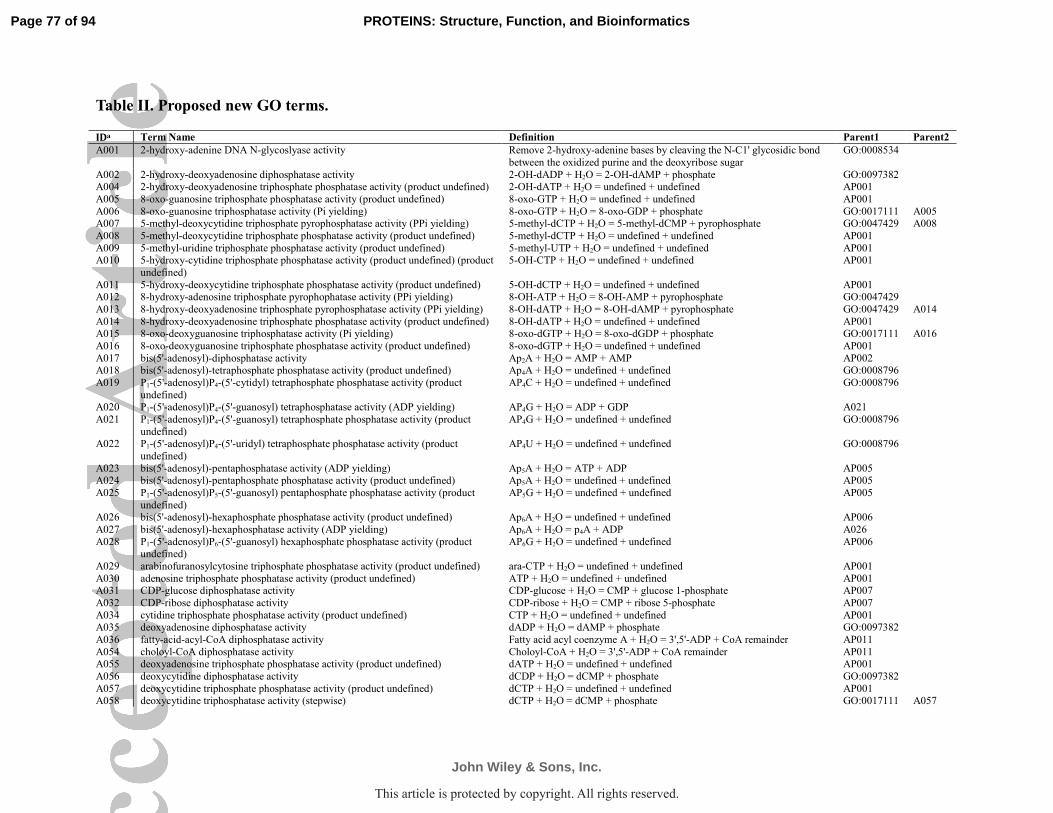
Table II. Proposed new GO terms.
IDa Term Name Definition Parent1 Parent2
A001 2-hydroxy-adenine DNA N-glycoslyase activity Remove 2-hydroxy-adenine bases by cleaving the N-C1' glycosidic bond
between the oxidized purine and the deoxyribose sugar
GO:0008534
A002 2-hydroxy-deoxyadenosine diphosphatase activity 2-OH-dADP + H2O = 2-OH-dAMP + phosphate GO:0097382 A004 2-hydroxy-deoxyadenosine triphosphate phosphatase activity (product undefined) 2-OH-dATP + H2O = undefined + undefined AP001
A005 8-oxo-guanosine triphosphate phosphatase activity (product undefined) 8-oxo-GTP + H2O = undefined + undefined AP001
A006 8-oxo-guanosine triphosphatase activity (Pi yielding) 8-oxo-GTP + H2O = 8-oxo-GDP + phosphate GO:0017111 A005 A007 5-methyl-deoxycytidine triphosphate pyrophosphatase activity (PPi yielding) 5-methyl-dCTP + H2O = 5-methyl-dCMP + pyrophosphate GO:0047429 A008
A008 5-methyl-deoxycytidine triphosphate phosphatase activity (product undefined) 5-methyl-dCTP + H2O = undefined + undefined AP001
A009 5-methyl-uridine triphosphate phosphatase activity (product undefined) 5-methyl-UTP + H2O = undefined + undefined AP001 A010 5-hydroxy-cytidine triphosphate phosphatase activity (product undefined) (product
undefined)
5-OH-CTP + H2O = undefined + undefined AP001
A011 5-hydroxy-deoxycytidine triphosphate phosphatase activity (product undefined) 5-OH-dCTP + H2O = undefined + undefined AP001 A012 8-hydroxy-adenosine triphosphate pyrophophatase activity (PPi yielding) 8-OH-ATP + H2O = 8-OH-AMP + pyrophosphate GO:0047429
A013 8-hydroxy-deoxyadenosine triphosphate pyrophosphatase activity (PPi yielding) 8-OH-dATP + H2O = 8-OH-dAMP + pyrophosphate GO:0047429 A014
A014 8-hydroxy-deoxyadenosine triphosphate phosphatase activity (product undefined) 8-OH-dATP + H2O = undefined + undefined AP001 A015 8-oxo-deoxyguanosine triphosphatase activity (Pi yielding) 8-oxo-dGTP + H2O = 8-oxo-dGDP + phosphate GO:0017111 A016
A016 8-oxo-deoxyguanosine triphosphate phosphatase activity (product undefined) 8-oxo-dGTP + H2O = undefined + undefined AP001
A017 bis(5'-adenosyl)-diphosphatase activity Ap2A + H2O = AMP + AMP AP002 A018 bis(5'-adenosyl)-tetraphosphate phosphatase activity (product undefined) Ap4A + H2O = undefined + undefined GO:0008796
A019 P1-(5'-adenosyl)P4-(5'-cytidyl) tetraphosphate phosphatase activity (product
undefined)
AP4C + H2O = undefined + undefined GO:0008796
A020 P1-(5'-adenosyl)P4-(5'-guanosyl) tetraphosphatase activity (ADP yielding) AP4G + H2O = ADP + GDP A021
A021 P1-(5'-adenosyl)P4-(5'-guanosyl) tetraphosphate phosphatase activity (product
undefined)
AP4G + H2O = undefined + undefined GO:0008796
A022 P1-(5'-adenosyl)P4-(5'-uridyl) tetraphosphate phosphatase activity (product
undefined)
AP4U + H2O = undefined + undefined GO:0008796
A023 bis(5'-adenosyl)-pentaphosphatase activity (ADP yielding) Ap5A + H2O = ATP + ADP AP005 A024 bis(5'-adenosyl)-pentaphosphate phosphatase activity (product undefined) Ap5A + H2O = undefined + undefined AP005
A025 P1-(5'-adenosyl)P5-(5'-guanosyl) pentaphosphate phosphatase activity (product
undefined)
AP5G + H2O = undefined + undefined AP005
A026 bis(5'-adenosyl)-hexaphosphate phosphatase activity (product undefined) Ap6A + H2O = undefined + undefined AP006
A027 bis(5'-adenosyl)-hexaphosphatase activity (ADP yielding) Ap6A + H2O = p4A + ADP A026
A028 P1-(5'-adenosyl)P6-(5'-guanosyl) hexaphosphate phosphatase activity (product undefined)
AP6G + H2O = undefined + undefined AP006
A029 arabinofuranosylcytosine triphosphate phosphatase activity (product undefined) ara-CTP + H2O = undefined + undefined AP001
A030 adenosine triphosphate phosphatase activity (product undefined) ATP + H2O = undefined + undefined AP001 A031 CDP-glucose diphosphatase activity CDP-glucose + H2O = CMP + glucose 1-phosphate AP007
A032 CDP-ribose diphosphatase activity CDP-ribose + H2O = CMP + ribose 5-phosphate AP007
A034 cytidine triphosphate phosphatase activity (product undefined) CTP + H2O = undefined + undefined AP001 A035 deoxyadenosine diphosphatase activity dADP + H2O = dAMP + phosphate GO:0097382
A036 fatty-acid-acyl-CoA diphosphatase activity Fatty acid acyl coenzyme A + H2O = 3',5'-ADP + CoA remainder AP011
A054 choloyl-CoA diphosphatase activity Choloyl-CoA + H2O = 3',5'-ADP + CoA remainder AP011 A055 deoxyadenosine triphosphate phosphatase activity (product undefined) dATP + H2O = undefined + undefined AP001
A056 deoxycytidine diphosphatase activity dCDP + H2O = dCMP + phosphate GO:0097382
A057 deoxycytidine triphosphate phosphatase activity (product undefined) dCTP + H2O = undefined + undefined AP001 A058 deoxycytidine triphosphatase activity (stepwise) dCTP + H2O = dCMP + phosphate GO:0017111 A057
Page 77 of 94
John Wiley & Sons, Inc.
PROTEINS: Structure, Function, and Bioinformatics
5051525354555657585960
This article is protected by copyright. All rights reserved.

A059 deoxyguanosine diphosphatase activity dGDP + H2O = dGMP + phosphate GO:0097382
A060 deoxyguanosine triphosphate phosphatase activity (product undefined) dGTP + H2O = undefined + undefined AP001 A061 CTP pyrophosphatase activity (PPi yielding) CTP + H2O = CMP + pyrophosphate GO:0047429 A034
A062 uridine triphosphate phosphatase activity (product undefined) UTP + H2O = undefined + undefined AP001
A063 deoxyuridine triphosphate phosphatase activity (product undefined) dUTP + H2O = undefined + undefined AP001 A064 deoxyuridine triphosphatase activity (stepwise) dUTP + H2O = dUMP + phosphate GO:0017111 A063
A065 m7G(5')ppp-mRNA diphosphatase activity (m7GDP yielding) m7G-ppp-mRNA + H2O = 7-methyl-GDP + p-mRNA AP013
A066 G(5')ppp-mRNA triphosphatase activity (GMP yielding) G-ppp-mRNA + H2O = GMP + p-mRNA AP013 A067 G(5')ppp-mRNA triphosphatase activity (GDP yielding) G-ppp-mRNA + H2O = GDP + p-mRNA AP013
A068 GDP-beta-fucose diphosphatase activity GDP-beta-fucose + H2O = GMP + fucose 1-phosphate AP008
A069 GDP-fructose diphosphatase activity GDP-fructose + H2O = GMP + fructose 1-phosphate AP008 A070 GDP-glucose diphosphatase activity GDP-glucose + H2O = GMP + glucose 1-phosphate AP008
A071 GDP-ribose diphosphatase activity GDP-ribose + H2O = GMP + ribose 5-phosphate AP008
A072 bis(5'-guanosyl)-diphosphatase activity Gp2G + H2O = GMP + GMP AP002 A073 bis(5'-guanosyl)-triphosphatase activity Gp3G + H2O = GDP + GMP AP003
A074 bis(5'-guanosyl)-tetraphosphate phosphatase activity (product undefined) GP4G + H2O = undefined + undefined GO:0008796
A075 bis(5'-guanosyl)-pentaphosphate phosphatase activity (product undefined) GP5G + H2O = undefined + undefined AP005 A076 guanosine triphosphate phosphatase activity (product undefined) GTP + H2O = undefined + undefined AP001
A077 guanosine triphosphatase activity (stepwise) GTP + H2O = GMP + phosphate GO:0017111 A076
A079 IDP-ribose diphosphatase activity IDP-ribose + H2O = IMP + ribose 5-phosphate AP014 A080 inosine triphosphate phosphatase activity (product undefined) ITP + H2O = undefined + undefined AP001
A081 m7G(5')ppp-snoRNA triphosphatase activity (m7GDP yielding) m7G-ppp-snoRNA + H2O = 7-methyl-GDP + p-snoRNA AP013
A082 N4-methyl-deoxycytidine triphosphate phosphatase activity (product undefined) N4-methyl-dCTP + H2O = undefined + undefined AP001 A084 O-acetyl-ADP-ribose diphosphatase activity O-acetyl-ADP-ribose + H2O = AMP + acetyl-ribose 5-phosphate GO:0019144
A085 oxothiamine-diphosphatase activity oxoThDP + H2O = oxoThMP + phosphate GO:0016462
A086 oxythiamine-diphosphatase activity oxyThDP + H2O = oxyThMP + phosphate GO:0016462 A087 thiamine triphosphate phosphatase activity (product undefined) ThTP + H2O = undefined + undefined GO:0016462
A088 adenosine tetraphosphate phosphatase activity (product undefined) P4A + H2O = undefined + undefined AP001 A089 adenosine tetraphosphatase activity (AMP yielding) P4A + H2O = AMP + triphosphate A088
A090 guanosine tetraphosphate phosphatase activity (product undefined) P4G + H2O = undefined + undefined AP001
A091 adenosine pentaphosphate phosphatase activity (product undefined) P5A + H2O = undefined + undefined AP001 A094 guanosine 3',5'-bis(diphosphate) diphosphate phosphatase activity (product
undefined)
ppGpp + H2O = undefined + undefined GO:0017110
A095 guanosine 3',5'-bis(diphosphate) diphosphatase activity (stepwise) ppGpp + H2O = pGp + phosphate A094 A096 malonyl-CoA diphosphatase activity malonyl-CoA + H2O = 3',5'-ADP + CoA remainder AP011
A098 trihydroxycoprostanoyl-CoA diphosphatase activity THCA-CoA + H2O = 3',5'-ADP + CoA remainder AP011
A099 thymidine triphosphate phosphatase activity (product undefined) TTP + H2O = undefined + undefined AP001 A100 thymidine triphosphatase activity (stepwise) TTP + H2O = TMP + phosphate GO:0017111 A099
A101 UDP-galactosamine diphosphatase activity UDP-galactosamine + H2O = UMP + galactoasmine phosphate GO:0008768
A102 UDP-galactose diphosphatase activity UDP-galactose + H2O = UMP + galactose 1-phosphate GO:0008768 A103 UDP-galacturonic acid diphosphatase activity UDP-galacturonic acid + H2O = UMP + phosphogalacturonic acid GO:0008768
A104 UDP-glucosamine diphosphatase activity UDP-glucosamine + H2O = UMP + glucoasmine phosphate GO:0008768
A105 UDP-glucose diphosphatase activity UDP-glucose + H2O = UMP + glucose 1-phosphate GO:0008768 A106 UDP-glucuronic acid diphosphatase activity UDP-glucuronic acid + H2O = UMP + phosphoglucuronic acid GO:0008768
A107 UDP-hexanolamine diphosphatase activity UDP-hexanolamine + H2O = UMP + hexanolamine phosphate GO:0016462
A108 UDP-mannose diphosphatase activity UDP-mannose + H2O = UMP + mannose 1-phosphate GO:0008768 A109 UDP-N-acetyl-galactosamine diphosphatase activity UDP-N-acetyl-galactosamine + H2O = UMP + N-acetyl-galactosamine
phosphate
GO:0008768
A110 UDP-N-acetyl-glucosamine diphosphatase activity UDP-N-acetyl-glucosamine + H2O = UMP + N-acetyl-glucosamine phosphate
GO:0008768
Page 78 of 94
John Wiley & Sons, Inc.
PROTEINS: Structure, Function, and Bioinformatics
5051525354555657585960
This article is protected by copyright. All rights reserved.

A111 UDP-N-acetyl-muramic acid diphosphatase activity UDP-N-acetyl-muramic acid + H2O = UMP + phospho-N-acetyl-muramic
acid
GO:0008768
A112 UDP-N-acetyl-muramoyl-L-alanine diphosphatase activity UDP-N-acetyl-muramoyl-L-Ala + H2O = UMP + N-acetyl-muramoyl-L-
Ala phosphate
GO:0016462
A113 uridine triphosphatase activity (stepwise) UTP + H2O = UMP + phosphate GO:0017111 A062 A114 3-methyl-3-hydroxyglutaryl-CoA diphosphatase activity 3-hydroxymethylglutaryl-CoA + H2O = 3',5'-ADP + 3-
hydroxymethylglutarylpantetheine 4'-phosphate
AP011
A115 deamino-NAD+ diphosphatase activity deamino-NAD+ + H2O = AMP + deamino-NMN+ AP012 A116 TDP-glucose diphosphatase activity TDP-glucose + H2O = TMP + glucose 1-phosphate AP009
A117 CDP-ethanolamine diphosphatase activity CDP-ethanolamine + H2O = CMP + phosphoethanolamine GO:0016462
A118 CoA-disulfide diphosphatase activity CoASSCoA + H2O = 3',5'-ADP + 4'-phosphopantetheine CoA disulfide AP011 A119 2'-phospho-ADP-ribose diphosphatase activity 2'-phospho-ADP-ribose + H2O = 2',5'-ADP + ribose 5-phosphate GO:0019144
A120 3'-dephospho-CoA diphosphatase activity 3'-dephospho-CoA + H2O = AMP + 4'-phosphopantetheine AP011
A121 ADP-mannose diphosphatase activity ADP-mannose + H2O = AMP + mannose 1-phosphate GO:0019144 A122 CoA-glutathione diphosphatase activity CoA-glutathione + H2O = 3',5'-ADP + glutathionylpantetheine 4'-
phosphate
AP011
A125 acetyl-CoA diphosphatase activity Acetyl-CoA + H2O = 3',5'-ADP + acetylpantetheine 4'-phosphate AP011 A126 NAADP+ diphosphatase activity NAADP+ + H2O = 2',5'-ADP + deamino-NMN+ AP012
A127 CDP-choline diphosphatase activity CDP-choline + H2O = CMP + phosphocoline GO:0016462
A128 succinyl-CoA diphosphatase activity Succinyl-CoA + H2O = 3',5'-ADP + succinylpantetheine 4'-phosphate AP011 A129 deamino-NADH diphosphatase activity deamino-NADH + H2O = AMP + deamino-NMNH AP012
A130 P1-(5'-adenosyl)P3-(5'-guanosyl) triphosphate phosphatase activity (product
undefined)
Ap3G + H2O = undefined + undefined AP003
A131 3'-amino-3'-dATP pyrophosphatase activity (PPi yielding) 3'-amino-3'-dATP + H2O = 3'-amino-3'-dAMP + pyrophosphate GO:0047429
A132 3'-amino-3'-TTP pyrophosphatase activity (PPi yielding) 3'-amino-3'-dTTP + H2O = 3'-amino-3'-dTMP + pyrophosphate GO:0047429
A133 thymidine-diphosphatase activity TDP + H2O = TMP + phosphate GO:0017110 A134 NADP+ diphosphatase activity NADP+ + H2O = 2',5'-ADP + NMN+ AP012
A135 bis(5'-adenosyl)-tetraphosphate phosphatase activity (AMP yielding) Ap4A + H2O = AMP + ATP A018 A136 adenosine pentaphosphate phosphatase activity (ATP yielding) P5A + H2O = ATP + PPi A091
A137 bis(5'-adenosyl)-hexaphosphatase activity (ATP yielding) Ap6A + H2O = ATP + ATP A026
AP001 nucleoside-polyphosphate phosphatase activity Hydrolysis of nucleoside polyphosphate at one pyrophosphate bound GO:0016462 AP002 dinucleoside-diphosphate phosphatase activity Hydrolysis of dinucleoside diphosphate into two nucleotide GO:0004551
AP003 dinucleoside-triphosphate phosphatase activity Hydrolysis of dinucleoside triphosphate into two nucleotide GO:0004551
AP005 dinucleoside-pentaphosphate phosphatase activity Hydrolysis of dinucleoside pentaphosphate into two nucleotide GO:0004551 AP006 dinucleoside-hexaphosphate phosphatase activity Hydrolysis of dinucleoside hexaphosphate into two nucleotide GO:0004551
AP007 CDP-sugar diphosphatase activity CDP-sugar + H2O = CMP + sugar 1-phosphate GO:0016462
AP008 GDP-sugar diphosphatase activity GDP-sugar + H2O = GMP + sugar 1-phosphate GO:0016462 AP009 TDP-sugar diphosphatase activity TDP-sugar + H2O = TMP + sugar 1-phosphate GO:0016462
AP011 general coenzyme A diphosphatase activity Hydrolysis of coenzyme A or its derivatives GO:0016462
AP012 general NAD diphosphatase activity Hydrolysis of NAD+ or its derivatives AP002 AP013 RNA decapping activity Hydrolysis of capped RNA into an uncapped RNA and a small molecule GO:0016462
AP014 IDP-sugar diphosphatase activity Hydrolysis of IDP-sugar derivatives GO:0016462 a Newly proposed children terms or parent terms associated with experimental data begin with A (e.g., A001), while parent terms
without direct experimental data begin with AP (e.g., AP001).
Page 79 of 94
John Wiley & Sons, Inc.
PROTEINS: Structure, Function, and Bioinformatics
5051525354555657585960
This article is protected by copyright. All rights reserved.

Table III. Proposed modification of current Gene Ontology.
Reason for
proposal
GO ID Current Namea Definitiona Proposed Name /
Definition
Current Parentb Proposed Parent Additional
Parent
misleading or imprecise name
GO:0004551 nucleotide diphosphatase activity
dinucleotide + H2O = 2 mononucleotides
dinucleotide-polyphosphate
phosphatase activity
misleading or imprecise
name, change
parent
GO:0050072 m7G(5')pppN diphosphatase activity
m7-G-ppp-polynucleotide + H2O = m7GMP +
polynucleotide
m7G(5')ppp-mRNA diphosphatase activity
(m7GMP yielding)
pyrophosphatase activity (GO:0016462)
RNA decapping activity (AP013)
GO:0052751 GDP-mannose hydrolase activity
GDP-mannose + H2O = GMP + mannose-1-P
GDP-mannose diphosphatase activity
pyrophosphatase activity (GO:0016462)
GDP-sugar diphosphatase activity
(AP008)
more specific
name (add product
description)
GO:0004170 dUTP diphosphatase
activity
dUTP + H2O = dUMP +
PPi
dUTP diphosphatase
activity (PPi yielding)
GO:0019177 dihydroneopterin
triphosphate
pyrophosphohydrolase activity
dihydroneopterin
triphosphate + H2O =
dihydroneopterin phosphate + PPi
dihydroneopterin
triphosphate
pyrophosphohydrolase activity (PPi yielding)
GO:0035870 dITP diphosphatase
activity
dITP + H2O = dIMP + PPi dITP diphosphatase
activity (PPi yielding)
GO:0036222 XTP diphosphatase
activity
XTP + H2O = XMP + PPi XTP diphosphatase
activity (PPi yielding)
GO:0044713 2-OH-ATP pyrophosphatase
activity
2-OH-ATP + H2O = 2-OH-AMP + PPi
2-OH-ATP pyrophosphatase activity
(PPi yielding)
more specific name and
definition
(different from GO:0044713)
GO:0044714 2-OH-(d)ATP pyroposphatase
activity
2-OH-(d)ATP + H2O = 2-OH-(d)AMP + PPi
2-OH-dATP pyrophosphatase activity
(PPi yielding);
2-OH-dATP + H2O = 2-OH-dAMP + PPi *
(proposed definition)
more specific name (add
product
description), change parent
GO:0004636 phosphoribosyl-ATP diphosphatase activity
1-(5-phospho-D-ribosyl)-ATP + H2O = 1-(5-
phosphonatoribosyl)-5'-
AMP + PPi
phosphoribosyl-ATP diphosphatase activity
(PPi yielding)
pyrophosphatase activity (GO:0016462)
nucleoside-triphosphate
diphosphatase activity
(PPi yielding) (GO:0047429)
GO:0008894 guanosine-5'-
triphosphate,3'-diphosphate
diphosphatase activity
guanosine 5'-
triphosphate,3'-diphosphate + H2O =
guanosine 5'-
diphosphate,3'-diphosphate + Pi
guanosine-5'-
triphosphate,3'-diphosphate
diphosphatase activity (Pi
yielding)
pyrophosphatase activity
(GO:0016462)
nucleoside-
triphosphatase activity (Pi yielding)
(GO:0017111)
GO:0017111 nucleoside-
triphosphatase activity
A nucleoside triphosphate
+ H2O = nucleoside diphosphate + Pi
nucleoside-triphosphatase
activity (Pi yielding)
pyrophosphatase activity
(GO:0016462)
nucleoside-
polyphosphate hydrolase activity
(AP001)
Page 80 of 94
John Wiley & Sons, Inc.
PROTEINS: Structure, Function, and Bioinformatics
5051525354555657585960
This article is protected by copyright. All rights reserved.

GO:0034431 bis(5'-adenosyl)-
hexaphosphatase activity
P1-P6-bis(5'-adenosyl)
hexaphosphate + H2O = AMP + p4A
bis(5'-adenosyl)-
hexaphosphatase activity (AMP yielding)
dinucleotide diphosphatase
activity (GO:0004551)
bis(5'-adenosyl)-
hexaphosphatase activity(A026)
GO:0034432 bis(5'-adenosyl)-
pentaphosphatase activity
P1-P6-bis(5'-adenosyl)
pentaphosphate + H2O = AMP + p4A
bis(5'-adenosyl)-
pentaphosphatase activity (AMP yielding)
dinucleotide diphosphatase
activity (GO:0004551)
bis(5'-adenosyl)-
pentaphosphatase activity(A024)
GO:0047429 nucleoside-
triphosphate diphosphatase activity
A nucleoside triphosphate
+ H2O = a nucleotide + PPi
nucleoside-triphosphate
diphosphatase activity (PPi yielding)
pyrophosphatase activity
(GO:0016462)
nucleoside-
polyphosphate hydrolase activity
(AP001)
GO:0047624 adenosine-tetraphosphatase
activity
p4A + H2O = ATP + Pi adenosine tetraphosphatase activity
(ATP yielding)
pyrophosphatase activity (GO:0016462)
adenosine 5'-tetraphosphatase
activity (A088)
GO:0050333 thiamine-triphosphatase activity
thiamine triphosphate + H2O = thiamine
diphosphate + Pi
thiamine-triphosphatase activity (Pi yielding)
nucleoside-triphosphatase activity (Pi yielding)
(GO:0017111)
thiamine triphosphate hydrolase activity
(A087)
more specific name (add
product
description), add parent
GO:0003924 GTPase activity GTP + H2O = GDP + Pi GTPase activity (Pi yielding)
nucleoside-triphosphatase activity (Pi yielding)
(GO:0017111)
GTP phosphatase activity (product
undefined) (A076)
GO:0008413 8-oxo-GTP pyrophosphatase
activity
8-oxo-GTP + H2O = 8-oxo-GMP + PPi
8-oxo-GTP pyrophosphatase activity
(PPi yielding)
nucleoside-triphosphate diphosphatase activity (PPi
yielding) (GO:0047429)
8-oxo-GTP phosphatase
activity (product
undefined) (A005) GO:0008828 dATP
pyrophosphohydrolase
activity
dATP + H2O = dAMP +
PPi
dATP
pyrophosphohydrolase
activity (PPi yielding)
nucleoside-triphosphate
diphosphatase activity (PPi
yielding) (GO:0047429)
dATP phosphatase
activity (product
undefined) (A055) GO:0016887 ATPase activity ATP + H2O = ADP + Pi ATPase activity (Pi
yielding)
nucleoside-triphosphatase
activity (Pi yielding)
(GO:0017111)
ATP phosphatase
activity (product
undefined) (A030) GO:0035539 8-oxo-dGTP
pyrophosphatase
activity
8-oxo-dGTP + H2O = 8-
oxo-dGMP + PPi
8-oxo-dGTP
pyrophosphatase activity
(PPi yielding)
nucleoside-triphosphate
diphosphatase activity (PPi
yielding) (GO:0047429)
8-oxo-dGTP
phosphatase
activity (product undefined) (A016)
GO:0036217 dGTP diphosphatase
activity
dGTP + H2O = dGMP +
PPi
dGTP diphosphatase
activity (PPi yielding)
nucleoside-triphosphate
diphosphatase activity (PPi yielding) (GO:0047429)
dGTP phosphatase
activity (product undefined) (A060)
GO:0036218 dTTP diphosphatase
activity
dTTP + H2O = dTMP +
PPi
TTP diphosphatase
activity (PPi yielding)
nucleoside-triphosphate
diphosphatase activity (PPi yielding) (GO:0047429)
TTP phosphatase
activity (product undefined) (A099)
GO:0036219 GTP diphosphatase
activity
GTP + H2O = GMP + PPi GTP diphosphatase
activity (PPi yielding)
nucleoside-triphosphate
diphosphatase activity (PPi yielding) (GO:0047429)
GTP phosphatase
activity (product undefined) (A076)
GO:0036220 ITP diphosphatase
activity
ITP + H2O = IMP + PPi ITP diphosphatase
activity (PPi yielding)
nucleoside-triphosphate
diphosphatase activity (PPi yielding) (GO:0047429)
ITP phosphatase
activity (product undefined) (A080)
GO:0036221 UTP diphosphatase
activity
UTP + H2O = UMP + PPi UTP diphosphatase
activity (PPi yielding)
nucleoside-triphosphate
diphosphatase activity (PPi yielding) (GO:0047429)
UTP phosphatase
activity (product undefined) (A062)
Page 81 of 94
John Wiley & Sons, Inc.
PROTEINS: Structure, Function, and Bioinformatics
5051525354555657585960
This article is protected by copyright. All rights reserved.

GO:0043273 CTPase activity CTP + H2O = CDP + Pi CTPase activity (Pi
yielding)
nucleoside-triphosphatase
activity (Pi yielding) (GO:0017111)
CTP phosphatase
activity (product undefined) (A034)
GO:0047693 ATP diphosphatase
activity
ATP + H2O = AMP + PPi ATP diphosphatase
activity (PPi yielding)
nucleoside-triphosphate
diphosphatase activity (PPi yielding) (GO:0047429)
ATP phosphatase
activity (product undefined) (A030)
GO:0047840 dCTP diphosphatase
activity
dCTP + H2O = dCMP +
PPi
dCTP diphosphatase
activity (PPi yielding)
nucleoside-triphosphate
diphosphatase activity (PPi yielding) (GO:0047429)
dCTP phosphatase
activity (product undefined) (A057)
GO:0050339 thymidine-
triphosphatase activity
TTP + H2O = TDP + Pi TTPase activity (Pi
yielding)
nucleoside-triphosphatase
activity (Pi yielding) (GO:0017111)
TTP phosphatase
activity (product undefined) (A099)
change
definition, change parent
GO:0004787 thiamine-
pyrophosphatase activity
TDP + H2O = TMP + Pi thiamine-diphosphate +
H2O = thiamine-phosphate + Pi *
(proposed definition)
nucleoside-diphosphatase
activity (GO:0017110)
pyrophosphatase
activity (GO:0016462)
change parent GO:0000210 NAD+ diphosphatase
activity
NAD+ + H2O = AMP +
NMN+
dinucleotide diphosphatase
activity (GO:0004551)
general NAD
diphosphatase activity (AP012)
GO:0008758 UDP-2,3-
diacylglucosamine hydrolase activity
H2O + UDP-2,3-bis(3-
hydroxymyristoyl)glucosamine = 2,3-bis(3-
hydroxymyristoyl)-beta-D-
glucosaminyl 1-phosphate + UMP
pyrophosphatase activity
(GO:0016462)
UDP-sugar
diphosphatase activity (GO:0008768)
GO:0010943 NADPH pyrophosphatase
activity
NADPH + H2O = NMNH + ADP
dinucleotide diphosphatase activity (GO:0004551)
general NAD diphosphatase activity
(AP012)
GO:0010945 CoA pyrophosphatase activity
coenzyme A + H2O = 3',5'-ADP + 4'-
phosphopantetheine
pyrophosphatase activity (GO:0016462)
general coenzyme A diphosphatase activity
(AP011)
GO:0017110 nucleoside-diphosphatase activity
a nucleoside diphosphate + H2O = a nucleotide + Pi
pyrophosphatase activity (GO:0016462)
nucleoside-polyphosphate
hydrolase activity
(AP001)
GO:0034353 RNA
pyrophosphohydrolase
activity
Catalysis of the removal of
a 5' terminal
pyrophosphate from the 5'-triphosphate end of an
RNA, leaving a 5'-
monophosphate end.
pyrophosphatase activity
(GO:0016462)
RNA decapping
activity (AP013)
GO:0035529 NADH
pyrophosphatase
activity
NADH + H2O = AMP +
NMNH + 2 H+
dinucleotide diphosphatase
activity (GO:0004551)
general NAD
diphosphatase activity
(AP012)
GO:0044715 8-oxo-dGDP
phosphatase activity
8-oxo-dGDP + H2O = 8-
oxo-dGMP + Pi
nucleoside-diphosphatase
activity (GO:0017110)
deoxynucleoside-
diphosphatase activity
(GO:0097382)
GO:0044717 8-hydroxy-dADP
phosphatase activity
8-OH-dADP + H2O = 8-
OH-dAMP + Pi
nucleoside-diphosphatase
activity (GO:0017110)
deoxynucleoside-
diphosphatase activity
(GO:0097382)
Page 82 of 94
John Wiley & Sons, Inc.
PROTEINS: Structure, Function, and Bioinformatics
5051525354555657585960
This article is protected by copyright. All rights reserved.

GO:0047884 FAD diphosphatase
activity
FAD + H2O = AMP +
FMN
dinucleotide diphosphatase
activity (GO:0004551)
dinucleoside
diphosphatase activity (AP002)
GO:0052840 inositol diphosphate
tetrakisphosphate diphosphatase activity
inositol diphosphate
tetrakisphosphate + H2O = inositol 1,3,4,5,6-
pentakisphosphate + Pi
pyrophosphatase activity
(GO:0016462)
diphosphoinositol-
polyphosphate diphosphatase activity
(GO:0008486)
GO:0052841 inositol bisdiphosphate tetrakisphosphate
diphosphatase activity
inositol bisdiphosphate tetrakisphosphate + H2O =
inositol diphosphate
pentakisphosphate + Pi
pyrophosphatase activity (GO:0016462)
diphosphoinositol-polyphosphate
diphosphatase activity
(GO:0008486)
GO:0052842 inositol diphosphate
pentakisphosphate
diphosphatase activity
inositol diphosphate
pentakisphosphate + H2O
= inositol hexakisphosphate + Pi
pyrophosphatase activity
(GO:0016462)
diphosphoinositol-
polyphosphate
diphosphatase activity (GO:0008486)
GO:0097382 deoxynucleoside-
diphosphatase activity
a deoxynucleoside
diphosphate + H2O = a deoxynucleotide + Pi
pyrophosphatase activity
(GO:0016462)
nucleoside-
polyphosphate hydrolase activity
(AP001)
remove (merge with
GO:0080041)
GO:0047631 ADP-ribose diphosphatase activity
ADP-ribose + H2O = AMP + D-ribose 5-phosphate.
a Current names and definitions defined by Gene Ontology version 2014.01.01 are shown for all GO terms in the table.
b Current parent of GO terms are omitted when the hierarchy of the term is not changed.
Page 83 of 94
John Wiley & Sons, Inc.
PROTEINS: Structure, Function, and Bioinformatics
5051525354555657585960
This article is protected by copyright. All rights reserved.

Page 84 of 94
John Wiley & Sons, Inc.
PROTEINS: Structure, Function, and Bioinformatics
5051525354555657585960
This article is protected by copyright. All rights reserved.

Table IV. Confidence score assignment for different types of experimental evidences.
Category Evidence Confidence Score
Genetic Phenotype shows the predicted physiology; in same species 0.99
Phenotype relates to the predicted physiology; in same species 0.7
Phenotype is inexplicable; in same species 0.1
Complementation in different species 0.5
Biochemical
kcat/Km 107 M
-1 s-1 0.99
106 M
-1 s-1 0.85
105 M
-1 s-1 0.5
104 M
-1 s-1 0.2
103 M
-1 s-1 0.1
0 0.01
Pseudo
kcat/Km
107 M
-1 s-1 0.7
106 M
-1 s-1 0.5
105 M
-1 s-1 0.25
104 M
-1 s-1 0.1
103 M
-1 s-1 0.05
0 0.01
Other
Biochemical
Data
100% Relative activity 0.1
Gel electrophoresis for rare substrates 0.5
Gel electrophoresis, HPLC for common substrates 0.05
X-ray structure with substrate for binding reaction 0.5
X-ray crystal structure with substrate 0.01
Positive activity (kcat, Km, and first order rate constants only) 0.01
Page 85 of 94
John Wiley & Sons, Inc.
PROTEINS: Structure, Function, and Bioinformatics
5051525354555657585960
This article is protected by copyright. All rights reserved.

Table V. Nudix homology domain structures in the 78-PDB structural and sequence alignments.
Molecular Function UniProt AC PDB Chain Species Reference
2-OH-dATP pyrophosphatase P36639 3ZR0 A Homo sapiens 115
3'-5' exonuclease Q81EE8 3Q4I A Bacillus cereus 116 5-methyl-dCTP pyrophosphatase P77788 2RRK A Escherichia coli (Kawasaki PDB ID: 2RRK)
8-oxo-GDP pyrophosphatase Q6ZVK8 3GG6 A Homo sapiens (Tresaugues PDB ID: 3GG6)
8-oxo-GTP pyrophosphatase P08337 3A6T A Escherichia coli 117 A:oxoG glycosylase P83847 1RRS A Geobacillus stearothermophilus 93
A:oxoG glycosylase Q9UIF7 1X51 A Homo sapiens (Tomizawa PDB ID: 1X51)
ADP-ribose pyrophosphatase O33199 1MQW A Mycobacterium tuberculosis 99 ADP-ribose pyrophosphatase O95848 3Q91 A Homo sapiens (Tresaugues PDB ID: 3Q91)
ADP-ribose pyrophosphatase Q55928 2QJO A Synechocystis sp. 107
ADP-ribose pyrophosphatase Q5NHR1 2QJT B Francisella tularensis 107 ADP-ribose pyrophosphatase Q5SJY9 2YVP A Thermus thermophilus 118
ADP-ribose pyrophosphatase Q84CU3 1V8M A Thermus thermophilus 119
ADP-ribose pyrophosphatase Q93K97 1KHZ A Escherichia coli 105 ADP-ribose pyrophosphatase Q9BW91 1QVJ A Homo sapiens 120
ADP-ribose pyrophosphatase Q9UKK9 2DSC A Homo sapiens 121
Ap3A pyrophosphatase P45799 1VHZ A Escherichia coli 98 Ap4A pyrophosphatase O04841 1JKN A Lupinus angustifolius 122
Ap4A pyrophosphatase P50583 3U53 A Homo sapiens 123
Ap4A pyrophosphatase Q9U2M7 1KTG A Caenorhabditis elegans 124 Ap6A pyrophosphatase Q75UV1 1VC8 A Thermus thermophilus (Iwai PDB ID: 1VC8)
CDP pyrophosphatase Q9RY71 2O5F A Deinococcus radiodurans 49
CoA pyrophosphatase Q9RV46 1NQZ A Deinococcus radiodurans 99 dGTP pyrophosphatase Q9RVK2 1SU2 A Deinococcus radiodurans 125
DHNTP pyrophosphatase P0AFC0 2O1C A Escherichia coli 63 FAD pyrophosphatase Q9RSC1 2W4E A Deinococcus radiodurans 126
GDP-mannose mannosyl hydrolase P32056 1RYA A Escherichia coli 38
GDP-mannose pyrophosphatase P37128 1VIU B Escherichia coli 98 GDP-mannose pyrophosphatase Q6XQ58 2I8T A Escherichia coli 100
HMP-pp pyrophosphatase P0AEI6 3SHD A Escherichia coli (Hong PDB ID: 3SHD)
IPP isomerase Q13907 2ICK A Homo sapiens 95 IPP isomerase Q46822 1PPV A Escherichia coli 97
IPP isomerase Q9BXS1 2PNY A Homo sapiens (Walker PDB ID: 2PNY)
mRNA binding O43809 2J8Q A Homo sapiens 103 mRNA decapping enzyme O13828 2QKM B Schizosaccharomyces pombe 127
mRNA decapping enzyme P53550 2JVB A Saccharomyces cerevisiae 128
mRNA decapping enzyme Q96DE0 2XSQ A Homo sapiens (Tresaugues PDB ID: 2XSQ) NADH pyrophosphatase P32664 1VK6 A Escherichia coli (JCSG PDB ID: 1VK6)
PP-InsP5 pyrophosphatase O95989 2FVV A Homo sapiens 129
PP-InsP5 pyrophosphatase Q8NFP7 3MCF A Homo sapiens (Tresaugues PDB ID: 3MCF) RNA pyrophosphohydrolase P0A776 2KDV A Escherichia coli (Bi PDB ID: 2KDV)
RNA pyrophosphohydrolase Q6MPX4 3FFU A Bdellovibrio bacteriovorus 130
snoRNA decapping enzyme Q6TEC1 2A8T A Xenopus laevis 101 transcription factor Q8EFJ3 3GZ8 A Shewanella oneidensis 46
(undetermined) A0REX4 3SMD A Bacillus thuringiensis (Palani PDB ID: 3SMD)
(undetermined) A0ZZM4 3FJY A Bifidobacterium adolescentis (Palani PDB ID: 3FJY)
Page 86 of 94
John Wiley & Sons, Inc.
PROTEINS: Structure, Function, and Bioinformatics
5051525354555657585960
This article is protected by copyright. All rights reserved.

(undetermined) B9WTJ0 3O8S A Streptococcus suis (JCSG PDB ID: 3O8S)
(undetermined) C3H476 3ID9 A Bacillus thuringiensis (Palani PDB ID: 3ID9) (undetermined) C8WVE1 3QSJ A Alicyclobacillus acidocaldarius (Michalska PDB ID: 3QSJ)
(undetermined) D4Q002 3SON A Listeria monocytogenes (JCSG PDB ID: 3SON)
(undetermined) O66548 3I7V A Aquifex aeolicus 119 (undetermined) O67435 2YYH A Aquifex aeolicus (Nakakaga PDB ID: 2YYH)
(undetermined) P53370 3H95 A Homo sapiens (Tresaugues PDB ID: 3H95)
(undetermined) P65556 2FKB A Escherichia coli (Nocek PDB ID: 2FKB) (undetermined) Q03S37 3EXQ A Lactobacillus brevis (Palani PDB ID: 3EXQ)
(undetermined) Q0TS82 3F6A A Clostridium perfringens (Palani PDB ID: 3F6A)
(undetermined) Q0TTC5 3FCM A Clostridium perfringens (Palani PDB ID: 3FCM) (undetermined) Q2RXE7 3R03 A Rhodospirillum rubrum (Zhang PDB ID: 3R03)
(undetermined) Q2RXX6 3DUP A Rhodospirillum rubrum (Patskovsky PDB ID: 3DUP)
(undetermined) Q3JWU2 4DYW A Burkholderia pseudomallei (Edwards PDB ID: 4DYW) (undetermined) Q5LBB1 3GWY A Bacteroides fragili (Patskovsky PDB ID: 3GWY)
(undetermined) Q6G5F4 3HHJ A Bartonella henselae 131
(undetermined) Q7NWQ3 3F13 A Chromobacterium violaceum (Bonanno PDB ID: 3F13) (undetermined) Q82VD6 3CNG A Nitrosomonas europaea (Osipiuk PDB ID: 3CNG)
(undetermined) Q82XR9 2B0V A Nitrosomonas europaea (Osipiuk PDB ID: 2B0V)
(undetermined) Q830S2 2FML A Enterococcus faecalis (Chang PDB ID: 2FML) (undetermined) Q836H1 2AZW A Enterococcus faecalis (Zhang PDB ID: 2AZW)
(undetermined) Q8AAV8 2FB1 A Bacteroides thetaiotaomicron (Chang PDB ID: 2FB1)
(undetermined) Q8PYE2 3GRN A Methanosarcina mazei (Patskovsky PDB ID: 3GRN) (undetermined) Q8R2U6 2DUK A Mus musculus (Hosaka PDB ID: 2DUK)
(undetermined) Q8ZM82 3HYQ A Salmonella typhimurium (Kim PDB ID: 3HYQ)
(undetermined) Q8ZNF5 3N77 A Salmonella typhimurium (Frydrysiak PDB ID: 3N77) (undetermined) Q8ZTD8 1K2E A Pyrobaculum aerophilum 132
(undetermined) Q92EH0 3I9X A Listeria innocua (Bonanno PDB ID: 3I9X) (undetermined) Q97QH6 2B06 A Streptococcus pneumoniae (Zhang PDB ID: 2B06)
(undetermined) Q97T37 2PQV A Streptococcus pneumoniae (Chang PDB ID: 2PQV)
(undetermined) Q9K704 3FK9 A Bacillus halodurans (Bonanno PDB ID: 3FK9) (undetermined) Q9X1A2 3E57 A Thermotoga maritima (Choi PDB ID: 3E57)
Page 87 of 94
John Wiley & Sons, Inc.
PROTEINS: Structure, Function, and Bioinformatics
5051525354555657585960
This article is protected by copyright. All rights reserved.

Table VI. Comparison between Gene Ontology Annotation (GOA) and our manual annotation of Nudix homology proteins.
Reason UniProt Name GOA Database Annotation with Evidence Codea Manual Annotation with Confidence Scoreb Reference
Activity not
stated in reference
NUD12_MOUSE GO:0000210 NAD+ diphosphatase activity IDA (NA) 54
NUD12_MOUSE GO:0035529 NADH pyrophosphatase activity IDA (NA) 54
Activity with
wrong substrate
assigned
NUD15_MOUSE GO:0008413 8-oxo-7,8-dihydroguanosine
triphosphate pyrophosphatase activity
IDA GO:0035539 8-oxo-dGTP pyrophosphatase activity (PPi
yielding)
0.86 133
PCD1_YEAST GO:0008413 8-oxo-7,8-dihydroguanosine
triphosphate pyrophosphatase activity
IDA GO:0035539 8-oxo-dGTP pyrophosphatase activity (PPi
yielding)
0.16 134
TNR3_SCHPO GO:0044715 8-oxo-dGDP phosphatase activity IDA GO:0035539 8-oxo-dGTP pyrophosphatase activity (PPi yielding)
0.19 135
YJ9J_YEAST GO:0044715 8-oxo-dGDP phosphatase activity IDA GO:0035539 8-oxo-dGTP pyrophosphatase activity (PPi
yielding)
0.07 135
NUD11_ARATH GO:0000210 NAD+ diphosphatase activity IDA GO:0035529 NADH pyrophosphatase activity 0.00 136
NUDT5_MOUSE GO:0017110 nucleoside-diphosphatase activity IDA GO:0080041 ADP-ribose pyrophosphohydrolase activity 0.85 137
Activity with
wrong products
assigned
NDX8_CAEEL GO:0003986 acetyl-CoA hydrolase activity IDA A125 acetyl-CoA diphosphatase activity 0.56 138
NUDT7_MOUSE GO:0003986 acetyl-CoA hydrolase activity IDA A125 acetyl-CoA diphosphatase activity 0.53 55 NUD26_ARATH GO:0034432 bis(5'-adenosyl)-pentaphosphatase
activity
IDA A023 bis(5'-adenosyl)-pentaphosphatase activity
(ADP yielding)
0.95 88
NUD27_ARATH GO:0034432 bis(5'-adenosyl)-pentaphosphatase activity
IDA A023 bis(5'-adenosyl)-pentaphosphatase activity (ADP yielding)
0.10 88
AP4A_HUMAN GO:0008803 bis(5'-nucleosyl)-tetraphosphatase
(symmetrical) activity
TAS A135 bis(5'-adenosyl)-tetraphosphate phosphatase
activity (AMP yielding)
1.00 51, 139
NUDI_ECOLI GO:0047840 dCTP diphosphatase activity IDA A058 deoxycytidine triphosphatase activity
(stepwise)
0.40 140
NUDI_ECOLI GO:0004170 dUTP diphosphatase activity IDA A064 deoxyuridine triphosphatase activity (stepwise)
0.76 140
8ODP_HUMAN GO:0003924 GTPase activity TAS GO:0036219 GTP diphosphatase activity (PPi yielding) 0.09 141 NUD11_ARATH GO:0008893 guanosine-3',5'-bis(diphosphate) 3'-
diphosphatase activity
IDA A095 guanosine 3',5'-bis(diphosphate)
diphosphatase activity (stepwise)
0.08 142
NUD15_ARATH GO:0008893 guanosine-3',5'-bis(diphosphate) 3'-diphosphatase activity
IDA A095 guanosine 3',5'-bis(diphosphate) diphosphatase activity (stepwise)
0.03 142
NUD25_ARATH GO:0008893 guanosine-3',5'-bis(diphosphate) 3'-
diphosphatase activity
IDA A095 guanosine 3',5'-bis(diphosphate)
diphosphatase activity (stepwise)
0.07 142
NUD26_ARATH GO:0008893 guanosine-3',5'-bis(diphosphate) 3'-
diphosphatase activity
IDA A095 guanosine 3',5'-bis(diphosphate)
diphosphatase activity (stepwise)
0.14 142
NDX8_CAEEL GO:0047994 hydroxymethylglutaryl-CoA hydrolase activity
IDA A114 3-methyl-3-hydroxyglutaryl-CoA diphosphatase activity
0.59 138
DCP2_HUMAN GO:0050072 m7G(5')pppN diphosphatase activity IDA A065 m7G(5')ppp-mRNA diphosphatase activity
(m7GDP yielding)
0.50 143
DCP2_SCHPO GO:0050072 m7G(5')pppN diphosphatase activity IDA A065 m7G(5')ppp-mRNA diphosphatase activity
(m7GDP yielding)
1.00 144
DCP2_YEAST GO:0050072 m7G(5')pppN diphosphatase activity IDA A065 m7G(5')ppp-mRNA diphosphatase activity (m7GDP yielding)
1.00 23
NUD16_HUMAN GO:0050072 m7G(5')pppN diphosphatase activity IDA A065 m7G(5')ppp-mRNA diphosphatase activity
(m7GDP yielding)
0.50 48
NUD16_XENLA GO:0050072 m7G(5')pppN diphosphatase activity IDA A081 m7G(5')ppp-snoRNA triphosphatase activity
(m7GDP yielding)
1.00 48
Page 88 of 94
John Wiley & Sons, Inc.
PROTEINS: Structure, Function, and Bioinformatics
5051525354555657585960
This article is protected by copyright. All rights reserved.

NDX8_CAEEL GO:0004778 succinyl-CoA hydrolase activity IDA A128 succinyl-CoA diphosphatase activity 0.58 138
Activity not specific
enough to
reflect the data in
reference
NUDE_ECOLI GO:0019144 ADP-sugar diphosphatase activity IDA GO:0080042 ADP-glucose pyrophosphohydrolase activity 0.03 91 NUDT5_HUMAN GO:0019144 ADP-sugar diphosphatase activity IDA GO:0080042 ADP-glucose pyrophosphohydrolase activity 0.72 145
AP4A_CAEEL GO:0004081 bis(5'-nucleosyl)-tetraphosphatase
(asymmetrical) activity
IDA A135 bis(5'-adenosyl)-tetraphosphate phosphatase
activity (AMP yielding)
0.98 59
NUD25_ARATH GO:0004081 bis(5'-nucleosyl)-tetraphosphatase
(asymmetrical) activity
IDA A135 bis(5'-adenosyl)-tetraphosphate phosphatase
activity (AMP yielding)
0.56 146
DDP1_YEAST GO:0008486 diphosphoinositol-polyphosphate diphosphatase activity
IDA GO:0052842 inositol diphosphate pentakisphosphate diphosphatase activity
0.96 147
NUDT3_HUMAN GO:0008486 diphosphoinositol-polyphosphate
diphosphatase activity
IDA GO:0052842 inositol diphosphate pentakisphosphate
diphosphatase activity
1.00 148
NUDT4_HUMAN GO:0008486 diphosphoinositol-polyphosphate
diphosphatase activity
IDA GO:0052842 inositol diphosphate pentakisphosphate
diphosphatase activity
1.00 108
Q7JVG2_DROME GO:0008486 diphosphoinositol-polyphosphate diphosphatase activity
IDA GO:0052842 inositol diphosphate pentakisphosphate diphosphatase activity
0.98 149
MUTY_ECOLI GO:0016787 hydrolase activity IDA GO:0034039 8-oxo-7,8-dihydroguanine DNA N-
glycosylase activity
1.00 41
NUDG_ECOLI GO:0016787 hydrolase activity IDA A004 2-hydroxy-deoxyadenosine triphosphate
phosphatase activity (product undefined)
1.00 150
Q5BE28_EMENI GO:0016787 hydrolase activity IMP GO:0000210 NAD+ diphosphatase activity 1.00 151 Y079_DEIRA GO:0016787 hydrolase activity IDA GO:0047840 dCTP diphosphatase activity (PPi yielding) 0.63 49
Q75UV1_THETH GO:0016818 hydrolase activity, acting on acid
anhydrides, in phosphorus-containing anhydrides
IDA A137 bis(5'-adenosyl)-hexaphosphatase activity
(ATP yielding)
0.96 152
NUD14_MOUSE GO:0008768 UDP-sugar diphosphatase activity IDA A105 UDP-glucose diphosphatase activity 0.06 153
Activity not sufficiently
supported by
experimental study
AP4A_CAEEL GO:0043135 5-phosphoribosyl 1-pyrophosphate pyrophosphatase activity
IDA GO:0043135 5-phosphoribosyl 1-pyrophosphate pyrophosphatase activity
0.06 139
NUD18_HUMAN GO:0044717 8-hydroxy-dADP phosphatase activity IDA GO:0044717 8-hydroxy-dADP phosphatase activity 0.11 154
NUD15_HUMAN GO:0008413 8-oxo-7,8-dihydroguanosine triphosphate pyrophosphatase activity
IDA GO:0008413 8-oxo-GTP pyrophosphatase activity (PPi yielding)
0.01 154
NUD10_ARATH GO:0047631 ADP-ribose diphosphatase activity IDA GO:0080041 ADP-ribose pyrophosphohydrolase activity 0.13 155
NUDT6_ARATH GO:0047631 ADP-ribose diphosphatase activity IDA GO:0080041 ADP-ribose pyrophosphohydrolase activity 0.10 155 8ODP_HUMAN GO:0047693 ATP diphosphatase activity IDA GO:0047693 ATP diphosphatase activity (PPi yielding) 0.02 156
DDP1_YEAST GO:0034431 bis(5'-adenosyl)-hexaphosphatase
activity
IDA GO:0034431 bis(5'-adenosyl)-hexaphosphatase activity
(AMP yielding)
0.12 157
DDP1_YEAST GO:0034432 bis(5'-adenosyl)-pentaphosphatase
activity
IDA GO:0034432 bis(5'-adenosyl)-pentaphosphatase activity
(AMP yielding)
0.06 157
NUD13_ARATH GO:0034432 bis(5'-adenosyl)-pentaphosphatase activity
IDA GO:0034432 bis(5'-adenosyl)-pentaphosphatase activity (AMP yielding)
0.08 88
NUD11_ARATH GO:0010945 CoA pyrophosphatase activity IDA GO:0010945 CoA pyrophosphatase activity 0.17 136
NUD15_ARATH GO:0010945 CoA pyrophosphatase activity IDA GO:0010945 CoA pyrophosphatase activity 0.11 88 NUDT1_ARATH GO:0019177 dihydroneopterin triphosphate
pyrophosphohydrolase activity
IDA GO:0019177 dihydroneopterin triphosphate
pyrophosphohydrolase activity (PPi yielding)
0.02 158
a The GOA version is 2013.12.11. Only included are experimental GOA (evidence code: IDA, IMP, IGI, IPI, EXP, and TAS) that are
not consistent with the manual annotation. b The false positive annotations, whose confidence scores are < 0.2, are indicated in grey.
Page 89 of 94
John Wiley & Sons, Inc.
PROTEINS: Structure, Function, and Bioinformatics
5051525354555657585960
This article is protected by copyright. All rights reserved.

Figure 1. Structural conservation within the Nudix homology clan. The overall architecture of the Nudix homology domain (~130 amino acids) is preserved across three enzyme families (hydrolase,
isopentenyl diphosphate isomerase, and adenine/guanine-specific adenine glycosylase) as well as Nudix
homology proteins having non-enzymatic functions. Despite the structural similarity, sequence identity within the Nudix homology domain between these proteins is low: E. coli ADP-ribose diphosphatase shares 11.4 % and 15.3 % amino acid sequence identity with human A/G-specific adenine glycosylase and E. coli isopentenyl diphosphate isomerase, respectively. And human A/G-specific adenine glycosylase shares 13.0
% identity with E. coli isopentenyl diphosphate. Representative structures (N and C-terminal residue numbers indicated) from the A. Nudix hydrolase family (represented by Escherichia coli ADP-ribose
diphosphatase – PDB1 ID: 1KHZ)105. The characteristic 23 amino acid Nudix pyrophosphohydrolase motif (residues 97–119; GX5EX7REUXEEXGU, where U is a hydrophobic residue and X is any amino acid)
constitutes a loop – helix – loop structure that is conserved across the Nudix hydrolases and is shown in red. B. The isopentenyl diphosphate isomerase family (represented by E. coli isopentenyl diphosphate isomerase
– PDB ID: 1NFS)97, with GX5EX7RRAXEEXGI in blue (residues 68–90), and C. A/G-specific adenine
glycosylase family (represented by human A/G-specific adenine glycosylase – PDB ID: 1X51)94, with VX2EX11QELXRWXG in light blue (residues 56–78). The structures were visualized with PyMOL v0.9967 and
graphics processed with Adobe Illustrator CS4113.
390x109mm (300 x 300 DPI)
Page 90 of 94
John Wiley & Sons, Inc.
PROTEINS: Structure, Function, and Bioinformatics
This article is protected by copyright. All rights reserved.

Figure 2. Reactions catalyzed by members of the Nudix homology clan. A. Nudix hydrolase catalyzed reaction (predominantly EC 3.6.1.-). Canonical Nudix hydrolase substrates are nucleoside diphosphates
linked to a variable moiety X. The nucleoside component (R) may include a variable number (n) of phosphate groups beyond the diphosphate, n = 0 – 4. Note that n may be zero, resulting in one
phosphorylated product (bottom; e.g., the Escherichia coli GDP-glycosyl hydrolase NudD cleaves GDP-glucose to yield GDP and glucose – EC 3.2.1.17)53. Hydrolysis of the diphosphate linkage may also result in
two phosphoryl-containing products (top; e.g., human diadenosine tetraphosphate hydrolase NUDT2 hydrolyzes Ap4A to yield AMP and ATP – EC 3.6.1.17)
51. B. Adenine glycosylase catalyzed reaction (EC
3.2.2.-). A/G–specific adenine glycosylases specifically catalyze the hydrolysis of the adenine-deoxyribose glycosidic bond from a DNA base-pair mismatch between 8-oxo-guanine and adenine, thereby excising the
base from dsDNA. C. Isopentenyl diphosphate isomerase (IDI) catalyzed reaction (EC 5.3.3.2). IDIs promote the interconversion between isopentenyl diphosphate and dimethylallyl diphosphate.
125x103mm (300 x 300 DPI)
Page 91 of 94
John Wiley & Sons, Inc.
PROTEINS: Structure, Function, and Bioinformatics
This article is protected by copyright. All rights reserved.
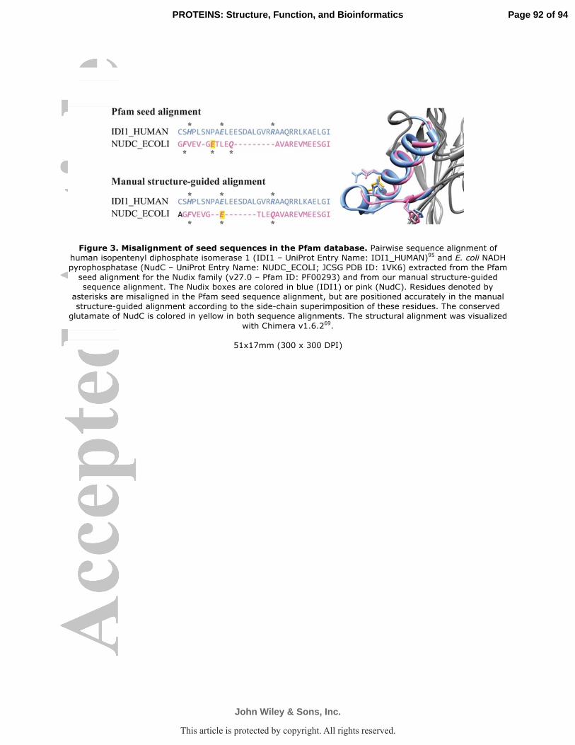
Figure 3. Misalignment of seed sequences in the Pfam database. Pairwise sequence alignment of human isopentenyl diphosphate isomerase 1 (IDI1 – UniProt Entry Name: IDI1_HUMAN)95 and E. coli NADH pyrophosphatase (NudC – UniProt Entry Name: NUDC_ECOLI; JCSG PDB ID: 1VK6) extracted from the Pfam seed alignment for the Nudix family (v27.0 – Pfam ID: PF00293) and from our manual structure-guided sequence alignment. The Nudix boxes are colored in blue (IDI1) or pink (NudC). Residues denoted by
asterisks are misaligned in the Pfam seed sequence alignment, but are positioned accurately in the manual structure-guided alignment according to the side-chain superimposition of these residues. The conserved
glutamate of NudC is colored in yellow in both sequence alignments. The structural alignment was visualized
with Chimera v1.6.269.
51x17mm (300 x 300 DPI)
Page 92 of 94
John Wiley & Sons, Inc.
PROTEINS: Structure, Function, and Bioinformatics
This article is protected by copyright. All rights reserved.

Figure 4. Specificity motifs reside in a loop (the X-loop) that contacts the X-moiety. A. UniProt Entry Names of selected enzymes from the structure-guided sequence alignment that exhibit specificity motifs in analogous loop regions downstream from the Nudix box motif. Motifs, either described by other
investigators or presented in this study, are colored according to function, where purple and orange refer to two distinct types of NDP-sugar diphosphatases, red refers to bis(5’-nucleosyl) polyphosphate hydrolase
activity, and blue refers to NADH diphosphatase activity. UniProt Entry Names follow each enzyme in parentheses and four enzymes are annotated with a PDB ID corresponding to the structure aligned in panel B. Enzymes under consideration include Escherichia coli ADP-ribose diphosphatase, E. coli GDP-mannose
diphosphatase, Homo sapiens ADP-sugar diphosphatase, Thermus thermophilus ADP-ribose diphosphatase, Synechocystis sp. ADP-ribose diphosphatase, Francisella tularensis subsp. tularensis ADP-ribose
diphosphatase, Methanococcus jannaschii ADP-ribose diphosphatase, Synechocystis sp. ADP-ribose diphosphatase, H. sapiens Ap4A hydrolase, Caenorhabditis elegans Ap4A hydrolase, Drosophila melanogaster
bis(5’-adenosyl) polyphosphate (ApnA) hydrolase, Lupinus angustifolius Ap4A hydrolase, E. coli NADH diphosphatase, Deinococcus radiodurans NADH diphosphatase, H. sapiens NAD(P)H diphosphatase, and
Saccharomyces cerevisiae NADH diphosphatase. B. MultiProt73 multiple structural alignment of Escherichia coli ADP-ribose diphosphatase105, Synchocystis sp. ADP-ribose diphosphatase107, E. coli NADH diphosphatase
(JCSG PDB ID: 2GB5), and Homo sapiens Ap4A hydrolase114. Specificity motifs associated with each enzyme’s function are colored according to panel A. These motifs all reside within the same structural region
that is in close proximity to the X-moiety of the Nudix hydrolase substrate. The co-crystallized substrate (ADP-ribose) of E. coli ADP-ribose diphosphatase is shown in purple to provide orientation. Two analogous
loops, one from each E. coli ADP-ribose diphosphatase monomer in the functional dimer, participate in contacting the terminal ribose moiety in the ADP-ribose substrate. C. DaliLite72 pairwise structural alignment
Page 93 of 94
John Wiley & Sons, Inc.
PROTEINS: Structure, Function, and Bioinformatics
This article is protected by copyright. All rights reserved.

of Escherichia coli (PDB ID: 1KHZ) Synechocystis sp. (PDB ID: 2QJO) ADP-ribose diphosphatases reveals general conservation with regard to the Nudix homology domain, but specific conformations with respect to
a loop that contacts the substrate’s ribose moiety (the “X-loop”). The bound substrate analogue, α/β-methylene-ADP-ribose (purple; with E. coli ADP-ribose diphosphatase), three magnesium cations (black;
with E. coli ADP-ribose diphosphatase), and ADP-ribose (orange;
156x150mm (300 x 300 DPI)
Page 94 of 94
John Wiley & Sons, Inc.
PROTEINS: Structure, Function, and Bioinformatics
This article is protected by copyright. All rights reserved.

Figure 5. Phylogeny of 347 structurally determined, functionally characterized, or phylogenetically important Nudix homology protein domains. The phylogenetic tree, constructed with RaxML78 with 1,000 bootstraps, was mid-point rooted. The bootstrap value of an internal node is placed next
to it. Inferred duplication events are represented by open diamonds on the corresponding internal nodes. Every leaf has domain of life, function, and ID notations. The domain of life of a leaf is indicated by a circle (eukaryota), a triangle (archaea), a square (virus), or is left blank (bacteria). The characterized functions, where known, are indicated with colored bars, whose lengths are proportional to the confidence scores of
function assignments. The colors of the bars are chosen to indicate the biochemical similarity (e.g., substrate structure, position of hydrolysis) of the functions. Abbreviations for annotated functions (usually substrate names) are listed in a pair of square brackets following the color bars. The UniProt Entry Name of
the protein and, if available, the associated PDB ID are noted in parentheses. If neither function nor structure is available for a leaf (as is the case for protein domains from the seed alignment of the Pfam
Nudix family (PF00293)), its UniProt Entry Name is omitted, and a cross is used as a placeholder. Red and
Page 95 of 94
John Wiley & Sons, Inc.
PROTEINS: Structure, Function, and Bioinformatics
This article is protected by copyright. All rights reserved.

cyan brackets with shorthand notation of functions indicate clades within which most characterized proteins share at least one common function. Red brackets have proteins from at least two domains of life, while blue
brackets mean that all proteins within the clades are from the same domain of life. Functionally monophyletic clades are noted by “☼,” and non-hydrolase functions are noted by “†,” both next to the
names of the clades. Protein clades that separate these designated clades are indicated by black brackets
with letters. The asterisks in the text of the color bar legend indicate non-hydrolase functions. A high-resolution version is available in Table S1, Resource 26. The phylogeny was visualized with Dendroscope
v3.2.883 and graphics processed with Adobe Illustrator CS4113.
228x298mm (300 x 300 DPI)
Page 96 of 94
John Wiley & Sons, Inc.
PROTEINS: Structure, Function, and Bioinformatics
This article is protected by copyright. All rights reserved.