the incidence of cancer in quantitatively transplanted
TRANSCRIPT
The etiology of mammary cancer in mice involvesgenetic factors (9), hormonal influences (39), the milkagent (14), and dietary and environmental conditions (37).The relative significance of the several contributing influences is controversial. Recently, Mühlbock and hisassociates (12, 39) strongly emphasized the dominant in
I Supported by grants from the Anna Fuller Fund, the American
Cancer Society Institutional Grants to Yale University School of
Medicine, and the Jane Coffin Childs Memorial Fund for MedicalResearch; by Grant C-343 from the National Cancer Institute,USPHS; and by a grant from the National Cancer Institute ofCanada.
2 Present address: Cancer Research Laboratory, University of
Western Ontario, London, Ontario, Canada.
Received for publication October 28, 1964; revised May 21,1965.
fluence of hormonal stimulation and Nandi (42) emphasized the importance of the milk agent in inducing hyPerPlastic alveolar nodules and eventual tumor. 1\Iicewithout the milk agent may be infected by ingestion orinjection of milk, by injection of semen (5, 38), or bytransplantation of mammary tumor and other tissues(7) so that they subsequentlyacquiremammarycancers.But milk-agent-free mice may also develop mammarycancer without being infected with the milk agent (23).
The infectivity of the milk agent and development ofmammary cancer in mammary grafts from milk-agentfree donors in agent-positive host mice or in mammaryglands of the hosts of milk-agent-free mice receiving graftsfrom milk-agent-positive donor mice were studied in conjunction with different hormonal influences.
1792
The Incidence of Cancer in Quantitatively TransplantedMammary Glands and Its Relation to Age and Milk Agent
of the Donor and Host Mice1
K. HosHINo,2 W. U. GARDNER,ANDR. A. PAWLIKOWSKIDepartment of Anatomy, Yale University School of Medicine, New Haven, Connecticut, and Cancer Research Laboratory, University
of Western Ontario, London, Ontario, Canada
SUMMARY
Two hundred fifty-four mammary segments, each 0.6 mm long with little surrounding adipose StI'OnTlal tissues, obtained from virgin female donor mice of the CBA orC57BL strains, were transplanted into the 4th mammary gland-free fat pads and thepararenal fat Pads of F1 hybrid (CBA x C57BL) hosts. Mammary grafts obtainedfrom milk-agent-free C57BL donors were I)laced in agent-positive hosts, and those derived from agent -positive CBA donors were placed ill agent-free host mice. i\Iammarygrafts from donors of less thati 4 months of age were placed in hosts 9@months old ormore, and grafts from 19-month-old or older donors were placed in the hosts that wereless than 4 months of age at the time of transplantation.
Mammary carcinomas developed in 34 of the 109 successfully transplanted marnmary segments; 10 tumors occurred ill 12 grafts in female hosts that reared several litters following transplantation, 4 occurred in 12 grafts in castrated male hosts treated
with estrogen and progesterone pellets for 12 to 17.5 months, and 20 occurred in 47grafts 111virgin female hosts bearing functional j)ituitary isografts. In the former 2groups, only mammary grafts derived from C57BL donors and transplanted into theagetit-positive hosts developed cancers. In the last group, mammary cancer did notappear iii the hosts iii which the j)ituitary grafts failed to survive. When 1)ituitary isografts were functional, 20 tumors developed 111mammary grafts obtained from eitherCS7BL or CBA donors and placed into either agent-positive or agent-free host mice.
i\iammary grafts from normal female CS7BL donors could he infected with themilk agent present in the environment in the milk-agent-positive hosts and could develop cancer. Also, excessive hormonal stimulation by pituitary isografts overcamelack of the milk agent in the host environment.
The incidence of tumors, the occurrence of histologically differeiit types of mainmary cancers 111grafts and in the hosts' glands, and the influence of pituitary isograftswere examined and discussed in conjunction with the l)rOblems of the milk agent andthe age of the animals.
Dow
nloaded from http://aacrjournals.org/cancerres/article-pdf/25/10/1792/2379416/cr0250101792.pdf by guest on 23 February 2022

H0sHIN0 et al.—Cancer Incidence in Transplanted Mammary Glands 1793
( FIt 4)
CHARTS 1—4.—Sketches of 4 mammary glands of donor mice marked to indicate the precise location of segmentsexcised for transplantation. White areas with arrows and letters are segments from which mammary t umors developed. A : Type A adenocarcinoma; B: Type B adenocarcinoma; C: Type C adenocarcinoma; 8: adenoacanthoma; and H: hyperplastic nodules. Black areas are segments that were successfully transplanted but@did notdevelop cancer. Shaded areas are segments that were not recovered from the hosts at autopsy.
CHART 1.—The right 3rd mammary gland of a 24-month-old female C57BL donor; the 20 segments were traiisplanted into 94-month-old female CCI hosts that were subsequently mated. Mammary cancers developed in 10transplanted segments.
CHART 2.—The right 3rd mammary gland of a 34-month-old female CBA donor; the 29 segments were traIlsplanted into 10-month-old female CC1 hosts that received 1 pituitary isograft each. Mammary cancers developedin 8 transplanted segments, and hyperplastic nodules developed in 3 segments.
CHART 3.—The left 1st mammary gland of a 19-month-old female CBA donor; the 20 segments were transplantedinto female CC1 hosts of 1—4months of age that each received 1 pituitary isograft. Mammary cancers developed
in 6 transplanted segments and hyperplastic nodules developed in 1 segment.CHART4.—The right 4th mammary gland of a 4-month-old female C57BL donor; the 24 segments were traiis
planted into castrated male CCI hosts of 104 to 13@months of age that subsequently received treatment withestrogen and progesterone pellets. Mammary cancers developed ill 4 transplanted segments and hyperplasticnodules in 6 segments.
MATERIALS AND i@dETHODS
Mice of 2 inbred strains, the CBA/SG and C57BL/Jax(29) (hereafter referred to as CBA and C57) were the donors, and their reciprocal F1 hybrids were the hosts usedfor the isologous transplantation of segments of mammaryducts. The donors and female hosts were virgin at thetime of transplantation. The CBA mice and F1 progenyof CBA females (designated as CC2) were considered tobe milk-agent positive, and the C57 mice and F1 hybridof C57 females (designated as CC1) as milk-agent free.
The donors were either less than 44 months old or olderthan 19 months. The hosts were either less than 4 monthsold or older than 94 months at the time of transplantation.Mammary segments obtained from milk-agent-free donorswere transplanted into milk-agent-positive hosts, and viceversa. In each combination of donor and host mice, thedonor was either younger or older than the host. The‘quantitative transplantation― technic (28) was used totransplant 254 mammary segments, each 0.6 mm long
and from a precisely located and recorded area of the do
Dow
nloaded from http://aacrjournals.org/cancerres/article-pdf/25/10/1792/2379416/cr0250101792.pdf by guest on 23 February 2022

DONORHOSTINCIDENCEPERIOD OF TRANSPLANTATION (mo)HOSTS'
AGE ATDEATH (mo)No.
OFPREG
NANCIESNo.
OFLITTERS
WEANEDIngraftsIn hosts' glands
(MCa/glandsexamined)SitedMCa/takes/grafts24-mo-old
@C57 (agent —)
19-mo-old@
CBA (agent +)10-mo-old
@CC2 (agent +)
1- to 4-mo-old
9 CC1(agent— )4mgffp
prfp
4mgffp
prfp5
(4)5/7 (5)/10 (5)5 (4)/5 (4)/10 (5)
0/11 (7)/20(10)
0/4 (3)/20 (10)3
(3)/40 (5)
0/80(10)11.6c
(8—14.5)
17.8(6.5—21.5)21.6
(18—24.5)
20.3(7.5—25.5)6.6
(4—9)
6.3 (3—9)4.4
(3—6)
5.7 (2—9)
Cancer Research Vol. 25, November 19651794
nor's mammary glands (Charts 1—4). The stroma surrounding each duct segment was removed as completelyas possible prior to transplantation. The transplants
were inserted separately by watchmakers' forceps into theadipose tissue of either the 4th mammary gland-free fatpads (prepared by the operation technic described in Ref.27) or the pararenal fat Pads of either one or both sidesof the host mice.
The female hosts were then mated with their male siblings (Group 1) or were given pituitary isografts from suchmales (Group 2). One pituitary gland was transplantedinto the right 4th mammary gland-free fat pad togetherwith the mammary transplant. In the former group, occurrence of pregnancy, abortion, still birth, parturition,and weanlings were recorded. In the latter group, vaginal smears were examined daily for 3—5weeks at intervals of 1—2months to determine evidence of the persistence of functioning pituitary grafts. The site of pituitarytransplant was examined at autopsy, and all questionabletissues were examined histologically.
The male hosts were orchidectomized when the 4thmammary gland parenchyma were removed. Followingtransplantation, they received s.c. implants of estrogenand progesterone pellets (Group 3). The pellets contained1 part estradiol benzoate and 1000 parts progesterone andweighed from 6.0 to 11.0 ing each. Pellets were implantedrepeatedly every 2—3months or when they could not bepalpated. At autopsy, the remaining portions of pelletswere removed, dried, and weighed. All experimentalammals were maintained under uniform conditions untilthey developed tumors or until death seemed imminent.When the hosts' own mammary glands developed tumorsand their general conditioll was satisfactory, the tumorswere removed surgically. When tumors appeared at locations that did not permit us to determine whether theorigm was from transplanted or from hosts' mammaryglands, the tumors were transplanted s.c. into intact female mice both of the same F1 hybrid and of the parentalstrains in order to study the histocompatibility of the tumor tissues. All mammary glands, both transplanted andnontransplanted, were l)rePared either as whole mountsor as histologic sections stained with hematoxylin andeosin after paraffin embedding. All mice were subjected
to complete autopsy, and all detected pathologic lesionswere examined histologically.
RESULTS
Mammary carcinomas developed from 34 of the 109successfully transplanted mammary segments, from 22 of75 successfully transplanted in the 4th mammary glandfree fat pads, and from 12 of 34 grafts in the pararenal fatpads.
The incidence of tumors in the hosts that subsequentlyreared young (Group 1).—Mammary cancers developedfrom 10 grafts, 5 at each of the 2 transplantation sites,when segments from young milk-agent-free, C57 femaledonors were transplanted into 10-month-old, agent-positive, CC2 female hosts that subsequently reared young(Table 1). No mammary cancer developed from graftsin agent-free CC1 female hosts, although mammary transplants were obtained from agent-positive, 19-month-oldCBA female donors. From the hosts' OW@ mammaryglands, 3 tumors developed in the CC2 mice, but none inthe CC1 mice. Hyperpla.stic nodules, however, were foundin both transplanted and nontransplanted hosts' mammaryglands in CC1, as well as in CC2, hosts.
The incidence of tumors in hosts that simultaneously received single pituitary isografts (Group 2).—Mammarycancers appeared ill 20 grafts, regardless of the presenceor absence of milk agent in the hosts and of the ages ofeither donor or host mice (Table 2). i\Iammary tumorsappeared in the grafts 12 months or more after transplantation. No tumors developed when the pituitary transplants failed to survive. Of 20 mammary tumors, 6 developed from the grafts in the hosts bearing nontumorouspituitary grafts, whereas 14 other tumors occurred fromthe grafts in the hosts that had enlarged and histologicallyadenomatous pituitary grafts (Fig. 1). Twenty-two of23 tumors that developed in the hosts' own mammaryglands occurred in mice with adenomatous pituitary grafts(Table3). Thehosts'ownmammaryglandsbecametumorous earlier than did the mammary grafts at 9 monthsor more after transplantation. Hyperplastic noduleswere observed in 7 mammary grafts in 3 CC1 and 3 CC2hosts and in 5 hosts' mammary glands in 3 mice, none ofwhich had mammary cancer. Reticulum cell sarcomas
TABLE 1
THE INCIDENCE OF MAMMARY CANCER (MCa) IN TRANSPLANTED AND HOsTS' OWN MAMMARY GLANDS
IN MICE THAT SUBSEQUENTLY REARED YOUNG
a 4mgffp, the 4th mammary gland-free fat pads; prfp, the pararenal fat pads.
‘,The number in parentheses represents the number of mice.
C The number given is an average; the range is given in parentheses.
Dow
nloaded from http://aacrjournals.org/cancerres/article-pdf/25/10/1792/2379416/cr0250101792.pdf by guest on 23 February 2022

Conditions ofpituitary isografts
at autopsy
AdenomatousNo.
ofhostmice
16MCa
in grafts,'transplants
14/60MCa
in hosts'glands/Ilosts' glands
22/131I
Days vaginal smears@ taken per mouse
@ 78@@195a(165)Days
of estrus/davssmears examined
av. ratio/mouse(%)12.7
± 0.7b(a)'Nontumorous86/271/6656—182(132)3.5 ±0.9(b)Not
recovered40/150/33133—191 (155)24.5 ± 5.6 (c)
Site of transplantationMammary cancersSuccessfultransplantsTransplantsRight
4th mammary gland-freefat pads with pituitarygrafts
Left 4th mammary gland-freefat pads
Right pararenal fat padsLeft pararenal fat pads
9
65017
1311628
212726
H0SHIN0 et al.—Cancer Incidence in Transplanted Mammary Glands 1795
TABLE 2
THE INCIDENCE OF MAMMARY CANCER (MCa) IN TRANSPLANTED AND HOSTS' OWN MAMMARY GLANDS
IN MICE BEARING PITUITARY ISOG1IAFTS
INCIDENCE
PITUITARY
GRAFTS (takes/transplants)
3/5
9/10
3/4
7/9
PERIOD OF TRAXSPLANTATION (mo)
HOSTS' AGE ATDEATh mo)
DONOR HOST In grafts In hosts' glands(MCa/glandsexamined)Site― MCa/takes/grafts
4 (1)/40(5) 13.6'(10.5—18.5)23.5 (20.5—28)94-mo-old 9 CC2(agent +)
10- to 18-mo-old 9CC1 (agent—)
94-mo-old 9 CCS
(agent +)1- to 4-mo-old 9CC1 (agent —)
4mgffpprfp4mgffpprfp4mgffpprfp4mgffp
prfp
2 (2)b/6(3)/10(5)
1 (1)/2 (2)/lO (5)6 (5)/12(9)/17(10)
2 (2)/9(6)/17(9)
1 (1)/4(4)/4(4)1 (1)/3(3)/8(4)6 (5)/8 (5)/18 (9)1 (1)/3 (3)/18 (9)
2k-mo-old@ C57
(agent —)34- to 44-mo-old 9CBA (agent+)
44-mnth-old
9 CBA (agent +)19-mo-old 9 CBA
(agent +)
4 (4)/82 (10)
3 (3)/36(4)
15.9 (8.5—18.5)
11.5(7—21.5)
27.6 (22.5—35)
20.4 (16—25)
22.7 (13—25.5)12 (7)/72 (9) 20.4 (12—21.5)
a 4mgffp, the 4th mammary gland-free fat pads; prfp, pararenal fat pads.
b The number in parentheses represents the number of mice.
C The number given is an average; the range is given in parentheses.
TABLE 3
THE FREQUENCY OF ESTRUS IN THE HOST MICE IN WHICH PITuI'rARY ISOGRAFTS WERE
FOUND AS ADENOMATOUS OR NONTUMOROL@S, OR WERE NOT FOUND AT AUTOPSY, AND
RELATION TO THE INCIDENCE OF MAMMARY CANCERS (MCa)
a ilange; the average is given in parentheses.
b S.E.
C Statistical differences: (a) < (c) and (b) < (c) with P < 0.001, but (a) 4 (b) (nolisignificant).
plants. The incidence of mammary tumors was similarOIl both right and left sides (Table 4). The incidences ofmam.mary tumors that developed from the hosts' manimary gland on the right side and the left side were similar:12 tumors from 118 mammary glands examined in theleft side, and 11 tumors from 112 glands in the right sideof the hosts. All mice with successful l)itUitarY grafts,particularly with adenomatous grafts, had both traIlsplanted and nontransplanted mammary glands that wereextremely well developed and that usually contained milkysecretion in dilated ducts (Figs. 2, 3). These conditionsmade it difficult to determine hyperplastic alveolar nodules. The number of the nodules observed was probablyminimal.
The largest adenomatous pituitary graft measured11 x 8 x 6 mm and weighed 260 mg. This was found in afemale CC1 host that also had 2 mammary cancers—onefrom transplanted mammary tissue and the other fromher own gland. Isografts of the pituitary adenoma wereplaced in the ventral subcutaneous areas of 16 intact female mice of 2—4months of age. Twelve months later,one grew large enough to be retransplanted into hosts of asucceeding generation. The tumorous mass measured
7.5 x 6.5 x 5.0 mm and weighed 120 tug. Histologicallythe tumor did not differ from the original tumor. The
TABLE 4
THE INCIDENCE OF TUMORS IN MAMMARY GRAFTS AT
DIFFERENT SITES IN THE HOSTS BEARING
PITUITARY ISOGRAFTS
appeared at sites of transplantation of 3 mammary graftsin the 4th mammary gland-free fat pads of 2 host mice andin areas of 3 hosts' mammary glands of 2 other mice.
When pituitary isografts persisted in the hosts, estrouscycles were prolonged or pseudo-pregnancy-like, or themice showed persistent diestrus. Contrarily, the micefrom which pituitary isografts were not recovered hadvaginal cycles of normal type (Table 3).
The pituitary grafts were placed in the right 4th mammary gland-free fat pads very close to the mammary trans
Dow
nloaded from http://aacrjournals.org/cancerres/article-pdf/25/10/1792/2379416/cr0250101792.pdf by guest on 23 February 2022

Type Type@ Type AdenoA B C acanthoma
1 1 1
4 1
2 2
3 1
1 2
6 4 1 1
Totals―15748131332
1796 Cancer Research
TABLE 5
Vol. 25, November 1965
THE INCIDENCE OF MAMMARY CANCER (MCa) IN TRANSPLANTED AND HOSTS' OWN MAMMARY GLANDS IN THE CASTRATED MALE HOSTS
TREATED WITH SEX HORMONES
INCIDENCE
ESTROGEN-PROGES
TERONE PELLETSABSORBED PER
MOUSE (tug)
PERIOD OF TRANSPLANTATION (mo)
HOSTS' AGE ATDEATH (mo)
DoNOR In grafts In hosts' glandsMCa/glands
examinedSite― MCa takes/grafts
4-mo-old 9 C57(agent —)
44-mo-old 9 CBA(agent +)
19-mo-old 9 CBA(agent +)
104-to 134-mo-old5―CC, (agent +)
9- to 94-mo-old@
CC1 (agent —)34- to 4-mo-old@
CC1 (agent —)
4mgffpprfp4mgffpprfp4mgffpprfp
2 (2)―/8(5)/12(6)
2 (2)/4(4)/12(6)
0/9 (6)/14(7)
0/1 (1)/14 (7)0/10 (6)/20 (10)0/3 (3)/20 (10)
5 (3)/36(6) 15.0'(12—17.5)27.0 (22.5—31)49.5 (44.1—55.0)
0/42 (6)
0/60 (10)
19.3 (16—21.5)
21.7 (20-22)
28.9 (25—31)
22.7 (21—23)
59.7 (50.9—68.0)
60.9 (52.9—63.6)
a 4mgffp, the 4th mammary gland-free fat pads; prfp, the pararenal fat pads.
b The number in parentheses represents the number of mice.
C The number given is an average; the range is given in parentheses.
TABLE 6
THE OCCURRENCE OF DIFFERENT TYPES OF TUMORS THAT DEVELOPED FROM MAMMARY
GRAFTS AND FROM HoSTS' OWN MAMMARYGLANDS IN MICE
TUMORS DEVELOPED FROM MAMMARYGRAFTS TUMORS DEVELOPED FROMOWN GLANDS
DONOR hosT―
Site of trans- Typeplantatiod' A
Type AdenoC acanthoma
1
1 4
1 41
TypeB
Older 9 CC2(rearing young)
Older@ CC2(E & P pellet')
Older 9 CC2(pituitary graft)
Older 9 CC1(pituitary graft)
Older 9 CC2(pituitary graft)
Young 9 CC1
(pituitary graft)
4mgffpprfp4mgffpprfp4mgffpprfp4mgffpprfp4mgffpprfp4mgffpprfp
441121
tation of 2 mammary grafts in the 4th mammary glandfree fat pads and 1 graft in the pararenal fat pad in 3 CC1host mice and in the area of 1 mammary gland of a castrated male CC1 mouse.
The origin of mammary grafts that developed carcinomasand the histopathologic classification of these tumors.—Thesegments of the mammary glands transplanted were from4 donors, and the ultimate fates of these transplants areillustrated in Charts 1—4. The histopathologic characteristics of the mammary tumors were classified accordingto the system described by Dunn (15). When a tumorconsisted of more than 1 type, it was classified as belonging to the most predominant type (Table 6). Mammarytransplants regenerated and mammary tumors of differenttypes developed from different portions of the donors'
mammary glands (Charts 1—4). In mammary grafts,
2
111
1
111
Young 9 C57
Young 9 C57
Young 9 C57
Young 9 CBA
Young 9 CBA
Older 9 CBA
a Treatment is given in parentheses.
b 4mgffp, the 4th mammary gland-free fat pads; prfp, the pararenal fat pads.
C Estrogen and progesterone pellet.
d In all, 34 tumors developed from mammary grafts and 31 from the hosts' own glands.
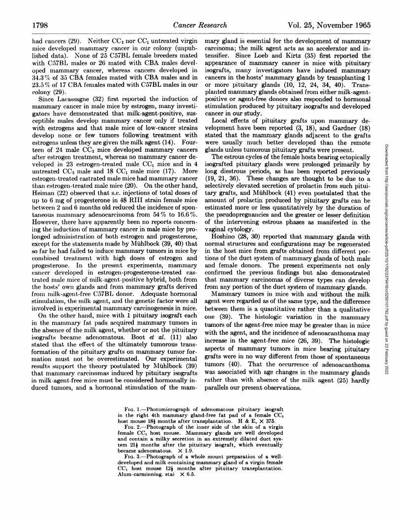
host mouse bearing the latter 1)ituitary tumor also hadmammary adenocarcinoma.
The incidence of tumors in the castrated male hosts subsequently treated with estrogen-progesterone pellets (Group 3).—Four mammary grafts became cancerous, and 6 othersdeveloped hyperplastic nodules in hormone-treated, castrated male CC2 hosts that received grafts from a youngC57 female donor (Table 5). Carcinomas arose in 5hosts' glands, and hyperplastic nodules appeared in 4glands. No mammary cancers developed in castratedmale CC1 hosts that received transplants from eitheryoung or old, agent-positive, CBA female donors, althoughthe period after transplantation in these hosts was considerably longer than that for the former group. Theincidence of hyperplastic nodules was less in the CC1 hosts.
Reticulum cell sarcomas developed at sites of transplan
Dow
nloaded from http://aacrjournals.org/cancerres/article-pdf/25/10/1792/2379416/cr0250101792.pdf by guest on 23 February 2022

HosHiNo et al.—Cancer Incidence in Transplanted Mammary Glands 1797
mammary tumors in mice by injections of extracts oftissues, mammary tumor tissues seem to be a relativelypoor and unreliable source of the milk agent. Bittner(6, 7) successfully transmitted the milk agent and subsequently induced mammary cancers in female mice ofC57BL strain and of ( 9 C57BL x cl@A)F1and F2 hybridsby the inoculation of normal tissues obtained from femaledonors of Strain A or (@ A x @C57BL)F1hybrid carryingthe agent. He inoculated splenic tissue (6) and splenic,thymic, or lactating mammary gland tissues (7). In thepresent experiments, mammary cancers did not appear inmilk-agent-free hosts that received grafts of mammarysegments obtained from the milk-agent-positive donorseveii when the hosts had repeated pregnancies or receivedprotracted estrogen-progesterone treatments after mammary transplantation (Tables 1 and 5).
The failure to transmit the milk agent from the grafts tothe host mice could not be determined critically in the absence of a progeny test (2). Mammary transplants 0.6mm long with little surrounding adipose tissue might havebeen too small to carry enough milk agent for survival andpropagation in the host environment. Propagation of themilk agent may require some autologous adipose tissue surrounding transplanted mammary parenchyma. Lasfargues et a!. (33) stated that the milk agent may not bemaintained ill nearly pure cultures of adult mammaryepithelium in roller tubes; the presence of some adiposetissue retained from the glandular stroma is necessary forextensive proliferation of the milk agent. The age of themice might also be of importance. Older mice required
larger quantities of tumor tissues containing tile mammarytumor agent in order to induce a comparable incidence oftumors (13). Susceptibility to the agent given by the injection of extracts of transplanted mammary cancer contaming the milk agent is influenced by the age of the mice(8). Adultfemalemicedevelopedfewor no tumorsafterinjections of the extracts that induced a high incidence ofmammaary cancer in young female mice. However, whenBittner (6, 7) induced mammary cancers by inoculatingnormal tissues, the hosts at the time of inoculation were4—8weeks of age or had had a 1st litter. In the presentstudy, 5 female and 10 castrated male hosts not developingmammary cancer were 34—4weeks of age at the time oftransplantation. Iti the experiments shown in Tables 1and 5, 19-month-old and 4k-month-old virgin femalemilk-agent-positive mice donated 80 and 28 mammarytralisplalits, respectively, iio@ie of @vhich developed 111ammary cancer or induced cancer in the hosts' mammaryglands. Feldnlall (16) found by electrohl Iflicroscopeneither A nor mature-type B virus-like particles in themammary glands of strain DBA virgin mice younger than6 months of age, hut she found both types of Particles inmice 6 months old or older and in young females duringtheir 1st pregnancy. Bittner used mice that had had 1 or2 litters as donors of inoculated lactating mammary gland,but as donors of inoculated normal splenic and thymictissues, he used 4- to 5-week-old mice and induced mammary cancers in the hosts (6, 7).
None of 18 breeding CC1 mice with an average of 8
pregnancies developed mammary cancer, whereas 78.8@of 33 CC2 female breeders with a@iaverage of 6 prcgnancies
acinar or medullary type of adenocarcinoma (Fig. 4)arose most frequently (15/34), papillary or trabeculartype (Fig. 5) less frequently (7/34), and cystic adenofibroma (Fig. 6) least frequently (4/34). These 3 types oftumors corresponded to Dunn's Types A, B, and C. Histologic variability was greater in tumors that developedfrom grafts 111 the hosts bearing pituitary isografts.Adenoacanthoma (Fig. 7) occurred only from grafts illagent-free CC1 hosts with pituitary isografts. The occurrences of Types A and B were equally common amongtumors that developed from the hosts' OWU mammaryglands (13/31 for each type). The incidence of adenoacanthoma in these tumors (2/31 ; 6.5 %) was less than intumors that developed from mammary grafts (8/34;23.6 %), but this type of tumor also occurred only in CC1host mice with pituitary isografts.
Histocompatibility of tumor tissues studied by retranspkzntation.—In order to study histocompatibility of tissuesof mice used in the present experiments, 20 segments ofnormal mammary glands obtained from both CC1 andCC2 females were transplanted quantitatively (each 0.6mm long) into the 4th mammary gland-free fat pads of 20mice of the CBA strain. All 40 mammary transplants inthe 20 host mice were rejected. Transplants of a tumorthat developed in the site of mammary grafts obtainedfrom a 19-month-old CBA donor and placed into the 4thmammary gland-free fat pad of a CC1 host. bearing apituitary isograft grew when placed in the s.c. areas of 3CC1 and 4 CC2 mice, but not in 4 mice of the CBA strain.This tumor was histologically a reticulum cell sarcoma.Tissues of a tumor that developed at the same site inanother CC1 host in the same experimental group grew in2 of 4 CBA and 1 of 3 CC1 mice. These tumor tissues werecomparable histologically to the original tumor, an adenoacanthoma, after being transplanted in both CBA and CC1mice for 100 days. One trabecular type adenocarcinoma,which developed from a mammary graft transplanted froma 34-month-old CBA donor into the 4th mammary glandfree fat pad of a CC1 host, grew in all of 4 CBA and in 3CC1 mice and retained its original histologic characteristics.
DISCUSSION
i\Iammary tissues tralisplanted from adult. femaleC57BL donor mice may be infected with the milk agentpresent it! the environment in the milk-agent-positivehosts and may subsequently develop mammary cancer.
l'sIice of low-cancer strains have been infected with themilk agent by transplantation of mammary tumors.Andervont (1) reported that 1 of 23 originally agent-freemice that carried a transj)lanted mammary cancer thathad arisen spontaneously in a milk-agent-infected donordeveloped mammary tumor in later life. Six mammarytumors developed in 66 milk-agent-free mice carryingtransplanted mammary tumor tissues that grew for a shortperiod in the ears of the hosts (31). Dmochowski (13) obtamed a high incidence of mammary cancer in F1 hybridfemale mice that received dried mammary tumor tissuesfrom their parental strain. Andervont (1) stated thattransplantation of mammary tissues is not an effectivemeans of infecting young susceptible mice with the milkagent. Barnum et al. (4) also noted that, for induction of
Dow
nloaded from http://aacrjournals.org/cancerres/article-pdf/25/10/1792/2379416/cr0250101792.pdf by guest on 23 February 2022
Cancer Research Vol. 25, November 19651798
had cancers (29). Neither CC2 nor CC1 untreated virginmice developed mammary cancer in our colony (unpublished data). None of 25 C57BL female breeders matedwith C57BL males or 26 mated with CBA males developed mammary cancer, whereas cancers developed in34.3 % of 35 CBA females mated with CBA males and in23.5 % of 17 CBA females mated with C57BL males in ourcolony (29).
Since Lacassagne (32) first reported the induction ofmammary cancer in male mice by estrogen, many investigators have demonstrated that milk-agent-positive, susceptible males develop mammary cancer only if treatedwith estrogens and that male mice of low-cancer strainsdevelop none or few tumors following treatment withestrogens unless they are given the milk agent (14). Fourteen of 24 male CC2 mice developed mammary cancers
after estrogen treatment, whereas no mammary cancer developed in 23 estrogen-treated male CC1 mice and in 4untreated CC2 male and 18 CC1 male mice (17). Moreestrogen-treated castrated male mice had mammary cancerthan estrogen-treated male mice (20). On the other hand,Heiman (22) observed that s.c. injections of total doses ofup to 6 mg of progesterone in 48 RIII strain female micebetween 2 and 6 months old reduced the incidence of spontaneous mammary adenocarcinoma from 54 % to 16.6 %.However, there have apparently been no reports concerning the induction of mammary cancer in male mice by prolonged administration of both estrogen and progesterone,except for the statements made by MUhlbock (39, 40) thatso far he had failed to induce mammary tumors in mice bycombined treatment with high doses of estrogen andprogesterone. In the present experiments, mammarycancer developed in estrogen-progesterone-treated castrated male mice of milk-agent-positive hybrid, both fromthe hosts' own glands and from mammary grafts derivedfrom milk-agent-free C57BL donor. Adequate hormonalstiInUlatiOll, the milk agent, and the genetic factor were all
involved in experimental mammary carcinogenesis in mice.On the other hand, mice with 1 pituitary isograft each
in the mammary fat 1)ads acquired mammary tumors in
the absence of the milk agent, whether or not the pituitaryisografts became adenomatous. Boot et al. (11) alsostated that the effect of the ultimately tumorous transformation of the pituitary grafts on mammary tumor fornlatioll must not be overestimated. Our experimentalresults support the theory postulated by Mühlbock (39)that mammary carcinomas induced by pituitary isograftsirk milk-agent-free mice must be considered hormonally in
duced tumors, and a hormonal stimulation of the mam
mary gland is essential for the development of mammarycarcinoma; the milk agent acts as an accelerator and intensifier. Since Loeb and Kirtz (35) first reported theappearance of mammary cancer in mice with pituitaryisografts, many investigators have induced mammarycancers in the hosts' mammary glands by transplanting 1or more pituitary glands (10, 12, 24, 34, 40). Transplanted mammary glands obtained from either milk-agentpositive or agent-free donors also responded to hormonalstimulation produced by pituitary isografts and developedcancer in our study.
Local effects of pituitary grafts upon mammary development have been reported (3, 18), and Gardner (18)stated that the mammary glands adj acent to the graftswere usually much better developed than the remoteglands unless tumorous pituitary grafts were present.
The estrous cycles of the female hosts bearing ectopicallyisografted pituitary glands were prolonged primarily bylong diestrous periods, as has been reported previously(19, 21, 36). These changes are thought to be due to aselectively elevated secretion of prolactin from such pituitary grafts, and MUhlbock (41) even postulated that theamount of prolactin produced by pituitary grafts can beestimated more or less quantitatively by the duration ofthe pseudopregnancies and the greater or lesser definitionof the intervening estrous phases as manifested in thevaginal cytology.
Hoshino (28, 30) reported that mammary glands withnormal structures and configurations may be regeneratedin the host mice from grafts obtained from different portions of the duct system of mammary glands of both maleand female donors. The present experiments not onlyconfirmed the previous findings but also demonstratedthat mammary carcinomas of diverse types can developfrom any portion of the duct system of mammary glands.
Mammary tumors in mice with and without the milkagent were regarded as of the same type, and the differencebetween them is a quantitative rather than a qualitativeone (39). The histologic variation in the mammarytumors of the agent-free mice may be greater than in micewith the agent, and the incidence of adenoacanthoma mayincrease in the agent-free mice (26, 39). The histologicaspects of mammary tumors in mice bearing pituitarygrafts were in no way different from those of spontaneoustumors (40) . That the occurrence of adenoacanthomawas associated with age changes in the mammary glandsrather than with absence of the milk agent (25) hardlyparallels our present observations.
FIG. 1 .—Photomicrograph of adenomatous pituitary isograftin the right 4th mammary gland-free fat pad of a female CC1host mouse 184 months after transplantation. H & E, X 375.
FIG. 2.—Photograph of the inner side of the skin of a virginfemale CC1 host mouse. Mammary glands are well developedand contain a milky secretion in an extremely dilated duct system 214 months after the pituitary isograft, which eventuallybecame adenomatous. X 1.9.
FIG. 3.—Photograph of a whole mount preparation of a welldeveloped and milk-containing mammary gland of a virgin femaleCC1 host mouse 124 months after pituitary transplantation.Alum-carminning. stai X 6.5.
Dow
nloaded from http://aacrjournals.org/cancerres/article-pdf/25/10/1792/2379416/cr0250101792.pdf by guest on 23 February 2022

.@
—@-- -a. *[email protected]., I,
,,
1799
:‘B@,@
Dow
nloaded from http://aacrjournals.org/cancerres/article-pdf/25/10/1792/2379416/cr0250101792.pdf by guest on 23 February 2022

FIG. 4.—Photomicrograph of Type A adenocarcinoma thatdeveloped in a transplanted segment derived from the glandillustrated in Chart 1. This tumor was taken 134 months aftertransplantation into the right pararenal fat pad of a female CCIhost mouse that reared 6 litters and also had spontaneous abortions3 times. H & E, X 100.
FIG. 5.—Photomicrograph of Type B adenocarcinoma thatdeveloped in a transplanted segment, indicated in Chart 2, 124months after transplantation into the right 4th mammary glandfree fat pad of a female CC1 host mouse who received a pituitaryisograft. H & E, X 110.
FIG. 6.—Photomicrograph of Type C adenocarcinoma thatdeveloped in a transplanted segment, indicated in Chart 3, 21months after transplantation into the left 4th mammary glandfree fat pad of a female CCI host mouse having adenomatouspituitary isograft. H & E, X 110.
FIG. 7.—Photomicrograph of adenoacanthoma that developedin a transplanted segment, indicated in Chart 3, 124 months aftertransplantation into the right 4th mammary gland-free fat padof a female CC1 host mouse whose pituitary graft became adenomatous. H & E, X 100.
1800
Dow
nloaded from http://aacrjournals.org/cancerres/article-pdf/25/10/1792/2379416/cr0250101792.pdf by guest on 23 February 2022

r4'@ ,@@ -..,. - .
@.-,. .—@....
@e,@@@ •@
.@:...‘,@ •.
@.;@‘-; @, - . , @2@ •;@.:,@t
@.3...(;,@@ .@
@F@@
-
l@@@ s.@@@
‘I@ : S—.@4@ . . -
@ I,..R@,@ .
@1@@@
I
1801
..@.“..@. .â€â€œ
@ .:;@:‘:$:@@@@@ ,:@ @.@ . .•.
&@ vi @,
. . .:@@@ @@/7e. . .@@
Dow
nloaded from http://aacrjournals.org/cancerres/article-pdf/25/10/1792/2379416/cr0250101792.pdf by guest on 23 February 2022
H0sHIN0 et al.—Cancer Incidence in Transplanted Mammary Glands 1803
Propionate on the Incidence of Mammary Cancer in Mice.Cancer Res., 6: 426—30,1945.
23. Heston, W. E. Mammary Tumors in Agent-free Mice. Ann.N.Y. Acad. Sci., 71: 931—42,1958.
24. . Induction of Mammary Gland Tumors in StrainC57BL/He Mice by Isografts of Hypophyses. J. Nati. CancerInst., 8f@:947—55,1964.
25. Heston, W. E., Deringer, @I.K., and Dunn, T. B. FurtherStudies on the Relationship between the Genotype and theMammary Tumor Agent in Mice. Ibid., 16: 1309-34, 1956.
26. Heston, W. E., Deringer, M. K., Dunn, T. B., and Levillain,W. D. Factors III the Development of Spontaneous MammaryGland Tumors in Agent-free Strain C3Hb Mice. Ibid., 10:1139—55,1950.
27. Hoshino, K. Morphogenesis and Growth Potentiality of Mammary Glands in Mice. I. Transplantability and Growth Potentiality of Mammary Tissue of Virgin Mice. mid., 59: 835.-Si,1962.
28. . Morphogenesis and Growth Potentiality of MammaryGlands in Mice. II. Quantitative Transplantation of Mammary Glands of Normal Male Mice. Ibid., SO: 585—91,1963.
29. . Strain l)ifferences in Normal and Abnormal Pregnancies in Mice and Relationship to Mammary Tumor Incidence.
Ibid., 52: 323—38,1964.30. . Regeneration and Growth of Quantitatively TraIls
planted Mammary Glands of Normal Female Mice. Anat.Record, 150: 221—36,1964.
31. Hummel, K. P., and Little, C. C. Studies on the @IouseMammary Tumor Agent. III. Survival and Propagation of the
Agent in Transplanted Tumors and in Hosts That Grew TheseTumors ill Their Tissues. Cancer Res., 9: 137—38,1949.
32. Lacassagne, A. Apparition de cancer de la mamelle chez Iasouris male soumise a de@ injections de folliculine. Compt.rend., 19,5:630—32,1932.
33. Lasfargues, E. Y., Murray, M. it., and Moore, I). H. Cult.ivat.ion of the Mouse Mammary Carcinoma Virus. J. NatI. Can
cer Inst. Monograph, 4: 151—66,1960.34. Liebelt, A. G., and Liebelt, H. A. Effects of a Single Pituitary
Isograft. on Mammary Tumorigenesis in Mice. Cancer Res.,
21: 86—91,1961.35. Loeb, L., and Kirtz, M. M. Effects of Transplants of Anterior
Lobes on Growth of Mammary Gland and on l)evelopment ofMammary Gland Carcinoma in Various Strains of Mice. Am.J.Cancer,36:56—82,1939.
36. Montemurro, I). G., and Gardner, W. U. Effects of Subcutaneous Transplants of Pituitary Gland and HypothalamusOIl the Estrous Cycle of the Mouse. Endocrinology, 78: 174—84,
1963.37. Morris, H. P. Diet and Some Other Environmental Influences
in the Genesis and Growth of Mammary Tumors in Mice. In:F. M. Moulton (ed.), Symposium on Mammary Tumors inMice, pp. 140-61. Washington, 1). C. : American Associationfor the Advancement of Science, 1945.
38. Mühlbock, 0. Mammary Tumor-Agent in the Sperm of HighCancer-StrainMaleMice. J. Natl. Cancer Inst., 10:861—64,1950.
39. - . The Hormonal Genesis of Mammary Cancer. Advan.Cancer Res., 4: 371—91,1956.
40. Muhlbock, 0., and Boot, L. M. Induction of Mammary Cancerin Mice without the Mammary Tumor Agent by Isografts ofHypophyses. Cancer Res., 19: 402—12,1959.
41. . Carcinogenesis in Relation to the Function of Endocrine Organ Transplants. In: M. J. Brennan and W. L. Simpson (eds.), Biological Interactions in Normal and NeoplasticGrowth, pp. 255-60. Boston : Little, Brown & Company, 1962.
42. Nandi, S. New Method for I)etection of Mouse MammaryTumor Virus. I. Influence of Foster Nursing on Incidence ofHyperplastic Mammary Nodules in BALB/Crgl Mice. J.Natl. Cancer Inst., 31: 57—73,1963.
REFERENCES
1. Andervont, H. B. Susceptibility of Young and of Adult Miceto the Mammary-Tumor Agent. J. NatI. Cancer Inst., @:397—401,1942.
2. . In Utero Transmission of the Mouse Mammary TumorAgent. Ibid., 31: 261—72,1963.
3. Bardin, C. W., Liebelt, A. G., and Liebelt, R. A. The DirectEffect of Pituitary Isografts on Mammary Gland Development in the Mouse. Proc. Soc. Exptl. Biol. Med., 110: 716—18,1962.
4. Barnum, C. P., Ball, Z. B., and Bittner, J. J. Partial Separation of the Mammary Tumor Agent and a Comparison ofVarious Sources of the Agent. Cancer Res., 7: 522—28,1947.
5. Bittner, J. J. Some Possible Effects of Nursing on the Mammary Gland Tumor Incidence in Mice. Science, 84: 162, 1936.
6. . Breast Cancer and Mother's Milk: Relation of Nursingto the Theory of Extrachromosomal Causation of Breast
Cancer in Mice—A Preliminary Report. J. Heredity, @8:363—65,1937.
7. . The Influence of Transplanted Normal Tissue on BreastCancer Ratios in Mice. Public Health Rept., 54: 1827—31,1939.
8. . Activity of the Mammary Tumor Agent in Mice ofDifferent Ages and Their Progeny. J. Natl. Cancer Inst., 18:65—76,1957.
9. . Genetic Concepts in Mammary Cancer in Mice. Ann.N.Y. Acad. Sci., 71: 943—75,1958.
10. Bittner, J. J., and Cole, H. L. Induction of Mammary Cancerin Agent-free Mice Bearing Pituitary Isografts Correlatedwith Inherited Hormonal Mechanisms. J. Natl. Cancer Inst..,27: 1273—84,1961.
11. Boot, L. M., Mühlbock, 0., Ropcke, G., and EbbenhorstTengbergen, W. van. Further Investigations on Induction ofMammary Cancer in Mice by Isografts of Hypophyseal Tissue.Cancer Res., 2L 713—27,1962.
12. Boot, L. M., Ropcke, G., and Muhlbock, 0. Mammary TumorInduction by Pituitary Isografts in Mice. Acta Unio Intern.Contra Cancrum, 18: 270-71, 1961.
13. Dmochowski, L. Age and Dosage in the Induction of Breast.Cancer in Mice by the Mouse Tumor Agent. Brit.. J. Exptl.Pathol.,26:192—97,1945.
14. @-.The Milk Agent in the Origin of Mammary Tumors inMice. Advan. Cancer Res., 1: 104—72,1953.
15. Dunn, T. B. Morphology of Mammary Tumors in Mice. In:F. Homberger (ed.), The Physiopathology of Cancer, Ed. 2,pp. 38—84.New York: Paul B. Hoeber, Inc., 1959.
16. Feldman, D. G. Origin and Distribution of Virus-like ParticlesAssociated with Mammary Tumors in DBA Straits Mice. I.Virus-like Particles in Mammary Gland Tissue. J. NatI.Cancer Inst., 50: 477—501,1963.
17. Gardner, W. U. The Effect of Estrogen on the Incidence ofMammary and Pituitary Tumors in Hybrid Mice. CancerRes., 1: 345—58,1941.
18. . Tumors in Transplanted Pituitary Glands in Mice.Proc. Am. Assoc. Cancer Res., 8: 113, 1960.
19. . Some Studies on Experimental Tumorigenesis: Tumorsin Transplanted Pituitary Glands. In: On Cancer and Hor
mones, pp. 89—106.Chicago: University of Chicago Press,1962.
20. Gardner, W. U., Pfeiffer, C. A., and Trentin, J. J. HormonalFactors in Experimental Carcinogenesis. In: F. Homberger(ed.), The Physiopathology of Cancer, Ed. 2, pp. 152-237.New York: Paul B. Hoeber, Inc., 1959.
21. Hagen, E. 0., and Rawlinson, H. E. The Duration of the Effectof Isologous Pituitary Implants on the Estrous Cycles in theIntact Mouse. Can. J. Biochem. Physiol., 41: 101-6, 1963.
22. Heiman, J. The Effect of Progesterone and Testosterone
Dow
nloaded from http://aacrjournals.org/cancerres/article-pdf/25/10/1792/2379416/cr0250101792.pdf by guest on 23 February 2022