the influence of repressor dna binding site …mbio.asm.org/content/5/5/e01684-14.full.pdfthe...
TRANSCRIPT

The Influence of Repressor DNA Binding Site Architecture onTranscriptional Control
Dan M. Park,* Patricia J. Kiley
Department of Biomolecular Chemistry, University of Wisconsin—Madison, Madison, Wisconsin, USA
* Present address: Dan M. Park, Lawrence Livermore National Laboratory, Livermore, California, USA.
ABSTRACT How the architecture of DNA binding sites dictates the extent of repression of promoters is not well understood.Here, we addressed the importance of the number and information content of the three direct repeats (DRs) in the binding andrepression of the icdA promoter by the phosphorylated form of the global Escherichia coli repressor ArcA (ArcA-P). We showthat decreasing the information content of the two sites with the highest information (DR1 and DR2) eliminated ArcA bindingto all three DRs and ArcA repression of icdA. Unexpectedly, we also found that DR3 occupancy functions principally in repres-sion, since mutation of this low-information-content site both eliminated DNA binding to DR3 and significantly weakened icdArepression, despite the fact that binding to DR1 and DR2 was intact. In addition, increasing the information content of any oneof the three DRs or addition of a fourth DR increased ArcA-dependent repression but perturbed signal-dependent regulation ofrepression. Thus, our data show that the information content and number of DR elements are critical architectural features formaintaining a balance between high-affinity binding and signal-dependent regulation of icdA promoter function in response tochanges in ArcA-P levels. Optimization of such architectural features may be a common strategy to either dampen or enhancethe sensitivity of DNA binding among the members of the large OmpR/PhoB family of regulators as well as other transcriptionfactors.
IMPORTANCE In Escherichia coli, the response regulator ArcA maintains homeostasis of redox carriers under O2-limiting condi-tions through a comprehensive repression of carbon oxidation pathways that require aerobic respiration to recycle redox carri-ers. Although a binding site architecture comprised of a variable number of sequence recognition elements has been identifiedwithin the promoter regions of ArcA-repressed operons, it is unclear how this variable architecture dictates transcriptional regu-lation. By dissecting the role of multiple sequence elements within the icdA promoter, we provide insight into the design princi-ples that allow ArcA to repress transcription within diverse promoter contexts. Our data suggest that the arrangement of recog-nition elements is tailored to achieve sufficient repression of a given promoter while maintaining appropriate signal-dependentregulation of repression, providing insight into how diverse binding site architectures link changes in O2 with the fine-tuning ofcarbon oxidation pathway levels.
Received 22 July 2014 Accepted 25 July 2014 Published 26 August 2014
Citation Park DM, Kiley PJ. 2014. The influence of repressor DNA binding site architecture on transcriptional control. mBio 5(5):e01684-14. doi:10.1128/mBio.01684-14.
Editor Susan Gottesman, National Cancer Institute
Copyright © 2014 Park and Kiley. This is an open-access article distributed under the terms of the Creative Commons Attribution-Noncommercial-ShareAlike 3.0 Unportedlicense, which permits unrestricted noncommercial use, distribution, and reproduction in any medium, provided the original author and source are credited.
Address correspondence to Patricia J. Kiley, [email protected].
This article is a direct contribution from a Fellow of the American Academy of Microbiology.
In Escherichia coli, the ArcAB two-component system, comprisedof the membrane-bound sensor kinase ArcB and the response
regulator ArcA, couples changes in the respiratory state of cells toa global transcriptional response (1). Under aerobic conditions,ArcB kinase activity is silenced, maintaining ArcA largely in theinactive, unphosphorylated state (1, 2). As O2 levels decrease, theproportion of phosphorylated ArcA increases accordingly, withmaximal phosphorylation occurring under anaerobic conditions(3). Upon phosphorylation, ArcA binds extensively across the ge-nome, regulating the expression of ~100 operons and acting pre-dominantly as a global repressor of nonfermentative carbon oxi-dation pathways (4). Although the mechanism of repression hasnot been well studied, ArcA binding sites within the promoters ofrepressed operons contain a variable number of direct repeat (DR)sequence elements while almost exclusively overlapping the �70
RNA polymerase (�70-RNAP) DNA recognition elements (4). De-fining how these ArcA cis-regulatory elements contribute to ArcADNA binding and repression is critical to understanding how theArcAB system coordinates this global reprogramming of tran-scription.
The DNA sequence determinants for ArcA binding have beenobscured by the long, degenerate DNA elements bound by ArcA invitro (5–10). Previous analyses of these footprinted regions haveproposed a 15-bp DNA site (5=-GTTAATTAAATGTTA-3=) con-sisting of two adjacent direct repeats (underlined) as the minimalArcA recognition site (11–14). However, recent analysis of thechromosomal binding regions of ArcA identified by chromatinimmunoprecipitation-DNA sequencing (ChIP-seq) suggestedthat the minimal ArcA binding site is composed of two 10-bpdirect repeat elements (5=-ATGTTAAAAA-1-ATGTTAAAAA-3=)
RESEARCH ARTICLE crossmark
September/October 2014 Volume 5 Issue 5 e01684-14 ® mbio.asm.org 1
m
bio.asm.org
on June 16, 2018 - Published by
mbio.asm
.orgD
ownloaded from
(Fig. 1A) separated by a single nucleotide spacer (11 bp, center tocenter [CTC]). Furthermore, the majority of ArcA binding sitescontain an additional one to three DR elements spaced by approx-imately one to two turns of the B-form DNA helix (11-bp or 22-bpCTC spacing) from the minimal two DR sites (4). DNase I foot-printing assays suggest that these additional DR elements dictatethe length of the ArcA binding site (4), providing an explanationfor the long ArcA-P footprints.
The abundance of ArcA binding sites with three DR elements
(4) raises the question of how ArcA dimers bind to a DNA site withan odd number of DR elements. Tandem direct repeat elementrecognition by an ArcA dimer is supported by the cocrystal struc-ture of the C-terminal DNA binding domain of the closely relatedresponse regulator PhoB bound to its tandem direct repeat site asa head-to-tail dimer (15). However, the structure of the activatedN-terminal regulatory domain of ArcA bound to a phosphate an-alog is also dimeric but with a symmetric mode of dimerization(16). These data have led to a model for ArcA and other OmpR/
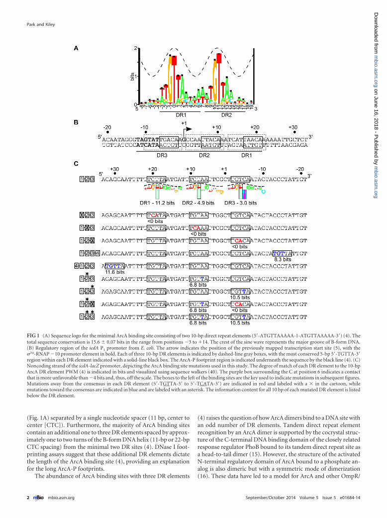
FIG 1 (A) Sequence logo for the minimal ArcA binding site consisting of two 10-bp direct repeat elements (5=-ATGTTAAAAA-1-ATGTTAAAAA-3=) (4). Thetotal sequence conservation is 15.6 � 0.07 bits in the range from positions �3 to �14. The crest of the sine wave represents the major groove of B-form DNA.(B) Regulatory region of the icdA P1 promoter from E. coli. The arrow indicates the position of the previously mapped transcription start site (5), with the�70-RNAP �10 promoter element in bold. Each of three 10-bp DR elements is indicated by dashed-line gray boxes, with the most conserved 5-bp 5=-TGTTA-3=region within each DR element indicated with a solid-line black box. The ArcA-P footprint region is indicated underneath the sequence by the black line (4). (C)Noncoding strand of the icdA-lacZ promoter, depicting the ArcA binding site mutations used in this study. The degree of match of each DR element to the 10-bpArcA DR element PWM (4) is indicated in bits and visualized using sequence walkers (40). The purple box surrounding the C at position 6 indicates a contactthat is more unfavorable than �4 bits and, thus, off the scale. The boxes to the left of the binding sites are the key used to indicate mutations in subsequent figures.Mutations away from the consensus in each DR element (5=-TGTTA-3= to 5=-TCATA-3=) are indicated in red and labeled with a � in the cartoon, whilemutations toward the consensus are indicated in blue and are labeled with an asterisk. The information content for all 10 bp of each mutated DR element is listedbelow the DR element.
Park and Kiley
2 ® mbio.asm.org September/October 2014 Volume 5 Issue 5 e01684-14
m
bio.asm.org
on June 16, 2018 - Published by
mbio.asm
.orgD
ownloaded from

PhoB response regulators that consists of the C-terminal DNAbinding domains of the dimer bound to two DRs in a head-to-tailorientation, connected via a flexible linker to the N-terminal reg-ulatory domains that are oriented symmetrically (head to head)along a common interface (16, 17). The recent structural charac-terization of full-length KdpE, another OmpR/PhoB family mem-ber, bound to its direct repeat site confirmed these different do-main symmetries while identifying an additional level ofasymmetry resulting from intramolecular contacts between thereceiver and DNA binding domains within one KdpE subunit(18). Nevertheless, full-length ArcA-P has been reported to formoligomers (19), as have both the isolated N-terminal andC-terminal domains (16), suggesting that although the minimalDNA binding unit is likely a dimer, as demonstrated for PhoB andOmpR (20–22), oligomerization beyond a dimer may explainbinding to multiple direct repeats.
To gain insight into the physiological function of multiple DRelement binding sites, we evaluated the role of each of the threepredicted 10-bp DR elements (DR1-1, DR2-1, DR3) in ArcA-PDNA binding and repression of icdA, encoding isocitrate dehy-drogenase of the tricarboxylic acid (TCA) cycle. These repeats aredirectionally oriented on the noncoding strand and are numberedon the basis of their order in the 5=-to-3= direction (Fig. 1B). Thisparticular three-DR binding site was chosen because all three DRelements were protected from DNase I cleavage when ArcA isbound (4, 5) despite both strong (DR1 [11.2-bit]) and weak (DR2[4.9-bit] and DR3 [3.0-bit]) matches to the position weight matrix(PWM) (Fig. 1) for a single DR element (4) and because ArcA-P isthe only annotated regulator of the primary icdA promoter (P1)(23). Thus, changes in ArcA DNA binding should change P1 re-pression. In addition, the positions of DR3 adjacent to the �10promoter element and of DR1 and DR2 downstream of the tran-scription start site (TSS) (5) (Fig. 1B) provided an opportunity todetermine if there were any specific effects of DR element posi-tioning on ArcA DNA binding and transcriptional regulation. Tounderstand the contribution of each DR in ArcA repression oficdA, they were mutated toward or away from 5=-TGTTA-3=(Fig. 1C), the most conserved sequence within each ArcA DR el-ement (hereinafter referred to as the consensus) and, based on thePhoB DNA cocrystal structure (15), the region likely contacted byArcA in the major groove. Mutant arcA alleles were used to deter-mine the phosphorylation dependence of this regulation. Ourdata reveal that all three DR elements are important for full anaer-obic repression of icdA and that degeneracy in these DR elementsis important for preserving O2-dependent regulation.
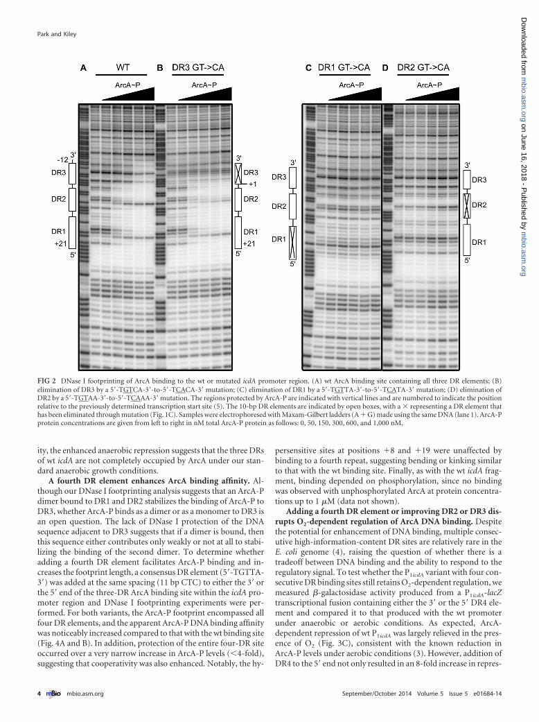
RESULTSAll three DR elements within the icdA promoter contribute toArcA-P DNA binding in vitro. To test the role of each of the threeDR elements in ArcA DNA binding to P1icdA, DNase I footprintingassays were performed using ArcA-P and either the wild type (wt)icdA promoter fragment or those in which each DR element wasindividually disrupted through mutation of highly conserved GTto CA (5=-TGTTA-3= to 5=-TCATA-3=), reducing the informationcontent of each DR element below the theoretical lowest limit ofbinding (0 bits) (24) (Fig. 1C). As previously observed (4), ArcA-Pprotected the three DR elements of the wt promoter region from�12 to �21 relative to the TSS (Fig. 2A). As expected from previ-ous results (4, 5), more ArcA-P (600 nM) was required to observemaximum occupancy of the lower-information-content site,
DR3 (3.0 bits), than the higher-information-content sites,DR1(11.2 bits) and DR2 (4.9 bits) (300 nM). Disruption of eitherDR1 or DR2 eliminated ArcA-P protection of all three DR ele-ments, even at the highest ArcA-P levels tested (Fig. 2C and D). Incontrast, when DR3 was mutated, ArcA-P binding to only DR3was eliminated (Fig. 2B). Furthermore, the amount of ArcA-Prequired for maximal binding of either DR1 or DR2 was not af-fected by disruption of DR3, suggesting that ArcA binding to DR1and DR2 is not enhanced by ArcA-P interactions with DR3 despitethe dependence of DR3 binding on ArcA-P interactions with DR1and DR2.
The mechanisms governing the occupancy of DR3 may becomplex, since we found that an N-terminal His tag variant ofArcA-P also eliminated binding to DR3, but not DR1 and DR2(data not shown), suggesting that protein-protein interactionsmay be important for stabilizing ArcA-P binding to DR3. We alsofound that disruption of DR3 weakened a hypersensitive band atposition �8 within DR2 (Fig. 2A and B). Because DNase I is sen-sitive to the minor groove width (25), this change in hypersensi-tivity may suggest that ArcA-P bends or kinks the DNA to a greaterdegree when bound to all three DR elements than when bound tojust DR1 and DR2. Thus, an ArcA-P dimer bound to DR1 andDR2 may also stabilize the binding of ArcA-P to DR3 by bendingthe DNA.
All three DR elements are required for repression of icdA invivo. How ArcA binding to each DR element contributes to icdArepression was determined by measuring �-galactosidase activityproduced from P1icdA-lacZ transcriptional fusions containing theGT-to-CA (5=-TGTTA-3= to 5=-TCATA-3=) mutations withineach DR element under anaerobic conditions. Basal promoter ac-tivity was not altered by any binding site mutation, as all variantsexhibited the same activity as the wt promoter in the absence ofArcA repression (data not shown). P1icdA was repressed 14-fold byArcA (Fig. 3A). However, disruption of either DR1 or DR2 com-pletely abolished ArcA-dependent repression (Fig. 3A), consistentwith the loss of DNA binding to all three DRs observed by DNaseI footprinting (Fig. 2C and D). In contrast, disruption of DR3,which did not perturb ArcA binding to DR1 and DR2 (Fig. 2B),showed an ~3.5-fold loss in repression (Fig. 3A). This result sug-gests that in vivo occupancy of DR1 and DR2 is sufficient to directa moderate amount of P1icdA repression but that additional occu-pancy of DR3 is required for maximal repression, perhaps becauseit overlaps the �10 promoter element.
The three DRs of P1icdA are suboptimal for maximal repres-sion. Since DR2 and DR3 contain a lower information contentthan DR1, we tested whether mutations that improve the infor-mation content affect repression under anaerobic conditions.Mutation of DR2 toward the consensus (5=-TGTAA-3= to 5=-TGTTA-3=) resulted in a 3-fold increase in anaerobic repression ofP1icdA (Fig. 3B). This repression still depends on DR3 function,since the additional disruption of DR3 (5=-TGTCA-3= to 5=-TCACA-3=) caused the same 3-fold reduction in repression as observedwhen DR3 was disrupted in an otherwise wt icdA sequence(Fig. 3A and B). When just DR3 was mutated toward the consen-sus (5=-TGTCA-3= to 5=-TGTTA-3=), repression was increased6-fold (Fig. 3B). Improving both DR2 and DR3 toward the con-sensus resulted in a level of repression similar to that observedwith a consensus DR3 element alone, suggesting that maximalP1icdA repression by ArcA had been achieved (Fig. 3B). Assumingthat these nucleotide changes simply improve DNA binding affin-
Repressor DNA Binding Architecture
September/October 2014 Volume 5 Issue 5 e01684-14 ® mbio.asm.org 3
m
bio.asm.org
on June 16, 2018 - Published by
mbio.asm
.orgD
ownloaded from
ity, the enhanced anaerobic repression suggests that the three DRsof wt icdA are not completely occupied by ArcA under our stan-dard anaerobic growth conditions.
A fourth DR element enhances ArcA binding affinity. Al-though our DNase I footprinting analysis suggests that an ArcA-Pdimer bound to DR1 and DR2 stabilizes the binding of ArcA-P toDR3, whether ArcA-P binds as a dimer or as a monomer to DR3 isan open question. The lack of DNase I protection of the DNAsequence adjacent to DR3 suggests that if a dimer is bound, thenthis sequence either contributes only weakly or not at all to stabi-lizing the binding of the second dimer. To determine whetheradding a fourth DR element facilitates ArcA-P binding and in-creases the footprint length, a consensus DR element (5=-TGTTA-3=) was added at the same spacing (11 bp CTC) to either the 3= orthe 5= end of the three-DR ArcA binding site within the icdA pro-moter region and DNase I footprinting experiments were per-formed. For both variants, the ArcA-P footprint encompassed allfour DR elements, and the apparent ArcA-P DNA binding affinitywas noticeably increased compared to that with the wt binding site(Fig. 4A and B). In addition, protection of the entire four-DR siteoccurred over a very narrow increase in ArcA-P levels (�4-fold),suggesting that cooperativity was also enhanced. Notably, the hy-
persensitive sites at positions �8 and �19 were unaffected bybinding to a fourth repeat, suggesting bending or kinking similarto that with the wt binding site. Finally, as with the wt icdA frag-ment, binding depended on phosphorylation, since no bindingwas observed with unphosphorylated ArcA at protein concentra-tions up to 1 �M (data not shown).
Adding a fourth DR element or improving DR2 or DR3 dis-rupts O2-dependent regulation of ArcA DNA binding. Despitethe potential for enhancement of DNA binding, multiple consec-utive high-information-content DR sites are relatively rare in theE. coli genome (4), raising the question of whether there is atradeoff between DNA binding and the ability to respond to theregulatory signal. To test whether the P1icdA variant with four con-secutive DR binding sites still retains O2-dependent regulation, wemeasured �-galactosidase activity produced from a P1icdA-lacZtranscriptional fusion containing either the 3= or the 5= DR4 ele-ment and compared it to that produced with the wt promoterunder anaerobic or aerobic conditions. As expected, ArcA-dependent repression of wt P1icdA was largely relieved in the pres-ence of O2 (Fig. 3C), consistent with the known reduction inArcA-P levels under aerobic conditions (3). However, addition ofDR4 to the 5= end not only resulted in an 8-fold increase in repres-
FIG 2 DNase I footprinting of ArcA binding to the wt or mutated icdA promoter region. (A) wt ArcA binding site containing all three DR elements; (B)elimination of DR3 by a 5=-TGTCA-3=-to-5=-TCACA-3=mutation; (C) elimination of DR1 by a 5=-TGTTA-3=-to-5=-TCATA-3=mutation; (D) elimination ofDR2 by a 5=-TGTAA-3=-to-5=-TCAAA-3=mutation. The regions protected by ArcA-P are indicated with vertical lines and are numbered to indicate the positionrelative to the previously determined transcription start site (5). The 10-bp DR elements are indicated by open boxes, with a � representing a DR element thathas been eliminated through mutation (Fig. 1C). Samples were electrophoresed with Maxam-Gilbert ladders (A � G) made using the same DNA (lane 1). ArcA-Pprotein concentrations are given from left to right in nM total ArcA-P protein as follows: 0, 50, 150, 300, 600, and 1,000 nM.
Park and Kiley
4 ® mbio.asm.org September/October 2014 Volume 5 Issue 5 e01684-14
m
bio.asm.org
on June 16, 2018 - Published by
mbio.asm
.orgD
ownloaded from

sion compared to the repression with the wt binding site underanaerobic conditions (Fig. 3B) but also increased repression byArcA under aerobic conditions to nearly the same magnitude ob-served under anaerobic conditions, indicating that ArcA repres-sion of this variant site was no longer O2 sensitive (Fig. 3C). Theaddition of DR4 to the 3= end disrupted promoter function, pre-venting assessment of ArcA repression (data not shown). The sim-plest interpretation of these results is that strengthening bindingaffinity disrupts O2-dependent regulation of ArcA DNA binding.
We also tested whether the degeneracy of DR2 and DR3(Fig. 1C) is important for maintaining O2-dependent regulationof P1icdA by assaying the variants where the sites were mutatedtoward the consensus. Improving DR2 or DR3 toward the con-
FIG 3 Effects of mutations on ArcA-dependent repression of P1icdA. Strainscontaining P1icdA-lacZ were grown in minimal medium with 0.2% glucose, andfold repression was calculated by dividing the �-galactosidase activity of a�arcA strain (e.g., 803 Miller units for wt P1icdA without O2) by the activity ofan arcA� strain (e.g., 57 Miller units for wt P1icdA without O2). The 10-bp DRelements are indicated by open boxes, with a � representing a DR element thathas been eliminated through mutation and an asterisk denoting DR elementsthat have been mutated toward the consensus (Fig. 1C). (A) Effects of muta-tions away from the consensus within each DR element assayed under anaer-obic conditions (�O2). (B) Effects of mutations toward the consensus withineach DR element assayed under anaerobic conditions. The dotted line repre-sents anaerobic ArcA-dependent repression of wt P1icdA. (C) Effects of muta-
(Continued)
Figure Legend Continued
tions toward the consensus within each DR element assayed under aerobicconditions. Error bars represent the standard errors of results from at leastthree independent replicates. We note that P1icdA expression in the constructwith a fourth DR element was about 18% higher in a �arcA background thanin the other strains tested (data not shown).
FIG 4 DNase I footprinting of ArcA binding to the icdA promoter regioncontaining four DR elements. (A) Fourth DR element (5=-TGTTA-3=) located5= of DR1; (B) fourth DR element (5=-TGTTA-3=) located 3= of DR3. Theregions protected by ArcA-P are indicated with vertical lines, with DR ele-ments indicated by open boxes. The numbers indicate positions relative to thepreviously determined transcription start site. Samples were electrophoresedwith Maxam-Gilbert ladders (A � G) made using the same DNA (lane 1).ArcA-P protein concentrations are given from left to right in nM total ArcA-Pprotein as follows: 0, 50, 150, 300, 600, and 1,000 nM.
Repressor DNA Binding Architecture
September/October 2014 Volume 5 Issue 5 e01684-14 ® mbio.asm.org 5
m
bio.asm.org
on June 16, 2018 - Published by
mbio.asm
.orgD
ownloaded from

sensus also increased aerobic P1icdA repression compared to thatwith the wild-type binding site, but the effect was more pro-nounced with a consensus DR3 element (~2.5-fold versus 7-foldrepression) (Fig. 3C). P1icdA with both consensus DR2 and DR3elements was even more repressed by ArcA under aerobic condi-tions (21-fold) than with either consensus site alone, suggestingthat there was an additive effect (Fig. 3C). Together, these resultssuggest that improving binding affinity through the use of con-sensus DR elements disrupts the signal-dependent regulation ofArcA DNA binding, suggesting that the degeneracy of DR2 andDR3 is important for balancing anaerobic repression with O2-dependent relief of repression.
Enhanced ArcA repression is still dependent on phosphory-lation. To test whether the enhanced repression of P1icdA withmutant ArcA binding sites is still dependent on phosphorylation,the aspartate residue at position 54 (site of phosphorylation [19])in the chromosomal copy of arcA was mutated to yield eitheralanine or glutamate, preventing phosphorylation from ArcB(19). The D54A variant reduced the repression of all P1icdA-lacZconstructs compared to that with the wt protein under both aer-obic and anaerobic growth conditions (Fig. 5A). This suggeststhat, independent of the strength of the binding site, repression islargely dependent on the phosphorylated form of ArcA. This re-sult is consistent with the failure of unphosphorylated ArcA tobind to the four DR sites in vitro (data not shown). Thus, theelevated aerobic repression with the strengthened ArcA bindingsites appears to result from increased occupancy of the smallamount of ArcA-P likely present during aerobic conditions.
We expected ArcA(D54E) to similarly reduce the repression ofP1icdA, since this substitution has previously been shown to pre-vent both phosphorylation from ArcB and binding to the pfl pro-moter (19). Surprisingly, ArcA(D54E) still strongly repressedP1icdA constructs with strengthened binding sites even though re-pression of wt P1icdA was largely eliminated; repression of the con-struct with a consensus DR3 element was reduced by only 2-fold,while repression of constructs with consensus DR2 and DR3 ele-ments or a fourth DR element was indistinguishable from thatobserved with the wt protein under anaerobic conditions
(Fig. 5B). Furthermore, under aerobic conditions, ArcA(D54E)repression of P1icdA was increased compared to that of wt ArcA forall binding sites tested (Fig. 5B). Thus, D54E ArcA appears topartially mimic phosphorylated ArcA. An aspartate-to-glutamatesubstitution has previously been shown to elicit constitutive activ-ity in some response regulators (26).
DISCUSSION
The results presented here provide new insight into the plasticityof the DNA elements that can control transcriptional repression.Our data suggest that for icdA, the arrangement of multiple DNAbinding elements appears to be tailored to achieve both sufficientDNA binding affinity and repression by ArcA while maintainingO2-dependent regulation. We propose that the distribution ofDNA binding information across several DR elements may be adesign principle to achieve the appropriate level of repression andto tune the signal-dependent regulation of target genes for bothArcA and other repressors.
Interaction of ArcA with three DR elements of icdA. Ouranalysis of the three DR elements of the icdA promoter indicatethat ArcA-P binding to the lowest-information-content site, DR3,is stabilized by ArcA-P bound to DR1 and DR2, suggestive of acooperative DNA binding mechanism. The lack of an observabledefect in binding to DR1 or DR2 when DR3 was eliminated sug-gests that the cooperative energy is predominantly partitioned to-ward binding of DR3, as expected for sites with large differences inintrinsic levels of binding energy (27). Since ~67 genomic siteshave an odd number of DR elements (4), cooperativity is likely animportant determinant for ArcA binding genome-wide.
An unanswered question is what the stoichiometry of ArcA-Pbinding to DR1, -2, and -3 is. It is possible that ArcA-P binds to theicdA promoter as a dimer of dimers; one dimer binds DR1 andDR2, as depicted in the PhoB and KdpE DNA cocrystal structures(15, 18), and the second dimer binds DR3 but only weakly toadjacent DNA sequence, such that no footprint is observed(Fig. 6). This model is supported by the requirement for phos-phorylation of ArcA to bind to DR3, which is also known to pro-mote dimer formation among OmpR/PhoB response regulators
FIG 5 Phosphorylation dependence of ArcA repression of P1icdA in strains with strengthened ArcA binding sites. The fold repression of P1icdA-lacZ in strainscontaining arcA-FRT-cat-FRT (white bars), arcA(D54A)-FRT-cat-FRT (light-gray bars), or arcA(D54E)-FRT-cat-FRT (dark-gray bars) was determined fromcells grown under anaerobic (A) or aerobic (B) conditions and calculated by dividing the �-galactosidase activity of a �arcA strain by the activity with each of thearcA alleles. Asterisks denote DR elements that have been mutated toward the consensus. Error bars represent the standard errors of results from at least threeindependent replicates.
Park and Kiley
6 ® mbio.asm.org September/October 2014 Volume 5 Issue 5 e01684-14
m
bio.asm.org
on June 16, 2018 - Published by
mbio.asm
.orgD
ownloaded from

(20, 21). However, phosphorylation may simply eliminate an in-teraction between the regulatory and DNA-binding domains, al-lowing ArcA-P to bind as a monomer to DR3 (Fig. 6). Thus, ad-ditional studies are necessary to determine the stoichiometry ofArcA binding to the icdA promoter and whether this stoichiome-try is shared among other ArcA sites with three DR elements.
In either scenario, the predominance of three DR sites with11-bp CTC spacing between each DR in the E. coli genome, to-gether with our previous finding that ArcA-P did not bind to apredicted DR3 element in which the CTC spacing was separatedby an additional bp (4), suggests that protein-protein interactionsbetween correctly spaced subunits is important for cooperativeArcA binding to multiple DRs. Because the C-terminal domain ofArcA binds as a dimer to two adjacent DRs, one can envision thatbinding of an ArcA-P dimer to DR1 and -2 stabilizes a seconddimer or a monomer via protein interactions with DR3. Addition-ally, the hypersensitive site observed when all three (or four) DRelements of icdA were occupied may indicate a requirement forDNA bending to facilitate these protein-protein interactions. Ourfinding that an N-terminally His-tagged variant of ArcA failed tostabilize binding to DR3 suggests that the His tag specifically dis-rupts the mechanism needed to enhance the energetics of DR3binding site occupancy. Additional work is needed to define themolecular interactions that stabilize ArcA-P binding to DR3 ele-ments, but tagged protein variants may not recapitulate this im-portant property of response regulators.
Maximizing repression by binding DR3. The analysis of theeffects of mutations eliminating individual DR elements suggeststhat DR1 and DR2 determine the overall strength of ArcA bindingand that all three DR elements contribute to repression. However,the fact that DR3 overlaps the �10 hexamer as opposed to DR1and DR2, which are located between positions �2 and �22(Fig. 1B), suggests that ArcA binding to DR3 may interfere withthe initial binding of RNA polymerase to form the closed complex,as has been shown for the Lac repressor bound to the Lac operatorthat overlaps the TSS (28). Furthermore, more-effective repres-sion was observed when the Lac and Tet operators overlapped the�10 and �35 promoter elements than when they overlappedthose placed downstream of the TSS (29–31). We do not expectthis particular role of DR3 in icdA to be broadly applicable to allmultiple-DR-element ArcA binding sites because of differences inboth the strengths and the locations of DR3 elements relative tothe TSS (4). Furthermore, because ArcA DNA elements are directrepeats, they can be found either in the same or in the oppositeorientation from the promoter elements, providing additionalflexibility for coding repressor information within a constrainedsequence space. There are several instances where all three ArcADR elements overlap the promoter elements or where DR3 isfound downstream of the TSS and may thus play a role more akinto those of DR1 and DR2 of icdA. This flexible property of re-sponse regulators may also be confined to repressors, since acti-
FIG 6 Model for ArcA-P binding to a three-DR binding site. The orientation and protein-protein contacts between the N- and C-terminal domains within anArcA-P dimer are based on crystallographic data from ArcA and PhoB, respectively (15, 16). Energetically favorable contacts are indicated in blue, while contactslikely to be less favorable are indicated in red. We propose that two ArcA-P dimers bind to a three-DR site in a cooperative manner; the first dimer binds to DR1and DR2, and a second dimer binds to DR3 and adjacent nonspecific sequences. A favorable energetic contribution from the interaction between ArcA-P dimersis likely required to overcome the poor binding affinity of an ArcA-P dimer to DR3 and adjacent nonspecific sequence. Alternatively, it is possible thatdimerization is not required for binding to DR3; ArcA-P may bind to DR3 as a monomer. Potential regions of interaction between ArcA-P molecules in bothscenarios are marked with question marks
Repressor DNA Binding Architecture
September/October 2014 Volume 5 Issue 5 e01684-14 ® mbio.asm.org 7
m
bio.asm.org
on June 16, 2018 - Published by
mbio.asm
.orgD
ownloaded from

vators are likely to be located in specific positions because of thetypical requirement to interact with RNA polymerase.
The combinatorial effect of weak versus strong DR elementscan create a range of responses to ArcA levels. At icdA, the differ-ences in binding affinity of an ArcA-P dimer for DR1/DR2 versusDR3 increases the amount of ArcA-P required for full occupancyin vitro. Assuming that ArcA-P binds the same way in vivo, thisbinding site architecture would extend the sensitivity to ArcA-Plevels by increasing the amount of ArcA-P required for maximalrepression. This property may be a feature shared with other re-sponse regulators, since in the case of OmpR, the binding of anOmpR-P dimer to box 1 (two DR elements) at the ompF andompC promoters occurs at a lower concentration of OmpR-P thandoes binding of OmpR-P to adjacent OmpR boxes (32, 33). Sim-ilarly, binding of PhoB-P to the upstream PhoB box at the pstSpromoter occurs at a lower concentration of PhoB-P than when itbinds to the adjacent, downstream box (34).
On the other hand, promoters with three or more DR elementsof high information content appear to result in ArcA-P occupancyover a very narrow range of protein concentrations. For example,when the disparity in ArcA-P binding affinities at icdA was re-duced by replacing the nonspecific sequence adjacent to DR3 witha fourth DR element, the increase in binding affinity resulted inthe promoter bound by ArcA-P in a highly cooperative manner. Asimilar switch-like occupancy of ArcA-P was also observed for thefour DR elements at the astC promoter (4). Data obtained usingan icdA-lacZ reporter fusion indicate that strengthening DR3 to-ward the consensus likely also enhances binding affinity. Indeed,all three DR elements at the acs promoter are bound over a narrowrange of ArcA-P levels, likely due to a greater energetic contribu-tion to ArcA-P binding provided by a stronger DR3 element (4).Thus, the combinatorial effect of strong or weak DR elements maybe used to either dampen or enhance the concentration-sensitiveoccupancy by ArcA-P compared to that of a site with only twoDRs.
Physiological significance of multiple DR binding sites. Theconfiguration of the ArcA DR elements may also provide a mech-anism for achieving a stepwise response to changes in O2, as sug-gested for OmpR-P dimer binding to the ompF and ompC pro-moters in response to changes in osmolarity (32). For example,under aerobic conditions, ArcA-P levels are likely insufficient forappreciable binding to the icdA promoter; thus, icdA expression ishigh, consistent with the need for isocitrate dehydrogenase forcarbon oxidation in the TCA cycle. However, as O2 becomes lim-iting, ArcA-P levels likely increase (3), perhaps allowing anArcA-P dimer to bind DR1 and DR2, reducing icdA expression toan intermediate level. As O2 is further depleted, ArcA-P levelslikely increase more, and we expect binding to all three DRs, re-ducing icdA expression to levels optimal for anaerobic metabo-lism. Experimental support for this model came from showingthat O2-dependent regulation was disrupted at icdA either by add-ing a fourth DR element or by improving DR2 and DR3. Thus,these results suggest that the degeneracy in DR2 and DR3 and theabsence of a recognizable fourth DR element is important formaintaining the balance between strong, but not complete, anaer-obic repression and O2-dependent relief of repression.
Given the function of the majority of ArcA-repressed operonsin aerobic respiratory metabolism, this balance between high-affinity ArcA-P binding and maintenance of O2-dependent regu-lation is likely widely applicable to genomic ArcA binding sites.
Furthermore, it may explain why there are many three-DR siteswithout identifiable fourth DR elements in the E. coli genome and,additionally, why the average strength of DR elements decreases asthe number of DR elements in the binding site increases (4). Nev-ertheless, both the strength of the promoter and the incorporationof other regulator binding sites should at least partially dictate thespecific ArcA binding site architecture required to achieve optimalregulation, with four DR sites apparently necessary at some pro-moters.
It will also be informative to determine how expression ofother ArcA-dependent promoters (e.g., acs and astC) with astrong DR3 and/or DR4 respond to changes in O2. The saturationof ArcA-P binding to these sites over a narrow range of ArcA-Pconcentrations in vitro (4) suggests that these promoters may re-spond to ArcA-P with a switch-like behavior as cells become lim-ited for O2. For the engineered icdA promoter containing afour-DR site, it seems likely that the affinity of ArcA-P for this siteis so strong that the concentration of ArcA-P present under aero-bic conditions is sufficient to occupy this site so that an O2-dependent change in repression cannot be observed. Nevertheless,our data provide a model for how the ArcA binding site architec-ture may be optimized to achieve regulatory logic schemes notpossible with a canonical two-DR binding site. This plasticity inthe promoter architecture likely plays an important role in linkingthe redox-sensing properties of the ArcAB two-component sys-tem with the fine-tuning of expression of carbon oxidation path-way levels.
The incorporation of plasticity in the binding site architecturesthat we observed for ArcA may be a common regulatory strategyfor other global transcriptional repressors (e.g., Fur, LexA). LikeArcA, Fur binding sites are variable in length (30 to 103 bp) andcontain multiple Fur recognition elements of differing predictedstrengths and locations with respect to the promoter elements(35). Although the physiological basis for this plasticity is un-known, it may similarly impose a differential sensitivity of regula-tory target expression to changes in Fe-Fur concentrations. Fur-thermore, although LexA-regulated genes typically have only oneLexA binding site, differences in the strengths and locations ofthese sites alter the absolute level and sensitivity of expression(36). In a few cases, adjacent LexA sites are bound in a cooperativemanner, further enhancing the sensitivity to changes in signal(36), as hypothesized for the ArcA binding sites located upstreamof acs and astC. Given the conserved dimerization mode and bind-ing of direct repeat DNA sites among response regulators withinthe OmpR/PhoB family (16), this architectural plasticity may be acommon regulatory strategy, particularly for regulators that act asrepressors at many targets.
MATERIALS AND METHODSStrain construction. An icdA promoter-lacZ fusion was constructed asdescribed previously (37) by amplifying the region from �50 to �330with respect to the start of translation using primers flanked by XhoI orBamHI restriction sites. The icdA fragment contains two promoters: onewhose expression is dependent on ArcA (P1) and a second whose expres-sion is dependent on FruR (P2) (5, 38). To examine icdA expression fromonly P1, transcription from P2 was eliminated using QuikChange site-directed mutagenesis (Stratagene) as described previously (39) to mutatethe �10 site from 5=-CATTAT-3= to 5=-CGGTGA-3=, generatingpPK9476. Mutations within the ArcA binding site of the icdA promoterwere similarly generated using pPK9476 as a template (mutations arenumbered with respect to P1 in Table 1). These lacZ promoter constructs
Park and Kiley
8 ® mbio.asm.org September/October 2014 Volume 5 Issue 5 e01684-14
m
bio.asm.org
on June 16, 2018 - Published by
mbio.asm
.orgD
ownloaded from

were then recombined into the chromosomal lac operon as previouslydescribed (37) and then transduced using P1 vir into MG1655 andPK9416 to form the strain derivatives listed in Table 1.
Chromosomally encoded arcA mutants in which aspartate at posi-tion 54 was replaced with glutamate or alanine were constructed inseveral steps. First, the arcA open reading frame (codons 1 to 238) wasamplified using primers flanked by HindIII and BamHI and clonedinto pBR322, generating pPK9965. The cat cassette from pKD32,which has flanking FRT (FLP recognition target) sites, was then clonedinto the BamHI site, 6 bp after the arcA termination codon. The arcAgene on the resulting plasmid, pPK9966, was then mutated using
QuikChange (Stratagene) site-directed mutagenesis to create theD54A and D54E mutants. The arcA-cat fragments were PCR amplifiedusing a primer with homology to the region upstream of arcA(5=-GGTAGCAAACATGCAGACCCCGCACATTCTTATCG-3=) anda primer with homology to the region downstream of arcA (5=-GCGCCGTTTTTTTTGACGGTGGTAAAGCCGATTAGTGTAGGCTGGAGCTGCTTC-3=), and the DNA was electroporated into BW25993/pKD46. The correct recombinants were selected for chloramphenicol(Cm) resistance, confirmed with DNA sequencing, and then trans-duced with P1 vir into the desired icdA promoter-lacZ fusion strains(Table 1). Placement of the cat cassette downstream of arcA did not
TABLE 1 Strains and plasmids used in this study
Strain or plasmid Description Source or reference
StrainsMG1655 F� �� rph-1 This laboratoryPK9416 MG1655 �arcA 4PK9483 MG1655 PicdA(-58GGTGA-54)-lacZ 4PK9484 PK9416 PicdA(-58GGTGA-54)-lacZ 4PK9494 MG1655 PicdA(-58GGTGA-54, 19TG20)-lacZ This studyPK9495 PK9416 PicdA(-58GGTGA-54, 19TG20)-lacZ This studyPK9486 MG1655 PicdA(-58GGTGA-54, 8TG9)-lacZ This studyPK9487 PK9416 PicdA(-58GGTGA-54, 8TG9)-lacZ This studyPK9496 MG1655 PicdA(-58GGTGA-54, -4TG-3)-lacZ This studyPK9497 PK9416 PicdA(-58GGTGA-54, -4TG-3)-lacZ This studyPK9915 MG1655 PicdA(-58GGTGA-54, 29AACA32)-lacZ This studyPK9916 PK9416 PicdA(-58GGTGA-54, 29AACA32)-lacZ This studyPK9917 MG1655 PicdA(-58GGTGA-54, -15ACA-13)-lacZ This studyPK9918 PK9416 PicdA(-58GGTGA-54, -15ACA-13)-lacZ This studyPK9924 MG1655 PicdA(-58GGTGA-54, -5A)-lacZ This studyPK9925 PK9416 PicdA(-58GGTGA-54, -5A)-lacZ This studyPK9941 MG1655 PicdA(-58GGTGA-54, 7A)-lacZ This studyPK9942 PK9416 PicdA(-58GGTGA-54, 7A)-lacZ This studyPK9943 MG1655 PicdA(-58GGTGA-54, 7A, -5A)-lacZ This studyPK9944 PK9416 PicdA(-58GGTGA-54, 7A, -5A)-lacZ This studyPK10967 MG1655 PicdA(-58GGTGA-54, 7A, -4TG-3)-lacZ This studyPK10968 PK9416 PicdA(-58GGTGA-54, 7A, -4TG-3)-lacZ This studyBW25993 lacIq �lacZWJ16 hsdR514 �araBADAH33 �rhaBADLD78 45PK9970 PK9483 arcA::cat This studyPK9973 PK9915 arcA::cat This studyPK9971 PK9924 arcA::cat This studyPK9972 PK9943 arcA::cat This studyPK9980 PK9483 arcA-D54A::cat This studyPK9983 PK9915 arcA-D54A::cat This studyPK9981 PK9924 arcA-D54A::cat This studyPK9982 PK9943 arcA-D54A::cat This studyPK9975 PK9483 arcA-D54E::cat This studyPK9978 PK9915 arcA-D54E::cat This studyPK9976 PK9924 arcA-D54E::cat This studyPK9977 PK9943 arcA-D54E::cat This study
PlasmidspKD46 Phage � gam-bet-exo genes under ParaB control B. L. WannerpKD13 FRT-kan-FRT K. A. Datsenko and B. L. WannerpKD32 FRT-cat-FRT B. L. WannerpPK7035 kan gene from pHP45� and BamHI-NdeI fragment from pRS1553 into pBR322 37pPK9476 pPK7035 PicdA(-58GGTGA-54)-lacZ 4pPK9477 pPK7035 PicdA(-58GGTGA-54 19TG20)-lacZ This studypPK9908 pPK7035 PicdA(-58GGTGA-54 8TG9)-lacZ This studypPK9909 pPK7035 PicdA(-58GGTGA-54 -4TG-3)-lacZ This studypPK9913 pPK7035 PicdA(-58GGTGA-54 29AACA32)-lacZ This studypPK9914 pPK7035 PicdA(-58GGTGA-54 -15ACA-13)-lacZ This studypPK15001 pPK7035 PicdA(-58GGTGA-54, 7A, -4TG-3)-lacZ This studypPK9965 arcA in pBR322 This studypPK9966 BamHI FRT-cat-FRT in pPK9965 This studypPK9431 Apr; His6-arcA cloned into the NheI and XhoI sites of pET-21d 4
Repressor DNA Binding Architecture
September/October 2014 Volume 5 Issue 5 e01684-14 ® mbio.asm.org 9
m
bio.asm.org
on June 16, 2018 - Published by
mbio.asm
.orgD
ownloaded from

alter ArcA activity, as icdA promoter-lacZ activity was comparable tothat of the wt arcA� strain for all binding sites tested (Fig. 3B and C and5A and B).
Determination of the information content of DR elements. A 10-bpArcA DR element, PWM, derived from the conservation of bases withinaligned DR1 and DR2 elements from 128 sequences bound by ArcA in vivo(4) was used to guide the design of binding site mutations. The informa-tion content of each mutant DR element was determined by the scanprogram (24) and is indicated in bits (Fig. 1C). Greater information con-tent should reflect stronger ArcA binding (24). Sequence walkers (40)were used to visualize how DR elements were evaluated by the PWM.Nucleotides extending upwards represent favorable DNA contacts, whileletters extending downward represent unfavorable contacts.
�-Galactosidase assays. All strains were grown in MOPS minimalmedium (41) with 0.2% glucose at 37°C and sparged with a gas mix of95% N2 and 5% CO2 (anaerobic) or 70% N2, 5% CO2, and 25% O2
(aerobic). Cells were harvested during mid-log growth (optical density at600 nm [OD600] of ~0.3 on a PerkinElmer Lambda 25 UV/visible-lightspectrophotometer). To terminate cell growth and any further proteinsynthesis, chloramphenicol (final concentration, 20 �g/ml) or tetracy-cline (final concentration, 10 �g/ml) was added, and cells were placed onice until assayed for �-galactosidase activity (42). �-Galactosidase assayswere repeated at least three times, and fold repression was calculated bydividing the �-galactosidase activity of a �arcA strain by the activity of anarcA� strain. Standard errors for data plotted as “fold repression” werecalculated using a formula for propagation of standard error (43).
Overexpression and purification of His6-ArcA. E. coli BL21(DE3)plysS, containing the PK9431 gene, was grown at 37°C until an OD600 of~0.4 was reached. A final concentration of 1 mM isopropyl �-D-1-thiogalactopyranoside (IPTG) was added, and cells were incubated at30°C. Cells were harvested, suspended in 5 mM imidazole buffer contain-ing 20 mM Tris-Cl (pH 7.9) and 0.5 M NaCl, and lysed by sonication.His6-ArcA was isolated from cell lysates by passing them over a Ni-nitrilotriacetic acid (NTA) column preequilibrated with 5 mM imidazole,washing the column extensively with the same buffer and then with 20 and50 mM imidazole, and then eluting with 100 mM imidazole. Fractionscontaining the overexpressed His6-ArcA, determined by electrophoresis,were dialyzed against 50 mM Tris-Cl, pH 7.5, 0.1 mM dithiothreitol(DTT), 0.1 mM EDTA, and 0.2 M NaCl. The His6 tag was removed fromArcA by overnight incubation with tobacco etch virus (TEV) protease at4°C and passage over a Ni-NTA-agarose column (Qiagen). The proteinconcentration of ArcA (reported here as monomers) was determined aspreviously described (4).
DNase I footprinting. icdA promoter fragments were isolated frompPK9476, pPK9477, pPK9908, pPK9909, pPK9913, pPK9914, andpPK15001 (Table 1) after digestion with XhoI and BamHI. Sequenaseversion 2.0 (USB Scientific) was used to 3=-end radiolabel the BamHI endof the fragment with [�-32P]dGTP (PerkinElmer). Labeled DNA frag-ments were isolated from a nondenaturing 5% acrylamide gel and weresubsequently purified with Elutip-d columns (Schleicher and Schuell).ArcA was phosphorylated by incubating it with 50 mM disodium car-bamyl phosphate (Sigma-Aldrich) in 50 mM Tris, pH 7.9, 150 mM NaCl,and 10 mM MgCl2 for 1 h at 30°C (6) and immediately used in the bindingassays. Footprinting assays were performed by incubating phosphorylatedArcA with labeled DNA (~5 nM) for 10 min at 30°C in 40 mM Tris(pH 7.9), 30 mM KCl, 100 �g/ml bovine serum albumin (BSA), and 1 mMDTT followed by the addition of 2 �g/ml DNase I (Worthington) for 30 s.The DNase I reaction was terminated by the addition of sodium acetateand EDTA to final concentrations of 300 mM and 20 mM, respectively.The reaction mix was ethanol precipitated, resuspended in urea loadingdye, heated for 60 s at 90°C, and loaded onto a 7 M urea– 8% polyacryl-amide gel in 0.5� Tris-borate-EDTA (TBE) buffer. An A�G ladder wasmade by formic acid modification of the radiolabeled DNA, followed bypiperidine cleavage (44). The reaction products were visualized by phos-phorimaging.
ACKNOWLEDGMENTS
We thank Wilma Ross for assistance with DNase I footprinting experi-ments and Erin Mettert for critically reading the manuscript.
This work was supported by NIH grant GM45844 to P.J.K. D.M.P. wassupported in part by the DOE BACTER Program (grant DE-FG02-04ER25627).
REFERENCES1. Malpica R, Sandoval GR, Rodríguez C, Franco B, Georgellis D. 2006.
Signaling by the arc two-component system provides a link between theredox state of the quinone pool and gene expression. Antioxid. RedoxSignal. 8:781–795. http://dx.doi.org/10.1089/ars.2006.8.781.
2. Malpica R, Franco B, Rodriguez C, Kwon O, Georgellis D. 2004.Identification of a quinone-sensitive redox switch in the ArcB sensor ki-nase. Proc. Natl. Acad. Sci. U. S. A. 101:13318 –13323. http://dx.doi.org/10.1073/pnas.0403064101.
3. Rolfe MD, Ter Beek A, Graham AI, Trotter EW, Asif HM, SanguinettiG, de Mattos JT, Poole RK, Green J. 2011. Transcript profiling andinference of Escherichia coli K-12 ArcA activity across the range of physi-ologically relevant oxygen concentrations. J. Biol. Chem. 286:10147–10154. http://dx.doi.org/10.1074/jbc.M110.211144.
4. Park DM, Akhtar MS, Ansari AZ, Landick R, Kiley PJ. 2013. Thebacterial response regulator ArcA uses a diverse binding site architectureto regulate carbon oxidation globally. PLoS Genet. 9:e1003839. http://dx.doi.org/10.1371/journal.pgen.1003839.
5. Chao G, Shen J, Tseng CP, Park SJ, Gunsalus RP. 1997. Aerobicregulation of isocitrate dehydrogenase gene (icd) expression in Escherichiacoli by the arcA and fnr gene products. J. Bacteriol. 179:4299 – 4304.
6. Lynch AS, Lin EC. 1996. Transcriptional control mediated by the ArcAtwo-component response regulator protein of Escherichia coli: character-ization of DNA binding at target promoters. J. Bacteriol. 178:6238 – 6249.
7. Cunningham L, Georgellis D, Green J, Guest JR. 1998. Co-regulation oflipoamide dehydrogenase and 2-oxoglutarate dehydrogenase synthesis inEscherichia coli: characterisation of an ArcA binding site in the lpd pro-moter. FEMS Microbiol. Lett. 169:403– 408. http://dx.doi.org/10.1111/j.1574-6968.1998.tb13347.x.
8. Pellicer MT, Lynch AS, De Wulf P, Boyd D, Aguilar J, Lin EC. 1999. Amutational study of the ArcA-P binding sequences in the aldA promoterof Escherichia coli. Mol. Gen. Genet. 261:170 –176. http://dx.doi.org/10.1007/s004380050954.
9. Pellicer MT, Fernandez C, Badía J, Aguilar J, Lin EC, Baldom L. 1999.Cross-induction of glc and ace operons of Escherichia coli attributable topathway intersection. Characterization of the glc promoter. J. Biol. Chem.274:1745–1752. http://dx.doi.org/10.1074/jbc.274.3.1745.
10. Drapal N, Sawers G. 1995. Purification of ArcA and analysis of its specificinteraction with the pfl promoter-regulatory region. Mol. Microbiol. 16:597– 607. http://dx.doi.org/10.1111/j.1365-2958.1995.tb02422.x.
11. Gerasimova AV, Gelfand MS, Makeev VY, Mironov AA, Favorov AV.2003. ArcA regulator of gamma-proteobacteria identification of the bind-ing signal and description of the regulon. Biophysics 48:S21–S25.
12. McGuire AM, De Wulf P, Church GM, Lin EC. 1999. A weight matrixfor binding recognition by the redox-response regulator ArcA-P of Esch-erichia coli. Mol. Microbiol. 32:219 –221. http://dx.doi.org/10.1046/j.1365-2958.1999.01347.x.
13. Liu X, De Wulf P. 2004. Probing the ArcA-P modulon of Escherichia coliby whole genome transcriptional analysis and sequence recognition pro-filing. J. Biol. Chem. 279:12588 –12597. http://dx.doi.org/10.1074/jbc.M313454200.
14. Wang X, Gao H, Shen Y, Weinstock GM, Zhou J, Palzkill T. 2008. Ahigh-throughput percentage-of-binding strategy to measure binding en-ergies in DNA-protein interactions: application to genome-scale site dis-covery. Nucleic Acids Res. 36:4863– 4871. http://dx.doi.org/10.1093/nar/gkn477.
15. Blanco AG, Sola M, Gomis-Rüth FX, Coll M. 2002. Tandem DNArecognition by PhoB, a two-component signal transduction transcrip-tional activator. Structure 10:701–713. http://dx.doi.org/10.1016/S0969-2126(02)00761-X.
16. Toro-Roman A, Mack TR, Stock AM. 2005. Structural analysis andsolution studies of the activated regulatory domain of the response regu-lator ArcA: a symmetric dimer mediated by the alpha4-beta5-alpha5 face.J. Mol. Biol. 349:11–26. http://dx.doi.org/10.1016/j.jmb.2005.03.059.
17. Gao R, Mack TR, Stock AM. 2007. Bacterial response regulators: versatile
Park and Kiley
10 ® mbio.asm.org September/October 2014 Volume 5 Issue 5 e01684-14
m
bio.asm.org
on June 16, 2018 - Published by
mbio.asm
.orgD
ownloaded from

regulatory strategies from common domains. Trends Biochem. Sci. 32:225–234. http://dx.doi.org/10.1016/j.tibs.2007.03.002.
18. Narayanan A, Kumar S, Evrard AN, Paul LN, Yernool DA. 2014. Anasymmetric heterodomain interface stabilizes a response regulator-DNAcomplex. Nat. Commun 5:3282. http://dx.doi.org/10.1038/ncomms4282.
19. Jeon Y, Lee YS, Han JS, Kim JB, Hwang DS. 2001. Multimerization ofphosphorylated and non-phosphorylated ArcA is necessary for the re-sponse regulator function of the Arc two-component signal transductionsystem. J. Biol. Chem. 276:40873– 40879. http://dx.doi.org/10.1074/jbc.M104855200.
20. Mack TR, Gao R, Stock AM. 2009. Probing the roles of the two differentdimers mediated by the receiver domain of the response regulator PhoB. J.Mol. Biol. 389:349 –364. http://dx.doi.org/10.1016/j.jmb.2009.04.014.
21. Barbieri CM, Wu T, Stock AM. 2013. Comprehensive analysis of OmpRphosphorylation, dimerization, and DNA binding supports a canonicalmodel for activation. J. Mol. Biol. 425:1612–1626. http://dx.doi.org/10.1016/j.jmb.2013.02.003.
22. Ritzefeld M, Walhorn V, Kleineberg C, Bieker A, Kock K, Herrmann C,Anselmetti D, Sewald N. 2013. Cooperative binding of PhoB(DBD) to itscognate DNA sequence—a combined application of single-molecule andensemble methods. Biochemistry 52:8177– 8186. http://dx.doi.org/10.1021/bi400718r.
23. Keseler IM, Collado-Vides J, Santos-Zavaleta A, Peralta-Gil M, Gama-Castro S, Muñiz-Rascado L, Bonavides-Martinez C, Paley S, Krummen-acker M, Altman T, Kaipa P, Spaulding A, Pacheco J, Latendresse M,Fulcher C, Sarker M, Shearer AG, Mackie A, Paulsen I, Gunsalus RP,Karp PD. 2011. EcoCyc: a comprehensive database of Escherichia colibiology. Nucleic Acids Res. 39:D583–D590. http://dx.doi.org/10.1093/nar/gkq1143.
24. Schneider TD. 1997. Information content of individual genetic se-quences. J. Theor. Biol. 189:427– 441. http://dx.doi.org/10.1006/jtbi.1997.0540.
25. Drew HR, Travers AA. 1984. DNA structural variations in the E. coli tyrTpromoter. Cell 37:491–502. http://dx.doi.org/10.1016/0092-8674(84)90379-9.
26. Smith JG, Latiolais JA, Guanga GP, Pennington JD, Silversmith RE,Bourret RB. 2004. A search for amino acid substitutions that universallyactivate response regulators. Mol. Microbiol. 51:887–901. http://dx.doi.org/10.1046/j.1365-2958.2003.03882.x.
27. Ackers GK, Shea MA, Smith FR. 1983. Free energy coupling withinmacromolecules. The chemical work of ligand binding at the individualsites in co-operative systems. J. Mol. Biol. 170:223–242. http://dx.doi.org/10.1016/S0022-2836(83)80234-4.
28. Schlax PJ, Capp MW, Record MT, Jr.. 1995. Inhibition of transcriptioninitiation by lac repressor. J. Mol. Biol. 245:331–350. http://dx.doi.org/10.1006/jmbi.1994.0028.
29. Elledge SJ, Davis RW. 1989. Position and density effects on repression bystationary and mobile DNA-binding proteins. Genes Dev. 3:185–197.http://dx.doi.org/10.1101/gad.3.2.185.
30. Lanzer M, Bujard H. 1988. Promoters largely determine the efficiency of
repressor action. Proc. Natl. Acad. Sci. U. S. A. 85:8973– 8977. http://dx.doi.org/10.1073/pnas.85.23.8973.
31. Cox RS, III, Surette MG, Elowitz MB. 2007. Programming gene expres-sion with combinatorial promoters. Mol. Syst. Biol. 3:145. http://dx.doi.org/10.1038/msb4100187.
32. Yoshida T, Qin L, Egger LA, Inouye M. 2006. Transcription regulationof ompF and ompC by a single transcription factor, OmpR. J. Biol. Chem.281:17114 –17123. http://dx.doi.org/10.1074/jbc.M602112200.
33. Rampersaud A, Harlocker SL, Inouye M. 1994. The OmpR protein ofEscherichia coli binds to sites in the ompF promoter region in a hierarchicalmanner determined by its degree of phosphorylation. J. Biol. Chem. 269:12559 –12566.
34. Makino K, Amemura M, Kawamoto T, Kimura S, Shinagawa H, NakataA, Suzuki M. 1996. DNA binding of PhoB and its interaction with RNApolymerase. J. Mol. Biol. 259:15–26. http://dx.doi.org/10.1006/jmbi.1996.0298.
35. Chen Z, Lewis KA, Shultzaberger RK, Lyakhov IG, Zheng M, Doan B,Storz G, Schneider TD. 2007. Discovery of fur binding site clusters inEscherichia coli by information theory models. Nucleic Acids Res. 35:6762– 6777. http://dx.doi.org/10.1093/nar/gkm631.
36. Butala M, Zgur-Bertok D, Busby SJ. 2009. The bacterial LexA transcrip-tional repressor. Cell. Mol. Life Sci. 66:82–93. http://dx.doi.org/10.1007/s00018-008-8378-6.
37. Kang Y, Weber KD, Qiu Y, Kiley PJ, Blattner FR. 2005. Genome-wideexpression analysis indicates that FNR of Escherichia coli K-12 regulates alarge number of genes of unknown function. J. Bacteriol. 187:1135–1160.http://dx.doi.org/10.1128/JB.187.3.1135-1160.2005.
38. Prost JF, Nègre D, Oudot C, Murakami K, Ishihama A, Cozzone AJ,Cortay JC. 1999. Cra-dependent transcriptional activation of the icd geneof Escherichia coli. J. Bacteriol. 181:893– 898.
39. Nesbit AD, Giel JL, Rose JC, Kiley PJ. 2009. Sequence-specific binding toa subset of IscR-regulated promoters does not require IscR Fe-S clusterligation. J. Mol. Biol. 387:28 – 41. http://dx.doi.org/10.1016/j.jmb.2009.01.055.
40. Schneider TD. 1997. Sequence walkers: a graphical method to display howbinding proteins interact with DNA or RNA sequences. Nucleic Acids Res.25:4408 – 4415. http://dx.doi.org/10.1093/nar/25.21.4408.
41. Neidhardt FC, Bloch PL, Smith DF. 1974. Culture medium for entero-bacteria. J. Bacteriol. 119:736 –747.
42. Miller JH. 1972. Experiments in molecular genetics. Cold Spring HarborLaboratory, Cold Spring Harbor, NY.
43. Ku HH. 1966. Notes on the use of propagation of error formulas. J. Res.Natl. Bureau Stand. 70C:263–273.
44. Maxam AM, Gilbert W. 1980. Sequencing end-labeled DNA with base-specific chemical cleavages. Methods Enzymol. 65:499 –560. http://dx.doi.org/10.1016/S0076-6879(80)65059-9.
45. Datsenko KA, Wanner BL. 2000. One-step inactivation of chromosomalgenes in Escherichia coli K-12 using PCR products. Proc. Natl. Acad. Sci.U. S. A. 97:6640 – 6645. http://dx.doi.org/10.1073/pnas.120163297.
Repressor DNA Binding Architecture
September/October 2014 Volume 5 Issue 5 e01684-14 ® mbio.asm.org 11
m
bio.asm.org
on June 16, 2018 - Published by
mbio.asm
.orgD
ownloaded from