the interplay between zinc, vitamin d and, il-17 in
TRANSCRIPT
Research ArticleThe Interplay between Zinc, Vitamin D and, IL-17 inPatients with Chronic Hepatitis C Liver Disease
Randa Reda,1 Amal A. Abbas,1 Mai Mohammed,1 Shahira F. El Fedawy,1 Hala Ghareeb,1
Rania H. El Kabarity,1 Rania A. Abo-Shady,1 and Doaa Zakaria2
1Clinical Pathology Department, Faculty of Medicine, Ain Shams University, Abbassia Square, P.O. Box 11566, Cairo, Egypt2Tropical Medicine Department, Faculty of Medicine, Ain Shams University, Abbassia Square, P.O. Box 11566, Cairo, Egypt
Correspondence should be addressed to Amal A. Abbas; dr amal [email protected]
Received 25 November 2014; Revised 2 January 2015; Accepted 4 January 2015
Academic Editor: Hasan Tarık Atmaca
Copyright © 2015 Randa Reda et al. This is an open access article distributed under the Creative Commons Attribution License,which permits unrestricted use, distribution, and reproduction in any medium, provided the original work is properly cited.
Objectives. To assess zinc (Zn) and vitamin D (Vit. D) status in chronic Hepatitis C virus- (HCV) infected patients and theirrelationship to interleukin- (IL-) 17 and disease severity and then investigate whether Zn and Vit. D3 modulate IL-17 expressionin chronic HCV patients. Methods. Seventy patients and fifty healthy subjects were investigated. Serum levels of Zn, Vit. D, andIL-17 were assessed in the patients group and subgroups. Patients lymphocytes were activated in vitro in the presence or absence ofZn or Vit. D3 and then intracellular IL-17 production was assessed using flow cytometry. Results. Zn and Vit. D were significantlydecreased in HCV patients. Increasing disease severity leads to more reduction in Zn level opposed by increasing IL-17 level. Znpotently reduced IL-17 production in a dose-related fashion; however it did not exert any toxic effects. Although Vit. D apparentlyincreases IL17 expression, it is unclear whether it is due to its toxic effect on cell count or lack of definite association between Vit.D and both IL-17 and disease severity. Conclusions. This study demonstrates that Zn modulates IL-17 expression and provides arationale for evaluating this compound as a supplementary agent in the treatment of chronic HCV.
1. Introduction
Hepatitis C virus infects primarily the hepatocytes, leads tothe development of fibrosis or cirrhosis of the liver, and is asignificant risk factor for the development of hepatocellularcarcinoma (HCC). Previous studies have demonstrated that Tcell immunoregulatory cytokines contribute to liver damage[1].
Human interleukin-17 (IL-17) producing CD4 T cells,Th17, comprise a proinflammatory T cell subset. Previousstudies have identified Th17 as a known arm of the CD4+ T-cell effector response [2] and several key cytokines, includingIL-1, IL-6, tumor necrosis factor alpha, and IL-23, createa cytokine milieu that regulates the differentiation andexpansion of human Th17 cell [3]. IL-17A can mobilize,recruit, and activate neutrophils, leading to massive tissueinflammation, and promote the progression of autoimmunedisease. Furthermore, serum IL-17 levels are increased andserve as a marker of the severity of acute hepatic injury [4, 5].
Zinc, one of the essential trace elements, is required bymany enzymes and transcription factors for their activity orthe maintenance of their structure. It has a variety of effectsin the immune system. Zn deficiency causes an imbalancebetween Th1 and Th2 function in periphery. Production ofIFN-gamma and IL-2, Th1 products, is decreased, whereasproduction of IL-4, IL-6, and IL-10 Th2 products is notaffected [6].
Themain zincmetabolismoccurs in the liver hepatocytes.In patients with Zn deficiency, reduced Zn concentrationsin the liver are one of the causes of impaired hepatocytesregeneration [7]. It has been demonstrated that Zn mayplay an important role as a negative regulator of HCVreplication in genome length RNA-replicating cells.Thus zincsupplementations appear to offer a novel approach for furtherstrategies in treatment of intractable chronic hepatitis C [8].Zinc uses are claimed to suppress Th17-mediated autoim-mune diseases at least in part by inhibiting the developmentof Th17 cells via attenuating STAT3 activation [9].
Hindawi Publishing CorporationJournal of Immunology ResearchVolume 2015, Article ID 846348, 11 pageshttp://dx.doi.org/10.1155/2015/846348
2 Journal of Immunology Research
Vitamin D is emerging as a critical factor involved in theregulation of the immune system, inflammatory response,and fibrogenesis [10]. Several studies on Egyptian patientswith hepatitis C virus showed a significant reduction ofVit. D and its active metabolite in HCVg4-infected patientscompared to healthy controls [11, 12]. Moreover they founda significant negative correlation between viral load and Vit.D status. Interestingly, low vitamin D levels have been relatedto poor liver function and stage of cirrhosis [10, 13]. VitaminD was shown to reduce the expression of collagen andprofibrotic factors leading to decreased fibrosis [14].The effectof Vit. D on the behavior of Th17 cells has been investigatedin different diseases and Vit. D suppresses the expressionof IL-17 and IL-23 [12, 15, 16], the regulatory effect on Th17cells by Vit. D occurs through the reduction of retinoic acid-related orphan receptor (ROR)𝛾t expression [15]. Therefore,this study was designed to assess Zinc and Vit. D status inchronic HCV-infected patients and its relationship to levelsof IL-17 as immune inflammatorymediators and to clarify theeffect of Zn and Vit. D in modulating the expression of IL-17in vitro.
2. Materials and Methods
2.1. Subjects. Prior to initiation, this study received approvalby the Ethical Committee of the Faculty of Medicine, AinShams University. The study recruited 70 patients withchronic HCV infection and they were diagnosed retrospec-tively by positivity of PCR and enzyme-linked immunosor-bent assay (ELISA) HCV antibodies, who were selected fromoutpatients and inpatients of Internal Medicine and TropicalMedicine Departments at Ain Shams University Hospitals.In addition fifty healthy normal persons matched for ageand sex as a control group were also included in the study.Inclusion criteria were based on a history of liver diseasewith HCV genotype 4 infection (as new patients or underfollow-up). Patients with other causes of viral hepatitis:hepatitis B virus (HBV) or coinfection with HBV and humanimmunodeficiency virus (HIV), cytomegalovirus (CMV),and Epstein–Barr virus (EBV), or having criteria suggestiveof fatty liver: Body Mass Index (BMI) >35, uncontrolled DM(HbA1c > 7), or history of taking hepatotoxic drugs for theprevious 6 months, were excluded. BMI was calculated asweight in kilograms divided by the square of height in meters[17]. (Obesity was defined as BMI > 30 kg/m2.)
All included patients underwent tests for liver function(ALT, AST, and bilirubin using Beckman Synchron CX7Delta Clinical System, prothrombin time, and INR usingstago analyzer), assessment of HCV levels, and abdominalultrasonography for detection of cirrhosis. Based on ultra-sonography results and liver function tests, the patients wereclassified into three subgroups: Group 1 includes 16 recentlydiagnosed chronically HCV-infected patients showing noevidence of hepatic cirrhosis or liver cell failure. Group 2includes 37 compensated chronically HCV-infected patientsshowing an evidence of hepatic cirrhosis but no evidenceof liver cell failure. Group 3 includes 17 decompensated
chronically HCV-infected patients showing an evidence ofhepatic cirrhosis and liver cell failure.
2.2. Measurement of Serum Interleukin-17, Zinc Level, and 25-OH Vitamin D. After subclassification, venous blood sam-ples (5mL) were obtained (after overnight fasting) from allpatients and controls. Samples were allowed to clot and serawere then separated by centrifugation (3500 rpm, 20min,25∘C) and then stored at −20∘C until used for serum analysisof the various parameters outlined below.
Commercially available ELISA kits (Labs Biotech, Inc,USA) were used for quantitative analysis of interleukin-17while determination of zinc level was done by zinc colorimet-ricmethod (kit supplied fromQuımicaClınicaAplicada S.A).Measurement of serum 25-OH vitamin D (as 25-OH vitaminD is themajor circulating form of vitamin D and is used as anindicator of vitamin D status) using a commercially available(ELISA) kit supplied by Calbiotech’s, Inc, USA, was done for20 selected patients from Group 2. Vitamin D deficiency wasdefined as a 25(OH) D serum level < 12 ng/mL, vitamin Dinsufficiency as 25(OH) D level 12–32 ng/mL, and vitamin Dsufficiency as > 32 ng/mL [18].
2.3. Assessment of Zn and Vit. D Effect on the Expression ofIL-17 in Cultured PBMCs
2.3.1. Reagents. Phorbol 12-myristate 13-acetate (PMA) andionomycin (IO) were both purchased from Serva Elec-trophoresis Germany, zinc sulphate was purchased fromElnasr, Pharmaceutical Chemical Industries, Egypt, vitaminD3 (Cholecalciferol) from Memphis, Pharmaceutical Chem-ical Industries, Egypt, and Intracellular Fixation & Permeabi-lization Buffer (plus Brefeldin A) kit from eBioscience, SanDiego, CA, USA.
2.3.2. Monoclonal Antibodies. Antibodies used includedCD3-PECy5, CD4-FITC, IL17-PE, and PE isotype control(eBioscience, San Diego, CA, USA).
2.3.3. Preparation of Peripheral Blood Mononuclear Cells(PBMCs). Twenty mL of peripheral blood were obtainedby venipuncture from the 20 selected patients of Group2 patients and collected into sterile EDTA tubes. ThePBMCs were immediately separated by density gradientcentrifugation over Ficoll–Hypaque (Lonza, BioWhittaker)and then washed twice with RPMI 1640. Cell count andviability were determined utilizing Guava ViaCount FlexReagent for Flow Cytometry (Merck Millipore, France).Viability was exceeding 95% in all studied cases. PBMCswere suspended in RPMI 1640 medium, supplementedwith 2mM l-glutamine, 25mM HEPES, 100U/mL benzyl-penicillin, 0.1mg/mL streptomycin, and 10%ABserum (com-plete medium) (Lonza, BioWhittaker). All cultures wereincubated.
2.3.4. Intracellular Cytokine Staining and Flow Cytometry.Peripheral blood mononuclear cells (PBMCs) from patientswere cultured at a concentration of 5 × 10⁵/well in 200𝜇L

Journal of Immunology Research 3
of complete medium in 96-well U bottom cell culture plates(Corning Incorporated, Corning, NY) and stimulated with10 ng/mL of PMAplus 1 𝜇g/mL IO, in the presence or absenceof vitamin D3 in two different concentrations (low 50 ng/mLand high 500 ng/mL) in some wells or Zn in the form ofzinc sulphate in both low and high conc. (3 𝜇mol/L and30 𝜇mol/L.) in other wells.The cells were incubated for 1 hourbefore the addition of Brefeldin A in a humidified incubatorat 37∘C and 5% CO
2. Then, the incubation was continued
for an additional 72 hours in the same circumstances. Afterincubation, the cells were washed twice with FACS bufferand stained for surface markers by incubation with CD3-PECy5 and CD4-FITC antibodies for 20min in the dark at4∘C. Cells were then washed twice with FACS buffer andresuspended in fix buffer for 30min at 4∘C in dark followedby 2-time wash with diluted PERM buffer.The permeabilizedcells then stained for intracellular cytokine (IL17) usingIL-17-PE antibodies then incubated in the dark at roomtemperature for 30min. After intracellular cytokine staining,the cells were washed and resuspended in 200𝜇L phosphate-buffered saline. Flow cytometry was then performed and datawas collected using a four-color Guava cytometer (MerckMillipore, France) and analysis was performed using FlowJosoftware (TreeStar, La Jolla, USA). The appropriate isotype-matchedmonoclonal antibodies were used to establish gatingparameters.
2.4. Assessment of HCV Levels. Quantitative reverse tran-scription polymerase chain reaction (RT-PCR) for HCV wasdone usingTaqMan technology by StratageneMx3000PReal-Time PCR System (Life Technologies, Applied Biosystems,USA); the RNA Isolation Kit (QIAamp minikit) and thereverse transcription and amplification Kit (Brilliant HCVQRT-PCR kit) were both purchased from Qiagen, Hilden,Germany. The RT-PCR had a limit of quantification (LOQ)of 25 IU/mL and a limit of detection (LOD) of 12 IU/mL.
2.5. Statistical Analyses. Data was analyzed using Prism5 software (GraphPad, La Jolla, CA). Patient and controlgroups were compared using Student’s t-test for parametricdata while two-tailed Mann-Whitney and Kruskal-Wallis testwere used for non parametric data. Correlations betweenparameters were determined using Spearman’s correlationcoefficient.
3. Results
3.1. Baseline Characteristics of ChronicHCVPatients. Seventychronic HCV patients were enrolled. Demographical andclinical characteristics of patients and controls are reportedin Table 1.
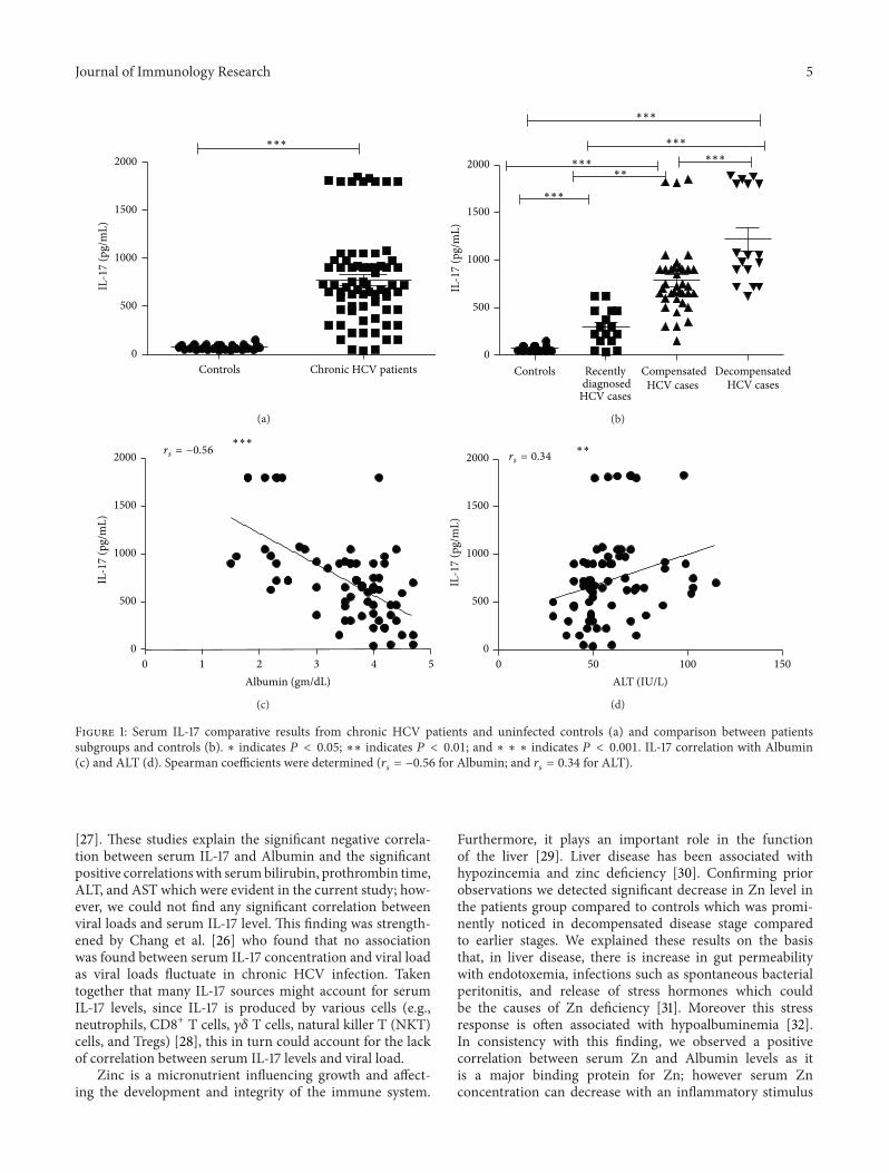
3.2. Elevated Levels of Serum IL-17 in Chronic HCV PatientsCorrelate with Severity of Liver Disease. Interleukin-17 is apotent mediator of delayed type reactions. It achieves thiseffect by elevating chemokine production in various tissueswhich, in turn, leads to recruitment of monocytes and neu-trophils to the site of inflammation [19]. IL-17 was measured
in the serum of chronic HCV patients (𝑛 = 70) and controls(𝑛 = 50). We observed significantly higher concentrationsof IL-17 in patients group compared to control group (𝑃 <0.001) (Figure 1(a)). To examine whether IL-17 was related toliver inflammation and fibrosis we stratified patients basedon ultrasonography results and liver function tests. Whencomparing Strata, the recently diagnosed, compensated, anddecompensated groups showed significantly higher levels ofIL-17 as compared to controls; meanwhile the compensatedand decompensated groups showed significantly higher levelsof IL-17 when compared to recently diagnosed HCV groupwith 𝑃 value < 0.001. Decompensated group also showedsignificantly higher levels of IL-17 as compared to compen-sated group (Figure 1(b)). In addition correlations betweenIL-17 and different laboratory parameters in patients groupwere done to show a significant negative correlation betweenIL-17 concentration and Albumin and a significant positivecorrelation with ALT, total and direct bilirubin, P.T, and INR(𝑃 < 0.001) (Table 2 and Figures 1(c) and 1(d)).
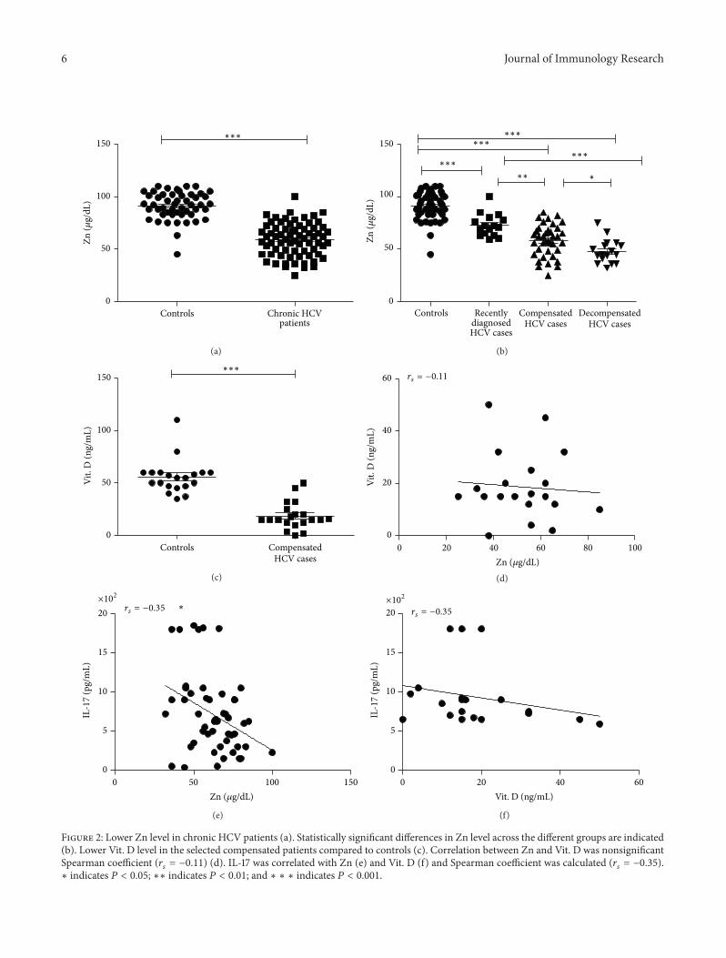
3.3. Zinc and Vitamin D Status in Chronic HCV Liver Diseaseand Their Relation with IL-17. The serum levels of zinc areoften decreased in HCV patients, and serum levels also tendto negatively correlate with hepatic reserve [20]. This wasobviously noticed in the patients group and subgroups whichshowed highly significant decrease in Zn level when com-pared to control group (Figure 2(a)). In addition data shownin Figure 2(b) indicate that compensated and decompensatedgroups had lower Zn levels compared to recently diagnosedgroup. Decompensated group also showed significantly lowerZn levels as compared to compensated group. Moreoverthere was a significant positive correlation between Zn andAlbumin and significant negative correlation with ALT, totaland direct bilirubin, P.T, and INR (Table 2). Because the liverplays a central role in Vit. D metabolism and its inadequacyis common in chronic liver diseases and correlates withdisease severity [21], we selected 20 chronic HCV casesfrom the compensated group to assess Vit. D status whichwas significantly lower than the controls level (Figure 2(c));however no correlations were found between vitamin Dserum levels, biochemical and virological data of the patients(data not shown), as well as serum Zn level (Figure 2(d)). Wedetermined a significant negative correlation between serumlevel of Zn and IL-17 (Figure 2(e)). There is also negativecorrelation between serum Vit. D level and IL-17; however,it is not significant (Figure 2(f)).
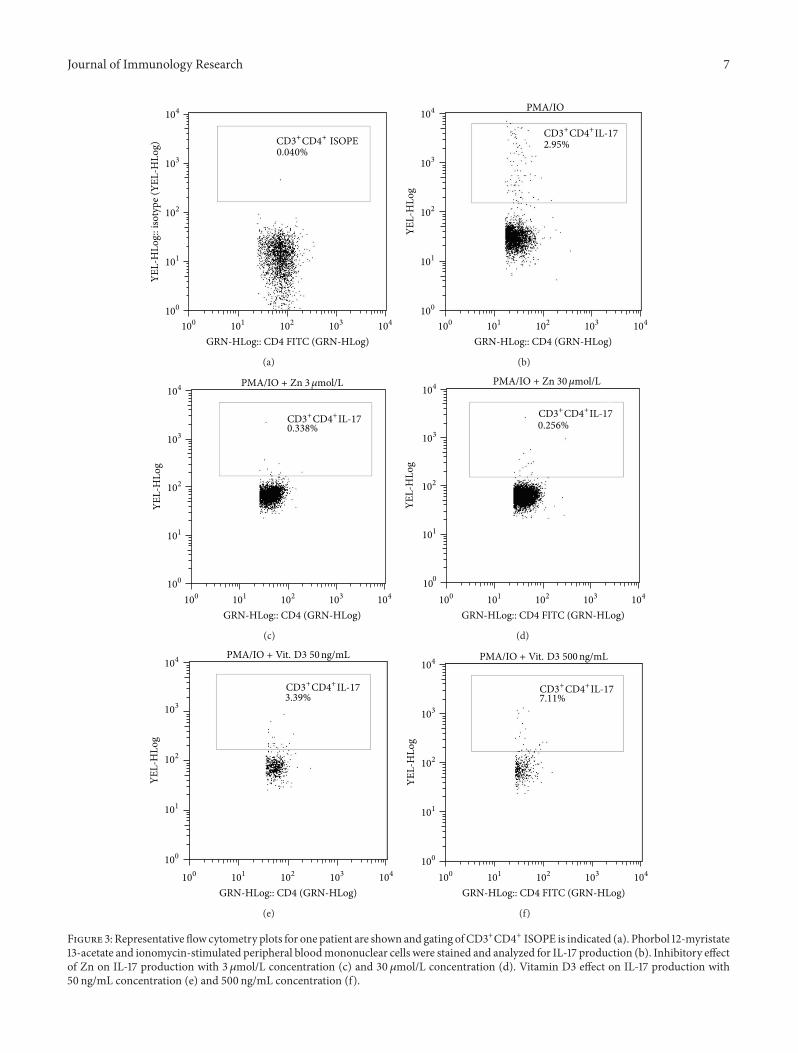
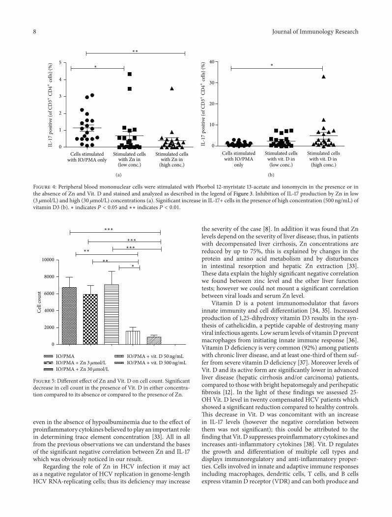
3.4. Role of Both Zn and Vit. D in Controlling IL-17 Expression.Both vitamin D and zinc play a role in innate and adaptiveimmune responses controlling inflammatory cytokine geneexpression [15, 22]. To test the effects of Zn and vitaminD3 on IL-17 cytokine production, PBMCs from only 20compensated cases were stimulated with PMA plus IO, inthe presence or absence of Zn (3 and 30 𝜇mol/L) or vitaminD3 (50 ng/mL and 500 ng/mL) then stained for intracellularIL-17, and analyzed by FACS (Figure 3). Data shown inFigure 4(a) indicate that the percentage of CD3+CD4+IL17+Tlymphocytes was significantly lower in the presence of Zn

4 Journal of Immunology Research
Table 1: Demographical and clinical characteristic of patients and controls.
Clinical characteristics All patients Recently diagnosed Compensated Decompensated ControlsAge, median (range) 48 (22–76) 30 (22–35) 48 (40–54) 63 (53–76) 31 (27–40)Sex (M/F) 35/35 9/7 17/20 9/8 22/28Albumin (g/dL), median(IQR) 3.6 (2.4–4) 4.2 (4–4.4) 3.6 (3.5–3.8) 2.3 (1.95–2.45) >4.1
Total Bilirubin (mg/dL),median (IQR) 0.9 (0.7–1.7) 0.7 (0.5–0.8) 0.7 (0.6–0.9) 3.14 (1.7–5.1) <1.2
Direct Bilirubin (mg/dL),median (IQR) 0.3 (0.2–0.9) 0.2 (0.1–0.3) 0.2 (0.1–0.3) 1.07 (0.8–2.35) <0.2
P.T# (sec), median (IQR) 13 (11.8–16.6) 11.9 (11.63–12.48) 12.5 (11.8–13) 17.4 (16.3–19.9) <12INR, median (IQR) 1.015 (0.98–1.5) 0.99 (0.9–1.04) 1 (0.9–1.07) 1.5 (1.48–1.7) 0.93 (0.9–1.02)ALT (IU/L), Mean ± SD 53.84 ± 12.36 56.5 ± 13 47.59 ± 11.87 58 ± 10.22 15.5 ± 7.65AST (IU/L), Mean ± SD 42.74 ± 10.59 47.44 ± 8.78 38.35 ± 9.32 42.71 ± 11.93 15.5 ± 6.25PCR (IU/mL), median(IQR) 31850 (22100–599000) 27600 (22100–646000) 36100 (22100–599000) 27600 (21200–630000) —
IL-17 (pg/mL), Mean ± SD 776.3 ± 57.39 295.8 ± 48.04 766.1 ± 62.90 1207 ± 113.8 72.17 ± 4.622ZN (𝜇g/dL), Mean ± SD 59.01 ± 1.86 72.81 ± 2.679 58.13 ± 2.445 48.00 ± 2.727 91.28 ± 1.838Vit. D (ng/mL), Mean ±SDa — — 18.65 ± 12.89 — 55.8 ± 16.27
aData available for 20 patients.P.T: prothrombin time; INR: international normalized ratio; ALT: alanine aminotransferase; AST: aspartate aminotransferase; IQR: the interquartile range;HCV: hepatitis C virus.
Table 2: Correlations between IL17 and Zn and different parametersin hepatitis C virus infected subjects.
IL-17 Zn𝑅 𝑃 value 𝑅 𝑃 value
Albumin (gm/dL) −0.561 <0.001 0.616 <0.001Total Bilirubin (mg/dL) 0.792 <0.001 −0.678 <0.001Direct Bilirubin (mg/dL) 0.649 <0.001 −0.584 <0.001P.T (sec) 0.656 <0.001 −0.632 <0.001INR 0.484 <0.001 −0.557 <0.001ALT (IU/L) 0.34 0.004 −0.304 0.032AST (IU/L) −0.059 0.684 −0.042 0.773PCR (IU/mL) −0.018 0.903 0.054 0.711
than in its absence and became more lower with increasingZn concentration which further support our suggestionregarding the link between Zn and IL-17. On the opposite sideaddition of Vit. D (in high concentration 500 ng/mL) leads toa significant increase in the percentage of CD3+CD4+IL17+cells compared to its absence (Figure 4(b)).
3.5. Effect of Zn and Vitamin D3 on Lymphocyte Count. Inorder to assess the in vitro effect of Zn and Vit. D on cellcount we compared the count of cells which stimulated inthe presence of Zn or Vit. D with those which stimulatedin their absence. We noticed that addition of Zn leads toincrease in the cell count especially with increasing the dose;however this effect was not significant; contrary to this result,addition of Vit. D leads to significant lowering in the cellcount compared to cells stimulated in its absence or in the
presence of Zn, and this effect seemed to be also dependenton the dose of vitamin D; however the 𝑃 value was nearlysignificant = 0.057 (Figure 5).
4. Discussions
Hepatitis C virus infection is a significant global public healthproblem. Persistent HCV infection eventually develops intoliver cirrhosis or hepatocellular carcinoma [23].Many reportsin HCV infections indicate a close correlation between virus-induced liver inflammations, infiltration, and activation ofTh17 cells and the amount of liver damage caused by theantiviral immune response. Moreover a shift from Th1 toTh17 seems to be potentially disadvantageous for the patientin terms of antiviral defense and liver disease progression[24, 25]. The present study showed that IL-17 was markedlyincreased inHCV-infected patients in comparison to controlsand this elevation became more evident with the progressionof the disease as shown upon comparing patients’ strata.These results were supported by the finding that increasingcirculating Th17, intrahepatic IL-17 positive cells, and HCV-specific Th17 cells were correlated with severity of liverinflammation in chronic HCV patients [26]. IL-17 and Th17seem to have an important role in viral infections andstronger Th17 responses are associated with higher viralplasma load, increased levels of serum transaminases, andenhanced activation of blood monocytes as well as livermacrophages [25]. Finally, it has been reported that antiviraltherapy with pegylated interferon and ribavirin in HCV-infected patients leads to a reduction of both Th1 and Th17responses, ameliorating HCV-mediated liver inflammation
Journal of Immunology Research 5
0
500
1000
1500
2000
Controls
IL-1
7 (p
g/m
L)
∗∗∗
Chronic HCV patients
(a)
0
500
1000
1500
2000
Controls
IL-1
7 (p
g/m
L)
∗∗∗
∗∗∗
∗∗∗
∗∗∗
∗∗
∗∗∗
DecompensatedHCV cases
CompensatedHCV cases
Recently
HCV cases diagnosed
(b)
0 1 2 3 4 50
500
1000
1500
2000
Albumin (gm/dL)
IL-1
7 (p
g/m
L)
∗∗∗rs = −0.56
(c)
0 50 100 1500
500
1000
1500
2000
ALT (IU/L)
IL-1
7 (p
g/m
L)∗∗
rs = 0.34
(d)
Figure 1: Serum IL-17 comparative results from chronic HCV patients and uninfected controls (a) and comparison between patientssubgroups and controls (b). ∗ indicates 𝑃 < 0.05; ∗∗ indicates 𝑃 < 0.01; and ∗ ∗ ∗ indicates 𝑃 < 0.001. IL-17 correlation with Albumin(c) and ALT (d). Spearman coefficients were determined (𝑟
𝑠= −0.56 for Albumin; and 𝑟
𝑠= 0.34 for ALT).
[27]. These studies explain the significant negative correla-tion between serum IL-17 and Albumin and the significantpositive correlationswith serumbilirubin, prothrombin time,ALT, and AST which were evident in the current study; how-ever, we could not find any significant correlation betweenviral loads and serum IL-17 level. This finding was strength-ened by Chang et al. [26] who found that no associationwas found between serum IL-17 concentration and viral loadas viral loads fluctuate in chronic HCV infection. Takentogether that many IL-17 sources might account for serumIL-17 levels, since IL-17 is produced by various cells (e.g.,neutrophils, CD8+ T cells, 𝛾𝛿 T cells, natural killer T (NKT)cells, and Tregs) [28], this in turn could account for the lackof correlation between serum IL-17 levels and viral load.
Zinc is a micronutrient influencing growth and affect-ing the development and integrity of the immune system.
Furthermore, it plays an important role in the functionof the liver [29]. Liver disease has been associated withhypozincemia and zinc deficiency [30]. Confirming priorobservations we detected significant decrease in Zn level inthe patients group compared to controls which was promi-nently noticed in decompensated disease stage comparedto earlier stages. We explained these results on the basisthat, in liver disease, there is increase in gut permeabilitywith endotoxemia, infections such as spontaneous bacterialperitonitis, and release of stress hormones which couldbe the causes of Zn deficiency [31]. Moreover this stressresponse is often associated with hypoalbuminemia [32].In consistency with this finding, we observed a positivecorrelation between serum Zn and Albumin levels as itis a major binding protein for Zn; however serum Znconcentration can decrease with an inflammatory stimulus
6 Journal of Immunology Research
0
50
100
150
Controls Chronic HCVpatients
∗∗∗
Zn (𝜇
g/dL
)
(a)
0
50
100
150∗∗∗
∗∗∗
∗∗∗∗∗∗
∗∗ ∗
Controls DecompensatedHCV cases
CompensatedHCV cases
Recently
HCV casesdiagnosed
Zn (𝜇
g/dL
)
(b)
0
50
100
150
Controls
Vit.
D (n
g/m
L)
∗∗∗
CompensatedHCV cases
(c)
0 20 40 60 80 1000
20
40
60Vi
t. D
(ng/
mL)
rs = −0.11
Zn (𝜇g/dL)(d)
0 50 100 1500
5
10
15
20
IL-1
7 (p
g/m
L)
∗×102
rs = −0.35
Zn (𝜇g/dL)
(e)
0 20 40 600
5
10
15
20
Vit. D (ng/mL)
IL-1
7 (p
g/m
L)
×102
rs = −0.35
(f)
Figure 2: Lower Zn level in chronic HCV patients (a). Statistically significant differences in Zn level across the different groups are indicated(b). Lower Vit. D level in the selected compensated patients compared to controls (c). Correlation between Zn and Vit. D was nonsignificantSpearman coefficient (𝑟
𝑠= −0.11) (d). IL-17 was correlated with Zn (e) and Vit. D (f) and Spearman coefficient was calculated (𝑟
𝑠= −0.35).
∗ indicates 𝑃 < 0.05; ∗∗ indicates 𝑃 < 0.01; and ∗ ∗ ∗ indicates 𝑃 < 0.001.
Journal of Immunology Research 7
104
103
102
101
100
104103102101100
YEL-
HLo
g:: i
soty
pe (Y
EL-H
Log)
GRN-HLog:: CD4 FITC (GRN-HLog)
0.040%CD3+CD4+ ISOPE
(a)
104
103
102
101
100
104103102101100
GRN-HLog:: CD4 (GRN-HLog)
YEL-
HLo
g
2.95%
PMA/IO
CD3+CD4+IL-17
(b)
104
103
102
101
100
104103102101100
YEL-
HLo
g
GRN-HLog:: CD4 (GRN-HLog)
0.338%
PMA/IO + Zn 3𝜇mol/L
CD3+CD4+IL-17
(c)
104
103
101
102
100
104103102101100
YEL-
HLo
g
GRN-HLog:: CD4 FITC (GRN-HLog)
0.256%
PMA/IO + Zn 30𝜇mol/L
CD3+CD4+IL-17
(d)
104
103
102
101
100
104103102101100
GRN-HLog:: CD4 (GRN-HLog)
YEL-
HLo
g
3.39%CD3+CD4+IL-17
PMA/IO + Vit. D3 50ng/mL
(e)
104
103
102
101
100
104103102101100
YEL-
HLo
g
GRN-HLog:: CD4 FITC (GRN-HLog)
7.11%CD3+CD4+IL-17
PMA/IO + Vit. D3 500ng/mL
(f)
Figure 3: Representative flow cytometry plots for one patient are shown and gating ofCD3+CD4+ ISOPE is indicated (a). Phorbol 12-myristate13-acetate and ionomycin-stimulated peripheral bloodmononuclear cells were stained and analyzed for IL-17 production (b). Inhibitory effectof Zn on IL-17 production with 3 𝜇mol/L concentration (c) and 30 𝜇mol/L concentration (d). Vitamin D3 effect on IL-17 production with50 ng/mL concentration (e) and 500 ng/mL concentration (f).
8 Journal of Immunology Research
0
1
2
3
4
5
∗∗
∗
Cells stimulatedwith IO/PMA only
Stimulated cellswith Zn in(low conc.)
Stimulated cellswith Zn in
(high conc.)
IL-17
posit
ive (
of C
D3+
CD4+
cells
) (%
)
(a)
0
10
20
30
40∗
Stimulated cellswith vit. D in(high conc.)(low conc.)
Stimulated cellswith vit. D inwith IO/PMA
Cells stimulated
only
IL-17
posit
ive (
of C
D3+
CD4+
cells
) (%
)
(b)
Figure 4: Peripheral blood mononuclear cells were stimulated with Phorbol 12-myristate 13-acetate and ionomycin in the presence or inthe absence of Zn and Vit. D and stained and analyzed as described in the legend of Figure 3. Inhibition of IL-17 production by Zn in low(3𝜇mol/L) and high (30 𝜇mol/L) concentrations (a). Significant increase in IL-17+ cells in the presence of high concentration (500 ng/mL) ofvitamin D3 (b). ∗ indicates 𝑃 < 0.05 and ∗∗ indicates 𝑃 < 0.01.
0
2000
4000
6000
8000
10000
Cel
l cou
nt
∗∗∗
∗∗∗
∗∗∗∗∗
∗∗∗
IO/PMAIO/PMA + Zn 3𝜇mol/LIO/PMA + Zn 30𝜇mol/L
IO/PMA + vit. D 500ng/mLIO/PMA + vit. D 50ng/mL
Figure 5: Different effect of Zn and Vit. D on cell count. Significantdecrease in cell count in the presence of Vit. D in either concentra-tion compared to its absence or compared to the presence of Zn.
even in the absence of hypoalbuminemia due to the effect ofproinflammatory cytokines believed to play an important rolein determining trace element concentration [33]. All in allfrom the previous observations we can understand the basesof the significant negative correlation between Zn and IL-17which was obviously noticed in our result.
Regarding the role of Zn in HCV infection it may actas a negative regulator of HCV replication in genome-lengthHCV RNA-replicating cells; thus its deficiency may increase
the severity of the case [8]. In addition it was found that Znlevels depend on the severity of liver disease; thus, in patientswith decompensated liver cirrhosis, Zn concentrations arereduced by up to 75%, this is explained by changes in theprotein and amino acid metabolism and by disturbancesin intestinal resorption and hepatic Zn extraction [33].These data explain the highly significant negative correlationwe found between zinc level and the other liver functiontests; however we could not mount a significant correlationbetween viral loads and serum Zn level.
Vitamin D is a potent immunomodulator that favorsinnate immunity and cell differentiation [34, 35]. Increasedproduction of 1,25-dihydroxy vitamin D3 results in the syn-thesis of cathelicidin, a peptide capable of destroying manyviral infectious agents. Low serum levels of vitaminDpreventmacrophages from initiating innate immune response [36].Vitamin D deficiency is very common (92%) among patientswith chronic liver disease, and at least one-third of them suf-fer from severe vitamin D deficiency [37]. Moreover levels ofVit. D and its active form are significantly lower in advancedliver disease (hepatic cirrhosis and/or carcinoma) patients,compared to those with bright hepatomegaly and perihepaticfibrosis [12]. In the light of these findings we assessed 25-OH Vit. D level in twenty compensated HCV patients whichshowed a significant reduction compared to healthy controls.This decrease in Vit. D was concomitant with an increasein IL-17 levels (however the negative correlation betweenthem was not significant); this could be attributed to thefinding thatVit. D suppresses proinflammatory cytokines andincreases anti-inflammatory cytokines [38]. Vit. D regulatesthe growth and differentiation of multiple cell types anddisplays immunoregulatory and anti-inflammatory proper-ties. Cells involved in innate and adaptive immune responsesincluding macrophages, dendritic cells, T cells, and B cellsexpress vitamin D receptor (VDR) and can both produce and
Journal of Immunology Research 9
respond to 1,25(OH)2D3[39]. Hepatocytes express only low
levels of VDR mRNA [40], so that vitamin D effects on theliver are most probably not conferred by direct signallingin parenchymal liver cells. In contrast, nonparenchymalhepatic cells such as sinusoidal endothelial cells, Kupffer cells,and hepatic stellate cells (HSC) do express VDR mRNAand functionally active VDR protein [41]. Many reportsindicate that 1,25(OH)
2D3suppresses Th17 driven cytokine
responses, induces Treg cells, induces IL-4 production (Th2),and enhances natural killer T-cell function [11, 42] but the keyimmunomodulatory property of 1,25(OH)
2D3is its ability
to inhibit expression of Th1 cytokines, whilst augmentingTh2 cytokines, with 1,25(OH)
2D3acting either directly via
effects on T lymphocytes or indirectly via effects on antigen-presenting cells (APCs). Moreover, elevated VDR expressionis also found on differentiated Th17 cells [43]. More recentstudies showed marked anti-inflammatory and antifibroticeffects of VDR-signalling in HSC. During inflammatoryliver injury after endotoxin injection, the activation of VDRsignalling by vitaminDattenuated liver damage in vivo. Vit. Dprovides protection against autoimmune and inflammatorydiseases, such as multiple sclerosis, type 1 diabetes, andinflammatory bowel disease partially due to its inhibitoryeffects on Th17 cells. The antiproliferative, prodifferentiative,antibacterial, immunomodulatory, and anti-inflammatoryproperties of synthetic VDR agonists could be exploitedto treat a variety of inflammatory and autoimmune dis-eases [39]. Treatment with VDR agonists inhibits the T-cell production of IL-17. Furthermore, IL-17 production issustained by IL-23, an IL-12 family member, the latter ofwhich is strongly inhibited byVDRagonists [11]. It was clearlydemonstrated that enhanced progression of liver fibrosis issignificantly and independently associated with both geneticVDR variants and low 25-OH vitamin D plasma levels. Thissuggests vitamin D substitution as a preventive measure forpatients with liver fibrosis [44].
In order to gain additional insight into the interplaybetween Zn, Vit. D, and IL-17 we have examined the abilityof Zn and Vit. D3 to interfere with Th17 activation andexpression of IL-17 in vitro. Noteworthy, IL-17 was suppressedsignificantly when Zn was added in both concentrations andthis was claimed to its suppressive effect on IL-6/STAT3(signal transducer and activator of transcription) signalingpathway which is a critical step for Th17 development. Znbinding changed the a-helical secondary structure of STAT3,disrupting the association of STAT3 with JAK2 kinase (Januskinase 2) and with a phosphopeptide that included a STAT3-binding motif from the IL-6 signal transducer gp130 [22].To test whether the effects of Zn on cytokine productionwere independent of toxic effects, cell count was examinedand interestingly we observed that, with addition of Zn,the count was increased compared to its absence especiallywith increasing Zn concentration, although this observationwas not significant; this effect could be mediated throughZn enhancement of DNA synthesis and RNA transcription,cell division, and cell activation as apoptosis (programmedcell death) is potentiated by Zn deficiency [45]; additionallyZn is an inhibitor of NADPH oxidases which catalyze theproduction of reactive oxygen species (ROS); on the other
side, Zn activates the dismutation of O2⋅− to H2O2by super
oxide dismutase which contains both copper and Zn [46];it also negatively regulates gene expression of inflammatorycytokines such as TNF-𝛼 and IL-1𝛽, which are known togenerate (ROS) and this may be one additional mechanismby which Znmay be functioning as an antioxidant in humans[47].
There is promising evidence that zinc may decreaseliver injury and provides antifibrotic effects in patients withchronic HCV. Himoto and Coworkers [20] used polaprezincas an antifibrotic therapy in patients with chronic HCV andshowed a decrease in noninvasive fibrosis markers.
One caveat of our studies was the effect of Vit. D onIL17 production. For reasons that remain unclear and despitethe apparent negative association between serum Vit. D andIL17, addition of Vit. D showed an apparent increase inthe percent of IL-17+ cells which was significant only withhigh concentration of Vit. D. Contrary to our finding anexperimental study on healthy human donor using CD4+T cells and mouse model for multiple sclerosis showed that1,25(OH)
2D3inhibits human IL-17A and suppresses mouse
IL-17A [48]; other studies in mice imply this regulatory effecton Th17 cells by Vit. D through the reduction of (ROR)𝛾texpression [15].
The reasons of these conflicting results still need to bedetermined, although we would contend that the interpreta-tion was somewhat misleading. Looking at the effect of Vit.D on cell count showed us a marked dose-related reductionin cell count compared to its absence or to the presence ofZn. Preclinical research indicates that vitamin D3 potentlyinhibited T cell proliferation in a dose-related fashion [49].The active metabolite of vitamin D, 1 alpha,25(OH)
2D3,
also known as calcitriol, has antiproliferative effects, acti-vates apoptotic pathways, and inhibits angiogenesis [50].The common antiproliferative vitamin D receptor (VDR)functions are associated with arrest at G0/G1 of the cell cycle,coupled with upregulation of a number of cell cycle inhibitorsincluding p21 and p27 [51]. On the other hand Th17 andotherTh17 and other IL-17 secreting cells could play a part inhepatic viral persistence bymeans of antiapoptoticmoleculesupregulation [26]. Consistently, IL-17 has been implicated inmodulating the expression levels of prosurvival Bcl-2 familyproteins, including Bcl-2 (B-cell lymphoma 2) [52] and Bfl-1/A1 (Bcl-2 family member) in some autoimmune diseasessuch as systemic lupus erythematosus [53]. Based on theseobservations it seems feasible that our preconceived notionregarding the impairment of apoptosis pathway in Th17 cellscan contribute to the rise of IL-17+ cells percent amongthe remained cells; however it remains to be determinedwhether the apparent increase is a true enrichment of IL-17producing cells or simply due to survival of IL-17+ cells fromthe toxic effect of Vit. D. However the clinical implicationof low vitamin D levels and HCV severity is still not clear.At least one study has found that serum Vit. D levels werenot associated with any parameter indicating disease severity.And they concluded that serum Zn but not serum vitamin Dlevels is strongly associated with disease severity and treat-ment response in chronic HCV [54]. This latter study mayalso account in part for the lack of significant correlations
10 Journal of Immunology Research
betweenVit. D serum levels, biochemical and virological dataof the patients, and serum Zn and IL17 levels that have beeninvestigated in the current study. However there are somelimitations in this study. An important limitation is the lackof assessment of Vit. D status in both recently diagnosedand decompensated groups which would give us a betterinformative insight on the interplay between Vit. D and IL-17in chronic HCV liver disease and whether it has a protectiverole in preventing liver fibrosis or not.
A final conclusion of our study concerns the potential useof Zn as an adjunct antifibrotic therapy and novel strategiesfor the treatment of chronic hepatitis C; it may be worthwhileexploring the benefit of zinc supplementation even with theadvent of novel direct antiviral agents. Meanwhile the role ofvitamin D is still a topic of debate and much work will berequired to understand the perturbations found in IL-17 andits relation to vitamin D at a mechanistic and genetic levels.
Ethical Approval
Written informed consent was obtained from each of theparticipants after approving the study protocol by the EthicsCommittee of Faculty of Medicine, Ain Shams University.
Conflict of Interests
There is no conflict of interests. All authors do not havea direct financial relation with the commercial identitiesmentioned in the paper.
Acknowledgment
This work was supported by Dr. MonaM. Rafik, research lab,Faculty of Medicine, Ain Shams University.
References
[1] N. V. Naoumov, “Hepatitis C virus-specific CD4+ T cells: dothey help or damage?”Gastroenterology, vol. 117, no. 4, pp. 1012–1014, 1999.
[2] E. Bettelli, M. Oukka, and V. K. Kuchroo, “TH-17 cells in thecircle of immunity and autoimmunity,”Nature Immunology, vol.8, no. 4, pp. 345–350, 2007.
[3] C. T. Weaver, R. D. Hatton, P. R. Mangan, and L. E. Harrington,“IL-17 family cytokines and the expanding diversity of effectorT cell lineages,” Annual Review of Immunology, vol. 25, pp. 821–852, 2007.
[4] Y. Yasumi, Y. Takikawa, R. Endo, and K. Suzuki, “Interleukin-17as a new marker of severity of acute hepatic injury,” HepatologyResearch, vol. 37, no. 4, pp. 248–254, 2007.
[5] D. B. O’Quinn, M. T. Palmer, Y. K. Lee, and C. T. Weaver,“Chapter 5 emergence of the Th17pathway and its role in hostdefense,” Advances in Immunology, vol. 99, no. 5, pp. 115–163,2008.
[6] A. S. Prasad, “Effects of zinc deficiency onTh1 andTh2 cytokineshifts,”The Journal of Infectious Diseases, vol. 182, supplement 1,pp. S62–S68, 2000.
[7] R. J. Cousins, “Absorption, transport, and hepatic metabolismof copper and zinc: special reference to metallothionein and
ceruloplasmin,” Physiological Reviews, vol. 65, no. 2, pp. 238–309, 1985.
[8] K. Yuasa, A. Naganuma, K. Sato et al., “Zinc is a negative regu-lator of hepatitis C virus RNA replication,” Liver International,vol. 26, no. 9, pp. 1111–1118, 2006.
[9] M. Nishihara, H. Ogura, N. Ueda et al., “IL-6-gp130-STAT3 inT cells directs the development of IL-17+ T
ℎwith a minimum
effect on that of Treg in the steady state,” International Immunol-ogy, vol. 19, no. 6, pp. 695–702, 2007.
[10] S. Petta, C. Camma, C. Scazzone et al., “Low vitamin C serumlevel is related to severe fibrosis and low responsiveness tointerferon-based therapy in genotype 1 chronic hepatitis C,”Hepatology, vol. 51, no. 4, pp. 1158–1167, 2010.
[11] N. M. El Husseiny, H. M. Fahmy, W. A. Mohamed, and H.Hisham, “Relationship between vitamin D and IL-23, IL-17 andmacrophage chemoattractant protein-1 as markers of fibrosis inhepatitis C virus Egyptians,” World Journal of Hepatology, vol.4, no. 8, pp. 242–247, 2012.
[12] M. F. Schaalan, W. A. Mohamed, and H. H. Amin, “VitaminD deficiency: Correlation to interleukin-17, interleukin-23 andPIIINP in hepatitis C virus genotype 4,” World Journal ofGastroenterology, vol. 18, no. 28, pp. 3738–3744, 2012.
[13] A. Monegal, M. Navasa, N. Guanabens et al., “Osteoporosisand bone mineral metabolism disorders in cirrhotic patientsreferred for orthotopic liver transplantation,” Calcified TissueInternational, vol. 60, no. 2, pp. 148–154, 1997.
[14] J. N. Artaza and K. C. Norris, “Vitamin D reduces the expres-sion of collagen and key profibrotic factors by inducing anantifibrotic phenotype in mesenchymal multipotent cells,” TheJournal of Endocrinology, vol. 200, no. 2, pp. 207–221, 2009.
[15] A.M.Mus, J. P. vanHamburg, P. Asmawidjaja et al., “Vitamin Dsuppresses Th17 cytokines via down regulation of RORgammatandNFATC2 and by differential regulation of GATA3,”Arthritis& Rheumatism, vol. 62, supplement 10, p. 38, 2010.
[16] S. Mehta, D. J. Hunter, F. M. Mugusi et al., “Perinatal outcomes,including mother-to-child transmission of HIV, and childmortality and their association with maternal vitamin D statusin Tanzania,” The Journal of Infectious Diseases, vol. 200, no. 7,pp. 1022–1030, 2009.
[17] World Health Organization, “Physical status: the use andinterpretation of anthropometry. Report of a WHO ExpertCommittee,” World Health Organization Technical Report 854,1995.
[18] O. Hochwald, I. Harman-Boehm, and H. Castel, “Hypovita-minosis D among inpatients in a sunny country,” The IsraelMedical Association Journal, vol. 6, no. 2, pp. 82–87, 2004.
[19] P.Miossec, T. Korn, andV. K. Kuchroo, “Interleukin-17 and type17 helper T cells,”TheNew England Journal of Medicine, vol. 361,no. 9, pp. 888–898, 2009.
[20] T. Himoto, N. Hosomi, S. Nakai et al., “Efficacy of zincadministration in patientswith hepatitis C virus-related chronicliver disease,” Scandinavian Journal of Gastroenterology, vol. 42,no. 9, pp. 1078–1087, 2007.
[21] L. Fisher andA. Fisher, “VitaminD and parathyroid hormone inoutpatients with noncholestatic chronic liver disease,” ClinicalGastroenterology andHepatology, vol. 5, no. 4, pp. 513–520, 2007.
[22] C. Kitabayashi, T. Fukada,M. Kanamoto et al., “Zinc suppressesTh17 development via inhibition of STAT3 activation,” Interna-tional Immunology, vol. 22, no. 5, pp. 375–386, 2010.
[23] D. Lavanchy, “Evolving epidemiology of hepatitis C virus,”Clinical Microbiology and Infection, vol. 17, no. 2, pp. 107–115,2011.
Journal of Immunology Research 11
[24] I. R. Wanless and K. Shiota, “The pathogenesis of nonalcoholicsteatohepatitis and other fatty liver diseases: a four-step modelincluding the role of lipid release and hepatic venular obstruc-tion in the progression to cirrhosis,” Seminars in Liver Disease,vol. 24, no. 1, pp. 99–106, 2004.
[25] A. Puel, R. Doffinger, A. Natividad et al., “Autoantibodiesagainst IL-17A, IL-17F, and IL-22 in patients with chronicmucocutaneous candidiasis and autoimmune polyendocrinesyndrome type I,” Journal of ExperimentalMedicine, vol. 207, no.2, pp. 291–297, 2010.
[26] Q. Chang, Y.-K.Wang, Q. Zhao, C.-Z.Wang, Y.-Z. Hu, and B.-Y.Wu, “Th17 cells are increasedwith severity of liver inflammationin patients with chronic hepatitis C,” Journal of Gastroenterologyand Hepatology, vol. 27, no. 2, pp. 273–278, 2012.
[27] M. A. Jimenez-Sousa, R. Almansa, C. de La Fuente et al.,“Increased Th1, Th17 and pro-fibrotic responses in hepatitis C-infected patients are down-regulated after 12 weeks of treatmentwith pegylated interferon plus ribavirin,” European CytokineNetwork, vol. 21, no. 2, pp. 84–91, 2010.
[28] A. G. Rowan, J. M. Fletcher, E. J. Ryan et al., “Hepatitis Cvirus-specific Th17 cells are suppressed by virus-induced TGF-𝛽,” Journal of Immunology, vol. 181, no. 7, pp. 4485–4494, 2008.
[29] M. A. Nazari, S. H. Malayeri, M. A. Pourhoseingholi, S. R.Mohebi, and M. R. Zali, “Evaluation of zinc plasma level inIranian cirrhotic patients due to hepatitis B and hepatitis C,”Hepatitis Monthly, vol. 10, no. 1, pp. 62–64, 2010.
[30] R. L. Vallee, W. E. Wacker, A. F. Bartholomay, and E. D.Robin, “Zinc metabolism in hepatic dysfunction. I. Serum zincconcentrations in Laennec’s cirrhosis and their validation bysequential analysis,” The New England Journal of Medicine, vol.255, no. 9, pp. 403–408, 1956.
[31] C. J. McClain, M. L. McClain, M. G. Boosalis, and B. Hennig,“Zinc and the stress response,” Scandinavian Journal of Work,Environment and Health, vol. 19, no. 1, pp. 132–133, 1993.
[32] L.M.Gaetke, C. J.McClain, R. T. Talwalkar, and S. I. Shedlofsky,“Effects of endotoxin on zinc metabolism in human volun-teers,” The American Journal of Physiology—Endocrinology andMetabolism, vol. 272, no. 6, pp. E952–E956, 1997.
[33] K. Grngreiff, “Zinc in liver disease,” The Journal of TraceElements in Experimental Medicine, vol. 15, no. 1, pp. 67–78,2002.
[34] H. F. DeLuca, “Overview of general physiologic features andfunctions of vitamin D,” The American Journal of ClinicalNutrition, vol. 80, no. 6, pp. 1689S–1696S, 2004.
[35] A. S. Dusso, A. J. Brown, and E. Slatopolsky, “Vitamin D,”American Journal of Physiology: Renal Physiology, vol. 289, no.1, pp. F8–F28, 2005.
[36] P. T. Liu, S. Stenger, H. Li et al., “Toll-like receptor triggering ofa vitamin D-mediated human antimicrobial response,” Science,vol. 311, no. 5768, pp. 1770–1773, 2006.
[37] J. Arteh, S. Narra, and S. Nair, “Prevalence of vitamin Ddeficiency in chronic liver disease,” Digestive Diseases andSciences, vol. 55, no. 9, pp. 2624–2628, 2010.
[38] B. D. Mahon, A. Wittke, V. Weaver, and M. T. Cantorna,“The targets of vitamin D depend on the differentiation andactivation status of CD4 positive T cells,” Journal of CellularBiochemistry, vol. 89, no. 5, pp. 922–932, 2003.
[39] L. Adorini and G. Penna, “Control of autoimmune diseasesby the vitamin D endocrine system,” Nature Clinical PracticeRheumatology, vol. 4, no. 8, pp. 404–412, 2008.
[40] C. Segura,M.Alonso, C. Fraga, T.Garcıa-Caballero, C.Dieguez,andR. Perez-Fernandez, “VitaminD receptor ontogenesis in ratliver,”Histochemistry andCell Biology, vol. 112, no. 2, pp. 163–167,1999.
[41] M. Wagner, G. Zollner, and M. Trauner, “Nuclear receptors inliver disease,” Journal of Hepatology, vol. 53, no. 3, pp. 1023–1034,2011.
[42] L. Steinman, “A brief history of TH17, the first major revisionin the TH1/TH2 hypothesis of T cell-mediated tissue damage,”Nature Medicine, vol. 13, no. 2, pp. 139–145, 2007.
[43] H. Zhang, D. Q. Shih, and X. Zhang, “Mechanisms underlyingeffects of 1,25-Dihydroxyvitamin D
3on the Th17 cells,” Euro-
pean Journal of Microbiology and Immunology, vol. 3, no. 4, pp.237–240, 2013.
[44] K. Baur, J. C. Mertens, J. Schmitt et al., “Combined effect of 25-OH vitamin D plasma levels and genetic Vitamin D receptor(NR 1I1) variants on fibrosis progression rate in HCV patients,”Liver International, vol. 32, no. 4, pp. 635–643, 2012.
[45] S. Overbeck, L. Rink, and H. Haase, “Modulating the immuneresponse by oral zinc supplementation: a single approachfor multiple diseases,” Archivum Immunologiae et TherapiaeExperimentalis, vol. 56, no. 1, pp. 15–30, 2008.
[46] P. A. Lachance, Z. Nakat, and W.-S. Jeong, “Antioxidants: anintegrative approach,” Nutrition, vol. 17, no. 10, pp. 835–838,2001.
[47] A. S. Prasad, B. Bao, F. W. J. Beck, O. Kucuk, and F. H. Sarkar,“Antioxidant effect of zinc in humans,” Free Radical Biology andMedicine, vol. 37, no. 8, pp. 1182–1190, 2004.
[48] S. Joshi, L.-C. Pantalena, X. K. Liu et al., “1,25-Dihydroxyvitamin D
3ameliorates Th17 autoimmunity via
transcriptional modulation of interleukin-17A,” Molecular andCellular Biology, vol. 31, no. 17, pp. 3653–3669, 2011.
[49] C. Almerighi, A. Bergamini, R. Lionetti et al., “Vitamin D3modulates T lymphocyte responses in hepatitis C virus-infectedliver transplant recipients,” Digestive and Liver Disease, vol. 44,no. 1, pp. 67–73, 2012.
[50] K. K. Deeb, D. L. Trump, and C. S. Johnson, “Vitamin Dsignalling pathways in cancer: potential for anticancer thera-peutics,”Nature Reviews Cancer, vol. 7, no. 9, pp. 684–700, 2007.
[51] J. Thorne and M. J. Campbell, “The vitamin D receptor incancer,” The Proceedings of the Nutrition Society, vol. 67, no. 2,pp. 115–127, 2008.
[52] M. Batten, J. Groom, T. G. Cachero et al., “BAFF mediatessurvival of peripheral immature B lymphocytes,”The Journal ofExperimental Medicine, vol. 192, no. 10, pp. 1453–1465, 2000.
[53] A. Doreau, A. Belot, J. Bastid et al., “Interleukin 17 acts insynergy with B cell-activating factor to influence B cell biologyand the pathophysiology of systemic lupus erythematosus,”Nature Immunology, vol. 10, no. 7, pp. 778–785, 2009.
[54] S. Kastens, K. Grungreiff, C. Terkamp et al., “Serum zinc butnot serum vitamin d levels are associated with disease severityand treatment response in chronic hepatitis c virus infection,”Journal of Hepatology, vol. 54, Supplement 1, p. S458, 2011.