the journal of biological chemistry © 2000 … small changes in conformation between the apo and...
TRANSCRIPT

X-ray Structures of the Apo and MgATP-bound States ofDictyostelium discoideum Myosin Motor Domain*
Received for publication, June 26, 2000, and in revised form, August 18, 2000Published, JBC Papers in Press, August 22, 2000, DOI 10.1074/jbc.M005585200
Cary B. Bauer, Hazel M. Holden, James B. Thoden, Robert Smith, and Ivan Rayment‡
From the Department of Biochemistry, University of Wisconsin, Madison, Wisconsin 53706-1544
Myosin is the most comprehensively studied molecu-lar motor that converts energy from the hydrolysis ofMgATP into directed movement. Its motile cycle con-sists of a sequential series of interactions between my-osin, actin, MgATP, and the products of hydrolysis,where the affinity of myosin for actin is modulated bythe nature of the nucleotide bound in the active site. Thefirst step in the contractile cycle occurs when ATP bindsto actomyosin and releases myosin from the complex.We report here the structure of the motor domain ofDictyostelium discoideum myosin II both in its nucleo-tide-free state and complexed with MgATP. The struc-ture with MgATP was obtained by soaking the crystalsin substrate. These structures reveal that both the apoform and the MgATP complex are very similar to thosepreviously seen with MgATPgS and MgAMP-PNP. More-over, these structures are similar to that of chicken skel-etal myosin subfragment-1. The crystallized protein isenzymatically active in solution, indicating that the con-formation of myosin observed in chicken skeletal myo-sin subfragment-1 is unable to hydrolyze ATP and mostlikely represents the pre-hydrolysis structure for themyosin head that occurs after release from actin.
Directed movement is one of the features common to livingorganisms. This property is provided by a suite of molecularmotors that includes, myosin, dyenin, and kinesin in eu-karyotes and bacterial flagella motors in prokaryotes. In eachmolecular motor the energy to drive this process is derived fromthe hydrolysis of ATP. For all of these motors, it has long beensuspected that conformational changes associated with differ-ential affinity of the motor protein for ATP and its hydrolysisproducts are responsible for energy transduction. After manyyears of study, it has proved to be difficult to identify the natureof the conformational changes that underlie directed move-ment, with the exception of myosin. However, even with myo-sin important questions remain.
Myosin is the most abundant and best known molecularmotor. Although initially identified in skeletal muscle, isoformsof this protein have been observed in all eukaryotic cells whereit fulfills a wide range of cellular functions from muscle con-
traction, cellular locomotion, and chemotaxis to organelletransport (1, 2). Indeed over 15 classes of myosin have nowbeen identified. All of these proteins contain a section of theirsequence that can be associated with a motor domain. Thecatalytic cycle of myosin reveals at first sight a counterintuitiverelationship between the chemical step of hydrolysis and theenergy transduction process. In myosin the energy transduc-tion step is associated with product release rather than ATPhydrolysis.
Kinetics studies have revealed that in the absence of ATP,myosin binds very tightly to actin to form the rigor state. Uponaddition of ATP, myosin rapidly changes to a weakly boundstate prior to nucleotide hydrolysis. Thereafter ATP is rapidlyhydrolyzed, but the hydrolysis products remain tightly boundto myosin. Rebinding to actin catalyzes the release of inorganicphosphate and triggers the start of the powerstroke (3, 4).Spectroscopic and fiber diffraction studies have shown thatconformational changes in the myosin motor domain and itsattached light chains are intimately involved in the energytransduction step (5–8). A full molecular understanding of thisbiological process requires a detailed structural view of each ofthe distinct states of the contractile cycle.
The structure of myosin has been determined for four organ-isms in a range of conformations. These include chicken skel-etal myosin subfragment-1 with sulfate in the active site (9),Dictyostelium discoideum myosin II truncated head (S1dC)1
with a variety of nucleotides bound in the active site (10–15),smooth muscle myosin head with its essential light chain andthe smooth muscle motor domain (16), and scallop myosin S1(17). A question inherent to all of these structures is theirrelationship to the conformational states that are transitionedthrough or populated within the contractile cycle.
The first structure for myosin subfragment-1 was that ofchicken skeletal myosin subfragment-1 (9). This protein wascrystallized from ammonium sulfate in the absence of nucleo-tide and was observed to contain a sulfate ion bound in theP-loop. Because this was the first determined structure, it wasunknown where this structure belonged in the catalytic cycle.Initially, it was believed to represent a point after release fromactin but prior to formation of the metastable state (18). How-ever because the overall structure seemed to fit a reasonablemodel for the actomyosin complex based on image reconstruc-tions it was subsequently ascribed by Holmes (19) to the rigorstate. Subsequent studies on a truncated myosin head or motordomain from D. discoideum revealed two conformations, one of
* This research was supported by National Institutes of Health GrantAR35186 (to I. R). The costs of publication of this article were defrayedin part by the payment of page charges. This article must therefore behereby marked “advertisement” in accordance with 18 U.S.C. Section1734 solely to indicate this fact.
The atomic coordinates and structure factors (code 1FMV/1FMW)have been deposited in the Protein Data Bank, Research Collaboratoryfor Structural Bioinformatics, Rutgers University, New Brunswick, NJ(http://www.rcsb.org/)..
‡ To whom correspondence should be addressed: Dept. of Biochemis-try, 433 Babcock Dr., Madison, WI 53706-1544. Tel.: 608-262-0529; Fax:608-265-2904; E-mail: [email protected].
1 The abbreviations used are: S1dC, D. discoideum myosin II motordomain, residues 1–762; Myosin S1, myosin subfragment-1; P-loop,phosphate binding loop or Walker A Motif; MgATPgS, magnesiumadenosine 59-O-thiotriphosphate; PEG, polyethylene glycol;S1dCzMgADPzBeFx, the beryllium fluoride-ADP complex of D. discoi-deum myosin motor domain; r.m.s., root mean square; nanolog, non-nucleotide analog; AMP-PNP, adenosine 59-(b,g-imino)triphosphate.
THE JOURNAL OF BIOLOGICAL CHEMISTRY Vol. 275, No. 49, Issue of December 8, pp. 38494–38499, 2000© 2000 by The American Society for Biochemistry and Molecular Biology, Inc. Printed in U.S.A.
This paper is available on line at http://www.jbc.org38494

which was obtained in the presence of MgADPzBeFx and wassimilar to that observed in chicken skeletal myosin subfrag-ment-1 (10). The other obtained in the presence of MgADPzAlF4
and MgADPzVO4 appeared to represent the conformation of themyosin head in the metastable state (10, 11).
Subsequent studies on the motor domain and myosin head ofsmooth muscle myosin demonstrated that the structure of themotor domain observed in the complex with MgADPzAlF4 andMgADPzVO4 are representative of the conformation of the my-osin head at the start of the powerstroke (16). Conversely amore recent structure of scallop myosin subfragment-1 com-plexed with MgADP revealed a similar conformation ofthe nucleotide binding pocket, but altered its orientation of thelight chain binding motif relative to that seen in any ofthe other structures (17). Thus the question still remains as tothe relationship between the structure observed in chickenskeletal myosin S1 and the contractile cycle.
To address the question of whether the conformation of themotor domain observed in chicken skeletal myosin S1 andDictyostelium myosin S1dCzMgADPzBeFx represents the struc-ture in the rigor conformation, we have determined the struc-ture of S1dC both in its apo state and bound to MgATP to highresolution. This establishes that the conformation seen inS1dCzMgADPzBeFx is unable to hydrolyze ATP and most likelyrepresents the initial state (pre-hydrolysis) after release ofmyosin from actin. It also shows that the protein undergoesvery small changes in conformation between the apo and ATP-bound states, which may account for the high binding affinityof myosin for ATP.
EXPERIMENTAL PROCEDURES
Protein Purification and Crystallization—The truncated D. discoi-deum myosin II head fragment, which consists of residues 2–762, wasprepared as described previously (10) with one exception. To ensurethat the final purified S1dC was free of magnesium pyrophosphate(MgPPi) in the active site, 1 mM ATP was used as the final elution bufferinstead of MgPPi and was followed by extensive dialysis.
Crystals of apoS1dC were grown by microbatch from 8.0% (w/v)PEG-8000, 50 mM Hepes, pH 7.0, and 1 mM dithiothreitol. The proteinconcentration in the final droplet was approximately 5.0 mg/ml. Theprotein/precipitant drops were immediately microseeded by streak-seeding from previous crystallizations and left at 4 °C. Small crystalsappeared overnight and grew to maximum dimensions of 0.1 3 0.4 30.4 mm3 over a period of 1 week.
Crystals of the S1dCzMgATP complex were prepared by transferringsingle crystals of apozS1dC to a solution containing 15% PEG-8000, 50mM Hepes, pH 7.0, 50 mM NaCl, 10 mM ATP, and 10 mM MgCl2. Thecrystals were stored in this solution for 10 min at 4 °C prior to initiatingthe rapid cooling process.
X-ray Data Collection and Processing—S1dC crystallizes in the spacegroup P21212 with final unit cell dimensions of a 5 103.5 Å, b 5 179.7Å, c 5 54.1 Å (apoS1dC) and a 5 104.1 Å, b 5 180.4 Å, c 5 54.2 Å(MgATP complex). High resolution x-ray data were collected at theStanford Synchrotron Radiation Laboratory (SSRL) beamline 7–1 witha MAR 30-cm imaging plate detector. Prior to data collection, crystalswere transferred stepwise into a cryoprotectant solution containing15% PEG-8000, 50 mM Hepes, pH 7.0, 25% ethylene glycol, and 300 mM
NaCl. The crystals were allowed to stand in this solution for 5 min atwhich point they were removed to a fresh droplet of cryoprotectant. Thisprocedure was repeated three times to assure no traces of mother liquorremained. The crystals were then picked up in a loop of surgical sutureand flash frozen in a stream of cold nitrogen gas (20). The similarprotocol was used to freeze the crystals of the S1dCzMgATP complex,except that MgATP was included in every solution. The crystals spenta minimum of 25 min in contact with solutions of MgATP prior to thefinal step of rapid cooling. Intensity data were processed and scaledwith the programs DENZO and SCALEPACK (21). X-ray data process-ing statistics are presented in Table I.
Structure Determination and Refinement—The atomic coordinatesfrom the isomorphous complex S1dCzMgAMP-PNP (without, MgAMP-PNP, and all solvent molecules, Protein Data Bank accession number1MMN) (14) were used as an initial model for refinement of bothstructures. Refinement was carried out with TNT (22) followed by
manual model rebuilding with Turbo FRODO (23). At this pointMg(OH2)ATP could be clearly seen in the Fo-Fc electron density map forthe ATP complex and was included in the model. For both structures,solvent molecules were added with the PEKPIK program of the TNTpackage (22) in locations where clear density and geometry consistentwith a water molecule were observed. Additional cycles of conjugategradient least squares were preformed with TNT until convergence wasachieved. The quality of the models were assessed with PROCHECK(24) and the PHIPSI program of the TNT package. The final refinementstatistics are given in Table II. The x-ray coordinates have been depos-ited in the Protein Data Bank with file names: apoS1dC, 1FMV andS1dCzMgATP, 1FMW. Root mean square differences (r.m.s.) betweencoordinates sets were determined with the program ALIGN (25).
RESULTS AND DISCUSSION
The three-dimensional structures of the S1dC myosin headfrom Dictyostelium in its apo state and complexed with MgATPwere determined to 2.1 and 2.15 Å resolution, respectively.Models contain a total of 743 and 738 amino acid residues of atotal of 761 for the apo and MgATP complex, respectively. Theelectron density for the apo structure is well defined except forthree short gaps, Arg203–Gly207, Ile500–Phe503, and Lys623–Asn626. In addition, 20 amino acid residue side chains weredisordered and built as alanines (Table II). For apoS1dC, 90.6and 9.2% of the conformational angles for the mainchain atomslie in the most favored and additionally favored regions of theRamachandran plot, respectively. One residue, Thr274, lies out-side these regions. Likewise the electron density for theS1dCzMgATP complex is well defined. There are three shortgaps in the structure; Arg202–Gly207, Asn500–Leu508, Lys623–Phe627. In addition, 18 amino acid residue side chains weredisordered and built as alanines (Table II). In the S1dCzMgATPcomplex, 90.3 and 9.6% of the residues exhibit conformationalangles that lie in the most favored and additionally favoredregions of the Ramachandran plot, respectively. Again, Thr274
lies outside these regions. The electron density surroundingthis residue in both complexes is well defined so there is noobvious reason for its unusual angles.
Structure of S1dCzMgATP—Although the structure ofS1dCzMgATP was determined subsequent to that of apoS1dC,this structure is described first because of its relationship tothe previously determined nucleotide complexes. As antici-pated, MgATP binds in the nucleotide binding pocket definedby the P-loop and the surrounding secondary structural ele-ments (Fig. 1). The electron density for MgATP is well defined(Fig. 2a). Examination of the electron density values associatedwith the g-phosphate and the temperature factors relative tothe other atoms of the nucleotide suggests that exceedinglylittle hydrolysis has occurred. The structure is very similar tothat of S1dCzMgATPgS and S1dCzMgAMP-PNP. The r.m.s.difference between 694 structurally equivalent a-carbons of
TABLE IX-ray data statistics
Compound S1dC z MgATP apoS1dCNumber of crystals used 1 1Maximum resolution (dmin, Å) 2.15 2.10Total reflections measured to dmin 149,338 202,575Independent reflections to dmin 54,467 59,189Rmerge (%)a 8.3 5.3Theoretical number of reflections to dmin 56,461 59,853
cumulative % completeness 96 99% completeness in highest resolutionshell
95b 98c
Unit cell parameters Åa 104.1 103.5b 180.4 179.7c 54.2 54.1a R 5 ¥uI 2 Iu /¥ I 3 100.b 2.23–2.15 Å.c 2.18–2.10 Å.
Structure of Dictyostelium Myosin MgATPzS1dC 38495

S1dCzMgATP and S1dCzMgATPgS complexes is 0.21 Åwhereas for 718 structurally equivalent a-carbons inS1dCzMgAMP-PNP the r.m.s. difference is 0.16 Å. The coordi-nation of the nucleotides in the binding pocket is remarkablysimilar (Fig. 3).
Careful examination of the hydrogen bonding pattern sur-rounding the g-phosphate in the complexes with MgATP andMgAMP-PNP reveals that there are essentially no differencesin the way that the phosphate interacts with the protein. Like-wise, apart from the presence of a sulfur atom in MgATPgS, thecoordination of the other oxygen atoms is identical to that ofMgATP. Most of the water molecules observed in the g-phos-phate binding pocket are common to all these complexes. Thisraises the question of why MgATP binds so much more tightlyto the active site of myosin relative to the commonly usednon-hydrolyzable analogs.
Thus far a total of three analogs that closely mimic ATP havebeen observed bound to Dictyostelium S1dC; these includeMgATPgS, MgAMP-PNP, and MgADPzBeFx. Each of theseshare much in common with MgATP but differs in an impor-tant way from the true substrate such that the binding affinity
or ability to be hydrolyzed is compromised. In the case ofMgAMP-PNP the g-phosphorous and its three non-bridgingoxygen atoms overlap the equivalent atoms of MgATP remark-ably well. The difference for MgAMP-PNP arises from thebridging nitrogen between b- and g-phosphate moieties. Thisinduces the amido side chain of Asn233 to rotate to accept ahydrogen bond from the proton on the bridging nitrogen (14).As a consequence, the hydrogen bonding network surroundingthe nucleotide is perturbed which accounts for the reduction inits binding affinity for myosin by a factor of at least 106 relativeto ATP (27).
MgATPgS is broadly considered to be the best analog ofMgATP; however, in the case of myosin this compound is slowlyhydrolyzed. Even so, MgATPgS binds very tightly to myosinand induces a state where myosin binds weakly to actin. Therate of hydrolysis of MgATPgS is comparable with the steadyrate of hydrolysis of ATP (28, 29) but is considerably slowerthan the actin-activated rate. The rate of hydrolysis ofMgATPgS is not enhanced by actin (28, 29), which suggeststhat the metastable state is not attained for ATPgS and thatthe rate-limiting step for hydrolysis of this nucleotide is bondcleavage rather than product release as for ATP. Comparison ofthe structures of S1dCzMgATP and S1dCzMgATPgS revealsthat all of the atoms of these nucleotides superimpose exceed-ingly well and only differ for the terminal sulfur atom onATPgS (Fig. 3). Interestingly the oxygen of ATP that falls in thesame location as the sulfur atom of ATPgS forms an ionicinteraction with Nz of Lys185 and hydrogen bonds to twosolvent molecules. A similar ionic interaction is formed by Sg ofATPgS; however, as might be expected there are no well de-fined hydrogen bonds to solvent molecules. In both cases, theoxygen of ATP and sulfur of ATPgS face toward the apex of thecleft that splits the 50-kDa region of the myosin heavy chain,which may account for the small penalty in reduced bindingaffinity by the replacement of an oxygen by a sulfur.
Structural comparison of MgATP to MgADPzBeFx is morecomplex because of the ill defined nature of the exact metallo-hydroxyfluoride species bound in the active site (30). As shownin Fig. 3, the positions of the fluorines or hydroxyl groups arevery close to those of the terminal oxygens of ATP. This con-
FIG. 1. Ribbon representation of S1dCzMgATP showing thelocation of MgATP. Figs. 1–4 were prepared with the programsMolscript and Bobscript (37, 26).
TABLE IIRefinement statisticsa
S1dC z MgATP apoS1dC
Resolution limits (Å) 30.0–2.15 30.0–2.10Final R-factorb 20.6 19.6R-free 28.6 28.0Number of reflections (working set) 52,350 57,680Number of reflections (test set) 5241 5739Number of protein atoms 5865 5908Number of solvent molecules 504 602Other molecules, ions 1 Mg1, 1 ATP 1 chlorideAverage B value-mainchain atoms 42.5 34.8
- all protein atoms 40.5 33.8- solvent atoms 47.6 42.7
Weighted r.m.s. deviations fromidealityBond lengths (Å) 0.014 0.013Bond angles (°) 2.46 2.68Planarity (trigonal) (Å) 0.005 0.008Planarity (others) (Å) 0.007 0.012Torsional angles (°) 17.9 17.1
Side chains built as alaninesK63, Q68, D69, K73,S208, K306, E312, K358,K361, E365, R445,L489V, K498, D505,F506, K622, K661, K670,P714, D715
K62, Q68, D69, S208, E291,K316, E322, K358, K361,E365, Q443, R445, L489V,K498, K622, K661, K670,K728
a TNT refinement.b R 5 ¥iFou 2 kuFci / ¥uFou.
Structure of Dictyostelium Myosin MgATPzS1dC38496

firms that MgADPzBeFx is a good structural analog of MgATPin that it serves to trap myosin in a conformation induced bythe true substrate. Biochemical evidence suggests thatMgADPzBeFx is more closely related to the pre-hydrolysis com-plex with ATP than the metastable state (31–33). Formation of
the MgADPzBeFx complex is sufficient to lower the bindingaffinity of myosin for actin (34). However, the complex withberyllium fluoride differs from the true substrate in that it isunable to induce the metastable state of myosin in solution andhence cannot drive the contractile cycle. Even so, it is a potent
FIG. 2. Stereoview of the difference density for MgATP (a) and the solvent electron density in apoS1dC (b). The location of ATP isshown superimposed on the solvent structure for the apoS1dC. The coefficients used in the calculation were of the form Fo-Fc where the substrateand water molecules were removed from the refinement.
FIG. 3. Stereoverlap of the triphosphate-like moiety of MgATP (black), MgATPgS (dark blue), MgAMP-PNP (green), andMgADPzBeFx (red). The coordinates of each of these complexes were superimposed with the program ALIGN (25) modified at the University ofWisconsin by Gary Wesenberg to allow selection of the segments used for alignment (available on request). The alignment was based on thea-carbons surrounding the nucleotide and consisted of residues 174–184, 235–245, and 450–455.
Structure of Dictyostelium Myosin MgATPzS1dC 38497
inhibitor of the ATPase activity in solution, which suggeststhat there is a small conformational difference in free energy ofthe protein between the structure of myosin in solution andthat bound to MgATP. This is confirmed by the structure ofapoS1dC described below.
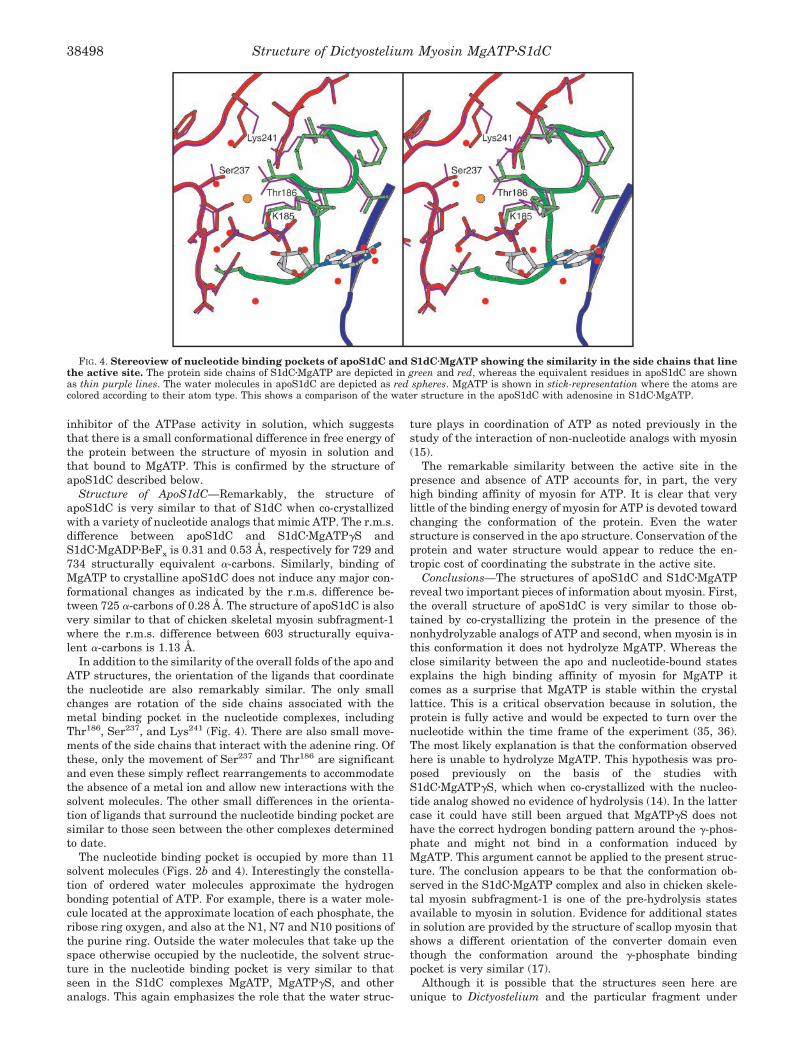
Structure of ApoS1dC—Remarkably, the structure ofapoS1dC is very similar to that of S1dC when co-crystallizedwith a variety of nucleotide analogs that mimic ATP. The r.m.s.difference between apoS1dC and S1dCzMgATPgS andS1dCzMgADPzBeFx is 0.31 and 0.53 Å, respectively for 729 and734 structurally equivalent a-carbons. Similarly, binding ofMgATP to crystalline apoS1dC does not induce any major con-formational changes as indicated by the r.m.s. difference be-tween 725 a-carbons of 0.28 Å. The structure of apoS1dC is alsovery similar to that of chicken skeletal myosin subfragment-1where the r.m.s. difference between 603 structurally equiva-lent a-carbons is 1.13 Å.
In addition to the similarity of the overall folds of the apo andATP structures, the orientation of the ligands that coordinatethe nucleotide are also remarkably similar. The only smallchanges are rotation of the side chains associated with themetal binding pocket in the nucleotide complexes, includingThr186, Ser237, and Lys241 (Fig. 4). There are also small move-ments of the side chains that interact with the adenine ring. Ofthese, only the movement of Ser237 and Thr186 are significantand even these simply reflect rearrangements to accommodatethe absence of a metal ion and allow new interactions with thesolvent molecules. The other small differences in the orienta-tion of ligands that surround the nucleotide binding pocket aresimilar to those seen between the other complexes determinedto date.
The nucleotide binding pocket is occupied by more than 11solvent molecules (Figs. 2b and 4). Interestingly the constella-tion of ordered water molecules approximate the hydrogenbonding potential of ATP. For example, there is a water mole-cule located at the approximate location of each phosphate, theribose ring oxygen, and also at the N1, N7 and N10 positions ofthe purine ring. Outside the water molecules that take up thespace otherwise occupied by the nucleotide, the solvent struc-ture in the nucleotide binding pocket is very similar to thatseen in the S1dC complexes MgATP, MgATPgS, and otheranalogs. This again emphasizes the role that the water struc-
ture plays in coordination of ATP as noted previously in thestudy of the interaction of non-nucleotide analogs with myosin(15).
The remarkable similarity between the active site in thepresence and absence of ATP accounts for, in part, the veryhigh binding affinity of myosin for ATP. It is clear that verylittle of the binding energy of myosin for ATP is devoted towardchanging the conformation of the protein. Even the waterstructure is conserved in the apo structure. Conservation of theprotein and water structure would appear to reduce the en-tropic cost of coordinating the substrate in the active site.
Conclusions—The structures of apoS1dC and S1dCzMgATPreveal two important pieces of information about myosin. First,the overall structure of apoS1dC is very similar to those ob-tained by co-crystallizing the protein in the presence of thenonhydrolyzable analogs of ATP and second, when myosin is inthis conformation it does not hydrolyze MgATP. Whereas theclose similarity between the apo and nucleotide-bound statesexplains the high binding affinity of myosin for MgATP itcomes as a surprise that MgATP is stable within the crystallattice. This is a critical observation because in solution, theprotein is fully active and would be expected to turn over thenucleotide within the time frame of the experiment (35, 36).The most likely explanation is that the conformation observedhere is unable to hydrolyze MgATP. This hypothesis was pro-posed previously on the basis of the studies withS1dCzMgATPgS, which when co-crystallized with the nucleo-tide analog showed no evidence of hydrolysis (14). In the lattercase it could have still been argued that MgATPgS does nothave the correct hydrogen bonding pattern around the g-phos-phate and might not bind in a conformation induced byMgATP. This argument cannot be applied to the present struc-ture. The conclusion appears to be that the conformation ob-served in the S1dCzMgATP complex and also in chicken skele-tal myosin subfragment-1 is one of the pre-hydrolysis statesavailable to myosin in solution. Evidence for additional statesin solution are provided by the structure of scallop myosin thatshows a different orientation of the converter domain eventhough the conformation around the g-phosphate bindingpocket is very similar (17).
Although it is possible that the structures seen here areunique to Dictyostelium and the particular fragment under
FIG. 4. Stereoview of nucleotide binding pockets of apoS1dC and S1dCzMgATP showing the similarity in the side chains that linethe active site. The protein side chains of S1dCzMgATP are depicted in green and red, whereas the equivalent residues in apoS1dC are shownas thin purple lines. The water molecules in apoS1dC are depicted as red spheres. MgATP is shown in stick-representation where the atoms arecolored according to their atom type. This shows a comparison of the water structure in the apoS1dC with adenosine in S1dCzMgATP.
Structure of Dictyostelium Myosin MgATPzS1dC38498

investigation, it is unlikely because the conformation of the50-kDa domains are very similar in chicken S1 and scallop S1.Rather, it suggests that the open conformation of the motordomain is a state that will belong to all pre-hydrolyis states.The present study refutes any proposals that the conformationof chicken skeletal myosin subfragment-1 belongs to the rigorstate. However, because the structure of chicken skeletal my-osin subfragment-1 approximately fits into image reconstruc-tions of actomyosin, it seems more likely that this conformationrepresents the state of myosin shortly after its release fromactin. This work does not conflict with the smooth muscleADPzBeFx structure, which adopts the closed conformation.The metastable state must be able to accommodate bothMgATP and MgADP-Pi. It should not be surprising that undersome crystallization conditions the closed conformation isfound in the presence of a MgATP analog. The question re-mains however, if MgATP were introduced into a crystal thatexhibits the closed conformation, would it be hydrolyzed in thelattice.
Acknowledgment—We thank Dr. Andrew M. Gulick for helpfuldiscussions.
REFERENCES
1. Mermall, V., Post, P. L., and Mooseker, M. S. (1998) Science 279, 527–5332. Sellers, J. R. (2000) Biochim. Biophys. Acta 1496, 3–223. Lymn, R. W., and Taylor, E. W. (1971) Biochemistry 10, 4617–46244. Dantzig, J. A., and Goldman, Y. E. (1985) J. Gen. Physiol. 86, 305–3275. Irving, M., St. Claire Allen, T., Sabido-David, C., Craik, J. S., Brandmeier, B.,
Kendrick-Jones, J., Corrie, J. E., Trentham, D. R., and Goldman, Y. E.(1995) Nature 375, 688–691
6. Baker, J. E., Brust-Mascher, I., Ramachandran, S., LaConte, L. E., andThomas, D. D. (1998) Proc. Natl. Acad. Sci. U. S. A. 95, 2944–2949
7. Ramachandran, S., and Thomas, D. D. (1999) Biochemistry 38, 9097–91048. Brust-Mascher, I., LaConte, L. E., Baker, J. E., and Thomas, D. D. (1999)
Biochemistry 38, 12607–126139. Rayment, I., Rypniewski, W. R., Schmidt-Base, K., Smith, R., Tomchick, D. R.,
Benning, M. M., Winkelmann, D. A., Wesenberg, G., and Holden, H. M.
(1993) Science 261, 50–5810. Fisher, A. J., Smith, C. A., Thoden, J., Smith, R., Sutoh, K., Holden, H. M., and
Rayment, I. (1995) Biochemistry 34, 8960–897211. Smith, C. A., and Rayment, I. (1996) Biochemistry 35, 5404–541712. Smith, C. A., and Rayment, I. (1995) Biochemistry 34, 8973–898113. Bauer, C. B., Kuhlman, P. A., Bagshaw, C. R., and Rayment, I. (1997) J. Mol.
Biol. 274, 394–40714. Gulick, A. M., Bauer, C. B., Thoden, J. B., and Rayment, I. (1997) Biochemistry
36, 11619–1162815. Gulick, A. M., Bauer, C. B., Thoden, J. B., Pate, E., Yount, R. G., and Rayment,
I. (2000) J. Biol. Chem. 275, 398–40816. Dominguez, R., Freyzon, Y., Trybus, K. M., and Cohen, C. (1998) Cell 94,
559–57117. Houdusse, A., Kalabokis, V. N., Himmel, D., Szent-Gyorgyi, A. G., and Cohen,
C. (1999) Cell 97, 459–47018. Rayment, I., Holden, H. M., Whittaker, M., Yohn, C. B., Lorenz, M., Holmes,
K. C., and Milligan, R. A. (1993) Science 261, 58–6519. Holmes, K. C. (1997) Curr. Biol. 7, R112–11820. Rodgers, D. W. (1997) Methods Enzymol. 276, 183–20321. Otwinowski, Z., and Minor, W. (1997) Methods Enzymol. 276, 307–32622. Tronrud, D. E. (1997) Methods Enzymol. 277, 306–31923. Roussel, A., and Cambillau, C. (1991) Silicon Graphics Geometry Partners
Directory, Vol. 86, Silicon Graphics24. Laskowski, R. A., MacArthur, M. W., Moss, D. S., and Thornton, J. M. (1993)
J. Appl. Crystallogr. 26, 283–29125. Cohen, G. H. (1997) J. Appl. Crystallogr. 30, 1160–116126. Esnouf, R. M. (1999) Acta Crystallogr. Sec. D 55, 938–94027. Greene, L. E., and Eisenberg, E. (1980) J. Biol. Chem. 255, 543–54828. Goody, R. S., and Mannherz, H. G. (1975) in Protein-Ligand Interactions
(Sund, H., and Blauer, G., eds) pp. 109–127, de Gruyter, Berlin29. Resetar, A. M., and Chalovich, J. M. (1995) Biochemistry 34, 16039–1604530. Henry, G. D., Maruta, S., Ikebe, M., and Sykes, B. D. (1993) Biochemistry 32,
10451–1045631. Ponomarev, M. A., Timofeev, V. P., and Levitsky, D. I. (1995) FEBS Lett. 371,
261–26332. Phan, B. C., Cheung, P., Stafford, W. F., and Reisler, E. (1996) Biophys. Chem.
59, 341–34933. Park, S., Ajtai, K., and Burghardt, T. P. (1997) Biochemistry 36, 3368–337234. Rostkova, E. V., Moiseeva, L. N., Teplova, M. V., Nikolaeva, O. P., and
Levitsky, D. I. (1999) Biochemistry (Mosc) 64, 875–88235. Itakura, S., Yamakawa, H., Toyoshima, Y. Y., Ishijima, A., Kojima, T., Harada,
Y., Yanagida, T., Wakabayashi, T., and Sutoh, K. (1993) Biochem. Cell Biol.196, 1504–1510
36. Kurzawa, S. E., Manstein, D. J., and Geeves, M. A. (1997) Biochemistry 36,317–323
37. Kraulis, P. J. (1991) J. Appl. Crystallogr. 24, 946–950
Structure of Dictyostelium Myosin MgATPzS1dC 38499