the nucleus and ribosomes - mrs. pietrangelo's...
TRANSCRIPT
1
THE NUCLEUS AND RIBOSOMES
© 2012 Pearson Education, Inc.
4.5 The nucleus is the cell’s genetic control center
The nucleus
– contains most of the cell’s DNA and
– controls the cell’s activities by directing protein synthesis by making messenger RNA (mRNA).
DNA is associated with many proteins in structures called chromosomes.
© 2012 Pearson Education, Inc.
4.5 The nucleus is the cell’s genetic control center
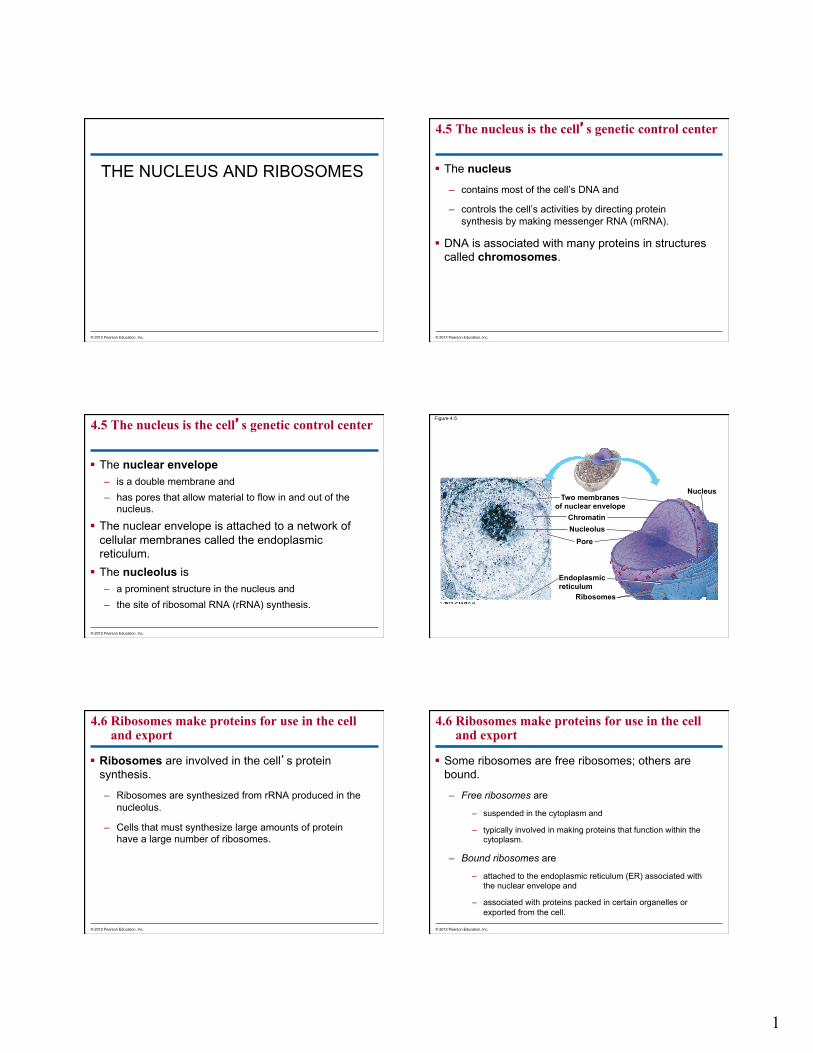
The nuclear envelope – is a double membrane and – has pores that allow material to flow in and out of the
nucleus.
The nuclear envelope is attached to a network of cellular membranes called the endoplasmic reticulum.
The nucleolus is – a prominent structure in the nucleus and – the site of ribosomal RNA (rRNA) synthesis.
© 2012 Pearson Education, Inc.
Figure 4.5
Two membranes of nuclear envelope
Nucleus
Chromatin Nucleolus
Pore
Endoplasmic reticulum
Ribosomes
4.6 Ribosomes make proteins for use in the cell and export
Ribosomes are involved in the cell’s protein synthesis.
– Ribosomes are synthesized from rRNA produced in the nucleolus.
– Cells that must synthesize large amounts of protein have a large number of ribosomes.
© 2012 Pearson Education, Inc.
4.6 Ribosomes make proteins for use in the cell and export
Some ribosomes are free ribosomes; others are bound.
– Free ribosomes are
– suspended in the cytoplasm and
– typically involved in making proteins that function within the cytoplasm.
– Bound ribosomes are
– attached to the endoplasmic reticulum (ER) associated with the nuclear envelope and
– associated with proteins packed in certain organelles or exported from the cell.
© 2012 Pearson Education, Inc.
2
Figure 4.6
Ribosomes ER Cytoplasm
Endoplasmic reticulum (ER) Free ribosomes Bound ribosomes
Diagram of a ribosome Protein
mRNA
Colorized TEM showing ER and ribosomes
THE ENDOMEMBRANE SYSTEM
© 2012 Pearson Education, Inc.
4.7 Overview: Many cell organelles are connected through the endomembrane system
Many of the membranes within a eukaryotic cell are part of the endomembrane system.
Some of these membranes are physically connected and some are related by the transfer of membrane segments by tiny vesicles (sacs made of membrane).
Many of these organelles work together in the – synthesis, – storage, and
– export of molecules.
© 2012 Pearson Education, Inc.
4.7 Overview: Many cell organelles are connected through the endomembrane system
The endomembrane system includes – the nuclear envelope,
– endoplasmic reticulum (ER),
– Golgi apparatus,
– lysosomes,
– vacuoles, and
– the plasma membrane.
John Kyrk Animation - http://www.johnkyrk.com/cellmembrane.html
© 2012 Pearson Education, Inc.
4.8 The endoplasmic reticulum is a biosynthetic factory
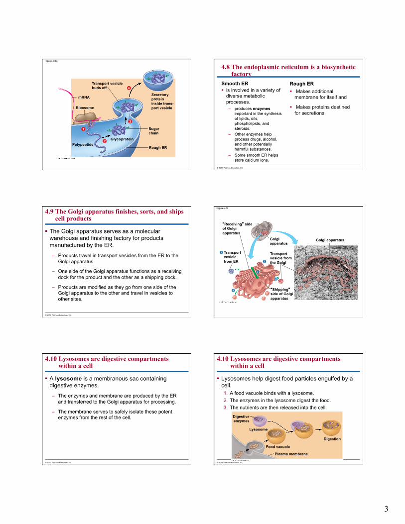
There are two kinds of endoplasmic reticulum—smooth and rough.
– Smooth ER lacks attached ribosomes.
– Rough ER lines the outer surface of membranes.
– Although physically interconnected, smooth and rough ER differ in structure and function.
© 2012 Pearson Education, Inc.
Figure 4.8A
Smooth ER
Rough ER
Ribosomes
Nuclear envelope
3
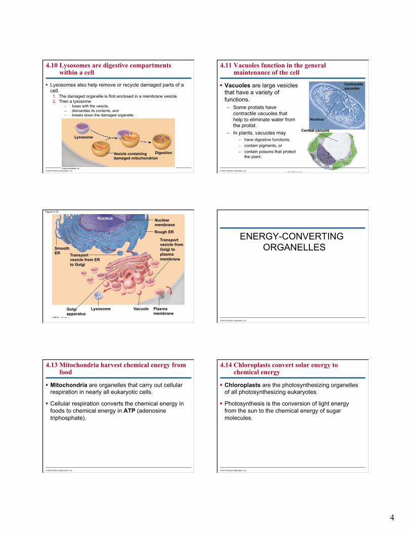
Figure 4.8B
Transport vesicle buds off
mRNA
Ribosome
Polypeptide Glycoprotein
Rough ER
Sugar chain
Secretory protein inside trans- port vesicle
4
3
2
1
4.8 The endoplasmic reticulum is a biosynthetic factory
Smooth ER is involved in a variety of
diverse metabolic processes. – produces enzymes
important in the synthesis of lipids, oils, phospholipids, and steroids.
– Other enzymes help process drugs, alcohol, and other potentially harmful substances.
– Some smooth ER helps store calcium ions.
Rough ER Makes additional
membrane for itself and
Makes proteins destined for secretions.
© 2012 Pearson Education, Inc.
4.9 The Golgi apparatus finishes, sorts, and ships cell products
The Golgi apparatus serves as a molecular warehouse and finishing factory for products manufactured by the ER.
– Products travel in transport vesicles from the ER to the Golgi apparatus.
– One side of the Golgi apparatus functions as a receiving dock for the product and the other as a shipping dock.
– Products are modified as they go from one side of the Golgi apparatus to the other and travel in vesicles to other sites.
© 2012 Pearson Education, Inc.
Figure 4.9
Golgi apparatus Golgi apparatus
Transport vesicle from the Golgi
“Shipping” side of Golgi apparatus
Transport vesicle from ER
“Receiving” side of Golgi apparatus
1
2
3
4
4
4.10 Lysosomes are digestive compartments within a cell
A lysosome is a membranous sac containing digestive enzymes.
– The enzymes and membrane are produced by the ER and transferred to the Golgi apparatus for processing.
– The membrane serves to safely isolate these potent enzymes from the rest of the cell.
© 2012 Pearson Education, Inc.
4.10 Lysosomes are digestive compartments within a cell
Lysosomes help digest food particles engulfed by a cell. 1. A food vacuole binds with a lysosome. 2. The enzymes in the lysosome digest the food. 3. The nutrients are then released into the cell.
© 2012 Pearson Education, Inc.
Digestive enzymes
Lysosome
Food vacuole
Plasma membrane
Digestion
4
4.10 Lysosomes are digestive compartments within a cell
Lysosomes also help remove or recycle damaged parts of a cell. 1. The damaged organelle is first enclosed in a membrane vesicle. 2. Then a lysosome
– fuses with the vesicle, – dismantles its contents, and – breaks down the damaged organelle.
© 2012 Pearson Education, Inc.
Lysosome
Vesicle containing damaged mitochondrion
Digestion
4.11 Vacuoles function in the general maintenance of the cell
Vacuoles are large vesicles that have a variety of functions. – Some protists have
contractile vacuoles that help to eliminate water from the protist.
– In plants, vacuoles may – have digestive functions, – contain pigments, or – contain poisons that protect
the plant.
© 2012 Pearson Education, Inc.
Contractile vacuoles
Nucleus
Central vacuole
Figure 4.12
Smooth ER
Nucleus
Transport vesicle from ER to Golgi
Golgi apparatus
Lysosome Vacuole Plasma membrane
Nuclear membrane
Rough ER
Transport vesicle from Golgi to plasma membrane
ENERGY-CONVERTING ORGANELLES
© 2012 Pearson Education, Inc.
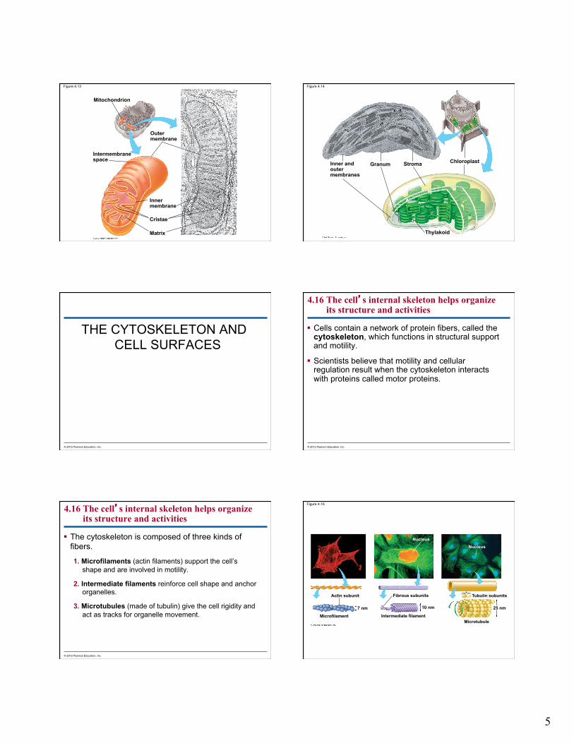
4.13 Mitochondria harvest chemical energy from food
Mitochondria are organelles that carry out cellular respiration in nearly all eukaryotic cells.
Cellular respiration converts the chemical energy in foods to chemical energy in ATP (adenosine triphosphate).
© 2012 Pearson Education, Inc.
4.14 Chloroplasts convert solar energy to chemical energy
Chloroplasts are the photosynthesizing organelles of all photosynthesizing eukaryotes.
Photosynthesis is the conversion of light energy from the sun to the chemical energy of sugar molecules.
© 2012 Pearson Education, Inc.
5
Figure 4.13
Matrix
Cristae
Inner membrane
Outer membrane
Mitochondrion
Intermembrane space
Figure 4.14
Inner and outer membranes
Granum Stroma Chloroplast
Thylakoid
THE CYTOSKELETON AND CELL SURFACES
© 2012 Pearson Education, Inc.
4.16 The cell’s internal skeleton helps organize its structure and activities
Cells contain a network of protein fibers, called the cytoskeleton, which functions in structural support and motility.
Scientists believe that motility and cellular regulation result when the cytoskeleton interacts with proteins called motor proteins.
© 2012 Pearson Education, Inc.
4.16 The cell’s internal skeleton helps organize its structure and activities
The cytoskeleton is composed of three kinds of fibers.
1. Microfilaments (actin filaments) support the cell’s shape and are involved in motility.
2. Intermediate filaments reinforce cell shape and anchor organelles.
3. Microtubules (made of tubulin) give the cell rigidity and act as tracks for organelle movement.
© 2012 Pearson Education, Inc.
Figure 4.16
Actin subunit
Nucleus Nucleus
Microfilament Intermediate filament
Fibrous subunits
7 nm 10 nm
Tubulin subunits
Microtubule
25 nm
6
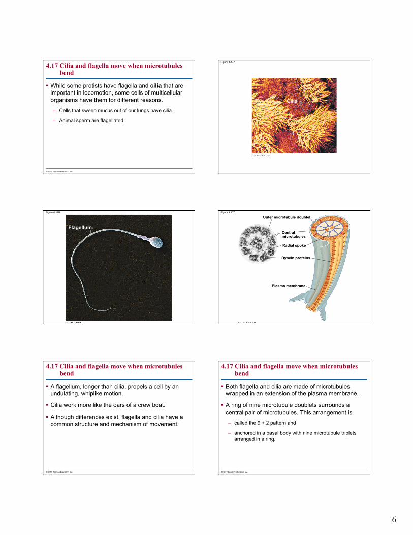
4.17 Cilia and flagella move when microtubules bend
While some protists have flagella and cilia that are important in locomotion, some cells of multicellular organisms have them for different reasons.
– Cells that sweep mucus out of our lungs have cilia.
– Animal sperm are flagellated.
© 2012 Pearson Education, Inc.
Figure 4.17A
Cilia
Figure 4.17B
Flagellum
Figure 4.17C
Outer microtubule doublet
Central microtubules
Radial spoke
Dynein proteins
Plasma membrane
4.17 Cilia and flagella move when microtubules bend
A flagellum, longer than cilia, propels a cell by an undulating, whiplike motion.
Cilia work more like the oars of a crew boat.
Although differences exist, flagella and cilia have a common structure and mechanism of movement.
© 2012 Pearson Education, Inc.
4.17 Cilia and flagella move when microtubules bend
Both flagella and cilia are made of microtubules wrapped in an extension of the plasma membrane.
A ring of nine microtubule doublets surrounds a central pair of microtubules. This arrangement is
– called the 9 + 2 pattern and
– anchored in a basal body with nine microtubule triplets arranged in a ring.
© 2012 Pearson Education, Inc.
7
4.17 Cilia and flagella move when microtubules bend
Cilia and flagella move by bending motor proteins called dynein feet.
– These feet attach to and exert a sliding force on an adjacent doublet.
– The arms then release and reattach a little further along and repeat this time after time.
– This “walking” causes the microtubules to bend.
© 2012 Pearson Education, Inc.
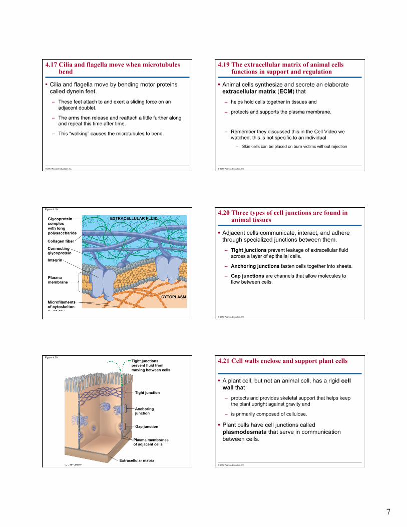
4.19 The extracellular matrix of animal cells functions in support and regulation
Animal cells synthesize and secrete an elaborate extracellular matrix (ECM) that
– helps hold cells together in tissues and
– protects and supports the plasma membrane.
– Remember they discussed this in the Cell Video we watched, this is not specific to an individual
– Skin cells can be placed on burn victims without rejection
© 2012 Pearson Education, Inc.
Figure 4.19
EXTRACELLULAR FLUID
CYTOPLASM Microfilaments of cytoskelton
Plasma membrane
Integrin
Connecting glycoprotein
Glycoprotein complex with long polysaccharide
Collagen fiber
4.20 Three types of cell junctions are found in animal tissues
Adjacent cells communicate, interact, and adhere through specialized junctions between them.
– Tight junctions prevent leakage of extracellular fluid across a layer of epithelial cells.
– Anchoring junctions fasten cells together into sheets.
– Gap junctions are channels that allow molecules to flow between cells.
© 2012 Pearson Education, Inc.
Figure 4.20 Tight junctions prevent fluid from moving between cells
Tight junction
Anchoring junction
Gap junction
Plasma membranes of adjacent cells
Extracellular matrix
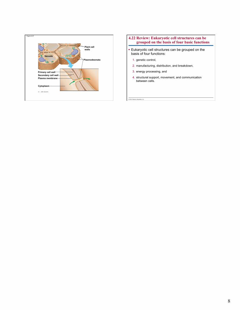
4.21 Cell walls enclose and support plant cells
A plant cell, but not an animal cell, has a rigid cell wall that
– protects and provides skeletal support that helps keep the plant upright against gravity and
– is primarily composed of cellulose.
Plant cells have cell junctions called plasmodesmata that serve in communication between cells.
© 2012 Pearson Education, Inc.
8
Figure 4.21
Vacuole
Plant cell walls
Plasmodesmata
Cytoplasm
Primary cell wall Secondary cell wall Plasma membrane
4.22 Review: Eukaryotic cell structures can be grouped on the basis of four basic functions
Eukaryotic cell structures can be grouped on the basis of four functions:
1. genetic control,
2. manufacturing, distribution, and breakdown,
3. energy processing, and
4. structural support, movement, and communication between cells.
© 2012 Pearson Education, Inc.