the open biotechnology journal
TRANSCRIPT

Send Orders for Reprints to [email protected]
36 The Open Biotechnology Journal, 2016, 10, (Suppl-1, M4) 36-41
1874-0707/16 2016 Bentham Open
The Open Biotechnology Journal
Content list available at: www.benthamopen.com/TOBIOTJ/
DOI: 10.2174/1874070701610010036
Epigenetic Reprogramming in the Mammalian Germ Line: PossibleEffects by Endocrine Disruptors on Primordial Germ Cells
Massimo De Felici* and Gina La Sala
Department of Biomedicine and Prevention, Section of Histology and Embryology, University of Rome "Tor Vergata",Rome, Italy
Abstract: The present work provides a brief review about evidence obtained in the past years mainly in our laboratory using themouse model, that germ cells and gonadal somatic cells may be direct target of endocrine disruptors (EDs) from very early stages ofgonad formation. Since it is now known that epigenetic pathways are crucial for germline development and that EDs are also able tointerfere with epigenetic mechanisms, we will discuss these results mostly in light of possible effects by such molecules on theepigenoma of the primordial germ cells (PGCs), the precursors of the adult gametes that transmit genetic information betweengenerations.
Keywords: Epigenetics, endocrine disruptors, primordial germ cells, xenoestrogens.
INTRODUCTION
On the basis of endogenous and exogenous environmental signals, epigenetic mechanisms of gene regulationinvolving DNA methylation, histone modifications, changes of chromatin structure and microRNA expression, organizethe genome into active and inactive domains, representing crucial players of gene expression. Key epigenetic modifiersof the genomic DNA are the methyltransferases (DNMTs), the methyl-CpG (MeC)-binding proteins and the histone-modifying enzymes. Among these, DNMT1 is a maintenance DNA methyltransferase whereas DNMT3A and DNMT3Bfunction in de novo methylation. Epigenetic regulation of genes encoding these enzymes, the MeC-binding proteins orthe microRNA expression by hormones provides ways of interplay between the endocrine system and epigenetics.These interactions can be insidiously modulated by a class of compounds termed as endocrine disruptors (EDs). EDsare environmental chemicals that mimic hormones or exert anti-hormone activities, and alter the physiologic function ofendogenous hormones. Plants are the sources of some of these chemicals, named phytoestrogens, while others arenatural substances such as heavy metals or synthetic molecules or drugs. Several reports indicate that in mammals and anumber of other species, EDs may be detrimental to reproduction by promoting abnormalities in sex differentiation andgonad functions, including testicular cancer in the male and ovarian diseases in the female (for a review, see [1]).However, the exact mechanisms of the ED action on the reproductive system are not completely known.
In the present work, we briefly review evidence coming mostly from results obtained in our laboratory in the mouse,that germ cells and gonadal somatic cells may be direct target of EDs from very early stages of gonad formation duringthe embryonic life. Since it is now emerging that epigenetic pathways are crucial for germline development and thatEDs are able to interfere with epigenetic mechanisms, we will discuss these results in light of possible effects by suchmolecules on the epigenoma of the primordial germ cells (PGCs), the precursors of the adult gametes. PGCs appear atearly stages of embryonic development and differentiate into oogonia/oocytes or prospermatogonia/gonocytes in thefemale and male foetal gonads, respectively. After birth, oocytes and gonocytes finally give rise to haploid oocytes andsperm that upon fertilization produces new individuals. In principle, alteration of epigenetic dynamics in PGCs may
* Address correspondence to this author at the Dipartimento di Biomedicina e Prevenzione, Università di Roma "Tor Vergata", Via Montpellier, 1,00133 Roma, Italy; Tel: 39-6-7259 6156; Fax: 39-6-7259 6172; E-mail: [email protected]

Epigenetic Reprogramming in the Mammalian Germ Line The Open Biotechnology Journal, 2016, Volume 10 37
lead not only to defects in germ cell development and adult fertility but it might be transmitted to the next generationwith possible onset of diseases in the adults.
Fig. (1). Schematic drawing of the ERα-dependent E2/ED activable pathways in mouse PGCs following the results described in [2].Binding of E2 or EDs to ERα allows the formation of a complex with MNAR that binds SRCs and leads to its activation; the complexbinds and phosphorylates (activates) PI3K and SRC. Upon activation PI3K mediates AKT phosphorylation (activation); SRC viaRAS and RAF are also able to trigger the activation pathway of ERKs (mainly ERK2). Eventually, SRC are responsible to cross-phosphorylation of the KIT receptor through an unknown pathway; this eventually also leads to the activation of the PI3K/AKTpathway.
ENDOCRINE DISRUPTORS AND REPRODUCTIVE TOXICOLOGY
There is little doubt that exposure to EDs may promote abnormalities in the reproductive system. For example, inmany species EDs alter sex differentiation and are regarded responsible for the decrease in sperm count and an increasein testicular cancer incidence in humans. Examples of the environmental EDs postulated to have adverse effects on thereproductive system in animals including humans are pesticides (e.g., methoxychlor), fungicides (e.g., vinclozolin), arange of xenoestrogens (EDs with estrogenic activity), like bisphenol A (BPA) and certain phthalates (for a review, see,[1]). Environmental xenoestrogens are likely to elicit their actions through the two canonical mammalian receptors forestrogens (ERα and ERβ), widely expressed in the gonads and the reproductive tracts. Several studies reported theexpression of ERα and ERβ in rodent and human prenatal and postnatal ovaries and testes both by germ cells andsomatic cells ([2] and references here in). Relevant for the present review, we found that ERα is expressed both byPGCs [2] and somatic cells [3] of mouse sex indifferent gonads. Moreover, results obtained in our and otherlaboratories showed that the development of mammalian PGCs and spermatogonia (namely their proliferation/survival),could be modulated and/or altered by estrogens and some EDs both directly or indirectly through neighbouring somaticcells , as the results of genomic and nongenomic effects ([2, 3] and references here in). In some cases, the molecularmechanisms underlying the ED effect on germ cells have been identified. For example, we found that 17-β-estradiol(E2) was able to rapidly stimulate AKT kinase, KIT receptor, ERK2 and SRC kinase phosphorylation in mouse PGCsand to promote their survival/proliferation [2] (Fig. 1). On the other hand, the level of AKT activity significantlydecreased in mouse PGCs exposed to lindane (γ-HCH) in vitro along with the increase in the number of apoptotic germcells either in culture and in the embryo [4]. Likewise, others found that a G-protein-coupled receptor-30 (GPR-30)mediates E2-induced proliferation of chicken PGCs through EGFR/AKT/β-catenin signaling pathway [5]. Other studiesshowed various responses by mouse PGCs to EDs likely through different mechanisms of action [6]. For example, invitro cultured PGCs exposed to mono (2-ethylhexyl) phthalate (MEHP) (the direct metabolite of the 2-ethyl-hexyl-phthalate or DEHP), a widespread plasticizer, affected PGC adhesion to cell monolayers likely causing alteration ofgerm cell-somatic cell interactions crucial for gonad development [7]. In order to identify gene deregulated by differentEDs and reprotoxicants, cDNA libraries prepared from small number of mouse PGCs followed by differentialscreening, showed altered gene expression in these cells after in vitro exposure to N-ethyl-N-nitrosurea (ENU),
38 The Open Biotechnology Journal, 2016, Volume 10 De Felici and La Sala
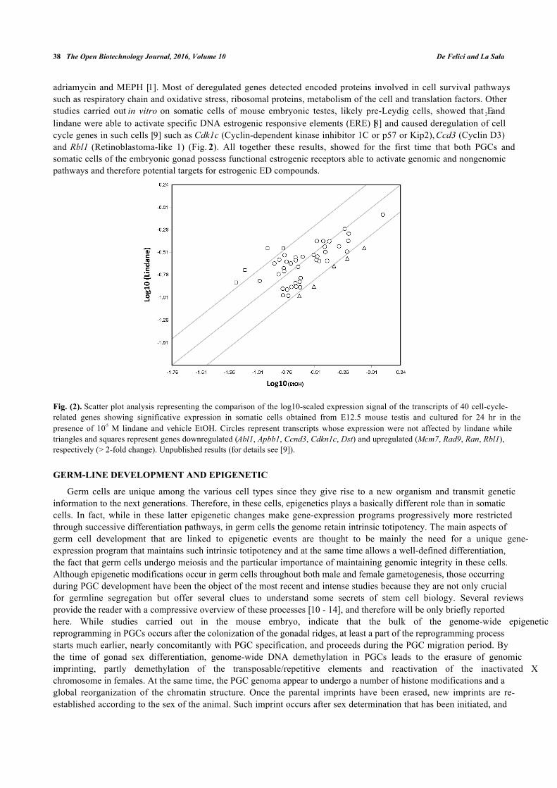
adriamycin and MEPH [1]. Most of deregulated genes detected encoded proteins involved in cell survival pathwayssuch as respiratory chain and oxidative stress, ribosomal proteins, metabolism of the cell and translation factors. Otherstudies carried out in vitro on somatic cells of mouse embryonic testes, likely pre-Leydig cells, showed that E2 andlindane were able to activate specific DNA estrogenic responsive elements (ERE) [8] and caused deregulation of cellcycle genes in such cells [9] such as Cdk1c (Cyclin-dependent kinase inhibitor 1C or p57 or Kip2), Ccd3 (Cyclin D3)and Rbl1 (Retinoblastoma-like 1) (Fig. 2). All together these results, showed for the first time that both PGCs andsomatic cells of the embryonic gonad possess functional estrogenic receptors able to activate genomic and nongenomicpathways and therefore potential targets for estrogenic ED compounds.
Fig. (2). Scatter plot analysis representing the comparison of the log10-scaled expression signal of the transcripts of 40 cell-cycle-related genes showing significative expression in somatic cells obtained from E12.5 mouse testis and cultured for 24 hr in thepresence of 10-5 M lindane and vehicle EtOH. Circles represent transcripts whose expression were not affected by lindane whiletriangles and squares represent genes downregulated (Abl1, Apbb1, Ccnd3, Cdkn1c, Dst) and upregulated (Mcm7, Rad9, Ran, Rbl1),respectively (> 2-fold change). Unpublished results (for details see [9]).
GERM-LINE DEVELOPMENT AND EPIGENETIC
Germ cells are unique among the various cell types since they give rise to a new organism and transmit geneticinformation to the next generations. Therefore, in these cells, epigenetics plays a basically different role than in somaticcells. In fact, while in these latter epigenetic changes make gene-expression programs progressively more restrictedthrough successive differentiation pathways, in germ cells the genome retain intrinsic totipotency. The main aspects ofgerm cell development that are linked to epigenetic events are thought to be mainly the need for a unique gene-expression program that maintains such intrinsic totipotency and at the same time allows a well-defined differentiation,the fact that germ cells undergo meiosis and the particular importance of maintaining genomic integrity in these cells.Although epigenetic modifications occur in germ cells throughout both male and female gametogenesis, those occurringduring PGC development have been the object of the most recent and intense studies because they are not only crucialfor germline segregation but offer several clues to understand some secrets of stem cell biology. Several reviewsprovide the reader with a compressive overview of these processes [10 - 14], and therefore will be only briefly reportedhere. While studies carried out in the mouse embryo, indicate that the bulk of the genome-wide epigeneticreprogramming in PGCs occurs after the colonization of the gonadal ridges, at least a part of the reprogramming processstarts much earlier, nearly concomitantly with PGC specification, and proceeds during the PGC migration period. Bythe time of gonad sex differentiation, genome-wide DNA demethylation in PGCs leads to the erasure of genomicimprinting, partly demethylation of the transposable/repetitive elements and reactivation of the inactivated Xchromosome in females. At the same time, the PGC genoma appear to undergo a number of histone modifications and aglobal reorganization of the chromatin structure. Once the parental imprints have been erased, new imprints are re-established according to the sex of the animal. Such imprint occurs after sex determination that has been initiated, and

Epigenetic Reprogramming in the Mammalian Germ Line The Open Biotechnology Journal, 2016, Volume 10 39
male and female germ-cell development diverges to give rise to sperm or eggs. In male, paternal methylation imprintsare progressively established in germ cells between the end of the fetal and the newborn stages. In the female germline,the initiation of DNA methylation imprinting occurs after birth, during the oocyte growth. The growing oocytes areblocked at the diplotene stage of meiotic prophase I, and the de novo methylation process is complete by the fully-grown oocyte stage. Besides DNA demethylation and methylation, several changes in histone occur during meiosis andthe final stages of both female and male gametogenesis that complete the dynamics of the germ cell epigenetics.
Fig. (3). Role of the PGCs in epigenetic transgenerational inheritance. EDs acting on the F0 generation gestating female influence thedeveloping F1 generation foetus and alter gonadal development to reprogram the PGC DNA methylation. This epigenetic alterationin the germline may become permanent and is transferred through the germline to subsequent generations. The embryo generatedfrom this germline posses an altered epigenome that affects developing somatic cells and tissues. This altered somatic celltranscriptome can then promote adult-onset disease associated with the transgenerational phenotype.
Given the evidence that EDs can affect epigenetics, for example by modifying the DNA methylation status and/orinhibiting histone deacetylase activity (for a review see [15]) and, as reported above, the presence functional estrogenicreceptors in PGCs and the companion somatic cells, it is reasonable to think that EDs, at least those with estrogenicactivity, might interfere with the germ cell reprogramming through epigenetic mechanisms. These effects can result inimmediate abnormalities in germ cell development and/or cause transgenerationaleffects on next generations. Thisprovides a unique epigenetic mechanism to promote a transgenerationalphenotype induced by an environmental factorincluding, besides testis and/or ovary abnormalities, tumours and pathological development in a variety of somatictissues [15].
Actually, there has always been much interest in the idea that some epigenetic marks can be inherited acrossgenerations. However, despite the fact that these marks are considered relatively stable during development, theyundergo resetting in PGCs and subsequently in the zygote at fertilization to ensure the totipotency of cells of the earlyembryo. For transgenerational epigenetic inheritance to occur this reprogramming must be, however, bypassed (Fig. 3).Recent results indicate that some sites of the genome can, actually, evade erasure of DNA methylation occurring inPGCs [16 - 18]. But evidence that EDs can impose transmissible epigenetic mark on PGCs is hotly debated.
Actually, some papers from the Skinner‘group support such a possibility. These authors reported that maternalexposure to certain environmental conditions and in particular to some EDs, result in transgenerational phenotypethroughout epigenetic alterations in the germ-line. In particular, they showed that transient male rat embryo exposure tovinclozolin at the time of PGC development remarkably caused a transgenerational phenotype in F1-F4 generations ofmale germ cell apoptosis and subfertility. This apparent epigenetic mechanism involves altered DNA methylation andpermanent re-programming of the male germline. In fact, a series of genes with altered DNA methylation andimprinting were identified in PGCs and later in sperm [19]. The rat model was also used to evaluate whether adult onsetovarian diseases could be induced transgenerationally after exposure of a gestating F0 generation female to knownenvironmental toxicants such as vinclozolin and a mixture of polycarbonate plastics such as BPA, dibulylphthalate(DBP) and bis(2-ethylexyl) phthalate (DEHP), during the period of PGC migration and gonadal ridges formation [20].The results showed that the environmental toxicants examined induced transgenerational ovarian adult-onset diseaseresembling human ovarian insufficiency (POI) and polycystic ovarian syndrome (PCOS), thus suggesting that suchovarian disease can have an epigenetic transgenerational etiology. Despite such results, others found that

40 The Open Biotechnology Journal, 2016, Volume 10 De Felici and La Sala
undernourishment during the prenatal life may compromise F1 sperm methylation but such change is not transmitted toF2 offspring; nonetheless, gene expression is altered in somatic cells of these F2 offspring at regions of F1 germlinedifferential methylation [21], leaving open the way to alternative possibilities for epigenetic trans-genarationaltransmission.
CONCLUSION
The epigenetic germ-line transgenerational disease hypothesis provides a unique perspective from which to viewadult onset disease caused by endocrine disruptors and ultimately offers new insights into novel prevention anddiagnostic and therapeutic approaches. Moreover, the researches in this field contribute to our understanding of theepigeneticmechanisms underlying imprinting erase and acquisition during germcell development and may haveimplications for assistedreproductive technologies. With the rapid advances in technologies for high-throughput globalscreening of DNA methylation, histone modifications, and small RNA profiling and the advent of next-generationsequencing technologies, we expect an explosive phase of growth in genome and epigenome science. Further studies onPGCs should focus on the molecular pathways activated by EDs leading to alteration of DNA methylation or otherepigenetic modifiers. Along with these advances will be significant for our understanding of the interplay of epigeneticsand genetics with environment compounds in modulating the endocrine system at the individual and population leveland of the etiology of several diseases.
CONFLICT OF INTEREST
The authors confirm that this article content has no conflict of interest.
ACKNOWLEDGEMENTS
Declared none.
REFERENCES
[1] Del Mazo J, Brieno-Enriquez MA. Endocrine disruptors, gene deregulation and male germ cell tumors. Int J Dev Biol 2013; 57: 225-9.
[2] La Sala G, Farini D, De Felici M. Rapid estrogen signaling in mouse primordial germ cells. Exp Cell Res 2010; 316: 1716-27.
[3] Moe-Behrens GH, Klinger FG, Eskild W, et al. AKT/PTEN signalling mediates estrogen-dependent proliferation of primordial germ cells invitro. Mol End 2003; 17: 2630-8.
[4] La Sala G, Farini D, De Felici M. Proapoptotic effects of lindane on mouseprimordial germ cells. Toxicol Sci 2009; 108: 445-51.
[5] Ge C, Yu M, Zhang C. G protein-coupled receptor 30 mediates estrogen-induced proliferation of primordial germ cells via EGFR/Akt/-catenin signaling pathway. Endocrinology 2012; 153: 3504-16.
[6] Lessard C, Pendola JK, Hartford SA, et al. New mouse genetic models for human contraceptiove development. Cytogenet Genome Res 2004;105: 222-7.
[7] Iona S, Klinger FG, Sisti R, et al. A comparative study of cytotoxic effects of N-ethyl-N-nitrosurea, adriamycin, and mono- (2-ethylhexyl)phthalate on mouse primordial germ cells. Cell Biol Toxicol 2002; 18(2): 131-45.[http://dx.doi.org/10.1023/A:1015336318623]
[8] La Sala G, Farini D, De Felici M. Estrogenic in vitro assay on mouse embryonic Leydig cells. Int J Dev Biol 2011; 54: 717-22.
[9] La Sala G. Effects of estrogens and endocrine disruptors on mouse embryonal germ cells and somatic gonadal cells. PhD Thesis. University ofRome Tor Vergata, Rome 2011.
[10] Hayashi K, Surani MA. Resetting the epigenome beyond pluripotency in the germline. Cell Stem Cell 2009; 4: 493-8.
[11] Sasaki H, Matsui Y. Epigenetic events in mammalian germ-cell development: reprogramming and beyond. Nat Rev Genet 2008; 9: 129-40.
[12] Kota S, Fell R. Epigenetic transitions in germ cell development and meiosis. Dev Cell 2010; 9: 675-86.
[13] Saitou M, Yamaji M. Primordial germ cells in mice. Cold Spring Harb Perspect Biol 2012; 4(11) .[http://dx.doi.org/10.1101/cshperspect.a0083752012]
[14] De Felici M. Nuclear reprogramming in mouse primordial germ cells: epigenetic contribution. Stem Cell Int 2011; 2011: 425863[http://dx.doi.org/10.4061/2011/425863]
[15] Skinner MK, Manikkam M, Guerrero-Bosagna C. Epigenetic transgenerational actions of environmental factors in disease etiology. TrendsEndocrinol Metab 2010; 21: 214-22.
[16] Lees-Murdock D, De Felici M, Walsh C. Methylation dynamics of repetitive DNA elements in the mouse germ cell lineage. Genomics 2003;82: 230-7.
[17] Seisenberg S, Andrews S, Krueger F, et al. The dynamics of genome-wide DNA methylation reprogramming in mouse primordial germ cells.

Epigenetic Reprogramming in the Mammalian Germ Line The Open Biotechnology Journal, 2016, Volume 10 41
Mol Cell 2012; 48: 849-62.
[18] Hackett JA, Sengupta R, Zylicz JJ, et al. Germline DNA demethylation dynamics and imprint erasure through 5-hydroxymethylcytosine.Science 2013; 339: 448-52.
[19] Skinner MK, Guerrero-Bosagna C, Haque M, et al. Environmentally induced transgenerational epigenetic reprogramming of primordial germcells and the subsequent germ line. PLoS One 2013; 8: e66318.
[20] Nilsson E, Larsen G, Manikkam M, et al. Environmentally induced epigenetic transgenerational Inheritance of ovarian disease. PLoS One2012; 7: e36129.
[21] Radford EJ, Ito M, Shi H, et al. In utero undernourishment perturbs the adult sperm methylome and intergenerational metabolism. Science2014; 345(6198): 1255903.
Received: June 2, 2014 Revised: May 15, 2015 Accepted: June 5, 2015
© De Felici and La Sala; Licensee Bentham Open.
This is an open access article licensed under the terms of the Creative Commons Attribution-Non-Commercial 4.0 International Public License (CCBY-NC 4.0) (https://creativecommons.org/licenses/by-nc/4.0/legalcode), which permits unrestricted, non-commercial use, distribution andreproduction in any medium, provided the work is properly cited.