the pathogenesis and management of achalasia: current ...the pathogenesis and management of...
TRANSCRIPT

Review
Gut and Liver, Vol. 9, No. 4, July 2015, pp. 449-463
The Pathogenesis and Management of Achalasia: Current Status and Future Directions
Fehmi Ates and Michael F. Vaezi
Division of Gastroenterology, Hepatology, and Nutrition, Center for Swallowing and Esophageal Disorders, Vanderbilt University Medical Center, Nashville, TN, USA
Achalasia is an esophageal motility disorder that is common-ly misdiagnosed initially as gastroesophageal reflux disease. Patients with achalasia often complain of dysphagia with sol-ids and liquids but may focus on regurgitation as the primary symptom, leading to initial misdiagnosis. Diagnostic tests for achalasia include esophageal motility testing, esophago-gastroduodenoscopy and barium swallow. These tests play a complimentary role in establishing the diagnosis of suspect-ed achalasia. High-resolution manometry has now identified three subtypes of achalasia, with therapeutic implications. Pneumatic dilation and surgical myotomy are the only defini-tive treatment options for patients with achalasia who can undergo surgery. Botulinum toxin injection into the lower esophageal sphincter should be reserved for those who can-not undergo definitive therapy. Close follow-up is paramount because many patients will have a recurrence of symptoms and require repeat treatment. (Gut Liver 2015;9:449-463)
Key Words: Pneumatic dilation; Surgical myotomy; Peroral esophageal myotomy
INTRODUCTION
Achalasia is a primary esophageal motor disorder of unknown etiology characterized manometrically by insufficient relaxation of the lower esophageal sphincter (LES) and loss of esophageal peristalsis; radiographically by aperistalsis, esophageal dilation, with minimal LES opening, “bird-beak” appearance, poor emp-tying of barium; and endoscopically by dilated esophagus with retained saliva, liquid, and undigested food particles in the ab-sence of mucosal stricturing or tumor. Achalasia occurs equally in both genders with prevalence that ranges up to 1 per 10,000 persons.1 There is no racial predilection. The majority of cases
are idiopathic, but the syndrome can be associated with malig-nancy (especially involving the gastroesophageal junction) and as a part of the spectrum of Chagas disease. Rarely, achalasia is genetically transmitted.2
Achalasia is an uncommon esophageal motility disorder de-fined traditionally by manometric criteria in the classic setting of dysphagia.3-7 The symptomatic consequence of this motility disorder is the classic presentation of dysphagia to solids and liquids associated with regurgitation of bland undigested food or saliva.3 Substernal chest pain during meals in the setting of dysphagia, weight loss, and even heartburn may be accompa-nying symptoms that often lead to misdiagnosis of achalasia erroneously as gastroesophageal reflux disease (GERD).8,9 Acha-lasia must be suspected in those with dysphagia to solids and liquids and in those with regurgitation unresponsive to initial trial of proton pump inhibitor (PPI) therapy.10 Endoscopic find-ings of retained saliva, liquid, and food in the esophagus with-out mechanical obstruction from stricture or mass should raise suspicion for achalasia. Conversely, other conditions may mimic achalasia both clinically and manometrically. These include pseudoachalasia from tumors in the gastric cardia or those infil-trating the myenteric plexus (adenocarcinoma of gastroesopha-geal junction, pancreatic, breast, lung, or hepatocellular cancers) or secondary achalasia from extrinsic processes such as prior tight fundoplication or laparoscopic adjustable gastric band-ing.11,12
1. History
Achalasia was first described and termed by Sir Thomas Willis in 1674, where he suggested that the disease is due to the loss of normal inhibition in the distal esophagus.13 Since then, the development of new diagnostic techniques stimulated new ideas on the etiology and pathophysiology of the disease leading to
This is an Open Access article distributed under the terms of the Creative Commons Attribution Non-Commercial License (http://creativecommons.org/licenses/by-nc/4.0) which permits unrestricted non-commercial use, distribution, and reproduction in any medium, provided the original work is properly cited.
Correspondence to: Michael F. VaeziDivision of Gastroenterology and Hepatology, Center for Swallowing and Esophageal Disorders, Vanderbilt University Medical Center, C2104-MCN, Nashville, TN 37232, USA Tel: +1-615-322-3739, Fax: +1-615-322-8525, E-mail: [email protected]
Received on November 14, 2014. Accepted on December 7, 2014.pISSN 1976-2283 eISSN 2005-1212 http://dx.doi.org/10.5009/gnl14446

450 Gut and Liver, Vol. 9, No. 4, July 2015
various theories in identifying the nature of motor disturbances in esophageal regions. This includes cardiospasm, esophageal muscle failure, and physical obstruction.14 In 1929, Sir Arthur Hurst coined the term “achalasia” suggesting that it may be due to the “loss of normal inhibition” in the distal esophagus.13 Subsequently, a body of evidence has emerged showing that idiopathic achalasia, characterized by the failure of the lower esophageal sphincter (LES) relaxation and aperistalsis, is indeed caused primarily by the loss of the inhibitory innervation of the esophageal myenteric plexus. However, the initiating cause is still elusive.
2. Esophageal motor innervation
Esophageal motor innervation is through the vagus nerve via the Myenteric or Meissner’s plexus (Fig. 1A). Neural innerva-tion differs in the proximal and distal esophagus. The striated muscle of the proximal esophagus is innervated by the somatic efferent fibers of the vagus nerve (Fig. 1B). The cell bodies for these fibers originate in the nucleus ambiguous and terminate on the motor end plate directly via cholinergic receptors.15,16 On the other hand, the smooth muscle of the distal esophagus is innervated by the preganglionic vagus nerve fibers with cell bodies located in the dorsal motor nucleus (DMN).17 Pregangli-onic fibers first innervate the myenteric plexus via cholinergic fibers.18 The esophageal wall and LES are subsequently inner-vated by the postganglionic neurons, consisting of excitatory and inhibitory neurons. The postganglionic excitatory neurons release acetylcholine while the inhibitory neurons release nitric oxide (NO) and vasoactive intestinal polypeptide (VIP) resulting in esophageal and LES contractions and relaxations, respec-tively (Fig. 1B).19,20
In addition to tonic contraction and relaxation, the inhibi-tory neurons are also vital to normal esophageal peristalsis. The esophagus, at baseline, is in a contractile state; however, with deglutition, the inhibitory neurons are excited to override the effect of excitatory neurons resulting in esophagus relaxation. Peristalsis is the net result of the coordinated relaxation and contraction mediated by the inhibitory and excitatory myenteric plexus neurons along the length of the esophagus.21 In achala-sia, there is loss of NO and VIP releasing inhibitory neurons.22,23 Thus, the loss of the inhibitory innervation in achalasia results in the manometric consequence of failure of LES relaxation as well as loss of esophageal peristalsis.
PATHOGENESIS
Pathophysiologically, the loss of the inhibitory innervation of the esophagus can be due to either extrinsic or intrinsic causes. Extrinsic causes may include central nervous system (CNS) le-sions involving the DMN or the vagal nerve fibers, while intrin-sic loss may be due to loss of the inhibitory ganglion cells in the myenteric plexus.
1. Extrinsic neuronal loss
Kimura24 was the first to suggest lesions in the CNS could ex-plain the clinical and manometric findings in achalasia. In 1929, he examined histologic sections of postmortem specimens of three achalasia patients. He discovered degenerated vagus nerve cells in the DMN. Similar DMN pathology was later reported by Cassella et al.25 This group conducted a histologic study of serial brain-stem sections of two achalasia patients and one control. They found a 34% to 43% decrease in the number of DMN neu-
A B
Meissner's plexus(submucosal)
Auerbach's plexus(myenteric)
Circular muscle
Vagus nerve
Longitudinal muscle
Vagus nerve
Dorsal motor nucleus
Nucleus ambiguus
Upper esophagus
Ach
Striated muscle fiber
+
+Ach
Ach+
Lower esophagus
Smooth muscle fiber
+Ach NO, VIP
Fig. 1. (A) Esophageal motor innervation by the vagus nerve; Auerbach’s and Meissner’s plexuses. (B) The striated muscle of the proximal esophagus is directly innervated by the somatic efferent cholinergic fibers of the vagus nerve originating from the nucleus ambiguus. In contrast, the smooth muscle of the distal esophagus is innervated by the preganglionic vagus nerve fibers from the dorsal motor nucleus. The preganglionic vagus fibers release acetylcholine, a neurotransmitter that affects two types of postganglionic neurons in the myenteric plexus, the excitatory cholinergic neurons and the inhibitory nitrinergic neurons.NO, nitric oxide; VIP, vasoactive intestinal polypeptide.

Ates F and Vaezi MF: The Pathogenesis and Management of Achalasia 451
rons bilaterally in the achalasia patients compared to the con-trol. To examine the effect of above observations, Higgs et al.26 prospectively induced bilateral DMN lesions on 13 cats using direct current. Nine out of the 13 cats with DMN lesions (69%) developed manometric and roentgenogram findings consistent with achalasia. Thus, these studies suggest that lesions located in the CNS may produce manometric findings of achalasia.
Abnormality in the vagal nerve fiber outside the CNS has also been associated with achalasia. Using an electron microscope, Cassella et al.25,27 detected vagus nerve abnormalities similar to Wallerian degeneration in achalasia patients. In addition, manometric findings of achalasia developed in a patient after a highly selective vagotomy for recurrent bleeding duodenal ulcer.28 However, most postvagotomy patients do not have symptoms or manometric findings of achalasia suggesting that such case reports are isolated atypical events. It is also possible that the vagal nerve degeneration and the loss of DMN neurons observed in the above achalasia patients are a secondary phe-nomenon caused by the loss of contact with the end organ, the myenteric plexus. In fact, extrinsic innervation abnormality is a rare finding in achalasia patients and is most likely not the pri-mary mechanism of the disease.29-31
2. Intrinsic neuronal loss
Studies suggest that the more likely neuronal abnormality in achalasia is the imbalance between the excitatory and inhibitory neurons of the myenteric plexus. Intact cholinergic excitatory neurons was shown by Holloway et al.32 in a case-control study of 27 achalasia patients and 21 healthy controls. Cholinergic and anticholinergic medications were administered to both groups followed by esophageal manometry. Anticholinergic medications decreased the LES pressure in both the groups,
while cholinergic medications increased it confirming that the cholinergic neurons in achalasia are preserved. This is the same mechanism through which botulinum toxin reduces the LES pressure and is shown to be efficacious in treating achalasia.33
However, unlike the intact excitatory innervation, many physiologic studies show either absent or abnormal inhibitory innervation in achalasia. Dodds et al.34 performed a case-control study in which 24 patients with achalasia received intravenous bolus doses of cholecystokinin-octapeptide (CCK-OP). The control group consisting of seven volunteers and 32 patients without evidence of idiopathic achalasia who were referred for esophageal manometry also received CCK-OP. In the control group, excitation of both inhibitory neurons and LES smooth muscle using CCK-OP produced the net effect of LES relaxation. This is because the inhibitory neurons override the direct stimu-lation of the LES smooth muscle. However, in patients with achalasia, administration of CCK-OP caused paradoxical in-crease in LES pressure due to the absence of inhibitory neurons resulting in unopposed direct excitatory effect of CCK-OP on the LES smooth muscle (Fig. 2), again, highlighting the absence of inhibitory neurons in achalasia patients. Hence, this test can be clinically used in patients with dysphagia postfundoplication suspected of having achalasia. If CCK-OP administration in this group results in increased resting LES pressure, it is likely that the patient has achalasia.34
Loss of inhibitory neurons as the primary pathology in idio-pathic achalasia was further strengthened by studies on inhibi-tory neurotransmitters. VIP as an inhibitory neurotransmitter of the esophageal myenteric plexus was shown to cause smooth muscle relaxation in vitro and LES relaxation in vivo.19,22,35-39 Subsequent studies showed that VIP containing fibers, which are present in normal esophageal myenteric plexus, were decreased or absent in patients with achalasia.22,40-44 More recent studies, however, point to NO as the primary inhibitory neurotransmitter in the myenteric plexus. Animal studies suggest that NO con-trols esophageal neuromuscular functions including LES relax-ation and normal peristalsis.20,45-54 For example, administration of NO synthase inhibitor, Nω-nitro-L-arginine methyl ester, to opossums resulted in a markedly diminished LES relaxation.53 Additionally, mice without neuronal NO synthase were shown to have impaired LES relaxation, similar to that seen in patients with achalasia.54
Human studies also suggest a significantly decreased or ab-sent NO innervation in the myenteric plexus of patients with achalasia. In one study, esophageal muscle strips were analyzed using nicotinamide-adenine dinucleotide phosphate diaphorase, a marker for NO synthase.55 In this study, achalasia patients were shown to have a significant decrease in NO neurons in LES compared to the controls. In another study, NO synthase activity was studied by measuring the transformation of 14C-L-arginine into 14C-L--citrulline. Again, significant loss of NO neurons was found in patients with achalasia.23 Lastly, when NO
Normal patientA B Achalasia patient
Preganglionicvagal fiber
Postganglionicfiber
+
LES muscle LES muscle
+
CCK-OP CCK-OP
Fig. 2. Both lower esophageal sphincter (LES) smooth muscle and the inhibitory neurons of the myenteric plexus have cholecystokinin receptors. (A) In a normal esophagus, administration of cholecys-tokinin-octapeptide (CCK-OP) results in LES relaxation because the inhibitory neurons override the direct excitation of the LES smooth muscle. (B) However, in achalasia, the LES smooth muscle excitation is unopposed due to the loss of the inhibitory neurons in the myen-teric plexus. As a result, CCK-OP causes LES contraction.

452 Gut and Liver, Vol. 9, No. 4, July 2015
was inactivated in healthy volunteers by the administration of recombinant human hemoglobin, manometric findings similar to achalasia were induced.56 Nitrinergic neurons and VIP neu-rons usually co-exist in the esophageal myenteric plexus and their loss occurring possibly concurrently results in the clinical consequences seen in patients with achalasia.57
There may be a spectrum of histopathological changes at different stages of achalasia. Early in the disease, there is myen-teric inflammation with ganglionitis without ganglion cell loss or neural fibrosis. This is consistent with the previous studies showing intact number of myenteric ganglion cells in the early stage of achalasia.25,58 During this early stage, vigorous achala-sia or now called type III achalasia by high-resolution manom-etry (HRM) may be the predominant finding. The disease then progresses to classic achalasia (types I and II) with progressive destruction of inhibitory neurons and neural fibrosis (Fig. 3).
ETIOLOGY
1. Familial
The existence of familial cases may suggest that in some achalasia is an inherited disease.59-62 Such familial cases have been mostly seen in the pediatric population, between siblings and in a few cases in monozygotic twins.59,60 There are also a few reports of a parent-child association for achalasia.61 Al-though these evidences suggest an autosomal recessive mode of inheritance for this disease,59-62 the rarity of familial occurrence does not support the hypothesis that genetic inheritance is a significant etiologic factor in most cases of achalasia. Instead, it
is proposed that genetic predisposition in such individuals prob-ably increases their susceptibility to acquiring achalasia after exposure to common environmental factors that may play a role in the pathogenesis.63
2. Infection
Several studies have suggested a possible association between viral infections and achalasia.64,65 In such studies, various viral antibodies were measured in sera of the patients with achalasia and the normal controls, and only measles and varicella zoster virus antibodies were found to be higher among a number of achalasia patients. On the other hand, in the clinical setting not all patients with measles and varicella will develop achalasia.63 Using polymerase chain reaction, other studies have demon-strated no evidence of any viral products in the esophageal tis-sue of patients with achalasia.66,67 In addition, even those studies that found evidence of a virus, could not establish a causal rela-tionship. In conclusion, available evidence suggests that infec-tion may not be a definite cause for esophageal achalasia. One strong piece of evidence in favor of infection in the pathogenesis of achalasia, however, is the fact that Chagas disease, caused by Trypanosoma cruzi, very closely mimics the pathophysiology of primary achalasia.68
3. Autoimmune
Increased prevalence of circulating antibodies against myen-teric plexus in some achalasia patients led to the suggestion of a role for autoantibodies in the pathogenesis of this disease;69,70 however, an another study by Moses et al.71 suggested that these circulatory antibodies are most likely the result of a nonspe-cific reaction to the disease process instead of being the cause of the disease. This idea was supported by detection of similar antibodies in patients without achalasia. Ultrastructural stud-ies of the esophageal tissue of patients with achalasia have also found inflammatory infiltrates around myenteric neurons, while in control group normal myenteric plexus was found without infiltration.72,73 Multiple case-control studies have reported a significant association with HLA class II antigens in idiopathic achalasia.74-76 Ruiz-de-León et al.77 also showed that achalasia patients with associated HLA allele were found to have higher prevalence of circulating antimyenteric autoantibodies, which supported the autoimmune etiology. HLA association also sug-gests immunogenetic predisposition for idiopathic achalasia; however, this should be taken with caution as not all the acha-lasia patients have associated HLA antigens. The most recent genetic association study in 4,242 controls and 1,068 achalasia patients imputed classical HLA haplotype and amino acid poly-morphisms suggesting immune mediated processes in idiopathic achalasia.78
Etiopathogenesis
Initialinsult (viral?)
Geneticpredisposition
Inflammationmyenteric plexus
Antimyentericantibodies
Destruction ofnerve cells
Achalasia
Fig. 3. In the early stage of achalasia, esophageal myenteric inflam-mation, caused by unknown host (genetic predisposition) and/or extrinsic (possibly viral) factors, may cause neuritis and ganglionitis with no ganglion cell loss or fibrosis. Functional esophageal dys-motility such as vigorous achalasia (type III achalasia) may be the predominant manifestation. Progressive destruction of the myenteric ganglion cells and neural fibrosis occurs, resulting in classic achalasia (types I and II).

Ates F and Vaezi MF: The Pathogenesis and Management of Achalasia 453
Table 1. Comparison of Manometric Abnormalities in Conventional and High-Resolution Manometry
Manometric featuresConventional manometry
Line tracing formatHigh-resolution manometry
Esophageal pressure topography
LES Impaired LES relaxation*
• Mean swallow induced fall in resting LES pressure to a nadir
value of >8 mm Hg above gastric pressure
• Complete relaxation to gastric baseline with a short duration (<6 sec)†
Basal pressure
• >45 mm Hg
Impaired EGJ relaxation
• Mean 4 sec IRP ≥10 mm Hg over test
swallows†
Esophageal peristalsis Aperistalsis in distal 2/3 of the esophagus
• No apparent contractions
• Simultaneous contractions with amplitudes <40 mm Hg
Aperistalsis
• Absent peristalsis (type I)
• Panesophageal pressurization (type II)
Atypical variants Vigorous
• Preserved peristalsis with esophageal contractions >40 mm Hg
• Simultaneous contractions >40 mm Hg
° Isobaric
° Nonisobaric
• Spastic achalasia (type III)
LES, lower esophageal sphincter; EGJ, esophagogastric junction; IRP, integrated relaxation pressure.*Required for diagnosis; †Supportive for the diagnosis.
A Bx4
100
0100
0100
0100
0
x4
100
0100
0100
0100
0
Length
alo
ng
the
esophagus
(cm
)
0
5
10
15
20
25
30
35
150
100
50
30
0
150
100
50
30
0
0
5
10
15
20
25
30
35
Length
alo
ng
the
esophagus
(cm
)
0 5 10 15 20 25
Seconds
E-sleeveMean pressure=39.1 mm Hg
UES
EGJ
5 s
C D
Fig. 4. Manometric tracings of achalasia by conventional water-perfused manometry: (A) simultaneous esophageal contractions associated with high lower esophageal sphincter (LES) pressure and (B) incomplete relaxation. High-resolution manometry tracings of (C) normal esophageal peri-stalsis and (D) achalasia showing simultaneous contractions along the esophagus with high E-sleeve LES pressure and incomplete relaxation. EGJ, esophagogastric junction; UES, upper esophageal sphincter.

454 Gut and Liver, Vol. 9, No. 4, July 2015
DIAGNOSIS AND DIFFERENTIAL DIAGNOSIS
The diagnosis of idiopathic achalasia is relatively straightfor-ward with a well-documented medical history, radiography, and esophageal motility testing.
1. History
In the early stages of the disease, dysphagia may be very subtle and can be misinterpreted as dyspepsia, poor gastric emptying, or stress. The presence of heartburn due to food stasis can add to this confusion. As the disease progresses, difficulty swallowing characteristically occurs with both solid foods, and liquids. The dysphagia is more to solids than liquids. To ease progression of the food bolus, patients usually modify their eat-ing habits: eating more slowly or use certain maneuvers such as raising the arms, or arching the back. The most common mis di-agnosis of achalasia is GERD since many patients’ regurgitation symptom is misinterpreted as reflux disease.8 It is important to ask about dysphagia or “hanging up” symptoms and be alert to the possible achalasia diagnosis in those who are not improved on PPI therapy post initial suspicion of GERD.
2. Esophageal manometry
By definition, an assessment of esophageal motor function is essential in the diagnosis of achalasia. Barium esophagram and esophagogastroduodenoscopy (EGD) are complementary tests to manometry in the diagnosis and management of achalasia. However, neither EGD nor barium esophagram alone is sensi-tive enough to make the diagnosis of achalasia with certainty. EGD may be supportive of a diagnosis of achalasia in only one-third of patients, whereas esophagram may be nondiagnostic in up to one-third of patients.79 Thus, “normal” findings on EGD or esophagram in patients suspected of having achalasia should prompt esophageal motility testing. However, in patients with classic endoscopic and/or esophagram findings, esophageal mo-tility would be considered supportive to confirm the diagnosis.
The manometric finding of aperistalsis and incomplete LES relaxation without evidence of a mechanical obstruction solidi-fies the diagnosis of achalasia in the appropriate setting (Table 1, Fig. 4).80 Other findings, such as an increased basal LES pres-sure, an elevated baseline esophageal body pressure, and si-multaneous nonpropagating contractions, may also support the diagnosis of achalasia, but these are not requirements for the diagnosis.7
The manometric techniques and equipment available in clini-cal practice range from conventional catheters with pressure sensors spaced anywhere from 3 to 5 cm apart utilizing solid-state technology or a water-perfused extrusion catheter to HRM assemblies that incorporate pressure sensors at 1 cm intervals with either a water-perfused extrusion or various solid-state technologies. Esophageal pressure topography has allowed for the differentiation of achalasia into three subtypes (Fig. 5) or variants with potential treatment outcome implications.81 To date, three separate retrospective cohort studies have shown that subtype II has the best prognosis, whereas subtype I is somewhat lower and subtype III can be difficult to treat.81-83 Although these subtypes can be defined with careful analysis of conventional tracings, it is easier and more reproducible with HRM. Future outcome studies are needed to determine the clini-cal impact of the three subtypes.
3. Timed barium esophagram
The diagnosis of achalasia is supported by esophagram find-ings including dilation of the esophagus, a narrow esophago-gastric junction (EGJ) with “bird beak” appearance, aperistalsis, and poor emptying of barium (Fig. 6). It may also be helpful in cases where esophageal manometry may be associated with equivocal findings. In addition to supporting the diagnosis of achalasia, an esophagram is also useful to assess for late- or end-stage achalasia changes (tortuosity, angulation, mega-esophagus) that have implications for treatment.
An additional role for radiological examination is to provide
Length
alo
ng
the
esophagus
(cm
)
0
5
10
15
20
25
30
35
150
100
50
30
0
Type I Type II Type III mm Hg
Fig. 5. High-resolution manometry of achalasia subtypes. Type I achalasia is associated with absent peristalsis and minimal esophageal body pres-surization. Type II achalasia is associated with panesophageal pressurization related to a compression effect. Type III achalasia has evidence of abnormal contractility (spastic).

Ates F and Vaezi MF: The Pathogenesis and Management of Achalasia 455
objective assessment of esophageal emptying after therapy. In many patients with achalasia, symptom relief does not always parallel esophageal emptying. This was initially demonstrated by measuring barium column height 1 and 5 minutes after up-right ingestion of a large barium bolus; an approach that has come to be known as the “timed barium esophagram” (TBE).84 Subsequent data suggested usefulness of TBE for the objective evaluation of achalasia patients after treatment, as it helps iden-tify patients who are more likely to fail treatment despite initial
symptomatic improvement.85-87 If timed barium esophagram is not available in a clinical setting then barium swallow may be employed to assess esophageal emptying in a patient with equivocal manometric findings and may also be used in follow up of patients post therapy.
4. Endoscopy
The primary role of EGD in the workup of achalasia is fo-cused on ruling out a mechanical obstruction or pseudoacha-
AA BB
CC DD
Fig. 7. Endoscopic appearance of achalasia: (A) foam in the esopha-gus is often suggestive of poor mo-tility and when combined with re-tained liquid (B) and food (C) along with a puckered gastroesophageal junction (D), should alert the endos-copist to the diagnosis of achalasia.
A B
Fig. 6. Timed barium swallow in achalasia. (A) Pretherapy retained barium in the esophagus at 1, 2, and 5 minutes after ingestion of barium. (B) Posttherapy barium swallow showing successful emptying of barium at all time intervals.

456 Gut and Liver, Vol. 9, No. 4, July 2015
lasia as they can mimic achalasia both clinically and mano-metrically.11,88,89 Similar to the manometric features in achalasia, mechanical obstruction can result in both impaired EGJ relax-ation and abnormal esophageal body function (aperistalsis or spastic contractions).90 Clinical presentation of dysphagia to solids and liquids in association with older age, weight loss, and a short duration of symptoms may clinically be suggestive of an infiltrating cancer; however, they are not sensitive or specific.91 Thus, patients presenting with a motor pattern or esophagram consistent with achalasia should be referred for endoscopic as-sessment with careful evaluation of the EGJ and gastric cardia on retroflexed view to rule out an infiltrating cancer.
Endoscopic evaluation can also be useful in raising initial suspicion for the diagnosis of achalasia in patients erroneously diagnosed with GERD (Fig. 7). In this group, endoscopic find-ings of a dilated esophagus with retained food or saliva and a puckered gastroesophageal junction are helpful in establishing the correct diagnosis. Endoscopic findings in achalasia may range from a seemingly normal examination to a tortuous dilat-ed sigmoid esophagus with retained food and secretions. Thus, endoscopy may not be sensitive in those with a nondilated esophagus, and esophageal motility test is indicated if there is clinical suspicion for achalasia. Endoscopy also plays a role post therapy in those who have return of their symptoms to evalu-ate for return of puckered EGJ versus reflux-induced stricturing from GERD.
MANAGEMENT
Achalasia is a chronic condition without cure. Current treat-ments options in achalasia are aimed at reducing the hypertonic-ity of the LES by pharmacologic, endoscopic, or surgical means. No intervention significantly affects esophageal peristalsis, and despite therapeutic interventions the LES hypertonicity returns over time, requiring repeat interventions. The goals in treating achalasia are to relieve patients’ symptoms, improve esopha-geal emptying, and prevent further dilation of the esophagus. To achieve these goals, the available therapeutic option must be tailored to patients with achalasia. Pharmacologic therapies including botulinum toxin injection have limited role in treating achalasia and are often reserved for those who are not candi-dates for definitive therapies such as pneumatic dilation (PD) or surgical myotomy. An extensive review of their limited role may be found in other published reviews92-94 and will not be discussed here.
1. Pneumatic dilation
PD is the most effective nonsurgical option for patients with achalasia.3 Bougienage or standard balloon dilations are not effective in fracturing the muscularis propria needed for symptomatic relief in this group of patients. However, many patients in clinical settings are treated with the standard bal-
loon dilators despite their lack of efficacy. It is highly recom-mended that patients with achalasia be referred to centers where definitive therapies are offered. All patients considered for PD must also be candidates for surgical intervention in the event of esophageal perforation needing repair. PD uses air pressures to intraluminally dilate and disrupt the circular muscle fibers of the LES. Today, the most commonly employed balloon dila-tor for achalasia is the nonradiopaque graded size polyethylene balloons (Rigiflex dilators). The procedure is always performed under sedation and traditionally under fluoroscopy, although data suggest that direct endoscopy-guided balloon positioning may also be employed.95,96 The dilators come in three different disposable balloon diameters (3.0, 3.5, and 4.0 cm). In compari-son, the largest standard through-the-scope balloons employed have a diameter size of 2.0 cm, which explains their lack of clinical effectiveness and inability to cause LES disruption. The most important aspects of PD are expertise of the operator and the presence of institutional backup for surgical intervention in case of perforation.97 After dilation, radiographic testing by gas-trograffin study followed by barium esophagram may be used to exclude esophageal perforation.98
Studies suggest that by using the graded dilator approach, good-to-excellent relief of symptoms is possible in 50% to 93% of patients.3,94,99,100 Cumulatively, dilation with 3.0, 3.5, and 4.0 cm balloon diameters results in good-to-excellent symptom-atic relief in 74%, 86%, and 90% of patients with an average follow-up of 1.6 years (range, 0.1 to 6 years). Furthermore, the rate of perforation may be lower with the serial balloon dilation approach. Initial dilation using a 3-cm balloon is recommended for most patients followed by symptomatic and objective assess-ment in 4 to 6 weeks. If patients continue to be symptomatic, the next size dilator may be employed. A small randomized trial of first PD comparing balloon size of 3.0 cm versus 3.5 cm and inflation time of 15 to 60 seconds showed that the more con-servative 3.0 cm balloon inflated for just 15 seconds delivered symptom response equal to the more aggressive approach of the larger dilator inflated over longer duration.101 The success of single PD was reported at 62% at 6 months and 28% at 6 years, whereas serial dilation resulted in symptom improvement in 90 % of patients at 6 months and 44% at 6 years.99 In a Euro-pean retrospective study in which serial dilation was performed with the goal of reducing the LES pressure below 15 mm Hg, a 3-year success of 78% to 85% was reported with PD.102 Over-all, a third of treated patients will experience symptom relapse over 4 to 6 years of follow-up. Predictors of favorable clinical response to PD include: older age (>45 years), female gender,103 narrow esophagus predilation, LES pressure after dilation of <10 mm Hg,104 and type II pattern on HRM.81,105 The serial approach in PD may not be as effective in younger males (age <45 years), possibly because of thicker LES musculature. In this group, it is recommended that the PD employing the 3.5 cm balloon or sur-gical myotomy may be the best initial approach. PD may also
Ates F and Vaezi MF: The Pathogenesis and Management of Achalasia 457
be employed postfailed Heller myotomy but larger balloon sizes may be needed to achieve better outcome.106
The most serious complication associated with PD is esopha-geal perforation with an overall median rate in experienced hands of 1.9% (range, 0% to 16%).99,107 Every patient undergo-ing PD must be aware of the risk and understand that surgical intervention is likely as a result of perforation. Early recogni-tion and management of perforation is key to better patient outcome. There are no predilation predictors of perforation; however, most perforations happen during the first dilation pos-sibly because of inappropriate positioning and distention of the balloon. GERD may occur after PD in 15% to 35% of patients and recurrence of dysphagia should exclude GERD-related distal esophageal stricture as a potential contributing complication. Thus, PPI therapy is indicated in those with GERD after PD.
2. Surgical myotomy
The original approach to surgical myotomy involved division of the muscle fibers of the LES (circular layer without disruption of the mucosa) through a thoracotomy.108 This achieved good to excellent results in 60% to 94% of patients followed for 1 to 36 years, and it remained the surgery of choice for many years.94 The technique initially involved laparotomy but subsequently replaced by minimally invasive techniques.108
There is variability of reports on the effectiveness of surgical modalities in achalasia with heterogeneity on follow-up length and definition of treatment success.100 As with PD, all data are based on prospective or retrospective cohort case/control stud-ies, with no randomized control trials comparing the different approaches to myotomy. In 13 studies of open transthoracic myotomy that included a total of 842 patients, symptom im-provement was achieved in a mean 83% of patients (range, 64% to 97%). For open transabdominal myotomy, symptom im-provement was achieved in 85% (range, 48% to 100%) of 732 patients in 10 studies. Data for thoracoscopic myotomy included 211 patients from eight studies, with symptom improvement in a mean 78% (range, 31% to 94%) of patients. Finally, in 39 studies of laparoscopic myotomy that included a total of 3,086 patients, symptom improvement was achieved in a mean 89% of patients (range, 77% to 100%).100 As with PD, the efficacy of Heller myotomy decreases with longer follow-up periods. In a series of 73 patients treated with Heller myotomy, excellent/good responses were reported in 89% and 57% of patients at 6 months and 6 years of follow-up, respectively.99 In addition, some have suggested that prior PD may result in a higher rate of intraoperative mucosal perforation but no change in the long-term symptomatic outcome.109
The development of GERD after myotomy is a frequent prob-lem and whether an antireflux procedure should be performed to prevent reflux has been the subject of extensive debate, es-pecially given concerns for increased postoperative dysphagia after a fundoplication. The average frequencies of GERD after
surgical myotomy without fundoplication for thoracotomy, lap-arotomy, thoracoscopy, and laparoscopy are similar: 29%, 28%, 28%, and 31%, respectively.100 Adding fundoplication after my-otomy decreases the risk of GERD for thoracotomy, laparotomy, and laparoscopy; 14%, 8%, and 9%, respectively. No study has included fundoplication after thoracoscopic myotomy.100 In a double-blinded randomized trial comparing myotomy with versus without fundoplication abnormal acid exposure by pH monitoring was demonstrated in 47% of patients without an antireflux procedure compared to only 9% in patients that had a posterior Dor fundoplication.110 A subsequent cost–utility analysis found that myotomy plus Dor fundoplication was more cost effective than myotomy alone.111 The most recent achalasia guidelines from the Society of American Gastrointestinal and Endoscopic Surgeons recommended that patients who undergo myotomy should have a fundoplication to prevent reflux.112
Although it has been fairly well established that adding a fun-doplication is beneficial for reducing the rate of GERD after my-otomy, there is less certainty on the best approach (anterior Dor or posterior Toupet). A recent multicenter randomized controlled trial comparing these two approaches found a nonsignificant higher percentage of abnormal pH test results in 24 patients with Dor compared with 19 patients with Toupet fundoplica-tion (41% vs 21%), with similar improvement in dysphagia and regurgitation symptoms in both groups.113 The rate of dyspha-gia appears to be independent of the presence or absence of fundoplication after myotomy.100 Given the likelihood of reflux symptoms and/or abnormal pH testing after myotomy despite added fundoplication, PPI therapy may be indicated in those who complain of heartburn.
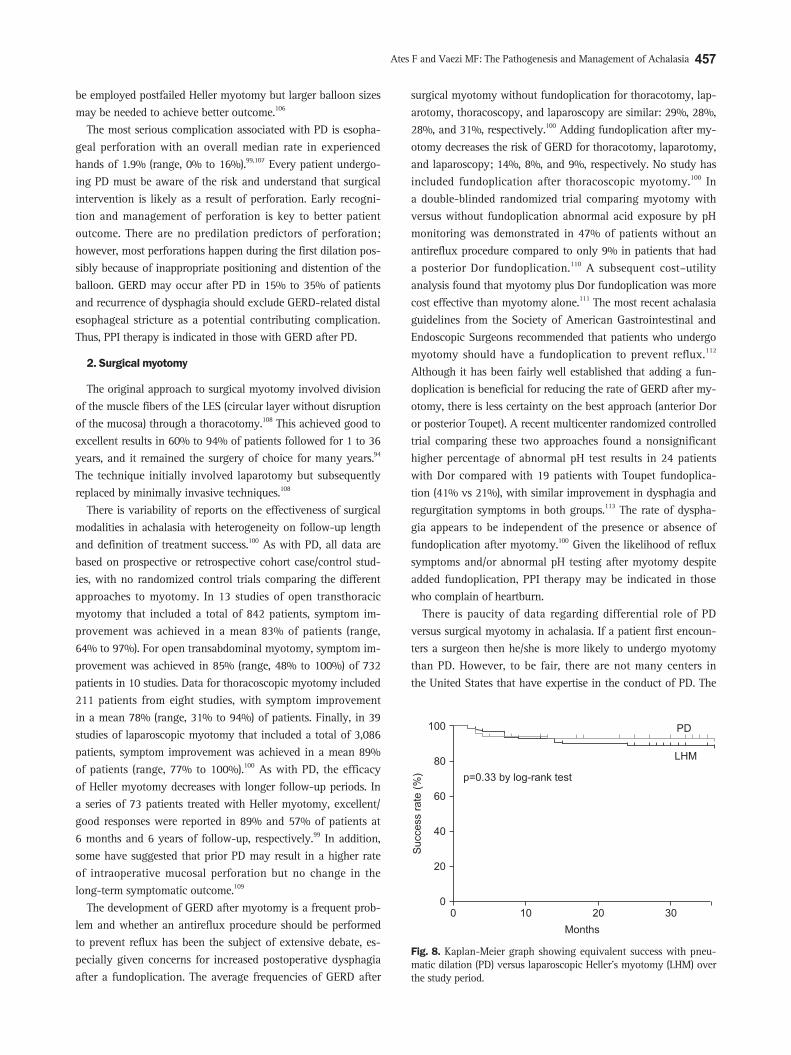
There is paucity of data regarding differential role of PD versus surgical myotomy in achalasia. If a patient first encoun-ters a surgeon then he/she is more likely to undergo myotomy than PD. However, to be fair, there are not many centers in the United States that have expertise in the conduct of PD. The
0
100
80
60
40
20
10 20 30
Success
rate
(%)
Months
0
p=0.33 by log-rank test
PD
LHM
Fig. 8. Kaplan-Meier graph showing equivalent success with pneu-matic dilation (PD) versus laparoscopic Heller’s myotomy (LHM) over the study period.

458 Gut and Liver, Vol. 9, No. 4, July 2015
only prospective randomized trial of PD versus surgical my-otomy showed equivalent benefit to both over the 2-year follow up period (Fig. 8).114 In this study patients from five different European countries were randomly allocated to Rigiflex dila-tion (n=94) or laparoscopic myotomy with Dor fundoplication (n=106). Symptom improvement, barium emptying and LES pressure reduction were similar for both groups at 2-years (86% and 90%, respectively). The results from this study further gives credence to the equal role of PD and myotomy in achalasia pa-tients. However, it is important to emphasize that if both options are not available in a given institution then the default should always be the treatment with the expertise.
3. Esophagectomy
In patients that may have had suboptimal treatment for long periods, “end-stage” achalasia may develop which is character-ized by megaesophagus or sigmoid esophagus and significant esophageal dilation and tortuosity. In this group of patients, PD is less effective but a surgical myotomy may be a reasonable ini-tial approach before consideration for esophagectomy. Two recent studies documented symptomatic improvement after myotomy in 92% and 72% of patients with megaesophagus.115, 116 However, in those unresponsive to therapy, esophageal resection is frequent-ly required.117 Esophagectomy is associated with a greater mor-bidity/mortality than laparoscopic Heller myotomy, and should be reserved for patients who have failed PD and/or myotomy and who are good candidates for surgery. Data from uncon-trolled studies show generally good response to esophagectomy, with symptom improvement in over 80% of patients with end-stage achalasia; mortality ranges between 0% and 5.4%.118
4. Peroral endoscopic myotomy
Peroral esophageal myotomy (POEM) is a recently developed endoscopic technique for treatment of patients with achalasia.119 The procedure involves endoscopic submucosal dissection with creation of a submucosal plane using a forward-viewing endo-scope to access the circular muscle fibers for performance of the myotomy. The myotomy is usually about 6 cm into the esopha-gus and 2 cm below the squamocolumnar junction. Overall, the success rate, defined by an improvement in symptoms and no requirement of additional medical or surgical treatment, in prospective cohorts have been >90%.120-122 A recent study in pa-tients post-POEM showed that there was no difference in patient outcome in those with had prior endoscopic or surgical therapy versus those who did not have such treatments.123 Having had prior therapies before POEM did not increase operative times. Mean operative procedure times in both groups were similar at 102 and 118 minutes, respectively. A single center comparison of myotomy versus PEOM for patients with achalasia showed similar 6-month symptom improvement but slightly higher (39%) rate of reflux in PEOM as compared to myotomy (32%).124 However, randomized prospective comparison trials with stan-
dard laparoscopic myotomy and/or PD are needed. Until then, POEM should only be performed in the context of clinical trials in centers with expertise with the technique.
5. Patient follow-up
The goals for the management of achalasia are focused on treatment of esophageal symptoms as well as the impaired esophageal emptying. Unfortunately, patient’s symptom may not be a reliable predictor of outcome as symptom improve-ment may occur without a significant improvement in esopha-geal emptying, placing the patient at risk for developing long-term complications of achalasia.85 Barium esophagram is an important tool in the management of achalasia both before and after intervention.85 Multiple studies have shown that the results of a post intervention timed barium studies can predict treatment success and requirement for future interventions. In 1999, Vaezi et al.85 presented data to support that there was a significant association between the results of the TBE and symptom resolution after PD. More importantly, however, they identified a group of patients who had poor esophageal empty-ing in the context of almost complete symptom resolution in which TBE predicted treatment failure at 1 year.86 Although the data do not support that an intervention should be performed based solely on the outcome of the TBE, it does support that follow-up should be more aggressive in patients with abnormal barium height regardless of symptoms as they may be at risk of symptomatic relapse. It is thus reasonable to repeat this test an-nually to assess for esophageal emptying. Achalasia is a chronic esophageal motility disorder and despite initial successful ther-apy most patients will eventually require repeat interventions. Thus, symptomatic and objective testing with barium swallow on a regularly scheduled interval may be needed to avoid end-stage achalasia/megaesophagus. There are limited data to sup-port routine screening for cancer. The overall number of cancers remains low and estimates have suggested that over 400 endos-copies would be required to detect one cancer.125 These numbers are further tempered by the fact that the survival of these pa-tients is poor once the diagnosis is made.126
In two recent studies of patients who were followed for a mean 5 to 6 years after laparoscopic Heller myotomy, 18% to 21% required additional treatment, most often with PD, but re-do myotomy, botulinum toxin injection, or smooth muscle re-laxing medications were also used.127,128 Similarly, in three recent studies of patients who were followed after successful graded PD, 23% to 35% underwent repeat treatment for symptomatic recurrence during a mean 5 to 7 years of follow-up, mostly with PD but some patients required surgery.102,127,129 The complexi-ties of managing achalasia, including treatment failures, were shown in a retrospective review of 232 achalasia patients who were followed after therapy for more than a period of 8 years.130 In this study, 93% of 184 patients did well after initial therapy, especially if combination therapy with more than one modality

Ates F and Vaezi MF: The Pathogenesis and Management of Achalasia 459
was employed. However, in those who failed initial myotomy, symptomatic management was more difficult. In this group, the rates of symptom response after PD and repeat myotomy were only 67% and 57%, respectively, with eight patients eventually requiring esophagectomy. PD after failed myotomy does not ap-pear to increase the risk of perforation, although data regarding this issue are limited.105
TREATMENT ALGORITHM
A tailored treatment algorithm for patients with achalasia is shown in Fig. 9. Symptomatic patients with achalasia who are good surgical candidates should be offered information about the risks and benefits of the two equally effective treatment op-tions of PD and myotomy. The choice between the procedures should depend on patient preference and institutional expertise. However, to maximize patient outcome, both procedures should be performed in centers of excellence with adequate volume and expertise. PD should be performed in a graded manner, starting with the smallest balloon (3.0 cm), except in younger males (<45 years old) who may benefit with the initial balloon size of 3.5 cm or surgical myotomy. In patients unresponsive to PD, surgical myotomy should be performed. Poor surgical candidates should initially undergo injection of the LES with botulinum toxin and should be aware that repeat therapy is often needed. Other medical therapies with nitrates or calcium channel blockers may be offered if there is no clinical response to botulinum toxin injection. Esophagectomy may be needed in those with dilated esophagus (>8 cm) with poor response to an initial myotomy.
CONFLICTS OF INTEREST
No potential conflict of interest relevant to this article was reported.
REFERENCES
1. Sadowski DC, Ackah F, Jiang B, Svenson LW. Achalasia: inci-
dence, prevalence and survival. A population-based study. Neu-
rogastroenterol Motil 2010;22:e256-e261.
2. Gockel HR, Schumacher J, Gockel I, Lang H, Haaf T, Nöthen
MM. Achalasia: will genetic studies provide insights? Hum Genet
2010;128:353-364.
3. Vaezi MF, Richter JE. Diagnosis and management of achalasia:
American College of Gastroenterology Practice Parameter Com-
mittee. Am J Gastroenterol 1999;94:3406-3412.
4. Francis DL, Katzka DA. Achalasia: update on the disease and its
treatment. Gastroenterology 2010;139:369-374.
5. Eckardt AJ, Eckardt VF. Treatment and surveillance strategies in
achalasia: an update. Nat Rev Gastroenterol Hepatol 2011;8:311-
319.
6. Richter JE, Boeckxstaens GE. Management of achalasia: surgery
or pneumatic dilation. Gut 2011;60:869-876.
7. Spechler SJ, Castell DO. Classification of oesophageal motility
abnormalities. Gut 2001;49:145-151.
8. Richter JE. The diagnosis and misdiagnosis of achalasia: it
does not have to be so difficult. Clin Gastroenterol Hepatol
2011;9:1010-1011.
9. Kessing BF, Bredenoord AJ, Smout AJ. Erroneous diagnosis of
gastroesophageal reflux disease in achalasia. Clin Gastroenterol
Hepatol 2011;9:1020-1024.
10. Katz PO, Gerson LB, Vela MF. Guidelines for the diagnosis and
management of gastroesophageal reflux disease. Am J Gastroen-
Patient with achalasia
Low surgical risk High surgical risk
PD MyotomyFailure
Males <45 yrBotulinum toxin
Nitrates
Ca channel blockers++
Repeat myotomy or PDesophagectomy
Failure
Failure
3.0 cm3.5 cm4.0 cm
3.5 cm4.0 cm
Fig. 9. Treatment algorithm for pa-tients with achalasia.PD, pneumatic dilation.

460 Gut and Liver, Vol. 9, No. 4, July 2015
terol 2013;108:308-328.
11. Tucker HJ, Snape WJ Jr, Cohen S. Achalasia secondary to car-
cinoma: manometric and clinical features. Ann Intern Med
1978;89:315-318.
12. Rozman RW Jr, Achkar E. Features distinguishing second-
ary achalasia from primary achalasia. Am J Gastroenterol
1990;85:1327-1330.
13. Birgisson S, Richter JE. Achalasia: what’s new in diagnosis and
treatment? Dig Dis 1997;15 Suppl 1:1-27.
14. Paterson WG. Etiology and pathogenesis of achalasia. Gastroin-
test Endosc Clin N Am 2001;11:249-266, vi.
15. Bieger D, Hopkins DA. Viscerotopic representation of the upper
alimentary tract in the medulla oblongata in the rat: the nucleus
ambiguus. J Comp Neurol 1987;262:546-562.
16. Toyama T, Yokoyama I, Nishi K. Effects of hexamethonium and
other ganglionic blocking agents on electrical activity of the
esophagus induced by vagal stimulation in the dog. Eur J Phar-
macol 1975;31:63-71.
17. Collman PI, Tremblay L, Diamant NE. The central vagal efferent
supply to the esophagus and lower esophageal sphincter of the
cat. Gastroenterology 1993;104:1430-1438.
18. Goyal RK, Rattan S. Nature of the vagal inhibitory innervation to
the lower esophageal sphincter. J Clin Invest 1975;55:1119-1126.
19. Goyal RK, Rattan S, Said SI. VIP as a possible neurotransmitter
of non-cholinergic non-adrenergic inhibitory neurones. Nature
1980;288:378-380.
20. Yamato S, Spechler SJ, Goyal RK. Role of nitric oxide in esopha-
geal peristalsis in the opossum. Gastroenterology 1992;103:197-
204.
21. Crist J, Gidda JS, Goyal RK. Intramural mechanism of esophageal
peristalsis: roles of cholinergic and noncholinergic nerves. Proc
Natl Acad Sci U S A 1984;81:3595-3599.
22. Aggestrup S, Uddman R, Jensen SL, et al. Regulatory peptides in
the lower esophageal sphincter of man. Regul Pept 1985;10:167-
178.
23. Mearin F, Mourelle M, Guarner F, et al. Patients with achalasia
lack nitric oxide synthase in the gastro-oesophageal junction. Eur
J Clin Invest 1993;23:724-728.
24. Kimura K. The nature of idiopathic esophagus dilatation. Jpn J
Gastroenterol 1929;1:199.
25. Cassella RR, Brown AL Jr, Sayre GP, Ellis FH Jr. Achalasia of the
esophagus: pathologic and etiologic considerations. Ann Surg
1964;160:474-487.
26. Higgs B, Kerr FW, Ellis FH Jr. The experimental production of
esophageal achalasia by electrolytic lesions in the medulla. J
Thorac Cardiovasc Surg 1965;50:613-625.
27. Cassella RR, Ellis FH Jr, Brown AL Jr. Fine-structure changes
in achalasia of the esophagus. I. vagus nerves. Am J Pathol
1965;46:279-288.
28. Duntemann TJ, Dresner DM. Achalasia-like syndrome presenting
after highly selective vagotomy. Dig Dis Sci 1995;40:2081-2083.
29. Atkinson M, Ogilvie AL, Robertson CS, Smart HL. Vagal function
in achalasia of the cardia. Q J Med 1987;63:297-303.
30. Eckardt VF, Krause J, Bolle D. Gastrointestinal transit and gastric
acid secretion in patients with achalasia. Dig Dis Sci 1989;34:665-
671.
31. Khajanchee YS, VanAndel R, Jobe BA, Barra MJ, Hansen PD,
Swanstrom LL. Electrical stimulation of the vagus nerve restores
motility in an animal model of achalasia. J Gastrointest Surg
2003;7:843-849.
32. Holloway RH, Dodds WJ, Helm JF, Hogan WJ, Dent J, Arndorfer
RC. Integrity of cholinergic innervation to the lower esophageal
sphincter in achalasia. Gastroenterology 1986;90:924-929.
33. Greaves RR, Mulcahy HE, Patchett SE, et al. Early experience
with intrasphincteric botulinum toxin in the treatment of achala-
sia. Aliment Pharmacol Ther 1999;13:1221-1225.
34. Dodds WJ, Dent J, Hogan WJ, Patel GK, Toouli J, Arndorfer RC.
Paradoxical lower esophageal sphincter contraction induced by
cholecystokinin-octapeptide in patients with achalasia. Gastroen-
terology 1981;80:327-333.
35. Biancani P, Walsh JH, Behar J. Vasoactive intestinal polypeptide:
a neurotransmitter for lower esophageal sphincter relaxation. J
Clin Invest 1984;73:963-967.
36. Rattan S. The non-adrenergic non-cholinergic innervation of the
esophagus and the lower esophageal sphincter. Arch Int Pharma-
codyn Ther 1986;280(2 Suppl):62-83.
37. Rattan S, Moummi C. Influence of stimulators and inhibitors of
cyclic nucleotides on lower esophageal sphincter. J Pharmacol
Exp Ther 1989;248:703-709.
38. Rattan S, Said SI, Goyal RK. Effect of vasoactive intestinal poly-
peptide. Proc Soc Exp Biol Med 1977;155:40-43.
39. Guelrud M, Rossiter A, Souney PF, Rossiter G, Fanikos J, Mu-
jica V. The effect of vasoactive intestinal polypeptide on the
lower esophageal sphincter in achalasia. Gastroenterology
1992;103:377-382.
40. Uddman R, Alumets J, Edvinsson L, Håkanson R, Sundler F.
Peptidergic (VIP) innervation of the esophagus. Gastroenterology
1978;75:5-8.
41. Wattchow DA, Furness JB, Costa M, O’Brien PE, Peacock M. Dis-
tributions of neuropeptides in the human esophagus. Gastroen-
terology 1987;93:1363-1371.
42. Aggestrup S, Uddman R, Sundler F, et al. Lack of vasoactive in-
testinal polypeptide nerves in esophageal achalasia. Gastroenter-
ology 1983;84(5 Pt 1):924-927.
43. Sigala S, Missale G, Missale C, et al. Different neurotransmitter
systems are involved in the development of esophageal achalasia.
Life Sci 1995;56:1311-1320.
44. Wattchow DA, Costa M. Distribution of peptide-containing nerve
fibres in achalasia of the oesophagus. J Gastroenterol Hepatol
1996;11:478-485.
45. Tøttrup A, Svane D, Forman A. Nitric oxide mediating NANC
inhibition in opossum lower esophageal sphincter. Am J Physiol
1991;260(3 Pt 1):G385-G389.
46. Murray J, Du C, Ledlow A, Bates JN, Conklin JL. Nitric oxide:
Ates F and Vaezi MF: The Pathogenesis and Management of Achalasia 461
mediator of nonadrenergic noncholinergic responses of opossum
esophageal muscle. Am J Physiol 1991;261(3 Pt 1):G401-G406.
47. Conklin JL, Du C, Murray JA, Bates JN. Characterization and
mediation of inhibitory junction potentials from opossum lower
esophageal sphincter. Gastroenterology 1993;104:1439-1444.
48. Fang S, Christensen J. Distribution of NADPH diaphorase in in-
tramural plexuses of cat and opossum esophagus. J Auton Nerv
Syst 1994;46:123-133.
49. Murray J, Bates JN, Conklin JL. Nerve-mediated nitric oxide
production by opossum lower esophageal sphincter. Dig Dis Sci
1994;39:1872-1876.
50. Murray JA, Clark ED. Characterization of nitric oxide synthase in
the opossum esophagus. Gastroenterology 1994;106:1444-1450.
51. Gaumnitz EA, Bass P, Osinski MA, Sweet MA, Singaram C. Elec-
trophysiological and pharmacological responses of chronically
denervated lower esophageal sphincter of the opossum. Gastroen-
terology 1995;109:789-799.
52. Kim CD, Goyal RK, Mashimo H. Neuronal NOS provides nitrergic
inhibitory neurotransmitter in mouse lower esophageal sphincter.
Am J Physiol 1999;277(2 Pt 1):G280-G284.
53. Paterson WG, Anderson MA, Anand N. Pharmacological char-
acterization of lower esophageal sphincter relaxation induced
by swallowing, vagal efferent nerve stimulation, and esophageal
distention. Can J Physiol Pharmacol 1992;70:1011-1015.
54. Sivarao DV, Mashimo HL, Thatte HS, Goyal RK. Lower esopha-
geal sphincter is achalasic in nNOS(-/-) and hypotensive in W/
W(v) mutant mice. Gastroenterology 2001;121:34-42.
55. De Giorgio R, Di Simone MP, Stanghellini V, et al. Esophageal
and gastric nitric oxide synthesizing innervation in primary
achalasia. Am J Gastroenterol 1999;94:2357-2362.
56. Murray JA, Ledlow A, Launspach J, Evans D, Loveday M, Conklin
JL. The effects of recombinant human hemoglobin on esophageal
motor functions in humans. Gastroenterology 1995;109:1241-
1248.
57. Singaram C, Sengupta A, Sweet MA, Sugarbaker DJ, Goyal RK.
Nitrinergic and peptidergic innervation of the human oesopha-
gus. Gut 1994;35:1690-1696.
58. Csendes A, Smok G, Braghetto I, et al. Histological studies of
Auerbach’s plexuses of the oesophagus, stomach, jejunum, and
colon in patients with achalasia of the oesophagus: correlation
with gastric acid secretion, presence of parietal cells and gastric
emptying of solids. Gut 1992;33:150-154.
59. Frieling T, Berges W, Borchard F, Lübke HJ, Enck P, Wienbeck
M. Family occurrence of achalasia and diffuse spasm of the oe-
sophagus. Gut 1988;29:1595-1602.
60. Stein DT, Knauer CM. Achalasia in monozygotic twins. Dig Dis
Sci 1982;27:636-640.
61. Annese V, Napolitano G, Minervini MM, et al. Family occurrence
of achalasia. J Clin Gastroenterol 1995;20:329-330.
62. Bosher LP, Shaw A. Achalasia in siblings: clinical and genetic
aspects. Am J Dis Child 1981;135:709-710.
63. Park W, Vaezi MF. Etiology and pathogenesis of achalasia: the
current understanding. Am J Gastroenterol 2005;100:1404-1414.
64. Jones DB, Mayberry JF, Rhodes J, Munro J. Preliminary report of
an association between measles virus and achalasia. J Clin Pathol
1983;36:655-657.
65. Robertson CS, Martin BA, Atkinson M. Varicella-zoster virus
DNA in the oesophageal myenteric plexus in achalasia. Gut
1993;34:299-302.
66. Niwamoto H, Okamoto E, Fujimoto J, Takeuchi M, Furuyama J,
Yamamoto Y. Are human herpes viruses or measles virus associ-
ated with esophageal achalasia? Dig Dis Sci 1995;40:859-864.
67. Birgisson S, Galinski MS, Goldblum JR, Rice TW, Richter JE.
Achalasia is not associated with measles or known herpes and
human papilloma viruses. Dig Dis Sci 1997;42:300-306.
68. de Oliveira RB, Rezende Filho J, Dantas RO, Iazigi N. The spec-
trum of esophageal motor disorders in Chagas’ disease. Am J
Gastroenterol 1995;90:1119-1124.
69. Storch WB, Eckardt VF, Wienbeck M, et al. Autoantibodies to
Auerbach’s plexus in achalasia. Cell Mol Biol (Noisy-le-grand)
1995;41:1033-1038.
70. Verne GN, Sallustio JE, Eaker EY. Anti-myenteric neuronal anti-
bodies in patients with achalasia: a prospective study. Dig Dis Sci
1997;42:307-313.
71. Moses PL, Ellis LM, Anees MR, et al. Antineuronal antibodies in
idiopathic achalasia and gastro-oesophageal reflux disease. Gut
2003;52:629-636.
72. Raymond L, Lach B, Shamji FM. Inflammatory aetiology of
primary oesophageal achalasia: an immunohistochemical and
ultrastructural study of Auerbach’s plexus. Histopathology
1999;35:445-453.
73. Clark SB, Rice TW, Tubbs RR, Richter JE, Goldblum JR. The
nature of the myenteric infiltrate in achalasia: an immunohisto-
chemical analysis. Am J Surg Pathol 2000;24:1153-1158.
74. Wong RK, Maydonovitch CL, Metz SJ, Baker JR Jr. Significant
DQw1 association in achalasia. Dig Dis Sci 1989;34:349-352.
75. De la Concha EG, Fernandez-Arquero M, Mendoza JL, et al. Con-
tribution of HLA class II genes to susceptibility in achalasia. Tis-
sue Antigens 1998;52:381-384.
76. Verne GN, Hahn AB, Pineau BC, Hoffman BJ, Wojciechowski
BW, Wu WC. Association of HLA-DR and -DQ alleles with idio-
pathic achalasia. Gastroenterology 1999;117:26-31.
77. Ruiz-de-León A, Mendoza J, Sevilla-Mantilla C, et al. Myenteric
antiplexus antibodies and class II HLA in achalasia. Dig Dis Sci
2002;47:15-19.
78. Gockel I, Becker J, Wouters MM, et al. Common variants in the
HLA-DQ region confer susceptibility to idiopathic achalasia. Nat
Genet 2014;46:901-904.
79. Howard PJ, Maher L, Pryde A, Cameron EW, Heading RC. Five
year prospective study of the incidence, clinical features, and di-
agnosis of achalasia in Edinburgh. Gut 1992;33:1011-1015.
80. Pandolfino JE, Kahrilas PJ. AGA technical review on the clinical
use of esophageal manometry. Gastroenterology 2005;128:209-
24.

462 Gut and Liver, Vol. 9, No. 4, July 2015
81. Pandolfino JE, Kahrilas PJ; American Gastroenterological As-
sociation. AGA technical review on the clinical use of esophageal
manometry. Gastroenterology 2005;128:209-224.
82. Salvador R, Costantini M, Zaninotto G, et al. The preoperative
manometric pattern predicts the outcome of surgical treatment
for esophageal achalasia. J Gastrointest Surg 2010;14:1635-1645.
83. Pratap N, Reddy DN. Can achalasia subtyping by high-resolution
manometry predict the therapeutic outcome of pneumatic bal-
loon dilatation? Author’s reply. J Neurogastroenterol Motil 2011;
17:205.
84. de Oliveira JM, Birgisson S, Doinoff C, et al. Timed barium swal-
low: a simple technique for evaluating esophageal emptying in
patients with achalasia. AJR Am J Roentgenol 1997;169:473-
479.
85. Vaezi MF, Baker ME, Richter JE. Assessment of esophageal emp-
tying post-pneumatic dilation: use of the timed barium esopha-
gram. Am J Gastroenterol 1999;94:1802-1807.
86. Vaezi MF, Baker ME, Richter JE. Assessment of esophageal emp-
tying post-pneumatic dilation: use of the timed barium esopha-
gram. Am J Gastroenterol 1999;94:1802-1807.
87. Andersson M, Lundell L, Kostic S, et al. Evaluation of the re-
sponse to treatment in patients with idiopathic achalasia by the
timed barium esophagogram: results from a randomized clinical
trial. Dis Esophagus 2009;22:264-273.
88. Dodds WJ, Stewart ET, Kishk SM, Kahrilas PJ, Hogan WJ. Radio-
logic amyl nitrite test for distinguishing pseudoachalasia from
idiopathic achalasia. AJR Am J Roentgenol 1986;146:21-23.
89. Kahrilas PJ, Kishk SM, Helm JF, Dodds WJ, Harig JM, Hogan
WJ. Comparison of pseudoachalasia and achalasia. Am J Med
1987;82:439-446.
90. Scherer JR, Kwiatek MA, Soper NJ, Pandolfino JE, Kahrilas PJ.
Functional esophagogastric junction obstruction with intact peri-
stalsis: a heterogeneous syndrome sometimes akin to achalasia. J
Gastrointest Surg 2009;13:2219-2225.
91. Sandler RS, Bozymski EM, Orlando RC. Failure of clinical criteria
to distinguish between primary achalasia and achalasia second-
ary to tumor. Dig Dis Sci 1982;27:209-213.
92. Vaezi MF, Pandolfino JE, Vela MF. ACG clinical guideline:
diagnosis and management of achalasia. Am J Gastroenterol
2013;108:1238-1249.
93. Boeckxstaens GE, Zaninotto G, Richter JE. Achalasia. Lancet
2014;383:83-93.
94. Vaezi MF, Richter JE. Current therapies for achalasia: comparison
and efficacy. J Clin Gastroenterol 1998;27:21-35.
95. Lambroza A, Schuman RW. Pneumatic dilation for achalasia
without fluoroscopic guidance: safety and efficacy. Am J Gastro-
enterol 1995;90:1226-1229.
96. Thomas V, Harish K, Sunilkumar K. Pneumatic dilation of acha-
lasia cardia under direct endoscopy: the debate continues. Gas-
trointest Endosc 2006;63:734.
97. Lynch KL, Pandolfino JE, Howden CW, Kahrilas PJ. Major
complications of pneumatic dilation and Heller myotomy for
achalasia: single-center experience and systematic review of the
literature. Am J Gastroenterol 2012;107:1817-1825.
98. Ott DJ, Richter JE, Wu WC, Chen YM, Castell DO, Gelfand DW.
Radiographic evaluation of esophagus immediately after pneu-
matic dilatation for achalasia. Dig Dis Sci 1987;32:962-967.
99. Vela MF, Richter JE, Khandwala F, et al. The long-term efficacy
of pneumatic dilatation and Heller myotomy for the treatment of
achalasia. Clin Gastroenterol Hepatol 2006;4:580-587.
100. Campos GM, Vittinghoff E, Rabl C, et al. Endoscopic and surgical
treatments for achalasia: a systematic review and meta-analysis.
Ann Surg 2009;249:45-57.
101. Gideon RM, Castell DO, Yarze J. Prospective randomized compar-
ison of pneumatic dilatation technique in patients with idiopathic
achalasia. Dig Dis Sci 1999;44:1853-1857.
102. Hulselmans M, Vanuytsel T, Degreef T, et al. Long-term outcome
of pneumatic dilation in the treatment of achalasia. Clin Gastro-
enterol Hepatol 2010;8:30-35.
103. Farhoomand K, Connor JT, Richter JE, Achkar E, Vaezi MF. Pre-
dictors of outcome of pneumatic dilation in achalasia. Clin Gas-
troenterol Hepatol 2004;2:389-394.
104. Eckardt VF, Aignherr C, Bernhard G. Predictors of outcome in
patients with achalasia treated by pneumatic dilation. Gastroen-
terology 1992;103:1732-1738.
105. Pratap N, Kalapala R, Darisetty S, et al. Achalasia cardia subtyp-
ing by high-resolution manometry predicts the therapeutic out-
come of pneumatic balloon dilatation. J Neurogastroenterol Motil
2011;17:48-53.
106. Guardino JM, Vela MF, Connor JT, Richter JE. Pneumatic dilation
for the treatment of achalasia in untreated patients and patients
with failed Heller myotomy. J Clin Gastroenterol 2004;38:855-
860.
107. Eckardt VF, Kanzler G, Westermeier T. Complications and their
impact after pneumatic dilation for achalasia: prospective long-
term follow-up study. Gastrointest Endosc 1997;45:349-353.
108. Ali A, Pellegrini CA. Laparoscopic myotomy: technique and
efficacy in treating achalasia. Gastrointest Endosc Clin N Am
2001;11:347-358, vii.
109. Morino M, Rebecchi F, Festa V, Garrone C. Preoperative pneu-
matic dilatation represents a risk factor for laparoscopic Heller
myotomy. Surg Endosc 1997;11:359-361.
110. Richards WO, Torquati A, Holzman MD, et al. Heller myotomy
versus Heller myotomy with Dor fundoplication for achalasia:
a prospective randomized double-blind clinical trial. Ann Surg
2004;240:405-412.
111. Torquati A, Richards WO, Holzman MD, Sharp KW. Laparoscopic
myotomy for achalasia: predictors of successful outcome after
200 cases. Ann Surg 2006;243:587-591.
112. Stefanidis D, Richardson W, Farrell TM, et al. SAGES guidelines
for the surgical treatment of esophageal achalasia. Surg Endosc
2012;26:296-311.
113. Rawlings A, Soper NJ, Oelschlager B, et al. Laparoscopic Dor ver-
sus Toupet fundoplication following Heller myotomy for achala-

Ates F and Vaezi MF: The Pathogenesis and Management of Achalasia 463
sia: results of a multicenter, prospective, randomized-controlled
trial. Surg Endosc 2012;26:18-26.
114. Boeckxstaens GE, Annese V, des Varannes SB, et al. Pneumatic
dilation versus laparoscopic Heller’s myotomy for idiopathic
achalasia. N Engl J Med 2011;364:1807-1816.
115. Sweet MP, Nipomnick I, Gasper WJ, et al. The outcome of lapa-
roscopic Heller myotomy for achalasia is not influenced by the
degree of esophageal dilatation. J Gastrointest Surg 2008;12:159-
165.
116. Mineo TC, Ambrogi V. Long-term results and quality of life after
surgery for oesophageal achalasia: one surgeon’s experience. Eur
J Cardiothorac Surg 2004;25:1089-1096.
117. Glatz SM, Richardson JD. Esophagectomy for end stage achalasia.
J Gastrointest Surg 2007;11:1134-1137.
118. Kadakia SC, Wong RK. Pneumatic balloon dilation for esophageal
achalasia. Gastrointest Endosc Clin N Am 2001;11:325-346, vii.
119. Inoue H, Minami H, Kobayashi Y, et al. Peroral endoscopic myot-
omy (POEM) for esophageal achalasia. Endoscopy 2010;42:265-
271.
120. Inoue H, Kudo SE. Per-oral endoscopic myotomy (POEM) for
43 consecutive cases of esophageal achalasia. Nihon Rinsho
2010;68:1749-1752.
121. von Renteln D, Inoue H, Minami H, et al. Peroral endoscopic my-
otomy for the treatment of achalasia: a prospective single center
study. Am J Gastroenterol 2012;107:411-417.
122. Swanström LL, Rieder E, Dunst CM. A stepwise approach and
early clinical experience in peroral endoscopic myotomy for the
treatment of achalasia and esophageal motility disorders. J Am
Coll Surg 2011;213:751-756.
123. Orenstein SB, Raigani S, Wu YV, et al. Peroral endoscopic myot-
omy (POEM) leads to similar results in patients with and without
prior endoscopic or surgical therapy. Surg Endosc 2015;29:1064-
1070.
124. Bhayani NH, Kurian AA, Dunst CM, Sharata AM, Rieder E,
Swanstrom LL. A comparative study on comprehensive, objective
outcomes of laparoscopic Heller myotomy with per-oral endo-
scopic myotomy (POEM) for achalasia. Ann Surg 2014;259:1098-
1103.
125. Sandler RS, Nyrén O, Ekbom A, Eisen GM, Yuen J, Josefsson S.
The risk of esophageal cancer in patients with achalasia: a popu-
lation-based study. JAMA 1995;274:1359-1362.
126. Leeuwenburgh I, Scholten P, Alderliesten J, et al. Long-term
esophageal cancer risk in patients with primary achalasia: a pro-
spective study. Am J Gastroenterol 2010;105:2144-2149.
127. Bonatti H, Hinder RA, Klocker J, et al. Long-term results of
laparoscopic Heller myotomy with partial fundoplication for the
treatment of achalasia. Am J Surg 2005;190:874-878.
128. Costantini M, Zaninotto G, Guirroli E, et al. The laparoscopic
Heller-Dor operation remains an effective treatment for esopha-
geal achalasia at a minimum 6-year follow-up. Surg Endosc
2005;19:345-351.
129. Zerbib F, Thétiot V, Richy F, Benajah DA, Message L, Lamouliatte
H. Repeated pneumatic dilations as long-term maintenance ther-
apy for esophageal achalasia. Am J Gastroenterol 2006;101:692-
697.
130. Eckardt VF, Hoischen T, Bernhard G. Life expectancy, complica-
tions, and causes of death in patients with achalasia: results of
a 33-year follow-up investigation. Eur J Gastroenterol Hepatol
2008;20:956-960.