the pre-hospital management of life-threatening chest injuries: a
TRANSCRIPT
Page 1 of 13
The pre-hospital management of life-threatening
chest injuries: a consensus statement Caroline Leech, Keith Porter, Richard Steyn, Colville Laird, Imogen Virgo, Richard Bowman, & David Cooper Chest injuries continue to be significant contributors to death from major trauma and are difficult to diagnose accurately in the pre-hospital environment. The incidence of life-threatening chest injuries is relatively small. Personal communication with the Trauma Audit Research Network provided figures for the frequency of chest injury diagnoses in UK major trauma patients reported to the database 2005 - September 2013 (see Table 1).
Condition Frequency in 183 232 major trauma patients Tension pneumothorax 1 in 250 (0.4%) Open pneumothorax 1 in 10 000 (0.01%) Massive haemothorax 1 in 1000 (0.1%) Flail chest (includes >3 rib fractures) 1 in 50 (2.2%) Cardiac tamponade 1 in 1250 (0.08%)
Table 1: Frequency of chest injuries in UK major trauma patients submitted to TARN “The pre-hospital management of chest injury: a consensus statement” was published by the Faculty of Pre-hospital Care, Royal College of Surgeons of Edinburgh in 2007.1 Since then new evidence has been published and adaptations have been made in pre-hospital management. To update the pre-existing guideline, a consensus meeting of stakeholders was held by the Faculty of Pre-hospital Care at Coventry in November 2013. A literature review was carried out prior to the consensus meeting, to inform the discussion process. Where no evidence existed, the practitioners discussed best practice based on expert opinion. This paper provides a guideline for the pre-hospital management of patients with the life-threatening chest injuries of tension pneumothorax, open pneumothorax, massive haemothorax, flail chest (including multiple rib fractures), and cardiac tamponade. General Management High-flow oxygen should be commenced with a reservoir mask for all patients who have sustained major trauma. A target oxygen saturation range of 94-98% should be maintained. 2 The optimal position for gas exchange is sitting up, or lying with the healthy lung down.3-6 This is unlikely to be possible in the case of patients who are hypovolaemic, in whom spinal fractures cannot be excluded, where lung injury has caused airway bleeding, or for practical reasons of safety during transfer. Early effective analgesia is essential for patients with a moderate or severe pain score. Pain from rib fractures or wounds will contribute to respiratory insufficiency and effective pain relief may obviate the need for invasive pre-hospital thoracic interventions.
Page 2 of 13
Tension Pneumothorax Making a diagnosis of tension pneumothorax in a spontaneously breathing trauma patient can be difficult. Decompensation is generally more progressive. Common features include universal symptoms of chest pain and respiratory distress, with tachycardia and ipsilateral decreased air entry found in 50-75% of cases. Signs
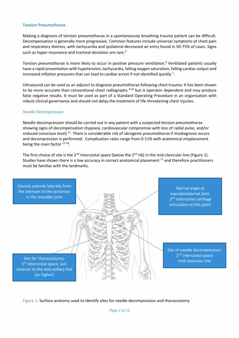
such as hyper-resonance and tracheal deviation are rare.7 Tension pneumothorax is more likely to occur in positive pressure ventilation.8 Ventilated patients usually have a rapid presentation with hypotension, tachycardia, falling oxygen saturation, falling cardiac output and increased inflation pressures that can lead to cardiac arrest if not identified quickly 7. Ultrasound can be used as an adjunct to diagnose pneumothorax following chest trauma. It has been shown to be more accurate than conventional chest radiographs 9,10 but is operator dependent and may produce false negative results. It must be used as part of a Standard Operating Procedure in an organisation with robust clinical governance and should not delay the treatment of life-threatening chest injuries. Needle Decompression Needle decompression should be carried out in any patient with a suspected tension pneumothorax showing signs of decompensation (hypoxia, cardiovascular compromise with loss of radial pulse, and/or reduced conscious level) 11. There is considerable risk of iatrogenic pneumothorax if misdiagnosis occurs and decompression is performed. Complication rates range from 0-11% with anatomical misplacement being the main factor 12-16. The first choice of site is the 2nd intercostal space (below the 2nd rib) in the mid-clavicular line (Figure 1). Studies have shown there is a low accuracy in correct anatomical placement 17 and therefore practitioners must be familiar with the landmarks. Figure 1: Surface anatomy used to identify sites for needl Figure 1: Surface anatomy used to identify sites for needle decompression and thoracostomy
Clavicle extends laterally from the sternum to the acromion
in the shoulder joint
Site of needle decompression: 2nd intercostal space mid-clavicular line
Sternal angle at manubriosternal joint.
2nd intercostal cartilage articulates at this point
Site for Thoracostomy: 5th intercostal space, just
anterior to the mid-axillary line (or higher)
Page 3 of 13
In the UK a standard 14G 4.5cm long cannula is commonly used to perform needle decompression. A number of studies have shown that the mean chest wall thickness at the 2nd intercostal space mid-clavicular line ranges from 3.06cm – 5.36cm and that a cannula of 4.5cm would fail to reach the pleural space in 4-100% of patients 12-28. The cannula may also fail to decompress the tension pneumothorax due to obstruction by blood, tissue or kinking. If there is no obvious air release on initial insertion a cannula should be inserted into the chest attached to a syringe and flushed with 2ml of air or water. Other causes of failure include a localized tension pneumothorax in the patient with pre-existing lung disease 29, or a large air leak in which the air collects in the pleural space quicker than can be drained by the narrow bore of the cannula 30. Needle decompression using a lateral approach (just anterior to the mid-axillary line) has been suggested as potentially more efficacious than the conventional anterior approach. Two studies showed that lateral chest wall thickness was thinner than that anteriorly, and four studies showed that the lateral chest wall thickness was greater than that anteriorly21,26, 31-34. Needle decompression should initially be performed in the 2nd intercostal space in the mid-clavicular line. In the presence of a presumed tension pneumothorax, with no clinical improvement, a 2nd attempt may be made with a 14G cannula in the 5th intercostal space just anterior to the mid-axillary line (Figure 2). It must be remembered that when the patient’s arms are elevated above their head the chest wall thickness may be thinner laterally, but the patient cannot be transported in this position. A cannula placed in the lateral site will be more prone to kinking when the arms are adducted by the patient’s side. There is also a potential increased risk of iatrogenic injury in the lateral site and therefore practitioners must undergo training to identify the correct landmarks. Over recent years, commercial products with MHRA approval such as the ThoraQuik device, have undergone testing with encouraging results 35, 36,. These devices may be preferable to the 14G 4.5cm cannula (which does not have MRHA approval) but are still early in clinical use. The landmarks for insertion are the same. Open thoracostomy Surgical incision and decompression of the pleural space with a thoracostomy is an effective method to treat tension pneumothorax37-40. It is diagnostic by the digital palpation of a deflated lung, which should re-inflate following decompression. Open thoracostomy may also be used to relieve severe surgical emphysema constricting respiration that may develop following a severe crush injury to the chest. Open thoracostomy should not be first line treatment for suspected tension pneumothorax in self-ventilating patients. Needle decompression with an approved device should be used first. If this fails, open thoracostomy can be considered and then followed by a chest drain (Figure 2). Pre-hospital insertion of a chest drain should be avoided where possible due to prolongation of on-scene time; risks of kinking, blocking or falling out during transfers; and long-term infection risks with non-sterile insertion techniques. It is accepted that chest drain insertion will be necessary in some circumstances eg high-altitude aero-medical retrieval. An open thoracostomy may be first-line treatment and can be left open for patients undergoing positive pressure ventilation (Figure 3). In positively pressure ventilated patients, who may have pronounced air leaks, it is potentially unsafe to cover an open thoracostomy with a commercial chest seal due to a risk of the seal blocking and the development of a tension pneumothorax. If the patient’s condition deteriorates, the thoracostomy should be reassessed and the tract re-opened with a clean, gloved finger to ensure patency. Packaging of the patient needs to avoid blockage of the thoracostomy holes by the patient’s arms.
Page 4 of 13
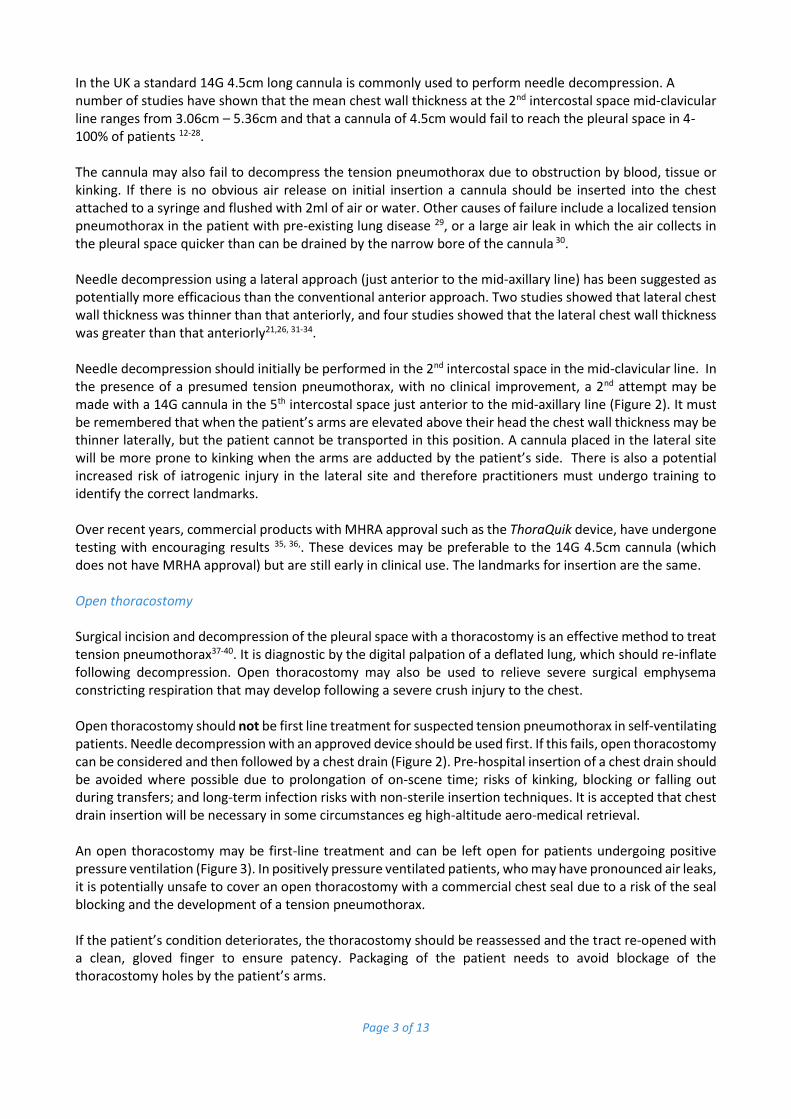
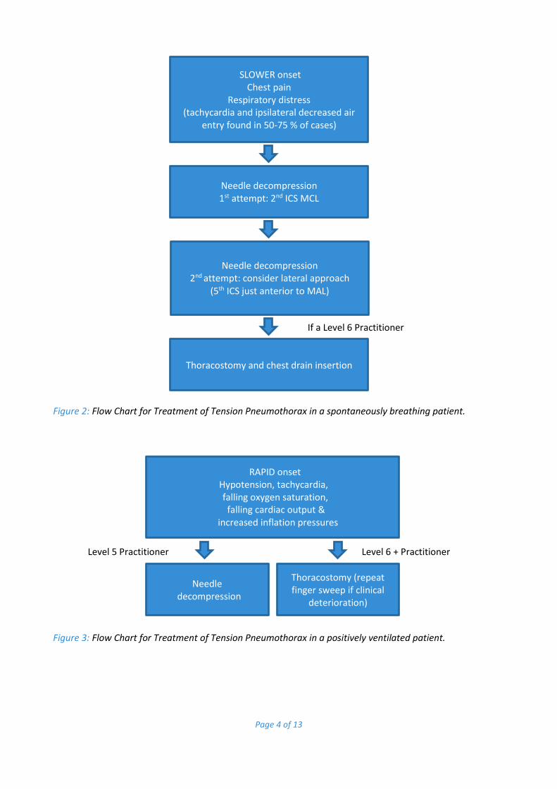
Figure 2: Flow Chart for Treatment of Tension Pneumothorax in a spontaneously breathing patient. Figure 3: Flow Chart for Treatment of Tension Pneumothorax in a positively ventilated patient.
Thoracostomy and chest drain insertion
SLOWER onset Chest pain
Respiratory distress (tachycardia and ipsilateral decreased air
entry found in 50-75 % of cases)
Needle decompression 1st attempt: 2nd ICS MCL
Needle decompression 2nd attempt: consider lateral approach
(5th ICS just anterior to MAL)
RAPID onset Hypotension, tachycardia,
falling oxygen saturation, falling cardiac output & increased inflation pressures
Needle decompression
Thoracostomy (repeat finger sweep if clinical
deterioration)
If a Level 6 Practitioner
Level 6 + Practitioner Level 5 Practitioner

Page 5 of 13
Procedure for thoracostomy
The patient’s arm is abducted and the 5th intercostal space identified just anterior to the mid-axillary line (see Figure 1). The site should be higher where landmarks are difficult (eg obese patients) or for pregnant patients who have a raised diaphragm. The practitioner should also confirm that the defined site is well within the ‘safe triangle’ (bordered by the anterior border of the latissimus dorsi, the lateral border of the pectoralis major muscle, and a line superior to the horizontal level of the nipple. 41
The chest is cleaned with chloraprep swabstick (or equivalent)
Just anterior to the mid axillary line, local anaesthetic is infiltrated into the skin, subcutaneous tissue, down to the pleura and surrounding area (this step is not required in traumatic cardiac arrest)
A 3-5cm transverse incision is made over the line of the 5th rib. Beware that the neurovascular bundle runs along the lower border of the rib
A tubular track is continued through the intercostal muscles using blunt dissection down to the pleura
The operator then inserts a finger along the track into the pleural cavity and sweeps around the space to detect the presence of any adhesions or bowel (in case of ruptured diaphragm) and whether the lung is inflated or deflated. It may also be possible to palpate the heart in a left thoracostomy to ascertain whether the heart is well filled or the presence of cardiac tamponade.
The complication rate of pre-hospital thoracostomy is not insignificant and estimated at 10-15% 44-48. It is imperative that any organisation performing pre-hospital thoracostomy has a standard operating procedure for the indications and technique, and all practitioners receive appropriate training. Pre-hospital organisations should develop robust methods of feedback with hospitals to enable audit of complication rates. There is no hard evidence for the prophylactic use of antibiotics for pre-hospital thoracostomy, although studies do exist looking at hospital tube thoracostomy 47-53. The British Thoracic Society guidelines recommend that antibiotic prophylaxis should be considered for trauma patients, especially after penetrating trauma, requiring chest drains54. Consensus opinion was that prophylactic antibiotics should be considered for pre-hospital thoracostomy, especially in cases of penetrating chest trauma, or with transport times >3 hours. There is no published evidence regarding the use of pre-hospital thoracostomies to insert emergency department chest drains versus a new incision in sterile conditions. The consensus view was to use the existing thoracostomy hole to place the intercostal drain, and to cover with intravenous antibiotics. Further incisions have the potential to cause more morbidity and are not justified. In the case of penetrating chest wall injury drains must not be inserted into the wound, even if this is at the appropriate anatomical site. Open pneumothorax Open pneumothorax is rare, with only 31 cases reported to TARN in an eight year period to 2013. This reflects the fact that in most cases of penetrating chest trauma, the tissues around the wound will self-seal. Diagnosis of an open pneumothorax is based on the clinical finding of a wound on the chest wall that is ‘sucking’ air and visibly bubbling, with evidence of an underlying pneumothorax. All practitioners should be able to recognise a tension pneumothorax, as a complication of open pneumothorax, or as a result of the treatment. Treatment recommendations The use of a three-sided first aid dressing is no longer deemed to be effective. 55
Page 6 of 13
The initial priority is to occlude the chest wound with a commercial chest seal. The majority of open pneumothoraces will not require any further treatment in the pre-hospital environment. Vented chest seals have been shown to be effective at preventing the development of a tension pneumothorax56 and are preferable to an unvented chest seal. Even a vented chest seal may become unvented due to occlusion with blood. If a chest seal is not available, the wound should be completely covered with a thin dry adherent dressing. If the patient develops signs of a tension pneumothorax following the application of a chest seal or dressing, then the seal or dressing should be immediately removed. If there is no improvement then needle decompression or thoracostomy should be performed as per the guidelines for tension pneumothorax. In some cases the wound will be completely occluded with a chest seal due to the position of the patient (eg a posterior wound with the patient supine). Consideration should be made as to the ideal transport position for the patient to allow removal of the dressing if required. Rapid sequence induction and positive pressure ventilation will need to be considered for large open pneumothoraces with respiratory failure. Prophylactic intravenous antibiotics should be administered for all cases of penetrating chest injury, where available.47, 54, 57, 58 Co-amoxiclav 1.2g is a suitable choice (or Gentamicin for Penicillin-allergic patients). Massive haemothorax The diagnosis of massive haemothorax may be made in the pre-hospital environment by the following means:
Clinical signs of haemothorax with hypovolaemia
Exsanguination from a thoracostomy or chest drain performed for suspected tension pneumothorax
Found at emergency thoracotomy for traumatic cardiac arrest from penetrating trauma The clinical signs of massive haemothorax will be very difficult to differentiate from a tension pneumothorax in the pre-hospital setting. Examination of the chest may reveal tachypnoea, reduced chest expansion, dullness to percussion, reduced air entry, reduced vocal resonance, and hypoxia. Circulatory shock will normally become apparent before respiratory compromise. Treatment recommendations Pre-hospital drainage of a massive haemothorax may be deleterious, by the mechanism of dislodging clots and promoting further internal thoracic haemorrhage, and should not routinely be performed. 42,59 If respiratory compromise is absent (tachypnoea, hypoxia) drainage of a haemothorax should be delayed until arrival at an Emergency Department where blood products, cell salvage, and cardiothoracic surgery are available. Where tension pneumothorax cannot be excluded, pre-hospital thoracostomy or chest drain insertion will need to be performed as per the Tension Pneumothorax guidelines above. If a thoracostomy reveals a significant haemorrhage from the thoracic cavity, a chest drain may be useful (to monitor the amount of ongoing blood loss and diagnose a massive haemothorax) but should not prolong on-scene time. Clamping a pre-hospital chest drain for exsanguinating haemothorax may be considered but is extremely high risk due to the likelihood of an underlying pneumo-haemothorax being present and the risk of accumulating a tension pneumothorax.
Page 7 of 13
Pre-hospital cell salvage is not currently used in the UK but should be considered immediately on arrival to hospital. As in all cases of internal haemorrhage, patients should be handled with minimal movements (including no log-roll) to minimise clot disruption.60 A loading dose of Tranexamic acid should be administered as soon as possible, and within three hours of injury.61 Fluid resuscitation should be administered according to existing principles of permissive hypotension, titrating 250ml boluses to maintain a systolic blood pressure of 90mmHg.62 Flail Chest The signs of flail chest may include pain on inspiration causing shallow breathing or a reduced inspiratory effort; tenderness of the chest wall; palpable crepitus of displaced rib fractures; deformity of the chest wall; or an underlying lung injury. Pulmonary contusion, pneumothorax or haemothorax may all co-exist with flail chest. 63 Paradoxical movement may not be seen in the pre-hospital assessment due to the position of the patient and compensation by involuntary splinting of the intercostal muscles. Pain and the underlying lung injury (rather than paradoxical movement) are the cause of respiratory compromise following blunt chest trauma. 64 Identifying that the patient has multiple rib fractures (> three) may be more important for management and triage decisions rather than specifically diagnosing a flail segment.63 Treatment recommendations There is no evidence that manual splinting of a flail segment using a hand or device will improve respiratory function and it may impair ventilation.62 An awake patient without respiratory compromise may find holding their fractured ribs with a hand helpful to manage pain during respiration. Deliberate positioning of the patient lying on an anterior or lateral flail segment is not feasible in transport or during spinal immobilisation. Where possible, sitting the patient up, may be beneficial to respiratory function. 3-6 Analgesia is critical in the early management of rib fractures. 64 A pain score should be performed early and repeated at frequent intervals. Analgesic agents such as intravenous Paracetamol, Morphine or Fentanyl, Ketamine or intranasal Diamorphine should all be considered depending on availability and expertise. Entonox cannot be recommended as approximately one third of patients with >3 rib fractures have an underlying pneumothorax (personal communication TARN). Intubation and mechanical ventilation are not obligatory for flail chest in the absence of respiratory failure. 65-71
Risk factors for a poor outcome following rib fractures includes age 65 years or more, three or more rib fractures, bilateral flail chest, chronic lung disease, co-existent underlying lung injury, pre-injury anticoagulant use, BMI >25, and oxygen saturation <90% in the Emergency Department.72 The presence of these factors may upgrade the triage decision to a trauma centre with cardiothoracic cover.
Page 8 of 13
Cardiac Tamponade There are no reliable pre-hospital signs or symptoms for cardiac tamponade. A high index of suspicion is based on the mechanism of penetrating injury and is confirmed by the presence of deterioration. Patients with penetrating chest injuries should be transported as soon as possible, with minimal intervention, to the nearest Major Trauma Centre. The presence of normal vital signs should not reassure the practitioner or delay transfer73. Performing RSI in patients who have cardiac tamponade as a suspected diagnosis may precipitate circulatory collapse and should be avoided where possible. There is no evidence for pre-hospital pericardiocentesis. The technique is difficult and associated with cardiac injury. It is unlikely that the clotted pericardial blood can be aspirated by a needle, and this technique does not stop continued bleeding from the ventricle into the pericardial sac. 74 Pre-hospital clam-shell thoracotomy should be considered as a ‘Resuscitative Thoracotomy’: an integral part of the life-saving resuscitation of a penetrating trauma patient. The indications for thoracotomy are:
Stab wounds to the chest or upper abdomen
Cardiac arrest with loss of vital signs ≤ 15 minutes
The suspected injury is suitable for temporary repair and control
A chain of survival exists for definitive management following Resuscitative Thoracotomy
The contra-indications to pre-hospital thoracotomy for chest trauma are:
Cardiac arrest secondary to blunt trauma
Cardiac arrest secondary to gunshot wound75
Loss of vital signs >15mins76-78
Unskilled practitioner The use of ultrasound is still unproven in determining the need for pre-hospital thoracotomy, but there are some pre-hospital systems that use ultrasound in the diagnosis of pericardial effusions for blunt trauma patients77. Pre-hospital ultrasound may be useful in the non-arrested patient or the blunt trauma patient with suspected tamponade. The technique of resuscitative thoracotomy is comprehensively described by Wise, et al. 79 Pre-hospital thoracotomy should only be performed by a skilled practitioner, within a system of training, continued education, audit and governance. The concept of the untrained practitioner undertaking the procedure is unethical. All cases of pre-hospital thoracotomy should automatically trigger network audit and review procedures. If the pre-hospital team do not have the skills to perform a thoracotomy for a patient in cardiac arrest, they should move to the hospital immediately. If a more advanced practitioner is responding to scene it may be prudent to rendezvous on route to or at hospital. The risks of a resuscitative thoracotomy should not be underestimated and include blood borne infections80; sharp injuries; and Post Traumatic Stress Disorder for emergency services personnel and bystanders.
Page 9 of 13
Conclusion This consensus statement forms an update to the 2007 FPHC guideline on the management of life-threatening chest injuries. It is intended that the document will be updated in the future to reflect ongoing developments in evidence and clinical practice. Acknowledgements We acknowledge the contribution of representatives from the following organisations: Association of First Aiders, BASICS, BASICS Scotland, Cardiothoracic Surgery – UHCW, Faculty of Pre-Hospital Care, FPHC Child Health & Paediatric representative, Mountain Rescue England & Wales, North East Ambulance Service, North West Ambulance Service, Northern Ireland Ambulance Service, Scottish Ambulance Service, South Central Ambulance Service, South East Coast Ambulance Service, UKSAR medical group, West Midlands Ambulance Service, Yorkshire Ambulance Service. Competing interests None declared. Funding The Consensus meeting was funded by the Faculty of Pre-hospital Care, Royal College of Surgeons of Edinburgh.
Page 10 of 13
References
1. Lee C, Revell M, Porter K, Steyn R. The pre-hospital management of chest injuries: a consensus statement. Faculty of Pre-hospital Care, Royal College of Surgeons of Edinburgh. Emerg Med J 2007;24(3):220-4.
2. O’Driscoll BR, Howard LS, Davison AG. Guideline for emergency oxygen use in adult patients. British Thoracic Society. Thorax 2008;63(Suppl 6):vi1-68.
3. Dean E. Effect of body position on pulmonary function. Phys Ther 1985; 65(5):613-618. 4. Clauss RH, Scalabrini BY, Ray JF, et al: Effects of changing body position upon improved ventilation-
perfusion relationships. Circulation 1968;37(Suppl2):214-217. 5. Remolina C, Khan AV, Santiago TV, et al. Positional hypoxemia in unilateral lung disease. N Engl J
Med 1981;304:523-525. 6. Sonnenblick M, Melzer E, Rosin AJ. Body positional effect on gas exchange in unilateral pleural
effusion. Chest 1983; 83:784-786. 7. Leigh-Smith S, Harris T. Tension pneumothorax-time for a re-think? Emerg Med J 2005; 22:8-16. 8. MacDuff A, Arnold A, Harvey J. Management of spontaneous pneumothorax: British Thoracic
Society pleural disease guideline 2010. Thorax Aug 2010;vol 65:supp II. 9. Chin EJ, Chan CH, Mortazavi R, Anderson CL, et al. A Pilot study examining the viability of a Pre-
hospital Assessment with Ultrasound for Emergencies (PAUSE) protocol. J Emerg Med 2013;44(1):142-9.
10. Jaffer U, Macauley D. Transthoracic ultrasonography to diagnose pneumothorax in trauma. BestBets, August 2005 (http://bestbets.org/bets/bet.php?id=328).
11. Battlefield Advanced Trauma Life Support, 4th edition, 2008. JSP 570. 12. Cullinane DC, Morris JA Jr, Bass JG, Rutherford EJ. Needle thoracostomy may not be indicated in the
trauma patient. Injury 2001;32:749-752. 13. Barton ED, Epperson M, Hoyt DB, et al. Pre-hospital needle aspiration and tube thoracostomy in
trauma victims: a six year experience with aeromedical crews. Journal of Emergency Medicine 1995;13:155-163.
14. Eckstein M, Suyehara D. Needle thoracostomy in the prehospital setting. Prehospital Emergency Care 1998;2:132-135.
15. Warner KJ, Copass MK, Bulger EM. Paramedic use of needle thoracostomy in the prehospital environment. Prehospital Emergency Care 2008;12:162-168.
16. Netto FA, Shulman H, Rizoli SB, et al, Are needle decompressions for tension pneumothoraces being performed appropriately for appropriate indications? American Journal of Emergency Medicine 2008;26:597-602.
17. Ferrie EP, Collum N, McGovern S. The right place in the right space? Awareness of site for needle thoracocentesis. Emerg Med J 2005;22:788-9.
18. Britten S, Palmer SH, Snow TM. Needle thoracocentesis in tension pneumothorax: insufficient cannula length and potential failure. Injury 1996;27:321-322.
19. Marinaro JL. Needle thoracostomy in trauma patients: What catheter length is adequate? Acad Emerg Med 2003;10:495.
20. Givens ML, Ayotte K, Manifold C. Needle thoracostomy: Implications of computed tomography chest wall thickness. Acad Emerg Med 2004;11:211-13.
21. Lander OM, Sanchez LD, Pedrosa I. Anterior Vs Lateral needle decompression of tension pneumothorax: a comparison by computed tomography chest wall management. Acad Emerg Med 2005;12(5 supp):66.
22. Zengerink I, Brink PR, Laupland KB, et al. Needle Thoracostomy in the treatment of a tension pneumothorax in trauma patients: What size needle? J Trauma 2008;64:111-114.
23. Harcke HT, Pearse LA, Levy AD, et al. Chest wall thickness in military personnel: implications for needle thoracentesis in tension pneumothorax. Military Med 2007;172:1260-3.
24. Stevens R. Needle Thoracostomy for tension pneumothorax. Prehospital Emergency Care 2009;13:14-17.
Page 11 of 13
25. Ball C. Thoracic needle decompression for tension pneumothorax: clinical correlation with catheter length. Can J Surg 2010;53:184-187.
26. McLean AR, Richards ME, Crandall CS, Marinaro JL. Ultrasound determination of chest wall thickness: implications for needle thoracostomy. American Journal of Emergency Medicine 2011:29:1173-1177.
27. Yangiwa T, Morita S, Yamamoto R, et al. Determination of the appropriate catheter length for needle thoracostomy by using computed tomography scans of trauma patients in Japan. Injury 2012;43:42-45.
28. Inaba K, Ives C, McClure K, et al. Radiologic evaluation of alternative sites for needle decompression of tension pneumothorax. Arch Surg 2012;147:813-17.
29. Mines D, Abbuhl S. Needle thoracostomy fails to detect a fatal tension pneumothorax. Ann Emerg Med 1993;22:836–6.
30. Jones R, Hollingsworth J. Tension pneumothoraces not responding to needle thoracocentesis. EMJ 2002;19:176–7.
31. Beckett A, Savage E, Pannell D, et al. Needle Decompression for Tension Pneumothorax in tactical casualty care: Do catheters placed in the midaxillary line kink more often than those in the midclavicular line? The Journal of Trauma 2011; 71: S408-s411.
32. Inaba K, Branco BC, Eckstein M, et al. Optimal positioning for Emergent Needle Thoracostomy: A Cadaver-Based Study. J Trauma 2011;71:1099-1103.
33. Sanchez LD, Straszewski S, Saghir A , et al. Anterior Versus Lateral Needle Decompression of Tension Pneumothorax: Comparison by CT Chest Wall Measurement. Academic Emergency Medicine 2011;18:1022-25.
34. Wax D, Leibowitz B. Radiologic Assessment of Potential Sites for Needle Decompression of a Tension Pneumothorax. Anesthesia & Analgesia 2007; 105:1385-88.
35. Rathinam S, Quinn DW, Bleetman A, et al. Evaluation of ThoraQuik: a new device for the treatment of pneumothorax and pleural effusion. Emerg Med J 2011;28:750-753.
36. Rathinam S, Grobler S, Bleetman A, et al. Evolved design makes ThoraQuik safe and user friendly in the management of pneumothorax and pleural effusion. Emerg Med J doi:10.1136/emermed-2012-201821, Published Online First 23 January 2013.
37. Deakin C, Davies G, Wilson A. Simple thoracostomy avoids chest drain insertion in prehospital trauma. J Trauma 1995; 39:373-4.
38. Massarutti D. Simple thoracostomy in prehospital trauma management is safe and effective: a 2 year experience by helicopter emergency medical crews. European Journal of Emergency Medicine 2006;13:276-280.
39. Waydhas C, Sauerland S. Pre-hospital pleural decompression and chest tube placement after blunt trauma: A systemic review. Resuscitation 2007;72:11-25.
40. Fitzgerald M. Pleural decompression and drainage during trauma reception and resuscitation. Injury 2008;39:9-20.
41. Laws D, Neville E, Duffy J. BTS guidelines for the insertion of a chest drain. Thorax2003;58 (suppl II) :ii55.
42. Aylwin CJ, Brohi K, Davies GD, Walsh MS. Pre-hospital and in-hospital thoracostomy: indications and complications. Ann R Coll Surg Engl 2008; 90:54-57.
43. Etoch SW, Bar-Natan MF, Miller FB, Richardson JD. Tube Thoracostomy. Factors relating to complications. Arch Surg 1995;130:521-5.
44. Millikan JS, Moore EE, Steiner E, et al. Complications of tube thoracostomy for acute trauma. Am J Surg 1980; 140:738-41.
45. Bailey RC. Complications of tube thoracostomy in trauma. J Accid Emerg Med 2000; 17:111-4. 46. Spanjersberg WR, Ringburg AN, Bergs EA, et al. Pre-hospital chest tube thoracostomy: effective
treatment or additional trauma? J Trauma 2005;59:788-93. 47. Bosman et al, Systematic Review and meta-analysis of antibiotic prophylaxis to prevent infections
from chest drains in blunt and penetrating thoracic injuries. British Journal of Surgery 2012;99:506-513.
Page 12 of 13
48. Sanabria A, Valdivieso E, Gomez G, et al. Prophylactic antibiotics in chest trauma: a meta analysis of high quality studies. Worl J Surg 2006;30:1843-1847.
49. Maxwell RA, Campbell DJ, Fabian TC, et al. Use of presumptive antibiotics following tube thoracostomy for traumatic hemopneumothorax in the prevention of empyema and pneumonia--a multi- center trial. J Trauma 2004; 57(4):742-748.
50. Gonzalez RP, Holevar MR. Role of Prophylactic antibiotics for tube thoracostomy in chest trauma. Am Surg 1998; 64:617-620.
51. Evans JT, Green JD, Carlin PE, Barrett LO. Meta-analysis of antibiotics in tube thoracostomy. Am Surg 1995; 61:215-9.
52. Fallon WF Jr, Wears RL. Prophylactic antibiotics for the prevention of infectious complications including empyema following tube thoracostomy for trauma: results of meta- analysis. J Trauma 1992; 33(1):110-116.
53. Moore FO, Duane TM, Hu CK, et al. Presumptive antibiotic use in tube thoracostomy for traumatic haemopneumothorax: An Eastern Association for the Surgery of Trauma Practice management guideline. J Trauma Acute Care Surgery 2012; Vol 73: S341-S344.
54. Havelock T, Teoh R, Laws D, et al. Pleural procedures and thoracic ultrasound. British Society Pleural Disease Guideline 2010. Thorax 2010;vol 65:supp II.
55. Butler FK, Dubose JJ, Otten EJ, et al. Management of Open Pneumothorax in Tactical Combat Casualty Care: TCCC Guidelines Change 13-02. Journal Special Operations Medicine 2013;13(3):81-86.
56. Kheirabadi BS, Terrazas IB, Koller A, et al. Vented versus unvented chest seals for treatment of pneumothorax and prevention of tension pneumothorax in a swine model. J Trauma Acute Care Surg 2013; 75(1):150-156.
57. Luchette FA, Barrie PS, Oswanski MF, et al. Practice Management Guidelines for Prophylactic Antibiotic Use in Tube Thoracostomy for Traumatic Hemopneumothorax: The EAST Practice Management Guidelines Work Group. J Trauma 2000; 48(4):753-7.
58. Martin GJ, Dunne JR, Cho JM, Solomkin JS. Prevention of Infections Associated With Combat-Related Thoracic and Abdominal Cavity Injuries. J Trauma 2011; 71(2):S270-S281.
59. Coats TJ, Wilson AW, Xeropotamous N. Pre-hospital management of patients with severe thoracic injury. Injury 1995;26(9):581-5.
60. Moss R, Porter K, Greaves I. Minimal patient handling: a Faculty of Pre-Hospital Care consensus statement. Emerg Med J 2013;30(12):1065-1066.
61. CRASH-2 trial collaborators. Effects of tranexamic acid on death, vascular occlusive events, and blood transfusion in trauma patients with significant haemorrhage (CRASH-2): a randomised, placebo-controlled trial. Lancet 2010; 376(9734):23-32.
62. Association of Ambulance Chief Executives. UK Ambulance Services Clinical Practice Guidelines 2013, Bridgwater 2013.
63. Sirmali M, Türüt H, Topçu S, Gülhan E, et al. A comprehensive analysis of traumatic rib fractures: morbidity, mortality and management. Eur J Cardiothorac Surg 2003; 24(1): 133-8.
64. Simon BJ, Cushman J, Barraco R, et al. Pain management guidelines for blunt thoracic trauma – EAST Practice Management Guidelines. J Trauma 2005; 59(5): 1256-67.
65. Simon B, Ebert J, Bokhari F, Capella J, et al. Eastern Association for the Surgery of Trauma. Management of pulmonary contusion and flail chest: an Eastern Association for the Surgery of Trauma practice management guideline. J Trauma Acute Care Surg 2012 Nov; 73(5 Suppl 4): S351-61.
66. Athanassiadi K, Theakos N, Kalantzi N, Gerazounis M. Prognostic factors in flail-chest patients. Eur J Cardiothorac Surg 2010 Oct; 38(4): 466-71.
67. Athanassiadi K, Gerazounis M, Theakos N. Management of 150 flail chest injuries: analysis of risk factors affecting outcome. Eur J Cardiothorac Surg 2004 Aug; 26(2): 373-6.
68. Barone JE, Pizzi WF, Nealon TF, et al. Indications for intubation in blunt chest wall trauma. Journal of Trauma-Injury Infection & Critical Care 1986; 26 (4): 334-8.
Page 13 of 13
69. Richardson JD, Adams L, Flint LM. Selective management of flail chest and pulmonary contusion. Ann Surg 1982; 196(4): 481-7.
70. Shackford SR, Smith DE, Zarins CK, et al. The management of flail chest. A comparison of ventilatory
and nonventilatory treatment. Am J Surg 1976; 132(6):759-62.
71. Trinkle JK, Richardson DJ, Franz JL, et al. Management of flail chest without mechanical ventilation.
Ann Thoracic Surgery 1975; 19(4): 355-63. 72. Battle C, Hutchings H, Evans PA. Blunt chest wall trauma: A review. Trauma 2013; 15(2): 156-175. 73. Seamon MJ, Fisher CA, Gaughan J, Lloyd M, et al. Prehospital procedures before emergency
department thoracotomy: ‘scoop and run’ saves lives. J Trauma 2007; 63(1): 113-20. 74. Pahle AS, Pedersen BL, Skaga NO, Pillgram-Larsen J. Emergency thoracotomy saves lives in a
Scandinavian hospital setting. J Trauma 2010; 68(3):599-603. 75. Seamon MJ, Shiroff AM, Franco M, et al. Emergency department thoracotomy for penetrating
injuries of the heart and great vessels: an appraisal of 283 consecutive cases from two urban trauma centres. J Trauma 2009; 67(6): 1250-1258.
76. Burlew CC, Moore EE, Moore FA, et al. Western Trauma Association Critical Decisions in Trauma: Resuscitative thoracotomy. J Trauma Acute Care Surg 2012; 73 (6):1359-63.
77. Sherren PB, Reid C, Habig K, Burns B. Algorithm for the resuscitation of traumatic cardiac arrest in a physician-staffed helicopter emergency medical service. Critical Care 2013; 17(Suppl 2):S105-106.
78. Lockey DJ, Lyon RM, Davies GE. Development of a simple algorithm to guide the effective management of traumatic cardiac arrest. Resuscitation 2013; 84(6): 738-742.
79. Wise D, Davies G, Coats T, Lockey T, et al. Emergency thoracotomy: how to do it. Emerg Med J 2005;22:22–24.
80. Soreide K, Petrone P, Asensio JA. Emergency thoracotomy in trauma: Rational, risks, and realities. Scand J Surg 2007; 96(1):4-10.