the prostaglandin h2 analog u-46619 improves the
TRANSCRIPT
Su et al. Stem Cell Research & Therapy (2018) 9:313 https://doi.org/10.1186/s13287-018-1061-4
RESEARCH Open Access
The prostaglandin H2 analog U-46619improves the differentiation efficiency ofhuman induced pluripotent stem cells intoendothelial cells by activating bothp38MAPK and ERK1/2 signaling pathways
Liping Su1†, Xiaocen Kong2†, Szeyun Lim1, Szejie Loo1, Shihua Tan1, Kiankeong Poh3, James Dutton4,Colin Stewart5, Stuart Cook1,6,7, Xiaofei Su2, Jianhua Ma2*, Jianyi Zhang8* and Lei Ye1*Abstract
Background: We have shown that the differentiation of human-induced pluripotent stem cells (hiPSCs) intoendothelial cells (ECs) is more efficient when performed with a 3-dimensional (3D) scaffold of biomaterial thanin monolayers. The current study aims to further increase hiPSC-EC differentiation efficiency by deciphering thesignaling pathways in 3D scaffolds.
Methods and results: We modified our 3D protocol by using U-46619 to upregulate both p38 mitogen-activatedprotein kinase (p38MAPK) and extracellular signal-regulated kinase 1/2 (ERK1/2) signaling, which increased thedifferentiation efficiency (as measured by CD31 expression) to as high as 89% in two established hiPSC lines.The differentiated cells expressed arteriovenous, but not lymphatic, markers; formed tubular structures and EClumen in vitro; had significantly shorter population-doubling times than monolayer-differentiated hiPSC-ECs;and restored perfusion and vascularity in a murine hind limb ischemia model. The differentiation efficiencywas also > 85% in three hiPSC lines that had been derived from patients with diseases or disease symptomsthat have been linked to endothelial dysfunction.
Conclusions: These observations demonstrate that activating both p38MAPK and ERK1/2 signaling pathwayswith U-46619 improves the efficiency of arteriovenous hiPSC-EC differentiation and produces cells with greaterproliferative capacity.
Keywords: Human-induced pluripotent stem cells, Endothelial differentiation, Signaling pathways
* Correspondence: [email protected]; [email protected];[email protected]†Liping Su and Xiaocen Kong contributed equally to this work.2Department of Endocrinology, Nanjing First Hospital, Nanjing MedicalUniversity, 68 Changle Road, Nanjing 210006, China8Department of Biomedical Engineering, The University of Alabama atBirmingham, Birmingham, AL 35294-2182, USA1National Heart Research Institute of Singapore, National Heart CentreSingapore, Singapore 117609, SingaporeFull list of author information is available at the end of the article
© The Author(s). 2018 Open Access This articInternational License (http://creativecommonsreproduction in any medium, provided you gthe Creative Commons license, and indicate if(http://creativecommons.org/publicdomain/ze
le is distributed under the terms of the Creative Commons Attribution 4.0.org/licenses/by/4.0/), which permits unrestricted use, distribution, andive appropriate credit to the original author(s) and the source, provide a link tochanges were made. The Creative Commons Public Domain Dedication waiverro/1.0/) applies to the data made available in this article, unless otherwise stated.

Su et al. Stem Cell Research & Therapy (2018) 9:313 Page 2 of 13
BackgroundVascular endothelial cells (ECs) form a physical barrierbetween the vessel wall and lumen, are metabolically ac-tive, and play a key role in the maintenance of cardiovas-cular homeostasis [1, 2] by producing molecules thatregulate vascular tone, cell adhesion, clotting, and fibrin-olysis [2]. Pathophysiological conditions, such as hyper-glycemia, hypercholesterolemia, hypertension, andstress, can lead to EC functional abnormalities that havebeen linked to atherosclerosis, coronary artery disease,diabetes, and hypertension, as well as normal physio-logical aging [1–4]. However, the availability of primaryhuman ECs for investigations of cell therapy or to serveas an in vitro platform for drug testing and disease mod-eling is limited. Human-induced pluripotent stem cells(hiPSCs) could relieve this scarcity because they can bedifferentiated into theoretically unlimited numbers ofany type of cell. Since hiPSCs are generated from a pa-tient’s own somatic cells, they carry genetic variationsthat may contribute to the development of the patient’sdisease [5–10].Traditional protocols for differentiating hiPSCs into ECs
(hiPSC-ECs) are performed in two-dimensional (2D) cul-ture systems [11–17], likely because the endothelium is a2D tissue. However, we have previously shown thathiPSC-EC differentiation can be remarkably efficient whenconducted in three-dimensional (3D) fibrin scaffolds [18];up to 45% of the hiPSCs assumed an EC phenotype, andthe phenotype remained stable for up to 4 weeks in vitro.Here, we investigated the pathways involved in hiPSC-ECdifferentiation to determine whether our protocol could bemade even more efficiently by targeting the p38 mitogen-activated protein kinase (p38MAPK) and extracellularsignal-regulated protein kinases 1 and 2 (ERK1/2) signalingpathways, which have been shown to contribute inde-pendently to the EC differentiation of pluripotentstem cells [19, 20]. Thus, we investigated the tem-poral dependence of our hiPSC-EC differentiationprotocol on these signaling pathways by treating thecells with selective inhibitors of p38MAPK (losmapi-mod [Losma]) [21] or ERK1/2 (SCH772984 [SCH])[22] during differentiation stages. We found that ourenhanced protocol can not only be used to generateECs from the non-disease hiPSCs, but also cells of patientswhose disease or disease symptoms have been linked toendothelial dysfunction, such as type 2 diabetes and ath-erosclerosis in patients with Hutchinson-Gilford progeriasyndrome [23–25], which have not been achieved withhigh differentiation efficiency.
Materials and methodshiPSC generationThe five hiPSC lines used in this study were reprogrammedfrom dermal fibroblasts by using non-integrating Sendai
virus and the reprogramming factors OCT4, SOX2, KLF4,and C-MYC, as described previously [26]. PCBC16iPS andGRiPS cells were reprogrammed from neonatal human der-mal fibroblasts (Lonza, USA) [18]. PG-608iPS cells were de-rived from a patient of the Coriell Institute for MedicalResearch (USA) who had Hutchinson-Gilford progeria syn-drome. DP1-C9iPS and DP3-C6iPSC cells were repro-grammed from the cells of two patients with type 2diabetes mellitus (T2DM). The procedures were approvedby the ethics committee of Nanjing Hospital, Nanjing,China, and the Centralised Institutional Review Board ofSingapore Health Services Pte Ltd., Singapore; informedconsent forms were signed by all patients. PCBC16iPSCswere used as representative hiPSCs in all experiments un-less stated otherwise. hiPSCs were cultured in a feeder-freesystem with a 1:1 mixture of E8/mTeSR (STEMCELLTechnologies, Canada) and passaged every 4 days with Ver-sene (Thermo Fisher, USA).
hiPSC-EC differentiation3D scaffoldsThis differentiation protocol was modified from a proto-col that has been described previously [18]. Briefly, stage1 began 2 days before initiating differentiation, whenhiPSCs were dissociated into single cells, seeded into a0.4-mL fibrin scaffold on a 24-well plate, and transferredto 6-well plates. Stage 2 of the protocol was initiated onday 0 by culturing the cell-containing fibrin/thrombinscaffold in EBM2 medium (Lonza, USA) supplementedwith B27 without insulin and CHIR99021 (CHIR) withor without U46619 for 24 h. The third stage began onday 1 when the medium was replaced with EBM2medium supplemented with B27 without insulin, vascu-lar endothelial growth factor-165 (VEGF), transforminggrowth factor β1 (TGFβ1), and erythropoietin (EPO);the cells were cultured for 48 h, the medium wasrefreshed on day 3, and the cells were cultured for an-other 48 h. On day 5, the differentiating hiPSCs were re-leased and cultured in EGM2-MV medium (Lonza,USA) supplemented with B27, VEGF, and SB-431542(SB). The medium was changed every 2 days, and differ-entiation efficiency was evaluated on day 11 viafluorescence-activated cell sorting (FACS); cells positivefor CD31 expression and for both CD31 and CD144 ex-pression were collected and expanded. For investigationsof p38MAPK and ERK1/2 inhibition, the inhibitors(10 μM Losma, an inhibitor of p38MAPK [21], and/or5 μM SCH, an inhibitor of ERK1/2 [22]) were added tothe differentiation medium 30 min before CHIR, VEGF/TGFβ1/EPO, or U46619 treatment was initiated.
2D monolayersThe monolayer culture protocol was identical to the 3Dculture protocol with the following exceptions. In stage
Su et al. Stem Cell Research & Therapy (2018) 9:313 Page 3 of 13
1, the dissociated hiPSCs were seeded into 6-well platesand cultured in monolayers, and on day 5, one well ofthe differentiating hiPSCs was harvested and cultured ina T-25 flask with EGM2-MV medium supplementedwith B27, VEGF, and SB. The medium was changedevery 2 days, and differentiation efficiency was evaluatedon day 11 via FACS.The EC population doubling time was calculated
within 7 days after cell sorting. Briefly, ECs were har-vested on day 2 after purification and cultured in 6-wellplates (2 × 10⁵ cells/well). The medium was changedevery 2 days, and ECs were harvested and counted onday 7.
Flow cytometryFlow cytometry analyses were conducted as described pre-viously [18, 27]. Briefly, the differentiated hiPSC-ECs weretrypsinized and re-suspended as single cells in glass tubes,incubated with 2% fetal bovine serum (FBS) inphosphate-buffered saline (PBS) containing primaryphycoerythrin (PE)- or allophycocyanin (APC)-conjugatedanti-human CD31 antibodies (clone WM59, BD Pharmin-gen, USA), FITC or PE-conjugated anti-human CD144antibodies (clone 55-7H1, BD Pharmingen, USA), or iso-type control antibodies for 30 min at 4 °C. To determineEC type, purified ECs on day 7 were incubated withFITC-conjugated anti-Eph-B4 antibodies, PE-conjugatedanti-human CXC chemokine receptor type 4 (CXCR4)antibodies, APC-conjugated anti-human delta-like 4(DLL4) or anti-human podoplanin antibodies (MiltenyiBiotec, Germany), or isotype control antibodies for30 min at 4 °C. The cells were washed with 2% FBS/PBS,re-suspended in 0.3 mL 2% FBS/PBS containing 5 μL ofpropidium iodide (10 μg/mL), and evaluated with a FACSAria instrument (BD Biosciences, USA).
Western blotPhosphorylated and non-phosphorylated p38MAPK andERK1/2 protein levels were determined by Western blotanalysis as described previously [28]. The cell lysate wasprepared with PhosphoSafe™ Extraction Reagent (Merck,Germany), and protein concentrations were determinedwith Bradford reagent (Bio-Rad Laboratories, USA). Pro-teins were separated, electrophoretically blotted ontonitrocellulose membranes, and washed with 10 mMTris-HCl buffer (pH 7.6) containing 0.05% Tween-20;then, the membranes were incubated in blocking buffer(5% non-fat dry milk, 10 mM Tris pH 7.5, 100 mM NaCl,0.1% Tween-20) at room temperature for 3 h and with di-luted primary antibodies (glyceraldehyde phosphate de-hydrogenase [GAPDH 1:2000, pERK1/2 [Thr202/Tyr204]1:1000, Santa Cruz Biotech, USA; p-p38MAPK [Thr180/Tyr182] 1:500, ERK1/2 1:1000, and p38MAPK 1:1000, CellSignaling, USA) at 4 °C overnight. Bound antibodies were
detected with HRP-conjugated anti-rabbit IgG (dilution1:1000 and 1:8000) and visualized with a ChemiDocTMMP Imaging System (Bio-Lab, USA) and Image Lab 5.1software (Bio-Lab, USA).
Quantitative RT-PCRRNA isolation and cDNA synthesis was performed as de-scribed previously [26, 29], and PCR thermal cycling wasconducted with the following primers: Brachyury, forward:AAAGAGATGATGGAGGAACCCGGA, reverse: AGGATGAGGATTTGCAGGTGGACA; Etv2, forward: GGGCTTGAAGGAGCCAAATTA, reverse: CAGGGATGAGCTTGTACCTTTC; Gata-2, forward: GACGACAACCACCACCTTAT, reverse: AGTCTGGATCCC TTCCTTCT; Tal-1,forward: AAATGGAGCAAAGTGGTAGGT, reverse: GTGACAACTCCAGCCTCTTAC; CD34, forward: TAGCCTGTCACCTGGAAATG, reverse: TGCCTTGATGTCACTTAGGATAG; and CD31, forward: TTGAGACCAGCCTGATGAAACCCT, reverse: TCCGTTTCCTGGGTTCAAGCGATA. Thermal cycling was performed 40 times, andeach cycle consisted of enzyme activation at 95 °C for15 min, denaturation at 95 °C for 30 s, and annealing at60 °C (for all PCR reactions) for 30 s and extension at 72 °Cfor 30 s. Endogenous GAPDH (forward: TCGACAGTCAGCCGCATCTTCTTT, reverse ACCAAATCCGTTGACTCCGACCTT) levels were used as an internal controlfor normalization [26]. Brachyury expression was presentedas a percentage of measurements obtained after Activin-A/BMP-4 treatment, and the expression of other genes waspresented as a percentage of their expression at day 0.
Dil-conjugated acetylated low-density lipoprotein uptakeand tube formationDil-conjugated acetylated low-density lipoprotein (Dil-a-ce-LDL) uptake and tube formation were evaluated asdescribed previously [18]. For the Dil-ace-LDL uptakeassay, hiPSC-ECs were incubated with DAPI overnight(1:1000 dilution) and then in EGM supplemented with10 μg/mL of Dil-ace-LDL (Life Technologies, USA) at37 °C for 12 h. For the tube formation assay, cells wereseeded in 48-well plates that had been coated withMatrigel (BD Pharmingen, USA) and incubated at 37 °Cfor 24 h. Numbers of node, junction, and mesh per lowmagnification (× 4) were quantified using angiogenesisanalyzer of ImageJ. For tube formation in 3D, 2 × 10⁴
hiPSC-ECs were seeded into 3D fibrin-thrombin scaf-folds composed of 50 μL of 25 mg/mL fibrinogen and50 μL of 20 U/mL thrombin and cultured in EGM sup-plemented with a × 100 dilution of B27, 100 ng/mLVEGF, SB, and 100 U/mL aprotinin.
Murine hind limb ischemia model and treatmentThe experimental protocol and animal maintenance pro-cedures were approved by the Institutional Animal Care

Su et al. Stem Cell Research & Therapy (2018) 9:313 Page 4 of 13
and Use Committee and performed in accordance withthe Animal Use Guidelines of Singapore Health ServicesPte Ltd. Eight-week-old NOD-SCID mice (InVivos,Singapore) were anesthetized with intraperitoneal injec-tions of ketamine (100 mg/kg) and xylazine (2.5 mg/kg).The right limb was shaved and disinfected with betadineand 70% alcohol; then, the femoral artery of the righthind limb was exposed and freed from the inguinal liga-ment via a longitudinal incision extending to a point justproximal to the patella. The artery and all branches fromthe inguinal ligament to the point where it bifurcatesinto the popliteal and saphenous arteries were closedwith 6-0 polypropylene sutures; then, the wound wasclosed, and the animals were allowed to recover. Keto-profen (2.5 mg/kg, subcutaneous) was administered forpain control and Baytril (15 mg/kg, intramuscular) toprevent infection for at least 3 days after the surgicalprocedure. Animals were randomly assigned to treat-ment with 1.5 × 10⁶ hiPSC-ECs in 0.2 mL EBM (i.e., thehiPSC-EC group, n = 8) or with 0.2 mL EBM (i.e., thebasal medium [BM] group, n = 9). The hiPSC-ECs hadbeen differentiated from the PCBC16 cell line, and thetreatments were administered 3 days after hind limb is-chemia (HLI) induction via four intramuscular injectionsinto the center of the ligated area and the surroundingregion along the femoral artery.
Laser Doppler imagingMice were anesthetized with intraperitoneal injections ofketamine (100 mg/kg) and xylazine (2.5 mg/kg), theirhind limbs were shaved, and laser Doppler imaging wasperformed with a PeriScan PIM 3 System (Perimed,Sweden). Measurements in the ischemic (right) limbwere normalized to measurements in the non-ischemic(left) limb and expressed as a percentage.
ImmunohistochemistryFor characterization of hiPSC-ECs in vitro, cells werefixed with 4% paraformaldehyde for 20 min at roomtemperature and then blocked with UltraV block (FisherScientific, USA) for 7 min. Primary antibodies (monoclo-nal anti-CD31 and mouse anti-CD144 [BD Pharmingen,USA]; 1:100 concentration) were added to the UltraVblock buffer and incubated overnight at 4 °C; then, thecells were incubated with PE-conjugated goatanti-mouse IgG secondary antibodies in PBS for 1 h atroom temperature, labeled with 4′,6-diamidino-2-pheny-lindole (DAPI), washed, and viewed under a fluorescencemicroscope (Olympus, Japan).To determine the neovascularization in ischemic limb,
mice limb muscles were collected, frozen, and cut into8-μm-thick sections; then, the sections were stained forCD31 expression (rabbit anti-CD31 [Abcam, USA]which targets both human and mouse ECs and goat
anti-rabbit IgG conjugated with FITC [Thermo FisherScientific, USA]) to evaluate total vessel density, forsmooth muscle actin (SMA) expression (Cy3-conjugatedmouse anti-SMA antibodies [Sigma-Aldrich] which tar-gets both human and mouse SMCs) to evaluate arterioledensity. Vascular structures that were positive for CD31expression (i.e., FITC fluorescence) and for both CD31and SMA expression (i.e., simultaneous FITC and Cy3fluorescence) were counted for all animals in bothgroups, three to four slides per animal, six to eight fieldsper slide.To identify transplanted hiPSC-ECs, a primary anti-
body specifically against human CD31 (hCD31, mouseanti-human CD31-Biotin) was used and visualized bymouse anti-Biotin-VioBright 515 (both from MiltenyiBiotec, Germany). Fluorescence images were taken withan Olympus IX71 fluorescence microscope.
StatisticsData are presented as mean ± standard deviation (SD).Comparisons among groups were analyzed for signifi-cance via one-way analysis of variance (ANOVA) withthe Tukey correction. Analyses were performed withSPSS software. A value of p < 0.05 was consideredsignificant.
ResultsCHIR99021 dose-dependently promotes the mesodermalspecification of hiPSCsThe 3D differentiation protocol consists of three stages(Fig. 1a). In stage 1, the cells were seeded into the 3D scaf-fold and maintained under standard hiPSC culture condi-tions for 2 days (i.e., from day − 2 to day 0). Differentiationbegins in stage 2 (day 0 to day 1) when the cells are di-rected toward an intermediate, mesodermal lineage; then,the final hiPSC-EC phenotype is induced during stage 3(day 1 to day 5) by exposing the cells to VEGF, TGFβ1, andEPO. In our previous report, stage 2 was initiated by cultur-ing the cells with 50 ng/mL Activin-A and 25 ng/mLBMP-4 [18]; however, mesodermal commitment can alsobe induced with the glycogen synthase kinase 3α/β inhibi-tor, CHIR [30], so we compared expression of the earlymesodermal marker, Brachyury, in hiPSCs cultured withvarying concentrations of CHIR. Brachyury mRNA levelsincreased logarithmically as CHIR concentrations wereraised from 5 to 15 μM in 5 μM increments, and after 24 hof differentiation, measurements were ~ 23-fold greaterwith 15 μM CHIR than when differentiation was initiatedwith Activin-A/BMP-4 (Fig. 1b). Furthermore, upon com-pletion of the entire hiPSC-EC differentiation protocol, flowcytometry assessments of CD31 expression (Fig. 1c) indi-cated that the efficiency of differentiation increased from 10to 58% over the same range of CHIR concentrations andwas ~ 2-fold greater with 15 μM CHIR than with
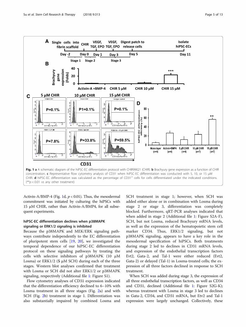
A
B
CD
Fig. 1 a A schematic diagram of the hiPSC-EC differentiation protocol with CHIR99021 (CHIR). b Brachyury gene expression as a function of CHIRconcentration. c Representative flow cytometry analysis of CD31 when hiPSC-EC differentiation was conducted with 5, 10, or 15 μMCHIR. d hiPSC-EC differentiation was calculated as the percentage of CD31+ cells for cells differentiated under the indicated conditions.(**p < 0.01 vs any other treatment)
Su et al. Stem Cell Research & Therapy (2018) 9:313 Page 5 of 13
Activin-A/BMP-4 (Fig. 1d, p < 0.01). Thus, the mesodermalcommitment was initiated by culturing the hiPSCs with15 μM CHIR, rather than Activin-A/BMP4, for all subse-quent experiments.
hiPSC-EC differentiation declines when p38MAPKsignaling or ERK1/2 signaling is inhibitedBecause the p38MAPK and MEK/ERK signaling path-ways contribute independently to the EC differentiationof pluripotent stem cells [19, 20], we investigated thetemporal dependence of our hiPSC-EC differentiationprotocol on these signaling pathways by treating thecells with selective inhibitors of p38MAPK (10 μMLosma) or ERK1/2 (5 μM SCH) during each of the threestages. Western blot analyses confirmed that treatmentwith Losma or SCH did not alter ERK1/2 or p38MAPKsignaling, respectively (Additional file 1: Figure S1).Flow cytometry analyses of CD31 expression indicated
that the differentiation efficiency declined to 6–10% withLosma treatment in all three stages (Fig. 2a) and withSCH (Fig. 2b) treatment in stage 1. Differentiation wasalso substantially impaired by combined Losma and
SCH treatment in stage 1; however, when SCH wasadded either alone or in combination with Losma duringstage 2 or stage 3, differentiation was completelyblocked. Furthermore, qRT-PCR analyses indicated thatwhen added in stage 2 (Additional file 1: Figure S2A-F),SCH, but not Losma, reduced Brachyury mRNA levels,as well as the expression of the hematopoietic stem cellmarker CD34. Thus, ERK1/2 signaling, but notp38MAPK signaling, appears to have a key role in themesodermal specification of hiPSCs. Both treatmentsduring stage 2 led to declines in CD31 mRNA levels,and expression of the endothelial transcription factorsEvt2, Gata-2, and Tal-1 were either reduced (Evt2,Gata-2) or delayed (Tal-1) in Losma-treated cells; the ex-pression of all three factors declined in response to SCHtreatment.When SCH was added during stage 3, the expression of
all three endothelial transcription factors, as well as CD34and CD31, declined (Additional file 1: Figure S2G-K);whereas treatment with Losma in stage 3 led to declinesin Gata-2, CD34, and CD31 mRNA, but Etv2 and Tal-1expression were largely unchanged. Collectively, these

Fig. 2 a Representative flow cytometry results and hiPSC-EC differentiation efficiency when 10 μM of the p38MAPK inhibitor losmapimod (Losma)was supplemented in differentiation stage 1, 2, or 3. b. Representative flow cytometry results and hiPSC-EC differentiation efficiency when 5 μMof the ERK1/2 inhibitor SCH was supplemented in differentiation stage 1, 2, or 3. c Representative flow cytometry results and mean hiPSC-ECdifferentiation efficiency when both Losma and SCH were supplemented in differentiation stage 1, 2, or 3. Assessments of CD31 levels werecompared with isotype controls. (***p < 0.001 vs any other treatment)
Su et al. Stem Cell Research & Therapy (2018) 9:313 Page 6 of 13
observations suggest that ERK1/2 signaling is crucial forhiPSC-EC differentiation, particularly during stages 2 and3 of our protocol, whereas the p38MAPK signaling path-way may play in more of an auxiliary role.
U-46619 improved hiPSC-EC differentiation efficiency byactivating both p38MAPK and ERK1/2 signalingBecause our observations indicated that both thep38MAPK and MEK/ERK pathways contribute tohiPSC-EC differentiation, we evaluated whether the effi-ciency of our hiPSC-EC differentiation protocol could beimproved by supplementing the medium with the prosta-glandin H2 analog U-46619, which has been shown to ac-tivate p38MAPK and ERK1/2 signaling [31, 32].Experiments were conducted with hiPSCs from thePCBC16iPS, which were reprogrammed from neonatalhuman dermal fibroblasts and have been used extensivelyin another investigation [18]. U-46619 did not improvedifferentiation efficiency when added during the first stageof our protocol; however, when 1 μM U-46619 was addedduring stage 2 or stage 3, > 85% (stage 2 89.1 ± 4.3%, stage3 85 ± 3.2%) of the differentiated cells expressed CD31(Fig. 3a). Higher U-46619 concentration (5 μM) was lesseffective at promoting hiPSC-EC differentiation; < 40% ofthe differentiated cells expressed CD31 when 5 μMU-46619 was added to the medium during protocol stage2 or stage 3 (Additional file 1: Figure S3). Differentiationalso declined to < 25% when U-46619 treatment was com-bined with Losma and was completely blocked whenU-46619 was combined with SCH or both SCH andLosma in stage 2 or 3 (Fig. 3b, c). Western blots confirmed
that Losma specifically inhibited U-46619-inducedp38MAPK activity, that SCH specifically inhibitedU-46619-induced ERK1/2 activity, and that the combin-ation of Losma and SCH inhibited the U-46619-inducedactivity of both pathways (Additional file 1: Figure S4).Furthermore, qRT-PCR analyses indicated that whenadded in stage 2 (Additional file 1: Figure S5A-F) or stage3 (Additional file 1: Figure S5G-J), U-46619 enhanced orprolonged Etv2, Gata-2, Tal-1, CD34, and CD31 gene ex-pression but not in the presence of SCH alone or com-bined SCH/Losma treatment. Losma impeded theexpression of all five markers when added to U-46619-treated cells in stage 2; however, when added in stage 3,Etv2 expression in U-46619-treated cells was prolonged,while the expression of Gata-2, Tal-1, CD34, and CD31gene expression was largely unchanged (Gata-2, Tal-1) oronly moderately reduced (CD34, CD31).The dramatically enhanced differentiation efficiency
achieved with U-46619 treatment prompted us to modifyour protocol by adding U-46619 (1 μM) to the mediumduring stage 2 (Fig. 4a). The modified protocol was testedin four additional hiPSC lines: GRiPS, which has been wellcharacterized in another study [18]; DP1-C9 and DP3-C6,which were derived from patients with T2DM; andPG-608, which was derived from a patient withHutchinson-Gilford progeria syndrome; all four lines werereprogrammed from dermal fibroblasts. The differenti-ation efficiency, as determined via flow cytometry analysisof CD31 expression, exceeded 85% in all lines tested(Fig. 4b). After purification, > 95% of the cells expressedthe arteriovenous EC marker CXCR4 [33, 34] and the

Fig. 3 a Representative flow cytometry results and hiPSC-EC differentiation efficiency when 1 μM U46619 was supplemented in differentiationstage 1, 2, or 3 (***p < 0.001 vs stage 1). Representative flow cytometry results and hiPSC-EC differentiation efficiency when U46619 wasco-supplemented with Losma, SCH, or both Losma and SCH during differentiation stage 2 (b) or 3 (c) (***p < 0.001 vs any other treatment).Assessments of CD31 levels were compared with isotype controls
Fig. 4 a A schematic diagram of the hiPSC-EC differentiation protocol with U46619 treatment in differentiation stage 2. b Representative flowcytometry results and hiPSC-EC differentiation efficiency in four hiPSC cell lines: GRiPSC, DP1-C9, DP3-C6, and PG-608. Assessments of CD31 levelswere compared with isotype controls. c Representative flow cytometry analysis for protein expression of delta-like canonical Notch ligand 4(DLL4), ephrin B4 (EphB4), podoplanin, and CXCR4 and the percentage of hiPSC-ECs that expressed DLL4, CXCR4, EphB4, and Podoplanin. (n = 4)
Su et al. Stem Cell Research & Therapy (2018) 9:313 Page 7 of 13

A1 A2 A3 A4 A5
B1 B2 B3 B4 B5
C1 C2 C3 C4 C5
D1 D2 D3 D4 D5
E1
F
E2 E3 E4 E5
Fig. 5 (See legend on next page.)
Su et al. Stem Cell Research & Therapy (2018) 9:313 Page 8 of 13

(See figure on previous page.)Fig. 5 hiPSC-ECs were differentiated from A PCBC16, B GRiPSC, C DP1-C9, D DP3-C6, and E PG-608 lines and evaluated for the expression of (1)CD31 and (2) CD144, for (3) Dil-ace-LDL uptake, for (4) tube formation on Matrigel, and for (5) EC lumen formation in fibrin-thrombin scaffolds.(Bar = 100 μm. Magnification of tubule formation and EC lumen = × 4). F Quantifications of numbers of nodes, junctions, and meshes formed byECs on Matrigel. (n = 5 each for PCBC16, GRiPSC, DP1-C9, and DP3-C6; n = 4 for PG-608). (*p < 0.05 and **p < 0.01 vs PG-608. ^p < 0.01 vs DP1-C9.#p < 0.05 vs DP3-C6)
Su et al. Stem Cell Research & Therapy (2018) 9:313 Page 9 of 13
arterial marker DLL4 [33, 35–37], while ~ 16% expressedthe venous marker EphB4 [38, 39], but expression of thelymphatic EC marker podoplanin [40, 41] was undetect-able (Fig. 4c). The differentiated cells also co-expressedCD31 and CD144, and functional assessments confirmedthat the cells were capable of Dil-ace-LDL uptake andformed tubular structures on Matrigel, as well as EClumen in fibrin-thrombin scaffolds (Fig. 5A–E). However,the formation of tubular structures and EC lumen was lessextensive for ECs differentiated from disease-specifichiPSC lines (particularly PG-608) rather than non-diseasehiPSC lines. Quantification showed that ECs derived fromPCBC and GRiPSCs had the highest numbers of nodesand junctions (both ~ 3-fold of PG-608), followed byDP1-C9 and DP3-C6 (both ~ 2-fold of PG-608) as com-pared with PG-608 (Fig. 5F). Furthermore, the numbers ofmeshes formed by PCBC16 and GRiPSCs were 7- or5.5-folds of PG-608, while the numbers of meshes formedby DP1-C9 or DP3-C6 were 3- or 4-folds of PG-608.When the same cell lines were differentiated in mono-
layers, the differentiation efficiencies were ~ 75% forPCBC16 and GRiPSC cells and 47–63% for the threedisease-specific hiPSC lines (Additional file 1: Figure S6).Population-doubling times were also 40–57% shorter inPCBC16-ECs and GRiPSC-ECs (p < 0.05), and tended tobe shorter in disease-specific hiPSC-ECs (but not signifi-cantly), when the cells were differentiated in 3D scaffoldsrather than in monolayers (Additional file 1: Figure S7).Thus, the modified 3D differentiation protocol was ex-ceptionally efficient, yielded arteriovenous, but notlymphatic ECs, and may produce cells that are moreproliferative than those achieved when the cells are dif-ferentiated in monolayers.
hiPSC-EC transplantation improves perfusion andvascularity in the ischemic limbs of miceThe hiPSC-ECs produced via our U-46619-enhanced 3Ddifferentiation protocol were also evaluated in a murineHLI model (Fig. 6a). HLI was surgically induced via per-manent ligation of the right femoral artery. Three dayslater, animals in the hiPSC-EC group (n = 8) were treatedwith injections of basal medium containing 1.5 × 10⁶
hiPSC-ECs, and animals in the BM group (n = 9) weretreated with an equivalent volume of cell-free basalmedium. The hiPSC-ECs had been differentiated fromPCBC16 cells, and injections were administered directlyinto the ischemic limb muscle. Perfusion was evaluated
in both the ischemic and non-ischemic contralaterallimbs immediately before HLI induction, at the time oftreatment administration and 2 weeks after treatmentvia laser Doppler imaging (Fig. 6b). Measurements inthe ischemic limb were normalized to measurements inthe non-ischemic contralateral limb.No evidence of perfusion was observable in images ob-
tained at the time of cell administration, and the ischemiclimbs of animals in the BM group were lost in four (out ofnine) mice by week 2. However, all limbs were retained byanimals in the hiPSC-EC group, and perfusion in their is-chemic limbs had recovered to approximately 50% of mea-surements in their non-ischemic limbs, which wassignificantly greater than the extent of recovery observed inthe five BM-treated mice that had not lost their ischemiclimbs (6%, p < 0.001) (Fig. 6c). Furthermore, assessments insections stained for CD31 and SMA (Fig. 6d) indicated thatmeasurements of total vessel density and arterial density inthe ischemic limbs of animals in the hiPSC-EC group were~ 80% (Fig. 6e) and ~ 90% (Fig. 6f), respectively, of mea-surements in their non-ischemic limbs and significantlyhigher than measurements in the ischemic limbs ofBM-treated mice. Tissue sections stained for SMA and thehuman-specific isoform of hCD31 indicated that the trans-planted hiPSC-ECs were present both in smooth muscle-containing vessels and in vessels that lacked smooth muscle(Fig. 6g), suggesting that transplanted hiPSC-ECs can con-tribute to capillary and arteriole formation. Collectively,these observations suggest that transplanted hiPSC-ECscan restore perfusion in the ischemic limb muscles of miceby promoting neovascularization.
DiscussionIn a previous report, we demonstrated that hiPSCs can bedifferentiated into ECs with reasonable efficiency (~ 45%)by culturing the cells in a 3D fibrin scaffold, rather than asa 2D layer [18]. The protocol consisted of three stages: instage 1, the hiPSCs were seeded into the scaffold andmaintained under standard conditions; then, in stage 2,the cells were directed toward the mesodermal lineage byculturing them with Activin-A and BMP4, and the ECphenotype was induced in stage 3 by exposing the cells toVEGF, TGF, and EPO. For the experiments reported here,we used the GSK-3α/β inhibitor CHIR rather thanActivin-A and BMP4 in stage 2, which modestly improvedthe efficiency of our protocol (to ~ 58%). Then, we identi-fied two signaling pathways, p38MAPK and MEK/ERK,

B
A
D
E
G
F
C
Fig. 6 (See legend on next page.)
Su et al. Stem Cell Research & Therapy (2018) 9:313 Page 10 of 13

(See figure on previous page.)Fig. 6 a A schematic diagram of the HLI model and treatment. b Laser Doppler imaging of mouse limbs before femoral artery ligation, 3 daysafter femoral artery ligation (i.e., at the time of treatment administration) and 17 days after treatment with basal medium or hiPSC-ECs. LaserDoppler imaging was performed with a PeriScan PIM 3 System with a similar setting. c Recovery of right limb perfusion was expressed as apercentage of measurements in the uninjured contralateral limb. d Fluorescence staining for CD31 and smooth muscle actin (SMA) in theischemic limb (right leg) and uninjured limb (left leg) of animals treated with basal medium or hiPSC-ECs after femoral artery ligation. e Vesseldensity and f arteriole density in ischemic limbs and uninjured contralateral limbs. g Fluorescence staining for human-specific CD31 and SMA inthe injured limbs of hiPSC-EC-treated animals (**p < 0.01 and ***p < 0.001; Bar: D = 100 μm, G = 50 μm)
Su et al. Stem Cell Research & Therapy (2018) 9:313 Page 11 of 13
that contributed to hiPSC-EC differentiation. We upregu-lated the activities of those pathways by adding the prosta-glandin H2 analog U-46619 to the medium in stage 2. Thedifferentiation efficiency achieved with this enhancedprotocol was as high as 89% when evaluated in an estab-lished hiPSC line and between 83 and 90% in all fivehiPSC lines tested. Three cell lines were reprogrammedfrom the cells of patients with diseases or disease symp-toms that are associated with endothelial dysfunction:T2DM (two lines) [23] and Hutchinson-Gilford progeriasyndrome (one line) [24, 25]. Analysis of the expression ofmarkers for arterial (DLL4 and CXCR4) [33–37], venous(EphB4 and CXCR4) [33, 34, 38, 39], and lymphatic(podoplanin) [40, 41] ECs indicated that the population ofdifferentiated cells contained only arteriovenous ECs.hiPSC-ECs formed tubular structures and EC lumensin vitro and restore perfusion and improve vascularity in amurine HLI model after transplantation in vivo.Although both ERK1/2 and p38MAPK signaling par-
ticipate in the differentiation of hiPSC-ECs, their in-volvement during our protocol appears to be of varyingimportance. Treatment with the ERK1/2 inhibitor SCHled to declines in Brachyury expression when the in-hibitor was added during stage 2 and in the expressionof Etv2, a master regulator of EC development [42, 43],when it was added in stage 3. Thus, ERK1/2 activity isrequired for both the mesodermal specification ofhiPSCs [44] and for inducing the terminal EC pheno-type. Some evidence suggests that p38MAPK activity isalso required for Etv2 gene expression during endothe-lial differentiation [19]. However, hiPSC-EC differenti-ation was only impaired, not blocked, by treatmentwith the p38MAPK inhibitor Losma, and Etv2 expres-sion levels only declined when Losma was added instage 2, but not when it was added in stage 3. Losmatreatment also reduced, while SCH treatment blocked,hiPSC-EC differentiation in the presence of U-46619,which activated p38MAPK and ERK1/2 [35, 36] and in-creased the magnitude and duration of the expressionof endothelial transcription factors such as Etv2,Gata-2, and Tal-1. Thus, although our observationsconfirm that both p38MAPK and ERK1/2 activitiescontribute to hiPSC-EC differentiation, ERK1/2 appearsto be indispensable, while p38MAPK likely serves in anauxiliary role.
Our hiPSC-EC differentiation protocol produced cellswith shorter population-doubling times, indicating thatthe 3D environment enhanced cell growth and prolifera-tion [45]. Furthermore, the efficiency of our differenti-ation protocol exceeded 85% when tested with hiPSCsthat had been reprogrammed from the cells of patientswhose disease or disease symptoms have been linked toendothelial dysfunction. These observations are notablebecause the biological activity of disease-specific stem/progenitor cells is often impaired [46–48]. The protocolmay also overcome epigenetic factors that the hiPSCs re-tain from their tissues of origin (i.e., epigenetic memory)[49] and, consequently, could improve hiPSC-EC differ-entiation in hiPSC lines that have been reprogrammedfrom non-endothelial cells.
ConclusionWe have developed an enhanced 3D protocol for differ-entiating hiPSCs into ECs that uses the prostaglandinH2 analog U-46619 to upregulate p38MAPK and ERK1/2 activity. The protocol produced populations of arterio-venous ECs that were up to 89% pure, formed tubularstructures and EC lumens in vitro, and restored perfu-sion and improved vascularity in a murine HLI modelafter transplantation in vivo. Collectively, these observa-tions have important implications for the use of ECs intissue engineering or as an in vitro platform for drugtesting and disease modeling.
Additional file
Additional file 1: Figure S1. Western blot analysis for proteinexpression of phosphorylated p38MAPK (p-p38MAPK), p38MAPK,phosphorylated ERK1/2 (p-ERK1/2), ERK1/2, and internal control GAPDH indifferentiating hiPSCs treated with CHIR, or Losma+CHIR, or SCH+CHIR atstage 2. Figure S2. (A) Brachyury gene expression level in presence ofSCH and/or Losma at stage 2. Gene expression levels of Etv2 (B), Gata-2(C), Tal-1 (D), CD34 (E), and CD31 (F) as a function of differentiation timewhen SCH and/or Losma was supplemented in differentiation stage 2.Gene expression levels of Etv2 (G), Gata-2 (H), Tal-1 (I), CD34 (J), and CD31(K) as a function of differentiation time when SCH and/or Losma was supple-mented in differentiation stage 3. Figure S3. (A) Typical flow cytometry resultof hiPSC-EC differentiation efficiency when 5 μM U46619 was supplementedin differentiation stages 2. The proportion of cells expressed CD31 were com-pared with respective isotype controls. (B) Mean differentiation efficiency ofhiPSC-ECs when 5 μM U46619 was supplemented in differentiation stage 2 or3. Figure S4. Western blot analysis for protein expression of p-p38MAPK,p38MAPK, p-ERK1/2, ERK1/2, and internal control GAPDH in differentiating

Su et al. Stem Cell Research & Therapy (2018) 9:313 Page 12 of 13
hiPSCs treated with U46619, or Losma+U46619, or SCH+U46619, or Losma+SCH+U46619. Figure S5. (A) Brachyury gene expression level in presence ofU46619, or SCH and/or Losma at stage 2. Gene expression levels of Etv2 (B),Gata-2 (C), Tal-1 (D), CD34 (E), and CD31 (F) as a function of differentiation timewhen U46619, or SCH and/or Losma was supplemented in differentiationstage 2. Gene expression levels of Etv2 (G), Gata-2 (H), Tal-1 (I), CD34 (J), andCD31 (K) as a function of differentiation time when U46619, or SCH and/orLosma was supplemented in differentiation stage 3. Figure S6. hiPSC-EC dif-ferentiation efficiencies when hiPSC were differentiated in monolayer. FigureS7. Cell doubling time of ECs differentiated in 3D or monolayers. (PPT 2185 kb)
Abbreviations3D: 3-Dimensional; APC: Allophycocyanin; BM: Basal medium; BMP-4: Bonemorphogenetic protein 4; CHIR: CHIR99021; CXCR4: C-X-C chemokinereceptor type 4; DAPI: 4′,6-Diamidino-2-phenylindole; Dil-ace-LDL: Dil-conjugated acetylated low-density lipoprotein; DLL4: Delta-like 4; E8BAC: E8medium supplemented 5 ng/mL BMP4, 25 ng/mL Activin-A, and 1 μM CHIR;EBM: Endothelial basal medium; EC: Endothelial cell;EDTA: Ethylenediaminetetraacetic acid; EGM: Endothelial growth medium;EphB4: Ephrin B4; EPO: Erythropoietin; ERK1/2: Extracellular signal-regulatedprotein kinases 1 and 2; FACS: Fluorescence-activated cell sorting; FBS: Fetalbovine serum; FGF2: Fibroblast growth factor 2; FITC: Fluorescein;FVIR: DMEM/F12 medium supplemented with 64 ng/mL L-ascorbic acid-2-phosphate-magnesium, 14 ng/mL sodium selenium, 534 μg/mL NaHCO3,10.7 μg/mL transferrin, 20 μg/mL insulin, 100 ng/mL fibroblast growth factor2 (FGF2), 50 ng/mL VEGF, 10 μM SB, and 5 μM RESV; GAPDH: Glyceraldehyde3-phosphate dehydrogenase; hCD31: Specific anti-human CD31 antibody.;hiPSC-ECs: Endothelial cells differentiated from human induced-pluripotentstem cells; hiPSCs: Human-induced pluripotent stem cells; HLI: Hind limbischemia; L690: L690330; Losma: Losmapimod; p38MAPK: p38 mitogen-activated protein kinase; PBS: Phosphate-buffered saline; PE: Phycoerythrin;qRT-PCR: Quantitative reverse transcription polymerase chain reaction;RESV: Resveratrol; SB: SB-431542; SCH: SCH772984; SD: Standard deviation;SMA: Smooth muscle actin; T2DM: Type 2 diabetes mellitus;TGFβ1: Transforming growth factor β1; VEGF: Vascular endothelial growthfactor 165
AcknowledgementsThe study was supported by Disease Model Core of National Heart ResearchInstitute Singapore. We thank Chenxu Wang for the technical support inPeriscan study and Zhonghao Tao for the technical support in angiogenesisanalyzer of ImageJ.
FundingThis work was supported by the Singapore National Medical ResearchCouncil grants CIRG/15may018 and OFIRG/16may039 and GOH ResearchFund-2014/0003.
Availability of data and materialsThe data supporting the conclusions of this article is included within thearticle.
Authors’ contributionsLPS, KXC, SYL, SJL, and SHT contributed to the collection and/or assembly ofdata and data analysis and interpretation. KKP wrote and edited themanuscript. JD, CS, XFS, and SC provided the study material. JHM providedstudy material and wrote the manuscript. JYZ contributed to theconceptualization and manuscript editing. LY contributed to theconceptualization and writing, performed the experiments and datacollection, and gave financial support. All authors read and approved thefinal manuscript.
Ethics approval and consent to participateThe procedures were approved by the ethics committee of Nanjing Hospital,Nanjing, China, and the Centralised Institutional Review Board of SingaporeHealth Services Pte Ltd., Singapore. Informed consent forms were signed byall patients. The animal experimental protocol and maintenance procedureswere approved by the Institutional Animal Care and Use Committee andperformed in accordance with the Animal Use Guidelines of SingaporeHealth Services Pte Ltd.
Consent for publicationAll authors agreed for the publication.
Competing interestsThe authors declare that they have no competing interests.
Publisher’s NoteSpringer Nature remains neutral with regard to jurisdictional claims inpublished maps and institutional affiliations.
Author details1National Heart Research Institute of Singapore, National Heart CentreSingapore, Singapore 117609, Singapore. 2Department of Endocrinology,Nanjing First Hospital, Nanjing Medical University, 68 Changle Road, Nanjing210006, China. 3Department of Cardiology, National University Health SystemSingapore and Yong Loo Lin School of Medicine, National University ofSingapore, Singapore, Singapore. 4Stem cell Institute, University of Minnesota,Minneapolis, MN, USA. 5Institute of Medical Biology, A*STAR, Singapore,Singapore. 6Programme in Cardiovascular & Metabolic Disorders,Duke-National University of Singapore, Singapore, Singapore. 7NHLI, ImperialCollege, London, UK. 8Department of Biomedical Engineering, The Universityof Alabama at Birmingham, Birmingham, AL 35294-2182, USA.
Received: 11 September 2018 Revised: 22 October 2018Accepted: 24 October 2018
References1. Deanfield JE, Halcox JP, Rabelink TJ. Endothelial function and dysfunction:
testing and clinical relevance. Circulation. 2007;115(10):1285–95.2. Cines DB, Pollak ES, Buck CA, Loscalzo J, Zimmerman GA, McEver RP, et al.
Endothelial cells in physiology and in the pathophysiology of vasculardisorders. Blood. 1998;91(10):3527–61.
3. Ludmer PL, Selwyn AP, Shook TL, Wayne RR, Mudge GH, Alexander RW,et al. Paradoxical vasoconstriction induced by acetylcholine inatherosclerotic coronary arteries. N Engl J Med. 1986;315(17):1046–51.
4. Tooke JE. Microvascular function in human diabetes. A physiologicalperspective. Diabetes. 1995;44(7):721–6.
5. Takahashi K, Tanabe K, Ohnuki M, Narita M, Ichisaka T, Tomoda K, et al.Induction of pluripotent stem cells from adult human fibroblasts by definedfactors. Cell. 2007;131(5):861–72.
6. Yu J, Vodyanik MA, Smuga-Otto K, Antosiewicz-Bourget J, Frane JL, Tian S,et al. Induced pluripotent stem cell lines derived from human somatic cells.Science. 2007;318(5858):1917–20.
7. Takahashi K, Yamanaka S. Induction of pluripotent stem cells from mouseembryonic and adult fibroblast cultures by defined factors. Cell. 2006;126(4):663–76.
8. Cowan CA, Atienza J, Melton DA, Eggan K. Nuclear reprogramming ofsomatic cells after fusion with human embryonic stem cells. Science. 2005;309(5739):1369–73.
9. Tada M, Takahama Y, Abe K, Nakatsuji N, Tada T. Nuclear reprogramming ofsomatic cells by in vitro hybridization with ES cells. Curr Biol. 2001;11(19):1553–8.
10. Wilmut I, Schnieke AE, McWhir J, Kind AJ, Campbell KH. Viable offspringderived from fetal and adult mammalian cells. Nature. 1997;385(6619):810–3.
11. Sahara M, Hansson EM, Wernet O, Lui KO, Spater D, Chien KR. Manipulationof a VEGF-Notch signaling circuit drives formation of functional vascularendothelial progenitors from human pluripotent stem cells. Cell Res. 2014;24(7):820–41.
12. Patsch C, Challet-Meylan L, Thoma EC, Urich E, Heckel T, O’Sullivan JF, et al.Generation of vascular endothelial and smooth muscle cells from humanpluripotent stem cells. Nat Cell Biol. 2015;17(8):994–1003.
13. Ikuno T, Masumoto H, Yamamizu K, Yoshioka M, Minakata K, Ikeda T, et al.Correction: efficient and robust differentiation of endothelial cells fromhuman induced pluripotent stem cells via lineage control with VEGF andcyclic AMP. PLoS One. 2017;12(4):e0176238.
14. Liu X, Qi J, Xu X, Zeisberg M, Guan K, Zeisberg EM. Differentiation offunctional endothelial cells from human induced pluripotent stem cells: anovel, highly efficient and cost effective method. Differentiation. 2016;92(4):225–36.
15. Harding A, Cortez-Toledo E, Magner NL, Beegle JR, Coleal-Bergum DP, HaoD, et al. Highly efficient differentiation of endothelial cells from pluripotent

Su et al. Stem Cell Research & Therapy (2018) 9:313 Page 13 of 13
stem cells requires the MAPK and the PI3K pathways. Stem Cells. 2017;35(4):909–19.
16. Sriram G, Tan JY, Islam I, Rufaihah AJ, Cao T. Efficient differentiation ofhuman embryonic stem cells to arterial and venous endothelial cells underfeeder- and serum-free conditions. Stem Cell Res Ther. 2015;6:261.
17. Ikuno T, Masumoto H, Yamamizu K, Yoshioka M, Minakata K, Ikeda T, et al.Efficient and robust differentiation of endothelial cells from human inducedpluripotent stem cells via lineage control with VEGF and cyclic AMP. PLoSOne. 2017;12(3):e0173271.
18. Zhang S, Dutton JR, Su L, Zhang J, Ye L. The influence of a spatiotemporal3D environment on endothelial cell differentiation of human inducedpluripotent stem cells. Biomaterials. 2014;35(12):3786–93.
19. Rasmussen TL, Shi X, Wallis A, Kweon J, Zirbes KM, Koyano-Nakagawa N,et al. VEGF/Flk1 signaling cascade transactivates Etv2 gene expression. PLoSOne. 2012;7(11):e50103.
20. Lian X, Bao X, Al-Ahmad A, Liu J, Wu Y, Dong W, et al. Efficient differentiationof human pluripotent stem cells to endothelial progenitors via small-moleculeactivation of WNT signaling. Stem Cell Rep. 2014;3(5):804–16.
21. Winder M, Wasen C, Aronsson P, Giglio D. Proliferation of the humanurothelium is induced by atypical beta1 -adrenoceptors. Auton AutacoidPharmacol. 2015;35(3):32–40.
22. Zhou Y, Zhang T, Wang X, Wei X, Chen Y, Guo L, et al. Curcumin modulatesmacrophage polarization through the inhibition of the toll-like receptor 4expression and its signaling pathways. Cell Physiol Biochem. 2015;36(2):631–41.
23. Hadi HA, Suwaidi JA. Endothelial dysfunction in diabetes mellitus. VascHealth Risk Manag. 2007;3(6):853–76.
24. Hamczyk MR, Del Campo L, Andres V. Aging in the cardiovascular system:lessons from Hutchinson-Gilford progeria syndrome. Annu Rev Physiol.2017; Epub 2017/09/22.
25. Gimbrone MA Jr, Garcia-Cardena G. Endothelial cell dysfunction and thepathobiology of atherosclerosis. Circ Res. 2016;118(4):620–36.
26. Ye L, Zhang S, Greder L, Dutton J, Keirstead SA, Lepley M, et al. Effectivecardiac myocyte differentiation of human induced pluripotent stem cellsrequires VEGF. PLoS One. 2013;8(1):e53764.
27. Ye L, Haider H, Esa WB, Law PK, Zhang W, Su L, et al. Nonviral vector-basedgene transfection of primary human skeletal myoblasts. Exp Biol Med(Maywood). 2007;232(11):1477–87.
28. Ye L, Lee KO, Su LP, Toh WC, Haider HK, Law PK, et al. Skeletal myoblasttransplantation for attenuation of hyperglycaemia, hyperinsulinaemia andglucose intolerance in a mouse model of type 2 diabetes mellitus.Diabetologia. 2009;52(9):1925–34.
29. Ye L, Zhang W, Su LP, Haider HK, Poh KK, Galupo MJ, et al. Nanoparticle baseddelivery of hypoxia-regulated VEGF transgene system combined with myoblastengraftment for myocardial repair. Biomaterials. 2011;32(9):2424–31.
30. Lian X, Zhang J, Azarin SM, Zhu K, Hazeltine LB, Bao X, et al. Directedcardiomyocyte differentiation from human pluripotent stem cells bymodulating Wnt/beta-catenin signaling under fully defined conditions. NatProtoc. 2013;8(1):162–75.
31. Minuz P, Gaino S, Zuliani V, Tommasoli RM, Benati D, Ortolani R, et al.Functional role of p38 mitogen activated protein kinase in plateletactivation induced by a thromboxane A2 analogue and by 8-iso-prostaglandin F2alpha. Thromb Haemost. 2002;87(5):888–98.
32. Miggin SM, Kinsella BT. Thromboxane A(2) receptor mediated activation ofthe mitogen activated protein kinase cascades in human uterine smoothmuscle cells. Biochim Biophys Acta. 2001;1539(1–2):147–62.
33. Pitulescu ME, Schmidt I, Giaimo BD, Antoine T, Berkenfeld F, Ferrante F,et al. Dll4 and Notch signalling couples sprouting angiogenesis and arteryformation. Nat Cell Biol. 2017;19(8):915–27.
34. Volin MV, Joseph L, Shockley MS, Davies PF. Chemokine receptor CXCR4expression in endothelium. Biochem Biophys Res Commun. 1998;242(1):46–53.
35. Bolla M, Matrougui K, Loufrani L, Maclouf J, Levy B, Levy-Toledano S, et al.p38 mitogen-activated protein kinase activation is required forthromboxane-induced contraction in perfused and pressurized ratmesenteric resistance arteries. J Vasc Res. 2002;39(4):353–60.
36. Tsai MH, Jiang MJ. Extracellular signal-regulated kinase1/2 in contraction ofvascular smooth muscle. Life Sci. 2005;76(8):877–88.
37. Shutter JR, Scully S, Fan W, Richards WG, Kitajewski J, Deblandre GA, et al.Dll4, a novel Notch ligand expressed in arterial endothelium. Genes Dev.2000;14(11):1313–8.
38. Bai J, Wang YJ, Liu L, Zhao YL. Ephrin B2 and EphB4 selectively mark arterialand venous vessels in cerebral arteriovenous malformation. J Int Med Res.2014;42(2):405–15.
39. Swift MR, Weinstein BM. Arterial-venous specification during development.Circ Res. 2009;104(5):576–88.
40. Keuschnigg J, Karinen S, Auvinen K, Irjala H, Mpindi JP, Kallioniemi O, et al.Plasticity of blood- and lymphatic endothelial cells and markeridentification. PLoS One. 2013;8(9):e74293.
41. Kong LL, Yang NZ, Shi LH, Zhao GH, Zhou W, Ding Q, et al. Theoptimum marker for the detection of lymphatic vessels. Mol Clin Oncol.2017;7(4):515–20.
42. Lee D, Park C, Lee H, Lugus JJ, Kim SH, Arentson E, et al. ER71 actsdownstream of BMP, Notch, and Wnt signaling in blood and vesselprogenitor specification. Cell Stem Cell. 2008;2(5):497–507.
43. Kataoka H, Hayashi M, Nakagawa R, Tanaka Y, Izumi N, Nishikawa S, et al.Etv2/ER71 induces vascular mesoderm from Flk1+PDGFRalpha+ primitivemesoderm. Blood. 2011;118(26):6975–86.
44. Na J, Furue MK, Andrews PW. Inhibition of ERK1/2 prevents neural andmesendodermal differentiation and promotes human embryonic stem cellself-renewal. Stem Cell Res. 2010;5(2):157–69.
45. Lee KW, Wang S, Dadsetan M, Yaszemski MJ, Lu L. Enhanced cell ingrowthand proliferation through three-dimensional nanocomposite scaffolds withcontrolled pore structures. Biomacromolecules. 2010;11(3):682–9.
46. van de Vyver M. Intrinsic mesenchymal stem cell dysfunction in diabetesmellitus: implications for autologous cell therapy. Stem Cells Dev. 2017;26(14):1042–53.
47. Cubbon RM, Kahn MB, Wheatcroft SB. Effects of insulin resistance onendothelial progenitor cells and vascular repair. Clin Sci (Lond). 2009;117(5):173–90.
48. Watson T, Goon PK, Lip GY. Endothelial progenitor cells, endothelialdysfunction, inflammation, and oxidative stress in hypertension. AntioxidRedox Signal. 2008;10(6):1079–88.
49. Saha K, Jaenisch R. Technical challenges in using human inducedpluripotent stem cells to model disease. Cell Stem Cell. 2009;5(6):584–95.