theuse ofplanarianstodissectthe molecularbasisof
TRANSCRIPT
The use of planarians to dissect the molecular basis ofmetazoan regeneration
ALEJANDRO SANCHEZALVARADO, PhD, PHILLIPA. NEWMARK, PhD
Freshwater planarians possess remarkable regenerative abilities that make them oneof the classic model organisms forthe study of regeneration . These free-living members of the phylum Platyhelminthes are representatives of the simplesttriploblastic organisms possessing bilateral symmetry and cephalization . Furthermore, planarians occupy an importantposition in the evolution of Metazoa, which allows for the possibility of vertically integrating molecular studies of regen-eration in this organism to other, more widely studied animal model systems. Because of their relative simplicity, devel-opmental plasticity, and evolutionary position, planarians are an attractive system to dissect the molecular processesunderlying regeneration . The objective of this article is to present a molecular strategy to identify and functionally ma-nipulate genes involved in the process of blastema-derived regeneration . Ultimately, the genes identified in planariansand their interactions during regeneration will define a series of useful molecular templates that may help unravel themore complex epigenetic processes of vertebrate regeneration and may perhaps uncoverthe factors that make re-generation permissive in some, but not all, metazoans. (WOUND REP REG 1998;6:413-420)
Regeneration is one of the most fascinating and in-triguing problems of biology, a fact that is clearlyshownby the continuous research that it has inspiredsince Trembley first reported its occurrence in hydraover 250 years ago. The timeless attraction of thisproblem springs from the unique set of questions itposes to the experimental biologist: How are polarityand pattern determined in regenerates? What are thepermissive and inhibitory factors required for regen-eration? How are cell-type-specific transcription fac-tors restricted during regeneration? How are size andproportion controlled? Obviously, the answers to suchquestionsgo further than understanding regenerationitself and impinge directly on some of today's mostintensively studied aspects of biology and develop-mental biology (e.g ., cell proliferation, morphogenesis,
From the Carnegie Institution of Washington, Departmentof Embryology, Baltimore, Md.
Reprint requests : Alejandro S6nchez Alvarado, PhD, De-partment of Embryology, Carnegie Institution ofWashington, 115 W. University Parkway, Balti-more, MD 21210.
Copyright ©1998 by The Wound Healing Society.1067-1927 $5.00 + 0
and organogenesis), yet the study of regeneration isstill in its molecular infancy. Hence, most, if not all,of these questions remain largely unanswered . In-deed, there is currently not one established modelsystem for the study ofregeneration that would allowfor a systematic elucidation of the molecular eventsunderpinning regeneration . The experiences derivedthus farfrom thestudyofregeneration in amphibians,combined with the limited regenerative abilities ofgenetic vertebrate models such as teleosts and mice,suggest that some of the answers to the key problemsof metazoan regeneration will, in all likelihood, notcome entirely from vertebrates.
Unfortunately, the same can be said of the avail-able invertebrate genetic systems that display limitedregenerative powers (e.g., imaginal disc regenerationin Drosophila) or no regeneration at all (Caenorhab-ditis elegans) . In order to study regeneration, it wouldbe best to identify an organism in which regenerationplays a prominent role in its life cycle and whosephysiological makeup is still complex enough to inte-grate vertically any molecular findings into more de-rived animal models . One such organism with thepotential of becoming a molecular-genetic model for
S-413

WOUND REPAIRAND REGENERATIONS-414 SANCHEZ ALVARADO AND NEWMARK
JULY-AUGUST 1998
0 Hour
0.5 Hour
1 HourFigure i Scanning electron micrographs of a planarian surface wound. 0 hour : surface of the uncovered wound immediately afteramputation (x601) . 0 .5 hour : epithelial cell with remnant ventral cilia losing its polarized morphology and beginning to migrate overthe wound surface (x122) . 1 hour: completely flattened out epithelial cells covering the surface of the wound (x202) . No prolifera-tion of epithelium is observed .
the study of regeneration is the planarian. The ob-jective of this article is to discuss the merits of rein-troducing the use of planarians as a regenerationsystem in which to dissect the molecular basis of epi-morphic regeneration in metazoans .
REGENERATION IN PLANARIANSPlanarians have been studied in great detail for theirregenerative abilities, and a large body of classic lit-erature exists on this problem. 1-3 The most commonlystudied order of planarians is Tricladida, so namedbecause of the one ascending and two descendingbranches of their gastrovascular organ system . TheTricladida are members of the class Turbellaria ofthe phylum Platyhelminthes, and members of eachof the three suborders (Paludicola, Maricola, and Ter-ricola) areknown to possess remarkable regenerativecapacities .' Flatwormsarethe stereotypical represen-tatives of the simplest organism in the tree of life,possessing three tissue layers (triploblastic), bilateralsymmetry, cephalization, and complex organ systems.These phenotypic traits place flatworms in a key po-sition of metazoan evolution, a fact supported by mo-lecular analyses oftheir 5S and 18S ribosomal RNA' 5and, more recently, by the studies of Hox phylogenyof Dr. Adoutte at theUniversity ofParis-Sudin France(personal communication).
Epimorphic regeneration in planarians, as in ver-tebrates, requires the formation of a bud or blastemathat subsequently grows and differentiates into themissing part(s). Similarly, regeneration in planariansbegins with the formation of a wound epitheliumwithin hours after amputation . 7 Nevertheless, the cel-lular dynamics of this process differ from that of ver-tebrates in that the wound is covered not by an activeproliferation of epithelial cells, but rather through aseries of drastic changes in both the morphology andthe migratory properties of the planarian epithelium(Figure 1) . Yet once formed, a gross morphologicalcomparison of planarian and vertebrate blastemasreveals several common characteristics, the mostprominent one being their well-defined epithelial andmesenchymal compartments (Figure 2) . In verte-brates, once the wound epithelium is formed, an apicalectodermal cap is formed . One important character-istic of the apical cap is that its underlying basallamina (adepidermal membrane) is disorganized andpartially absent . Normally, the basal lamina is closelyadherent to the epithelium at the interface with theunderlying mesoderm, physically separating thesetwo compartments . The absence of this barrier at theapical cap permits a direct contact between the cap'sepithelial cells and the underlying mesenchyme. 8Such contact is necessary for the normal progressionof regeneration,9,10 indicating the need for an ongoing

WOUND REPAIR AND REGENERATIONVOL 6, NO, 4
sANCHEZ ALVARADO AND NEWMARK S-415
(Brondsted, 1969
(Thornton,1953)
(A. Sanchez)after Dubois, 1948 )
Figure 2 Histological comparison of regenerating blastemas and normal limb bud illustrate the shared epithelial (blue arrow)lmes-enchymal (red arrow) interactions taking place during regeneration (A and B) and limb bud formation (C) . A, Camera lucidadrawing of a planarian (Dugesia lugubris) blastema . (Reprinted from Brondsted 3 after Dubois . 38) B, Regenerating limb blastema ofa salamander (Ambystoma opacum) . (Reprinted from Thornton . 13 ) C, Normal limb bud of Rona temporaries.
molecular interaction between these two compart-ments . In planarians, the wound epithelium is anal-ogous to the apical cap in vertebrates in that it alsono longer adheres to a basal membrane. Thus, a directphysical contact is established between the mesen-chymal and epithelial components of the blastema,11allowing possible molecular interactions to occur.
One important factor that contributes to theprogress of regeneration in vertebrates is innerva-tion . 12 Innervation ofregenerating tissues is necessaryfor the maintenance of the apical cap13 and the pro-motion of cell proliferation . 14 Also, injuring and deflec-tion ofbrachial or sciatic nerves in salamanders resultin the generation of ectopic structures such as limbs,fins, or tails, 15,16 all of which rely for their normal andinduced ontogeny on the formation of an epithelial-mesenchymal bud similar in structure to that of theregeneration blastema . Whether innervation plays arole in the establishment and/or progression of regen-eration in planarians is unclear. However, neuronalinput effects have been described in the annelid wormSpirographis spallanzanii, in which cutting the nervecord and deflecting the resulting severed ends to thebody wall result in polar-specific regenerates. Thus,the nerve originatingfrom the head and whose severedend faces caudally before deflection will produce a tailregenerate, whereas the other posterior halffacing to-ward the head will induce a cephalic regenerate afterits deflection to the body wall . 17
Several experiments have shown that apicalcap maintenance and cell proliferation in verte-brates and blastema polarity in some worms requireunidentified trophic factors released by injured
neurons . 18 However, such factors may not be nervespecific after all, as demonstrated by limb-regen-eration studies in aneurogenic animals obtainedthrough parabiosis . Aneurogenic limbs can be gen-erated experimentally by the surgical posteriortwinning of two tailbud-stage salamander embryos,in which the neural tube and adjacent neuralcrest from the anterior trunk and hindbrain levelare removed from one of the parabionts . 19,20 Fore-limbs of both host and parasite develop, the latterlacking innervation, 21 and both are capable ofregenerating normally and completely.19 Transplan-tation experiments have shown that skin graftsfrom aneurogenic limbs can completely replace thecontribution of neurons to regenerating limbs,22
indicating that in aneurogenic animals, the trophicfactors required for regeneration are produced bythe skin . These observations may explain oneapparent discrepancy between vertebrate and pla-narian regeneration : planarians do not need anerve cord to be present in order to regenerate,because as little as 1/300th of the organism(approximately 1 x 10⁴ cells) devoid of this neuronaltissue is still capable of regenerating a wholeplanarian . 1,3 Hence, it is possible that if trophicfactors are required for regeneration to occur inplanarians, they may also be found in the woundepithelia . The characterization of such factors inplanarians may ultimately result in the identifi-cation of the long sought-after neurotrophic factor(s)of vertebrate regeneration .
The establishment during regeneration of histo-logically similar epithelial and mesenchymal interac-

S-41 6 SANCHEZALVARADO AND NEWMARK
tions in vertebrates and Platyhelminthes may reflectthe evolutionary conservation of a molecular plan forthe definition of cellular identities during morpho-genesis. Although the role of the wound epitheliumin planarians in regeneration has not been conclu-sively established, several lines of experimentationsuggest that it plays a pivotal organizing role in theblastema . One interesting feature of planarian blas-temas is their ability to maintain polarity so that aposterior blastema will give rise to a tail, whereas ananterior blastema differentiates into a head (Figure
3) . Chandebois noted that in planarians, anterior am-putation wounds are covered by dorsal epithelium,whereas posterior wounds are covered by ventral ep-ithelium .23 Also, it hasbeen observed that the identityof the blastema may be determined by its positionalong the anterior/posterior axis of the organism24
and that such position may be established by theasymmetric distribution of proteins on the epithe-lium .7°25 These observations combined with the factthat blastema epithelium is known to activate Hoxgenes in the mesenchyme of both vertebrate 26 andplanarian blastemas27 suggest an active role for theepithelium in the early patterning of the regenerate .
THE PLANARIAN NEOBLASTUnlike vertebrates, no regression oftissues, dediffer-entiation, or both have been observed in planarianregeneration .28 Regression and dedifferentiation invertebrate regeneration are required for the forma-
tion of the pluripotential cells that make up the bulk
WOUND REPAIR AND REGENERATIONJULY-AUGUST 1998
Figure 3 Polarity maintenance in regen-erating blastemas of planarians (afterMorgan' and Hoy39) .
of the blastema . In planarians, however, stem cellsresiding in the parenchyma (mesenchyme) undergoa short-range migration (200-300 lim) from the siteof amputation to the wound epithelium and give riseto the blastema .28 These stem cells, or neoblasts, makeup 20%-30% ofthe cell population in adultplanariansand are the only mitotically active cells in these or-ganisms. Thus, it appears that blastema formationin planarians bypasses the dedifferentiation requiredin vertebrates by virtue of a pre-existing populationof undifferentiated cells.
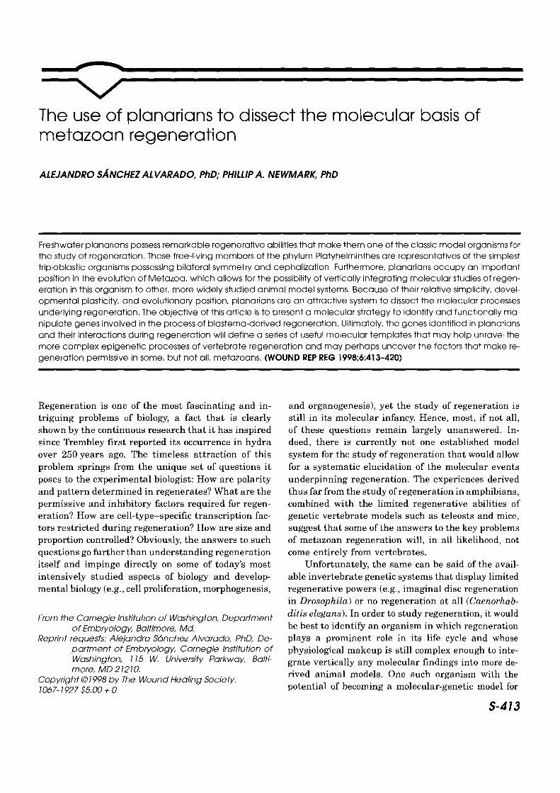
Neoblasts serve two purposes . First, they con-stantly replace the dying, nonproliferating differen-tiated cells ofthe adultorganism . 29 Second, they makeup the bulk of the mesenchymal component of theregeneratingblastema .28 Baguna et al . elegantlydem-onstrated the totipotential nature of neoblasts30 byinjecting cell fractions highly enriched in neoblastsinto x-ray irradiated planarians (Figure 4) . A dose of8,000 rads is sufficient to stop cell division in planar-ians almost immediately and causes death after 4-6 weeks. Irradiation also results in the abrogation ofregeneration as early as 3 days after treatment. In-jection of purified neoblasts from normal donors intoirradiated hosts results in the survival of the hostand in the restoration of regenerative abilities.30 Thetotipotentiality of neoblasts is further demonstratedwhen donor neoblasts from a sexually reproducingstrain are injected into irradiated asexual hosts. Theneoblasts not only repopulate the host and reactivateits regenerative abilities, but also "transform" it intoa sexual planarian because functional reproductive
WOUND REPAIR AND REGENERATIONVOL 6, NO. 4
SANCHEZALVARADO AND NEWMARK S-417
Total Cell
Donor
D%000
Differentiated Cells
Irradiated Host
O O
~0F-00' OW-0,\_
Total CelO
0 0
O000
O
T"O 7o %P
-1 Neoblasts
Figure 4 Schematic of the procedure employed to introduce donor cells into irradiated host planarians . Disaggregation of hostcells is accomplished in a calcium/magnesium-free medium . The cells are then size-fractionated through Nytex sieves of differentpore sizes . A triangular window is made in the donor, and cells are injected into the parenchyma of the host using a micropipette .The triangular tissue piece is replaced and allowed to heal . If successful, the host will survive and will in 2-3 weeks be able to re-generate its tissues again (adapted from Bagund et al. 3°) .
organs and copulatory apparatus are formed .31 Theseexperimental results confirm the role of neoblasts inplanarian regeneration and provide evidence for thetotipotent, stem-cell nature of this parenchymal cellpopulation .
GENE ISOLATION AND TRANSGENESIS INPLANARIANS: CURRENTANDFUTUREDIRECTIONSOur laboratory has applied a gene expression screendevised by Wang and Brown at the CarnegieInstitution32 to regenerating and nonregeneratingplanarian tissues in order to identify differentiallyexpressed genes. This method has the unique advan-tage of being able to estimate the number of upreg-ulated and downregulated genes in any givenscreen .32 The methodology requires small amounts of
Injection of Cells
the obtained poly(A)+ RNA populations; these are re-verse transcribed, restricted, ligated to linkers, andamplified by polymerase chain reaction . The cDNAsare then subjected to a series of subtractive hybrid-izations to enrich for differentially expressed tran-scripts.32 Thus far, we have identified a total of 59unique cDNA fragments whose expression is modu-lated by regenerative events . Examples of such frag-ments are shown in Figure 5 . The data indicate thatit is not only possible to enrich for regeneration-mod-ulated genes that may be required for the generationof a planarian blastema (Figure 5, a), but also toidentify genes that are specific to either cephalic orcaudal blastemas, that is, polarity-specific genes (Fig-ure 5, b and c) . Currently, our laboratory is engagedin the sequencing and characterization of the tempo-ral and spatial expression patterns ofthese andotherisolated transcripts.

S-478 SANCHEZ ALVARADO AND NEWMARK
HHBTTB HHBTTB HHBTTB
a b cFigure 5 Regeneration modulated cDNA fragments duringplanarian regeneration . Individual fragment obtained fromthe subtractions are radioactively labeled and hybridized tofilters containing 1 ug per lane of either head (H), head blas-tema (H8), tail (T), or tail blastema (TB) subtraction enrichedcDNA fragments . a, Blastema upregulated fragment . b, Tailblastema upregulated fragment . c, Head blastema upregu-lated fragment .
The ability to study gene function in vivo is crucialand defines an organism's usefulness as an experi-mental model system . The study of regeneration hassuffered under this tenet because organisms that are
well suited to genetic manipulations (mouse, ze-brafish, Drosophila, and C. elegans) display limitedor no regenerative powers, and those that are widelyused to study regeneration (axolotls, salamanders,and Pleurodeles) are quite refractory to genetic anal-
yses . Perhaps the most attractive feature of planari-ans is that their biology allows for the real possibilityofgenerating transgenic lines in which to study those
genes that may be involved in the process of epimor-
phic regeneration . The biology of planarians and theirdevelopmental plasticity make it possible to test ef-
fectively the viability of several well-establishedtransgenic methodologies currently being used inother animal model systems .
As previously discussed, the totipotency of pla-narian neoblasts, combined with their ease of rein-
troduction into irradiated animals (Figure 4), makes
these cells prime vectors for the introduction of ex-
ogenous DNAs into naive individuals. If one were to
homologize planarian neoblasts with murine embry-
onic stem cells, for example, it is not difficult to en-
vision the use of several well-establishedmethodologies such as electroporation, germ-cell in-
jections, and even transposable-element-driventransgenesis in order to generate recombinant cells.Hence, genetically modified neoblasts may be used to
repopulate irradiated animals, creating, in essence,
a transgenic animal whose cells originated from theintroduced recombinant neoblasts. We are attempting
to take advantageofthe pluripotentiality of planarian
SUMMARY
WOUND REPAIR AND REGENERATIONJULY-AUGUST 1998
neoblasts, as well as of these well-established meth-odologies to introduce exogenous DNA into these or-ganisms for the production of transgenic lines.
What does one hope to learn from the study of regen-eration in planarians? An indication ofthe exceptionalbiological secrets guarded so jealously by the Turbel-larians is exemplified by one remarkable property ofthe regenerating blastema : its morphogenetic equi-potency. One of the earliest discoveries of experimen-tal embryology was the production of more than onenormal larva by the physical fragmentation of early-stage echinoderm embryos, an observation that ledDriesch33 to postulate the idea of "harmonic equipo-tential systems." This idea was extended into thestudy of vertebrate embryology and eventually led tothe discovery of the mosaic nature of the early ver-tebrate embryo .34 The term mosaic was chosen be-cause a series of heterotopic and heterochronictransplantation schemes demonstrated that the mor-phologically homogeneous mesodermal layer of theearly embryo was already subdivided into areas fatedto give rise to various organs later during develop-ment . These observations led to the idea that organ-ogenesis has its ontogeny at developmental stages inwhich no overt signs of differentiation can be dis-cerned .
The most complete studies on this matter werecarried out by Harrison 35 and Detwiler36 using
the limb as a model. They noted that defined,undifferentiated mesodermal areas of early tailbud and midgastrulating stage embryos could de-velop into limbs. Harrison referred to these areasas morphogenetic fields . He termed them auton-omous because they could assemble organs bythemselves and equipotent because any part withinthe field could give rise to the whole organ. These"organ fields" resemble the whole organism in thepremosaic stage (pregastrulation), in combining ageneral determination with an epigenetic mode ofdevelopment.
The regenerating blastema in planarians can beconceived of as a morphogenetic field. First, it pro-
duces either a head or a tail, depending on its location .
Second, it is autonomous because its transplantation
gives rise to the appropriate structure. 3 Third, theblastema is equipotent because its parts are able togenerate a complete rather than an incomplete struc-ture (Figure 6) . Considering the evolutionary positionof planarians, the autonomy and equipotency of theirblastemas point to a set ofwidely conserved properties

WOUND REPAIR AND REGENERATIONVOL 6, NO. 4
of morphogenetic fields among very distant phyla of
the animal kingdom . Planarians provide us with anexample of a triploblastic organism whose morpho-genesis occurs in the absence of embryogenesis. Infact, regeneration in planarians is morphogenesis .Planarians that reproduce by fission do not have theluxury of gastrulation to establish their anterior/pos-terior and dorsal/ventral axes epigenetically. Hence,
the adult organism must rely on a defined mecha-
nism(s) to maintain polarity in the absence of any
kind ofembryonic development. Such amechanism(s)
may evolutionarily precede embryogenesis properand, if conserved in sexually reproducing animals,may provide unique insights into aspects of pro-
tostome and deuterostome embryogenesis. Thus, it
seems likely that a molecular study of planarian re-generation could shed light on the molecular basis ofmorphogenetic field establishment, as well as on themechanisms used for its differentiation. Nevertheless,and most likely the result of a historical accident,the study of planarians at the molecular level hasbeen largely neglected. Therefore, identifying thosegenes that are under temporal and spatial regulation
during the formation and differentiation ofplanarianblastemas, that is, regeneration, may ultimately pro-vide us with the molecular skeleton at the root of thecomplex morphogenetic events that occur in higherorganisms.
ACKNOWLEDGMENTSThis work was supported in part by National Insti-tutes of Health grant RO1 GM57260-01 to A.S.A . andby postdoctoral fellowship DRG-1322 of the CancerResearch Fund of the Damon Runyon-Walter Winch-ell Foundation to P.A.N . The authors would like toexpress their gratitude to Mike Sepanski for his in-valuable and expert assistance in the preparation ofsamples and the gathering of scanning electron mi-croscope images .
REFERENCES1. Morgan TH . Regeneration . New York : The Macmillan Com-
pany, 1901 .2. Child CM.
The physiological gradients .
Protoplasma1929 ;5:447-76.
3 . Brgndsted HV Planarian regeneration. Oxford : PergamonPress, 1969 .
4 . Hori H, Muto A, Osawa S, Takai M, Lue K-Y, Kawakatsu M.Evolution of Turbellaria as deduced from 5S ribosomal RNAsequences. In : Free-living and symbiotic Platyhelminthes. Go-ettingen, Germany: Prog Zool/Fortschr Zool, 1991 .
5. Riutort M, Field KG, Turbeville JM, Raff RA, Baguna J. 18SRNA sequences and phylogeny Platyhelminthes. Can J Zool1992;70 :1425-39 .
SANCHEZ ALVARADO AND NEWMARK S-419
6. Balavoine G. Identification of members of several homeoboxgenes in a planarian using a ligation-mediated polymerasechain reaction technique. Nucleic Acids Res 1996 ;24:1547-53 .
7 . Baguna J, Sa16 E, Romero R, Garcia-Fernandez J, Bueno D,Munoz-Marmol AM, Bayascas-Ramirez JR, Casali A. Regen-eration and pattern formation in planarians : cells, moleculesand genes. Zool Sci Jpn 1994;11:781-95.
8 . Bryant SV, Fyfe D, Singer M. The effects of denervation onthe ultrastructure ofyounglimb regenerates in the newt 7}itu-rus. Dev Biol 1974;24:577-95.
9 . Taube E. Regeneration mit beteiligung ortsfremder haut beitritonen . Wilhelm Roux' Arch 1921 ;49 :269-315 .
10 . Goss RJ. Regenerative inhibition following limb amputationand immediate insertion into the body cavity. Anat Rec1956;126:15-27 .
11 . Baguha J, Sal6 E, Collet J, Auladell MC, Ribas M. Cellular,molecular and genetic approaches to regeneration and patternformation in planarians . Fortschr Zool 1988 ;36:65-78 .
12 . Singer M. Induction of regeneration of the forelimb of thepostmetamorphic frog by augmentation of the nerve supply.J Exp Zool 1954;126:419-72.
13 . Thornton CS. Histological modifications in denervated injuredforelimbs ofAmblystoma larvae . J Exp Zool 1953 ;112:119-50.
14 . Bantle JA, Tassava RA . The neurotrophic influence on RNAprecursor incorporation into polyribosomes of regeneratingadult newt forelimbs. J Exp Zool 1974;189:101-13.
15. Locatelli P L'influenza del sistema nervoso sui processi diregenerazione . Arch Sci Biol ; 5 :362-78 .
16. GuyenotE. Territoires de regeneration chez le lezard (Lacertamuralis) . C.R . Soc Biol 1928 ;99127 .
17. Kiortsis V, Moraitou M. Factors of regeneration in Spirogra-phis spallanzanii . In : Kiortsis V, Trampusch HAL, editors .Regeneration in animals and related problems . Amsterdam:North-Holland, 1965 :250-61.
18. Singer M, Maier CE, McNutt WS. Neurotrophic activity ofbrain extracts in forelimb regeneration ofthe urodele 7Niturus .J Exp Zool 1976 ;196:131-50.
19. Yntema CL . Regeneration in sparsely innervated and aneuro-genic forelimbs of Amblystoma larvae. J Exp Zool1959;140 :101-24.
20. Yntema CL . Blastema formation in sparsely innervated andaneurogenic forelimbs of Amblystoma larvae. J Exp Zool1959;142 :423-40.
21 . Egar M, Yntema CL, Singer M. The nerve fiber content ofAmbystoma aneurogenic limbs. J Exp Zool 1977;186 :91-6.
22. Thornton CS, Thornton MT. Recuperation ofregeneration indenervated limbs of Ambystoma larvae . J Exp Zool1970;173:293-302.
23. Chandebois R. The dynamics of wound closure and its role inthe programming ofplanarian regeneration . 1 . Blastema emer-gence. Dev Growth Differ 1980;22:693-704 .
24. Slack JM . The source of cells for regeneration. Nature1980;286:5775-60 .
25. Bueno D, Baguna J, Romero R. A central body region definedby a position-specific molecule in the planarian Dugesia (Gi-rardia) tigrina : spatial and temporal variations during regen-eration . Dev Biol 1996;178:446-58.
26. Gardiner, GM, Blumberg, B, Komine, Y, Bryant, SV Regula-tion ofHoxA expression in developing and regenerating axolotllimbs. Development 1995 21:1731-1741
27. BayascasJR, Castillo E, Munoz-Marm61 AM, Salo E. PlanarianHox genes: novel patterns of expression during regeneration .Development 1997;124 :141-8 .
28. Sal6 E, Baguna J. Regeneration and pattern formation inplanarians . 11 . Local origin and role of cell movements inblastema formation. Development 1989 :107;69-76 .
29. Baguna J, Romero J. Quantitative analysis of cell types duringgrowth, degrowth and regeneration in the planarians Dugesia
S-420 SANCHEZALVARADO AND NEWMARK
mediterranea and Dugesia tigrina . Hydrobiologia1981;84:181-94.
30 . Baguna J, Sa16 E, Auladell C. Regeneration and pattern for-mation in planarians . III . Evidence that neoblasts are totipo-tent stem cells and the source of blastema cells . Development1989 ;107 :77-86 .
31 . Baguna J, Romero R, Salo E, Collet J, Auladell C, Ribas M,Riutort M, Garcia-Fernandez J, Burgaya F, Bueno D. Growth,degrowth and regeneration as developmental phenomena inadult freshwater planarians . In : Martin H-J, editor. Experi-mental embryology in aquatic plants and animals. NewYork :Plenum Press, 1990 .
32 . Wang Z, BrownDDA. Gene expression screen. Proc Natl AcadSci USA 1991;88:11505-9 .
33. Driesch H. Die Isolirten blastomeren des echinidenkeimes .Arch Entw 1900 ;10:361 .
34. Huxley JS, De Beer GR. The elements of experimental em-bryology. Cambridge: Cambridge University Press, 1934 .
WOUND REPAIR AND REGENERATIONJULY-AUGUST 1998
35. Harrison R. Experiments on the development of the fore-limbof Amblystoma, a self-differentiating equipotential system. JExp Zool 1918 ;25 :413-61.
36. Detwiler SR. On the time of determination of the anteropos-terior axis of the forelimb of Amblystoma. J Exp Zool1933 ;64 :405-14.
37. Mitman G, Fausto-Sterling A. Whatever happened to pla-naria? C. M. Child and the physiology of inheritance . In :Clarke, AE, Fujimura, JH editors. The right tools for the job:at work in 20th-century life sciences . Princeton, NJ : PrincetonUniversity Press, 1992 .
38. Dubois F. Sur une nouvelle methode permettant de mettre enevidence la migration des cellules de regeneration chez lesPlanaires . C R Acad Sci 1948;226:1316-7.
39. Hay DE . Regeneration . New York : Holt, Rinehart and Win-ston, 1966.