transverse myelopathy following intrathecal administration ... · transverse myelopathy following...
TRANSCRIPT
Seediscussions,stats,andauthorprofilesforthispublicationat:https://www.researchgate.net/publication/6501866
Transversemyelopathyfollowingintrathecaladministrationofchemotherapy
ArticleinSingaporemedicaljournal·March2007
Source:PubMed
CITATIONS
12
READS
126
3authors,including:
Chooi-FunLeong
HospitalUniversitiKebangsaanMalaysia(HUKM)
80PUBLICATIONS317CITATIONS
SEEPROFILE
AllcontentfollowingthispagewasuploadedbyChooi-FunLeongon20December2014.
Theuserhasrequestedenhancementofthedownloadedfile.Allin-textreferencesunderlinedinblueareaddedtotheoriginaldocument
andarelinkedtopublicationsonResearchGate,lettingyouaccessandreadthemimmediately.

Singapore Med J 2007; 48(2) : e46C a s e R e p o r t
ABSTRACTTransverse myelopathy is one of the rare complications following administration of intrathecal chemotherapy. We report two cases of transverse myelopathy following administration of intrathecal methotrexate and cytarabine arabinoside. One patient was a 17-year-old Malay man who had lymphoblastic lymphoma in the leukaemic phase, while the other patient was a 40-year-old Malay man with relapsed Hodgkin’s lymphoma. Both cases demonstrated variability in onset of symptoms, clinical progression and final outcome from the complication.
Keywords : chemotherapy complications, cytarabine arabinoside, intrathecal methotrexate, transverse myelopathy
Singapore Med J 2007; 48(2):e46–e49
INTRODUCTIONNeurological complications of anti-cancer therapy
may result from direct toxic effects on the nervous
system, or indirectly from drug-induced metabolic
derangements or cerebrovascular accidents.(1-4) Their
recognition is important because of potential confusion
with metastatic disease. Furthermore, discontinuation
of the offending agent may prevent irreversible injury.
Common neurological complications of chemotherapy
include peripheral neuropathy, cranial neuropathies,
acute encephalopathy, acute vasculopathies, headaches
and seizures.(1-4) Transverse myelopathy, a much
less common sequela following intrathecal (IT)
administration of chemotherapy, is defined as the
development of isolated spinal cord dysfunction over
hours or days in the absence of a compressive lesion.
It has been rarely reported following IT methotrexate
(MTX), cytarabine arabinoside (Ara-C) and thiotepa.(5-7)
CASE REPORTSCase 1A 17-year-old Malay man was diagnosed to have
lymphoblastic lymphoma in the leukaemic phase in
July 2004, and underwent autologous peripheral
blood stem cell transplantation (PBSCT) five months
later (Fig. 1). Six weeks after PBSCT, he developed
sudden onset of isolated left facial nerve palsy.
Leukaemic infiltration of the central nervous system
(CNS) was confirmed with the presence of
malignant lymphoid cells noted in the cytospin
Teh H S, Fadilah S A W, Leong C F
Haematology Unit,Department of Medicine,Faculty of Medicine,University Kebangsaan Malaysia,Jalan Yaacob Latif,Bandar Tun Razak,Cheras,Kuala Lumpur 56000,Malaysia
Teh HS, MD, MMedClinical Specialist
Fadilah SAW, MD, MMed, PhDAssociate Professor
Leong CF, MD, MPath, FRCPAAssociate Professor
Correspondence to:Dr Teh Hiok Seng,Tel: (60) 3 9170 2391Fax: (60) 3 9173 7829Email: hiokseng2005 @gmail.com
Transverse myelopathy following intrathecal administration of chemotherapy
Fig. 1 Timeline of events for Case 1. Hyper-CVAD: Hyperfractionated cyclophosphamide, vincristine, doxorubicin and dexamethasone.
Singapore Med J 2007; 48(2) : e47
of the cerebrospinal fluid (CSF). He was given
the first IT (via the third and fourth lumbar
intervertebral space) MTX 12.5 mg immediately
following the diagnostic lumbar puncture. After
confirming CNS relapse, he was given IT MTX
12.5 mg, Ara-C 40 mg and dexamethasone 4 mg about
one week after the first IT MTX. Soon after completion
of IT injection, he felt numbness of his left lower
limb, followed by development of paraplegia and
paraesthesia of the lower parts of the body. There was
no urinary or bladder incontinence.
Physical examination revealed that the blood
pressure was high (200/120 mmHg) and he was
tachycardic, pulse rate of 120 beats per minute.
Sensory loss was noted from lower extremities up to T4
vertebral level. Intravenous (IV) hydrocortisone
100 mg and piriton 10 mg were given immediately.
Contrast-enhanced computed tomography of the brain
was done on the same day and was normal. The power
of his lower limbs improved from 0/5 to 3/5 (Medical
Research Council Scale) 45 minutes after the IT
injection. Power and sensory deficit of the lower limb
were normalised after the next day.
As the neurological complication occurred
following IT administration of MTX in combination
with Ara-C and not MTX alone, Ara-C was thought to be
the most likely offending agent. However, he developed
paraplegia a week later, following IT MTX 12.5 mg
and dexamethasone 4 mg, in the absence of Ara-C.
The symptoms completely resolved 30 minutes after
IV hydrocortisone 100 mg and piriton 10 mg.
Magnetic resonance (MR) imaging of the brain and
thoracolumbar spine revealed no abnormality. Repeated
CSF examination during the neurological event was
unremarkable.
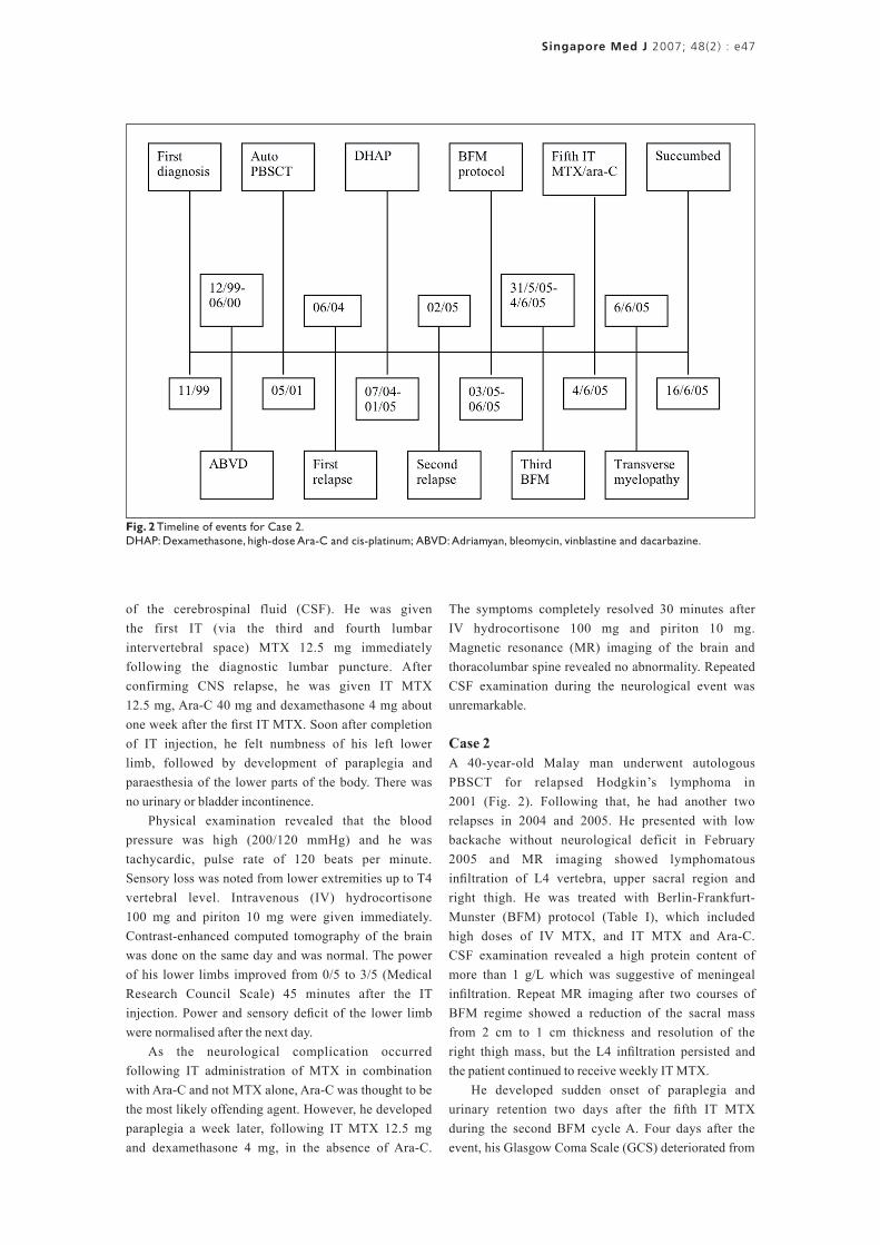
Case 2A 40-year-old Malay man underwent autologous
PBSCT for relapsed Hodgkin’s lymphoma in
2001 (Fig. 2). Following that, he had another two
relapses in 2004 and 2005. He presented with low
backache without neurological deficit in February
2005 and MR imaging showed lymphomatous
infiltration of L4 vertebra, upper sacral region and
right thigh. He was treated with Berlin-Frankfurt-
Munster (BFM) protocol (Table I), which included
high doses of IV MTX, and IT MTX and Ara-C.
CSF examination revealed a high protein content of
more than 1 g/L which was suggestive of meningeal
infiltration. Repeat MR imaging after two courses of
BFM regime showed a reduction of the sacral mass
from 2 cm to 1 cm thickness and resolution of the
right thigh mass, but the L4 infiltration persisted and
the patient continued to receive weekly IT MTX.
He developed sudden onset of paraplegia and
urinary retention two days after the fifth IT MTX
during the second BFM cycle A. Four days after the
event, his Glasgow Coma Scale (GCS) deteriorated from
Fig. 2 Timeline of events for Case 2. DHAP: Dexamethasone, high-dose Ara-C and cis-platinum; ABVD: Adriamyan, bleomycin, vinblastine and dacarbazine.

Singapore Med J 2007; 48(2) : e48
15/15 to 10/15, and he developed respiratory distress.
He was electively intubated for respiratory distress
and poor GCS. Repeat MR imaging of the spine and
brain showed no evidence of disease progression, spinal
cord compression or brain metastasis. He continued to
deteriorate and succumbed ten days later.
DISCUSSIONTransverse myelopathy, an unusual complication of
IT MTX/Ara-C, is defined as the development of
isolated spinal cord dysfunction over hours or days in
the absence of a compressive lesion.(5-7) The incidence
was reported in approximately 3% of all cases treated
with IT MTX or Ara-C. Our patients demonstrated a
typical transverse myelopathy with paraplegia, level of
sensory deficit as well as bladder and respiratory muscle
involvement following administration of IT MTX and
Ara-C. The symptoms developed within few minutes
and two days, respectively, after completion of the
medication. This was similar to other patients previously
described who developed symptoms within minutes
to 48 hours after treatment, although some may even
persist up to two weeks.(5-7) Repeated MR imaging of the
spine and brain which excluded a compressive lesion,
close temporal relationship and lumbar puncture site
via L3/L4 vertebral level which was free of spinal cord,
supported that the transverse myelopathy was related
to IT administration of MTX and Ara-C.
In the first patient, all the symptoms (weakness and
sensory loss) started to improve in less than one hour
and completely resolved in less than 24 hours. The rapid
development and resolution of the symptoms suggest
that it is most likely a neuropraxia caused by sudden
osmolality changes in CSF, or direct toxic or irritation
by the IT medication. Hence, recovery occurred rapidly
when the offending agents were cleared from the spinal
CSF. This was consistent with the half-lives of IT
administration of MTX and Ara-C in CSF, which are
4.5–14 hours(8,9) and 2–11 hours, respectively. Although
the majority of cases reported showed clinical
improvement, the extent of recovery was variable.(5-7)
In contrast to the first case that developed
neurological symptoms after the second IT MTX,
the second patient developed the symptoms only after
the fifth IT MTX. Additionally, the neurological deficit
was more extensive and progressive. The more severe
neurological manifestation may be partly attributed
to systemic IV MTX in high doses. Besides, the pre-
existing CNS involvement observed in the second
case also contributed to the increased risk and severity
of neurotoxicity of IT MTX.(5-7)
Documented risk factors for the development of
MTX-related transverse myelopathy include high
dose MTX of more than 50 mg given intrathecally,
repeated injection with close interval less than one
week, concurrent cranial radiotherapy, systemic MTX,
active CNS disease (e.g. meningeal leukaemia).(8,9)
Since the occurrence and clinical course of IT-induced
transverse myelopathy is unpredictable, we should
try to avoid high dose IT MTX, close interval of
administration, concurrent cranial radiotherapy and
systemic MTX as far as possible. Those high-risk
patients should be counselled properly about the
possible side effects before administration of the
medication. Any further administration of IT MTX
or Ara-C must be stopped once the complication has
occurred.(5-7) The patients should be reassured regarding
Table I. Chemotherapy regime.
Chemotherapy agents Manufacturer City/state Country
HyperCVAD cycle A
Cyclophosphamide Baxter Frankfurt Germany
Vincristine MaynePharma Mulgrane Australia
Doxorubicin Dabur Solan India
Dexamethasone ZydusCadila Ahmedabad India
HyperCVAD cycle B
Methotrexate MaynePharma Mulgrane Australia
Ara-C Dabur Solan India
ICE
Ifosfamide Baxter Frankfurt Germany
Carboplatin MaynePharma Mulgrane Australia
Etopoxide Dabur Solan India
ABVD
Doxorubicin Dabur Solan India
Bleomycin NipponKayaku Tokyo Japan
Vincristine MaynePharma Mulgrane Australia
Dacarbazine MaynePharma Mulgrane Australia
DHAP
Dexamethasone ZydusCadila Ahmedabad India
Cis-platinum Pfizer Bentley Australia
Ara-C Dabur Solan India
BFM cycle A
Dexamethasone ZydusCadila Ahmedabad India
Vincristine Pfizer Bentley Australia
Ifosphamide Baxter Frankfurt Germany
Methotrexate MaynePharma Mulgrane Australia
Etopoxide Dabur Solan India
Ara-C Dabur Solan India
BFM cycle B
Dexamethasone ZydusCadila Ahmedabad India
Vincristine Pfizer Bentley Australia
Cyclophosphamide Baxter Frankfurt Germany
Methotrexate MaynePharma Mulgrane Australia
Adriamycin Dabur Solan India
Singapore Med J 2007; 48(2) : e49
the clinical course and given appropriate supportive
treatment accordingly. It is advisable to monitor the
CSF MTX level to make sure that it has declined to
an acceptable value before the subsequent dose is
administered.
In conclusion, history of uncomplicated IT
administration of MTX or Ara-C does not exclude
one from developing transverse myelopathy during
subsequent chemotherapy as illustrated in the second
case. The onset can vary from a few minutes to a few
hours after IT injection. The clinical course also can
vary from complete resolution of symptoms to continued
deterioration, merely paraplegia to respiratory muscle
involvement and even death.
REFERENCES1. Rottenberg DA, ed. Neurological Complications of Cancer
Treatment. Boston: Butterworth-Heinemann, 1991.
2. Gilbert MR. The neurotoxicity of cancer chemotherapy. Neurologist 1998; 4:43.
3. Keime-Guibert F, Napolitano M, Delattre JY. Neurological complications of radiotherapy and chemotherapy. J Neurol 1998; 245:695-708.
4. Wen PY. Central nervous system complications of cancer therapy. In: Schiff D, Wen PY, eds. Cancer Neurology in Clinical Practice. Boston: Humana Press, 2002.
5. Gagliano RG, Costanzi JJ. Paraplegia following intrathecal methotrexate: report of a case and review of the literature. Cancer 1976; 37:1663-8.
6. Dunton SF, Nitschke R, Spruce WE, Bodensteiner J, Krous HF. Progressive ascending paralysis following administration of intrathecal and intravenous cytosine arabinoside. A Pediatric Oncology Group study. Cancer 1986; 57:1083-8.
7. Werner RA. Paraplegia and quadriplegia after intrathecal chemotherapy. Arch Phys Med Rehabil 1988; 69:1054-6.
8. Bleyer WA, Dedrick RL. Clinical pharmacology of intrathecal methotrexate. I. Pharmacokinetics in nontoxic patients after lumbar injection. Cancer Treat Rep 1977; 61:703-8.
9. Miller KT, Wilkinson DS. Pharmacokinetics of methotrexate in the cererospinal fluid after intracerebroventricular administration in patients with meningeal carcinomatosis and altered cerebrospinal fluid flow dynamics. Ther Drug Monit 1989; 11:231-7.
View publication statsView publication stats