two somatic tsh receptor mutations in a patient with toxic...
TRANSCRIPT

Endocrine-Related Cancer (2003) 10 591–600
Two somatic TSH receptor mutations in apatient with toxic metastasising follicularthyroid carcinoma and non-functionallung metastases
D Fuhrer, A Tannapfel1, O Sabri2, P Lamesch3 and R PaschkeIII Medical Department, 1Institute of Pathology, 2Department of Nuclear Medicine and 3Department of Surgery,University of Leipzig, Leipzig, Germany
(Requests for offprints should be addressed to R Paschke; Email: [email protected])
Abstract
In a 59-year-old patient, thyroid follicular cancer was diagnosed in two right-sided toxic thyroidnodules, which had presented clinically as unilateral thyroid autonomy. In addition, the patient hadhistologically proven lung metastases of thyroid cancer; however, these failed to exhibit iodine uptakeand were resistant to radioiodine treatment. The functional activity of the thyroid nodules promptedus to screen for TSH receptor (TSHR) mutations, and the histological diagnosis of follicular carcinomaled us to search for the PAX8-PPARγ1 rearrangement and mutations in the ras genes. Each thyroidnodule harboured a different TSHR mutation (large nodule, Asp633Tyr; small nodule, Phe631Ile).Presence of both mutations in one sample suggestive of local invasion of a thyroid carcinoma couldnot be demonstrated, although several specimens from different nodule locations were screened.Only the wild-type TSHR sequence was identified in the histologically normal left thyroid lobe, andno genetic alterations were found in the other investigated genes. No TSHR mutations were detectedin the pulmonary metastases.
This is the first case report of a patient with toxic follicular thyroid carcinoma harbouring twodifferent TSHR mutations and presenting with non-functional lung metastases.
Endocrine-Related Cancer (2003) 10 591–600
Introduction
Gain-of-function mutations in the thyrotropin receptor gene(TSHR) have been identified in various clinical conditions,namely toxic thyroid nodules and subsets of toxic multinodu-lar goitres caused by somatic TSHR mutations, in addition toautosomal dominant non-autoimmune hyperthyroidism andgestational thyrotoxicosis attributable to germline TSHRmutations (Parma et al. 1993, Paschke & Ludgate 1997).Constitutive thyrotropin receptor activation by thesemutations confers TSH-independent thyroid hormone pro-duction and thyroid growth, resulting in hyperthyroidism andthyroid hyperplasia.
In addition, to date, somatic TSHR mutations have beenreported in nine differentiated thyroid carcinomas, five ofwhich mimicked the phenotype of toxic thyroid nodules(Russo et al. 1995, 1997, 1999, Spambalg et al. 1996, Cama-cho et al. 2000, Mircescu et al. 2000). We describe the firstpatient with metastasising toxic follicular thyroid cancer, har-bouring two different somatic TSHR mutations.
Endocrine-Related Cancer (2003) 10 591–600 Online version via http://www.endocrinology.org1351-0088/03/010–591 2003 Society for Endocrinology Printed in Great Britain
Case report
A 59-year-old patient presented with a 4-year history of atoxic right-sided goitre and a recent event of cardiac failureand atrial fibrillation. His thyroid function tests showedsubclinical hyperthyroidism (TSH < 0.01 mU/l, free tri-iodothyronine and free thyroxine (T4) in the upper normalrange) while he was receiving carbimazole 20 mg/day. Ultra-sonography demonstrated two thyroid nodules (3.5 · 2 · 5 cm,solid with calcifications, and 1.5 · 2 · 2 cm, solid) in the rightlobe (34 ml) and a normal left thyroid lobe (2 ml). Increaseduptake of technetium on scintiscan (6.76%) in the entire rightlobe, with complete suppression of the left lobe, was com-patible with two right-sided toxic nodules (Fig. 1). Afterrecompensation of cardiac failure, thyroid surgery wasplanned.
However, a left-sided pulmonary lesion was noted on thepreoperative chest X-ray and confirmed on computedtomography (CT) scan (Fig. 2). Because the patient had along-standing history of heavy smoking, bronchoscopy was

Fuhrer et al.: Hot thyroid cancer
Figure 1 Scintiscan: increased circumscribed uptake of technetium in the right thyroid lobe, and suppression of the leftthyroid lobe.
performed, whereby an additional left-sided endobronchialtumour (2 cm) and several suspicious mucosal lesions werefound and harvested for biopsy. Histopathological analysisof these specimens gave the unexpected diagnosis of multiplemetastases of follicular thyroid carcinoma.
The patient underwent total thyroidectomy and receivedablative radioiodine therapy with 5.698 GBq (154 mCi)iodine-131 (TSH concentration 21.07 mU/l; serum thyroglo-bulin concentration 10.3 ng/ml; thyroglobulin antibodies 27IU/l (normal range < 60 IU/l)). At day 5, a post-therapeutictotal-body scan was performed, which showed uptake ofradioiodine in the thyroid bed, but not over the lungs.
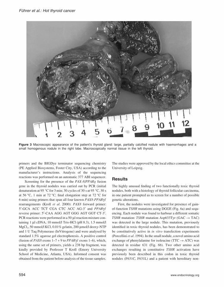
The macroscopic appearance of the thyroid specimenwas that of a calcified nodule filling the right thyroid lobe,with compression of normal thyroid tissue at the margins anda separate right-sided small solid thyroid nodule. The isthmusand the left lobe were of normal size and appearance (Fig. 3).
Histological analysis of the thyroid specimen showedfollicular neoplasia with capsule invasion, in addition to vas-cular invasion being present in both nodules in several loca-tions, suggesting a diagnosis of follicular carcinoma in bothnodules (Figs 3 and 4). Apart from the two nodules, onlynormal thyroid tissue was found in the remainder of the rightlobe and the entire left thyroid lobe.
592 www.endocrinology.org
At 4 months follow-up, a diagnostic 131I total-body scanwas performed (647.5 MBq (17.5 mCi) 131I) after withdrawalof L-T4 treatment (TSH concentration 26.5 mU/l). The scanwas completely negative, as were serum thyroglobulin con-centrations ( < 0.3 ng/ml) measured at the same time.
At 8 months follow-up, the patient was generally well,with undetectable serum thyroglobulin concentrations whilereceiving TSH-suppressive L-T4 treatment. However, arepeat CT scan revealed persistence of the pulmonarylesions, with an unchanged appearance on bronchoscopy andthe consistent finding of histologically thyroglobulin-positivepulmonary metastasis (Fig. 5). Subsequently, the patientunderwent extensive thoracic surgery to remove all macro-scopically visible thyroid metastases.
Materials and methods
DNA extraction
Genomic DNA was isolated from the two thyroid nodules,from surrounding normal thyroid tissue of the right lobe, andleft lobe normal thyroid tissue, using a Qiagen Tissue Kitaccording to the manufacturers’ instructions. In addition,DNA was extracted from the paraffin-embedded tissue sec-

Endocrine-Related Cancer (2003) 10 591–600
Figure 2 Preoperative CT scan: left-sided pulmonary lesion with pleural adhesion. An identical result was obtained at 8months follow-up after total thyroidectomy and ablative radioiodine treatment.
tions of the lung metastases (Fig. 5). Thus paraffin-embeddedtissue sections 20 µm thick were deparaffinized in xylol,rehydrated in an alcohol series, air-dried, and resuspended in100 µl lysis buffer (500 mM Tris-HCl, 10 mM NaCl, 20 mMEDTA, 1% SDS, pH 8.9). DNA extraction was performed at56 °C by the addition of 1 µl proteinase K (50 mg/ml) andthen following the Qiagen Tissue Kit procedure.
RNA extraction
Total RNA was extracted from the two nodules and normaltissue using the method of Chomczynski & Sacchi (1987).Complementary DNAs were prepared by reverse transcriptionof 1 µg total RNA in a 20 µl reaction mixture containing 5×first-strand buffer (50 mM Tris-HCl (pH 8.3), 375 mM KCland 15 mM MgCl2, 0.5 mM dNTPs, 5 mM dithiothreitol),
www.endocrinology.org 593
0.15 U RNase inhibitor, 2.5 µM oligo dT primer and 2 UMMLV reverse transcriptase (all reagents from Promega).cDNA synthesis was performed at 42 °C for 1 h, followedby an incubation at 95 °C for 10 min (MG Research Cycler).
PCR, denaturating gradient gelelectrophoresis and sequencing
Exons 9 and 10 of the TSHR gene and exons 1 and 2 of theH- and K-ras genes were amplified from genomic DNA byPCR (Fuhrer et al. 1997, Krohn et al. 2001). Screening forTSHR mutations was performed by denaturating gradient gelelectrophoresis (DGGE) as described previously (Trulzsch etal. 2001). TSHR PCR fragments with abnormal gel shiftingpatterns and ras PCR products were sequenced on bothstrands using the respective PCR primers as sequencing
Fuhrer et al.: Hot thyroid cancer
Figure 3 Macroscopic appearance of the patient’s thyroid gland: large, partially calcified nodule with haemorrhages and asmall homogenous nodule in the right lobe. Macroscopically normal tissue in the left thyroid.
primers and the BIGDye terminator sequencing chemistry(PE Applied Biosystems, Foster City, USA) according to themanufacturer’s instructions. Analysis of the sequencingreactions was performed on an automatic 377 ABI sequencer.
Screening for the presence of the PAX-8/PPARg fusiongene in the thyroid nodules was carried out by PCR (initialdenaturation at 95 °C for 3 min; 30 cycles of 30 s at 95 °C, 30 sat 56 °C, 1 min at 72 °C; final elongation step at 72 °C for6 min) using primers that span all four known PAX8-PPARγ1rearrangements (Kroll et al. 2000): PAX8 forward primer:5′-GCA ACC TCT CGA CTC ACC AG-3′ and PPARγ1reverse primer: 5′-CAA AGG AGT GGG AGT GGT CT-3′.PCR reactions were performed in a 50 µl reaction mixture con-taining 1 µl cDNA, 10 mmol/l Tris-HCl (pH 8.3), 1.5 mmol/lMgCl2, 50 mmol/l KCl, 0.01% gelatin, 200 µmol/l deoxy-NTPand 1 U Taq Polymerase (InVitrogene) and were analysed bystandard 1.5% agarose gel electrophoresis. A positive control(fusion of PAX8 exons 1–7 + 9 to PPARγ1 exons 1–6), which,using the same set of primers, yields a 238 bp fragment, waskindly provided by Professor T Kroll (Emory UniversitySchool of Medicine, Atlanta, USA). Informed consent wasobtained from the patient before analysis of the tissue samples.
594 www.endocrinology.org
The studies were approved by the local ethics committee at theUniversity of Leipzig.
Results
The highly unusual finding of two functionally toxic thyroidnodules, both with a histology of thyroid follicular carcinoma,in one patient prompted us to screen for a number of possiblegenetic alterations.
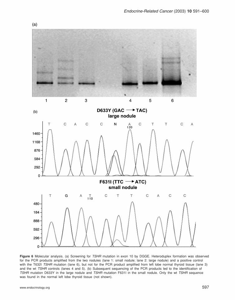
First, the nodules were investigated for presence of gain-of-function TSHR mutations using DGGE (Fig. 6a) and sequ-encing. Each nodule was found to harbour a different somaticTSHR mutation: TSHR mutation Asp633Tyr (GAC � TAC)was detected in the large nodule. This mutation, previouslyidentified in toxic thyroid nodules, has been demonstrated tobe constitutively active in in vitro transfection experiments(Porcellini et al. 1994). In the small nodule, a novel amino acidexchange of phenylalanine for isoleucine (TTC � ATC) wasdetected in residue 631 (Fig. 6b). Two other amino acidexchanges resulting in constitutive TSHR activation havepreviously been described in this codon in toxic thyroidnodules (F631C, F631L) and a patient with hereditary non-

Endocrine-Related Cancer (2003) 10 591–600
Figure 4 Histology of the thyroid specimen. (a) Large nodule; (b) small nodule; (c) small nodule. Original magnification ×10(haematoxylin & eosin stain). Invasion of the capsule is demonstrated in both nodules, consistent with the diagnosis offollicular carcinoma. Only normal thyroid tissue was found in the left thyroid lobe (not shown).

Fuhrer et al.: Hot thyroid cancer
Figure 5 Diffuse infiltration of follicular thyroid cancer (arrows) in surgically removed lung tissue (haematoxylin & eosinstain).
autoimmune hyperthyroidism (F631L) (Porcellini et al. 1994,Kopp et al. 1995). In contrast, only the wild-type (wt TSHR)sequence was found in both the right and left lobe normalthyroid tissue (not shown).
Secondly, we hypothesised that only one of the ‘toxicthyroid nodules’ was the follicular carcinoma exhibiting localinvasion of the neighbouring toxic follicular adenoma. How-ever, serial sections of the right thyroid lobe showed thatthe two nodules were completely separated from each other,without evidence of invasion. In addition, genomic DNAsextracted from five different locations within each nodulewere investigated. DGGE was used for TSHR mutationscreening because of its superior sensitivity for detection ofheterozygous mutations in samples with varying degrees ofnormal and diseased tissue components (Trultzsch et al.2001). However, although one or other of the two TSHRmutations was detected in every sample, the presence of bothmutations in one sample could not be demonstrated.
Thirdly, we screened for mutations in the H- and K-rasgenes, implicated in various thyroid pathologies and associ-ated in vitro with a follicular phenotype (Bond et al. 1994,
596 www.endocrinology.org
Wynford-Thomas et al. 1997, Gire & Wynford-Thomas2000). However, no ras mutations were identified in eitherof the two nodules.
Fourthly, we investigated the presence of a PAX8-PPARγ1 fusion gene, previously reported as specific for fol-licular thyroid carcinoma (Kroll et al. 2000). Using primers,which allow detection of all four known PAX8-PPARγ1rearrangements, we were unable to identify a PAX8-PPARγ1fusion gene in the cDNAs prepared from both nodules.
Fifthly, we aimed to clarify the molecular origin of thelung metastases. Thus we extracted and sequenced genomicDNA from the initial transbronchial biopsy specimen that ledto the diagnosis of metastasising thyroid cancer, but foundonly wt TSHR sequences. The same results were obtainedwhen we later screened surgically removed lung tissue withmacroscopically visible tumour infiltration.
Discussion
In this paper we present a patient with unilateral thyroidautonomy attributable to two right-sided toxic nodules, both
Endocrine-Related Cancer (2003) 10 591–600
Figure 6 Molecular analysis. (a) Screening for TSHR mutation in exon 10 by DGGE. Heteroduplex formation was observedfor the PCR products amplified from the two nodules (lane 1: small nodule; lane 2: large nodule) and a positive controlwith the T632I TSHR mutation (lane 6), but not for the PCR product amplified from left lobe normal thyroid tissue (lane 3)and the wt TSHR controls (lanes 4 and 5). (b) Subsequent sequencing of the PCR products led to the identification ofTSHR mutation D633Y in the large nodule and TSHR mutation F631I in the small nodule. Only the wt TSHR sequencewas found in the normal left lobe thyroid tissue (not shown).
www.endocrinology.org 597

Fuhrer et al.: Hot thyroid cancer
of which exhibited the histological features of thyroid follicu-lar carcinomas (Fig. 4) and were first diagnosed through theirlung metastases (Figs 2 and 5). Consistent with their func-tional activity, somatic TSHR mutations (D633Y and F631I)were identified in each nodule (Fig. 6b). Other genetic altera-tions, e.g. ras mutations and PAX8-PPARγ1 rearrangement,that could be expected to contribute to the phenotype of fol-licular thyroid carcinoma were not found.
To our knowledge, this is the first patient with differen-tiated toxic thyroid cancer harbouring two different TSHRmutations. In view of the unusual clinical, pathological andmolecular findings, we were highly critical of this diagnosis.However, the diagnosis was established by: (i) demonstrationof both capsular (Fig. 4) and vascular invasion in both nod-ules; (ii) repeated finding of lung metastases (Fig. 5) of thy-roid follicular cancer, which exhibited weak thyroglobulinstaining; (iii) failure to demonstrate local invasion of a fol-licular cancer into a hot thyroid nodule, by the absence ofco-localisation of both TSHR mutations in one DNA sample,even though different locations within the nodules wereinvestigated and DGGE was used as a highly sensitivemethod of screening (Fig. 6a). Because the patient is male,clonality analysis of the thyroid tumours was not possible;(iv) failure to detect, other than in the two nodules, any otherlesions on either macroscopic inspection or histological ana-lysis of the patient’s thyroid specimen.
The association of TSHR mutations with thyroid malig-nancy, described to date in nine thyroid carcinomas involvingdifferent histologies such as follicular, papillary and insularthyroid cancer, remains an unresolved molecular puzzle.Thus it is unclear whether (certain) TSHR mutations mighthasten the onset of thyroid malignancy or, perhaps morelikely, whether the occurrence of an activating TSHRmutation in a thyroid cancer could be the incidental result ofan increased mutation rate resulting from another molecularalteration.
Activating TSHR mutations, which have been reported in57–82% of hyperfunctioning thyroid nodules, are, accordingto general perception, invariably linked to benign thyroiddisease (Parma et al. 1993, 1997, Krohn & Paschke 2001,Trultzsch et al. 2001). In agreement with this, continuouscAMP activation by thyroid-specific expression of either theA2 adenosine receptor, gsp or cholera toxin A1 in transgenicmice has been shown to cause stimulation of differentiatedthyroid growth and hyperthyroidism (Ledent et al. 1992,Michiels et al. 1994, Zeiger et al. 1997). However, foci oftransformed thyrocytes have been described in old A2 aden-osine transgenics (Van Sande et al. 1995) and, interestingly,transgenics with a mutant α1B-adrenergic receptor exhibitedan aggressive phenotype of rapidly developing hyperfunc-tioning thyroid nodules, with frequent evolution towardsmetastasising thyroid malignancy, most probably as a resultof dual pathway (cAMP and phosphokinase C) activation(Ledent et al. 1997). Constitutive stimulation of the phospho-
598 www.endocrinology.org
kinase C pathway has been described for a small number ofTSHR mutations in vitro; however, these included only twoof the nine TSHR mutations identified in ‘hot’ thyroid can-cers (M486F and T632I; Van Sande et al. 1995). Finally,possibly the strongest argument against a constitutive TSHRmutation propagating evolution of thyroid cancer in humansis the fact that thyroid malignancy has been reported in onlyone among more than 150 patients of the 10 families andnine individuals with activating TSHR germline mutationsdescribed to date (Leclere et al. 1997, www.uni-leipzig.de/innere/tshr).
Which hypotheses could then be established on the basisof the molecular findings in our patient, in particular for therecurrent lung metastases with absence of TSHR mutations,which are difficult to reconcile with TSHR mutation bearingprimary thyroid malignancies?
First, there may initially have been one primary thyroidtumour, which did not harbour a TSHR mutation and forwhich we were unable to demonstrate a specific genetic alter-ation – for example, we did not identify ras mutations or aPAX8-PPARγ1 rearrangement. Hence, the occurrence of twodifferent somatic TSHR mutations would represent secondaryevents conferring autonomous function exclusively to thetwo thyroid nodules. Besides the known genetic heterogen-eity of primary tumours, a very high degree of genetic diver-gence in early disseminated tumour cells has recently beenreported (Klein et al. 2002). Subsequently, this seems to bereduced with the emergence of clinically evident metastases,suggesting that clonal selection of cells leading to metastasestakes place after dissemination. This remains speculative, butone could thus hypothesise that occurrence of activatingTSHR mutations in such tumour cells provides a furthergrowth advantage driving the manifestation of a ‘hot’ cancer-ous nodule.
Secondly, the patient could harbour a combination ofbenign and malignant lesions in his right thyroid lobe – thatis, two (one hot, one benign) thyroid nodules, each with adifferent somatic mutation, and a third lesion representingthe metastasising thyroid follicular cancer (with no TSHRmutation). However, on serial sections we did not observeinvasion of a thyroid cancer into the two (hot) nodules.
Thirdly, this patient might have the unique characteristicof having possessed three different thyroid cancer cell lines(one without a TSHR mutation) from the outset. In this case,one can again only speculate as to the localisation of thethyroid cancer without a TSHR mutation. Interestingly, thesmall nodule, which presented as a homogenous solid tumour(Fig. 3), showed a predominance of wt TSHR over the F631Iallele in all sequencing reactions (Fig. 6b), as opposed to thelarger, macroscopically heterogenous thyroid nodule with analmost equal distribution of wt TSHR and D633Y alleles(Fig. 6b).
Although we were not able to resolve the molecularpuzzle in this patient, it is noteworthy that his other medical
Endocrine-Related Cancer (2003) 10 591–600
history has, apart from arterial hypertension, been uneventful,and that there was no increased prevalence of cancer in thefamily history. We have since followed up this patientclosely; at 24 months after initial diagnosis and 12 monthsafter extensive lung surgery, he has remained well, with nosigns of recurrent thyroid malignancy.
In summary, we describe the first patient with metasta-sising toxic follicular carcinoma harbouring two differentsomatic TSHR mutations in the TSHR gene and presentingwith non-functional pulmonary metastases.
Acknowledgements
We wish to thank Professor Todd Kroll, Emory UniversitySchool of Medicine, Atlanta, USA for providing the PAX8-PPARg fusion gene. We are grateful to M Gutknecht andB Jessnitzer for excellent technical assistance and toDr Krohn for critical discussion of the manuscript.
Funding
This project was supported by grants from the DeutscheForschungsgemeinschaft (Fu 356/1–1 and Pa 423/3–2). DFuhrer is a fellow of the Emmy Noether program fundedby the Deutsche Forschungsgemeinschaft.
References
Bond JA, Wyllie FS, Rowson J, Radulescu A & Wynford-Thomas D 1994 In vitro reconstruction of tumour initiation ina human epithelium. Oncogene 9 281–290.
Camacho P, Gordon D, Chiefari E, Yong S, DeJong S, Pitale S,Russo D & Filetti S 2000 A Phe 486 thyrotropin receptormutation in an autonomous functioning follicular carcinomathat was causing hyperthyroidism. Thyroid 11 1009–1012.
Chomczynski P & Sacchi N 1987 Single step method of RNAisolation by acid guanidinium thiocyanate-phenol-chlorofomextraction. Analytical Biochemistry 162 156–159.
Fuhrer D, Holzapfel HP, Wonerow P, ScherbaumWA& Paschke R1997 Somatic mutations in the thyrotropin receptor gene and not inthe Gs alpha protein gene in 31 toxic thyroid nodules. Journal ofClinical Endocrinology and Metabolism 82 885–891.
Gire V &Wynford-Thomas D 2000 Ras oncogene activation inducesproliferation in normal human thyroid epithelial cells without lossof differentiation. Oncogene 19 737–744.
Klein CA, Blankenstein TJF, Schmidt-Kittler O, Petronia M, PolzerB, Stoecklein NH & Riethmuller G 2002 Genetic heterogeneity ofsingle disseminated tumour cells in minimal residual cancer.Lancet 360 683–689.
Kopp P, van Sande J, Parma J, Duprez L, Gerber H, Joss E, JamesonJL, Dumont JE & Vassart G 1995 Brief report: congenitalhyperthyroidism caused by a mutation in the thyrotropin receptorgene. New England Journal of Medicine 332 150–154.
Krohn K & Paschke R 2001 Progress in understanding the aetiologyof thyroid autonomy. Journal of Clinical Endocrinology andMetabolism 86 3336–3345.
www.endocrinology.org 599
Krohn K, Reske A, Ackermann F, Mueller A & Paschke R 2001 Rasmutations are rare in solitary cold and toxic thyroid nodules.Clinical Endocrinology 55 241–248.
Kroll TG, Sarraf P, Pecciarini L, Chen CJ, Mueller E, SpiegelmanBM& Fletcher JA 2000 Pax8-PPARy1 fusion oncogene humanthyroid carcinoma. Science 289 1357–1360.
Leclere J, Bene MC, Aubert V, Klein M, Pascal-Vigneron V, WeryhaG & Faure G 1997 Clinical consequences of activating germlinemutations of TSH receptor, the concept of toxic hyperplasia.Hormone Research 47 158–162.
Ledent C, Dumont JE, Vassart G & Parmentier M 1992 Thyroidexpression of an A2 adenosine receptor transgene induces thyroidhyperplasia and hyperthyroidism. EMBO Journal 11 537–542.
Ledent C, Denef JF, Cottecchia S, Lefkowitz R, Dumont J, VassartG & Parmentier M 1997 Costimulation of adenylyl cyclase andphospholipase C by a mutant alpha 1B-adrenergic receptortransgene promotes malignant transformation of thyroid follicularcells. Endocrinology 138 369–378.
Michiels FM, Caillou B, Talbot M, Dessarps-Freichey F, MaunouryMT, Schlumberger M, Mercken L, Monier R & Feunteun J 1994Oncogenic potential of guanine nucleotide stimulatory factor αsubunit in thyroid glands of transgenic mice. PNAS 91 10488–10492.
Mircescu H, Parma J, Huot C, Deal C, Oligny LL, Vassart G & VanVliet G 2000 Hyperfunctioning malignant thyroid nodule in an11-year-old girl: pathologic and molecular studies. Journal ofPaediatrics 137 585–587.
Parma J, Duprez L, Van Sande J, Cochaux P, Gervy C, Mockel J,Dumont J & Vassart G 1993 Somatic mutations in the thyrotropinreceptor gene cause hyperfunctioning thyroid adenomas. Nature365 649–651.
Parma J, Duprez L, Van Sande J, Hermans J, Rocmans P, Van VlietG, Costagliola S, Rodien P, Dumont JE & Vassart G 1997Diversity and prevalence of somatic mutations in the thyrotropinreceptor and Gs alpha genes as a cause of toxic thyroid adenomas.Journal of Clinical Endocrinology and Metabolism 82 2695–2701.
Paschke R & Ludgate M 1997 The thyrotropin receptor in thyroiddisease. New England Journal of Medicine 337 1675–1681.
Porcellini A, Ciullo I, Laviola L, Amabile G, Fenzi G &Avvedimento VE 1994 Novel mutations of thyrotropin receptorgene in thyroid hyperfunctioning adenomas. Journal of ClinicalEndocrinology and Metabolism 79 657–661.
Russo D, Arturi F, Schlumberger M, Caillou B, Monier R, Filetti S &Suarez HG 1995 Activating mutations of the TSH receptor indifferentiated thyroid carcinomas. Oncogene 11 1907–1911.
Russo D, Arturi F, Suarez HG, Schlumberger M, Du Villard JA,Crocetti U & Filetti S 1996 Thyrotropin receptor gene alterationsin thyroid hyperfunctioning adenomas. Journal of ClinicalEndocrinology and Metabolism 81 1548–1551.
Russo D, Tumino S, Arturi F, Vigneri P, Grasso G, Pontecorvi A,Filetti S & Belfiore A 1997 Detection of an activating thyrotropinreceptor mutation in a case of an autonomously hyperfunctioningthyroid insular carcinoma. Journal of Clinical Endocrinology andMetabolism 82 725–738.
Russo D, Wong MG, Cetani G, Costante G, Chiefari E, Treseler PA,Arturi F, Filetti S & Clark OH 1999 A Val677 activating mutationof the thyrotropin receptor in a Huerthle cell cancer associated withthyrotoxicosis. Thyroid 1 13–17.
Spambalg D, Sharifi N, Elisei R, Gross JL, Medeiros-Neto G & FaginJA 1996 Structural studies of the thyrotropin receptor and Gsα in

Fuhrer et al.: Hot thyroid cancer
human thyroid cancers: low prevalence of mutations predictsinfrequent involvement in malignant transformation. Journal ofClinical Endocrinology and Metabolism 81 898–901.
Trulzsch B, Krohn K, Wonerow P, Chey S, Holzapfel HP,Ackermann F, Fuhrer D & Paschke R 2001 Detection ofthyroid-stimulating hormone receptor and Gs alpha mutationsin 75 toxic thyroid nodules by denaturing gradient gelelectrophoresis. Journal of Molecular Medicine 78 684–691.
Van Sande J, Parma J, Tonacchera M, Swillens S, Dumont J &Vassart G 1995 Somatic and germline mutations of the TSH
600 www.endocrinology.org
receptor gene in thyroid disease. Journal of ClinicalEndocrinology and Metabolism 80 2577–2585.
Wynford-Thomas D 1997 Origin and progression of thyroidepithelial tumours. Cellular and molecular mechanism.Hormone Research 47 145–157.
Zeiger MA, Saji M, Gusev Y, Westra WH, Takiyama Y,Dooley WC, Kohn LD & Levine MA 1997 Thyroid specificexpression of cholera toxic A1 subunit causes thyroidhyperplasia and hyperthyroidism in transgenic mice.Endocrinology 138 3133–3140.