universiti putra malaysia molecular …psasir.upm.edu.my/8462/1/fsmb_2002_2_a.pdfuniversiti putra...
TRANSCRIPT
UNIVERSITI PUTRA MALAYSIA
MOLECULAR CHARACTERISTICS OF VIBRIO CHOLERAE 01 FROM MIRI, SARAWAK
MICKY AK VINCENT
FSMB 2002 2
MOLECULAR CHARACTERISTICS OF VIBRIO CHOLERAE 01 FROM MIRI, SARAWAK
By
MICKY AK VINCENT
Thesis Submitted in Fulfilment of the Requirement for the Degree of Master of . Science in the Faculty of Food Science and Biotechnology
Universiti Putra Malaysia
March 2002

· . . , MY SOURCES OF STRENGTH & ENCOURAGEMENT . . . .
MY FAITHFUL GOD
MAK
APAK
YEK
NETT & WATT
BABY GIRL & BABY BOY
HOUSEMATES
FRIENDS
. . . .. SHAPERS OF MY LIFE . . . .
2

3
Abstract of thesis presented to the Senate ofUniversiti Putra Malaysia as fulfilment of the requirement for the degree of Master of Science
MOLECULAR CHARACTERISTICS OF VIBRIO CHOLERAE 01 FROM MIRI, SARAWAK
By
MICKY VINCENT
March 2002
Chairman : Associate Professor Dr. Son Radu
Faculty : Food Science and Biotechnology
The purpose of this case study was to evaluate the molecular characteristics of Vibrio
cholerae 0 1 isolated during the biggest cholerae outbreak in Miri between November
1 997 to April 1 998. A total of 33 strains were examined. The randomly selected strains
from over 1 ,000 fecal samples were studied for their antibiotic resistance, the occurrence
of plasmids, RAPD-peR fingerprinting and the presence of the Ace, Ctxa, Ctxb and Zot
genes. This study has shown that all strains were found to be resistant to four or more of
the nineteen antibiotics and antimicrobial agents tested with MAR indices ranging
between 0.21 to 0.74. These high MAR indexes suggest that all the strains originated
from high-risk sources. The isolates exhibited high resistance to bacitracin (96.97%),
cefuroxim (96.97%), cephalotin (90.9 1 %), streptomycin (87.88%), rifampin (75 .76%) and
tetracycline (72.73%). The isolates in this study also demonstrated various degrees of
resistance toward other antimicrobial agents used such as, carbenicillin (69.70%),
amikacin (57.58%), ampicillin (54.54%), erythromycin (5 1 .52%), nalidixic acid
(5 1 .52%), kanamycin (48.48%), oxacillin (33 .33%), penicillin G (27.27%), ceftriaxone

4
(2 1 .2 1 %), gentamycin (2 1 .2 1 %), vancomycin (2 1 .2 1%) and cefoperazone (3 .03%). All
strains were, however, susceptible to chloramphenicol. According to the plasmid profile
analysis, only one plasmid pattern was observed among the plasmids harboring isolates
with the plasmid DNA bands ranging in sizes from 1 .3 to 1 .6 megadalton. Randomly
amplified polymorphic DNA (RAPD) analysis was used .to analyze the genetic
differentiation and relatedness of the 33 Vibrio cholerae 0 1 strains, using two arbitrary
primers (GEN15003 and GENI5005), after screening a set of 1 0 primers. The two
primers generated polymorphism in all 33 strains, producing typeable and reproducible
results. The RAPD profiles revealed a wide variability and high level of DNA sequence
diversity within the Vibrio cholerae 0 1 strains tested. This revealed no correlation with
the source of isolation. The results from the RAPD-PCR fingerprinting were used to
construct a dendrogram. From the dendrogram generated, three main clusters were
observed and further subdivided into several subc1usters defining the genetic
heterogeneity among the isolates. The detection of the specific genes by PCR yielded the
following results; 32 of 33 (96.97%) isolates were positive for the Ace, Ctxa, Ctxb and
Zot genes. Only 1 (3 .03%) of the isolates exhibited the absences of the respective genes.
The observations from all the investigations done on the isolates may indicate that
multiple pathogenic strains of Vibrio cholerae 0 1 , rather than a single type of infective
strain cause these infections during the Miri cholera outbreak of 1 997 and 1 998.

Abstrak tesis yang dikemukakan kepada Senat Universiti Putra Malaysia sebagai memenuhi keperluan untuk ijazah Master Sains
CIRI-CIRI MOLEKULAR VIBRIO CHOLERAE 01 DAR! MIRI, SARA WAK
Oleb
MICKY AK VINCENT
Mac 2002
Pengerusi : Professor Madya Dr. Son Radu
Fakulti : Fakulti Sains Makanan dan Bioteknologi
Matlamat kajian ini dijalankan adalah untuk mengkaji ciri-ciri molekular Vibrio
cholerae 0 1 yang telah dipencilkan semasa wabak kolera yang terbesar di Miri, Sarawak
sepanjang tempoh November, 1 997 sehingga April, 1 998. Sebanyak 33 pencilan telah
diuji. Kesemua pencilan-pencilan ini mempakan kutipan secara rambang daripada
sebanyak 1 ,000 sampel najis dan telah dikaji untuk kerintangan terhadap antibiotik dan
agen antimikrobial, kehadiran plasmid, pencirian RAPD-PCR serta kewujudan gen-gen
Ace, Ctxa, Ctxb and Zot. Hasil kajian ini telab mendapati bahawa kesemuan pencil an
mempunyai kerintangan terhadap empat atau lebih antibiotik serta agen antimikrobial
yang telah diuji dengan nilai MAR dari julat 0.21 ke 0 .74. Nilai-nilai MAR yang tinggi
ini mencadangkan bahawa semua pencilan ini berasal dari sumber berisiko tinggi.
Pencilan-pencilan ini menunjukkan kerintangan yang tinggi terhadap bacitracin (96.97%),
cefuroxim (96.97%), cephalotin (90.91 %), streptomycin (87.88%), rifampin (75.76%) dan
tetracycline (72.73%). Pencilan-pencilan ini juga didapati menunjukkan pelbagai tahap
kerintangan terhadap lain-lain agen antimikrobial dan antibiotik yang diuji seperti
carbenicillin (69.70%), amikacin (57.58%), ampicillin (54.54%), erythromycin (5 1 .52%),

6
nalidixic acid (5 1 .52%), kanamycin (48.48%), oxacillin (33 .33%), penicillin G (27.27%),
ceftriaxone (2 1 .21 %), gentamycin (2 1 .2 1 %), vancomycin (2 1 .2 1 %) dan cefoperazone
(3 .03%). Walau bagaimanapun, kesemua pencilan ini adalah sensitif terhadap
chloramphenicol. Menurut hasil daripada kajian terhadap kehadiran plamid, hanya satu
pol a yang diperolehi yang menunjukkan kehadiran dua plasmid bersaiz 1 .3 dan 1 .6
Megadalton. Analisis RAPD (Randomly amplified polymorphic DNA) telah dijalankan
untuk mengkaji perbezaaan serta kesamaan genetik ke atas kesemua 33 pencilan dengan
menggunakan dua primer rawak (GEN1 5003 dan GEN 1 5005) setelah menguji set
sebanyak 1 0 primer. Kedua-dua primer yang telah digunakan ini telah menunjukkan
polimorfisma terhadap kesemua 33 pencilan yang diuji dengan mendapat keputusan yang
boleh-percaya dan dapat dihasilkan semula. Profil RAPD menunjukkan variasi yang
pelbagai dan corak diversiti yang tinggi di antara pencilan V. cholerae 0 1 yang telah diuji .
Oleh sebab itu, tiada korelasi dapat dikaitkan dengan sumber pencilan. Keputusan
daripada ujian RAPD telah digunakan untuk menghasilkan sebuah dendrogram. Daripada
dendrogram yang telah dibina, tiga kumpulan utama telah didapati sebelum pembahagian
kepada sub-divisi yang lebih kecil yang menunjukkan kepelbagaian genetik di antara
pencilan-pencilan. Kajian terhadap kehadiran gen-gen yang spesifik pula telah mendapati
bahawa 32 daripada 33 (96.97%) pencilan adalah positif terhadap kehadiran gen Ace,
Ctxa, Ctxb dan Zot. Hanya 1 (3 .03%) pencilan yang menunjukkan ketiadaan keempat
empat gen yang diuji. Kajian yang telah dibuat ke atas kesemua pencilan telah
menunjukkan bahawa strain V. cholerae 0 1 yang mempunyai kepelbagai sifat patogenik
dipercayai telah menyebabkan wabak kolera di Miri, Sarawak pada tahun 1 997 and 1 998 .

7
ACKNOWLEGDEMENTS
God Is Good .. . . His Mercies Endureth Forever I!!
I start my acknowlegdement with one of the greatest phrase I 've known to be true to
reflect on The One who has always been faithful. .
I thank Associate Professor Dr. Son Radu for trusting me and giving me one of the
greatest opportunities of a lifetime to do my MSc. His patience, guidance and constant
encouragement I will always remember.
My gratitude and appreciation to Dr. Kasing Apun and ever so sweet Dr. Raha Abdul
Rahim for their support.
To my family; my late Father, whom I would trully love to savour the moment with when
I 'get up there', my mother for her unending support and love, my sisters; Nett and Yek
for their financial support which took me this far and Watt, Baby Girl, Baby Boy for
being part of the support team. Their prayers have urshered me to what God have
planned for me.
To my lab buddies; first and foremost, Lesley whose ever-ready hands and ears assisted in
the finishing of my work; Sam, Zainuri "boss", Naseer, Wai Ling, Kqueen, Yousr, Lim,
Yoke Ann, Wu, Yanti, Zaza, Nizam, Apitnya Vanichpun, Sal, Pak Yuherman and Yap,
thank you for all your companionship, jokes, food. The lab is never boring with all of
you around.
My friends - Eleanor, Uding, Lenny, Jade, Jovita, Chris, Mina, Joche, Jaq, Pendy,
Punidha, Cecillia "sulu", PPPSIBPJ . . . . . Keep on serving Him !!!
God Is Good . . . . His Mercies Endureth Forever I!!

8
Approval Sheet No.1.
I certify that an Examination Committee has met on 1 9th February, 2002 to conduct the final examination of Micky Ak Vincent on his Master of Science thesis entitled "Molecular Characteristics of Vibrio cholerae 01 from Miri, Sarawak." in accordance with Universiti Putra Malaysia (Higher Degree) Act of 1980 and Universiti Putra Malaysia (Higher Degree) Regulations 198 1 . The Committee recommended that the candidate be awarded the relevant degree. The Committee Members for the candidate are as follows:
Associate Professor Dr. Son Radu, Ph.D, Associate Professor, Faculty of Food Science and Biotechnology Universiti Putra Malaysia (Chairman)
Dr. Raha Abdul Rahim, Ph.D, Faculty of Food Science and Biotechnology Universiti Putra Malaysia (Member)
Dr. Kasing Apun, Ph.D, Faculty of Resource Science and Technology Universiti Malaysia Sarawak (Member)
Dr. Hirzun Mohd Yussof, Ph.D. Professor, Faculty of Food Science and Biotechnology Universiti Putra Malaysia (Independent Examiner)
MOHD. GHAZALI MOHA YJDIN, Ph.D, Professor/Deputy Dean of Graduate School, Universiti Putra Malaysia
Date:
9
Approval Sheet No. 2 .
This thesis submitted to the Senate of Universiti Putra Malaysia and was accepted as fulfillment of the requirements for the degree of Master of Science.
KAMIS A WANG, Ph.D, Associate Professor Dean of Graduate School, Universiti Putra Malaysia
Date:

1 0
DEC LARA TION
I hereby declare that the thesis is based on my original work except for quotations and citations which have been duly acknowledged. I also declare that it has not been previously of concurrently submitted for any other degree at UPM or other institutions.
�\� . . . . . . . . . . . . . . . . . . . . . . . . . . . . . . . . . .
Candidate Micky Ak. Vincent 20 March, 2002

L I S T O F C O N T E N T S
DEDICATION ABSTRACT ABSTRAK ACKNOWLEDGEMENTS APPRO V AL SHEETS DECLARATION FORM LIST OF CONTENTS LIST OF TABLES LIST OF FIGURES LIST OF PLATES LIST OF ABBREVIATIONS
CHAPTER
I INTRODUCTION
II LITERATURE REVIEW Cholera Vibrio spp. And Vibrio cholera 01 Cholera Toxin
Ctxa and Ctxb Genes Zonula occludens toxin (Zot) Accessory cholera enterotoxin (Ace)
Miri, Sarawak Antibiotics and Antimicrobial Agents Resistance Plasmid Profile Polymerase Chain Reaction RAPD-PCR
III MATERIALS AND METHODS Bacterial Strains Plasmids Strorage and Maintainance of Bacterial Strains Materials Antibiotic Susceptibility and Resistance Tests Plasmid Extraction Technique DNA Preparation for PCR analysis Randomly Amplified Polymorphic DNA (RAPD) PCR Primer of RAPD-PCR Reaction RAPD-PCR Cocktail Mixture Ace, Ctxa, Ctxb and Zot Detection Ace, Ctxa, Ctxb and Zot Gene PCR Coctail Mixture Agarose Gel Electrophoresis RAPD Analysis
Page
2 3 5 7 8
10 1 1 1 3 14 1 5 1 6
1 8
26 26 30 34 35 38 39 40 41 5 1 56 62
66 66 66 66 67 68 69 70 72 73 73 74 75 76 78
II

1 2
IV RESULTS 79
Antibiotics Susceptibility and Resistance Test 79 Plasmid Profiles 85 RAPD-PCR Analisys 89 Ace, Ctxa, Ctxb and Zot Detection 94
V DISCUSSION 98
VI CONCLUSION 1 10
REFERENCES 1 14 APPENDICES 1 25 BIODATA OF THE AUTHOR 1 33

LIST OF TABLES
Table
1
2
3
4
5
6
Sequence of Ace, Ctxa, Ctxb and Zot primers.
Cocktail mixture and annealing conditions for each sets of Ace, Ctxa, Ctxb and Zot primers used.
.
Result for antibiotics and antimicrobial agents susceptibility and resistance test.
Results for the Antibiotics and Antimicrobial Agents studies for the thirty-three strains of V. cholerae 01 isolates.
Results of plasmid profiling of V. cholerae 01 strains (MI-M34).
Results of Ace, Ctxa, Ctxb and Zot genes detected from the V. cholerae 01 isolates (MI -M34).
13
Page
74
75
81
82
88
97

LIST OF FIGURES
Figure
1
2
3
4
The relationship between the biotypes and serotypes of V. cholerae Ol.
Graph showing percentage of resistance of V. cholerae 0 1 isolates against types of antibiotics and antimicrobial agents used.
Pie chart showing percentage of resistance of V. cholerae 0 1 isolates against the total numbers of antibiotics and antimicrobial agents used.
Dendrogram based on RAPDistance ( 1 994) genetic distances showing relationships between the 33 strains of V. cholerae 01 isolated from the cholera outbreak in Miri, Sarawak.
1 4
Page
33
83
84
93
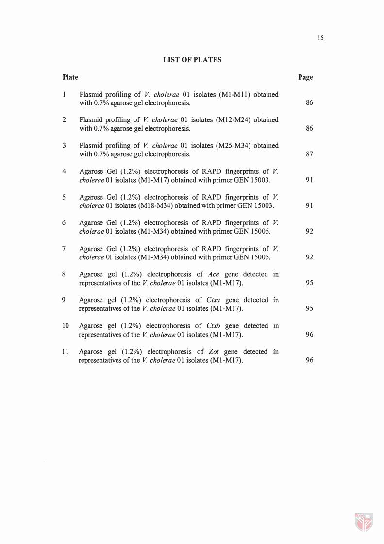
LIST OF PLATES
Plate Page
Plasmid profiling of V cholerae 0 1 isolates (MI-M l l ) obtained with 0.7% agarose gel electrophoresis. 86
2 Plasmid profiling of V cholerae 0 1 isolates (MI 2-M24) obtained with 0.7% agarose gel electrophoresis. 86
3 Plasmid profiling of V cholerae 0 1 isolates (M25-M34) obtained with 0.7% ag(lrose gel electrophoresis. 87
4 Agarose Gel ( 1 .2%) electrophoresis of RAPD fingerprints of V cholerae 0 1 isolates (MI -M I 7) obtained with primer GEN 1 5003 . 9 1
5 Agarose Gel (1 .2%) electrophoresis of RAPD fingerprints of V cholerae 01 isolates (MI 8-M34) obtained with primer GEN 1 5003 . 9 1
6 Agarose Gel ( 1 .2%) electrophoresis of RAPD fingerprints of V cholerae 01 isolates (MI -M34) obtained with primer GEN 1 5005. 92
7 Agarose Gel ( 1 .2%) electrophoresis of RAPD fingerprints of V cholerae 01 isolates (MI -M34) obtained with primer GEN 1 5005. 92
8 Agarose gel ( 1 .2%) electrophoresis of Ace gene detected m representatives of the V cholerae 01 isolates (MI -M I 7). 95
9 Agarose gel ( 1 .2%) electrophoresis of Ctxa gene detected m representatives of the V cholerae 0 1 isolates (MI -M I 7). 95
10 Agarose gel ( 1 .2%) electrophoresis of Ctxb gene detected m representatives of the V cholerae 0 1 isolates (MI -M I 7) . 96
1 1 Agarose gel ( 1 .2%) electrophoresis of Zot gene detected m representatives of the V cholerae 0 1 isolates (MI-M1 7) . 96
15
LIST OF ABBREVIATIONS
Abbreviations
Amp B bp C Cb Cfp Cro Cxm ccc Da dATP dCTP dGTP dH20 DNA dTTP E EDTA EtBr G Gm I.e. K kbp Kf LB M MDa ml mm mM
).lg ).ll mol Na NaCI NaOH Ox P Psi R Ra RAPD RNA Rpm s
Ampicillin Bacitracin base pair Chloramphenicol Carbenicillin Cefoperazone Ceftriaxone Cefuroxim covalently closed circular Dalton (unit of molecular mass) deoxyadenosine triphosphate deoxycytosine triphosphate deoxyguanosine triphosphate distilled water deoxyribonucleic acid deoxythymidine triphosphate Erythromycin Ethylenediamine tetra-acetic acid Ethidium bromide gram Gentamycin that/example Kanamycin kilobase pair Cephalotin Luria Bertani Molar or molarity (moles of solute per liter of solution) Megadalton milliliter millimeter millimolar mIcrogram microliter mole Nalidixic acid Sodium chloride Sodium hydroxide Oxacillin Penicillin G Pound(s) per square inch (lb/in2) Resistant Rifampin Randomly Amplified Polymorphic DNA Ribonucleic acid revolution per minute sensitive
1 6

S sdH20 SDS Taq TBE Te Tris UV V Va
Streptomycin sterile distilled water Sodium dodecyl sulphate 'l'hermus aquaticus DNA (polymerase) Tris-Borate EDT A electrophoresis buffer Tetracycline Tris (hydroxymethyl) methylamine ultraviolet volts Vancomycin
1 7

18
CHAPTERl
INTRODUCTION
Vibrio cholerae is the bacterium that causes cholera, a potentially epidemic and life
threatening secretory diarrhea characterized by numerous, voluminous watery stools,
often accompanied by vomiting, and resulting in hypovolemic shock and acidosis, and
sometimes muscle cramps (Son et aI., 1 999; Heilpem and Waldor, 2000). Cholera is an
ancient disease, caused by certain members of the species Vibrio cholerae that can also
cause mild or inapparent infections. The World Health Organization describes cholera as
a tragedy because this theoretically "most preventable disease" is one of the top causes of
human morbidity and mortality in the world in many areas of Asia, Africa, and Latin
America (Osawa et al. , 1 997). The incidence of cholera is estimated to exceed five
million cases each year (Jiang et al., 2000).
Vibrio cholerae is a natural inhabitant of aquatic environments. It is a Gram-negative
bacterium that belongs to the subdivision of the family Proteobacteriaceae, from the
genus of Vibrio and the family of Vibrionacea, (Trucksis et ai., 1 998; Beltran et ai., 1 999;
Walia et al. , 1 999). Vibrios are one of the most common organisms in surface waters of
the world, occurring in both marine and freshwater habitats and in associations with
aquatic animals, often with a variety of algae and crustaceans (Byun et ai. , 1 999). This
bacterium, in its extreme manifestation, can cause one of the most rapidly fatal diarrheal
known. Cholera by itself has contributed to millions of death worldwide ever since it was
first recorded. Outbreaks of cholera caused death are estimated at 1 20,00 worldwide
annually, affecting mainly children (Faruque et aI., 1 998). A healthy person may become
hypotensive within an hour of the onset of symptoms and may die within 2-3 hours if no

1 9
treatment is provided. More commonly, the disease progresses from the first liquid stool
to shock in 4- 12 hours, with death following in 1 8 hours to several days later (Merson et
al., 1 978). Untreated cholera frequently results in high mortality rates (50-60%).
Records of cholera pandemics started in 1 8 1 7. However, incidences of cholera or at
least cholera like outbreaks is not new nor is it just discovered during the modem-time
era. Descriptions dating back to pre-Christian calendar (B.e.) have been found written in
Sanskrit (�500 to 400 B.C.) from Sushruta Samshita in India. In fact, cholera existed in
India for centuries without leaving the subcontinent until 1 8 1 7. Gaspar Correa wrote in
1 503, that when Vasco da Gama landed on the southwestern coast of India in 1498, about
20,000 men of Cali cut died of "a disease which struck them sudden-like in the belly, so
that some of them died in 8 hours" (Colwell, 1 996).
From an epidemiological standpoint, the species has been divided into serogroup 01
and serogroup non-O 1 strains, which were long believed to differ in ability to cause
epidemic cholera. Historically, 01 strains ha-,e been responsible for all major epidemics,
including seven pandemics (Beltran et aI., 1 999). V. cholerae has the ability to cause
global epidemics, or pandemics. It is believed that the first six cholera pandemics were
caused by the classical V. cholerae biotype, while the seventh pandemic was caused by
the EI Tor biotype (Kimsey and Waldor, 1998; Provenzano et al., 2000). The first
pandemic is believed to have been fairly limited in its scope, being only about 7 years in
duration ( 1 8 1 7 - 1 823). This first pandemic was related to two wars at the time - the
Oman war and the war between Persia and Turkey (Colwell, 1 996). The second pandemic
is believed to have started in Russia in 1 829 and before ending its reign in 1 85 1 , spreaded
to the Americas, British Isles in 1 830-1 849 in London due to the mixing of drinking water
and sewage waste in the streets of London (Faruque et aI., 1 998). The person responsible

20
for the stopping of the London outbreak was Dr. John Snow who was also the first person
to connect the disease's spread through un sanitized drinking water (Merson et al., 1978).
His action was an important understanding of the epidemiology of cholera. During the
third pandemic which lasted from 1852- 1859, records showed that the illness was
rampant in the United States where it was known to be hitting cities and towns along the
Mississippi, Missouri and Ohio rivers until the outbreak ended in 1870's during the end of
the forth pandemic (Faruque et al., 1998). The fifth pandemic was marked by high
incidences of death in Argentina, Chile and Peru when it spreaded to South America. It
was during the fifth pandemic that Koch first isolated what he then referred to as "comma
bacilli" from stools of patients between 1883 and 1884 in Egypt and India respectively.
The sixth pandemic was presumably caused by V. cholera of the classical type marking
its reappearance as a large outbreak in Egypt. It was also recorded that during the 1899-
1923 pandemic, south and Southeast Asia, and also East and the Balkan peninsular was
also affected by the disease (Colwell, 1996).
The seventh pandemic was initiated by t!le emergence of the "EI Tor" biotype
(distinguished from the classical type by production of he moly sins) in 196 1 on the island
of Sulewasi in Indonesia, also producing a major epidemic in the Philipines. By the end of
1962, it had affected the entire Southeast Asian Archipelago spreading like a storm to
Borneo, Java and Taiwan. This particular pandemic is the most extensive of all the
pandemics in geographical spread and duration (Faruque et aI, 1998).
Until recently, cholera outbreaks have been attributed only to Vibrio cholera 01 of
the Vibrio species, but in October 1992, epidemic cholera reported in Madras and other
places in India and in Southern Bangladesh shocked the world by the appearance of a new
cholera sinister (Karaolis et al., 1995; Beltran et al., 1999). This V. cholerae non-O 1

2 1
serogroup spreaded like wild fire in Bangladesh to the entire country b y December 1992,
with clinical syndrome typical that of cholera, affecting thousands of people, mainly
adults and causing many deaths (Albert et ai., 1993; Boyd and Waldor, 1999). This new
serogroup of V. choierae was defined as 0139, with the synonym Bengal, to indicate its
first isolation from coastal areas of the Bay of Bengal. Diarrhea cases due to this new
serogroup then started to spread to other countries like Pakistan, Nepal, China, Thailand,
Kazakhstan, Afghanistan and Malaysia with more reports from the United Kingdom and
the United States. Epidemiologists believe that if outbreaks due to this new serogroup
should affect more countries, this could initiate the eighth pandemic (Faruque et ai.,
1998b).
Humans apparently are the only natural host for the cholera vibrios (Merson et al.,
1978). Cholera is acquired by the ingestion of water or food contaminated with the feces
of an infected individual. After oral ingestion of contaminated food or water and even
person-to-person transmission, this bacterium colonizes the human small intestine where
it secretes a cholera enterotoxin (CT) (Blake et ai., 1980; De Paola, 1981; Jafrul et al.,
1994; Kimsey and Waldor, 1998). In fact, the pathogenicity of cholera is mainly
associated with their ability to produce this toxin, which is encoded in its "virulence
cassette" region of the chromosome that consists of the Ace, Ctxa, Ctxb and Zot genes
(Trucksis et at., 1993). Since not all Vibrio cholerae or Vibrio cholerae 01 are toxigenic,
regular surveillance and examination of isolates for their potential to produce CT are
needed to obtain a clearer picture of the public health hazard caused by toxigenic strains
(Almeida et ai., 1990). Recently, CT was found to be encoded in the genome of an
unusual lysogenic filamentous phage, name CTX� (Boyd and Waldor, 1999).
22
Malaysia is no exception for cholera outbreak cases. The period of 1991 to 1994 has
seen serious outbreaks of the disease (Mahalingam et aI., 1994). In order to maintain the
health security of our land, evolutionary and epidemiology research has to be carried out
continuously either in the present or the future. Even if there were no emergence of new
mutated strains, this effort is worthwhile since every year there are still sporadic
outbreaks of cholera cases and there are trends of increasing cholera cases in recent years.
This is proven from the fact that increased trend had been observed from various areas in
Peninsular Malaysia that are well known for cholera outbreak cases. Penang, for
instance, until the month of May in 1996, a total of 476 cases of cholera cases had been
reported. Another report came from Shah Alam, Selangor, where in 1998 about 10
outbreak of cholera cases were reported. A cholera outbreak in an army camp in the
month of December the same year had later been confirmed to be caused by the new
mutated strain, V. cholerae serotype 0139. A statistics made by the Minisitry of Health
from the year 1995 to 1999 reported a total of 5,915 cholera cases with 62 fatalities
(Appendices). Hence, few alarming trends have shown the great importance of studies
8eing conducted for V. cholerae in Malaysia. Various reports had also indicated sporadic
cases of cholera in Kelantan, Putra Jaya and Kajang in the year 2000. This latest outbreak
of cholera in Kota Barn, Kelantan started on the 26th of September, 2000, reaching
Machang, Pasir Mas on the 8th of October, 2000 with about 180 reported infections.
All this while, identification and characterization of bacterial strains have been done
with the more conventional method of biotyping, namely on the biochemical
characteristics and serologic identification (Mahalingam et ai., 1994). But sceptics had
proven that with the conventional methods, there are bound to be some loopholes that
cause inaccuracies in studies. These are especially true, with the method focusing in
detecting more on the phenotypic outlooks of bacteria strains. This would prove costly

23
since phenotypic differences would only reveal part of the full potential characterization
of a certain bacteria strain. In addition, most biotyping methods have shown to be very
tedious and time consuming. The recent global resurgence of cholera underscores the
urgent need for effective and rapid epidemiological surveillance. Epidemiologic
investigation of cholera requires the characterization of V. cholerae isolates by typing
systems, which also allow determination of isolates relatedness.
Recently, various molecular biology-based techniques have been used to study the
relationships among clinical and environmental isolate. Molecular tools have been
considered as an alternative tools especially with the creation of Polymerase Chain
Reaction (PCR) by Kary Mullis in 1987. Molecular tools are considered to be
characterising or determining strains from the most basic structure of all living beings
which is the genetic structure. Creation of PCR technique have improved in the
specification, amplification and shortening the time factor of molecular techniques.
Together with these molecular tools, traditional and molecular techniques has created a
whole new armada of bacterial typing fleet which include randomly amplified
polymorphic DNA (RAPD) (Yuherman et aI., 1 998), multilocus enzyme electrophoresis
(MLEE) (Beltran et aI., 1999), pulsed-field gel electrophoresis (PFGE) (Cameron et al.,
1994; Mahalingam et al. 1994), ribotyping (Popovic et al., 1993) and amplified fragment
length polymorphism fingerprinting (ALFP) (Jiang et at., 2000). All these tools had
differentiated isolates of the V. cholerae population into different electrophoretic (ETs or
zymovars), PFGE types, ribotypes, ALFP types and RAPD fingerprint types, respectively.
Applications of these molecular tools has shown the existence within the V. cholerae
popUlation of several pathogenic clones (Byun et aI., 1999).

24
It is now common to use rapid, practical and economical phenotypic and genotypic
techniques for the characterization of organisms, and among them plasmid profiling,
antibiotic resistance patterns, randomly amplified polymorphic DNA (RAP D) and
specific PeR-based assays. A rapid diagnosis of cholera is important both for the
immediate management of the patient with severe diarrhea and for epidemiological
tracking. Hence, this research work done on V. cholerae 01 isolates from Miri, Sarawak
has included molecular tools as part of the characterization criteria. In the work reported
here, V. cholerae 01 isolated from the biggest and longest cholera outbreak from
November 1997 to April 1998 in Miri, Sarawak were characterized by antibiotic
resistance, plasmid profiling, randomly amplified polymorphic DNA (RAPD) and
detection of the cholera toxin (CT) gene (consisting of the detecting Ace, Ctxa, Ctxb and
Zot genes) by using specific primers in PCR.