universiti putra malaysia production of hepatitis b viral antigens and
TRANSCRIPT

UNIVERSITI PUTRA MALAYSIA
PRODUCTION OF HEPATITIS B VIRAL ANTIGENS AND ANTIBODIES USING PHAGE DISPLAY TECHNOLOGY FOR THE
DEVELOPMENT OF A DIAGNOSTIC TEST
TAN GEOK HUN.
FBSB 2006 10
PRODUCTION OF HEPATITIS B VIRAL ANTIGENS AND ANTIBODIES USING PHAGE DISPLAY TECHNOLOGY FOR THE DEVELOPMENT OF
A DIAGNOSTIC TEST
TAN GEOK HUN
DOCTOR OF PHILOSOPHY UNIVERSITI PUTRA MALAYSIA

PRODUCTION OF HEPATITIS B VIRAL ANTIGENS AND ANTIBODIES USING PHAGE DISPLAY TECHNOLOGY FOR THE DEVELOPMENT OF
A DIAGNOSTIC TEST
TAN GEOK HUN
Thesis Submitted to the School of Graduate Studies, Universiti Putra Malaysia, in Fulfilment of the Requirement for the Degree of Doctor of Philosophy
April 2006

DEDICATION
I dedicate this work to my beloved mother Goh Beng Eng,
darling husband Chau Sawang, brothers (Tan Kee Song, Tan Ke
Chuan) and sisters (Tan Geok Huwai, Tan Hui Geok) for their love,
support and encouragement during my study.
Thank you so much.. . . . ..
Abstract of thesis presented to the Senate of Universiti Putra Malaysia in fulfilment of the requirement for the degree of Doctor of Philosophy
PRODUCTION OF HEPATITIS B VIRAL ANTIGENS AND ANTIBODIES USING PHAGE DISPLAY TECHNOLOGY FOR THE DEVELOPMENT OF
A DIAGNOSTIC TEST
TAN GEOK HUN
April 2006
Chairman
Faculty
: Associate Professor Tan Wen Siang, PhD
: Biotechnology and Biomolecular Sciences
Hepatitis B is one of the most common infectious diseases in the world. It is caused
by the hepatitis B virus (HBV) which is estimated to infect more than one third of
the world's population and there are about 400 million carriers of HBV worldwide.
The infection can now be prevented through immunization with a vaccine based on
the surface antigen (HBsAg) produced in yeast by genetic engineering. In order to
provide additional means to control the disease, rapid, easier and cheaper diagnostic
assay have been developed using phage display technology in these studies.
From the present studies, bacteriophage T7 was employed to display the
immunodominant region of S-HBsAg, amino acid residues 1 1 1-1 56, on the exterior
of the phage particles. The expressed immunodominant region of S-HBsAg
remained antigenic and it was displayed on the coat protein of the recombinant
phage particle, T7-HBsAglll-lj6, which has the potential to be used as an

immunological reagent for the detection of anti-HBsAg antibody in human serum
samples at as low as 0.25 mIUIml. In addition, the hsion phage also applied on dot-
blot for detection of anti-HBsAg antibody. However, the sensitivity of this assay is
low as compared to ELISA method.
A phage heptapeptide random library was used to identify peptide ligands that
interact with HBsAg. From the third round of panning, 75% of phages screened
carried the peptide sequence C-ETGAKPH-C which is the most frequently
identified phage clones in this round. The phage clone was characterized and a
cyclic synthetic peptide bearing the identical peptide sequence was synthesized.
The phage was able to compete with anti-HBsAg monoclonal antibody as well as
the synthetic peptide for the binding site on HBsAg. The optimum pH and
temperature for phage binding was around 4 to 8 and 4"C, respectively. An
equilibrium binding assay in solution showed that the phage binds tightly to HBsAg
with a relative dissociation constant (KC') of 2.9 f 0.9 nM, illustrating that the
phage bearing ETGAKPH has the potential to be used as a diagnostic reagent for
detecting HBsAg in human sera.
As a preliminary effort to study the detection of HBcAg in the serum samples,
single chain variable fragment (scfv) of anti-HBcAg antibody library was
constructed by fusion to gpIII protein of bacteriophage M13, which allows for the
display of the fusion protein (scfv) at the tip of the filament. Truncated HBcAg was
inoculated into female BalbIC mice before the antibody and spleen cells were
harvested for the construction of library. Multiple reactions of PCR were carried out,
and the size of the antibody library was 2.18 x lo7 cfidml. This library was further
panned against HBcAg to get the specific phage clone that interacts with HBcAg.
The phage clone with higher absorbance value was fbrther rescued by helper phage
M13K07, and the phagemid was digested to determine the insert of scfi. In this
study, a phage-ELISA assay was established and the minimum amount of HBcAg
that can be detected was about 10 ng with 1.0 x 1 012 pfidml of purified fusion phage.
This hsion phage scfv showed a promising result for the detection of HBcAg in the
human treated serum samples.
In conclusion, development of phage-ELISA for the detection of anti-HBsAg
antibody, HBsAg and HBcAg based on phage display can be an alternative choice
to reduce the cost of detection kits. This study also provides a model in the
development of diagnostic test for the detection of other biological samples based
upon phage display technology.

Abstrak tesis yang dikemukakan kepada Senat Universiti Putra Malaysia sebagai memenuhi keperluan untuk ijazah Doktor Falsafah
PENGHASILAN ANTIGEN DAN ANTIBODI VIRUS HEPATITIS B DENGAN MENGGUNAKAN TEKNOLOGI PAMERAN FAJ UNTUK
PERKEMBANGAN UJIAN DIAGNOSIS
Oleh
TAN GEOK HUN
April 2006
Pengerusi : Profesor Madya Tan Wen Siang, PhD
Fakulti : Bioteknologi dan Sains Biomolekular
Hepatitis B merupakan salah satu penyakit be rjangkit merbahaya di dunia. Penyakit
tersebut yang disebabkan oleh virus hepatitis B (HBV) dianggarkan telah
menjangkiti lebih daripada satu per tiga daripada penduduk dunia dan lebih 400 juta
penduduk dunia dikategorikan sebagai pembawa. Pada masa kini, jangkitan
daripada virus HBV boleh dielakkan dengan menggunakan vaksin berdasarkan
antigen permukaan virus hepatitis B (HBsAg) yang dibentuk daripada kejuruteraan
genetik yis. Untuk mengawal penularan penyakit ini, satu teknik diagnosis yang
cepat, senang dan murah telah dibangunkan berdasarkan kepada teknologi pameran
faj.
Dalam kajian ini, bacteria faj telah dieksplotasikan bagi mempamerkan bahagian
imrnunodominan antigen permukaan virus pada bahagian luar faj. Bahagian
imrnunodominan antigen permukaan virus ini kekal menjadi antigenik dan
vii
dipersembahkan pada bahagian permukaan protein faj rekombinan T7-HBsAg, 11-156
yang berfungsi untuk mengesan antibody anti-HBsAg yang terdapat di dalam
sampel serum manusia sehingga 0.25 mIUlml. Selain itu, faj ini juga digunakan
dalam dot-blok asai. Namun demikian, tahap kepekaan asai ini adalah kurang kalau
dibandingkan dengan cara ELISA.
Suatu perpustakaan faj digunakan untuk mengenalpasti ligan peptida yang boleh
bertindak balas dengan HBsAg. Tujuh puluh lima peratus daripada jumlah faj yang
disaring membawa jujukan asid amino C-ETGAKPH-C yang menunjukkan
kebarangkalian paling tinggi dalam pusingan ketiga pernilihan berafiniti. Pencirian
faj tersebut telah dilaksanakan dan peptida berkonformasi sintetik yang beridentikal
dengan faj tersebut telah disintesiskan. Faj ini didapati mampu bersaing dengan
antibodi monoklon anti-HBsAg dan peptida sintetik untuk tapak pengikatan pada
HBsAg. pH dan suhu yang optima untuk pengikatan faj masing-masing ialah sekitar
4-8 dan 4°C. Esai keseimbangan pengikatan dalam cecair menunjukkan faj tersebut
mengikat dengan kuat kepada HBsAg dengan pemalar penceraian relative (&=I)
iaitu 2.9 + 0.9 nM. Ini menggambarkan bahawa faj pembawa ETGAKPH
berpotensi sebagai reagen diagnostik bagi pengesanan HBsAg dalam serum
manusia.
Sebagai permulaan untuk kajian mengesan HBcAg dalam sarnpel darah,
penghasilan perpustakaan antibodi anti-HBcAg yang mengandungi scfv terikat
protein gpIII pada faj M13 telah dijalankan. Mencit betina Balb/C diinokulasikan
dengan HBcAg sebelum limpanya dikeluarkan dan digunakan untuk penyediaan

. . . V l l l
perpustakaan tersebut. Pelbagai tindak balas PCR telah dijalankan dan saiz
perpustakaan antibodi tersebut dianggarkan sebanyak 2.18 x lo7 cfulml.
Perpustakaan antibodi tersebut seterusnya dipilih melalui afinitinya terhadap
HBcAg untuk mendapatkan faj yang bertindak balas secara spesifik dengan HBcAg.
Faj klon yang menunjukkan penyerapan OD yang tinggi diselamatkan dengan faj
pembantu M13K07 dan fajrnid diuraikan untuk menentukan kandungan scfv.
Dalam kajian ini, faj ELISA sudah diperkenalkan dan minimum kuantiti HBcAg
yang dapat dikesan oleh faj pada 1.0 x 10¹² pfulml ialah sekitar 10 ng. Faj ini
menunjukkan keputusan yang memberangsangkan dalarn pengesanan HBcAg
dalam sampel darah manusia.
Pengembangan esai faj untuk ELISA pengesanan antibodi anti-HBsAg, HBsAg dan
HBcAg berdasarkan pameran faj berupaya berfungsi sebagai alternatif bagi
mengurangkan kos pengesanan. Selain daripada itu, kajian ini juga menyediakan
satu model bagi pengembangan diagnosis untuk pengesanan sample biologi lain
berdasarkan teknologi pameran faj .
ACKNOWLEDGEMENTS
I wish to acknowledge generous individuals whose valuable support made this
study a success and the completion of this thesis possible. First and foremost, I wish
to convey my most sincere gratitude to my friendly and helpful supervisors, Assoc.
Prof. Dr. Tan Wen Siang, Prof. Datin Dr. Khatijah Yusoff and Prof. Dr. Seow Heng
Fong for their invaluable advices, excellent guidance and constructive criticisms
during this study. This project wouldn't have been possible without them.
Special thanks to all the staffs of the Department of Microbiology and members of
Virology Lab 143 and small lab 134 for not only helping me with my study but also
making my stay in the lab a very pleasurable and memorable one. I thank Assoc.
Prof. Dr. Sheila Nathan, Dr. Majid Eshagi and Dr. Azri for their useful comments
and discussions during the study. Wai Ling, Dr. Wong, Dr. Priadarishni, Eni, Dr.
Khoo, Rafidah, Lalita, Eddie, Thong Chuan, Suhana, Nazrien, Zul, Onie, Ina,
Andrew, Wai Kit, Kah Fai, Salwa, Taznim, Firooze, Budy, Max, Mokrish and all
the others, thank you very much.
Last but not least, I would like to express my deepest appreciation to my beloved
father who always in my memory, my dearest mum, my dearest darling husband,
brothers and sisters for their encouragement, patience, sacrifices and love that
brought me to the end of this study. Finally, I would like to thank the Ministry of
Science, Technology and Environment of Malaysia for providing me the National
Science Fellowship.

I certifL that Examination Committee has met on 3rd April 2006 to conduct the final examination of Tan Geok Hun on her Doctor of Philosophy thesis entitiled "Production of Hepatitis B Viral Antigens and Antibodies Using Phage Display Technology for the Development of A Diagnostic Test" in accordance with the Universiti Pertanian Malaysia (Higher Degree) Act 1980 and Universiti Pertanian Malaysia (Higher Degree) Regulations 1981. The Committee recommends that the candidate be awarded the relevant degree. Members of the Examination Committee are as follows:
JANNA ONG ABDULLAH, PhD Lecturer Faculty of Biotechnology and Biomolecular Sciences Universiti Putra Malaysia (Chairman)
NORHANI ABDULLAH, PhD Professor Faculty of Biotechnology and Biomolecular Sciences Universiti Putra Malaysia (Internal Examiner)
MOHD HAIR BEJO, PhD Associate Professor Faculty of Veterinary Medicine Universiti Putra Malaysia (Internal Examiner)
THONG KWAI LIN Professor Faculty of Science Universiti Malaya (External Examiner)
School of Graduate Studies Universiti Putra Malaysia
Date: 26 APR 2006

This thesis submitted to the Senate of Universiti Putra Malaysia has been accepted as fulfilment of the requirement for the degree of Doctor of Philosophy. The members of the Supervisory Committee are as follows:
TAN WEN SIANG, PhD Associate Professor Faculty of Biotechnology and Biomolecular Sciences Universiti Putra Malaysia (Chairman)
DATIN KHATIJAH YUSOFF, PhD Professor1 Dean Faculty of Biotechnology and Biomolecular Sciences Universiti Putra Malaysia (Member)
SEOW HENG FONG, PhD Professor Faculty of Medicine and Health Sciences Universiti Putra Malaysia (Member)
AINI IDERIS, PhD Professor / Dean School of Graduate Studies Universiti Putra Malaysia
DECLARATION
I hereby declare that the thesis is based on my original work except for quotations and citations, which have been duly acknowledged. I also declare that it has not been previously or concurrently submitted for any other degree at UPM or other institutions.
TAN GEOK HUN

... Xll l
TABLE OF CONTENTS
DEDICATION ABSTRACT ABSTRAK ACKNOWLEDGEMENTS APPROVAL DECLARATION LIST OF TABLES LIST OF FIGURES LIST OF ABBREVIATIONS
CHAPTER
1
2
INTRODUCTION
Page . . 11 ... 111
vi ix X
xii xvii XX
xxiii
LITERATURE REVIEW 5 2.1 Hepatitis B Virus 5
2.1.1 Historical Aspects 5 2.1.2 HBV Morphology 8 2.1.3 Genomic Organization and Viral
Transcription 11 2.1.4 Surface Protein (HBsAg) 14 2.1.5 Core Protein (HBcAg) 19 2.1.6 E Protein (HBeAg) 23 2.1.7 Virus Replication 24 2.1.8 Pathogenesis of HBV Infection 26
2.2 Serological Assays of HBV Infection 27 2.3 Diagnostic Test for HBV Antigens and Antibodies 30 2.4 Phage Display Technology 3 2
2.4.1 Morphology and Structure of Filamentous Phage 3 5
2.4.2 Morphology and Structure of Bacteriophage T7 3 7
2.4.3 Recombinant Antibody Libraries 40
MATERIALS AND METHODS 44 3.1 Materials 44 3.2 Display of Immunodominant Region of S-HBsAg
on T7 46 3.2.1 Amplification of DNA Encoding S-HBsAg 46 3.2.2 Purification of PCR Product 46 3.2.3 Restriction Enzyme (RE) Digestion of the
Purified PCR Product 48 3.2.4 In vitro Packaging of T7 DNA 48
xiv
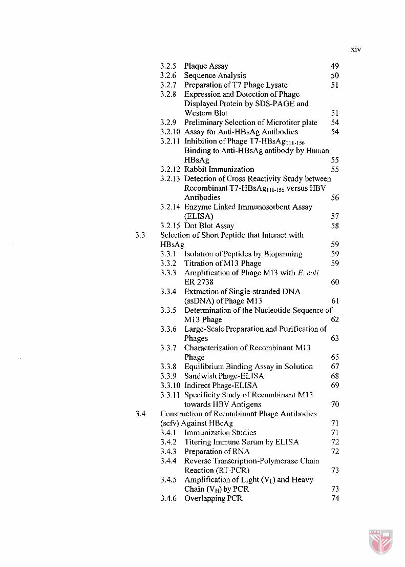
3.2.5 Plaque Assay 3.2.6 Sequence Analysis 3.2.7 Preparation of T7 Phage Lysate 3.2.8 Expression and Detection of Phage
Displayed Protein by SDS-PAGE and Western Blot 5 1
3.2.9 Preliminary Selection of Microtiter plate 54 3.2.10 Assay for Anti-HBsAg Antibodies 54 3.2.1 1 Inhibition of Phage T7-HBsAgl 1-1 56
Binding to Anti-HBsAg antibody by Human HBsAg 5 5
3.2.12 Rabbit Immunization 5 5 3.2.13 Detection of Cross Reactivity Study between
Recombinant T7-HBsAg l I 1.156 versus HBV Antibodies 56
3.2.14 Enzyme Linked Immunosorbent Assay (ELISA) 5 7
3.2.15 Dot Blot Assay 58 Selection of Short Peptide that Interact with HBsAg 5 9 3.3.1 Isolation of Peptides by Biopanning 5 9 3.3.2 Titration of M13 Phage 59 3.3.3 Amplification of Phage M 13 with E. coli
ER 2738 60 3.3.4 Extraction of Single-stranded DNA
(ssDNA) of Phage M 1 3 6 1 3.3.5 Determination of the Nucleotide Sequence of
M13 Phage 62 3.3.6 Large-Scale Preparation and Purification of
Phages 63 3.3.7 Characterization of Recombinant M 13
Phage 65 3.3.8 Equilibrium Binding Assay in Solution 67 3.3.9 Sandwish Phage-ELISA 68 3.3.10 Indirect Phage-ELISA 69 3.3.1 1 Specificity Study of Recombinant M 13
towards HBV Antigens 70 Construction of Recombinant Phage Antibodies (scfv) Against HBcAg 7 1 3.4.1 Immunization Studies 7 1 3.4.2 Titering Immune Serum by ELISA 72 3.4.3 Preparation of RNA 72 3.4.4 Reverse Transcription-Polymerase Chain
Reaction (RT-PCR) 73 3.4.5 Amplification of Light (VL) and Heavy
Chain (VH) by PCR 73 3.4.6 Overlapping PCR 74

3.4.7 Cloning of scfv to pComb3x Phagemid 77 3.4.8 Preparation of Competent Cells and
Transformation 77 3.4.9 Rescue of scfv Displaying Phages 78 3.4.10 Affinity Selection of Recombinant Antibody
Library 79 3.4.1 1 Screening of Recombinant Phagemid 79 3.4.12 Inhibition Study of Phage scfv by Cyclic
Peptide 3.4.13 Specificity Test of Phage-ELISA 3.4.14 Development of Phage-ELISA
DISPLAY OF IMMUNODOMINANT REGION OF HBsAg ON T7 PHAGE 8 5 4.1 Introduction 85 4.2 Objectives 86 4.3 Methodology 86 4.4 Results 8 7
4.4.1 Cloning of the Coding Region of HBsAgl I 1 - 1 ~ ~
into T7 Vector 8 7 4.4.2 Expression of Recombinant T7-HBsAgl t 1-156 90
4.4.3 Antigenicity Analysis
4.4.4 Sensitivity of Phage ELISA 4.4.5 HBsAg-T7-HBsAgl 1 i-iS6 Competition
Assay 4.4.6 Immunogenic Properties of Recombinant
T7 Phage 98 4.4.7 Determination of Cross Reactivity of
Recombinant T7-HBsAgl 11-1 56 100 4.4.8 Detection of Anti-HBsAg antibody with
Recombinant T7 versus Commercial HBsAg 102 4.5 Discussion 108
SELECTION OF SHORT PEPTIDES THAT INTERACT WITH HBsAg 5.1 Introduction 5.2 Objectives 5.3 Methodology 5.4 Results
5.4.1 Isolation of Peptide by Biopanning 5.4.2 Characterization of Phage Carrying
ETGAKPH Peptide 5.4.3 Detection of HBsAg by Sandwich
Phase-ELISA

xvi
5.4.4 Detection of HBsAg by Indirect Phage-ELISA 129
5.4.5 Specificity Study of Fusion M13 towards HBV Antigens 132
5.4.6 Testing of Phage Peptide with HBsAg Positive Human Sera 132
Discussion 136
CONSTRUCTION OF RECOMBINANT SCFV ANTIBODIES AGAINST HBcAg 142 6.1 Introduction 142 6.2 Objectives 143 6.3 Methodology 143 6.4 Results 144
6.4.1 Construction of Recombinant Antibody Library 144
6.4.2 Biopanning of the Recombinant Antibody Library scfv 149
6.4.3 Screening of Positive Phagemid 149 6.4.4 Sensitivity Study of Fusion Phage scfv 155 6.4.5 Determination of Specificity of Fusion Phage
scfv towards HBV Antigens 157 6.4.6 Phage-peptide Inhibition Assay 157 6.4.7 Comparison of Fusion Phage scfv and
Anti-HBcAg antibody for Detection of HBcAg in Human Serum Samples 16 1
6.5 Discussion 164
GENERAL DISCUSSION AND CONCLUSION
REFERENCES APPENDICES BIODATA OF THE AUTHOR PUBLICATIONS
xvii
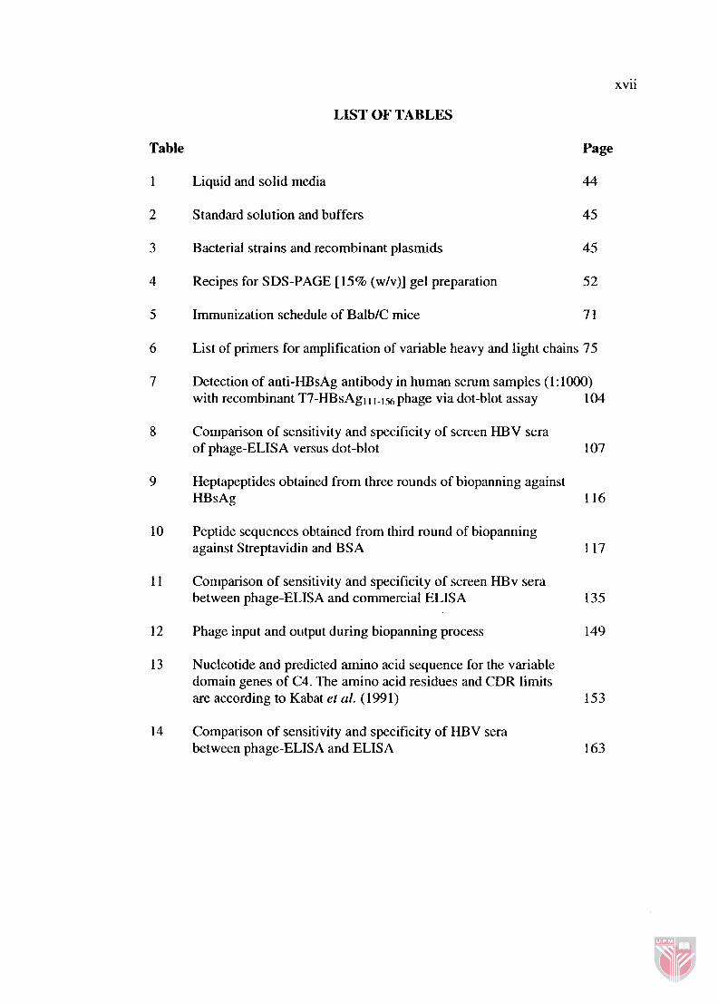
LIST OF TABLES
Table
1
2
Liquid and solid media
Standard solution and buffers
Bacterial strains and recombinant plasrnids
Recipes for SDS-PAGE [15% (wlv)] gel preparation
Immunization schedule of BalblC mice
Page
44
45
List of primers for amplification of variable heavy and light chains 75
Detection of anti-HBsAg antibody in human serum samples (1:1000) with recombinant T7-HBsAg, 11.156 phage via dot-blot assay 104
Comparison of sensitivity and specificity of screen HBV sera of phage-ELISA versus dot-blot
Heptapeptides obtained from three rounds of biopanning against HBsAg 116
Peptide sequences obtained from third round of biopanning against Streptavidin and BSA
Comparison of sensitivity and specificity of screen HBv sera between phage-ELISA and commercial ELISA
Phage input and output during biopanning process
Nucleotide and predicted amino acid sequence for the variable domain genes of C4. The amino acid residues and CDR limits are according to Kabat et al. (199 1)
Comparison of sensitivity and specificity of HBV sera between phage-ELISA and ELISA

LIST OF FIGURES
Figure Page
Electron micrographs of HBV particles found in the serum of infected patient
Schematic diagram of a virion structure of HBV
The structural and functional features of HBV DNA
Envelope proteins of HBV
Conformational model of the major hydrophilic region (MHR) of HBsAg 18
Three-dimensional maps of hepatitis core particles
Polypeptide fold of the HBcAg monomer
Immune response against HBV infection
Diagnostic test for HBV antigens and antibodies
10 Biopanning procedure
11 A filamentous bacteriophage particles
12 Structure of the T7 phage particle
13 Schematic diagram of T7 Select 4 15- 1 b phage display and T7 wild-type
14 Schematic diagram of the basic antibody structure
15 Scheme of amplification and cloning procedure of antibody library
16 Amplification and digestion of the coding region of HBsAgl 1 1-156 subtype (adw)
17 Sequencing results of recombinant T7 containing S 1 1 1 - 156
1 8 SDS-PAGE [15% (wlv)] analysis and Coomassie Blue staining of recombinant T7-HBsAg 111-156 phage, T7 wild-type and purified human HBsAg 9 1

xxi
19 Western Blot analysis of recombinant T7-HBsAg and wild-type T7 phages 92
20 Antigenicity of recombinant T7 phage as measured by ELISA 94
2 1 Sensitivity of phage-ELISA assay
22 Inhibition of recombinant T7 phage against anti-HBsAg antibody with human HBsAg
23 Immunogenicity of recombinant T7
24 Specificity study of recombinant T7-HBsAgll 1-156 phage towards three different monoclonal antibodies of HBV, namely anti-HBsAg antibody, anti-HBcAg antibody and anti-HBeAg antibody 10 1
25 Detection of anti-HBsAg antibody in human sera by recombinant T7- HBsAgl 11-1 j6 versus HBsAg
26 Densitometric analysis of fusion T7-HBsAgl 11-156 for detection of anti-HBsAg antibody in human sera by dot-blot assay
27 Sequence analysis of the insert peptide from disulfide constrained heptapeptide phage library of MI 3
28 Inhibition of antibodies binding to HBsAg with phage bearing ETGAKPH
29 Peptide-Phage Competition Assay 121
30 Interaction of ETGAKPH phage with HBsAg at different pH 122
3 1 Interaction of phage bearing ETGAKPH at different temperatures 123
32 Time course study 125
33 Input-output experiment 125
34 Interaction of HBsAg with ETGAKPH phage
35 Sandwich phage-ELISA for detecting HBsAg
36 Sensitivity of the sandwich phage-ELISA
37 Indirect phage-ELISA for detecting HBsAg
3 8 Sensitivity of indirect phage-ELISA
xxii
Specificity study of phage bearing peptide ETGAKPH by three different type of HBV antigens, namely HBsAg, HBcAg and HBeAg
Detection of HBsAg in HBV positive and negative sera by hsion phage and anti-HBsAg antibody via phage-ELISA and commercial ELISA 134
Immunization of BalbIC mice
The assembly strategy of single chain variable fragment into pComb3x phagemid vector
Amplification of light and heavy chain fragments of antibody with a set of primers
Phage-ELISA of phage clones selected randomly on LB containing carbenicillin plate 151
Restriction enzyme analysis of recombinant pComb3x phagemid 152
Sensitivity test of the phage-ELISA and ELISA
Specificity study of fusion phage scfv towards HBV antigens, namely HBsAg, HBcAg and HBeAg
Specificity study of fusion phage scfv towards HBV antigens, namely, HBsAg, HBcAg and HBeAg
Comparison of fusion phage scfv with anti-HBcAg antibody for detection of HBcAg in human serum

xxiii
pmole
ALT
Amp
ATP
LIST OF ABBREVIATIONS
alpha
beta
kappa
lambda
degree centrigrade
microgram (1 o6 g)
microlitre (1 o - ~ 1)
micromolar M)
picomole
alanine aminotransferase
ampicillin
adenosine triphosphate
anti-HBsAg HBsAg antibody
anti-HBcAg HBcAg antibody
anti-HBeAg HBeAg antibody
bp basepair
BSA bovine serum albumin
ccc covalently closed circular
C-terminus carboxy terminus
Carb Carbenicillin
DHBV duck hepatitis B virus
deoxy-ribonucleic acid DNA
xxiv
DR
dsDNA
DTT
DMF
ELISA
ER
g
GSHV
h
HHBV
HBcAg
HBsAg
HBeAg
HBV
HCC
HIV
HSV
IPTA
IgG
Kan
kb
Kd
kDa
deoxynucleoside triphosphate
direct repeat
double-stranded DNA
1,4-dithiothreitol
dimethylformamide
enzyme-linked irnmunoabsorbent assay
endoplasmic reticulum
gram
ground squirrel hepatitis virus
hour
herons hepatitis B virus
hepatitis B core protein
hepatitis B surface antigen
hepatitis B e protein
hepatitis B virus
hepatocellular carcinoma
human immunodeficiency virus
herpes simplex virus
isopropyl-P-d-thiogalactopyranoside
Immunoglobulin G
Kanamycin
kilobase
dissociation constant
kilodalton
xxv
1
IFN
LB
L-HBsAg
MHR
mAb
mg
mIU/ml
relative dissociation constant
litre
interferon
Luria broth
large surface antigen
molar
major hydrophilic region
monoclonal antibody
milligram g)
miliinternational unit
ORF
PAGE
PBS
PEG
M-HBsAg medium surface antigen
MSC multiple cloning site
min minute
ml millilitre (10" 1)
millimeter m)
mRNA messenger ribonucleic acid
NDV Newcastle disease virus
nM nanomolar (1 o - ~ M)
N-terminus amino terminus
OD optical density
open reading frame
polyaccrylamide gel electrophoresis
phosphate buffered saline
polyethylene glycol