university of jordan1 the eye: i. optics of vision faisal i. mohammed, md, phd
TRANSCRIPT
University of Jordan 1
The Eye: I. Optics of Vision
Faisal I. Mohammed , MD, PhD
University of Jordan 2
Objectives Describe the visual receptors List the types of lenses and recognize how they work Determine the power of lenses Describe accommodation for near vision and far vision Recognize nearsightedness and farsightedness and
determine its correction Describe visual acuity and its abnormalities Determine intraocular pressure and glaucoma
University of Jordan 3
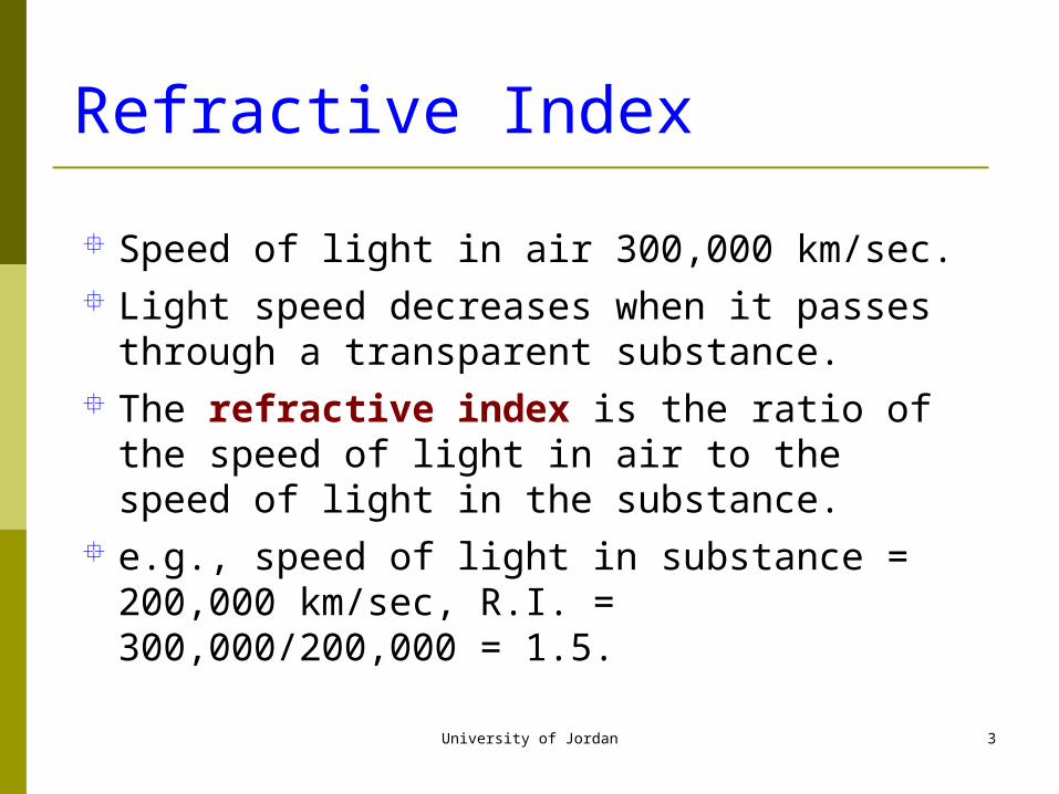
Refractive Index
Speed of light in air 300,000 km/sec. Light speed decreases when it passes through a
transparent substance. The refractive index is the ratio of the speed of
light in air to the speed of light in the substance. e.g., speed of light in substance = 200,000 km/sec,
R.I. = 300,000/200,000 = 1.5.
University of Jordan 4
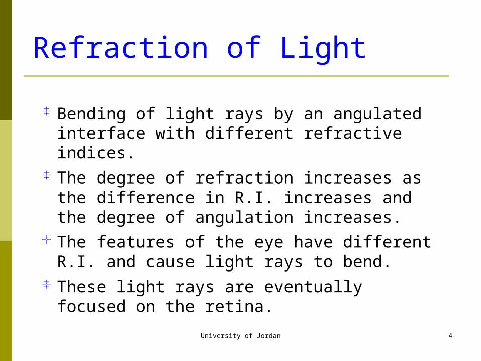
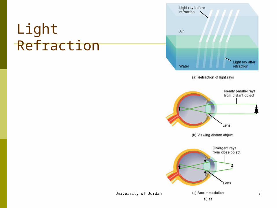
Refraction of Light
Bending of light rays by an angulated interface with different refractive indices.
The degree of refraction increases as the difference in R.I. increases and the degree of angulation increases.
The features of the eye have different R.I. and cause light rays to bend.
These light rays are eventually focused on the retina.
University of Jordan 5
Light Refraction
University of Jordan 6

University of Jordan 7
University of Jordan 8
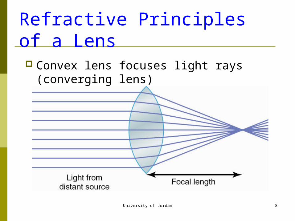
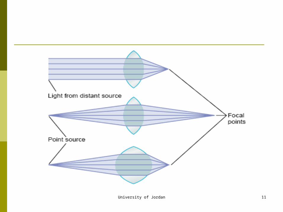
Refractive Principles of a Lens
Convex lens focuses light rays (converging lens)
University of Jordan 9
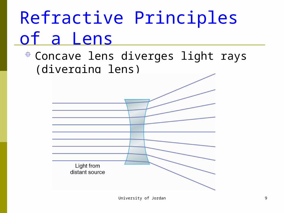
Refractive Principles of a Lens Concave lens diverges light rays (diverging lens)
University of Jordan 10
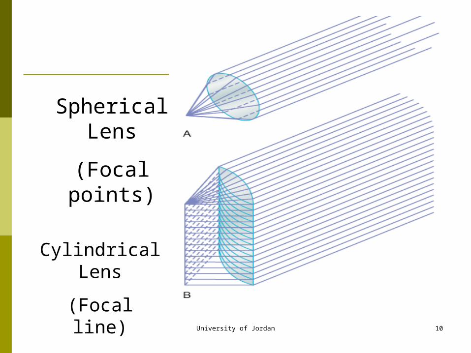
Spherical Lens
(Focal points)
Cylindrical Lens
(Focal line)
University of Jordan 11
University of Jordan 12
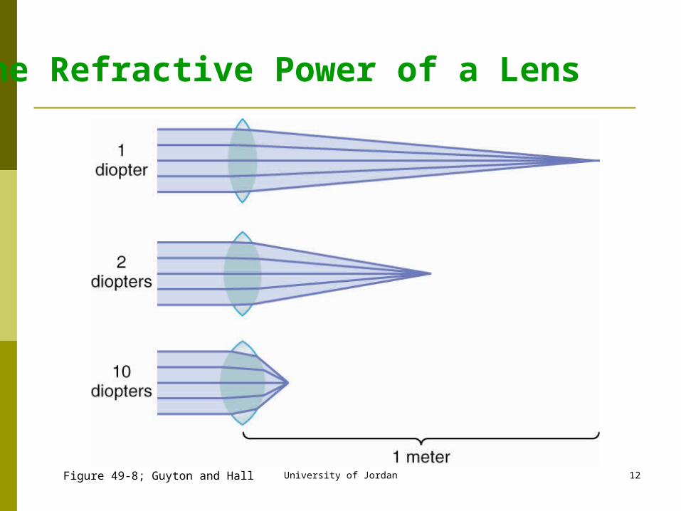
The Refractive Power of a Lens
Figure 49-8; Guyton and Hall
University of Jordan 13
Focusing Power of the Eye
Most of the refractive power of the eye results from the surface of the cornea. a diopter is a measure of the power of a lens 1 diopter is the ability to focus parallel light rays at a
distance of 1 meter, it is a measure of power of lenses Diopter = 1/ focal length in meters i.e the power of a
lens with focal length 0.5 meter is 2 (more convex) the retina is considered to be 17 mm behind the
refractive center of the eye therefore, the eye has a total refractive power of 59
diopters (1000/17)
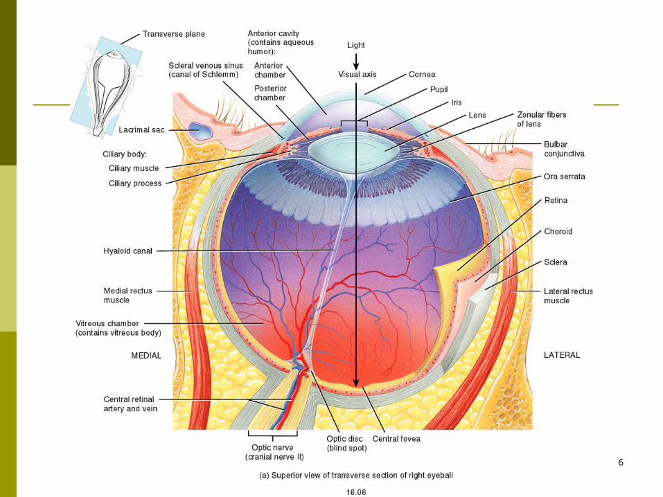
Image formation on the retina-requirements
Light refraction or bending the light by the refractive media – Cornea, Aqueous humor, Lens and Vitreous humor
Accommodation: An increase in the curvature of the lens for near vision,
The near point of vision is the minimum distance from the eye an object can be clearly focused with maximum accommodation
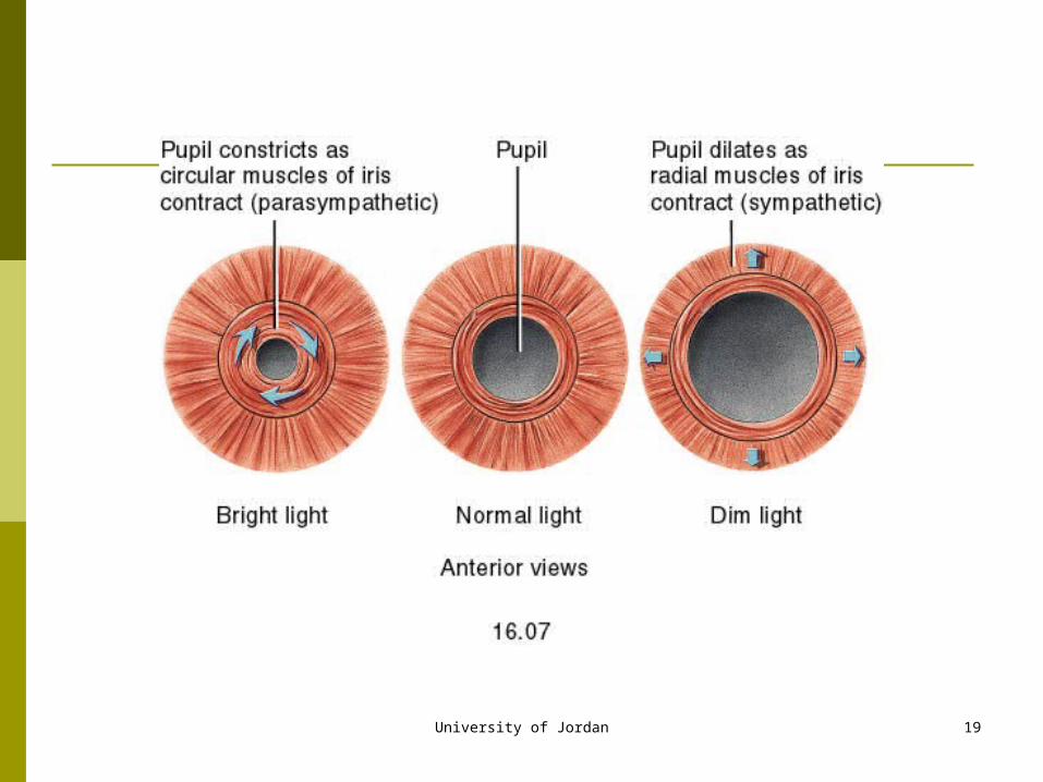
Constriction (meiosis) and dilation (Mydriasis) of the pupil
Convergence and divergence of the eyes for binocular vision
University of Jordan 14
University of Jordan 15
Accommodation
Refractive power of the lens is 20 diopters. Refractive power can be increased to 34 diopters
by changing shape of the lens - making it fatter (more convex).
This is called accommodation. Accommodation is necessary to focus the image
on the retina. Normal image on the retina is upside down.
University of Jordan 16
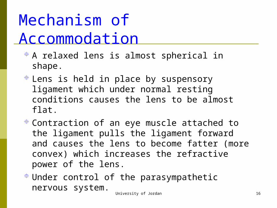
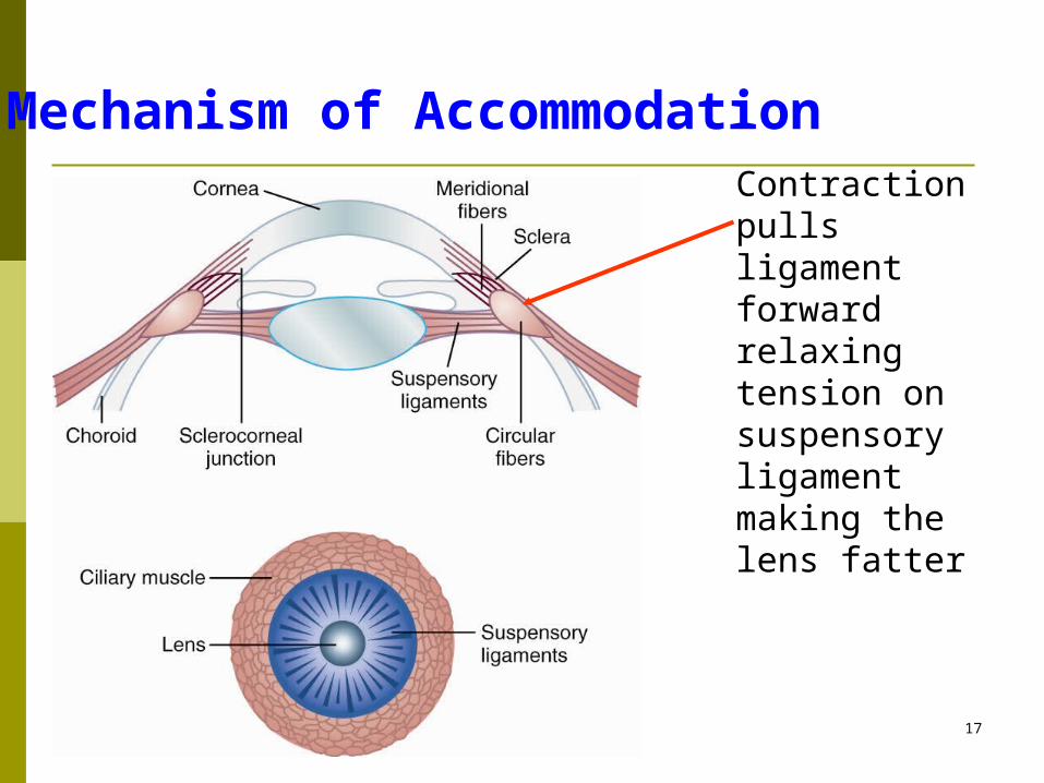
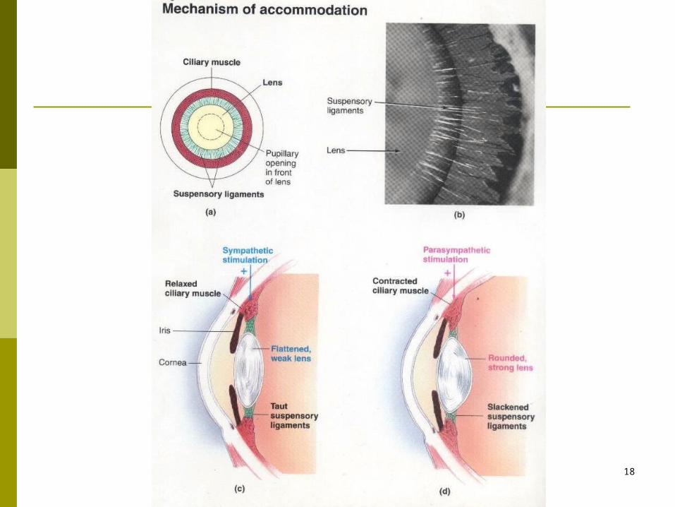
Mechanism of Accommodation
A relaxed lens is almost spherical in shape. Lens is held in place by suspensory ligament
which under normal resting conditions causes the lens to be almost flat.
Contraction of an eye muscle attached to the ligament pulls the ligament forward and causes the lens to become fatter (more convex) which increases the refractive power of the lens.
Under control of the parasympathetic nervous system.
University of Jordan 17
Mechanism of AccommodationContraction pulls ligament forwardrelaxing tension on suspensory ligamentmaking the lens fatter
University of Jordan 18
University of Jordan 19
University of Jordan 20
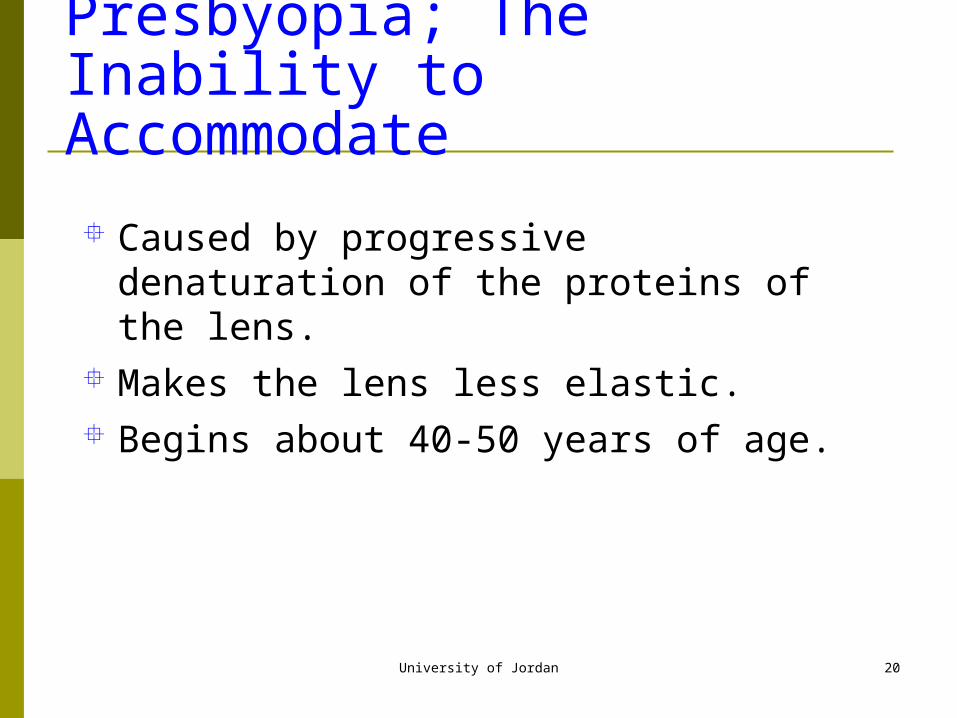
Presbyopia; The Inability to Accommodate
Caused by progressive denaturation of the proteins of the lens.
Makes the lens less elastic. Begins about 40-50 years of age.
University of Jordan 21
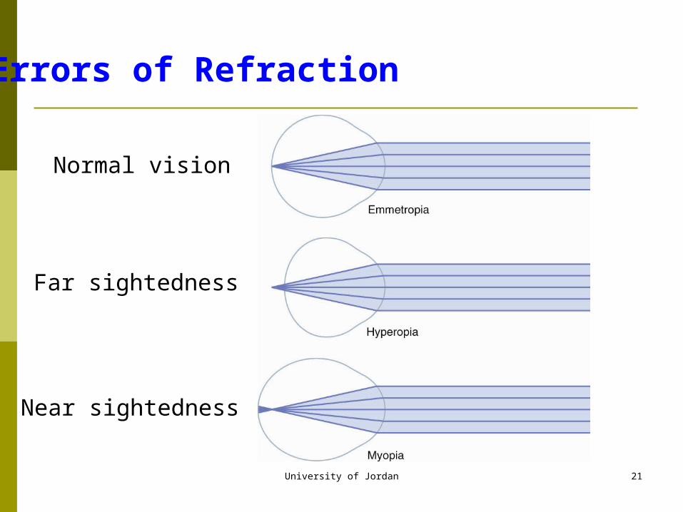
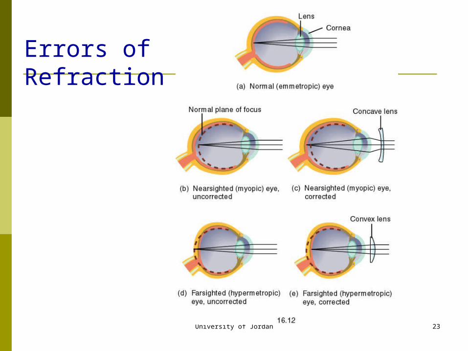
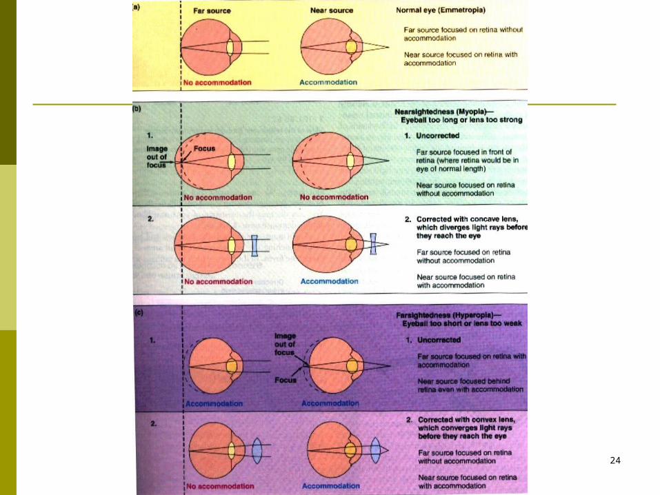
Errors of Refraction
Normal vision
Far sightedness
Near sightedness
University of Jordan 22
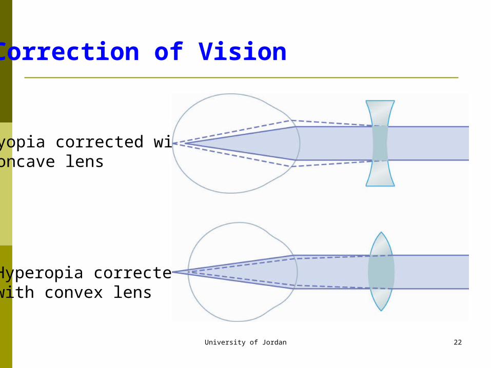
Correction of Vision
Myopia corrected withconcave lens
Hyperopia corrected with convex lens
University of Jordan 23
Errors of Refraction
University of Jordan 24
University of Jordan 25
Other Errors of Vision
Astigmatismunequal focusing of light rays due to an oblong
shape of the cornea Cataracts
cloudy or opaque area of the lenscaused by coagulation of lens proteins
University of Jordan 26
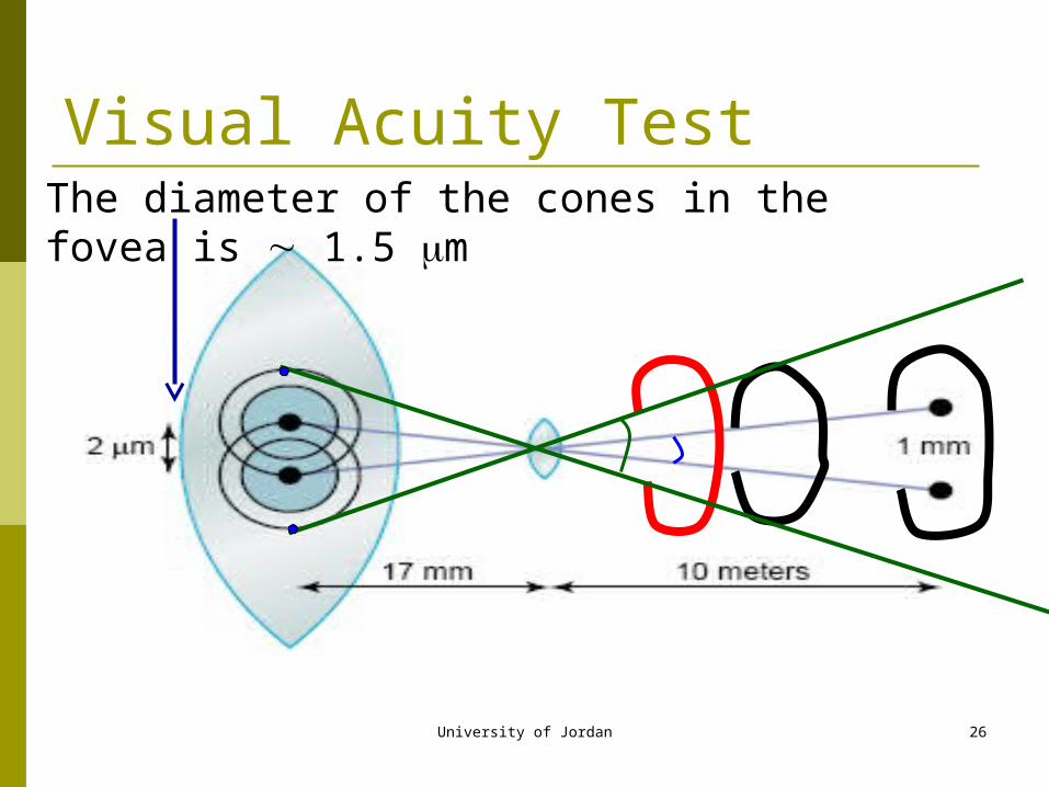
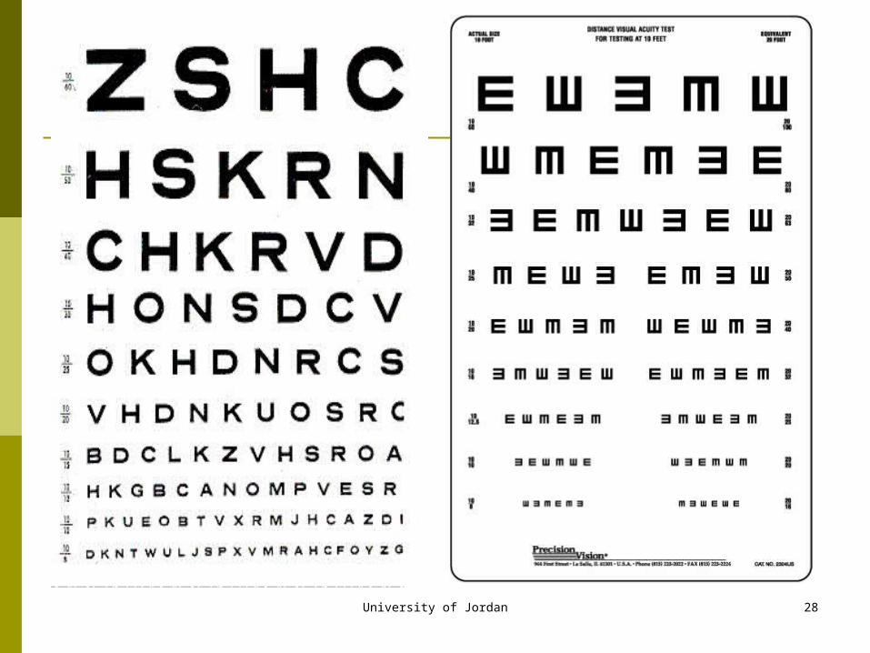
Visual Acuity TestThe diameter of the cones in the fovea is 1.5 m
University of Jordan 27
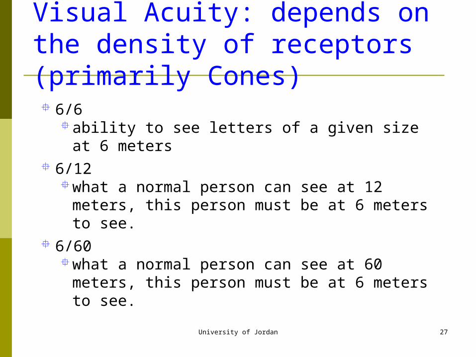
Visual Acuity: depends on the density of receptors (primarily Cones)
6/6ability to see letters of a given size at 6 meters
6/12what a normal person can see at 12 meters, this
person must be at 6 meters to see. 6/60
what a normal person can see at 60 meters, this person must be at 6 meters to see.
University of Jordan 28
University of Jordan 29
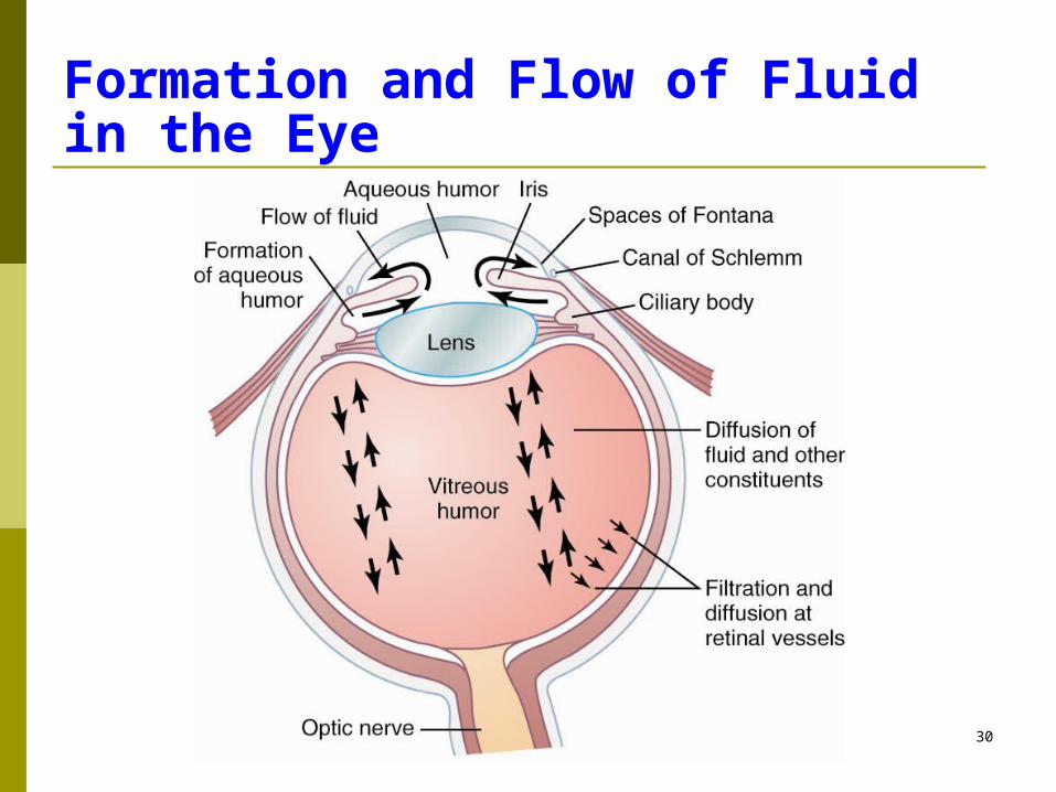
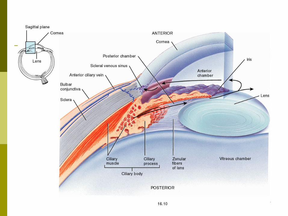
Fluid System of the Eye
Intraocular fluid keeps the eyeball round and distended.
2 fluid chambers:aqueous humor which is in front of the lens
freely flowing fluidvitreous humor which is behind the lens
gelatinous mass with little flow of fluid
University of Jordan 30
Formation and Flow of Fluid in the Eye
University of Jordan 31
University of Jordan 32
Formation of Aqueous Humor
Produced by the ciliary processes of the ciliary body at a rate of 2-3 microliters/min.
Flows between the ligaments of the lens, through the pupil into the anterior chamber, goes between the cornea and the iris, through a meshwork of trabeculae to enter the canal of schlemm which empties into aqueous veins and then into extraocular veins.

University of Jordan 33
Intraocular Pressure Normally 15 mm Hg with a range of 12-20 mm Hg. The level of pressure is determined by the resistance
to outflow of aqueous humor in the canal of schlemm. increase in intraocular pressure caused by an
increase in resistance to outflow of aqueous humor through a network of trabeculae in the canal of schlemm (Glaucoma)
can cause blindness due to compression of the axons of the optic nerve

University of Jordan 34
Thank YouThank You
University of Jordan 35
The Eye: II. Receptor and Neural Function of the Retina
Faisal I. Mohammed, MD,PhD
University of Jordan 36
Objectives
Describe visual receptors and characterize them List the layers of the retina and its cellular makeup Explain visual transduction mechanism Outline light and dark adaptation Describe vitamin A importance for vision Explain color blindness
University of Jordan 37
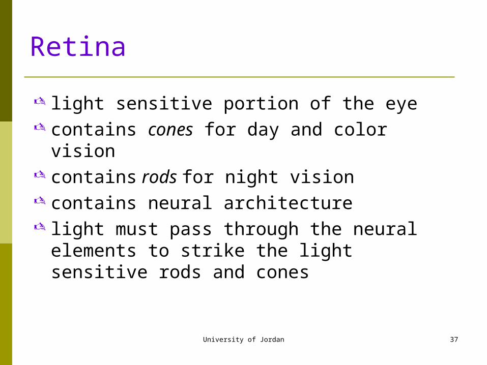
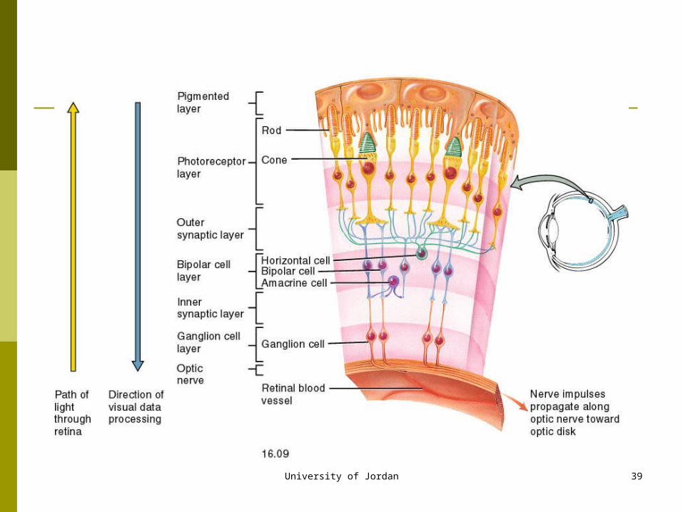
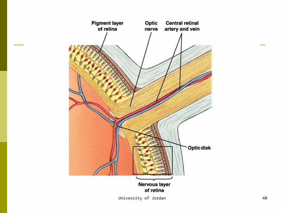
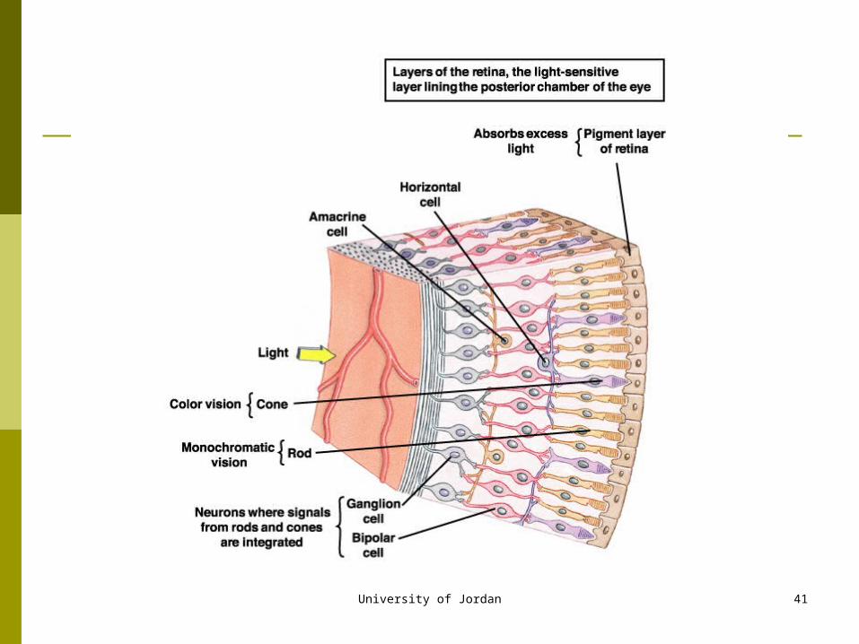
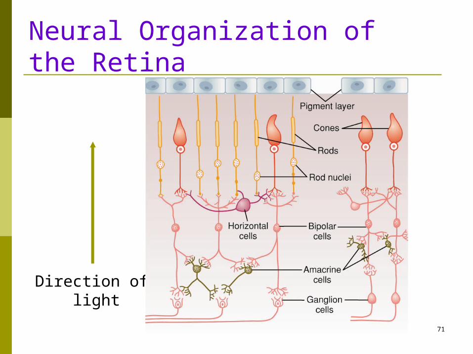
Retina
light sensitive portion of the eye contains cones for day and color vision contains rods for night vision contains neural architecture light must pass through the neural elements to
strike the light sensitive rods and cones
University of Jordan 38
University of Jordan 39
University of Jordan 40
University of Jordan 41
University of Jordan 42
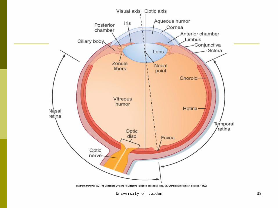
The Fovea A small area at the center of the retina about 1 sq
millimeter The center of this area, “the central fovea,” contains only
conesthese cones have a special structureaid in detecting detail
In the central fovea the neuronal cells and blood vessels are displaced to each side so that the light can strike the cones directly.
This is the area of greatest visual acuity
University of Jordan 43
Rods, Cones and Ganglion Cells
Each retina has 100 million rods and 3 million cones and 1.6 million ganglion cells.
60 rods and 2 cones for each ganglion cell At the central fovea there are no rods and the
ratio of cones to ganglion cells is 1:1. May explain the high degree of visual acuity
in the central retina
University of Jordan 44
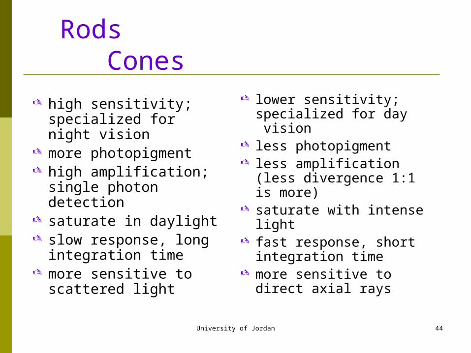
Rods Cones
high sensitivity; specialized for night vision
more photopigment high amplification; single
photon detection saturate in daylight slow response, long
integration time more sensitive to scattered
light
lower sensitivity; specialized for day vision
less photopigment less amplification (less
divergence 1:1 is more) saturate with intense light fast response, short
integration time more sensitive to direct
axial rays
University of Jordan 45
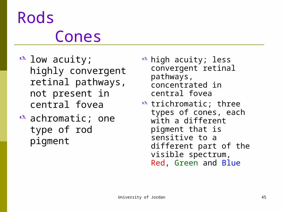
Rods Cones
low acuity; highly convergent retinal pathways, not present in central fovea
achromatic; one type of rod pigment
high acuity; less convergent retinal pathways, concentrated in central fovea
trichromatic; three types of cones, each with a different pigment that is sensitive to a different part of the visible spectrum, Red, Green and Blue
University of Jordan 46
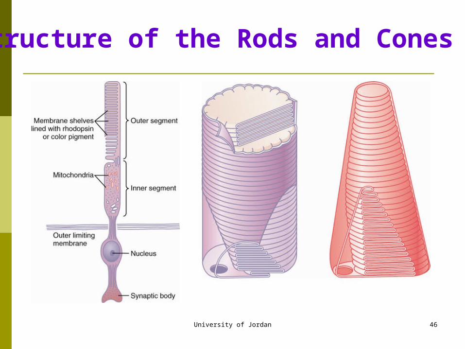
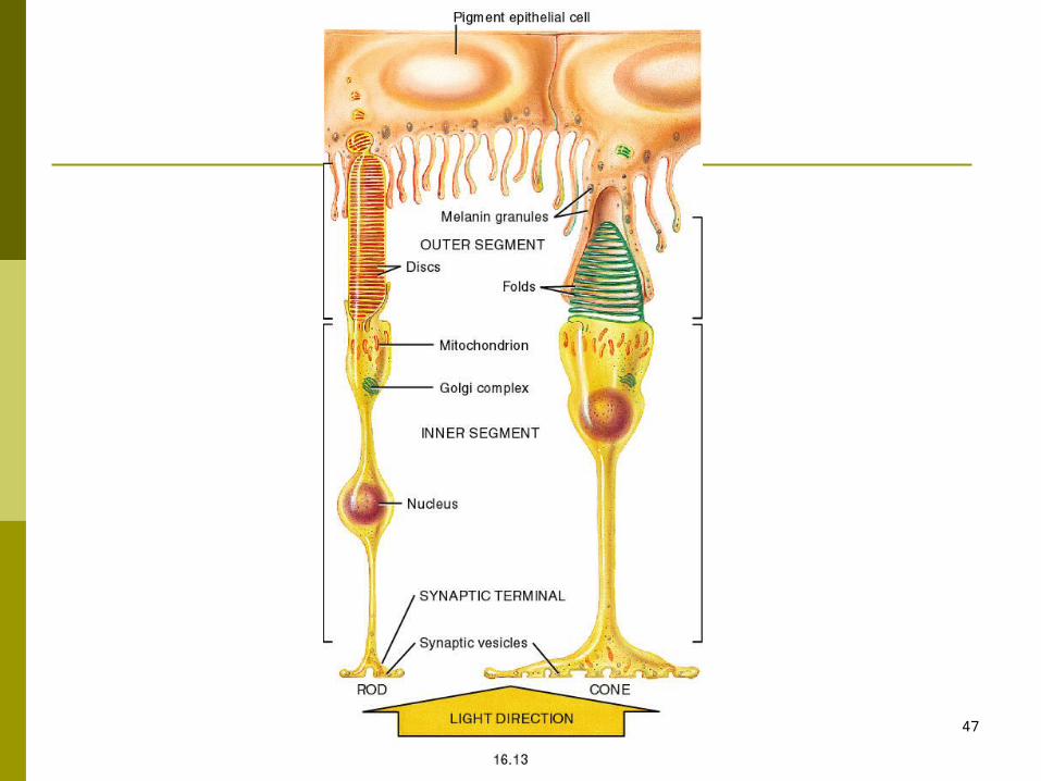
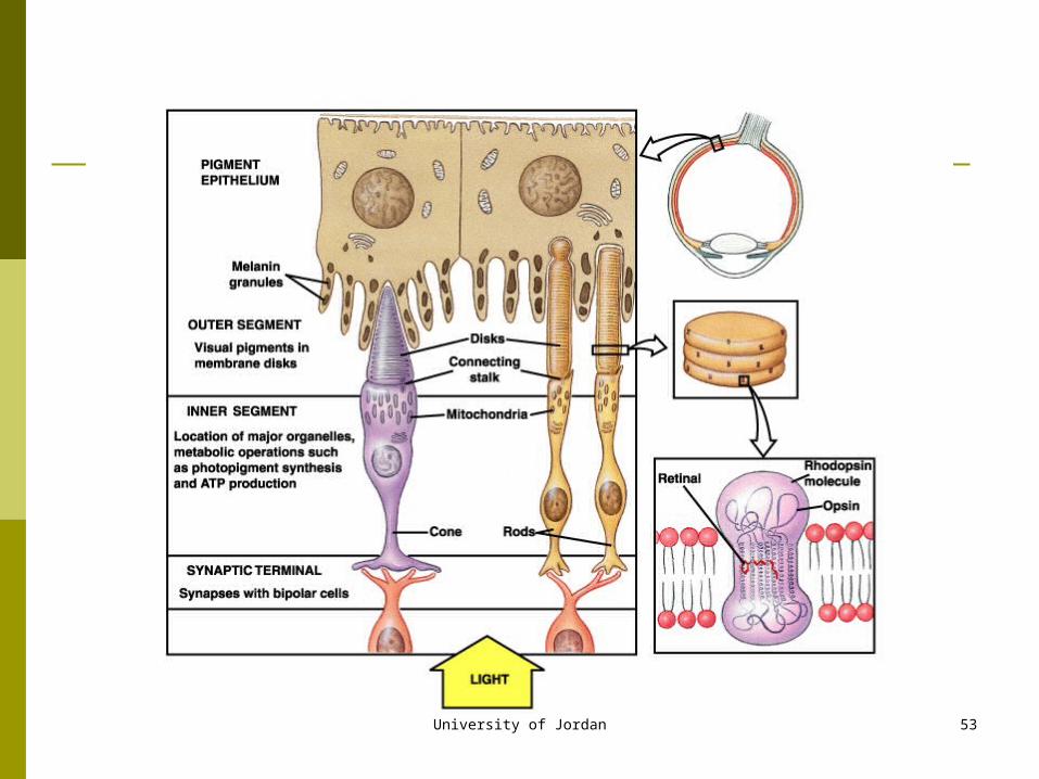
Structure of the Rods and Cones
University of Jordan 47
University of Jordan 48
Pigment Layer of Retina
Pigment layer of the retina is very important Contains the black pigment melanin Prevents light reflection in the globe of the eye Without the pigment there would be diffuse
scattering of light rather than the normal contrast between dark and light.
This is what happens in albinos (genetic absence of melanocyte activity) poor visual acuity because of the scattering of light
University of Jordan 49
Photochemistry of Vision Rods and cones contain chemicals that decompose on
exposure to light. This excites the nerve fibers leading from the eye. The membranes of the outer-segment of the rods contain
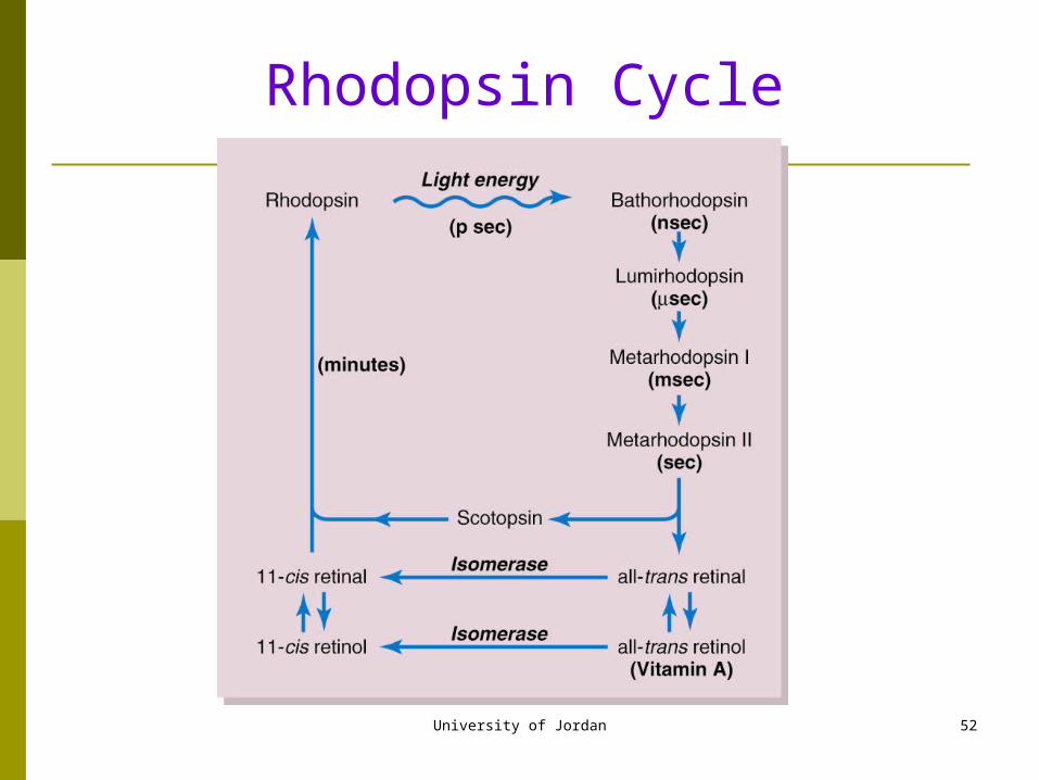
rhodopsin or visual purple. Rhodopsin is a combination of a protein called scotopsin and a
pigment, retinal (Vitamin A derivative) The retinal is in the cis configuration. Only the cis configuration can bind with scotopsin to form
rhodopsin.
University of Jordan 50
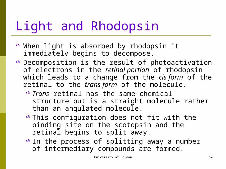
Light and Rhodopsin When light is absorbed by rhodopsin it immediately begins to
decompose. Decomposition is the result of photoactivation of electrons in
the retinal portion of rhodopsin which leads to a change from the cis form of the retinal to the trans form of the molecule. Trans retinal has the same chemical structure but is a
straight molecule rather than an angulated molecule. This configuration does not fit with the binding site on the
scotopsin and the retinal begins to split away. In the process of splitting away a number of intermediary
compounds are formed.
University of Jordan 51
University of Jordan 52
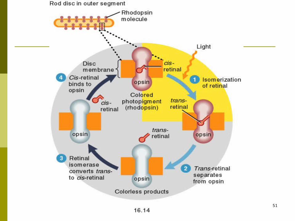
Rhodopsin Cycle
University of Jordan 53
University of Jordan 54
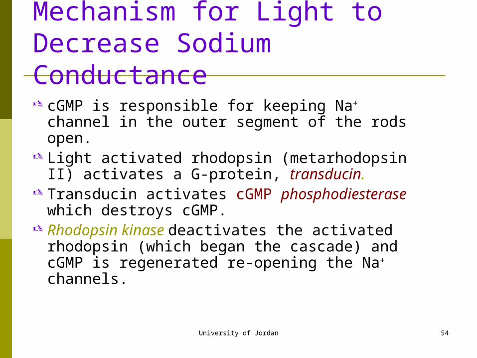
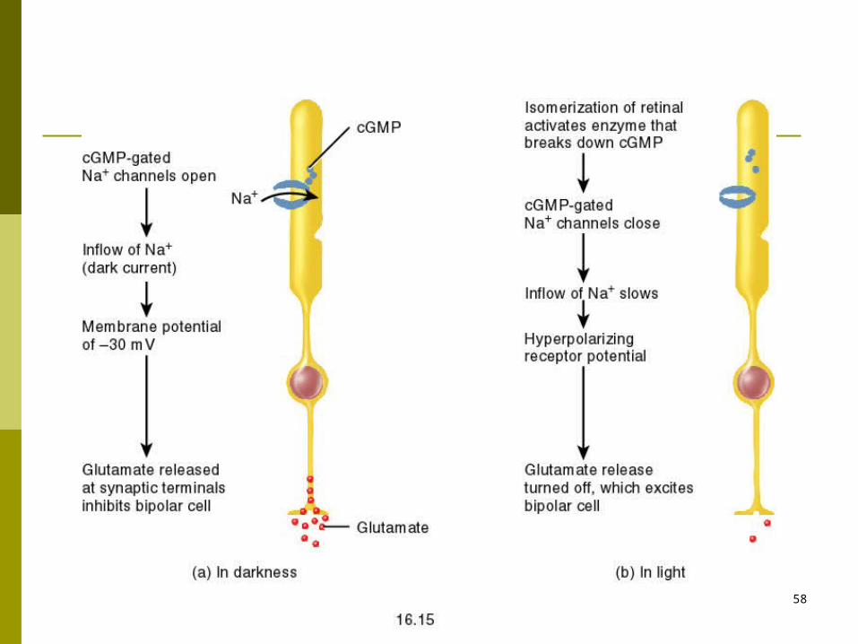
Mechanism for Light to Decrease Sodium Conductance
cGMP is responsible for keeping Na+ channel in the outer segment of the rods open.
Light activated rhodopsin (metarhodopsin II) activates a G-protein, transducin.
Transducin activates cGMP phosphodiesterase which destroys cGMP.
Rhodopsin kinase deactivates the activated rhodopsin (which began the cascade) and cGMP is regenerated re-opening the Na+ channels.
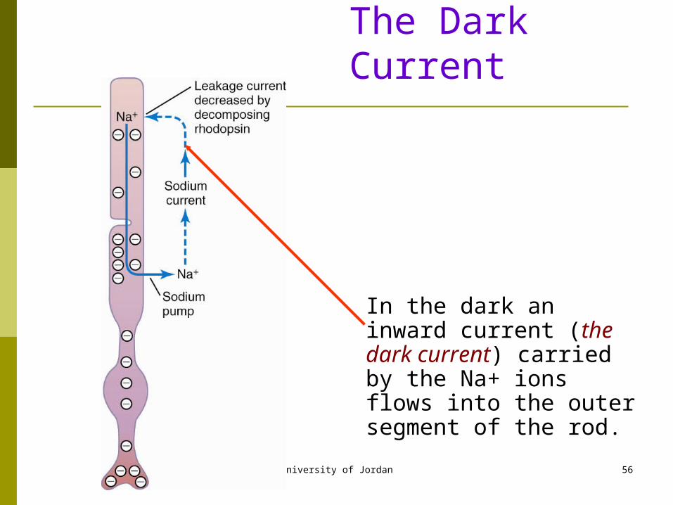
University of Jordan 56
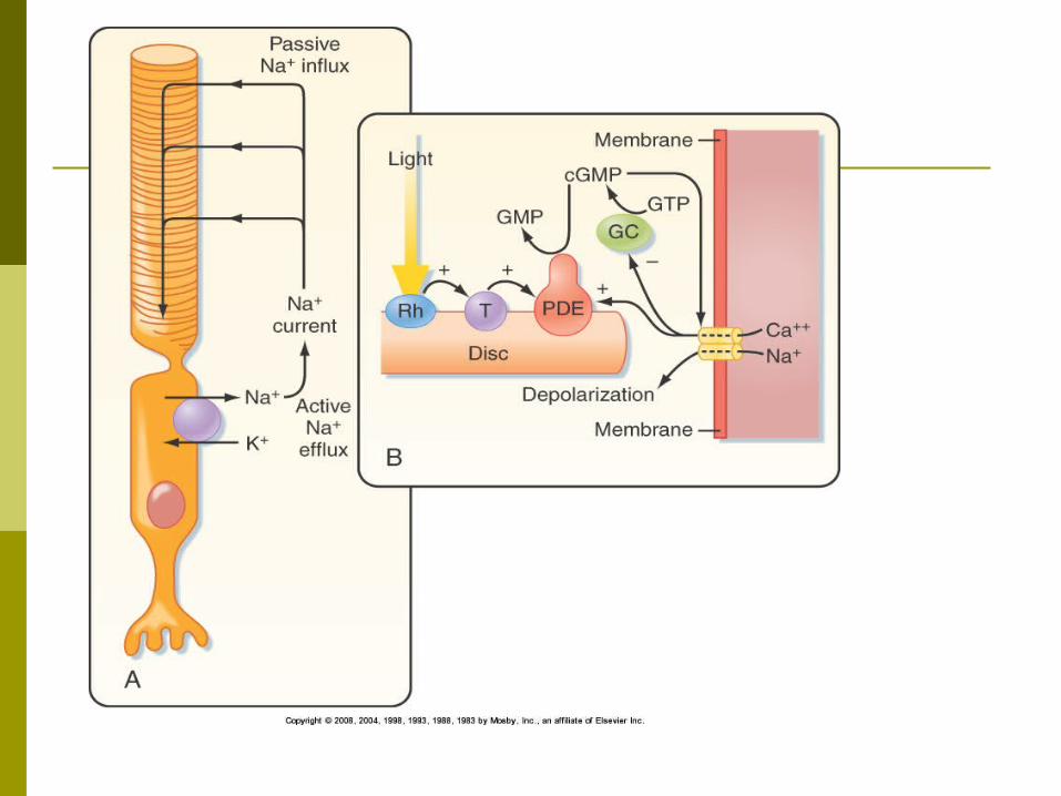
The Dark Current
In the dark an inward current (the dark current) carried by the Na+ ions flows into the outer segment of the rod.
University of Jordan 57
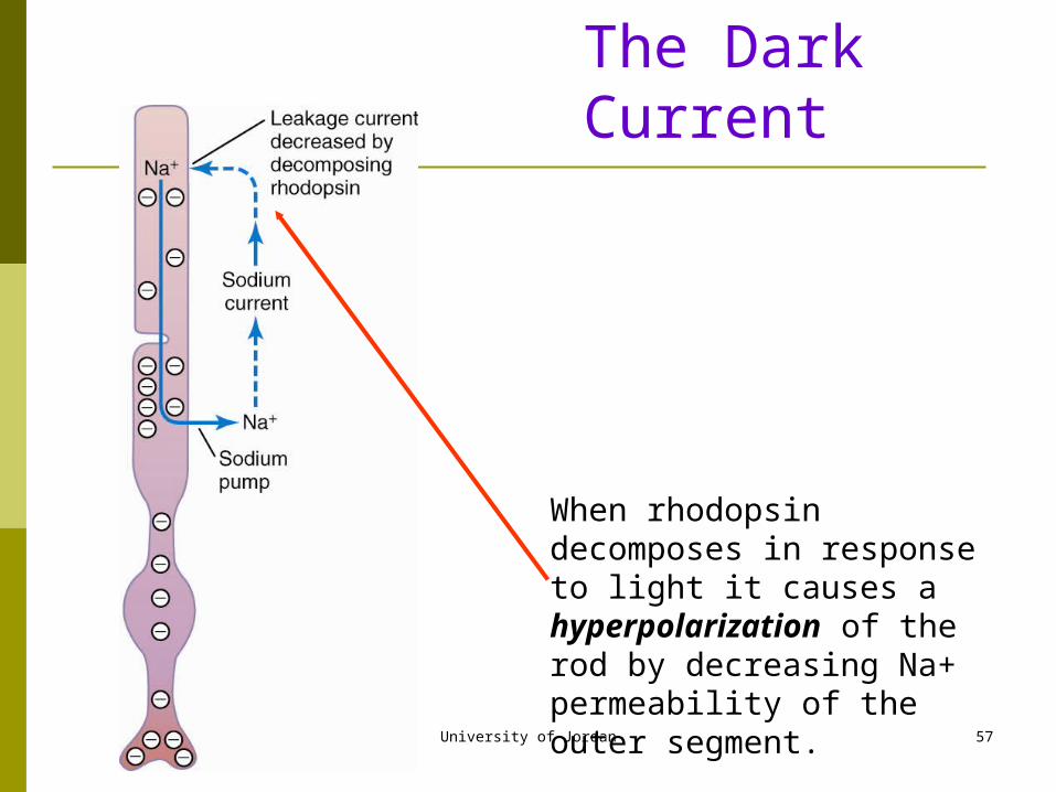
When rhodopsin decomposes in response to light it causes a hyperpolarization of the rod by decreasing Na+ permeability of the outer segment.
The Dark Current
University of Jordan 58
University of Jordan 59
Rod Receptor Potential (Cont’d)
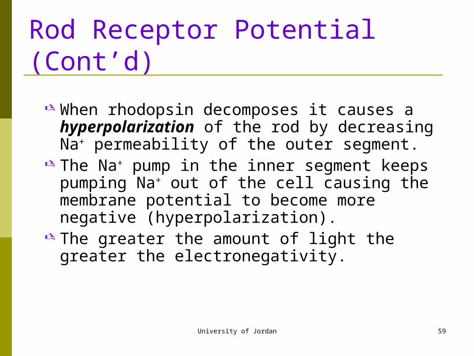
When rhodopsin decomposes it causes a hyperpolarization of the rod by decreasing Na+ permeability of the outer segment.
The Na+ pump in the inner segment keeps pumping Na+ out of the cell causing the membrane potential to become more negative (hyperpolarization).
The greater the amount of light the greater the electronegativity.
University of Jordan 60
The Rod Receptor Potential
Normally about -40 mV Normally the outer segment of the rod is very
permeable to Na+ ions. In the dark an inward current (the dark current)
carried by the Na+ ions flows into the outer segment of the rod.
The current flows out of the cell, through the efflux of K+, ions in the inner segment of the rod.
University of Jordan 61
Duration and Sensitivity of the Receptor Potential A single pulse of light causes activation of the rod
receptor potential for more than a second. In the cones these changes occur 4 times faster. Receptor potential is proportional to the logarithm
of the light intensity. very important for discrimination of the light
intensity
University of Jordan 62
Role of Vitamin A
Vitamin A is the precursor of all-trans-retinal, the pigment portion of rhodopsin.
Lack of vitamin A causes a decrease in retinal.
This results in a decreased production of rhodopsin and a lower sensitivity of the retina to light or night blindness.
University of Jordan 63
Dark and Light Adaptation
In light conditions most of the rhodopsin has been reduced to retinal so the level of photosensitive chemicals is low.
In dark conditions retinal is converted back to rhodopsin.
Therefore, the sensitivity of the retinal automatically adjusts to the light level.
Opening and closing of the pupil also contributes to adaptation because it can adjust the amount entering the eye.
University of Jordan 64
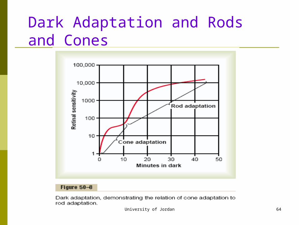
Dark Adaptation and Rods and Cones
University of Jordan 65
Importance of Dark and Light Adaptation
The detection of images on the retina is a function of discriminating between dark and light spots.
It is important that the sensitivity of the retina be adjusted to detect the dark and light spots on the image.
Enter the sun from a movie theater, even the dark spots appear bright leaving little contrast.
Enter darkness from light, the light spots are not light enough to register.
University of Jordan 66
Dark Adaptation
Gradual increase in photoreceptor sensitivity when entering a dark room. Maximal sensitivity reached in 20 min.
Increased amounts of visual pigments produced in the dark. Increased pigment in cones produces slight dark
adaptation in 1st 5 min. Increased rhodopsin in rods produces greater
increase in sensitivity.100,000-fold increase in light sensitivity in rods.
University of Jordan 67
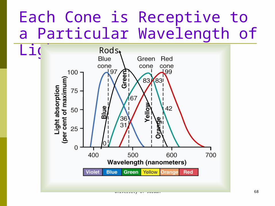
Color Vision
Color vision is the result of activation of cones. 3 types of cones:
blue conegreen cone red cone
The pigment portion of the photosensitive molecule is the same as in the rods, the protein portion is different for the pigment molecule in each of the cones.
Makes each cone receptive to a particular wavelength of light
University of Jordan 68
Each Cone is Receptive to a Particular Wavelength of Light
Rods
University of Jordan 69
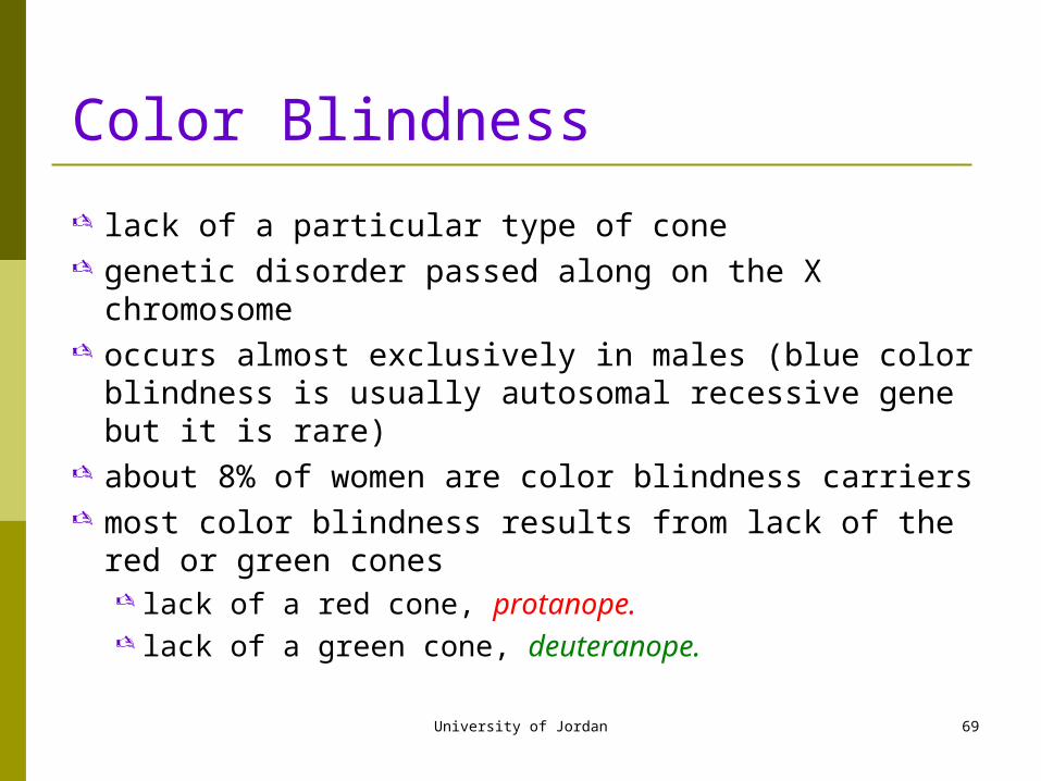
Color Blindness
lack of a particular type of cone genetic disorder passed along on the X chromosome occurs almost exclusively in males (blue color blindness is
usually autosomal recessive gene but it is rare) about 8% of women are color blindness carriers most color blindness results from lack of the red or green
cones lack of a red cone, protanope. lack of a green cone, deuteranope.
University of Jordan 70
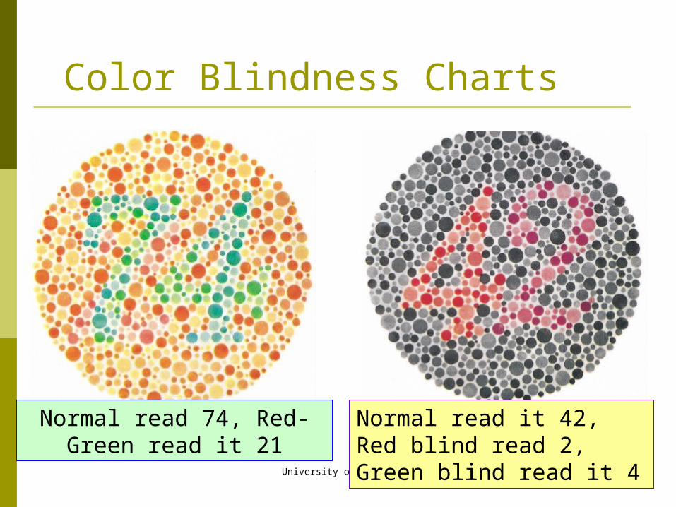
Color Blindness Charts
Normal read 74, Red-Green read it 21
Normal read it 42, Red blind read 2, Green blind read it 4
University of Jordan 71
Neural Organization of the Retina
Direction of light

University of Jordan 72
University of Jordan 73
Signal Transmission in the Retina
Transmission of signals in the retina is by electrotonic conduction.
Allows graded response proportional to light intensity.
The only cells that have action potentials are ganglion cells and amacrine cells.send signals all the way to the brain
University of Jordan 74
Lateral Inhibition to Enhance Visual Contrast
horizontal cells connect laterally between the rods and cones and the bipolar cells
output of horizontal cells is always inhibitory prevents the lateral spread of light excitation on the
retina have an excitatory center and an inhibitory
surround essential for transmitting contrast borders in the
visual image
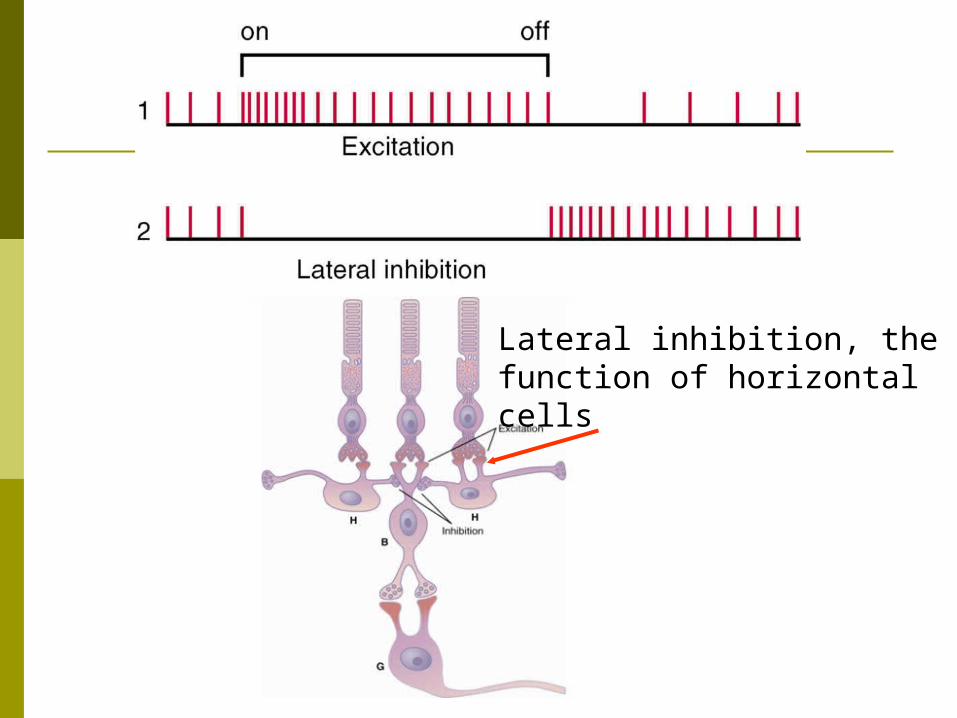
Lateral inhibition, the function of horizontal cells
University of Jordan 76
Function of Amacrine Cells
About 30 different types Some involved in the direct pathway from rods to bipolar
to amacrine to ganglion cells Some amacrine cells respond strongly to the onset of the
visual signal, some to the extinguishment of the signal Some respond to movement of the light signal across the
retina Amacrine cells are a type of interneuron that aid in the
beginning of visual signal analysis.
University of Jordan 77
Three Types of Ganglion Cells
W cells (40%) receive most of their excitation from rod cells. sensitive to directional movement in the visual field
X cells (55%) small receptive field, discrete retinal locations, may be responsible for the transmission of the visual image itself, always receives input from at least one cone, may be responsible for color transmission.
Y cells (5%) large receptive field respond to instantaneous changes in the visual field.
University of Jordan 78
Excitation of Ganglion Cells
spontaneously active with continuous action potentials
visual signals are superimposed on this background
many excited by changes in light intensity respond to contrast borders, this is the way
the pattern of the scene is transmitted to the brain

University of Jordan 79