unmethylated and methylated cpg … de jong et al 2010.pdfthe journal of immunology the...
TRANSCRIPT

of August 16, 2012.This information is current as Endosomes
Ability To Colocalize with TLR9 in Late Oligodeoxynucleotide Is Dependent on TheirUnmethylated and Methylated CpG The Immunostimulatory Activity of
Kazem, Pieter Cullis, Wilf Jefferies and Ying TamSusan D. de Jong, Genc Basha, Kaley D. Wilson, Mikameh
http://www.jimmunol.org/content/184/11/6092doi: 10.4049/jimmunol.0802442April 2010;
2010; 184:6092-6102; Prepublished online 28J Immunol
MaterialSupplementary
2.DC1.htmlhttp://www.jimmunol.org/content/suppl/2010/04/28/jimmunol.080244
Referenceshttp://www.jimmunol.org/content/184/11/6092.full#ref-list-1
, 9 of which you can access for free at: cites 24 articlesThis article
Subscriptionshttp://jimmunol.org/subscriptions
is online at: The Journal of ImmunologyInformation about subscribing to
Permissionshttp://www.aai.org/ji/copyright.htmlSubmit copyright permission requests at:
Email Alertshttp://jimmunol.org/cgi/alerts/etocReceive free email-alerts when new articles cite this article. Sign up at:
Print ISSN: 0022-1767 Online ISSN: 1550-6606. Immunologists, Inc. All rights reserved.Copyright © 2010 by The American Association of9650 Rockville Pike, Bethesda, MD 20814-3994.The American Association of Immunologists, Inc.,
is published twice each month byThe Journal of Immunology
at University of B
ritish Colum
bia on August 16, 2012
http://jimm
unol.org/D
ownloaded from
The Journal of Immunology
The Immunostimulatory Activity of Unmethylated andMethylated CpG Oligodeoxynucleotide Is Dependent on TheirAbility To Colocalize with TLR9 in Late Endosomes
Susan D. de Jong,* Genc Basha,*,†,1 Kaley D. Wilson,*,1 Mikameh Kazem,*
Pieter Cullis,* Wilf Jefferies,†,2 and Ying Tam‡,2
TLR9 recognizes CpG motifs present in pathogenic DNA and triggers potent immune responses. It is generally accepted that
TLR9 distinguishes pathogenic DNA based, in part, on methylation status, where TLR9 binds unmethylated but not methylated
CpG. However, we showed that methylated CpG induces potent TLR9-mediated responses when delivered in lipid nanoparticles.
In this article, we report that methylation dictates the ability of free CpG DNA to colocalize with TLR9 in late endosomes.
However, when delivered in lipid nanoparticles, CpG DNA and TLR9 colocalize, regardless of methylation status. Therefore, it is
proposed that the ability of immune cells to distinguish unmethylated pathogenic from methylated mammalian DNA is controlled
by a mechanism that regulates TLR9 mobilization and colocalization rather than a differential binding affinity. The Journal of
Immunology, 2010, 184: 6092–6102.
The mammalian immune system has evolved highly con-served pattern recognition receptors (PRRs), such as theTLR family, which recognize specific molecular patterns,
expressed by a diverse group of infectious microorganisms, as dan-ger signals of infection and trigger potent, protective immuneresponses (1–6). Inherent to this detection system is the ability todistinguish pathogen-associated patterns from those encounteredduring benign or beneficial self and environmental interactions.Although this can be easily conceptualized for TLRs 4, 2/6, and 5,all of which recognize structurally complex and unique ligands,such as LPS, peptidylglycan, and flagellin, it is somewhat less intu-itive for TLR9, which specifically recognizes CpG DNA motifs (7).Distinguishing pathogenic from eukaryotic DNA is a multifac-
torial process, relying on localization of TLR9 to the endosomalcompartment (8, 9), suppression of CpG frequency in eukaryoticDNA, the occurrence of eukaryotic CpGs within immunosup-pressive flanking sequences, and methylation status (2, 10, 11).With regard to methylation status, TLR9 was shown to specifically
respond to unmethylated CpG motifs, such as those present inbacterial DNA, compared with eukaryotic CpGs, of which 70–80% are methylated (12). Furthermore, it was demonstrated thatmethylation of immunostimulatory CpG DNA abrogates activity,which is commonly attributed to the specificity of TLR9 for un-methylated motifs and its relative inability to bind methylatedsequences. Direct evidence for this comes from surface plasmonresonance studies that reported higher-affinity binding of TLR9with unmethylated CpG oligodeoxynucleotide (ODN) and CpG-containing plasmid DNA compared with the methylated forms(10, 13).Conversely, it was demonstrated that when expressed on the cell
surface, a chimeric TLR9 is capable of responding to self-DNA (9),and our group and other investigators observed that methylatedCpG (mCpG) ODN possesses TLR9-mediated immunostimulatorypotential when delivered in a lipid carrier system (14–16). Spe-cifically, we recently reported that for natural phosphodiester andmodified phosphorothioate ODN, lipid nanoparticle (LN) deliveryendows mCpG ODN with immunostimulatory activity similar toor greater than the equivalent unmethylated ODN through a TLR9-mediated mechanism (17).The ability of mCpG DNA to induce TLR9-mediated immune
responses suggests that methylation status may determine the im-munostimulatory activity of CpGDNA bymechanisms other than byaffecting binding properties. We show in this article that althoughmethylated andunmethylatedCpGODNin free formare takenupandtraffic to the endosome similarly, only unmethylated ODN promoteseffective mobilization of TLR9 to the late endosomal compartment,allowing for colocalization with its CpG ligand. Interestingly, ad-ministration of methylated and unmethylated CpG ODN in LNs, aswell as empty LNs, promotes the mobilization of TLR9 to theendosome. We further demonstrate that following exposure to freeunmethylated CpG ODN and LNs, colocalization occurs through ansrc-family kinase (SFK)-mediated signaling pathway. These resultssuggest that the lack of immunological activity for free methylatedODN is largely due to a failure to localize with TLR9 in the lateendosomal compartment, rather than an inability to bind and activateTLR9. Finally, we confirm this hypothesis by showing that predosingwith empty LNs to induce TLR9 mobilization to the endosomal
*Department of Biochemistry and Molecular Biology and †Biomedical ResearchCentre, University of British Columbia, Vancouver; and ‡Tekmira Pharmaceuticals,Burnaby, British Columbia, Canada
1G.B. and K.D.W. contributed equally to this work.
2W.J. and Y.T. are cosenior authors.
Received for publication July 24, 2008. Accepted for publication March 19, 2010.
This work was supported in part by Canadian Institutes for Health Research operatinggrants to P.R.C. and W.J. S.D.deJ. and K.D.W. were recipients of Natural Sciencesand Engineering Research Council and Natural Sciences and Engineering ResearchCouncil, Canadian Institutes for Health Research, and Michael Smith Foundation forHealth Research postgraduate awards, respectively.
Address correspondence and reprint requests to Dr. Ying K. Tam at the currentaddress: AlCana Technologies, Inc., 2714 West 34th Avenue, Vancouver, BritishColumbia, V6L 2A1 Canada. E-mail address: [email protected]
The online version of this article contains supplemental material.
Abbreviations used in this paper: 1,19dioctadecyl3, 3,39,39 tetramethylindocarbocyanineperchlorate; BMDC, bone marrow dendritic cell; DC, dendritic cell; DilC18, E, early;EEA1, early endosomeAg 1; ER, endoplasmic reticulum; LAMP1, lysosomal-associatedmembrane protein 1; L, late; LN, lipid nanoparticle; mCpG,methylated CpG;MFI, meanfluorescence intensity; ODN, oligodeoxynucleotide; PP2, pyrazol pyrimidine type 2;PRR, pattern recognition receptor; SFK, src-family kinase.
Copyright� 2010 by TheAmericanAssociation of Immunologists, Inc. 0022-1767/10/$16.00
www.jimmunol.org/cgi/doi/10.4049/jimmunol.0802442
at University of B
ritish Colum
bia on August 16, 2012
http://jimm
unol.org/D
ownloaded from

compartment endows free mCpG ODN with immunostimulatoryactivity.The ability to differentiate between pathogenic and eukaryotic
DNA represents a vital element of the eukaryotic immune system,promoting rapid and vigorous immune responses to protect againstpathogenic attack while avoiding inappropriate and pathologicimmune responses to self-DNA during normal processes, such asdevelopment, growth, and maintenance. Based on these data, wepropose a new model in which the differential immunostimulatoryactivity of unmethylated and methylated CpG ODN, and, byextension, pathogenic and self-DNA, is determined by their abilityto effectively induce mobilization of TLR9 and to colocalize withthe receptor in the endosomal compartment, rather than differ-ential affinities for TLR9. We suggest that discrimination betweenfree methylated and unmethylated CpG ODN occurs upstream ofTLR9 by recognition of the methylation status of free CpG ODN,triggering an SFK-signaling pathway that induces TLR9 mobi-lization to the late endosomal compartment. Uptake of LN sys-tems also triggers TLR9 mobilization to the endosome, effectivelybypassing this discrimination process and allowing for colocali-zation of the nucleic acid payload with TLR9, irrespective ofmethylation status.
Materials and MethodsReagents
These studies were performed with phosphorothioated unmethylated andmethylated (CpG motif shown in bold, 59 methylcytosine denoted by p)INX-6295 (59-TAACpGTTGAGGGGCAT-39), INX-5001 (59-AACpGTT-39), CpG-2006 (59-TCpGTCpGTTTTGTCpGTTTTGTCpGTT-39), andG3139 (59-TCTCCCAGCpGTGCpGCCAT-39). Specific references to CpGand mCpG ODN in this section and Results refer to INX-6295, unlessotherwise noted. Of note is the 39 G-tetrad motif in INX-6295. Althoughimmunostimulatory activity has been ascribed to these motifs, deletion oralteration did not have any impact on the immunostimulatory activity ofINX-6295, whereas alteration of the CpG motif effectively abrogated ac-tivity. Unlabeled and 59 FAM-labeled ODN were synthesized by TrilinkBioTechnologies (San Diego, CA). Tissue culture media, L-glutamine, FBS,penicillin G, and streptomycin sulfate were from Invitrogen (Burlington,Ontario, Canada), and recombinant mouse IL-4 and GM-CSF were fromCedarlane Laboratories (Burlington, Ontario, Canada). BioPlex multiplexcytokine bead analysis reagents were purchased from Bio-Rad (Hercules,CA), and Abs for ELISA, cytometric bead array sets, and all Abs for flowcytometry were purchased from BD Biosciences (Mississauga, Ontario,Canada). Poly-D-lysine was from Sigma-Aldrich (Oakville, Ontario, Can-ada), Ficoll-Paque was from Amersham (Baie d’Urfe, Quebec, Canada),and collagenase D was obtained from Roche Applied Sciences (Laval,Quebec, Canada) The SFK inhibitor, pyrazol pyrimidine type 2 (PP2), wasobtained from Calbiochem (San Diego, CA), and anti-CD11c MACS beadswere fromMiltenyi Biotec (Auburn, CA). Goat anti-mouse early endosomeAg 1 (EEA1) and rat anti-mouse lysosomal-associated membrane protein 1(LAMP1) were from Santa Cruz Biotechnology (Santa Cruz, CA); rabbitanti-mouse to TLR3, TLR7, and TLR9 were obtained from Abcam(Cambridge, MA). Texas Red-conjugated dextran, 1,19dioctadecyl3,3,39,39tetramethylindocarbocyanine perchlorate (DiIC18), and all secondary Abswere purchased from Invitrogen.
Mice and cell lines
Eight- to 10-wk-old female ICR and C57BL/6 mice were obtained fromCharles River Laboratories (Wilmington, MA) and held in a pathogen-freeenvironment. All procedures were approved by the appropriate institutionalanimal care committee (University of British Columbia and/or TekmiraPharmaceuticals) and performed in accordance with the guidelines estab-lished by the Canadian Council on Animal Care. The murine macrophagecell line, RAW264.7, was obtained from the American Type Culture Col-lection (Manassas, VA) and cultured inDMEMsupplemented with penicillinG (100 U/ml), streptomycin sulfate (100 mg/ml), 2 mM L-glutamine, and10% FBS. Bone marrow dendritic cells (BMDCs) were derived from bonemarrow cells collected from the long bones of ICR mice cultured with IL-4and GM-CSF for 7 d in compete medium consisting of RPM1 1640 sup-plemented with penicillin G (100 U/ml), streptomycin sulfate (100 mg/ml),2 mM L-glutamine, and 10% FBS. Resultant cells were .85% CD11c+ and
displayed a myeloid phenotype (CD11c+, CD11b+, Mac3lo), as assessed byflow cytometry.
Preparation of liposomal nanoparticles
ODNs were encapsulated in LNs containing an ionizable aminolipidusing an ethanol dialysis procedure, as previously described (18). DiIC18
(0.5 mol%) was used in the formulation of empty LNs. Prior to formu-lation, CpG ODN was heated to eliminate any multimers (as assessed byPAGE) that may have formed, particularly for INX-6295 as a conse-quence of the 39 G-tetrad. ODN concentrations were determined by UVspectroscopy (260 nm) on a Beckman DU 640 spectrophotometer, andlipid concentrations were determined using an inorganic phosphorusassay after separation of the lipids from the ODN by a Bligh and Dyerextraction (19). The ODN/lipid ratio was typically 0.10–0.13 (w/w), witha particle size of 100 6 25 nm, as determined by quasi-elastic lightscattering using a NICOMP Model 370 submicron particle sizer (ParticleSizing Systems, Santa Barbara, CA).
Cell uptake and immune response
For in vitro analysis, RAW264.7 or BMDCs were incubated for 1, 4, 12, or24 h with 2–50 mg/ml fluorescently labeled free or encapsulated CpGODN or mCpG ODN. Cells were harvested, washed, and then analyzedfor uptake using flow cytometry. For ex vivo assessment, ICR mice wereinjected s.c. with 5 mg/kg fluorescently labeled free or encapsulated CpGODN or mCpG ODN. Spleens and/or lymph nodes were obtained frommice 1, 4, 7, and 24 h postadministration. To demonstrate the role of SFKsignaling in the immunostimulatory activity of CpG ODN, LN-CpGODN, and LN-mCpG ODN, C57BL/6 mice were treated i.p. with 1 mg/kg PP2 twice per day for 7 d. PP2 has been extensively described andused in the literature as an SFK-specific inhibitor. Mice were injected i.v.with 20 mg/kg free or encapsulated fluorescently labeled CpG ODN ormCpG ODN, and tissue samples were collected 12 h postadministration.Cells were processed to single-cell suspensions, as previously described(20). For plasma cytokine analysis, blood was collected and processedto plasma by centrifugation and frozen at 220˚C until assayed. Plasmaconcentrations of IL-6, MCP-1, and IFN-g were determined usingELISA or cytometric bead array (BD Biosciences or Bio-Rad), as per themanufacturer’s instructions. For empty LN predosing studies, C57BL/6mice were dosed i.v. with free mCpG ODN alone, empty LN alone, orwith empty LN 0.5 h prior to the i.v. administration of free mCpG ODNat ODN and lipid doses of 20 and 200 mg/kg, respectively; tissue sam-ples were collected at 4 and 24 h. Spleens and blood were processed andanalyzed as previously described.
Flow cytometry
Cell uptake (as judged by the intensity of the fluorescently labeled ODNon a per-cell basis) was assessed in specific immune cell populations (asdetermined by phenotype analysis; cell suspensions were stained with PE-conjugated anti-CD11b or allophycocyanin-conjugated anti-CD11c Abs,and analyzed using a four-color FACSort flow cytometer and CellQuest Prosoftware, both from BD Biosciences). For a determination of immuneactivation, cell suspensions were stained with FITC- or allophycocyanin-labeled phenotype Abs (against CD11b, CD11c, Mac3, CD8, and B220/CD45R) in combination with PE-conjugated Abs directed against the ac-tivation markers CD69 or CD86. Cell activation was assessed using anLSRII flow cytometer and FACS Diva software (BD Biosciences).
Endosomal trafficking and localization
Endosomal localization of free and encapsulated CpG ODN and mCpGODN was assessed by incubating RAW264.7 cells with 10 mg/ml free orencapsulated CpG ODN or mCpG ODN for 4 h prior to the addition of1 mg/ml subcellular compartmental marker, Texas Red-conjugated dextran10,000 m.w., for an additional 2 h. BMDCs were grown on poly-D-lysine–precoated coverslips and incubated with 5 mg/ml free or encapsulated CpGODN or mCpG ODN for 4 h. To determine the possibility of preferentialtrafficking of unmethylated CpG ODN specifically to TLR9-containingendosomes, thus allowing for interaction with TLR9 and im-munostimulatory activity compared with mCpG ODN, C57BL/6 micewere injected i.v. with 20 mg/kg free or encapsulated, methylated or un-methylated fluorescently labeled CpG ODN or 150 mg/kg DiI–labeledempty LNs. To demonstrate the role of SFK signaling in the colocalizationof CpG ODN, LN-CpG ODN, and LN-mCpG ODN, C57BL/6 mice wereinitially injected i.p. with 1 mg/kg specific SFK inhibitor PP2 twice perday for 7 d. Following the last treatment, mice were injected i.v. with20 mg/kg free or encapsulated, methylated or unmethylated fluorescentlylabeled CpG ODN. After 4 h, all mice were euthanized, spleens were
The Journal of Immunology 6093
at University of B
ritish Colum
bia on August 16, 2012
http://jimm
unol.org/D
ownloaded from

disrupted, and dendritic cells (DCs) were isolated and processed for vi-sualization, as outlined previously.
Immunofluorescence
Following incubation, RAW264.7 cells were washed and incubated inOptimem prior to live cell visualization. BMDCswere treated with 2%BSAin PBS, fixed in 2% paraformaldehyde, and permeabilized with 0.1% sa-ponin and 2% BSA in PBS. Cells were stained with rat anti-mouse LAMP1,followed by Alexa 647-conjugated rabbit anti-rat Ab as a detection reagent.For ex vivo studies, spleens were dissociated by injection of 1 ml RPMI1640 containing 5% FBS and 1 mg collagenase D and incubated for 30 minat 37˚C. Subsequently, DC-enriched cell populations were obtained bycentrifugation of cell suspensions on Ficoll-Paque gradients. DCs werethen purified by positive selection with anti-CD11c MACS beads, with theresulting population being .98% CD11c+. Splenic DCs were resuspendedin complete media and grown on poly-D-lysine–precoated coverslips for3 h. Attached DCs were then treated with 2% BSA in PBS, followed byfixation with 2% paraformaldehyde. The cells were then permeabilizedwith 0.1% saponin and 2% BSA in PBS, followed by incubation withgoat anti-mouse EEA1 or rat anti-mouse LAMP1. Secondary Alexa 647-conjugated rabbit anti-goat and goat anti-rat Abs were used, as detectionreagents. To examine the colocalization of EEA1 and LAMP1+ compart-ments with TLR3, TLR7, or TLR9, mouse anti-mouse or rabbit anti-mouseTLR3, TLR7, or TLR9 polyclonal Abs, followed by rabbit anti-mouse andor goat anti-rabbit, both coupled to Alexa 568 were used, respectively.
Confocal microscopy
For RAW264.7 and BMDCs, sections 0.1 mm in thickness were capturedusing a Bio-Rad Radiance 2000 laser scanning or Nikon C1 immunoflu-orescent confocal microscope. Data were analyzed using ImageJ v1.37(National Institutes of Health, Bethesda, MD), to select images with a totalthickness of 0.2–0.3 mm, and processed with Adobe Photoshop CS2(Adobe Systems, San Jose, CA). For ex vivo studies, sections of 0.15 mmwere captured with a Nikon-C1, TE2000-E immunofluorescent confocalmicroscope (Nikon Instruments, Melville, NY). Data were analyzed asdescribed previously, and stacks with a total thickness of 0.6 mm wereprocessed with Adobe Photoshop CS2. Isotype control Abs were used inall confocal microscopy experiments to confirm the specificity of Abstaining. Thirty to 50 images were collected for each treatment. The per-centage of colocalization of combinations of free or encapsulated CpGODN or mCpG ODN, TLR9, and LAMP1/EEA1, for all studies, was as-sessed in OpenLab (PerkinElmer, Woodbridge, Ontario, Canada).
Statistical analyses
All statistical analyses were performed using SPSS version 14.0 (SPSS,Chicago, IL). Initially, one-way ANOVA was used to statistically evaluatethe differences among treatment groups. In the case of statistically sig-nificant results, unless otherwise stated, the differences among treatmentgroups were assessed through the use of Bonferroni adjusted t tests, a posthoc test that controls for family-wise error rate. A p value ,0.05 wasconsidered significant.
ResultsMethylation status does not influence ODN uptake orintracellular trafficking by immune cells when in free orLN-encapsulated form
The uptake and trafficking characteristics of unmethylated andmethylated CpG ODN were assessed to clarify their roles asdeterminants of immunostimulatory activity. Results from in vitrouptake studies offluorescently labeledODN in culturedmacrophagecells and BMDCs demonstrated that, regardless of the duration ofexposure or ODN concentration (Supplemental Fig. 1), free mCpGODN and CpG ODN are taken up in a similar manner in both celltypes (Fig. 1Aa, 1Ab, respectively; Supplemental Fig. 4A). Like-wise, an in vivo analysis of uptake following s.c. administration oflabeled ODN (Fig. 1B, Supplemental Fig. 4A) showed that freeunmethylated and methylated CpG ODN are taken up similarly byAPCs, as demonstrated in CD11b+ cells (Fig. 1Ba, 1Bc), CD11c+
cells (Fig. 1Bb, 1Bd), and, to a lesser extent, B220+ cells (Sup-plemental Fig. 2) from spleen (Fig. 1Ba, 1Bb, Supplemental Fig.2A) and lymph nodes (Fig. 1Bc, 1Bd, Supplemental Fig. 2B). As
expected, similar uptake for LN-encapsulated unmethylated andmethylated ODN was also observed in vitro and in vivo.Intracellular trafficking was also evaluated for a potential role in
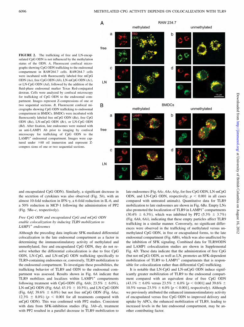
determining the relative immunostimulatory activities of CpG andmCpG ODN. Endosomal trafficking was assessed in RAW264.7cells and BMDCs (data not shown) using the fluid-phase sub-cellular compartmental marker Texas Red-conjugated dextran inconjunction with fluorescently labeled ODN. As shown in Fig. 2A,mCpG and CpG ODN efficiently localized to the endosomalcompartment in free (Fig. 2Aa, 2Ab) or LN (Fig. 2Ac, 2Ad) form.Similarly, free and LN-mCpG and CpG ODN were delivered ef-ficiently to the endosomal compartment of BMDCs, as shown bythe colocalization with EEA1 (data not shown) and LAMP1+ (Fig.2B). As expected, uptake and trafficking were not dependent onCpG motifs, because non-CpG ODN exhibited identical in vitroand in vivo uptake and intracellular trafficking characteristics(data not shown).
Free CpG, LN-CpG, and LN-mCpG ODN colocalize with TLR9in the late endosomal compartment, but free mCpG does not
The similar uptake and endosomal trafficking characteristics in-dicate that the differential immunostimulatory activity of freeunmethylated and methylated CpG ODN cannot be attributed toeither of these two processes. To evaluate the potential impact ofother aspects of intracellular trafficking on immunostimulatoryactivity, an ex vivo examination of the relative ability of free CpGand mCpG ODN to colocalize with TLR9 in the endosomalcompartment was undertaken. Specifically, the colocalization ofCpG ODN with TLR9 in early and late endosomes was assessed inDCs isolated from the spleens of mice injected with fluorescentlylabeled free unmethylated or methylated CpG ODN.Consistent with immunostimulatory activity, mice treated with
free CpG ODN showed colocalization of CpG ODN with TLR9 inthe LAMP1+ compartment (Fig. 3Ab) but not for free mCpG ODN(Fig. 3Aa), whereas the administration of CpG ODN encapsulatedin LNs resulted in effective colocalization with TLR9 in the lateendosomal compartment, regardless of ODN methylation status(Fig. 3Ac, 3Ad). Quantitative analysis (Fig. 3B, Supplemental Fig.4B for combined data) and statistical analyses confirmed that freeCpG ODN (16.8% 6 4.0%), as well as LN-CpG (24.1% 6 6.8%)and LN-mCpG ODN (22.3% 6 6.6%), effectively colocalizedwith TLR9 in the late endosome, whereas free mCpG ODN(7.3% 6 3.2%) did not (p , 0.001 for all three compared withfree mCpG ODN). Of note, LN-mCpG ODN and LN-CpG ODNdemonstrated significantly greater colocalization with TLR9 inLAMP1+ compartments than did free CpG ODN (p , 0.05 andp , 0.001, respectively), with no difference in colocalizationbetween the two encapsulated forms. Interestingly, the uptake ofLNs themselves, regardless of payload, seems to be a sufficienttrigger for colocalization, because empty LNs administered i.v.colocalized with TLR9 in LAMP1+ endosomes (Fig. 4Ba). Theempty LNs labeled with the fluorescent marker DiI showed sta-tistically significantly greater colocalization with TLR9 in theLAMP1+ compartment (17.2% 6 5.7%) compared with freemCpG ODN (p , 0.001).Significantly greater colocalization of CpG ODN with TLR9
occurred in late LAMP1+ endosomes (Fig. 3Ca) compared withearly EEA1+ endosomes following the administration of free CpG,LN-CpG, and LN-mCpG (p , 0.001, p , 0.05, and p , 0.001,respectively). This is despite the fact that similar levels of free andLN-CpG ODN are found in early and late endosomes (Fig. 3Cb).These data show that the ability of free CpG ODN to colocalize
with TLR9 in late endosomes is dependent on methylation statusand that delivery in LN form overcomes this discrimination and
6094 METHYLATED CPG ACTIVITY DEPENDS ON COLOCALIZATION WITH TLR9
at University of B
ritish Colum
bia on August 16, 2012
http://jimm
unol.org/D
ownloaded from

allows for colocalization, regardless of the methylation status (oreven the presence) of the payload.
Colocalization with TLR9 and immunostimulatory activity aremediated via SFK
It was reported that CpG motifs induce a sequence-specific, TLR9-independent SFK-signaling cascade at the plasma membrane that isultimately required for CpG engagement and the activation ofTLR9-MyD88 (21). Therefore, we investigated whether SFKsignaling could mediate the colocalization of free CpG ODN (andpotentially, LN-CpG and LN-mCpG ODN) with TLR9 in theLAMP1+ compartment. To demonstrate the role of SFK signalingin colocalization, mice were pretreated with PP2, an SFK-specificinhibitor, prior to the administration of fluorescently labeled freeor encapsulated CpG ODN and mCpG ODN.As shown in Fig. 4A, pretreatment with PP2 effectively abol-
ished the colocalization of free CpG ODN with TLR9 in the en-dosomal compartment compared with untreated controls (Fig. 4Abversus 4Aa; 2.6% 6 3.2% compared with 16.1 6 8.1%; p ,0.001). Similarly, PP2 was effective in preventing colocalizationof TLR9 with LN-CpG ODN (Fig. 4Ac; 4.7% 6 5.4%) and LN-
mCpG ODN (Fig. 4Ad; 2.7% 6 1.7%) in LAMP1+ compartments,as well as with empty LNs (Fig. 4Bb; 17.3% 6 5.7%) comparedwith non–PP2-treated controls (Fig. 4Ba; 1.3% 6 1.7%). Quan-titative data for the free and LN unmethylated and methylatedCpG and for empty LNs are shown in Fig. 4Ca and 4Ab, re-spectively (Supplemental Fig. 4B for combined data). Concomi-tant with a significant reduction in the colocalization of free CpGODN, LN-CpG ODN, and LN-mCpG ODN with TLR9 in the lateendosomal compartment following treatment with PP2, a reduc-tion in immune cell activation and cytokine secretion was ob-served (Fig. 5A, 5B, respectively; Supplemental Fig. 4C forcombined data). The most notable effects of SFK suppressionresulting from the administration of PP2 were on the upregulationof CD86 and CD69 on Mac3+ and CD11c+ cells. As shown in Fig.5A, a significant reduction was observed in the expression ofCD69 on Mac3+ (Fig. 5Ac; p , 0.05 and p , 0.005 for free andencapsulated CpG ODN, respectively), CD8+ (Fig. 5Ad; p , 0.05for encapsulated CpG ODN), and CD11c+ cells (SupplementalFig. 3, p , 0.005) and the expression of CD86 on B220+ (Fig.5Ab; p , 0.05 and p , 0.001 for free and encapsulated CpGODN, respectively) and CD11c+ cells (Fig. 5Aa; p , 0.05 for free
0
100
200
300
400
500
600
4 8 12 16 20 24
Time (h)
0
200
400
600
800
4 8 12 16 20 24
Time (h)
OD
N U
pta
ke (
MF
I)
A
B
0
2
4
6
8
10
12
14
0 4 8 12 16 20 24 0
4
8
12
16
20
0 4 8 12 16 20 24
0
10
20
30
40
50
60
0 4 8 12 16 20 24
Time (h)
0
20
40
60
80
100
120
140
0 4 8 12 16 20 24
Time (h)
OD
N U
pta
ke (
MF
I)O
DN
Up
take
(M
FI)
a
c d
b
a b
RAW 234.7 BMDCs
Spleen
Lymph Node
CD11b+ve CD11c+ve
FIGURE 1. The uptake of free and LN-encapsulated CpG ODN is not influenced by the methylation status of the ODN. A, In vitro uptake of free and LN
unmethylated and methylated CpG ODN by murine RAW264.7 macrophage cells and BMDCs. RAW264.7 cells (Aa) and BMDCs (Ab) were incubated
with 10 and 5 mg/ml of fluorescently labeled (5-FAM) free mCpG ODN (d), free CpG ODN (n), LN-mCpG ODN (s), or LN-CpG ODN (N), respectively.
Cells were analyzed for uptake of ODN (as judged by MFI) by flow cytometry. Levels of uptake at 4˚C and background fluorescence levels were subtracted
from the data (MFIs of 54 for mCpG ODN, 64 for CpG ODN, 2.7 for LN-mCpG ODN, and 2.9 for LN-CpG ODN [Aa] and MFIs of 22 for mCpG ODN, 26
for CpG ODN, 4.2 for LN-CpG ODN, and 5.1 for LN-mCpG ODN [Ab]). Data are representative of three separate experiments. The combined data for
in vitro ODN uptake in RAW234.7 and BMDCs are shown in Supplemental Fig. 4A. B, In vivo uptake of free and LN unmethylated and methylated CpG
ODN by spleen- and lymph node-resident immune cells. Fluorescently labeled (5-FAM) free mCpG ODN (d), free CpG ODN (n), LN-mCpG ODN (s),
and LN-CpG ODN (N) was administered s.c. to mice (four animals/group). Mice were euthanized at the indicated time points, and spleens (Ba, Bb) and
lymph nodes (Bc, Bd) were harvested and processed to single cells. Samples were analyzed for uptake of the ODN (as judged by MFI) by specific cell types
(as judged by expression of the phenotype markers CD11b [Ba, Bc] and CD11c [Bb, Bd]) by flow cytometry. Background fluorescence levels (MFIs of 18.1,
17.8, 11.8, and 13.2 for Ba–d, respectively) were subtracted from the data. Data are representative of at least three independent experiments. The combined
data for in vivo ODN uptake in splenic and lymph node CD11b and CD11c cells are shown in Supplemental Fig. 4A.
The Journal of Immunology 6095
at University of B
ritish Colum
bia on August 16, 2012
http://jimm
unol.org/D
ownloaded from
and encapsulated CpG ODN). Similarly, a significant decrease inthe secretion of cytokines was also observed (Fig. 5b), with analmost 10-fold reduction in IFN-g, a 6-fold reduction in IL-6, anda 50% reduction in MCP-1 following the administration of PP2(Fig. 5Ba–c, respectively).
Free CpG ODN and encapsulated CpG and mCpG ODNenable colocalization by inducing TLR9 mobilization toLAMP1+ endosomes
Although the preceding data implicate SFK-mediated differentialcolocalization in the late endosomal compartment as a factor indetermining the immunostimulatory activity of methylated andunmethylated, free and encapsulated CpG ODN, they do not re-solve whether the differential colocalization is due to free CpGODN, LN-CpG, and LN-mCpG ODN trafficking specifically toTLR9-containing endosomes or, conversely, TLR9 mobilization tothe endosomal compartment. To investigate these possibilities, thetrafficking behavior of TLR9 and ODN to the endosomal com-partment was assessed. Results shown in Fig. 6A indicate thatTLR9 mobilizes and localizes within LAMP1+ compartmentsfollowing treatment with CpG-ODN (Fig. 6Ab; 23.5% 6 6.0%),LN-mCpG ODN (Fig. 6Ad; 43.1% 6 10.5%), and LN-CpG ODN(Fig. 6Af; 39.6% 6 6.0%) but not free mCpG ODN (Fig. 6Aa;12.3% 6 8.0%) (p , 0.001 for all treatments compared withmCpG ODN). This was confirmed with PP2 studies. Consistentwith data from SFK-inhibition studies (Fig. 3Ab–d), treatmentwith PP2 resulted in a parallel decrease in TLR9 mobilization to
late endosomes (Fig. 6Ac, 6Ae, 6Ag, for free CpG ODN, LN-mCpGODN, and LN-CpG ODN, respectively; p , 0.001 in all casescompared with untreated animals). Quantitative data for TLR9mobilization to late endosomes are shown in Fig. 6Ba. Empty LNsalso promoted the localization of TLR9 in LAMP1+ compartments(30.4% 6 6.3%), which was inhibited by PP2 (5.3% 6 3.7%)(Fig. 6Ah, 6Ai), indicating that these empty particles affect TLR9trafficking in a similar manner. Conversely, no significant differ-ences were observed in the trafficking of methylated versus un-methylated CpG ODN, in free or encapsulated forms, to the lateendosomal compartment (Fig. 6Bb), which was also unaffected bythe inhibition of SFK signaling. Combined data for TLR9/ODNand LAMP1 colocalization studies are shown in SupplementalFig. 4D. These data indicate that the administration of free CpG(but not mCpG) ODN, as well as LN, promotes an SFK-dependentmobilization of TLR9 to LAMP1+ compartments that is respon-sible for colocalization rather than differential CpG trafficking.It is notable that LN-CpG and LN-mCpG ODN induce signif-
icantly greater mobilization of TLR9 to the endosomal compart-ment compared with an equivalent dose of free CpG ODN(43.1% 6 6.0% versus 23.5% 6 6.0% [p , 0.001] and 39.6% 610.5% versus 23.5% 6 6.0% [p , 0.001], respectively). Althoughwe previously attributed the enhanced immunostimulatory activityof encapsulated versus free CpG ODN to improved delivery anduptake by APCs, the enhanced mobilization of TLR9, leading toincreased levels in the late endosomal compartment, may be an-other contributing factor.
FIGURE 2. The trafficking of free and LN-encap-
sulated CpG ODN is not influenced by the methylation
status of the ODN. A, Fluorescent confocal micro-
graphs showing CpG ODN trafficking to the endosomal
compartment in RAW264.7 cells. RAW264.7 cells
were incubated with fluorescently labeled free mCpG
ODN (Aa), free CpG ODN (Ab), LN-mCpG ODN (Ac),
or LN-CpG ODN (Ad), followed by the addition of the
fluid-phase endosomal marker Texas Red-conjugated
dextran. Cells were analyzed by confocal microscopy
for trafficking of CpG ODN to the endosomal com-
partment. Images represent Z-compressions of one or
two sequential sections. B, Fluorescent confocal mi-
crographs showing CpG ODN trafficking to endosomal
compartment in BMDCs. BMDCs were incubated with
fluorescently labeled free mCpG ODN (Ba), free CpG
ODN (Ba), LN-mCpG ODN (Bc), or LN-CpG ODN
(Bd). After fixation, late endosomes were stained with
an anti-LAMP1 Ab prior to imaging by confocal
microscopy for trafficking of CpG ODN to the
LAMP1+ endosomal compartment. Images were cap-
tured under 360 oil immersion and represent Z-
compres sions of one or two sequential sections.
6096 METHYLATED CPG ACTIVITY DEPENDS ON COLOCALIZATION WITH TLR9
at University of B
ritish Colum
bia on August 16, 2012
http://jimm
unol.org/D
ownloaded from

Predosing with empty LNs endows immunostimulatory activityon free mCpG ODN
Overall, these data indicated that the immunostimulatory activity ofCpG DNA (free unmethylated, as well as LN-CpG and mCpG) isdependent on its ability to induce TLR9 mobilization to the lateendosomal compartment rather than its receptor-binding affinity. Incontrast, free mCpG ODN fails to induce mobilization and, thus, isimmunologically inactive. Based on this model of CpGDNA/TLR9activity, it can be predicted that if TLR9 were present in theendosomal compartment, free CpG ODN, regardless of methyla-tion status, would be immunostimulatory. Therefore, to confirm ourhypothesis, we tested this prediction by predosing mice with emptyLNs to mobilize TLR9 to the endosomal compartment prior totreatment with free mCpG ODN.As previously demonstrated, the administration of free mCpG
ODN or empty LNs failed to induce immune stimulation, as
assessed by immune cell activation at 4 or 24 h (Fig. 7A). However,
as predicted, the administration of empty liposomes, followed
0.5 h later by free mCpG ODN, induced the expression of the acti-
vation markers CD69 (Fig. 7Aa, 7Ac, 7Ae, 7Ag) and CD86 (Fig.
7Ab, 7Ad, 7Af) in a number of immune cell populations, including
CD11b+ (Fig. 7Aa, 7Ab), CD11c+ (Fig. 7Ac, 7Ad), CD45R/B220+
(Fig. 7Ae, 7Af), and DX5+ (Fig. 7Ag) cells by 24 h, compared with
control animals, as well as those treated with mCpG ODN and
empty liposomes alone (p , 0.001 for all groups and parameters
measured). A similar and statistically significant upregulation of
cell-activation marker expression on all immune cell populations
examined was seen on a per-cell basis, as judged by mean fluo-
rescence intensity (MFI) (Supplemental Fig. 5).Consistent with these data, the analysis of plasma cytokine levels
showed that pretreatment with empty LNs endowed immune ac-
tivity on free mCpG ODN. Plasma levels of IL-6 (p , 0.001) and
FIGURE 3. Free CpG, LN-CpG, and LN-mCpG
ODN colocalize with TLR9 in LAMP1+ compartments
in vivo, but free mCpG ODN does not. A, Fluorescent
confocal micrographs showing CpG ODN–TLR9 co-
localization in LAMP1+ endosomes of splenic DCs.
Fluorescently labeled free mCpG ODN (Aa), free CpG
ODN (Ab), LN-mCpG ODN (Ac), or LN-CpG ODN
(Ad) was administered i.v. to mice (five animals/group).
Animals were euthanized, spleens were harvested, and
CD11c+ cells were isolated and permeabilized. After
fixation, cells were stained directly with anti-TLR9 and
anti-LAMP1 (late endosomal marker protein) Abs and
imaged by confocal microscopy for colocalization of
CpG ODN with TLR9 in LAMP1+ endosomes. Images
were captured under 360 oil immersion and represent
Z-compressions of three or four sequential sections.
Data are representative of at least three independent
experiments. B, Colocalization of CpG ODN with
TLR9 in LAMP1+ endosomes of splenic DCs. Cells
were visualized using confocal microscopy. Colocali-
zation of fluorescently labeled ODN, TLR9, and
LAMP1 (percentage of colocalization 6 SD) was
quantified using OpenLab. Data are representative of
three separate experiments. The combined colocaliza-
tion data in splenic DCs are shown in Supplemental Fig.
4B. C, Colocalization of free CpG, LN-CpG, and LN-
mCpG ODN with TLR9 occurs predominantly in
LAMP1+ compartments in vivo. Fluorescently labeled
free and LN-mCpG and CpG ODN was administered
i.v. to mice (five animals/group). After 4 h, animals were
euthanized, spleens were harvested, and CD11c+ cells
were isolated and permeabilized. After fixation, cells
were stained directly with Abs directed against the early
(E) EEA1 or the late (L) LAMP1 endosomal markers in
the presence (Ca) or absence (Cb) of Abs against TLR9
and imaged by confocal microscopy; the percentage of
colocalization 6 SD was quantified using OpenLab.
Data are representative of at least three independent
experiments.
The Journal of Immunology 6097
at University of B
ritish Colum
bia on August 16, 2012
http://jimm
unol.org/D
ownloaded from

IL-12p70 (p , 0.001) at 4 h and IFN-g (p , 0.001) and MCP-1(p , 0.01) at 24 h were significantly elevated compared withcontrol animals (Fig. 7Ba–d, respectively), as well as those treatedwith mCpG ODN and empty liposomes alone, for which little orno effect was observed. Similar data were observed at 4 and/or24 h following administration for the majority of other cytokines,chemokines, and growth factors examined, including IL-1, -3, -4,and -10; TNF-a; MIP-1a; RANTES; G-CSF; and GM-CSF,whereas treatment with free mCpG ODN or empty LNs generallydid not result in significant changes in plasma cytokine levelscompared with those observed for the saline-treated control.Additional studies were used to further test this hypothesis with
various other CpG ODNs, including INX-5001, a hexameric CpGODN, and CpG 2006, a human optimized 24mer containing threeCpG motifs. As clearly shown (Supplemental Fig. 6), althoughempty LNs or free mCpG ODN alone was not immunostimulatory,the administration of LNs followed by free mCpG ODN resulted inthe activation of a variety of immune cell populations, includingmacrophages, DCs, B lymphocytes, and NK cells. The similarresults from these studies demonstrating that prearming endosomeswith TLR9 endows otherwise inactive, free mCpG ODN with
immune activity further validates this hypothesis and confirms it asa general model for determining unmethylated versus methylatedCpG DNA/TLR9 immune activity.
DiscussionEukaryotic organisms have evolved systems to rapidly elaborateprotective immune responses to combat pathogenic invasion basedon PRRs that recognize highly conserved molecular patterns as-sociated with pathogens. Although most PRRs bind ligands thatare structurally complex and unique, TLR9 recognizes CpG mo-tifs within pathogenic DNA and synthetic ODNs. Distinguishingpathogenic sequences from eukaryotic DNA has been proposed tobe a multifactorial process based, in part, on methylation status;it is generally accepted that TLR9 specifically recognizes un-methylated CpG motifs, and methylated DNA is nonimmuno-stimulatory because of its inability to interact with TLR9 (2, 10,11, 13, 22). Recently, however, several separate lines of researchindicated that mCpGs are able to induce immune responses viaTLR9 (9, 14, 15), including work from this laboratory demon-strating that LN delivery endows mCpG ODN with immuno-stimulatory potential through a TLR9-dependent mechanism (17).
FIGURE 4. SFK inhibitor PP2 inhibits the lo-
calization of TLR9 to LAMP1+ endosomes. A,
Fluorescent confocal micrographs showing inhi-
bition of CpG ODN and TLR9 colocalization in
LAMP1+ endosomes of splenic DCs by PP2.
C57BL/6 mice (five animals/group) were treated
i.p. with PP2. Following the final treatment, mice
were injected i.v. with fluorescently labeled free
CpG ODN (Aa, Ab), LN-CpG ODN (Ac) or LN-
mCpG ODN (Ad) in control (Aa) or PP2-treated
(Ab–d) mice. Animals were euthanized, spleens
were harvested, and CD11c+ cells were isolated
and permeabilized. After fixation, cells were
stained directly with anti-TLR9 and LAMP1 Abs
and imaged by confocal microscopy for colocali-
zation of CpG ODN with TLR9 in LAMP1+ en-
dosomes. Images were captured under 360 oil
immersion and represent Z-compressions of three
or four sequential sections. Data are representative
of two separate experiments. B, Fluorescent con-
focal micrographs showing colocalization of empty
LNs with TLR9 in LAMP1+ endosomes of splenic
DCs. C57BL/6 mice (five animals/group) were
initially treated i.p. with PP2. Following final
treatment, DiI-labeled empty LNs were adminis-
tered i.v. to control (Ba) and PP2-treated (Bb) mice.
Animals were euthanized, spleens were harvested,
and CD11c+ cells were isolated and permeabilized.
After fixation, cells were stained directly with anti-
TLR9 and anti-LAMP1 Abs and imaged by con-
focal microscopy for colocalization of empty LNs
with TLR9 in LAMP1+ endosomes. Images cap-
tured under 360 oil immersion. Data are repre-
sentative of two separate experiments. C, Inhibition
of CpG ODN or LN colocalization with TLR9 in
LAMP1+ endosomes of splenic DCs by PP2. Cells
were visualized by confocal microscopy, and co-
localization of fluorescently labeled ODN or LN
with TLR9 and LAMP1 (percentage of colocali-
zation 6 SD) was quantified using OpenLab. Data
are representative of two separate experiments. The
combined colocalization data in splenic DCs are
shown in Supplemental Fig. 4B.
6098 METHYLATED CPG ACTIVITY DEPENDS ON COLOCALIZATION WITH TLR9
at University of B
ritish Colum
bia on August 16, 2012
http://jimm
unol.org/D
ownloaded from

These results showed that mCpG ODN can act through TLR9to stimulate an immune response and indicated that the immu-nostimulatory activity of unmethylated versus methylated CpGODN is regulated by a mechanism that does not involve differ-ential TLR9 affinity. The data presented in this article show thatthe dependence of immunopotency on methylation status arisesfrom the ability of free unmethylated (and the inability offree methylated) CpG ODN to induce TLR9 mobilization andcolocalization in the late endosomal compartment via an SFK-signaling cascade. In contrast, nanoparticulate delivery allowsfor effective CpG ODN–TLR9 colocalization in late endosomes,regardless of methylation status, also via an SFK-signalingpathway, resulting in immunostimulatory activity. This wasconfirmed by studies in which the induction of TLR9 mobili-zation to the late endosomal compartment was sufficient to en-dow a range of free mCpG ODNs with immunostimulatorycapacity.
Although the localization of TLR9 in the endoplasmic reticulum(ER) of resting APCs and its rapid trafficking to the endosomal/lysosomal compartments upon cellular activation have been welldescribed (21), the mechanisms that control TLR9 trafficking arepoorly understood. The data presented in this article demonstrat-ing differential trafficking of TLR9 in response to free un-methylated and methylated CpG ODN points to a cellular mecha-nism that can distinguish CpG methylation status and triggerTLR9 mobilization, thus allowing for the colocalization of CpGODN and TLR9 in the endosomal compartment, with subsequentbinding and immunogenic signaling. Sanjuan et al. (21) reportedon a CpG-dependent, TLR9-independent SFK-signaling pathwayinitiated by a plasma membrane-bound, sequence-specific receptorat the plasma membrane upstream of TLR9 that induces cyto-skeletal reorganization and is ultimately required for TLR9 en-gagement and immunostimulatory activity. Although a definitivemechanistic answer awaits further studies, we propose that the
0
10
20
30
40
50
pg/m
l
0
5
10
15
20
25
30
pg/m
l
0
100
200
300
400
500
CpG ODN LN - mCpG ODN LN - mCpG ODN+ PP2
pg/m
l
100
200
300
400
MF
I
0
40
80
120
160
0
20
40
60
80
CpG ODN LN - mCpG ODN LN - mCpG ODN + PP2
0
10
20
30
40
CpG ODN LN - mCpG ODN LN - mCpG ODN + PP2
MF
I
CD11c/CD86+ve B220/CD86+ve
Mac3/CD69+ve CD8/CD69+ve
IFNγ
IL-6
MCP-1
A
B
a
a
b
c
c d
b
FIGURE 5. CpG-mediated immune stimulation is
an SFK-dependent process. A, Inhibition of CpG-
mediated cell activation marker expression on APCs
by PP2. C57BL/6 mice (four animals/group) were
treated i.p. with PP2. Following the final treatment,
free or LN-CpG ODN was administered i.v. to mice.
After 12 h, animals were euthanized, and spleens were
harvested. Splenocytes were analyzed for the expres-
sion of CD69 and CD86 cell surface-activation mark-
ers in conjunction with phenotype markers by flow
cytometry. Background MFI levels in the absence of
CpG ODN of 80.1, 34.0, 8.2, and 6.7 were subtracted
from the data presented in Aa–d, respectively. Data are
representative of three separate experiments. The
combined activation marker expression data on splenic
immune cells are shown in Supplemental Fig. 4C. B,
Inhibition of CpG-mediated plasma cytokine levels by
PP2. C57BL/6 mice (four animals/group) were treated
i.p. with PP2. Following the final treatment, free or
LN-CpG ODN was administered i.v. to mice. After
12 h, animals were euthanized, blood was collected by
cardiac puncture and processed to collect plasma, and
levels of IFN-g (Ba), IL-6 (Bb), and MCP-1 (Bc) were
determined by cytometric bead array. Data are repre-
sentative of three separate experiments. The combined
plasma cytokine data are shown in Supplemental Fig.
4C.
The Journal of Immunology 6099
at University of B
ritish Colum
bia on August 16, 2012
http://jimm
unol.org/D
ownloaded from

SFK-mediated, methylation-dependent mobilization and migra-tion of TLR9 described in this article and the pathway proposedby Sanjuan et al. (20) may represent a common pathway initiatedby an unidentified surface receptor capable of distinguishingmethylated versus nonmethylated and CpG versus non-CpGODN.It is noteworthy that LN uptake, regardless of payload, also acts
via an SFK-signaling cascade to trigger TLR9 mobilization to theendosomal compartment, thus allowing methylated and unme-thylated CpG ODN to colocalize with and engage TLR9. Althoughnot capable of initiating an immune response (20, 23), LNs car-rying no payload efficiently colocalize with TLR9 in LAMP1+
compartments in an SFK-dependent manner. Although receptor-mediated and macropinocytotic/phagocytic uptake of free and LN-CpG ODN, respectively, represent distinct and divergent events,it seems likely that both processes converge through a commonSFK-signaling cascade that results in TLR9 mobilization from theER to the late endosome.
The results presented in this article suggest a newmodel wherebythe relative immunopotency of unmethylated and methylated CpGand, by extension, self- and pathogenic DNA, is determined. In thismodel, free self-DNA liberated as a result of certain pathologicalconditions would fail to mobilize TLR9 and induce immune re-sponsiveness, whereas advanced infections resulting in free patho-genic DNA would induce DNA–TLR9 colocalization and allowfor immunostimulatory activity. Importantly, in the early stages ofpathogenic infection in which the primary exposure to pathogenicDNA is likely to be through ingested bacterial and viral particles,phagocytic uptake would result in TLR9 mobilization and endo-somal colocalization with pathogenic DNA, allowing for immu-nostimulatory responses. However, because a common route ofexposure to self-DNA is through phagocytosis of apoptotic cells,additional mechanisms that specifically dampen immune res-ponses to self-DNA acquired by phagocytosis are likely to beinvolved and could include receptors for lipid components uniqueto apoptotic cells, such as phosphatidylserine.
FIGURE 6. Colocalization is mediated by TLR9
mobilization and trafficking to the LAMP1+ compart-
ment. A, Fluorescent confocal micrographs showing
TLR9 mobilization to LAMP1+ endosomes of splenic
DCs, which was inhibited by PP2. C57BL/6 mice (five
animals/group) were treated i.p. with PP2 (Ac, Ae, Ag,
Ai). Following the final treatment, free mCpG ODN
(Aa), free CpG ODN (Ab, Ac), LN-mCpG OD 281–
2150 N (Ad, Ae), LN-CpG ODN (Af, Ag), or DiI LN
(Ah, Ai) was administered i.v. to mice. Animals were
euthanized, spleens were harvested, and CD11c+ cells
were isolated and permeabilized. After fixation, cells
were stained directly with anti-TLR9 and LAMP1 and
imaged by confocal microscopy for TLR9 mobilization
to LAMP1+ endosomes. To enhance the ability to de-
tect differences in colocalization, the color of the im-
ages was modified from magenta and cyan to red and
green. Images were captured under 360 oil immersion
and represent Z-compressions of three or four se-
quential sections. Data are representative of two sep-
arate experiments. B, Mobilization of TLR9 and CpG
ODN to LAMP1+ endosomes of splenic DCs in control
and PP2-treated mice. Cells were visualized by con-
focal microscopy and colocalization of immunostained
TLR9 or fluorescently labeled ODN with LAMP1
(percentage of colocalization 6 SD) was quantified
using OpenLab. Black bars represent TLR9 or ODN
and LAMP1+ colocalization data from control animals,
whereas white bars represent data from PP2-treated
animals. Data are representative of two separate ex-
periments. The combined colocalization data in splenic
DCs are shown in Supplemental Fig. 4D.
6100 METHYLATED CPG ACTIVITY DEPENDS ON COLOCALIZATION WITH TLR9
at University of B
ritish Colum
bia on August 16, 2012
http://jimm
unol.org/D
ownloaded from

Recently, it was proposed that the immunostimulatory activity ofnatural phosphodiester DNA via TLR9 is not dependent on thepresence of CpG motifs, but rather is mediated by the deoxyribosebackbone (24). Together with the findings reported in this article,these data seem to indicate that although DNA methylation im-pacts upstream events controlling TLR9 mobilization, it is not themethylation of CpG motifs, specifically, that is relevant.In summary, we showed that TLR9-mediated recognition of
bacterial DNA is more regulated than previously thought, withTLR9 being sequestered until activated by a stimulation providedby free unmethylated CpG DNA or particulate uptake. The resultspresented indicate that the SFK-dependent migration of TLR9 from
the ER to the late endosomal/lysosomal compartment is a pivotalstep for determining the immunostimulatory activity of free meth-ylated and unmethylated CpG ODN, suggesting that the ability todistinguish self from non-self DNA does not reside within TLR9itself, but rather occurs through strict regulation of its subcellulardistribution. Similarly, particulate delivery, regardless of payload,primes APCs for immunostimulatory activity by promoting theSFK-dependent mobilization and trafficking of TLR9 from the ERto late endosomal compartments.Overall, ourfindings are consistentwith work showing the importance of intracellular localization inregulating TLR9 activity and specificity (9), unify a number ofconcepts regarding TLR9-mediated immunostimulatory activity,
0
400
800
1200
1600
2000
IL-6
Pla
sm
a L
ev
els
(p
g/m
l)
0
200
400
600
800
IL-1
2p
70
Pla
sm
a L
ev
els
(p
g/m
l)
0
200
400
600
800
1000
IFN
-γγ
Pla
sm
a L
ev
els
(p
g/m
l)
0%
10%
20%
30%
40%
50%
0%
5%
10%
15%
20%
25%
30%
35%
% o
f C
D11
b+
ve
Ce
lls
0%
5%
10%
15%
20%
25%
30%
35%
0%
5%
10%
15%
20%
% o
f C
D11
c+
ve
Ce
lls
0%
5%
10%
15%
20%
25%
Control mCpG ODN
Empties Empties + mCpG
NA 24h
0%
5%
10%
15%
20%
% o
f B
22
0+
ve
Ce
lls
A B
a b
c d
e f
0%
4%
8%
12%
16%
20%
Control mCpG ODN
Empties Empties + mCpG
NA 24h
% o
f D
X-5
+ve
Ce
lls
g
CD69 CD86a
b
c
0
5000
10000
15000
20000
25000
Control mCpG ODN Empties Empties + mCpG
mCpG ODN Empties Empties + mCpG
4 h 24 h
MC
P-1
Pla
sm
a L
ev
els
(p
g/m
l)
d
FIGURE 7. Predosing with empty LNs endowed free mCpG ODN with immunostimulatory activity. Immune activation in animals predosed with empty
LNs prior to administration of free mCpGODN. C57BL/6mice (three animals/group) were treated i.v. with empty LNs followed 30 min later with free mCpG
ODN. After 4 and 24 h, animals were euthanized, and blood and spleens were harvested. A, Cell activation-marker expression on immune cells 24 h after
treatment with empty LNs prior to administration of free mCpG ODN. Splenocytes were analyzed for the expression of CD69 (Aa, Ac, Ae, Ag) and CD86
(Ab, Ad, Af, Ah) cell surface-activation markers in conjunction with CD11b (Aa, Ab), CD11c (Ac, Ad), CD45R/B220 (Ae, Af), and DX5 (Ag, Ah) phenotype
markers by flow cytometry. B, Plasma cytokine/chemokine levels in animals 4 and/or 24 h after treatment with empty LNs prior to administration of free
mCpG ODN. Blood was collected and processed to plasma for cytokine/chemokine/growth factor levels by multiplex bead array. Plasma levels are shown for
the cytokines IL-6 (Ba) and IL12p70 (Bb) 4 h after administration and the cytokine IFN-g (Bc) and chemokine MCP-1 (Bd) 24 h after administration.
The Journal of Immunology 6101
at University of B
ritish Colum
bia on August 16, 2012
http://jimm
unol.org/D
ownloaded from

and provide insight into the mechanisms and processes that regulateTLR9 localization and signaling.Finally, these results provide insight into the design of optimized,
immune-based CpG clinical therapies, providing a mechanisticrationale for the administration of LN-mCpG ODN. Furthermore,results from this work may have direct implications in the de-velopmentof other nucleic acid drugs, suchasplasmidDNAforgenetherapy and antisense, ribozyme, and siRNA oligonucleotides fortargeted gene downregulation, which have significant requirementsfor systemic-delivery technologies. Understanding the mechanismsthat regulate the immunostimulatory activity of particulate nucleicacids will provide guidance for the rational design, evaluation, anddevelopment of these therapeutics.
DisclosuresS.D.deJ. is an employee of Tekmira Pharmaceuticals Corporation, which is
focused on the development of lipid-nanoparticulate nucleic acid therapeu-
tics. P.R.C. is a founder of Tekmira, and P.R.C and Y.K.T. have financial
interests in Tekmira.
References1. Yamamoto, S., T. Yamamoto, S. Shimada, E. Kuramoto, O. Yano, T. Kataoka,
and T. Tokunaga. 1992. DNA from bacteria, but not from vertebrates, induces
interferons, activates natural killer cells and inhibits tumor growth. Microbiol.
Immunol. 36: 983–997.2. Krieg, A. M., A. K. Yi, S. Matson, T. J. Waldschmidt, G. A. Bishop, R. Teasdale,
G. A. Koretzky, and D. M. Klinman. 1995. CpG motifs in bacterial DNA trigger
direct B-cell activation. Nature 374: 546–549.3. Klinman, D. M., A. K. Yi, S. L. Beaucage, J. Conover, and A. M. Krieg. 1996.
CpG motifs present in bacteria DNA rapidly induce lymphocytes to secrete in-
terleukin 6, interleukin 12, and interferon gamma. Proc. Natl. Acad. Sci. USA 93:
2879–2883.4. Yi, A. K., J. H. Chace, J. S. Cowdery, and A. M. Krieg. 1996. IFN-gamma
promotes IL-6 and IgM secretion in response to CpG motifs in bacterial DNA
and oligodeoxynucleotides. J. Immunol. 156: 558–564.5. Yi, A. K., D. M. Klinman, T. L. Martin, S. Matson, and A. M. Krieg. 1996. Rapid
immune activation by CpG motifs in bacterial DNA. Systemic induction of IL-6
transcription through an antioxidant-sensitive pathway. J. Immunol. 157: 5394–
5402.6. Van Uden, J. H., C. H. Tran, D. A. Carson, and E. Raz. 2001. Type I interferon is
required to mount an adaptive response to immunostimulatory DNA. Eur. J.
Immunol. 31: 3281–3290.7. Kaisho, T., and S. Akira. 2006. Toll-like receptor function and signaling. J.
Allergy Clin. Immunol. 117: 979–987, quiz 988.8. Ahmad-Nejad, P., H. Hacker, M. Rutz, S. Bauer, R. M. Vabulas, and H. Wagner.
2002. Bacterial CpG-DNA and lipopolysaccharides activate Toll-like receptors
at distinct cellular compartments. Eur. J. Immunol. 32: 1958–1968.
9. Barton, G. M., J. C. Kagan, and R. Medzhitov. 2006. Intracellular localization ofToll-like receptor 9 prevents recognition of self DNA but facilitates access toviral DNA. Nat. Immunol. 7: 49–56.
10. Rutz, M., J. Metzger, T. Gellert, P. Luppa, G. B. Lipford, H. Wagner, and S. Bauer.2004. Toll-like receptor 9 binds single-stranded CpG-DNA in a sequence- and pH-dependent manner. Eur. J. Immunol. 34: 2541–2550.
11. Takeshita, F., C. A. Leifer, I. Gursel, K. J. Ishii, S. Takeshita, M. Gursel, andD. M. Klinman. 2001. Cutting edge: Role of Toll-like receptor 9 in CpG DNA-induced activation of human cells. J. Immunol. 167: 3555–3558.
12. Bird, A. P., M. H. Taggart, R. D. Nicholls, and D. R. Higgs. 1987. Non-meth-ylated CpG-rich islands at the human alpha-globin locus: implications for evo-lution of the alpha-globin pseudogene. EMBO J. 6: 999–1004.
13. Cornelie, S., J. Hoebeke, A. M. Schacht, B. Bertin, J. Vicogne, M. Capron, andG. Riveau. 2004. Direct evidence that toll-like receptor 9 (TLR9) functionallybinds plasmid DNA by specific cytosine-phosphate-guanine motif recognition.J. Biol. Chem. 279: 15124–15129.
14. Yasuda, K., Y. Ogawa, I. Yamane, M. Nishikawa, and Y. Takakura. 2005.Macrophage activation by a DNA/cationic liposome complex requires endo-somal acidification and TLR9-dependent and -independent pathways. J. Leukoc.Biol. 77: 71–79.
15. Yasuda, K., M. Rutz, B. Schlatter, J. Metzger, P. B. Luppa, F. Schmitz, T. Haas,A. Heit, S. Bauer, and H. Wagner. 2006. CpG motif-independent activation ofTLR9 upon endosomal translocation of “natural” phosphodiester DNA. Eur. J.Immunol. 36: 431–436.
16. Yasuda, K., P. Yu, C. J. Kirschning, B. Schlatter, F. Schmitz, A. Heit, S. Bauer,H. Hochrein, and H. Wagner. 2005. Endosomal translocation of vertebrate DNAactivates dendritic cells via TLR9-dependent and -independent pathways. J.Immunol. 174: 6129–6136.
17. Chikh, G., S. D. de Jong, L. Sekirov, S. G. Raney, M. Kazem, K. D. Wilson,P. R. Culllis, J. P. Dutz, and Y. K. Tam. 2009. Synthetic methylated CpG ODNsare potent in vivo adjuvants when delivered in liposomal nanoparticles. Int.Immunol. 21: 757–767.
18. Semple, S. C., S. K. Klimuk, T. O. Harasym, N. Dos Santos, S. M. Ansell,K. F. Wong, N. Maurer, H. Stark, P. R. Cullis, M. J. Hope, and P. Scherrer. 2001.Efficient encapsulation of antisense oligonucleotides in lipid vesicles usingionizable aminolipids: formation of novel small multilamellar vesicle structures.Biochim. Biophys. Acta 1510: 152–166.
19. Bligh, E. G., and W. J. Dyer. 1959. A rapid method of total lipid extraction andpurification. Can. J. Biochem. Physiol. 37: 911–917.
20. de Jong, S., G.Chikh, L. Sekirov, S.Raney, S. Semple, S.Klimuk,N.Yuan,M.Hope,P. Cullis, and Y. Tam. 2007. Encapsulation in liposomal nanoparticles enhances theimmunostimulatory, adjuvant and anti-tumor activity of subcutaneously adminis-tered CpG ODN. Cancer Immunol. Immunother. 56: 1251–1264.
21. Sanjuan, M. A., N. Rao, K. T. Lai, Y. Gu, S. Sun, A. Fuchs, W. P. Fung-Leung,M. Colonna, and L. Karlsson. 2006. CpG-induced tyrosine phosphorylationoccurs via a TLR9-independent mechanism and is required for cytokine secre-tion. J. Cell Biol. 172: 1057–1068.
22. Latz, E., A. Schoenemeyer, A. Visintin, K. A. Fitzgerald, B. G. Monks,C. F. Knetter, E. Lien, N. J. Nilsen, T. Espevik, and D. T. Golenbock. 2004.TLR9 signals after translocating from the ER to CpG DNA in the lysosome. Nat.Immunol. 5: 190–198.
23. Li, W. M., M. B. Bally, and M. P. Schutze-Redelmeier. 2001. Enhanced immuneresponse to T-independent antigen by using CpG oligodeoxynucleotides en-capsulated in liposomes. Vaccine 20: 148–157.
24. Haas, T., J. Metzger, F. Schmitz, A. Heit, T. Muller, E. Latz, and H. Wagner.2008. The DNA sugar backbone 29 deoxyribose determines toll-like receptor 9activation. Immunity 28: 315–323.
6102 METHYLATED CPG ACTIVITY DEPENDS ON COLOCALIZATION WITH TLR9
at University of B
ritish Colum
bia on August 16, 2012
http://jimm
unol.org/D
ownloaded from