using engineered bacteria to characterize infection...
TRANSCRIPT
Brief Report
Using Engineered Bacteria to Characterize Infection
Dynamics and Antibiotic Effects In VivoGraphical Abstract
Highlights
d E. coli engineered with a genetic toggle switch to monitor
bacterial behavior in vivo
d Many bacteria continue to replicate during chronic infection
d Antibiotic treatment enriches for non-dividing bacteria
in vitro, but not in vivo
d Non-replicating bacteria do not necessarily confer antibiotic
tolerance in vivo
Certain et al., 2017, Cell Host & Microbe 22, 263–268September 13, 2017 ª 2017 The Authors. Published by Elsevierhttp://dx.doi.org/10.1016/j.chom.2017.08.001
Authors
Laura K. Certain, Jeffrey C. Way,
Matthew J. Pezone, James J. Collins
In Brief
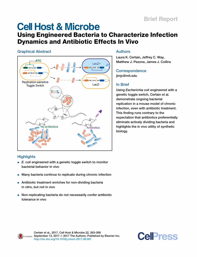
Using Escherichia coli engineered with a
genetic toggle switch, Certain et al.
demonstrate ongoing bacterial
replication in a mouse model of chronic
infection, even with antibiotic treatment.
This finding runs contrary to the
expectation that antibiotics preferentially
eliminate actively dividing bacteria and
highlights the in vivo utility of synthetic
biology.
Inc.
Cell Host & Microbe
Brief Report
Using Engineered Bacteria to CharacterizeInfection Dynamics and Antibiotic Effects In VivoLaura K. Certain,1,2 Jeffrey C. Way,1 Matthew J. Pezone,1 and James J. Collins1,3,4,5,6,7,8,*1Wyss Institute for Biologically Inspired Engineering, Harvard University, 3 Blackfan Circle, Boston, MA 02115, USA2Division of Infectious Diseases, Massachusetts General Hospital, 55 Fruit Street, Boston, MA 02114, USA3Institute for Medical Engineering and Science4Department of Biological Engineering5Synthetic Biology Center
MIT, Cambridge, MA 02139, USA6Harvard-MIT Program in Health Sciences and Technology, Cambridge, MA 02139, USA7Broad Institute of MIT and Harvard, 415 Main Street, Cambridge, MA 02142, USA8Lead Contact
*Correspondence: [email protected]
http://dx.doi.org/10.1016/j.chom.2017.08.001
SUMMARY
Synthetic biology has focused on engineering mi-crobes to synthesize useful products or to serve asliving diagnostics and therapeutics. Here we utilizea host-derived Escherichia coli strain engineeredwith a genetic toggle switch as a research tool toexamine in vivo replicative states in a mouse modelof chronic infection, and to compare in vivo andin vitro bacterial behavior. In contrast to the effectof antibiotics in vitro, we find that the fraction ofactively dividing bacteria remains relatively highthroughout the course of a chronic infection in vivoand increases in response to antibiotics. Moreover,the presence of non-dividing bacteria in vivo doesnot necessarily lead to an antibiotic-tolerant infec-tion, in contrast to expectations from in vitro experi-ments. These results demonstrate the utility of engi-neered bacteria for querying pathogen behaviorin vivo, and the importance of validating in vitrostudies of antibiotic effects with in vivo models.
Much of our understanding of how antibiotics affect bacteria
comes from in vitro work, but we do not always know how well
these findings translate into in vivo settings. Recent work using
fluorescent reporters has begun to examine the behavior of path-
ogens in animal models of infection (Claudi et al., 2014; Helaine
et al., 2014; Manina et al., 2015; Saliba et al., 2016). Synthetic
biology offers a complementary approach, in which synthetic
gene circuits can be constructed that register complex inputs,
such as a count of multiple events (Friedland et al., 2009) or
memory of a much earlier event (Kotula et al., 2014). In this pa-
per, we used Escherichia coli engineered with a genetic toggle
switch to determine the proportion of bacteria that are actively
dividing in a mouse model of chronic infection, so as to charac-
terize infection dynamics and antibiotic effects in vivo, as well as
to compare in vivo and in vitro bacterial behavior.
The E. coli strain PAS133 carries a bistable genetic toggle
switch in the form of an inducible lacZ element, such that upon
Cell Host & Microbe 22, 263–268, SepteThis is an open access article under the CC BY-N
exposure to anhydrotetracycline (ATC) the bacteria switch from
lacZ� to lacZ+, and remain lacZ+ even after ATC is removed
(Figure 1A) (Gardner et al., 2000; Kobayashi et al., 2004; Kotula
et al., 2014; Lee et al., 2016). The toggle switch is based on
the cI/Cro system from lambda phage, and at baseline is in the
cI (lacZ�) state. Because the switch from lacZ� to lacZ+ re-
quires the concentration of cI protein to fall by dilution, the design
of the circuit predicts that response to the ATC trigger requires
about three to four cell divisions (Kotula et al., 2014; Shea and
Ackers, 1985). Therefore, by treating a population of PAS133
cells with ATC, and then plating on indicator media to distinguish
lacZ� from lacZ+ cells, one can determine the proportion of cells
that were actively dividing at the time of ATC exposure. We first
confirmed this predicted behavior in vitro, and then used it to
study the behavior of E. coli PAS133 in amousemodel of chronic
orthopedic hardware infection.
To confirm in vitro that E. coli PAS133 switches from lacZ�to lacZ+ upon exposure to ATC during active growth, but not
during stationary phase or halted-growth conditions, we
tested the response to ATC under various growth conditions
and at various points of the growth curve. As predicted,
actively dividing bacteria rapidly switched from lacZ� to
lacZ+, but bacteria whose growth had been stopped by low
temperatures, lack of nutrients (culturing in saline), or reaching
stationary phase did not change from lacZ� to lacZ+ in the
presence of ATC (Figures 1B and 1C). Furthermore, bacteria
growing more slowly changed to lacZ+ more slowly (Figures
S1A and S1B), supporting the claim that the change to
lacZ+ state depends on cell division.
We next used this strain to explore the in vitro response of bac-
teria to antibiotics. Quinolones are more effective in vitro against
actively dividing bacteria (Zeiler, 1985), and therefore we pre-
dicted that treating with levofloxacin (a quinolone antibiotic)
would enrich the bacterial population for non-dividing cells.
Indeed, treatment with levofloxacin caused a decrease in the
proportion of bacteria that changed to lacZ+ upon exposure to
ATC (Figures 1D and 1E). Moreover, the increase in antibiotic
tolerance seen as PAS133 approached stationary phase corre-
lated with the decreasing response to ATC (Figure 1C), further
validating the toggle switch as a marker of cell division and
mber 13, 2017 ª 2017 The Authors. Published by Elsevier Inc. 263C-ND license (http://creativecommons.org/licenses/by-nc-nd/4.0/).
A
cI
cI
cItetR Cro
Cro LacZ
Cro
LacZCro
LacZ
ATC Actively dividing
Non-dividing
Baseline: LacZ OFF No response to ATC: LacZ OFF
Cell remembers ATC: LacZ ON
Toggle switch
Trigger
D E
No antibioticsLevofloxacin
B
ATC for 4 hours
>99% lacZ+ >99% lacZ- >95% lacZ-
Log phase Log phase in PBS
Log phaseon ice
>97% lacZ-
ATC 4,24 hrs
Stationary phase
100% LacZ+
38±3% LacZ+
0% LacZ+
C
Log reduction in cfu/ml after 24h levofloxacin
Time levofloxacin added
0 2 4 6 8 1010 5
10 6
10 7
10 8
10 9
10 10
Time (h)
cfu/
ml
59±5% LacZ+
0246810
Early log
Late log
Stationary
-1 0 1 2 3 40.0
0.40.7
0.8
0.9
1.0
Hours of ATC
Frac
tion
LacZ
+
-1 0 1 2 3 410 5
10 6
10 7
10 8
10 9
10 10
Hours of ATC
cfu/
ml
No antibioticsLevofloxacin
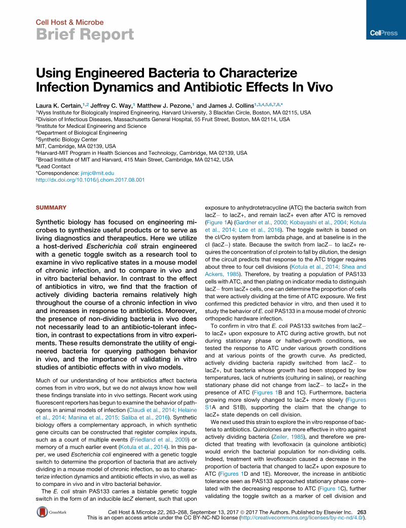
Figure 1. Engineered E. coli PAS133 Reports Bacterial Cell Division In Vitro
(A) Diagram of the genetic toggle switch and trigger. Exposure to ATC causes actively dividing E. coli PAS133 to switch to the Cro/lacZ+ state; non-dividing E. coli
PAS133 stay in the cI/lacZ� state.
(B) Schematic of the in vitro experiments. For log-phase bacteria, >7,000 colonies were observed over four independent experiments; two colonies were lacZ�.
For stationary-phase bacteria treated with ATC for 4 hr, >6,000 colonies were observed over four independent experiments; three were lacZ+. For stationary-
phase bacteria treated with ATC for 24 hr, 350 bacteria were observed (three biological replicates); seven were lacZ+ (2%). For the log-phase bacteria placed on
ice, 152 colonies were observed (three biological replicates); none were lacZ+. For the log-phase bacteria in PBS, 601 colonies were observed (six biological
replicates); 23 were lacZ+ (3.8%).
(C) Correlation of growth phase with response to ATC (1 hr of treatment at 1 mg/mL) and susceptibility to levofloxacin (24 hr of treatment at 500 ng/mL). Data
represent the mean and SD of six biological replicates (two independent experiments each with three biological replicates). The fraction lacZ+ at T = 5 is shown
separately for each experiment.
(D and E) Effect of low-dose levofloxacin (50 ng/mL) on bacterial population (D) and response to ATC (E) in vitro; mean and SD of three biological replicates. See
also Figure S1.
264 Cell Host & Microbe 22, 263–268, September 13, 2017
demonstrating the greater susceptibility of actively dividing bac-
teria to quinolones.
After confirming the behavior of E. coli PAS133 in vitro, we
used this strain to determine the growth status of bacteria in a
mousemodel of chronic orthopedic hardware infection (Figure 2)
and compared the in vivo behavior in this infection model to the
in vitro results. In humans, orthopedic hardware infections are
notoriously recalcitrant, often requiring surgical removal of the
infected device, as antibiotics alone do not reliably cure these in-
fections (Tande and Patel, 2014). This refractoriness to treatment
is typically explained by two hypotheses: the antibiotic cannot
get to the bacteria at adequate concentrations (due to biofilm
formation or sequestration inside cells) (Archer et al., 2011; Ka-
linka et al., 2014; Loffler et al., 2014; Tuchscherr et al., 2016),
and/or the bacteria are not actively replicating and are therefore
tolerant of antibiotic exposure (Allison et al., 2011; Cohen et al.,
2013; Conlon et al., 2016; Grant andHung, 2013).We usedE. coli
PAS133 to explore this second hypothesis—specifically, we
determined the proportion of bacteria in a mouse model of
chronic orthopedic hardware infection that were actively
dividing, and how treatment with levofloxacin affected this pro-
portion. Though the most common causes of orthopedic infec-
tions in humans are staphylococci (Tande and Patel, 2014),
E. coli can also cause device-associated infections (Zmistowski
et al., 2011), and we confirmed histologically that infection with
E. coli PAS133 causes an inflammatory response in this animal
model (Figures S2A and S2B).
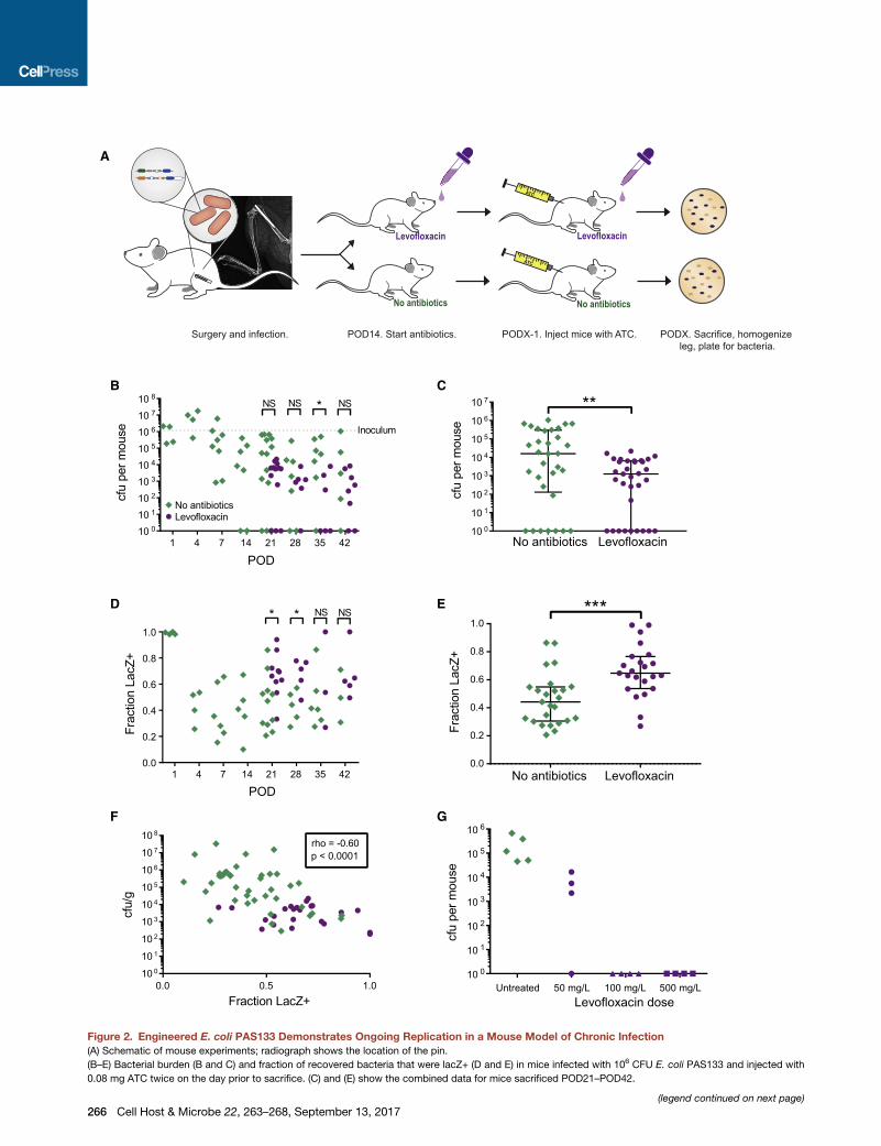
Mice had a plastic pin implanted in the femur, and PAS133
was inoculated into the surgical site during surgery (Figure 2A)
(Bernthal et al., 2010). Every week, a subset of mice was in-
jected with ATC and then euthanized the following day. Bacte-
ria from the infected legs were plated to determine bacterial
burden in the mouse and the fraction of the bacteria that
were lacZ+ (Figures 2B–2E). At the beginning of the infection
(first 24 hr), all of the bacteria changed from lacZ� to lacZ+
in response to ATC, i.e., all were actively dividing, as in early
log-phase growth in vitro. However, by post-operative day
four (POD4), the fraction of bacteria that responded to ATC
dropped to about half and remained at that proportion for the
course of the infection (Figure 2D). This result indicates that
by POD4 about half of the bacteria have entered a non-dividing
state. Interestingly, the corollary is that many of the bacteria
(approximately half of the population) continue to divide, even
6 weeks after the initial infection event. Since the bacterial pop-
ulation at the site of infection is not increasing, this ongoing cell
division is presumably balanced by bacterial death or clear-
ance by the immune system. These findings indicate that bac-
teria in a chronic infection do not mimic stationary phase bac-
teria in culture, even when the total bacterial population size
remains relatively constant.
To determine the effect of antibiotics on the response to ATC
in vivo, the infection was allowed to progress for 2 weeks, and
then half of the mice were started on levofloxacin. If levofloxacin
preferentially kills the actively dividing (ATC-responsive) bacte-
ria, the proportion of bacteria that respond to the ATC trigger
should decrease on antibiotic therapy, as we saw in vitro. Levo-
floxacin treatment did cause a decrease in total number of
bacteria recovered from the mouse (median colony-forming
units [CFU] 16,000 versus 1,300, p = 0.01 by Mann-Whitney
test; Figure 2C). However, the proportion of surviving bacteria
that responded to the ATC trigger failed to decrease, and in
fact increased from 44% to 65% (p = 0.0004, Mann-Whitney
test; Figure 2E). In other words, though mice on levofloxacin
had a lower bacterial burden at the site of infection, the antibiotic
treatment did not enrich for non-dividing bacteria and instead
enriched for actively dividing bacteria, in contrast to the in vitro
results (compare Figure 1E with Figure 2E).
One explanation for these results is that a subset of the bacte-
rial population in the mouse has evolved resistance and is there-
fore better able to replicate in the face of antibiotics. To test this
hypothesis, we screened bacteria isolated from the infected
mouse legs for antibiotic resistance. We determined that the
starting levofloxacin MIC (minimum inhibitory concentration) of
E. coli PAS133 was 50 ng/mL. We screened 366 E. coli colonies
frommice treated with levofloxacin for 1–4weeks (189 lacZ+ and
177 lacZ�), and none showed robust growth at levofloxacin
50 ng/mL (Figure S2C). These results indicate that the emer-
gence of resistance does not explain the enrichment of actively
dividing bacteria at an infection site during antibiotic treatment.
Additionally, these findings are in contrast to the relative ease
with which quinolone resistance emerges in vitro (Lee et al.,
2010; Ling et al., 2015).
Our results suggest that levofloxacin treatment does not pref-
erentially kill dividing bacteria in vivo, in contrast to its effects
in vitro. It is possible that some of the non-dividing bacteria in
the mouse switch into a dividing state, thus becoming suscepti-
ble to killing. The heterogeneity in the data is consistent with this
hypothesis, as we may be observing different states of a contin-
uously fluctuating ecosystem. It may even be that the reduction
in bacterial population by antibiotics is what triggers dormant
bacteria to begin dividing, as suggested by our finding that (for
time points from POD4 onward) the proportion of lacZ+ bacteria
was inversely correlated with bacterial burden in the mouse (Fig-
ure 2F). Of note, with a higher dose of levofloxacin we were able
to cure the mice (Figure 2G), indicating either that all of the bac-
teria divide at some point during the antibiotic treatment and the
lower dose just wasn’t reaching them all, or that the non-dividing
bacteria can be easily killed with a slight increase in antibiotics.
Either explanation runs counter to the typical perception of
chronic infections, in which quiescent, non-dividing bacteria
are presumed to be part of the population and to survive high-
dose antibiotic treatment. Importantly, our findings indicate
that the presence of non-dividing bacteria at an infection site
does not necessarily lead to an antibiotic-tolerant infection, in
contrast to expectations from in vitro experiments.
An assumption made by this study is that ATC is able to reach
all of the bacteria, and therefore the bacteria that fail to change to
lacZ+ are non-dividing, not simply unreachable by small mole-
cules. Because in some mice all the recovered bacteria had
changed to lacZ+, we are confident that the dose of ATC is
adequate to reach the infecting E. coli. However, it is still
possible that the bacteria that remain lacZ� reflect a seques-
tered population, rather than a non-dividing one (e.g., in a bio-
film). In theory, one could test this hypothesis by doing immuno-
histochemical staining for the beta-galactosidase enzyme and
determining if the lacZ+ bacteria are distributed differently in
the tissue from the lacZ� bacteria. Unfortunately, in this mouse
model the bacterial burden is too low for this technique to be
Cell Host & Microbe 22, 263–268, September 13, 2017 265
Surgery and infection. POD14. Start antibiotics.
ATC
PODX-1. Inject mice with ATC.
No antibiotics
Levofloxacin
PODX. Sacrifice, homogenize leg, plate for bacteria.
ATC
No antibiotics
Levofloxacin
A
D
B
E
No antibiotics Levofloxacin10 0
10 1
10 2
10 3
10 4
10 5
10 6
10 7
cfu
perm
ouse
**C
1 4 7 14 21 28 35 4210 0
10 1
10 2
10 3
10 4
10 5
10 6
10 7
10 8
POD
cfu
perm
ouse Inoculum
*NS NSNS
No antibioticsLevofloxacin
1 4 7 14 21 28 35 420.0
0.2
0.4
0.6
0.8
1.0
POD
Frac
tion
LacZ
+
* * NS NS
Untreated 50 mg/L 100 mg/L 500 mg/LLevofloxacin dose
F G
0.0 0.5 1.010 0
10 1
10 2
10 3
10 4
10 5
10 6
10 7
10 8
Fraction LacZ+
cfu/
g
rho = -0.60p < 0.0001
No antibiotics Levofloxacin0.0
0.2
0.4
0.6
0.8
1.0
Frac
tion
Lac Z
+
***
10 0
10 1
10 2
10 3
10 4
10 5
10 6
cfu
perm
ouse
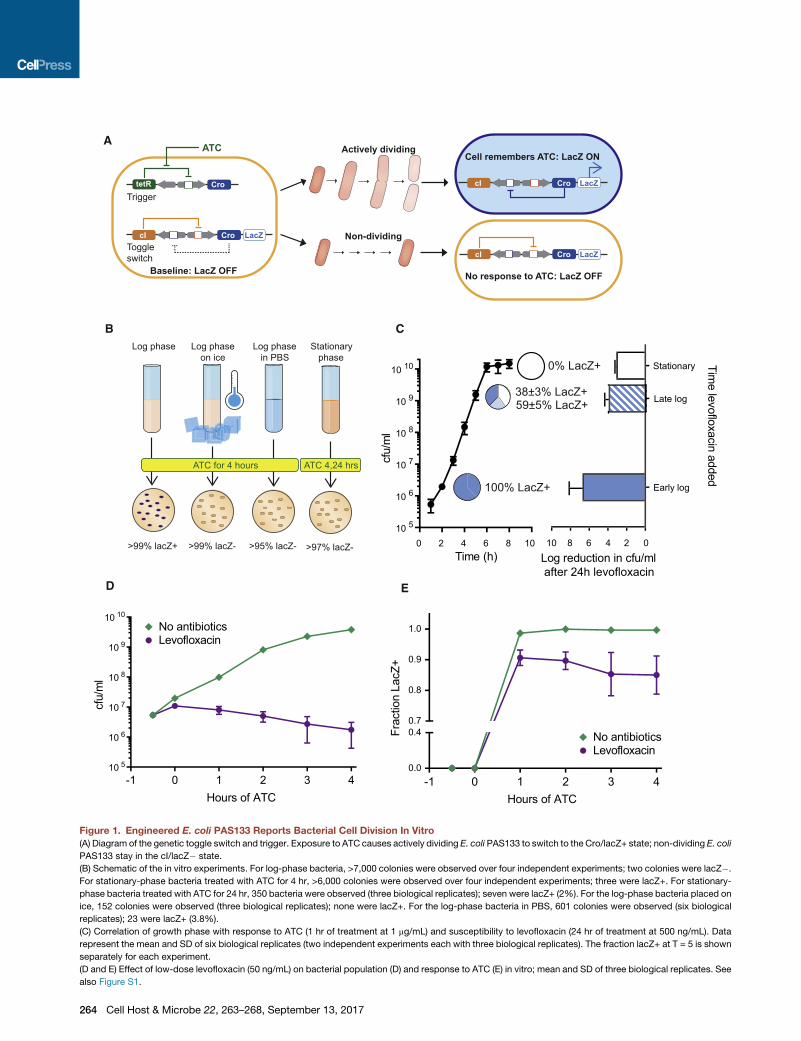
Figure 2. Engineered E. coli PAS133 Demonstrates Ongoing Replication in a Mouse Model of Chronic Infection
(A) Schematic of mouse experiments; radiograph shows the location of the pin.
(B–E) Bacterial burden (B and C) and fraction of recovered bacteria that were lacZ+ (D and E) in mice infected with 106 CFU E. coli PAS133 and injected with
0.08 mg ATC twice on the day prior to sacrifice. (C) and (E) show the combined data for mice sacrificed POD21–POD42.
(legend continued on next page)
266 Cell Host & Microbe 22, 263–268, September 13, 2017
useful (too few bacteria per histological section); however, in
other animal models of infection it could work well.
Our study demonstrates the value of synthetic biology tools for
probing bacterial behavior in vivo and illustrates that in vitro re-
sponses to antibiotic treatment do not always predict the in vivo
responses. The lacZ+/� output of the genetic toggle switch used
here is easily determined with standard microbiological tech-
niques, can be engineered in various bacteria, and is adaptable
to a variety of animal models of infection to study bacterial repli-
cation at the single-cell level. It requires no special equipment,
such as a confocal microscope or cell sorter. Much work in syn-
thetic biology to date has focused on engineering diagnostic or
therapeutic devices (Braff et al., 2016; Daeffler et al., 2017; Dan-
ino et al., 2015; Hwang et al., 2017; Kojima et al., 2016; Riglar
et al., 2017; Slomovic et al., 2015). We hope that future work
will expand the use of engineered organisms as research tools
for exploring the complexities of in vivo settings and the dynamic
actions of antibiotics and other therapeutic agents.
STAR+METHODS
Detailed methods are provided in the online version of this paper
and include the following:
d KEY RESOURCES TABLE
d CONTACT FOR REAGENT AND RESOURCE SHARING
d EXPERIMENTAL MODEL AND SUBJECT DETAILS
B Bacteria
B Experimental Animals
d METHOD DETAILS
B In Vitro Experiments
B In Vivo Experiments
d QUANTIFICATION AND STATISTICAL ANALYSIS
SUPPLEMENTAL INFORMATION
Supplemental Information includes two figures and can be found with this
article online at http://dx.doi.org/10.1016/j.chom.2017.08.001.
AUTHOR CONTRIBUTIONS
L.K.C. conceived the project, designed and performed the experiments, and
wrote the manuscript. J.C.W. contributed to project conception and edited
the manuscript. M.J.P. assisted with the animal experiments and reviewed
themanuscript. J.J.C. contributed to project conception, experimental design,
and preparation of the manuscript.
ACKNOWLEDGMENTS
The authors would like to acknowledge Richard Losick (Harvard University) for
providing lab space near the animal facility for preparation of bacteria; James
Weaver (Wyss Institute) for 3D printing the plastic pins for the mice; Amanda
Martinot (Harvard University) for assistance with the histology photos; Caleb
Bashor (MIT) for assistance with figure design; and Nadia Cohen, Arnaud Gu-
tierrez, Michael Lobritz, Rebecca Shapiro, Jonathan Stokes, and three anon-
(C) Bacterial burden in mice off (n = 33; 15 males and 18 females) and on (n = 34
(E) Fraction of recovered bacteria that were lacZ+ in mice off (n = 25; 10 males a
Bars in (C) and (E) indicate median and 25th/75th percentiles. NS, no significant
(F) Spearman correlation of bacterial burden (CFU/g) with the fraction of E. coli P
(G) Bacterial burden in mice sacrificed on POD21; treated for 1 week with 50, 10
See also Figure S2.
ymous reviewers for thoughtful comments on the manuscript. This work was
supported by the Paul G. Allen Frontiers Group, the Defense Threat Reduction
Agency grant HDTRA1-15-1-0051, and the Wyss Institute for Biologically
Inspired Engineering. J.J.C. is scientific co-founder and SAB chair of EnBiotix,
which is an antibiotics startup company. J.C.W. is an inventor on a patent
application that relates to the genetically engineered system used in this work.
Received: January 24, 2017
Revised: May 17, 2017
Accepted: August 1, 2017
Published: August 31, 2017
REFERENCES
Ackers, G.K., Johnson, A.D., and Shea, M.A. (1982). Quantitative model for
gene regulation by lambda phage repressor. Proc. Natl. Acad. Sci. USA 79,
1129–1133.
Allison, K.R., Brynildsen, M.P., and Collins, J.J. (2011). Metabolite-enabled
eradication of bacterial persisters by aminoglycosides. Nature 473, 216–220.
Archer, N.K., Mazaitis, M.J., Costerton, J.W., Leid, J.G., Powers, M.E., and
Shirtliff, M.E. (2011). Staphylococcus aureus biofilms: properties, regulation,
and roles in human disease. Virulence 2, 445–459.
Bachmanov, A.A., Reed, D.R., Beauchamp, G.K., and Tordoff, M.G. (2002).
Food intake, water intake, and drinking spout side preference of 28 mouse
strains. Behav. Genet. 32, 435–443.
Bernthal, N.M., Stavrakis, A.I., Billi, F., Cho, J.S., Kremen, T.J., Simon, S.I.,
Cheung, A.L., Finerman, G.A., Lieberman, J.R., Adams, J.S., and Miller, L.S.
(2010). Amousemodel of post-arthroplasty Staphylococcus aureus joint infec-
tion to evaluate in vivo the efficacy of antimicrobial implant coatings. PLoSOne
5, e12580.
Braff, D., Shis, D., and Collins, J.J. (2016). Synthetic biology platform technol-
ogies for antimicrobial applications. Adv. Drug Deliv. Rev. 105 (Pt A), 35–43.
Claudi, B., Sprote, P., Chirkova, A., Personnic, N., Zankl, J., Sch€urmann, N.,
Schmidt, A., and Bumann, D. (2014). Phenotypic variation of Salmonella in
host tissues delays eradication by antimicrobial chemotherapy. Cell 158,
722–733.
Cohen, N.R., Lobritz, M.A., and Collins, J.J. (2013). Microbial persistence and
the road to drug resistance. Cell Host Microbe 13, 632–642.
Conlon, B.P., Rowe, S.E., Gandt, A.B., Nuxoll, A.S., Donegan, N.P., Zalis, E.A.,
Clair, G., Adkins, J.N., Cheung, A.L., and Lewis, K. (2016). Persister formation
in Staphylococcus aureus is associated with ATP depletion. Nat. Microbiol.
1, 16051.
Daeffler, K.N., Galley, J.D., Sheth, R.U., Ortiz-Velez, L.C., Bibb, C.O., Shroyer,
N.F., Britton, R.A., and Tabor, J.J. (2017). Engineering bacterial thiosulfate and
tetrathionate sensors for detecting gut inflammation. Mol. Syst. Biol. 13, 923.
Danino, T., Prindle, A., Kwong, G.A., Skalak, M., Li, H., Allen, K., Hasty, J., and
Bhatia, S.N. (2015). Programmable probiotics for detection of cancer in urine.
Sci. Transl. Med. 7, 289ra84.
Friedland, A.E., Lu, T.K., Wang, X., Shi, D., Church, G., and Collins, J.J. (2009).
Synthetic gene networks that count. Science 324, 1199–1202.
Gardner, T.S., Cantor, C.R., and Collins, J.J. (2000). Construction of a genetic
toggle switch in Escherichia coli. Nature 403, 339–342.
Gottesman, S. (2003). Proteolysis in bacterial regulatory circuits. Annu. Rev.
Cell Dev. Biol. 19, 565–587.
Grant, S.S., and Hung, D.T. (2013). Persistent bacterial infections, antibiotic
tolerance, and the oxidative stress response. Virulence 4, 273–283.
; 11 males and 23 females) 50 mg/L levofloxacin.
nd 15 females) and on (n = 23; 8 males and 15 females) 50 mg/L levofloxacin.
difference; *p < 0.05, **p = 0.005, ***p = 0.0004 by Mann-Whitney test.
AS133 that were lacZ+ from POD4 onward (n = 63; 18 males and 45 females).
0, or 500 mg/L levofloxacin; or left untreated.
Cell Host & Microbe 22, 263–268, September 13, 2017 267
Helaine, S., Cheverton, A.M., Watson, K.G., Faure, L.M., Matthews, S.A., and
Holden, D.W. (2014). Internalization of Salmonella by macrophages induces
formation of nonreplicating persisters. Science 343, 204–208.
Hwang, I.Y., Koh, E., Wong, A., March, J.C., Bentley, W.E., Lee, Y.S., and
Chang, M.W. (2017). Engineered probiotic Escherichia coli can eliminate and
prevent Pseudomonas aeruginosa gut infection in animal models. Nat.
Commun. 8, 15028.
Kalinka, J., Hachmeister, M., Geraci, J., Sordelli, D., Hansen, U., Niemann,
S., Oetermann, S., Peters, G., Loffler, B., and Tuchscherr, L. (2014).
Staphylococcus aureus isolates from chronic osteomyelitis are character-
ized by high host cell invasion and intracellular adaptation, but still induce
inflammation. Int. J. Med. Microbiol. 304, 1038–1049.
Kobayashi, H., Kaern, M., Araki, M., Chung, K., Gardner, T.S., Cantor, C.R.,
and Collins, J.J. (2004). Programmable cells: interfacing natural and engi-
neered gene networks. Proc. Natl. Acad. Sci. USA 101, 8414–8419.
Kojima, R., Aubel, D., and Fussenegger, M. (2016). Toward a world of thera-
nostic medication: programming biological sentinel systems for therapeutic
intervention. Adv. Drug Deliv. Rev. 105 (Pt A), 66–76.
Kotula, J.W., Kerns, S.J., Shaket, L.A., Siraj, L., Collins, J.J., Way, J.C., and
Silver, P.A. (2014). Programmable bacteria detect and record an environ-
mental signal in the mammalian gut. Proc. Natl. Acad. Sci. USA 111,
4838–4843.
Lee, H.H., Molla, M.N., Cantor, C.R., and Collins, J.J. (2010). Bacterial charity
work leads to population-wide resistance. Nature 467, 82–85.
Lee, J.W., Gyorgy, A., Cameron, D.E., Pyenson, N., Choi, K.R., Way, J.C.,
Silver, P.A., Del Vecchio, D., and Collins, J.J. (2016). Creating single-copy ge-
netic circuits. Mol. Cell 63, 329–336.
Ling, L.L., Schneider, T., Peoples, A.J., Spoering, A.L., Engels, I., Conlon, B.P.,
Mueller, A., Sch€aberle, T.F., Hughes, D.E., Epstein, S., et al. (2015). A new anti-
biotic kills pathogens without detectable resistance. Nature 517, 455–459.
Loessner, H., Leschner, S., Endmann, A., Westphal, K., Wolf, K., Kochruebe,
K., Miloud, T., Altenbuchner, J., and Weiss, S. (2009). Drug-inducible remote
control of gene expression by probiotic Escherichia coli Nissle 1917 in intes-
tine, tumor and gall bladder of mice. Microbes Infect. 11, 1097–1105.
Loffler, B., Tuchscherr, L., Niemann, S., and Peters, G. (2014). Staphylococcus
aureus persistence in non-professional phagocytes. Int. J. Med. Microbiol.
304, 170–176.
Manina, G., Dhar, N., and McKinney, J.D. (2015). Stress and host immunity
amplify Mycobacterium tuberculosis phenotypic heterogeneity and induce
nongrowing metabolically active forms. Cell Host Microbe 17, 32–46.
268 Cell Host & Microbe 22, 263–268, September 13, 2017
Marx, J.O., Vudathala, D., Murphy, L., Rankin, S., and Hankenson, F.C. (2014).
Antibiotic administration in the drinking water of mice. J. Am. Assoc. Lab.
Anim. Sci. 53, 301–306.
Osmon, D.R., Berbari, E.F., Berendt, A.R., Lew, D., Zimmerli, W., Steckelberg,
J.M., Rao, N., Hanssen, A., and Wilson, W.R.; Infectious Diseases Society of
America (2013). Diagnosis and management of prosthetic joint infection: clin-
ical practice guidelines by the Infectious Diseases Society of America. Clin.
Infect. Dis. 56, e1–e25.
Pakula, A.A., Young, V.B., and Sauer, R.T. (1986). Bacteriophage lambda cro
mutations: effects on activity and intracellular degradation. Proc. Natl. Acad.
Sci. USA 83, 8829–8833.
Riglar, D.T., Giessen, T.W., Baym, M., Kerns, S.J., Niederhuber, M.J.,
Bronson, R.T., Kotula, J.W., Gerber, G.K., Way, J.C., and Silver, P.A. (2017).
Engineered bacteria can function in the mammalian gut long-term as live diag-
nostics of inflammation. Nat. Biotechnol. 35, 653–658.
Saliba, A.E., Li, L., Westermann, A.J., Appenzeller, S., Stapels, D.A., Schulte,
L.N., Helaine, S., and Vogel, J. (2016). Single-cell RNA-seq ties macrophage
polarization to growth rate of intracellular Salmonella. Nat. Microbiol. 2, 16206.
Shea, M.A., and Ackers, G.K. (1985). The OR control system of bacteriophage
lambda. A physical-chemical model for gene regulation. J. Mol. Biol. 181,
211–230.
Slomovic, S., Pardee, K., and Collins, J.J. (2015). Synthetic biology devices for
in vitro and in vivo diagnostics. Proc. Natl. Acad. Sci. USA 112, 14429–14435.
Tande, A.J., and Patel, R. (2014). Prosthetic joint infection. Clin. Microbiol. Rev.
27, 302–345.
Tuchscherr, L., Kreis, C.A., Hoerr, V., Flint, L., Hachmeister, M., Geraci, J.,
Bremer-Streck, S., Kiehntopf, M., Medina, E., Kribus, M., et al. (2016).
Staphylococcus aureus develops increased resistance to antibiotics by form-
ing dynamic small colony variants during chronic osteomyelitis. J. Antimicrob.
Chemother. 71, 438–448.
Wiegand, I., Hilpert, K., and Hancock, R.E. (2008). Agar and broth dilution
methods to determine the minimal inhibitory concentration (MIC) of antimicro-
bial substances. Nat. Protoc. 3, 163–175.
Zeiler, H.J. (1985). Evaluation of the in vitro bactericidal action of ciprofloxacin
on cells of Escherichia coli in the logarithmic and stationary phases of growth.
Antimicrob. Agents Chemother. 28, 524–527.
Zmistowski, B., Fedorka, C.J., Sheehan, E., Deirmengian, G., Austin, M.S., and
Parvizi, J. (2011). Prosthetic joint infection caused by gram-negative organ-
isms. J. Arthroplasty 26 (6 Suppl), 104–108.
STAR+METHODS
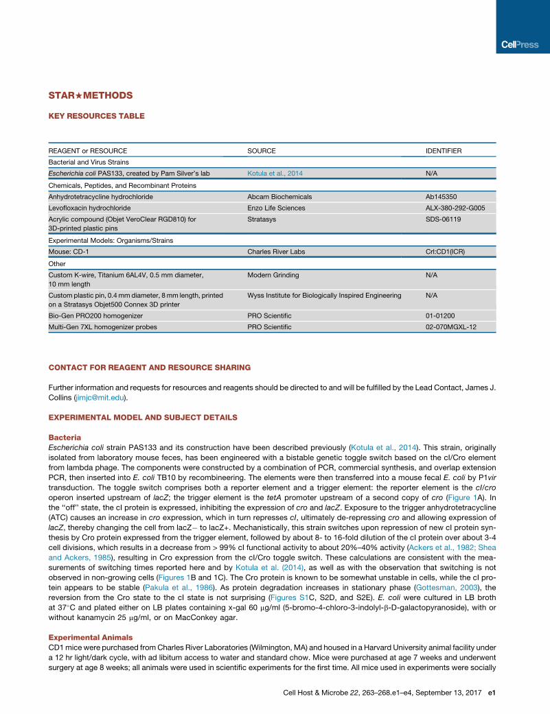
KEY RESOURCES TABLE
REAGENT or RESOURCE SOURCE IDENTIFIER
Bacterial and Virus Strains
Escherichia coli PAS133, created by Pam Silver’s lab Kotula et al., 2014 N/A
Chemicals, Peptides, and Recombinant Proteins
Anhydrotetracycline hydrochloride Abcam Biochemicals Ab145350
Levofloxacin hydrochloride Enzo Life Sciences ALX-380-292-G005
Acrylic compound (Objet VeroClear RGD810) for
3D-printed plastic pins
Stratasys SDS-06119
Experimental Models: Organisms/Strains
Mouse: CD-1 Charles River Labs Crl:CD1(ICR)
Other
Custom K-wire, Titanium 6AL4V, 0.5 mm diameter,
10 mm length
Modern Grinding N/A
Custom plastic pin, 0.4mmdiameter, 8mm length, printed
on a Stratasys Objet500 Connex 3D printer
Wyss Institute for Biologically Inspired Engineering N/A
Bio-Gen PRO200 homogenizer PRO Scientific 01-01200
Multi-Gen 7XL homogenizer probes PRO Scientific 02-070MGXL-12
CONTACT FOR REAGENT AND RESOURCE SHARING
Further information and requests for resources and reagents should be directed to and will be fulfilled by the Lead Contact, James J.
Collins ([email protected]).
EXPERIMENTAL MODEL AND SUBJECT DETAILS
BacteriaEscherichia coli strain PAS133 and its construction have been described previously (Kotula et al., 2014). This strain, originally
isolated from laboratory mouse feces, has been engineered with a bistable genetic toggle switch based on the cI/Cro element
from lambda phage. The components were constructed by a combination of PCR, commercial synthesis, and overlap extension
PCR, then inserted into E. coli TB10 by recombineering. The elements were then transferred into a mouse fecal E. coli by P1vir
transduction. The toggle switch comprises both a reporter element and a trigger element: the reporter element is the cI/cro
operon inserted upstream of lacZ; the trigger element is the tetA promoter upstream of a second copy of cro (Figure 1A). In
the ‘‘off’’ state, the cI protein is expressed, inhibiting the expression of cro and lacZ. Exposure to the trigger anhydrotetracycline
(ATC) causes an increase in cro expression, which in turn represses cI, ultimately de-repressing cro and allowing expression of
lacZ, thereby changing the cell from lacZ� to lacZ+. Mechanistically, this strain switches upon repression of new cI protein syn-
thesis by Cro protein expressed from the trigger element, followed by about 8- to 16-fold dilution of the cI protein over about 3-4
cell divisions, which results in a decrease from > 99% cI functional activity to about 20%–40% activity (Ackers et al., 1982; Shea
and Ackers, 1985), resulting in Cro expression from the cI/Cro toggle switch. These calculations are consistent with the mea-
surements of switching times reported here and by Kotula et al. (2014), as well as with the observation that switching is not
observed in non-growing cells (Figures 1B and 1C). The Cro protein is known to be somewhat unstable in cells, while the cI pro-
tein appears to be stable (Pakula et al., 1986). As protein degradation increases in stationary phase (Gottesman, 2003), the
reversion from the Cro state to the cI state is not surprising (Figures S1C, S2D, and S2E). E. coli were cultured in LB broth
at 37�C and plated either on LB plates containing x-gal 60 mg/ml (5-bromo-4-chloro-3-indolyl-b-D-galactopyranoside), with or
without kanamycin 25 mg/ml, or on MacConkey agar.
Experimental AnimalsCD1mice were purchased fromCharles River Laboratories (Wilmington, MA) and housed in a Harvard University animal facility under
a 12 hr light/dark cycle, with ad libitum access to water and standard chow. Mice were purchased at age 7 weeks and underwent
surgery at age 8 weeks; all animals were used in scientific experiments for the first time. All mice used in experiments were socially
Cell Host & Microbe 22, 263–268.e1–e4, September 13, 2017 e1
housed, except for the first three days after surgery, when they were housed individually. The animal protocol was approved by the
Institutional Animal Care and Use Committee (IACUC) of Harvard University Faculty of Arts and Sciences, and all animal experiments
were performed in accordance with the NIH Guide for the Care and Use of Laboratory Animals. Both sexes of mice were used, with
the exact numbers of each sex in each experiment indicated below or in the figure legends. Female mice were preferred, due to ease
of long-term cohabitation; however, male mice were also included in keeping with new guidelines requiring the use of both sexes in
animal experiments. The study was not designed or powered to detect differences in outcome between the sexes, as the focus was
on the behavior of the bacteria, not themice; however, no differences were noted. Mice were assigned to antibiotic treatment groups
at the time of surgery. Theywere not randomized, but rather were assigned to groups (antibiotics versus no antibiotics) such that each
group had mice from each day of surgery (for experiments involving more than 12 mice, which took place over multiple days). There
was no blinding with regards to treatment group, as researchers were responsible for administering the antibiotic treatment. After
recovering from the surgery, the mice appeared healthy throughout the course of the experiment; they had appropriate weight
gain, were well-groomed, and moved about the cage without difficulty.
METHOD DETAILS
In Vitro ExperimentsEffect of Growth Phase
To characterize the behavior of E. coli PAS133 in vitro (Figure 1B), bacteria were grown overnight in LB. For characterization of sta-
tionary-phase bacteria, these overnight cultures were used without further adjustment. For characterization of actively dividing bac-
teria, the overnight culture was diluted 1:1000 in fresh, pre-warmed LB media and incubated with agitation at 37�C for two hours.
Response to ATCwas tested by adding 1 mg/ml ATC to the bacterial cultures and incubating for four hours. Stationary-phase bacteria
were incubated for 24 hr in addition to four hours. Alternatively, log-phase cultures (i.e., those that had been diluted and then cultured
for two hours) were placed on ice for 3-4 hr, or re-suspended in phosphate buffered saline (PBS) for 3-4 hr, prior to the addition
of ATC.
To correlate growth phase with response to ATC and with susceptibility to levofloxacin (Figure 1C), overnight cultures were diluted
1:10,000 in pre-warmed LB at time 0 and put in a 37�C shaker. Every hour, spot dilutions were made for quantification of cfu/ml. At
times 2, 5, and 8 hr, 1 mL samples were removed from the culture (2 samples per culture flask) and transferred to culture tubes. Levo-
floxacin or ATC was added, to a final concentration of 500 ng/ml or 1 mg/ml, respectively. These treated samples were cultured at
37�C with agitation. After one hour (ATC-treated samples) or 24 hr (levofloxacin-treated samples), they were removed from the incu-
bator. The ATC-treated samples were diluted and plated on LB-kan-x-gal plates for quantification of fraction lacZ+. The levofloxacin-
treated samples were centrifuged, media removed, and re-suspended in an equal volume of PBS before making spot dilutions for cfu
quantification. For the early-log samples, instead of spot dilutions the entire sample was plated. The degree of killing was calculated
by comparing the cfu/ml at the time levofloxacin was added to the cfu/ml after 24 hr of levofloxacin treatment. For the early-log sam-
ples, five of six replicates had no remaining bacteria, and therefore for the purposes of calculating the logs of killing we used a value of
0.1 cfu/ml. The remaining sample had about 200 cfu/ml.
Effect of Antibiotic Treatment
To characterize the effect of levofloxacin on the behavior of E. coli in vitro (Figures 1D and 1E), overnight cultures were diluted 1:1000
in LB and incubated at 37�C with agitation for 90 min, at which point levofloxacin was added, to a final concentration of 50 ng/ml (at
the MIC). Thirty minutes later, ATC was added, to a final concentration of 1 mg/ml. Cells were plated every hour for quantification of
cfu/ml and fraction lacZ+.
Effect of Growth Rate
To determine the effect of different growth media on PAS133 (Figures S1A and S1B), overnight cultures were rinsed twice with PBS
and resuspended in M9 media with 0.5% casamino acids. They were then diluted 1:1000 in M9-casamino acids with a variable
amount of dextrose (0.3, 3, or 30 mM), or in LB. They were grown at 37�C for two hours, then induced with ATC (time 0). To measure
the rate of return to the lacZ� state once the bacteria reached stationary phase (Figure S1C), log-phase bacteria were induced with
ATC for four hours, then rinsed and resuspended in LB. They were diluted 1:10 at time 0 (resulting concentration 23 108 cfu/ml) then
monitored for the loss of the lacZ+ state over time.
Determining Minimum Inhibitory Concentration
We determined the MIC of E. coli PAS133 by standard liquid media techniques (Wiegand et al., 2008). Briefly, bacteria were
grown overnight, then diluted 1:1000 in fresh media containing two-fold serial dilutions of antibiotic. These bacterial cultures
were incubated in a 96-well microplate at 37�C, with agitation, for 24 hr and then observed for growth. The lowest concentration
that inhibited visible growth was taken as the MIC, which for E. coli PAS133 was 50 ng/ml. We screened the bacteria recovered
from the mice for antibiotic resistance by growing cultures in a 96-well microplate for �6 hr and diluting 1:30, and then pinning
the diluted cultures onto LB agar plates containing levofloxacin 50 ng/ml. If there were a meaningful increase in MIC then we
would expect the bacteria to demonstrate robust growth at this concentration. As a control, we pinned onto plates containing
no antibiotic to confirm technique; we pinned first onto the antibiotic plate and then onto the no-antibiotic plate without reloading
the pins.
e2 Cell Host & Microbe 22, 263–268.e1–e4, September 13, 2017
In Vivo ExperimentsSurgical Technique
Eight-week-old CD1 mice underwent surgery as outlined by Bernthal et al. (2010). Briefly, mice were anesthetized with isoflurane,
prepped and draped for surgery in a sterile fashion, and a 1 cm pin was inserted retrograde into the intramedullary canal. For the
purposes of the radiograph in Figure 2A, the pin was made of titanium (manufactured by Modern Grinding, Port Washington, WI).
For mice infected with E. coli, it was made of biocompatible plastic (3D-printed at the Wyss Institute, Harvard University). The
pins were 3D-printed attached to a solid base, with 12 pins per block. Just prior to surgery, the pins were sterilized by immersion
of the block in 95% ethanol, then detached from the block using sterile scissors. After pin insertion, the end of the pin was inoculated
with 106 cfu E. coliPAS133. Bacterial counts were confirmed by plating 104-fold dilutions of the infecting stock, incubating at 37�C for
24 hr, and counting cfu.
Antibiotic Treatment of Mice
For mice treated with levofloxacin, treatment began on post-operative day (POD) 14 and was administered in the drinking water at a
concentration of 50 mg/L (unless otherwise specified). The antibiotic-treated water was changed twice per week. This antibiotic was
chosen because it had a low MIC for the target strains and is commonly used to treat human infections with E. coli (Osmon et al.,
2013). In addition, it has excellent oral bioavailability and is water soluble, facilitating the administration of antibiotics via the drinking
water (Marx et al., 2014). To determine the best dose of levofloxacin to use (Figure 2G), we gave four mice levofloxacin 50 mg/L, four
mice levofloxacin 100 mg/L, four mice levofloxacin 500 mg/L, and left five mice untreated; all 17 of these mice were female. All the
mice treated with 100mg/L or 500mg/L were cured; one of themice treated with levofloxacin 50mg/L was cured; none of the control
mice were cured. We therefore used the lowest concentration of levofloxacin for E. coli-infected mice because it reduced the
bacterial burden in the mouse but did not cure the infection. Levofloxacin 50 mg/L provides an estimated dose of 10 mg/kg/day
(Bachmanov et al., 2002), which is comparable to human dosing.
Administering Anhydrotetracycline
Mice infectedwith E. coliPAS133were injectedwith ATC 0.08mg intraperitoneally (IP) twice on the day prior to sacrifice unless other-
wise indicated in the results (Loessner et al., 2009); the doses were given approximately 8 hr apart, and the second dose preceded
euthanasia by at least 16 hr. By waiting 16 hr from the second dose, we ensured that there was not enough residual ATC in the tissues
to cause the bacteria to switch on the plate. This was validated by injecting uninfected mice (n = 3 female) with ATC, sacrificing 16 hr
after the second dose, homogenizing their legs, then mixing the homogenates with log-phase E. coli PAS133 prior to plating on in-
dicator media; none of the resulting colonies were lacZ+. In contrast, if we sacrificed mice (n = 4 female) only four hours after a single
dose of ATC, homogenized the legs, and mixed the homogenates with log-phase E. coli PAS133, then all the colonies were lacZ+.
Likewise, if we injected with 0.24mg ATC x 2, rather than 0.08mg, sacrificed 16 hr after the second dose, homogenized the legs, then
mixed the homogenates with E. coli PAS133, about 20% of colonies were lacZ+ (n = 1 male, 1 female; 57 blue cfu out of 252 total for
one, 55 out of 265 for the other). We did not wait longer than a day after ATC injection because the lacZ+ state slowly reverts to lacZ�in themouse (Figure S2D, n = 9 female; Figure S2E, n = 17 female, 14male). To confirm that the E. coli did not to switch to lacZ+ in the
absence of ATC treatment, we sacrificedmice (n = 4males, 3 females) on POD20 without administering ATC; no colonies were lacZ+
(> 1000 cfu observed from 2 mice on levofloxacin and 5 mice not on antibiotics).
Processing of Mouse Tissue
After euthanasia, the mouse’s infected hind limb was drenched with iodine then removed at the hip with sterile scissors and forceps.
The skin was removed as well as the distal leg (from themid-tibia down). The entire remaining leg – bone, muscle, cartilage, pin – was
morcellated with scissors then homogenized in 3 mL sterile phosphate buffered saline (PBS). The volume of the homogenate (usually
about 4.0ml) was recorded to allow calculation of the total bacterial burden in themouse leg. Serial dilutions of the homogenate were
plated onto LB agar containing 60 mg/ml x-gal, with or without kanamycin, and incubated at 37�C for 24 hr.
To confirm that the bacteria recovered from the mice were PAS133, we screened a subset of the colonies recovered from each
mouse for kanamycin resistance and response to ATC. (Kanamycin is the selectable marker in the toggle switch.) We screened
763 colonies from 38 mice, and found 48 colonies from eight mice that were kanamycin sensitive. Data from these eight mice
were not included in the results, and we subsequently plated the leg homogenates on LB-x-gal agar containing kanamycin, to avoid
skewing the results with contaminating bacteria. We additionally screened 189 kanamycin-resistant lacZ- colonies from 19 mice for
response to ATC; all 189 flipped to lacZ+ upon exposure to ATC. For mice from whom fewer than five colonies of bacteria were
recovered (n = 5 mice), the fraction that were lacZ+ was not included in the results, as it is too few colonies to reflect an accurate
percentage.
Histology
Mice infected with E. coli PAS133, or mice that underwent surgery but were inoculated with sterile liquid, were sacrificed on POD23.
Their legs were fixed in formalin for 72 hr, then transferred to 70% ethanol and transferred to the Rodent Histopathology Core at Har-
vard Medical School. There, they were decalcified in EDTA, sliced along the sagittal plane, embedded in paraffin, and sectioned.
QUANTIFICATION AND STATISTICAL ANALYSIS
Initial estimates of how many mice would be needed per group to see a significant difference were based on prior work using this
mouse model (Bernthal et al., 2010). All p-values are two-sided. For comparing groups, we used the Mann-Whitney test rather
than a t test, due to small sample size/non-normal distribution. For the in vivo experiments, visual inspection of the data suggested
Cell Host & Microbe 22, 263–268.e1–e4, September 13, 2017 e3
that there was no time-dependent effect of antibiotics. That is, the bacterial burden of mice on antibiotics was the same regardless of
the length of antibiotic treatment; this impression was confirmed by finding no difference by either the Mann-Whitney test or the
Kolmogorov-Smirnov test for any pairwise comparison of time-points from POD21 onward for mice within the same treatment group.
We therefore combined data for POD21-42 to increase power to detect a difference between treated and untreated mice. Data from
each time-point may be seen in Figures 2B and 2D.
e4 Cell Host & Microbe 22, 263–268.e1–e4, September 13, 2017