vatech imaging systems - cloudinary · vatech will provide a trade in value for your 3d imaging...
TRANSCRIPT

2 Contents
About Vatech America
Vatech Assurance
PaX-i
PaX-i Insight
i3D Smart
PaX-i3D
Green CT
Green CT 2
i3D Premium
Ez3D-i
EzDent-i
EzRay Air Wall
HD Sensor
3
4
6
12
16
20
26
32
36
42
45
46
50
VATECH IMAGING SYSTEMS

Vatech is a leading manufacturer of radiographic imaging solutions for the medical, dental, and veterinary �elds with o�ces in over 70 countries worldwide.
2005 2007 2008 2009 2013
Launched World's First3 in 1 Digital X-ray System :
Picasso-Trio
Launched World's FirstAuto-Switching System :
PaX-Duo3D
World's FirstOne Shot Cephalometric :
PaX-Uni3D
World's FirstFree FOV System : PaX-Reve3D
GroundbreakingLow Radiation System:
Green CT
2017
41-Layer DigitalPanoramic Radiograph
PaX-i Insight
About Vatech America 3
As the US subsidiary of Vatech Inc, Vatech America is committed to providing the industry with innovative dental x-ray imaging solutions while maintaining a primary focus on ultimately enhancing the quality of patient care.
From the world’s �rst 3-in-1 digital x-ray system to the latest in high resolution, low radiation CBCT devices, Vatech America is the clinician's preferred vendor for their diagnostic imaging endeavors.
MISSION: With honesty and integrity, we strive to be the industry leader and preferred partner by providing innovative imaging solutions and �rst class client services that ultimately enhance the quality of patient care.

What is Vatech Assurance?
Vatech Assurance, a core value of Value Added Technologies, is our promise to our customers that we
will not only provide a premium dental imaging system to suit your needs, but also to ensure the value
of your investment from becoming obsolete by providing multiple options for upgrading to the newest
technologies as they are developed. Whether it’s a defined transition strategy to newer technologies, or
protection of currently owned technology, Vatech Assurance provides industry leading service, support,
warranty coverage, as well as added value that reaches beyond the x-ray itself.
No Asterisks | No Legalese | Just Vatech Assurance
4 Vatech Assurance
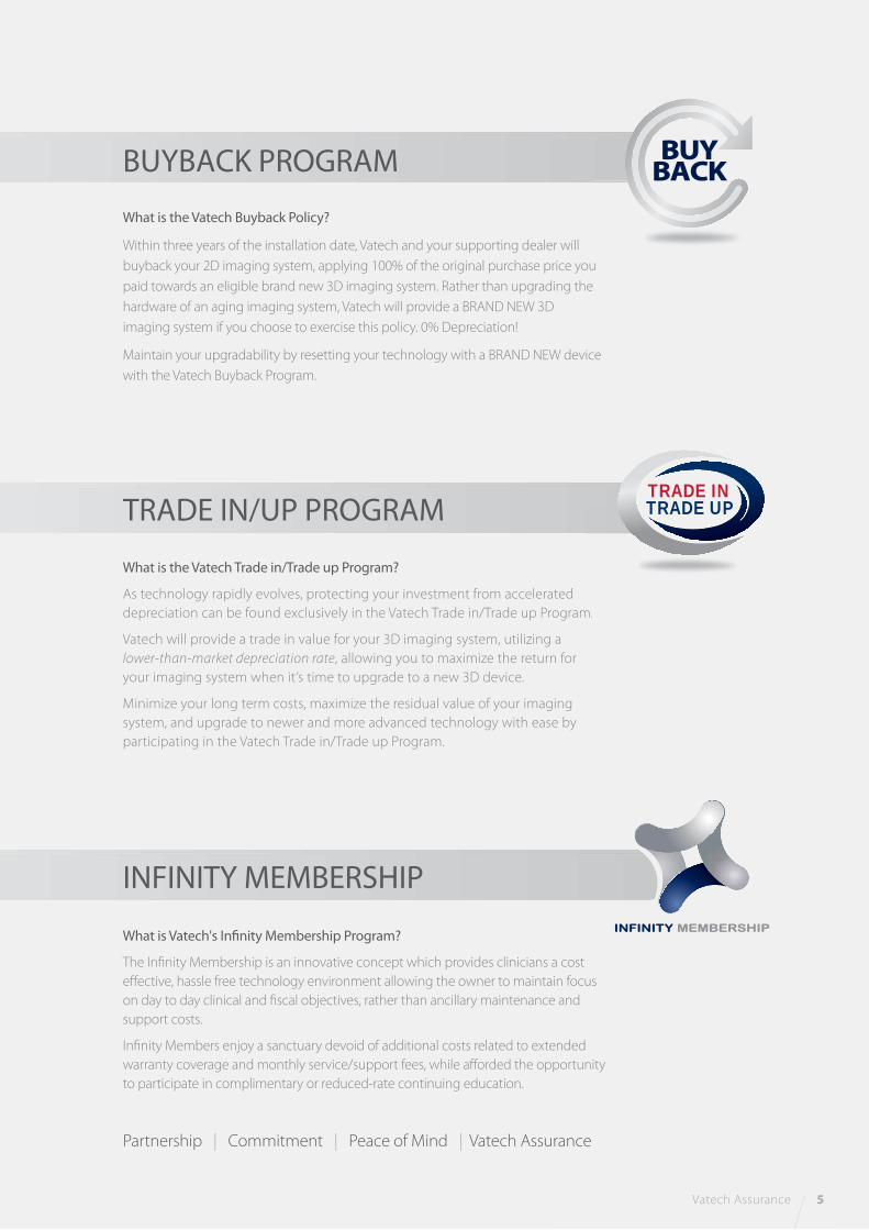
What is Vatech's Infinity Membership Program?
The Infinity Membership is an innovative concept which provides clinicians a cost
effective, hassle free technology environment allowing the owner to maintain focus
on day to day clinical and fiscal objectives, rather than ancillary maintenance and
support costs.
Infinity Members enjoy a sanctuary devoid of additional costs related to extended
warranty coverage and monthly service/support fees, while afforded the opportunity
to participate in complimentary or reduced-rate continuing education.
What is the Vatech Buyback Policy?
Within three years of the installation date, Vatech and your supporting dealer will
buyback your 2D imaging system, applying 100% of the original purchase price you
paid towards an eligible brand new 3D imaging system. Rather than upgrading the
hardware of an aging imaging system, Vatech will provide a BRAND NEW 3D
imaging system if you choose to exercise this policy. 0% Depreciation!
Maintain your upgradability by resetting your technology with a BRAND NEW device
with the Vatech Buyback Program.
What is the Vatech Trade in/Trade up Program?
As technology rapidly evolves, protecting your investment from accelerated
depreciation can be found exclusively in the Vatech Trade in/Trade up Program.
Vatech will provide a trade in value for your 3D imaging system, utilizing a
lower-than-market depreciation rate, allowing you to maximize the return for
your imaging system when it’s time to upgrade to a new 3D device.
Minimize your long term costs, maximize the residual value of your imaging
system, and upgrade to newer and more advanced technology with ease by
participating in the Vatech Trade in/Trade up Program.
Vatech Assurance 5
u
e
BUYBACK PROGRAM
TRADE IN/UP PROGRAM
m.
TRADE INTRADE UP
INFINITY MEMBERSHIP
Partnership | Commitment | Peace of Mind | Vatech Assurance
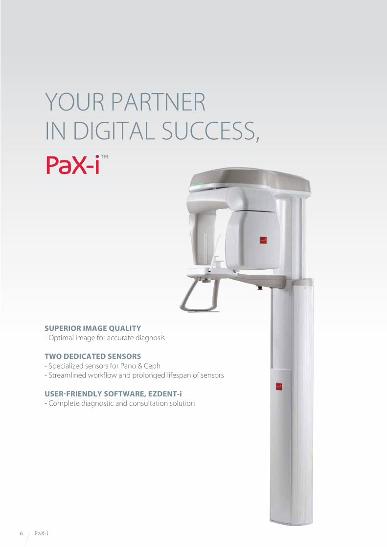
YOUR PARTNER IN DIGITAL SUCCESS,
SUPERIOR IMAGE QUALITY- Optimal image for accurate diagnosis
TWO DEDICATED SENSORS- Specialized sensors for Pano & Ceph
- Streamlined workflow and prolonged lifespan of sensors
USER-FRIENDLY SOFTWARE, EZDENT-i- Complete diagnostic and consultation solution
6 PaX-i
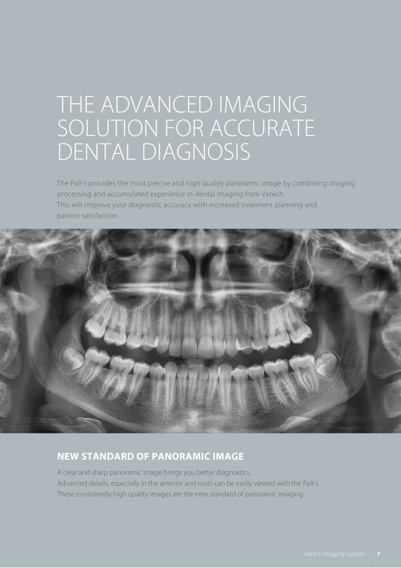
THE ADVANCED IMAGINGSOLUTION FOR ACCURATE DENTAL DIAGNOSISThe PaX-i provides the most precise and high quality panoramic image by combining imaging
processing and accumulated experience in dental imaging from Vatech.
This will improve your diagnostic accuracy with increased treatment planning and
patient satisfaction.
NEW STANDARD OF PANORAMIC IMAGEA clear and sharp panoramic image brings you better diagnostics.
Advanced details, especially in the anterior and roots can be easily viewed with the PaX-i.
These consistently high quality images are the new standard of panoramic imaging.
Vatech Imaging System 7

Ceph Sensor
Pano Sensor
TWO DEDICATED SENSORS The PaX-i offers two dedicated and embedded sensors for Pano and Ceph.
This not only allows you to capture an optimal image from each sensor
but it also creates efficient workflow.
8 PaX-i
Bitewing Mode TMJ Mode
Standard / Right / Front / LeftPANO EXAMINATION Narrow / Normal
Wide / Child
Orthogonal
NormalSPECIAL EXAMINATION
SELECTION ARCH
Orthogonal Standard / Right / Front / Left
Bitewing Standard / Right / Front / Left
TMJ LAT Open / Close
TMJ PA Open / Close
Sinus LAT / PA
EXAMINATION MODE
MAKE YOUR DIAGNOSIS EASY AND EFFICIENT WITHVARIOUS CAPTURE MODESThe PaX-i has various capture modes to meet your diagnostic needs. You can choose any capture
mode based on your diagnostic needs.

THE ADVANCED IMAGE SOLUTIONFOR ORTHODONTIC DIAGNOSIS AND TREATMENT PLANNING EXTENDED DIAGNOSTIC VIEW FOR WIDE INSIGHT
CEPHALOMETRIC (SCAN CEPH)The PaX-i provides optimal images exclusively designed for orthodontics.
There are two image sizes available, Lateral and Full Lateral, allowing you to choose your image size
based on your diagnostic needs.
Vatech Imaging System 9
LATERAL
EXAMINATION PROGRAM SCAN TIME IMAGE SIZE
LATERAL
FULL LATERAL
12.9 sec
16.9 sec
21x23 cm (8.3”x9.1”)
27x23 cm (10.6”x9.1”)
FULL LATERAL
Provide specialized high quality images to suit
orthodontics and maxillofacial surgeries.
A full lateral image size is 30% wider and shows the occipital
area of the patient, which enables comprehensive diagnosis.
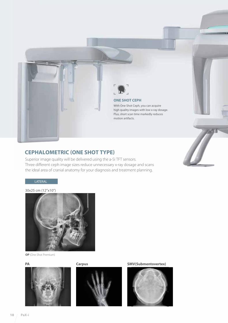
Superior image quality will be delivered using the a-Si TFT sensors.
Three different ceph image sizes reduce unnecessary x-ray dosage and scans
the ideal area of cranial anatomy for your diagnosis and treatment planning.
ONE SHOT CEPHWith One Shot Ceph, you can acquire
high quality images with low x-ray dosage.
Plus, short scan time markedly reduces
motion artifacts.
10 PaX-i
PA Carpus
OP (One Shot Premium)
SMV(Submentovertex)
CEPHALOMETRIC (ONE SHOT TYPE)
LATERAL
30x25 cm (12”x10”)

PaX-i
PaX-i SC
PaX-i OP
Standing / Wheel-Chair Accessible
Pano : HD 13.5 sec / Normal 10.1 secCeph : Scan 12.9 sec / One-shot 0.9 sec
0.5 mm 14 bit
50-90 kVp / 4-10 mA
Ceph FOV Size
Gray Scale
Patient Positioning
Scan Time
Focal Spot
Tube Voltage/Current
62
.99
”
90
.55
”
27
.56
”
62
.99
”
90
.55
”
27
.56
”
62
.99
”
90
.55
”
27
.56
”
20.8”
38.35”
46
.06
”
75.2”
20.8”
46
.06
”
75.43”
20.8”
46
.06
”
38.35”38.35”
Function
TOP VIEW
FRONT VIEW
TOP VIEW
FRONT VIEW
TOP VIEW
FRONT VIEW
Pano + Ceph SC 8.3”x9.1” [LAT, PA, SMV, Waters View, Carpus] 10.6“x9.1” [Full LAT]
OP 12“x10” [LAT, PA, SMV, Waters View, Carpus]
PANOCEPH
SCAN ONE SHOT
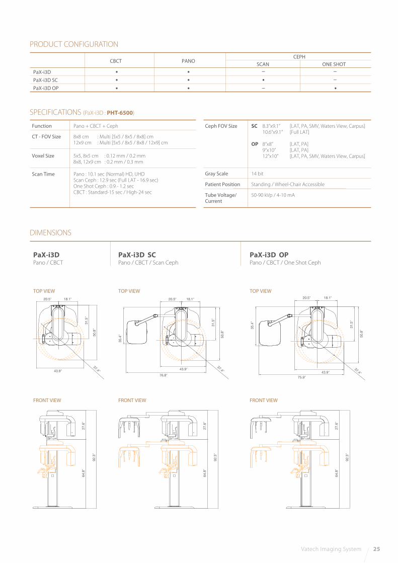
PRODUCT CONFIGURATION
SPECIFICATIONS (PaX-i : PCH-2500)
DIMENSIONS
PaX-iPano
PaX-i SCPano / Scan Ceph
PaX-i OPPano / One Shot Ceph
Vatech Imaging System 11

BEYOND 2D, DEPTH ADDED PANORAMA
12 PaX-i Insight
INSIGHT PAN- The next evolutionary step forward in panoramic imaging with insight pan
RAPID CEPH- 1.9 second acquisition time produces superb image quality- Reduced motion artifacts and faster work�ow
USER-FRIENDLY EZDENT-I SOFTWARE- Powerful diagnostic value with Insight feature- Complete solution for consultation- Easy to learn, easy to use
MULTILAYERS
41
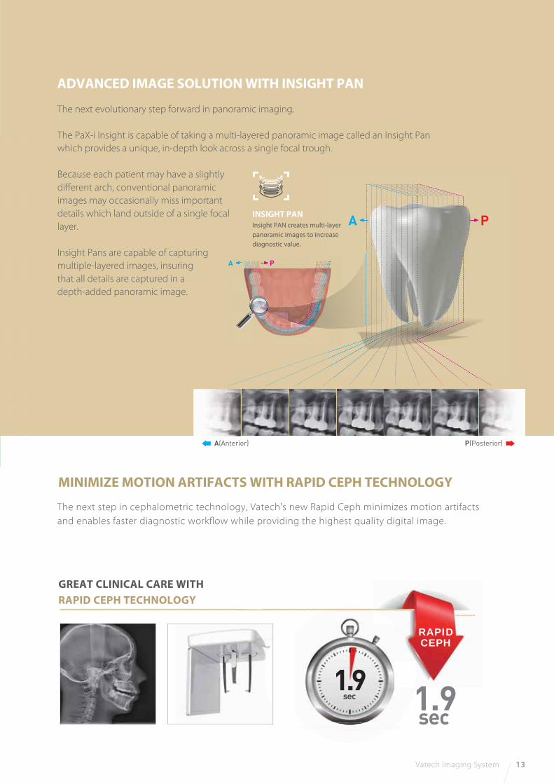
The next evolutionary step forward in panoramic imaging.
The PaX-i Insight is capable of taking a multi-layered panoramic image called an Insight Pan
which provides a unique, in-depth look across a single focal trough.
Because each patient may have a slightly
different arch, conventional panoramic
images may occasionally miss important
details which land outside of a single focal
layer.
Insight Pans are capable of capturing
multiple-layered images, insuring
that all details are captured in a
depth-added panoramic image.
The next step in cephalometric technology, Vatech's new Rapid Ceph minimizes motion artifacts
and enables faster diagnostic workflow while providing the highest quality digital image.
Vatech Imaging System 13
ADVANCED IMAGE SOLUTION WITH INSIGHT PAN
MINIMIZE MOTION ARTIFACTS WITH RAPID CEPH TECHNOLOGY
GREAT CLINICAL CARE WITH RAPID CEPH TECHNOLOGY
1.9sec
1.9sec
A(Anterior) P(Posterior)
INSIGHT PAN Insight PAN creates multi-layer
panoramic images to increase
diagnostic value.
A P
PA
RAPIDCEPH
14 PaX-i Insight
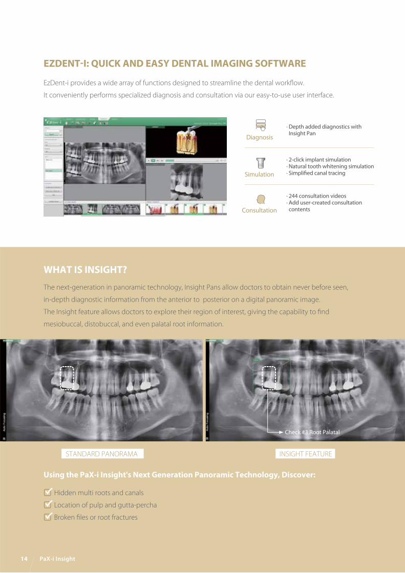
EZDENT-I: QUICK AND EASY DENTAL IMAGING SOFTWARE
EzDent-i provides a wide array of functions designed to streamline the dental workflow.
It conveniently performs specialized diagnosis and consultation via our easy-to-use user interface.
· Depth added diagnostics with Insight Pan
· 2-click implant simulation· Natural tooth whitening simulation· Simplified canal tracing
· 244 consultation videos· Add user-created consultation contents
Diagnosis
Simulation
Consultation
WHAT IS INSIGHT?
Using the PaX-i Insight's Next Generation Panoramic Technology, Discover:
The next-generation in panoramic technology, Insight Pans allow doctors to obtain never before seen,
in-depth diagnostic information from the anterior to posterior on a digital panoramic image.
The Insight feature allows doctors to explore their region of interest, giving the capability to find
mesiobuccal, distobuccal, and even palatal root information.
Hidden multi roots and canals
Location of pulp and gutta-percha
Broken files or root fractures
Check #3 Root Palatal
STANDARD PANORAMA INSIGHT FEATURE

Vatech Imaging System 15
DIMENSIONS
TOP VIEW
With CEPH unit Without CEPH unit
FRONT VIEW
PRODUCT CONFIGURATION
SPECIFICATIONS (PaX-i Insight : PCH-30CS)
PANO CEPH
PaX-i Insight
PaX-i Insight SC
Function Pano + Ceph
Focal Spot 0.5 mm (IEC60336)
Scan TimePano
Normal 10.4 / 14.0 / 21.0 sec
Insight PAN 10.4 sec
Ceph 1.9 / 3.9 sec
Gray Scale 14 bit
Tube Voltage / Current 60 ~ 99 kV / 4 ~ 10 mA
Weight
Without Ceph unit209.4 lbs. – without Base
297.6 lbs. – with Base
With Ceph unit264.5 lbs. – without Base
352.7 lbs. – with Base
Dimensions Without Ceph unit 38.98 Inch (L) x 47.24 Inch (W) x 90.55 Inch (H)
With Ceph unit 75.98 Inch (L) x 47.24 Inch (W) x 90.55 Inch (H)
* The specifications are subject to change without prior notice.
33.07”
33.07”
38.19”
38.98”75.98”
20.79”
5.12”
47
.24
”2
7.5
6”
63
”
90
.55
”
27
.56
”6
3”
90
.55
”
29
.4”
38.19”
38.98”
20.79”
5.12”
47
.24
”29
.4”
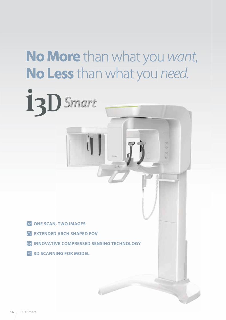
No More than what you want,No Less than what you need.
16 i3D Smart
ONE SCAN, TWO IMAGES
EXTENDED ARCH SHAPED FOV
INNOVATIVE COMPRESSED SENSING TECHNOLOGY
3D SCANNING FOR MODEL

ONE SCAN, TWO IMAGES
One scan with the i3D Smart gives you not just a CT image but also an Auto Pano image. This means,
patients who require both images do not need to undergo two x-ray scans. Also, CT and Auto Pano
images are displayed within one viewer.
SMART INNOVATION
2D 3D
* Conventional panorama mode is provided.
[ 2D AND 3D IN ONE VIEWER ]
Viewing 2D and 3D images together provides many benefits.
There is no need to utilize two different software programs
and the one viewer feature presents a professional look for
your patients.
This layout helps patients better understand the images,
which will eventually result in increasing acceptance rates.
Vatech Imaging System 17
18 i3D Smart
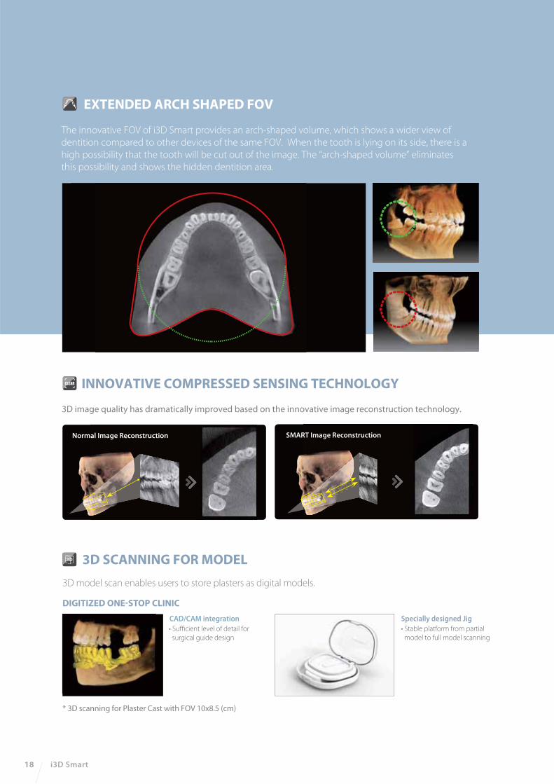
EXTENDED ARCH SHAPED FOV
The innovative FOV of i3D Smart provides an arch-shaped volume, which shows a wider view of
dentition compared to other devices of the same FOV. When the tooth is lying on its side, there is a
high possibility that the tooth will be cut out of the image. The “arch-shaped volume” eliminates
this possibility and shows the hidden dentition area.
3D model scan enables users to store plasters as digital models.
3D SCANNING FOR MODEL
DIGITIZED ONE-STOP CLINIC
* 3D scanning for Plaster Cast with FOV 10x8.5 (cm)
• Sufficient level of detail for
surgical guide design
CAD/CAM integration Specially designed Jig
• Stable platform from partial
model to full model scanning
Normal Image Reconstruction SMART Image Reconstruction
3D image quality has dramatically improved based on the innovative image reconstruction technology.
INNOVATIVE COMPRESSED SENSING TECHNOLOGY
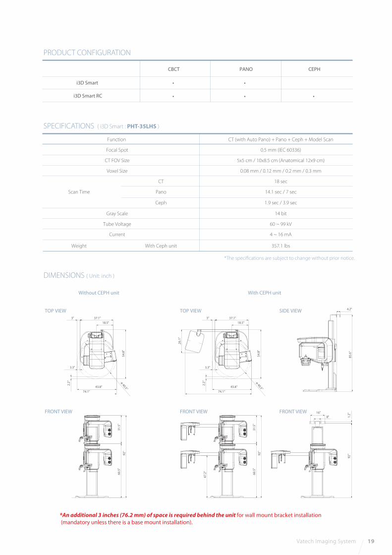
DIMENSIONS ( Unit: inch )
SPECIFICATIONS ( i3D Smart : PHT-35LHS )
Function
Focal Spot
CT FOV Size
Voxel Size
Tube Voltage
Gray Scale
Current
CT (with Auto Pano) + Pano + Ceph + Model Scan
0.5 mm (IEC 60336)
5x5 cm / 10x8.5 cm (Anatomical 12x9 cm)
0.08 mm / 0.12 mm / 0.2 mm / 0.3 mm
18 sec
14.1 sec / 7 sec
1.9 sec / 3.9 sec
14 bit
60 ~ 99 kV
4 ~ 16 mA
CT
Pano
Ceph
Scan Time
PRODUCT CONFIGURATION
i3D Smart
i3D Smart RC
CBCT
• •
PANO
• • •
CEPH
Vatech Imaging System 19
FRONT VIEW FRONT VIEW
TOP VIEW SIDE VIEW
Weight With Ceph unit 357.1 lbs
4.2"
85
.6"
31
.5"
60
.5"
92
"
47
.2"
16"
8" 1.2
"9
2"
3" 37.1"
18.5"
3.3"
43.8"
74.1"
2.2
"
26
.1"
54
.8"
ø 40.5"
FRONT VIEW
TOP VIEW
31
.5"
60
.5"
92
"
3" 37.1"
18.5"
3.3"
43.8"
74.1"
2.2
"
54
.8"
ø 40.5"
*The specifications are subject to change without prior notice.
*An additional 3 inches (76.2 mm) of space is required behind the unit for wall mount bracket installation
(mandatory unless there is a base mount installation).
With CEPH unitWithout CEPH unit
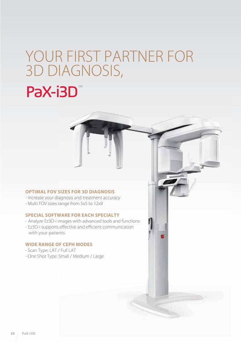
OPTIMAL FOV SIZES FOR 3D DIAGNOSIS- Increase your diagnosis and treatment accuracy
- Multi FOV sizes range from 5x5 to 12x9
SPECIAL SOFTWARE FOR EACH SPECIALTY - Analyze Ez3D-i images with advanced tools and functions
- Ez3D-i supports effective and efficient communication
with your patients
WIDE RANGE OF CEPH MODES- Scan Type: LAT / Full LAT
- One Shot Type: Small / Medium / Large
20 PaX-i3D
YOUR FIRST PARTNER FOR3D DIAGNOSIS,

50 mm
50
mm
50
mm 80
mm
90
mm
80 mm
80 mm120 mm
FLEXIBLE 3D IMAGING WITH MULTI FOV SELECTIONThe PaX-i3D provides 4 multi FOV sizes ranging from 5x5 to 12x9.
By selecting the appropriate FOV size, you can view the optimal image size for
your diagnostic needs, reducing unnecessary x-ray radiation for patients.
FOV 5x5 FOV 8x5
POWERFUL DIAGNOSTIC VALUEWITH 3D IMAGES
5x5 images are useful for a specific area diagnosis with minimal
x-ray exposure for patients. It can especially increase the accuracy
of endodontic diagnosis by specifically checking the number of
root canals and abnormal root canal shapes, such as C-shapes
that are difficult to check when using a 2D x-ray system.
8x5 images can provide more extended oral information on
maxillary or mandibular areas. An accurate treatment plan can
be established by taking into account the major anatomical
structures like mandibular nerve, mental foramen or maxillary
sinus.
Vatech Imaging System 21

FOV 8x8
FOV 12x912x9 images can provide the most optimal information for
oral diagnosis fully covering both maxillary and mandibular
structures including the 3rd molar region in a single scan.
It is suitable for most oral surgery cases as well as multiple
implant surgery.
8x8 images enable comprehensive diagnosis and treatment
planning including both maxillary and mandibular areas in a
single scan. It is useful for complex implant surgery as well
as left or right TMJ diagnosis.
PATENTED AUTO-SWITCHING TECHNOLOGYThe PaX-i3D offers convenient and safe
‘Patented Auto-Switching Technology’ system between CBCT
and Panoramic sensors. This smart system conveniently
prevents sensor damage from accidental dropping.
22 PaX-i3D
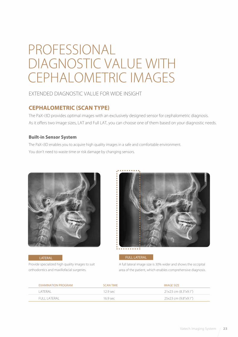
LATERAL
EXAMINATION PROGRAM SCAN TIME IMAGE SIZE
LATERAL
FULL LATERAL
12.9 sec
16.9 sec
21x23 cm (8.3”x9.1”)
25x23 cm (9.8”x9.1”)
FULL LATERAL
CEPHALOMETRIC (SCAN TYPE)The PaX-i3D provides optimal images with an exclusively designed sensor for cephalometric diagnosis.
As it offers two image sizes, LAT and Full LAT, you can choose one of them based on your diagnostic needs.
Built-in Sensor SystemThe PaX-i3D enables you to acquire high quality images in a safe and comfortable environment.
You don’t need to waste time or risk damage by changing sensors.
EXTENDED DIAGNOSTIC VALUE FOR WIDE INSIGHT
PROFESSIONALDIAGNOSTIC VALUE WITHCEPHALOMETRIC IMAGES
Provide specialized high quality images to suit
orthodontics and maxillofacial surgeries.
A full lateral image size is 30% wider and shows the occipital
area of the patient, which enables comprehensive diagnosis.
Vatech Imaging System 23
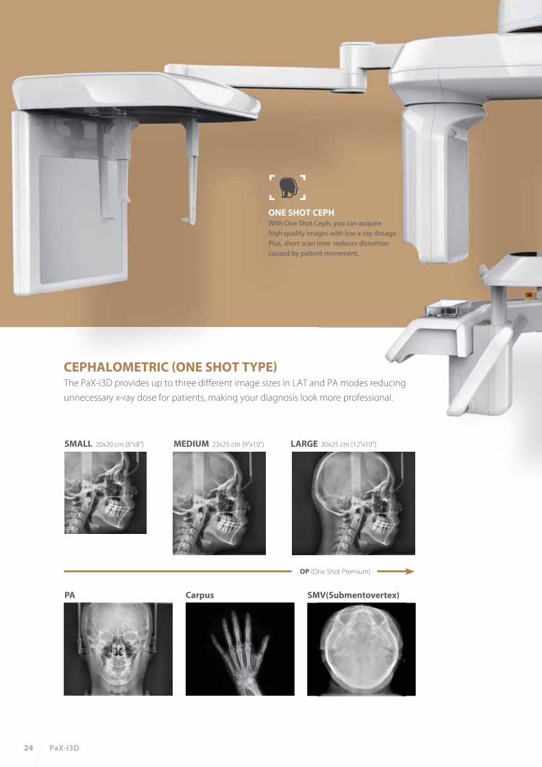
ONE SHOT CEPHWith One Shot Ceph, you can acquire
high quality images with low x-ray dosage.
Plus, short scan time reduces distortion
caused by patient movement.
SMALL 20x20 cm (8”x8”) MEDIUM 23x25 cm (9”x10”)
PA Carpus
OP (One Shot Premium)
SMV(Submentovertex)
LARGE 30x25 cm (12”x10”)
CEPHALOMETRIC (ONE SHOT TYPE)The PaX-i3D provides up to three different image sizes in LAT and PA modes reducing
unnecessary x-ray dose for patients, making your diagnosis look more professional.
24 PaX-i3D
Function Ceph FOV Size
Gray Scale
Patient Position
Tube Voltage/Current
CT - FOV Size
Voxel Size
Scan Time 14 bit
Standing / Wheel-Chair Accessible
50-90 kVp / 4-10 mA
SC 8.3”x9.1” [LAT, PA, SMV, Waters View, Carpus] 10.6“x9.1” [Full LAT]
OP 8“x8” [LAT, PA] 9“x10” [LAT, PA] 12“x10” [LAT, PA, SMV, Waters View, Carpus]
PaX-i3D
PaX-i3D SC
PaX-i3D OP
TOP VIEW
FRONT VIEW
TOP VIEW
FRONT VIEW
TOP VIEW
FRONT VIEW
PANOCBCTCEPH
SCAN ONE SHOT
PRODUCT CONFIGURATION
SPECIFICATIONS (PaX-i3D : PHT-6500)
DIMENSIONS
PaX-i3DPano / CBCT
PaX-i3D SCPano / CBCT / Scan Ceph
PaX-i3D OPPano / CBCT / One Shot Ceph
Pano + CBCT + Ceph
8x8 cm : Multi [5x5 / 8x5 / 8x8] cm12x9 cm : Multi [5x5 / 8x5 / 8x8 / 12x9] cm
5x5, 8x5 cm : 0.12 mm / 0.2 mm8x8, 12x9 cm : 0.2 mm / 0.3 mm
Pano : 10.1 sec (Normal) HD, UHDScan Ceph : 12.9 sec (Full LAT - 16.9 sec)One Shot Ceph : 0.9 - 1.2 secCBCT : Standard-15 sec / High-24 sec
Vatech Imaging System 25
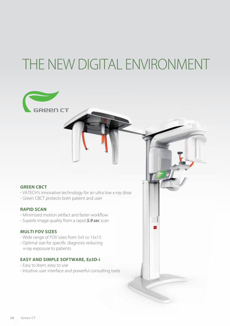
THE NEW DIGITAL ENVIRONMENT
GREEN CBCT- VATECH’s innovative technology for an ultra low x-ray dose
- Green CBCT protects both patient and user
RAPID SCAN- Minimized motion artifact and faster workflow
- Superb image quality from a rapid 5.9 sec scan
MULTI FOV SIZES- Wide range of FOV sizes from 5x5 to 15x15
- Optimal size for specific diagnosis reducing
x-ray exposure to patients
EASY AND SIMPLE SOFTWARE, Ez3D-i- Easy to learn, easy to use
- Intuitive user interface and powerful consulting tools
26 Green CT
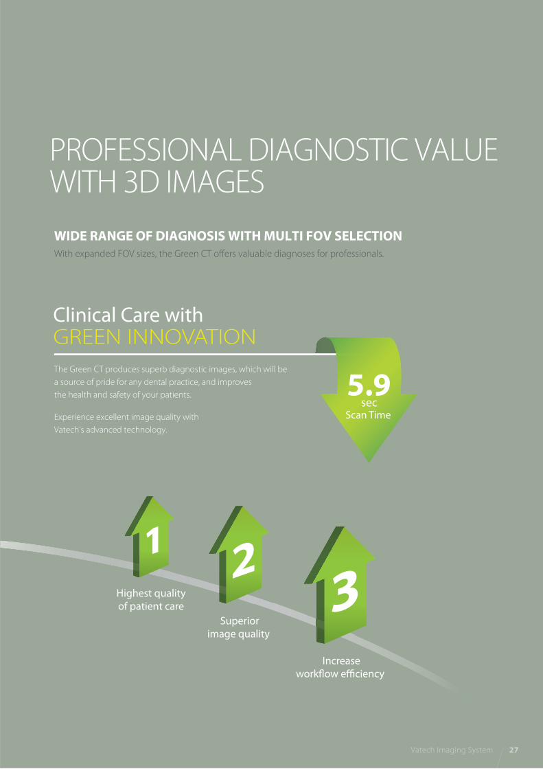
Highest qualityof patient care
Superiorimage quality
Increaseworkflow efficiency
11
1
WIDE RANGE OF DIAGNOSIS WITH MULTI FOV SELECTIONWith expanded FOV sizes, the Green CT offers valuable diagnoses for professionals.
PROFESSIONAL DIAGNOSTIC VALUEWITH 3D IMAGES
The Green CT produces superb diagnostic images, which will be
a source of pride for any dental practice, and improves
the health and safety of your patients.
Experience excellent image quality with
Vatech’s advanced technology.
Clinical Care with GREEN INNOVATION
Vatech Imaging System 27
5.9 sec
Scan Time

FOV 8x8
FOV 12x9
FOV 15x15
8x8 images enable comprehensive diagnosis and treatment
planning including both maxillary and mandibular areas in a
single scan. It is useful for complex implant surgery as well as
left or right TMJ diagnosis.
12x9 images can provide the most optimal information for
oral diagnosis fully covering both maxillary and mandibular
structures including the 3rd molar region in a single scan.
It is suitable for most oral surgery cases as well as multiple
implant surgery.
15x15 images from the Green CT enable you to do a comprehensive
diagnosis including oral and maxillofacial surgery.
This perfect FOV size will be helpful for complex orthognathic,
implant, and orthodontic surgery.
PATENTED AUTO-SWITCHING TECHNOLOGYThe Green CT offers convenient and safe
‘Patented Auto-Switching Technology’ system between CBCT
and Panoramic sensors. This smart system conveniently
prevents sensor damage from accidental dropping.
TECHNOe
system betw
em convenie
al dropping
OLOGY
ween CBCTCT
ently
.
28 Green CT
80
mm
90
mm
80 mm
120 mm
150 mm
15
0 m
m
50 mm
50
mm
50
mm
80 mm

LATERAL
EXAMINATION PROGRAM SCAN TIME IMAGE SIZE
LATERAL
FULL LATERAL
3.9 sec
16.9 sec
21x23 cm (8.3”x9.1”)
27x23 cm (10.6”x9.1”)
FULL LATERAL
CEPHALOMETRIC (SCAN TYPE)The Green CT provides optimal images with an exclusively designed sensor for cephalometric
diagnosis. As it offers two image sizes, LAT and Full LAT, you can choose one of them based on
the purposes of your diagnostic needs.
Built-in Sensor SystemThe Green CT enables you to acquire high quality images in a safe and comfortable environment.
Best of all, you don’t need to waste time or risk damage by changing sensors.
EXTENDED DIAGNOSTIC VALUE FOR WIDE INSIGHT
PROFESSIONALDIAGNOSTIC VALUE WITHCEPHALOMETRIC IMAGES
Provide specialized high quality images to suit
orthodontics and maxillofacial surgeries.
A full lateral image size is 30% wider and shows the occipital
area of the patient, which enables comprehensive diagnosis.
Vatech Imaging System 29
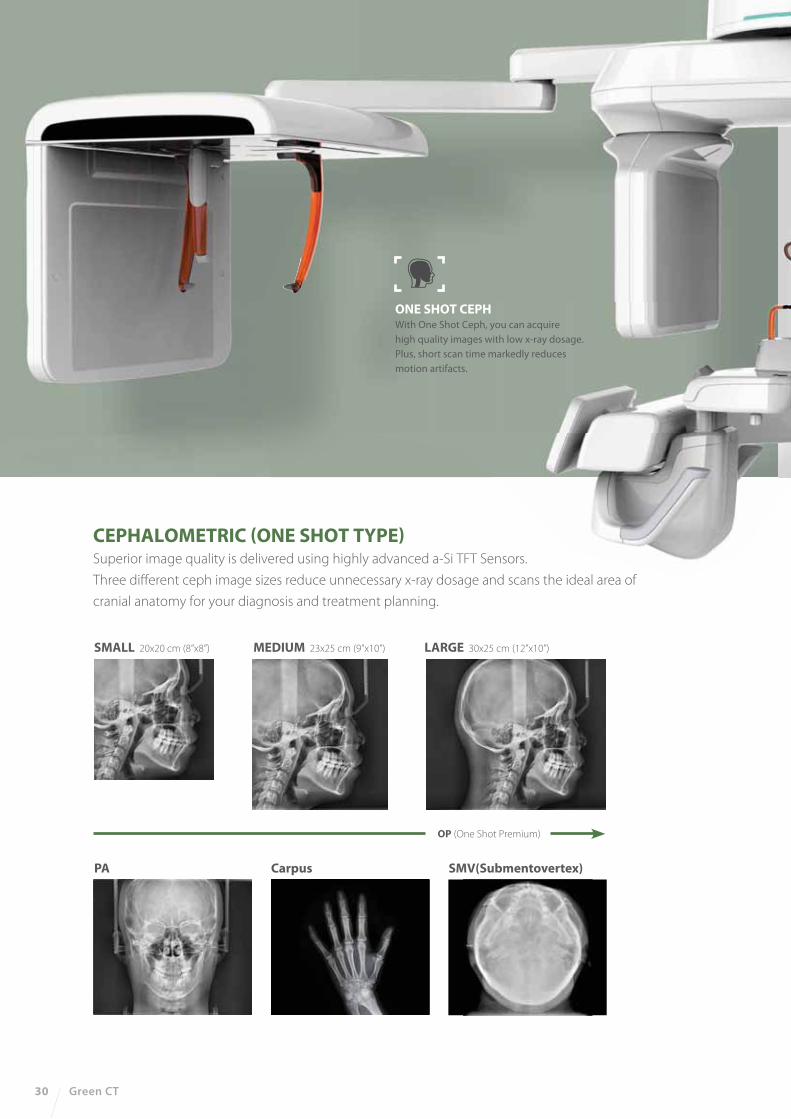
SMALL 20x20 cm (8”x8”) MEDIUM 23x25 cm (9”x10”)
PA Carpus
OP (One Shot Premium)
SMV(Submentovertex)
LARGE 30x25 cm (12”x10”)
ONE SHOT CEPHWith One Shot Ceph, you can acquire
high quality images with low x-ray dosage.
Plus, short scan time markedly reduces
motion artifacts.
30 Green CT
CEPHALOMETRIC (ONE SHOT TYPE)Superior image quality is delivered using highly advanced a-Si TFT Sensors.
Three different ceph image sizes reduce unnecessary x-ray dosage and scans the ideal area of
cranial anatomy for your diagnosis and treatment planning.

47.2”
52
.2”
18”
39.8”
41.7”
36.2”
18”
78.1”
47.2”
52
.2”
34
.6”
3.5”
41.7”41.7”
36.2”
18”
78.1”
47.2”
52
.2”
34
.6”
3.5”
27
.6”
64
.6”
92
.1”
27
.6”
64
.6”
92
.1”
27
.6”
64
.6”
92
.1”
Function Ceph FOV Size
Gray Scale
Patient Position
Tube Voltage/Current
CT - FOV Size
Voxel Size
Scan Time 14 bit
Standing / Wheel-Chair Accessible
50-100 kVp(1 kV step) / 4-16 mA(0.1 mA step)
SC 8.3”x9.1” [LAT, PA, SMV, Waters View, Carpus]
10.6“x9.1” [Full LAT]
OP 8“x8” [LAT, PA]
9“x10” [LAT, PA]
12“x10” [LAT, PA, SMV, Waters View, Carpus]
Green CT
Green CT SC
Green CT OP
TOP VIEW
FRONT VIEW
TOP VIEW
FRONT VIEW
TOP VIEW
FRONT VIEW
PANOCBCTCEPH
SCAN ONE SHOT
PRODUCT CONFIGURATION
SPECIFICATIONS (Green CT : PHT-60CFO)
DIMENSIONS
Green CTPano / CBCT
Green CT SCPano / CBCT / Scan Ceph
Green CT OPPano / CBCT / One Shot Ceph
Pano + CBCT + Ceph
15x15 cm : Multi [5x5 / 8x5 / 8x8 / 12x9 / 15x15 cm]
5x5 cm : 0.08 mm / 0.2 mm
8x8, 12x9 cm : 0.2 mm / 0.3 mm
15x15 cm : 0.25 mm / 0.3 mm
Pano : 10.1 sec
Scan Ceph : 3.9 sec
One Shot Ceph : 0.9 - 1.2 sec
CBCT : 5.9 sec, 9 sec
* An additional 7.5 inches (191 mm) of space is required behind the unit for wall mount bracket installation
(mandatory unless there is a base mount installation).
Vatech Imaging System 31
32 Green CT 2
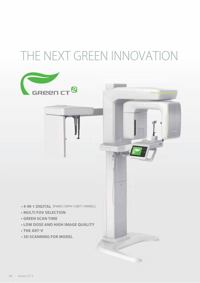
THE NEXT GREEN INNOVATION
• 4-IN-1 DIGITAL • MULTI FOV SELECTION• GREEN SCAN TIME• LOW DOSE AND HIGH IMAGE QUALITY• THE ART-V• 3D SCANNING FOR MODEL
�PANO | CEPH | CBCT | MODEL�
Vatech Imaging System 33
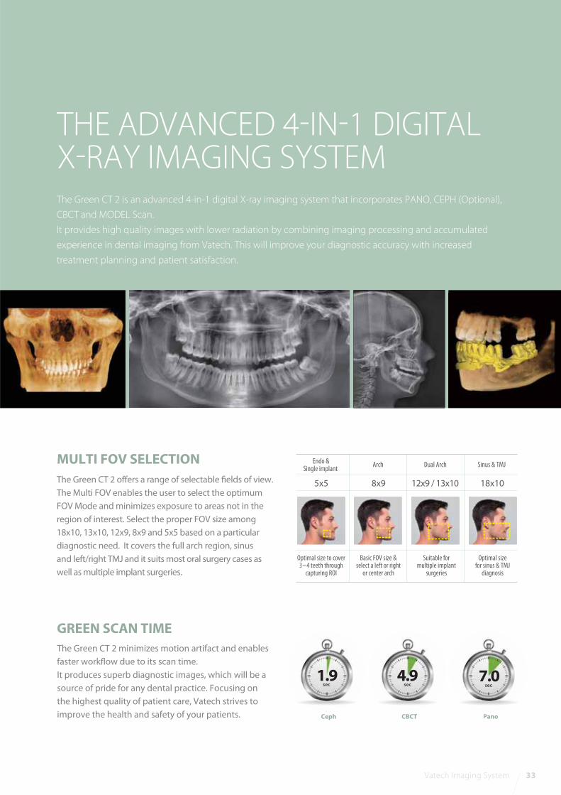
MULTI FOV SELECTION
The Green CT 2 is an advanced 4-in-1 digital X-ray imaging system that incorporates PANO, CEPH (Optional),
CBCT and MODEL Scan.
It provides high quality images with lower radiation by combining imaging processing and accumulated
experience in dental imaging from Vatech. This will improve your diagnostic accuracy with increased
treatment planning and patient satisfaction.
The Green CT 2 offers a range of selectable fields of view.
The Multi FOV enables the user to select the optimum
FOV Mode and minimizes exposure to areas not in the
region of interest. Select the proper FOV size among
18x10, 13x10, 12x9, 8x9 and 5x5 based on a particular
diagnostic need. It covers the full arch region, sinus
and left/right TMJ and it suits most oral surgery cases as
well as multiple implant surgeries.
THE ADVANCED 4-IN-1 DIGITALX-RAY IMAGING SYSTEM
The Green CT 2 minimizes motion artifact and enables
faster workflow due to its scan time.
It produces superb diagnostic images, which will be a
source of pride for any dental practice. Focusing on
the highest quality of patient care, Vatech strives to
improve the health and safety of your patients.
GREEN SCAN TIME
Ceph
1.9sec
CBCT
4.9sec
7.0sec
Pano
18x1012x9 / 13x10 8x95x5
Dual Arch
Suitable formultiple implant
surgeries
Sinus & TMJ
Optimal sizefor sinus & TMJ
diagnosis
Arch
Basic FOV size &select a left or right
or center arch
Endo &Single implant
Optimal size to cover3~4 teeth through
capturing ROI

34 Green CT 2
3D model scan enables users to store plasters as digital models.
3D SCANNING FOR MODEL
*3D scanning for Plaster Cast with FOV 8x9 (cm)
DIGITIZED ONE-STOP CLINIC
CAD/CAM integration
• Sufficient level of detail for surgical
guide design
Specially designed Jig
• Stable protection from partial model
to full model
Metal artifact hinders visualization and naturally reduces
diagnostic confidence.
Clear image gives you less stress and more confidence which
leads to accurate diagnosis for implant planning.
THE ART-V
*ART-V is the new name of Vatech’s MAR function.
(Artifact Reduction Technology of Vatech)
ART-V On ART-V Off
What has been developed at Vatech breaks many conventions in dental radiography.
It was always believed that with low radiation comes inferior image quality, which renders it useless in clinical diagnosis.
However, the Green CT 2 provides clinically diagnosable x-ray scans at a low x-ray dosage. With low dose x-ray radiography,
achieving clinically diagnosable image quality is the new golden-standard.
LOW DOSE AND HIGH IMAGE QUALITY
Effective Dose Data
Others
Vatech Imaging System 35
DIMENSIONS
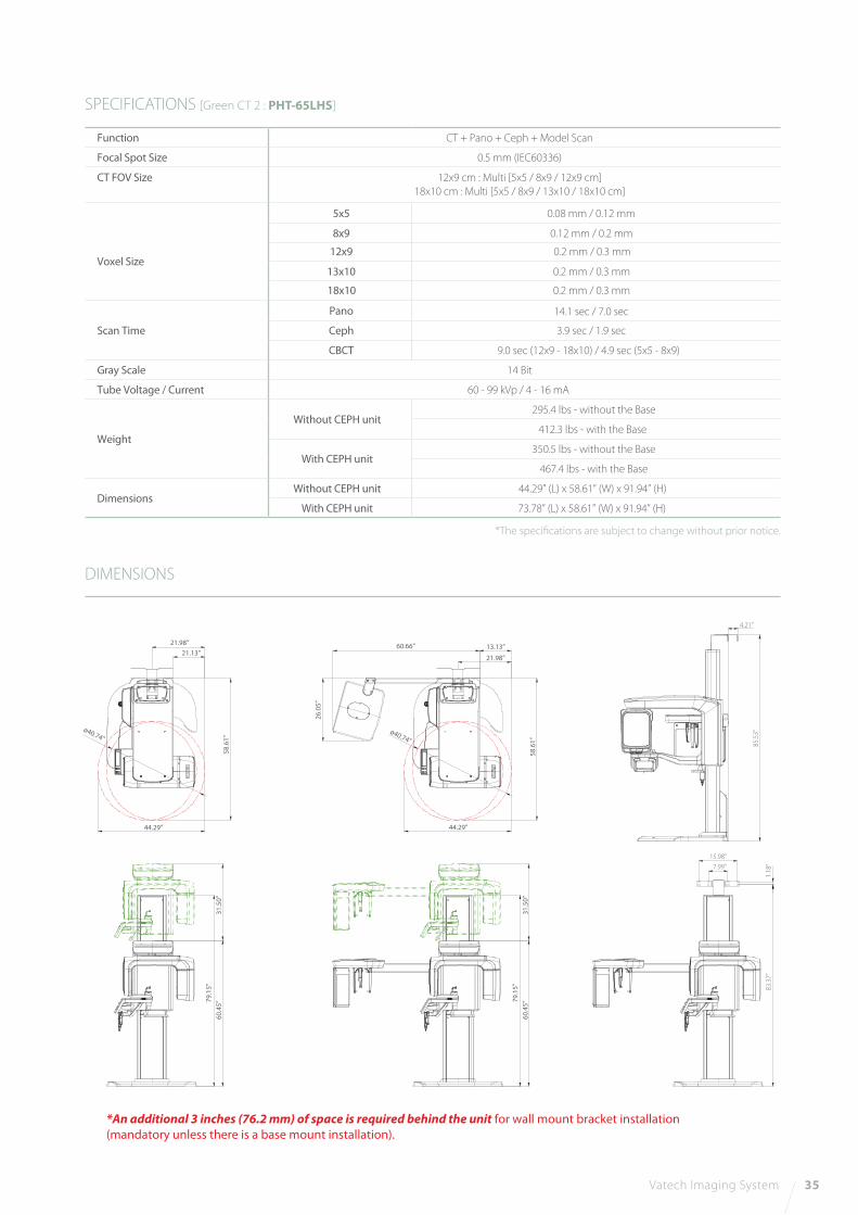
SPECIFICATIONS [Green CT 2 : PHT-65LHS]
Function CT + Pano + Ceph + Model Scan
Focal Spot Size 0.5 mm (IEC60336)
CT FOV Size 12x9 cm : Multi [5x5 / 8x9 / 12x9 cm]18x10 cm : Multi [5x5 / 8x9 / 13x10 / 18x10 cm]
Voxel Size
18x10 0.2 mm / 0.3 mm
13x10 0.2 mm / 0.3 mm
8x9 0.12 mm / 0.2 mm
12x9 0.2 mm / 0.3 mm
5x5 0.08 mm / 0.12 mm
Scan Time
Pano 14.1 sec / 7.0 sec
Ceph 3.9 sec / 1.9 sec
CBCT 9.0 sec (12x9 - 18x10) / 4.9 sec (5x5 - 8x9)
Gray Scale 14 Bit
Tube Voltage / Current 60 - 99 kVp / 4 - 16 mA
Weight
Without CEPH unit295.4 lbs - without the Base
412.3 lbs - with the Base
With CEPH unit350.5 lbs - without the Base
467.4 lbs - with the Base
Dimensions Without CEPH unit 44.29” (L) x 58.61” (W) x 91.94” (H)
With CEPH unit 73.78” (L) x 58.61” (W) x 91.94” (H)
*The speci�cations are subject to change without prior notice.
21.98”21.13”
58.6
1”
58.6
1”
ø40.74”ø40.74”
44.29” 44.29”
26.0
5”
60.66” 13.13”
21.98”
4.21”
85.5
3”
1.18
”
15.98”7.99”
83.3
7”
60.4
5”79
.15”
31.5
0”
60.4
5”79
.15”
31.5
0”
*An additional 3 inches (76.2 mm) of space is required behind the unit for wall mount bracket installation(mandatory unless there is a base mount installation).

RAISING THE BARFOR EXCELLENCE
36 i3D Premium
LARGE 21X19 FOV FOR COMPLETEDIAGNOSTIC IMAGING NEEDS
THE OPTIMAL SOLUTION FORAIRWAY AND ENT DIAGNOSIS
AUTOMATICALLY GENERATES UP TO6 TYPES OF IMAGES IN 1 SCAN
THE MOST SUITABLE FOV SIZEFOR A COMPLETE DIAGNOSIS
Vatech Imaging System 37
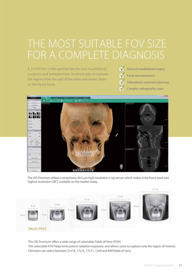
A 21x19 FOV is the optimal size for oral maxillofacial
surgeons and orthodontists. Anatomically, it captures
the regions from the roof of the orbits and nasion down
to the hyoid bone.
The i3D Premium utilizes a proprietary 49.5 μm high resolution x-ray sensor which makes it the finest pixel and
highest resolution CBCT available on the market today.
The i3D Premium offers a wide range of selectable Fields of View (FOV).
This selectable FOV helps limit patient radiation exposure, and allows users to capture only the region of interest.
Clinicians can select between 21x19, 17x15, 17x11, 12x9 and 8X8 fields of view.
Oral and maxillofacial surgery
Facial reconstructions
Orthodontic treatment planning
Complex orthognathic cases
8 cm
8 cm
9 cm
12 cm
15 cm
17 cm
155 c11 cm
17 cm
19 cm
21 cm
[Multi FOV]
38 i3D Premium
THE OPTIMAL SOLUTION FORAIRWAY AND ENT DIAGNOSIS
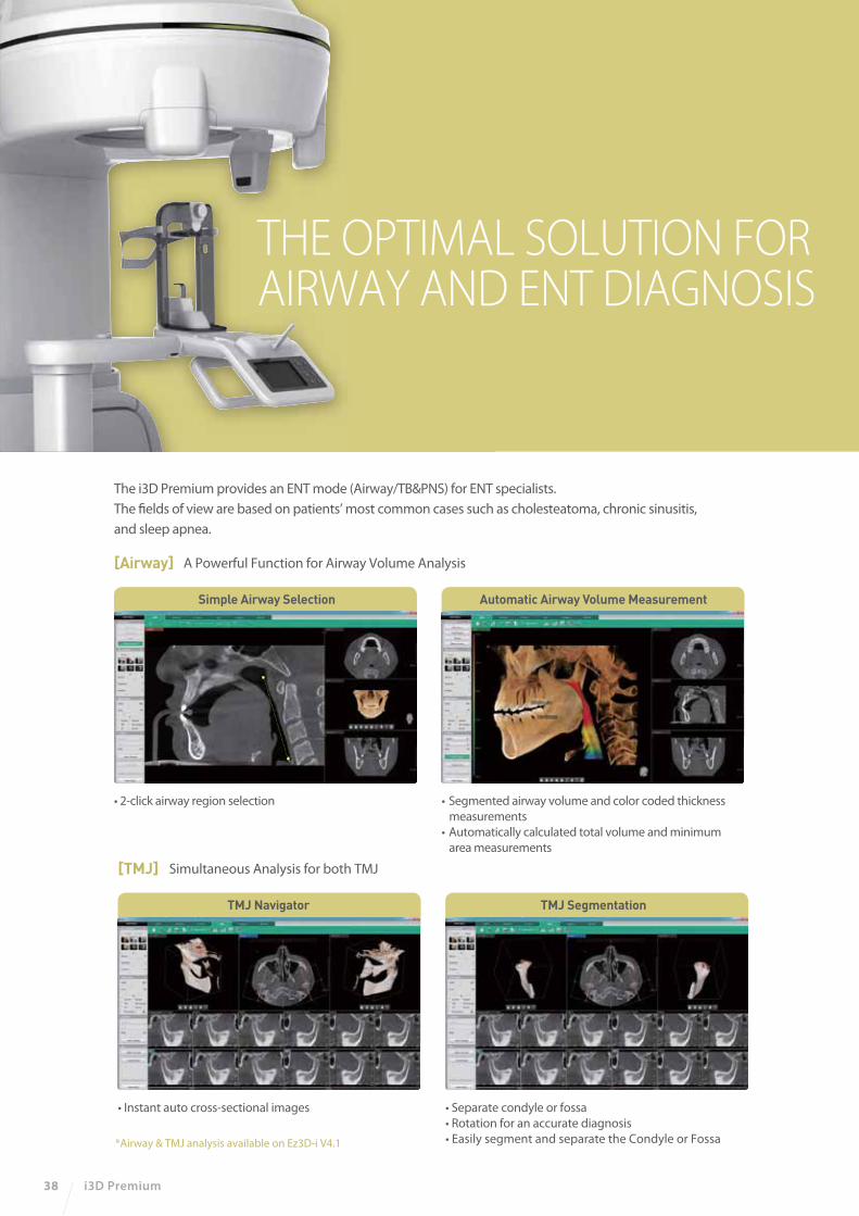
The i3D Premium provides an ENT mode (Airway/TB&PNS) for ENT specialists.
The fields of view are based on patients’ most common cases such as cholesteatoma, chronic sinusitis,
and sleep apnea.
[Airway] A Powerful Function for Airway Volume Analysis
• Segmented airway volume and color coded thickness
measurements
• Automatically calculated total volume and minimum
area measurements
Automatic Airway Volume Measurement
• 2-click airway region selection
Simple Airway Selection
*Airway & TMJ analysis available on Ez3D-i V4.1
[TMJ] Simultaneous Analysis for both TMJ
• Instant auto cross-sectional images
TMJ Navigator
• Separate condyle or fossa
• Rotation for an accurate diagnosis
• Easily segment and separate the Condyle or Fossa
TMJ Segmentation
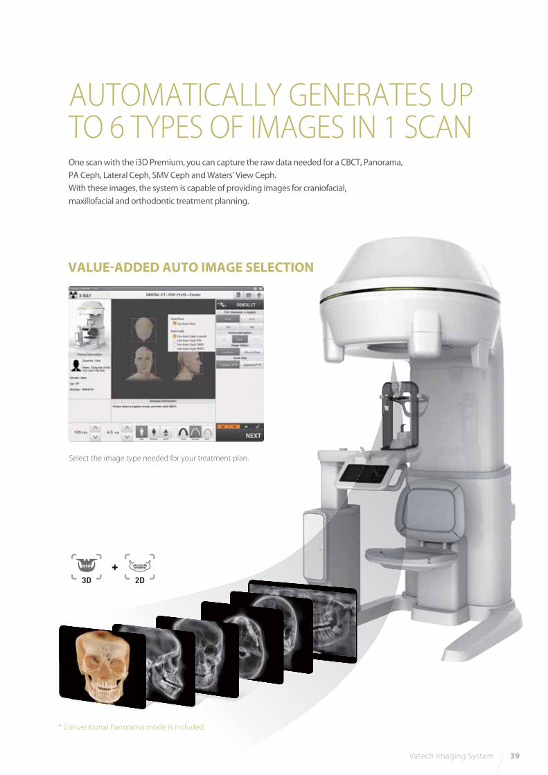
AUTOMATICALLY GENERATES UPTO 6 TYPES OF IMAGES IN 1 SCAN
Vatech Imaging System 39
One scan with the i3D Premium, you can capture the raw data needed for a CBCT, Panorama,
PA Ceph, Lateral Ceph, SMV Ceph and Waters' View Ceph.
With these images, the system is capable of providing images for craniofacial,
maxillofacial and orthodontic treatment planning.
Select the image type needed for your treatment plan.
2D3D+
VALUE-ADDED AUTO IMAGE SELECTION
* Conventional Panorama mode is included.
40 i3D Premium
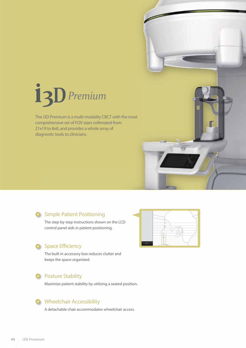
Simple Patient Positioning
The step-by-step instructions shown on the LCD
control panel aids in patient positioning.
Space Efficiency
The built in accessory box reduces clutter and
keeps the space organized.
Posture Stability
Maximize patient stability by utilizing a seated position.
Wheelchair Accessibility
A detachable chair accommodates wheelchair access.
The i3D Premium is a multi-modality CBCT with the most
comprehensive set of FOV sizes collimated from
21x19 to 8x8, and provides a whole array of
diagnostic tools to clinicians.
Vatech Imaging System 41
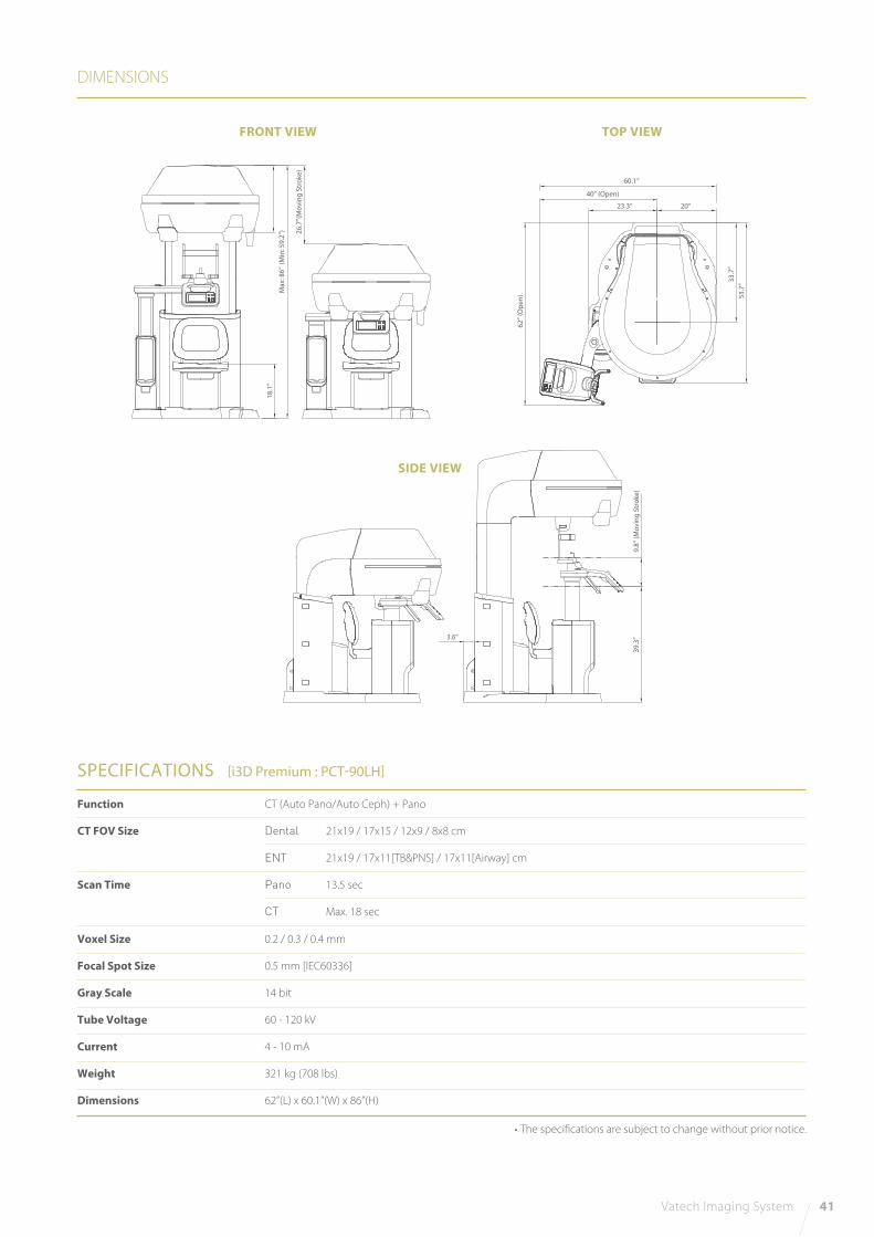
DIMENSIONS
SPECIFICATIONS [i3D Premium : PCT-90LH]
FRONT VIEW TOP VIEW
Function
CT FOV Size
Scan Time
Voxel Size
Focal Spot Size
Gray Scale
Tube Voltage
Current
Weight
Dimensions
CT (Auto Pano/Auto Ceph) + Pano
Dental 21x19 / 17x15 / 12x9 / 8x8 cm
ENT 21x19 / 17x11[TB&PNS] / 17x11[Airway] cm
Pano 13.5 sec
CT Max. 18 sec
0.2 / 0.3 / 0.4 mm
0.5 mm [IEC60336]
14 bit
60 - 120 kV
4 - 10 mA
321 kg (708 lbs)
62”(L) x 60.1”(W) x 86”(H)
• The specifications are subject to change without prior notice.
SIDE VIEW
3.6”3
9.3
”9
.8”
(Mo
vin
g S
tro
ke)
62
” (O
pe
n)
26
.7”(
Mo
vin
g S
tro
ke)
Ma
x: 8
6”
18
.1”
(Min
: 59
.2”)
60.1”
40” (Open)
23.3” 20”
33
.7”
53
.7”
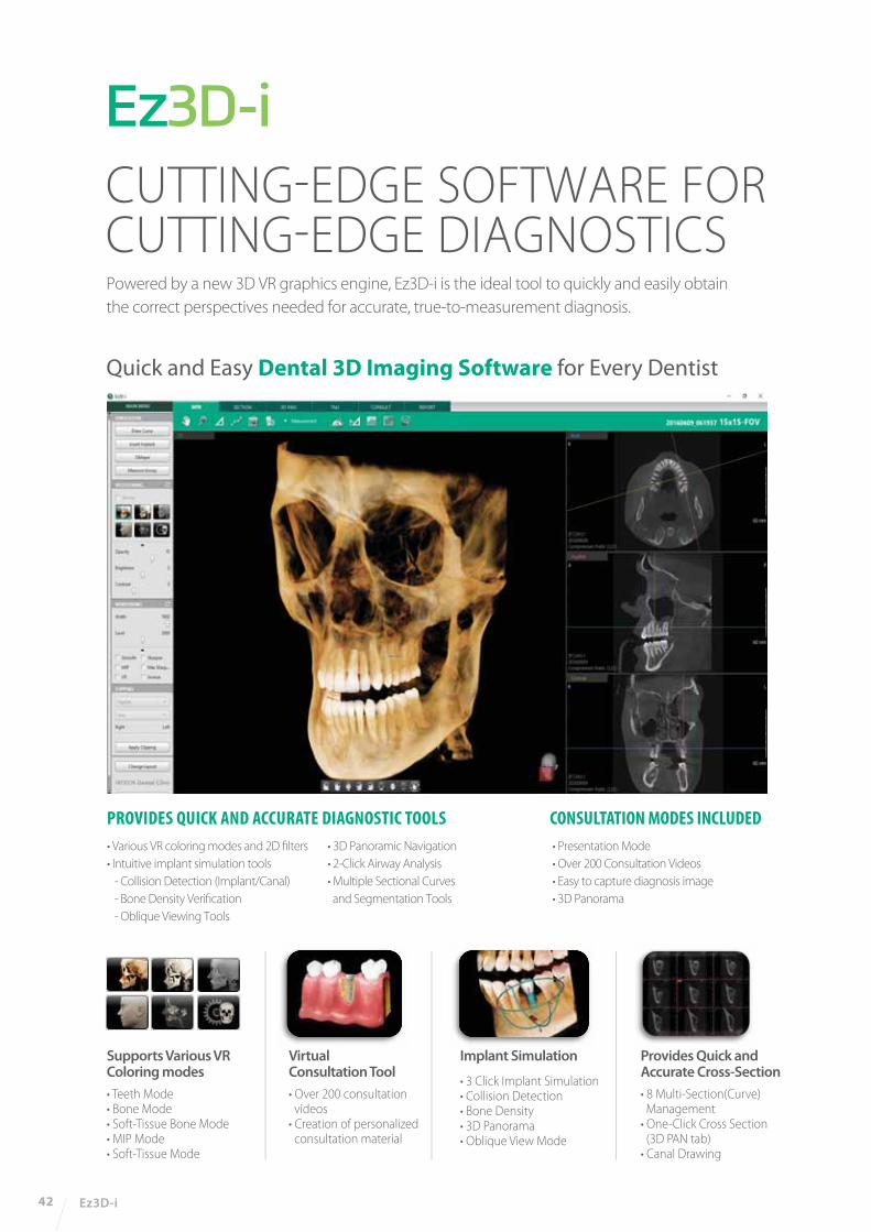
CUTTING-EDGE SOFTWARE FOR CUTTING-EDGE DIAGNOSTICS
42 Ez3D-i
Supports Various VRColoring modes
• Teeth Mode• Bone Mode• Soft-Tissue Bone Mode• MIP Mode• Soft-Tissue Mode
Virtual Consultation Tool
• Over 200 consultation videos• Creation of personalized consultation material
Implant Simulation
• 3 Click Implant Simulation• Collision Detection• Bone Density• 3D Panorama• Oblique View Mode
Provides Quick andAccurate Cross-Section
• 8 Multi-Section(Curve) Management• One-Click Cross Section (3D PAN tab)• Canal Drawing
Quick and Easy Dental 3D Imaging Software for Every Dentist
PROVIDES QUICK AND ACCURATE DIAGNOSTIC TOOLS CONSULTATION MODES INCLUDED• Various VR coloring modes and 2D filters
• Intuitive implant simulation tools
- Collision Detection (Implant/Canal)
- Bone Density Verification
- Oblique Viewing Tools
• 3D Panoramic Navigation
• 2-Click Airway Analysis
• Multiple Sectional Curves
and Segmentation Tools
• Presentation Mode
• Over 200 Consultation Videos
• Easy to capture diagnosis image
• 3D Panorama
Powered by a new 3D VR graphics engine, Ez3D-i is the ideal tool to quickly and easily obtain
the correct perspectives needed for accurate, true-to-measurement diagnosis.
Ez3D-i 43
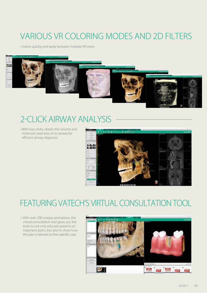
2-CLICK AIRWAY ANALYSIS • With two clicks, obtain the volume and
minimum axial area of an airway for
efficient airway diagnosis
VARIOUS VR COLORING MODES AND 2D FILTERS• Switch quickly and easily between multiple VR views
FEATURING VATECH'S VIRTUAL CONSULTATION TOOL
• With over 200 unique animations, the
virtual consultation tool gives you the
tools to not only educate patients on
treatment plans, but also to show how
this plan is relevant to their specific case

44 Ez3D-i
3D PANORAMIC NAVIGATION• Easily navigate and obtain a sectional view by utilizing our new and
intuitive 3D panoramic navigation mode
• Simply click and drag our viewing window over the
3D panorama to obtain a sectional view of that region
• Angulation made easy
By clicking 3D Navigator and positioning to ROI,
it’s easy to verify 2D sectional images
IMPLANT SIMULATION • Available in all viewing modes in Ez3D-i (MPR/Section/3DPan)
• Colorized bone density viewing modes available
• Adjustable automatic implant collision detection function between multiple implants and/or nerve canal
MPR tab
SECTION tab
3D PAN tab
MULTI-CURVE MANAGEMENT• Draw sectional curves from either the MPR View or Sectional View
• Easily manage and up to 8 different sectional curves
• Intuitive click-and-drag sectional view manipulation
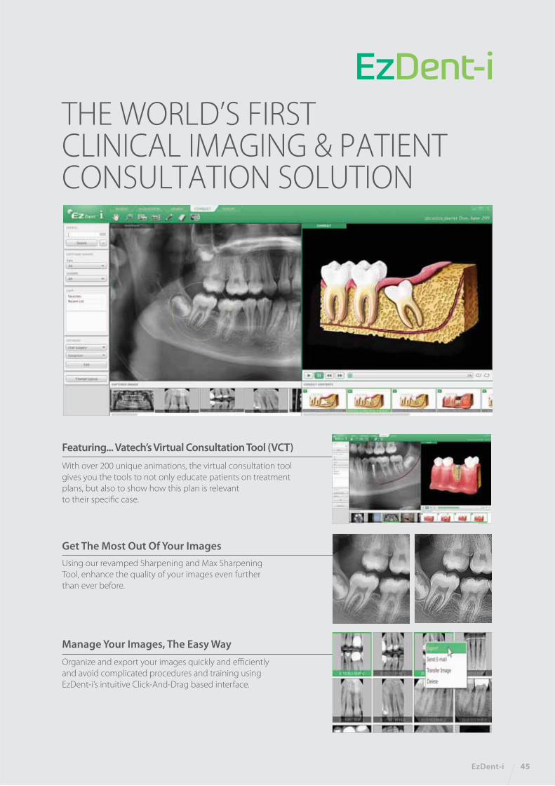
THE WORLD’S FIRST CLINICAL IMAGING & PATIENT CONSULTATION SOLUTION
EzDent-i 45
Featuring... Vatech’s Virtual Consultation Tool (VCT)
With over 200 unique animations, the virtual consultation tool
gives you the tools to not only educate patients on treatment
plans, but also to show how this plan is relevant
to their specific case.
Using our revamped Sharpening and Max Sharpening
Tool, enhance the quality of your images even further
than ever before.
Get The Most Out Of Your Images
Organize and export your images quickly and efficiently
and avoid complicated procedures and training using
EzDent-i’s intuitive Click-And-Drag based interface.
Manage Your Images, The Easy Way
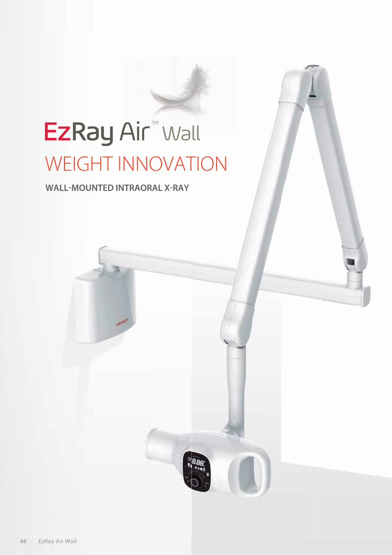
WALL-MOUNTED INTRAORAL X-RAY
46 EzRay Air Wall
WEIGHT INNOVATION
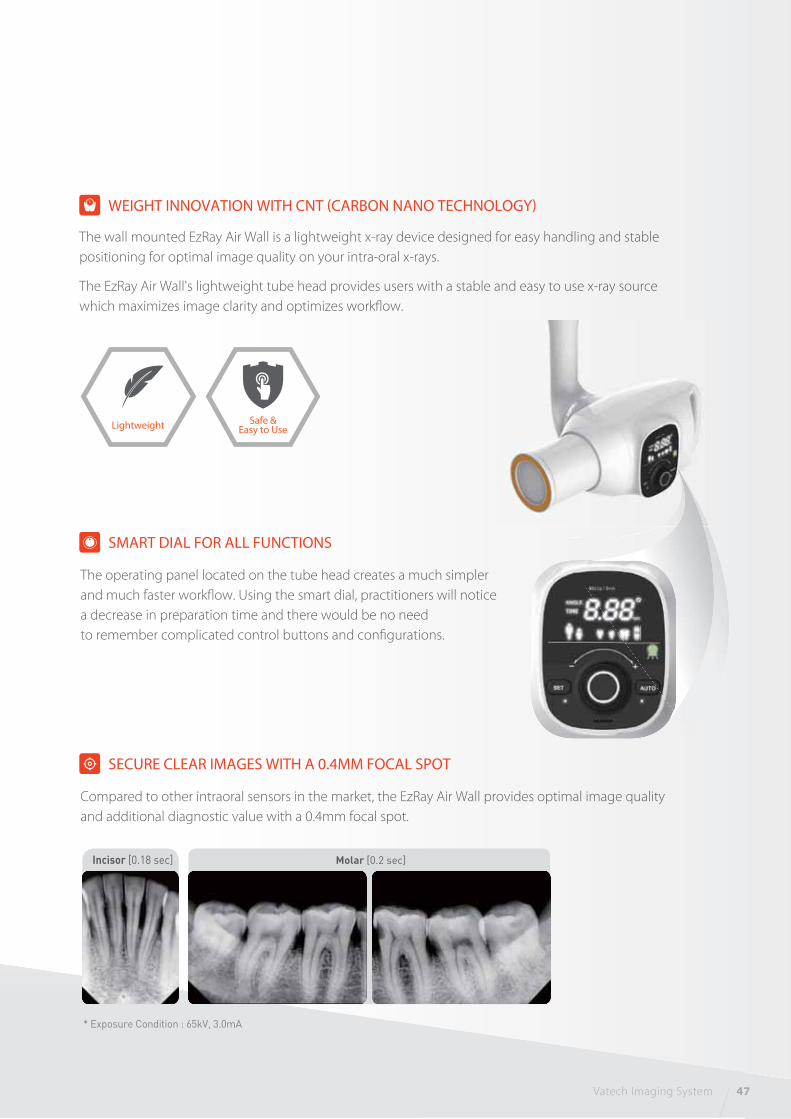
* Exposure Condition : 65kV, 3.0mA
Molar [0.2 sec] Incisor [0.18 sec]
The wall mounted EzRay Air Wall is a lightweight x-ray device designed for easy handling and stable
positioning for optimal image quality on your intra-oral x-rays.
The EzRay Air Wall's lightweight tube head provides users with a stable and easy to use x-ray source
which maximizes image clarity and optimizes workflow.
WEIGHT INNOVATION WITH CNT (CARBON NANO TECHNOLOGY)
Safe &Easy to UseLightweight
SMART DIAL FOR ALL FUNCTIONS
The operating panel located on the tube head creates a much simpler
and much faster workflow. Using the smart dial, practitioners will notice
a decrease in preparation time and there would be no need
to remember complicated control buttons and configurations.
SECURE CLEAR IMAGES WITH A 0.4MM FOCAL SPOT
Compared to other intraoral sensors in the market, the EzRay Air Wall provides optimal image quality
and additional diagnostic value with a 0.4mm focal spot.
Vatech Imaging System 47

ERGONOMIC DESIGNERGONOMIC DESIGN
[ Round Shape Design ]
The round shape design adds a modern yet simple touch
to the EzRay Air Wall.
[ One-Handed Grip ]
A hand grip provides an accurate method
of positioning the EzRay Air Wall.
[ Stable Arm ]
The x-ray unit arm firmly supports the positioning of the tube head.
This allows practitioners to produce superb images
at any given time.
48 EzRay Air Wall
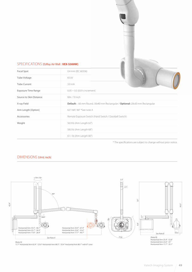
DIMENSIONS [Unit: inch]
SPECIFICATIONS [EzRay Air Wall : VEX-S300W]
Focal Spot
Tube Voltage
Tube Current
Exposure Time Range
Source to Skin Distance
X-ray Field
Accessories
Weight
0.4 mm (IEC 60336)
65 kV
3.0 mA
0.05 ~ 0.5 (0.01s increment)
Min. 7.9 inch
Default: : 60 mm Round, 30x40 mm Rectangular / Optional: 20x30 mm Rectangular
Remote Exposure Switch (Hand Switch / Doorbell Switch)
Arm Length [Option] 62”/ 68”/ 80” *See note A
56.9 Ib (Arm Length 62")
58.0 Ib (Arm Length 68")
61.1 Ib (Arm Length 80")
* The specifications are subject to change without prior notice.
42
.4"
33
"9
.4"
2.3
"
45
.3"
2.3"
84º
38º
218º
+70º/-70º
660º
2.1"
Horizontal Arm 35.5" : 46.1"
Horizontal Arm 23.7" : 34.3"
Horizontal Arm 17.8" : 28.4"
Horizontal Arm 34.4" : 67.4"
Horizontal Arm 23.6" : 55.6"
Horizontal Arm 17.7" : 49.7"
ment)
Vatech Imaging System 49
[Note A]
17.7” Horizontal Arm 62.4” / 23.6“ Horizontal Arm 68.3” / 35.4“ Horizontal Arm 80.1” with 8” cone
[Note B]
Horizontal Arm 35.4" : 52.8"
Horizontal Arm 23.6" : 41"
Horizontal Arm 17.7" : 35.1"
See Note B
See Note A

REDEFINING INTRAORAL SENSORS
50 HD Sensor
1.5
EXPERIENCE THE HIGHEST RESOLUTION
4.8 MM ULTRA-SLIM DESIGN
NEW CONTRAST FILTERS FOR YOUR PERFECT IMAG
Rounded Corner
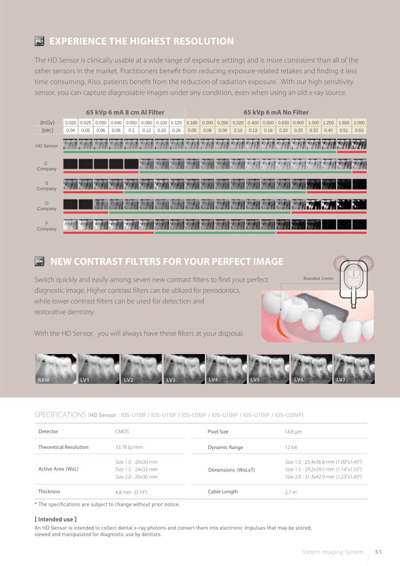
NEW CONTRAST FILTERS FOR YOUR PERFECT IMAGE
RAW LV1 LV2 LV3 LV4 LV5 LV6 LV7
Vatech Imaging System 51
Switch quickly and easily among seven new contrast filters to find your perfect
diagnostic image. Higher contrast filters can be utilized for periodontics,
while lower contrast filters can be used for detection and
restorative dentistry.
With the HD Sensor, you will always have these filters at your disposal.
SPECIFICATIONS
Detector
Theoretical Resolution
Active Area (WxL)
Thickness Cable Length
CMOS
33.78 lp/mm
Size 1.0 : 20x30 mm
Size 1.5 : 24x33 mm
Size 2.0 : 26x36 mm
4.8 mm (0.19”) 2.7 m
Pixel Size 14.8 μm
Dynamic Range 12 bit
Dimensions (WxLxT)
Size 1.0 : 25.4x36.8 mm (1.00”x1.45”)
Size 1.5 : 29.2x39.5 mm (1.14”x1.55”)
Size 2.0 : 31.3x42.9 mm (1.23”x1.69”)
(HD Sensor : IOS-U10IF / IOS-U15IF / IOS-U20IF / IOS-U10VF / IOS-U15VF / IOS-U20VF)
* The specifications are subject to change without prior notice.
[ Intended use ]An HD Sensor is intended to collect dental x-ray photons and convert them into electronic impulses that may be stored,
viewed and manipulated for diagnostic use by dentists.
EXPERIENCE THE HIGHEST RESOLUTION
The HD Sensor is clinically usable at a wide range of exposure settings and is more consistent than all of the
other sensors in the market. Practitioners benefit from reducing exposure-related retakes and finding it less
time consuming. Also, patients benefit from the reduction of radiation exposure. With our high sensitivity
sensor, you can capture diagnosable images under any condition, even when using an old x-ray source.
65 kVp 6 mA 8 cm Al Filter(mGy)(sec)
0.020
0.04
0.025
0.05
0.030
0.06
0.040
0.08
0.050
0.1
0.060
0.12
0.100
0.20
0.125
0.26
0.160
0.05
0.200
0.06
0.250
0.08
0.320
0.10
0.400
0.13
0.500
0.16
0.630
0.20
0.800
0.25
1.000
0.32
1.250
0.40
1.600
0.51
2.000
0.63
HD Sensor
C Company
SCompany
OCompany
FCompany
65 kVp 6 mA No Filter