vegf and notch in tip and stalk cell...
TRANSCRIPT
VEGF and Notch in Tip and Stalk Cell Selection
Raquel Blanco and Holger Gerhardt
Vascular Biology Laboratory, London Research Institute, Lincoln’s Inn Fields Laboratories, LondonWC2A 3LY, United Kingdom
Correspondence: [email protected]
Sprouting angiogenesis is a dynamic process in which endothelial cells collectively migrate,shape new lumenized tubes, make new connections, and remodel the nascent network intoa hierarchically branched and functionally perfused vascular bed. Endothelial cells in thenascent sprout adopt two distinct cellular phenotypes—known as tip and stalk cells—withspecialized functions and gene expression patterns. VEGF and Notch signaling engage inan intricate cross talk to balance tip and stalk cell formation and to regulate directed tipcell migration and stalk cell proliferation. In this article, we summarize the current knowl-edge and implications of the tip/stalk cell concepts and the quantitative and dynamic inte-gration of VEGF and Notch signaling in tip and stalk cell selection.
ANGIOGENESIS: TIP AND STALKPHENOTYPES
Angiogenesis is the process of expandingexisting blood vessel networks mainly by
sprouting new branches that connect and sub-sequently remodel into a functional vascularcircuit. The process is generally initiated in thecontext of tissue growth, either during develop-ment or cancer, during inflammation andwound healing, or during increasing physiolog-ical metabolic tissue demands in physical train-ing. Once the new vessels establish nutrient andoxygen supplies that meet the metabolic tissuedemand, the vessels will become quiescent(Risau 1997).
A key trigger of angiogenesis is local tissuehypoxia, which activates local cellular pro-duction of proangiogenic growth factors suchas vascular endothelial growth factor (VEGF)
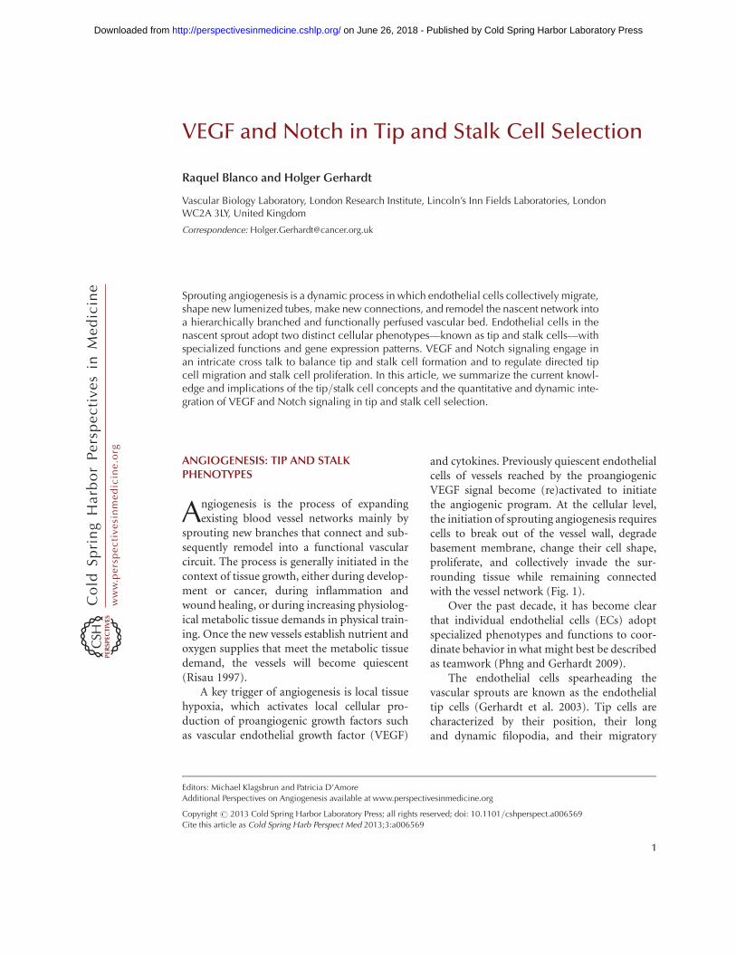
and cytokines. Previously quiescent endothelialcells of vessels reached by the proangiogenicVEGF signal become (re)activated to initiatethe angiogenic program. At the cellular level,the initiation of sprouting angiogenesis requirescells to break out of the vessel wall, degradebasement membrane, change their cell shape,proliferate, and collectively invade the sur-rounding tissue while remaining connectedwith the vessel network (Fig. 1).
Over the past decade, it has become clearthat individual endothelial cells (ECs) adoptspecialized phenotypes and functions to coor-dinate behavior in what might best be describedas teamwork (Phng and Gerhardt 2009).
The endothelial cells spearheading thevascular sprouts are known as the endothelialtip cells (Gerhardt et al. 2003). Tip cells arecharacterized by their position, their longand dynamic filopodia, and their migratory
Editors: Michael Klagsbrun and Patricia D’Amore
Additional Perspectives on Angiogenesis available at www.perspectivesinmedicine.org
Copyright # 2013 Cold Spring Harbor Laboratory Press; all rights reserved; doi: 10.1101/cshperspect.a006569
Cite this article as Cold Spring Harb Perspect Med 2013;3:a006569
1
ww
w.p
ersp
ecti
vesi
nm
edic
ine.
org
on June 26, 2018 - Published by Cold Spring Harbor Laboratory Press http://perspectivesinmedicine.cshlp.org/Downloaded from
behavior (Gerhardt et al. 2003). Analogous toan axonal growth cone, the tip cells integrateattractive and repulsive directional cues pre-sented by the environment and thereby definethe route in which the new sprout grows (Ger-hardt et al. 2003). Moreover, the tip cells arerequired to create new connections between dif-ferent sprouts to generate an interconnectedand functional vascular network (Fig. 1) (Isogaiet al. 2003).
Following the tip cells are the endothelialstalk cells, which produce fewer filopodia, arehighly proliferative, establish adherent and tight
junctions to ensure the stability of the newsprout, and form the nascent vascular lumen(Fig. 1) (Gerhardt et al. 2003; Dejana et al.2009; Iruela-Arispe and Davis 2009; Phng andGerhardt 2009).
Additionally to the morphologic and func-tional differences, tip and stalk cells exhibitdistinct gene expression profiles. Several recenttranscriptional profiling efforts established alist of genes enriched in tip cells, including cell-surface receptors, growth factors, neuronalguidance receptors, and matrix-degrading pro-teases, as well as matrix components and more.
Quiescent endothelium stimulated by VEGF-AA
Blood flow
EC activation and ECM degradationB
Tip and stalk cell selection
Stalk cell Stalk cellTip cell
C
Stalk elongation
Lumen formation
DFusion
Pericytes or SMC ECM
EC VEGF-A
EMaturation and stabilization
F
Figure 1. Schematic model of sprout initiation, vessel branching, and maturation. Angiogenesis is activated inresponse to local tissue hypoxia. The hypoxic tissue releases endothelial growth factor, that is, VEGF-A, which(re)activates the quiescent endothelial cells (ECs). (A) At the cellular level, the angiogenic initiation requires thedegradation of the extra cellular matrix (ECM) (B) as well as the specification of the (re)activated ECs into tipand stalk cells (C). ECs proliferate and collectively invade the hypoxic tissue while they remain connected to theoriginal vascular network. (D) In the nascent sprout, the tip cells, characterized by their migratory behavior anddynamic filopodia, lead the sprout toward the VEGF-A source, whereas the stalk cells proliferate to supportsprout elongation. The tip cells connect the new sprouts into a functional vessel loop. (E) The new connectionbetween different sprouts occurs through tip cell fusion (anastomosis). Formation of the vascular lumen ini-tiates blood flow, increases tissue oxygenation, and, in turn, reduces the release of endothelial growth factors,supporting the establishment of quiescence. (F) Vessel maturation and stabilization proceed with the recruit-ment of mural cells (pericytes) and the deposition of ECM.
R. Blanco and H. Gerhardt
2 Cite this article as Cold Spring Harb Perspect Med 2013;3:a006569
ww
w.p
ersp
ecti
vesi
nm
edic
ine.
org
on June 26, 2018 - Published by Cold Spring Harbor Laboratory Press http://perspectivesinmedicine.cshlp.org/Downloaded from

Examples are Dll4, VEGFR2, VEGFR3, PDGFB,Ang-2, UNC5B, CXCR4, and Nidogen-2 (Ger-hardt et al. 2003; Claxton and Fruttiger 2004;Lu et al. 2004; Tammela et al. 2008). Currentefforts aim to decipher the functional relevanceof the differential expression between tip andstalk cells for the coordinated behavior of theendothelial cells in the nascent sprout.
One of the first questions that arise from theobservation of different endothelial cell pheno-types in the vascular sprout is: How are thesecellular phenotypes specified? Studies in mouseand zebrafish development, in in vitro sprout-ing assays and in tumor angiogenesis, illustratethat the VEGF and Notch signaling pathwaysare fundamental for the specification of theendothelial cells into tip and stalk cells duringthe angiogenic sprouting process in physiologi-cal and pathological conditions (Gerhardt et al.2003; Noguera-Troise et al. 2006; Ridgway et al.2006; Hellstrom et al. 2007b; Leslie et al. 2007;Lobov et al. 2007; Siekmann and Lawson 2007;Suchting et al. 2007; Phng and Gerhardt 2009;Phng et al. 2009). Before discussing how VEGFand Notch engage in this process, we will brieflysummarize key aspects of each signaling pathway.
VEGF SIGNALING PATHWAY
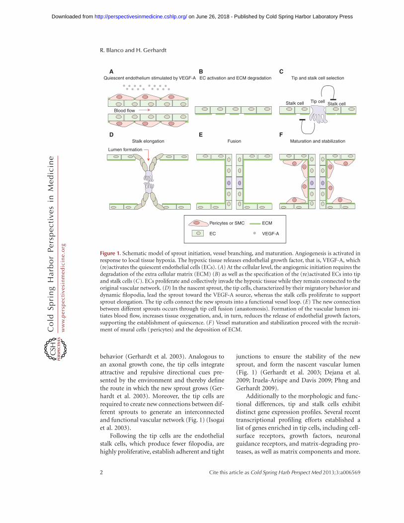
The vascular endothelial growth factor (VEGF)signaling pathway is essential for the regulationof sprouting angiogenesis in physiological andpathological conditions, controlling multipleaspects of endothelial behavior such as endo-thelial differentiation, migration, proliferation,survival, and permeability control (Fig. 2) (Fer-rara et al. 2003).
In mammals, the VEGF family of growthfactors encompasses six secreted dimeric glyco-proteins (VEGF-A, -B, -C, -D, -E, and placentagrowth factor [PlGF]), which display distinctaffinities for three tyrosine kinase receptors(VEGF receptor 1 [VEGFR1], 2 [VEGFR2],and 3 [VEGFR3]) (Fig. 2). The VEGF ligandsare expressed in many cells and tissues, whereasthe receptors were initially considered endothe-lial-specific. VEGFR1 and 2 are most stronglyexpressed in vascular endothelial cells but arealso expressed in myeloid cells and neurons
(Barleon et al. 1996; Bellon et al. 2010; Mura-matsu et al. 2010). In the adult, VEGFR3 isrestricted to the lymphatic endothelium. How-ever, activated blood endothelial cells in theembryo and during postnatal and tumor angio-genesis up-regulate VEGFR3 expression andsignaling (Tammela et al. 2008).
Among the different ligands, VEGF-A ismost prominently involved in the angiogenicprocess. VEGF-A expression is induced byhypoxic conditions (in a hypoxia-induciblefactor Hif1/2–dependent manner), cytokines,growth factors, hormones, oncogenes, andtumor-suppressor genes (Dvorak 2005). Differ-ent VEGF-A splice isoforms have been reportedin human and mouse (Tischer et al. 1991).These splice variants differ in their tissuedistribution because of the presence or absenceof carboxy-terminal heparan-sulfate-bindingretention motifs that mediate interactionswith cell surface and extracellular matrix pro-teoglycans (Park et al. 1993). All VEGF-A iso-forms interact specifically with VEGFR1 andVEGFR2 but show highest affinity for bindingto VEGFR1. Nevertheless, the tyrosine kinaseactivity of VEGFR2 is the main mediator ofVEGF-A signaling during sprouting angiogene-sis (Fig. 2) (Takahashi and Shibuya 2005; Ols-son et al. 2006). VEGF-C-activating VEGFR3has also been shown to mediate proangiogenicsignaling, and the processed form of VEGF-Ccan also activate VEGFR2. In brief, the VEGFligand–receptor interaction induces dimeriza-tion of the VEGF receptor, which, in turn,results in the autophosphorylation of differenttyrosine residues that recruit SH-2-containingadaptors and downstream kinases. Successively,this process activates a variety of downstreamsignaling pathways that regulate endothelialcell migration, survival, proliferation, and tubeformation (Fig. 2).
In contrast to VEGFR2, VEGFR1 has veryweak kinase activity and acts as a decoy receptor,competitively reducing VEGF binding toVEGFR2 and therefore limiting the activity ofVEGF pathway in the vascular endothelial cells(Park et al. 1994). In addition, alternative splic-ing of VEGFR1 produces a soluble isoform ofthis receptor (sVEGFR1) that sequesters VEGF
VEGF and Notch in Tip and Stalk Cell Selection
Cite this article as Cold Spring Harb Perspect Med 2013;3:a006569 3
ww
w.p
ersp
ecti
vesi
nm
edic
ine.
org
on June 26, 2018 - Published by Cold Spring Harbor Laboratory Press http://perspectivesinmedicine.cshlp.org/Downloaded from
in the extracellular medium and thereby modu-lates VEGF signaling in the vascular endothe-lium (Tanaka et al. 1997). Furthermore, thecell-surface glycoproteins neuropilin1 (Nrp1)and neuropilin2 (Nrp2), originally identifiedas receptors for semaphorins, also modulatethe VEGF signaling output. Both proteins actas VEGF non-kinase coreceptors, enhancingthe binding activity of VEGF to VEGFR1 and2 in the case of Nrp1, and of VEGF-C toVEGFR3 in the case of Nrp2 (Fig. 2) (Ferraraet al. 2003; Takahashi and Shibuya 2005).
Direct evidence for the implication of VEGFligand and receptors in vasculogenesis andangiogenesis comes from the study of knockoutmice for the VEGF ligand and the differentVEGFRs genes. In general, homozygous dele-tion for the VEGF-A gene or any of the VEGFRsgenes involved in the VEGF signaling pathwayleads to embryonic death in utero betweenembryonic days 8.5 (E8.5) and E10.5, as a con-sequence of abnormal vascular development(Fong et al. 1995; Shalaby et al. 1995; Carmelietet al. 1996; Ferrara et al. 1996; Dumont et al.
Dimerization orbinding domain
A
B
Tyrosinekinase domain
a1a2
c1
b1b2 VEGF binding domain
VEGFR1 VEGFR2 VEGFR3
S-S
S-S S-S S-S
AngiogenesisVasculogenesis
NRP1/2
LymphangiogenesisAngiogenesis
VEGF-AVEGF-BPIGF
VEGF-AVEGF-BPIGF
VEGF-AVEGF-CVEGF-DVEGF-E
VEGF-AVEGF-CVEGF-D
VEGF-CVEGF-D
sVEGFR1
NRP2 NRP2NRP1 NRP1
VEGFR2VEGFR1 HeterodimerVEGFR2/3
VEGFR3
Endothelial cell:
Migration Proliferation Cell survival Vascular permeability
Figure 2. VEGFs, VEGF receptors, and coreceptors. (A) Outline of the structural domains of the differentVEGFRs and coreceptors Nrp1 and Nrp2. (B) Schematic representation of the binding specificity of the differentVEGF family members (VEGF-A, -B, -C, -D, -E, and PlGF) to the tyrosine kinase receptors, VEGFR1, VEGFR2,and VEGFR3. Interactions between VEGFRs and the coreceptors Nrp1 or Nrp2 are also shown in the figure.Activation of VEGFR1 and VEGFR2 regulates vasculogenesis and angiogenesis. However, activation of VEGFR3stimulates lymphangiogenesis and embryonic angiogenesis. In particular, during sprouting angiogenesisVEGFR2 is the principal mediator of VEGF-A signaling, activating a variety of downstream signaling pathwaysthat regulate endothelial cell migration, survival, proliferation, and tube formation. In contrast, VEGFR1 orsVEGFR1, acting as a decoy receptor, limits the VEGF activity in the vascular endothelium.
R. Blanco and H. Gerhardt
4 Cite this article as Cold Spring Harb Perspect Med 2013;3:a006569
ww
w.p
ersp
ecti
vesi
nm
edic
ine.
org
on June 26, 2018 - Published by Cold Spring Harbor Laboratory Press http://perspectivesinmedicine.cshlp.org/Downloaded from
1998). Importantly, embryonic lethality be-tween days E11 and E12 has also been observedassociated with the heterozygous deletion ofVEGF-A (Carmeliet et al. 1996; Ferrara et al.1996). As is discussed below, VEGF-A andDll4 (Notch pathway ligand) are the onlyproteins of both pathways for which the hetero-zygosity results in a lethal embryonic pheno-type, highlighting the essential and uniquerole of both proteins during angiogenesis. Incontrast, deletion of VEGF-B, -C, or PlGF genesproduces only subtle vascular phenotypes dur-ing development (Autiero et al. 2003; Reicheltet al. 2003).
NOTCH SIGNALING PATHWAY
The Notch pathway is an evolutionarily con-served intercellular contact-dependent signal-ing mechanism involved in multiple cell fatedecisions and patterning processes duringnormal development. Among other functions,Notch signaling has been implicated in cellfate determination and differentiation of epi-thelial, neuronal, bone, blood, and mostrecently, endothelial cells (Karsan 2005; Bray2006; Louvi and Artavanis-Tsakonas 2006;Radtke et al. 2006).
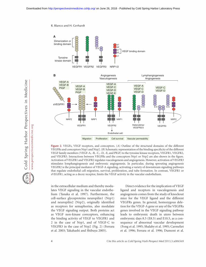
In vertebrates, four Notch receptors (fromNotch1 to Notch4) and five Notch transmem-brane ligands (Jagged1 and Jagged2, homo-logs to Serrate, and Delta-like ligand 1 [Dll1],3 [Dll3], and 4 [Dll4]) constitute the core com-ponents of the Notch pathway (Ellisen et al.1991; Weinmaster et al. 1992; Lardelli et al.1994; Bettenhausen and Gossler 1995; Lindsellet al. 1995; Shawber et al. 1996; Uyttendaeleet al. 1996; Dunwoodie et al. 1997; Rao et al.2000; Shutter et al. 2000; Mailhos et al. 2001).Notch signaling begins with receptor–ligandinteractions between neighboring cells. Thisinteraction triggers a series of proteolytic cleav-ages of the Notch receptors. The final one, cata-lyzed by the g-secretase complex, releases theactive Notch intercellular domain (NICD)from the cell membrane (Weinmaster 1998;Mumm and Kopan 2000). Subsequently,NICD translocates to the nucleus, where itinteracts directly with the transcription factor
CSL (also known as CBF1, Su [H], or Lag2).In absence of NICD, CSL functions as a tran-scriptional repressor through association witha corepressor complex that contains, amongother components, histone deacetylases (Kaoet al. 1998). Following binding of NICD toCSL, the corepressor complex is convertedinto an activation complex by displacing core-pressors and recruiting coactivators. NICD,CSL, Mastermind-like (MAML) polypeptides,and the histone acetylase CBP/p300 are keycomponents of the Notch transcriptional acti-vator complex (Fryer et al. 2002; Jeffries et al.2002). Recruitment of the coactivator complexto promoters containing CSL-binding elementsinduces transcriptional gene activation. Thissignaling mechanism is known as the “canoni-cal Notch pathway” (Fig. 3). Hairy/enhancerof split (HES) and HES-related proteins(HEY/HRT/HERP) family genes are well-characterized direct target genes of Notch sig-naling (Nakagawa et al. 2000; Davis and Turner2001; Iso et al. 2003). Moreover, the Dll4 liganditself is also a target gene regulated by the Notchsignaling pathway (Uyttendaele et al. 1996;Sainson et al. 2005).
Multiple observations indicate that theNotch signaling pathway plays a key role at dif-ferent stages of vascular development. TheNotch receptors 1 and 4 and also four of thefive different ligands are expressed in the vascu-lature (Hofmann and Luisa Iruela-Arispe2007). Furthermore, global knockout mice forNotch1 and Notch1/Notch4, as well as Jagged1, die in utero, between E9.5 and E10.5, display-ing severe vascular abnormalities produced bythe lack of capacity to remodel the primordialvascular plexus into a hierarchical network(Xue et al. 1999; Krebs et al. 2000). Similarlyto knockout studies, mice with a constitutiveendothelium-specific expression of an activatedallele for Notch4 (Notch4/int3) die duringembryonic development, at day E10.5, andshow vascular remodeling abnormalities remi-niscent of those observed in Notch1-deficientmice (Uyttendaele et al. 2001). Moreover,mice deficient in Dll4 ligand die during earlyembryogenesis from vascular defects similarto those of Notch1/Notch4-deficient mice.
VEGF and Notch in Tip and Stalk Cell Selection
Cite this article as Cold Spring Harb Perspect Med 2013;3:a006569 5
ww
w.p
ersp
ecti
vesi
nm
edic
ine.
org
on June 26, 2018 - Published by Cold Spring Harbor Laboratory Press http://perspectivesinmedicine.cshlp.org/Downloaded from

Surprisingly, the genetic requirement of Dll4 isdose-dependent, and heterozygous Dll4 dele-tion is also lethal on most genetic backgrounds.Haploinsufficiency of Dll4 ligand results in vas-cular remodeling defects, arteriovenous malfor-mation, and an incomplete formation ofarteries (Duarte et al. 2004; Gale et al. 2004;Krebs et al. 2004). Moreover, Dll4þ/ – miceshow an increase in the number of vascularsprouts and vessel branches in the growing frontof some vascular beds, such as yolk sac (Galeet al. 2004). It is important to note that theDll4 ligand is the only Notch pathway compo-nent showing haploinsufficiency (Duarte et al.2004; Gale et al. 2004; Krebs et al. 2004), high-lighting the particular importance of Dll4 in
vascular development. Furthermore, endothe-lial-specific deletion of CSL leads to similardefects as in Notch1, Notch1/Notch4, or Dll4knockout mice. Additionally, simultaneousinactivation of two transcriptional genes regu-lated by the Notch signaling pathway, Hey1and Hey2, also results in embryonic lethalitydue to vascular defects (Fisher et al. 2003).
In sum, these different phenotypes showthat the Notch pathway must be tightlyregulated to ensure proper vascular formationduring embryonic development, postnatalangiogenesis, and, as we will discuss below, dur-ing pathological angiogenesis (Limbourg et al.2005; Noguera-Troise et al. 2006; Ridgwayet al. 2006; Li et al. 2007; Takeshita et al. 2007).
MAMLADAMγ-secretase p300
NICD
NICD translocateto the nucleus
Endothelial receiving cell or S
TALK
cell
CoRs
CSL
Notch receptor
Delta/jagged ligandEnd
othe
lial s
endi
ng c
ell o
r TIP
cel
l
NICDCSL
Figure 3. Canonical Notch signaling pathway. The Notch pathway is an evolutionarily conserved intercellularsignaling mechanism implicated in cell fate determination and differentiation of endothelial cells. Notchsignaling begins with receptor–ligand interaction between neighboring cells. This interaction triggers aseries of proteolytic cleavages of the Notch receptor. The final one, catalyzed by the g-secretase complex, releasesthe active Notch intercellular domain (NICD) from the cell membrane. NICD is translocated to the nucleus,where it interacts directly with the transcription factor CSL. Following binding of NICD to CSL, the repressorcomplex is converted into an activating complex by displacing corepressors and recruiting coactivators. Afterthe coactivator complex is recruited, the transcription of promoters that contains CSL-binding elements isinduced. Hairy/enhancer of split (HES) and HES-related proteins (HEY/HRT/HERP) family genes areincluded among the target genes of Notch signaling. Dll4 ligand is also a target gene regulated by the Notch sig-naling pathway.
R. Blanco and H. Gerhardt
6 Cite this article as Cold Spring Harb Perspect Med 2013;3:a006569
ww
w.p
ersp
ecti
vesi
nm
edic
ine.
org
on June 26, 2018 - Published by Cold Spring Harbor Laboratory Press http://perspectivesinmedicine.cshlp.org/Downloaded from

IMPLICATION OF VEGF AND NOTCHPATHWAY ON TIP/STALK SELECTION
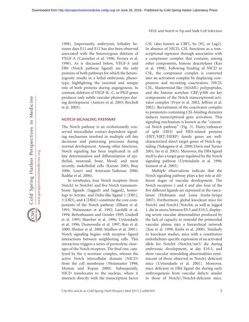
VEGF-A and Notch signaling pathways are keyplayers governing tip and stalk cell behavior(Fig. 4).
Normal sprouting angiogenesis in themouse retina requires a gradient of the pro-angiogenic factor VEGF-A (Ruhrberg et al.2002; Gerhardt et al. 2003). This local VEGF-Agradient is produced by the preformed astrocyte
network that serves as a guiding scaffold forthe developing retinal plexus (Fruttiger 2002;Gerhardt et al. 2003). In fact, the tip cells areclosely attached to the astrocytes, and theirfilopodia extend along the astrocytes towardhigher VEGF-A concentrations in this gradient(Gerhardt et al. 2003). Interestingly, if VEGF-Astimulation is blocked using sVEGFR1 orneutralizing the signaling through VEGFR2with antibodies, the tip cell filopodia are com-pletely retracted in the sprouting retinal front
Tip to stalk lateral inhibition
VEGF-AVEGF gradient
VEGFreceptor signaling
High DII4
STA
LK c
ell
Low Dll4
Jagged 1
SIRT1High Notch signaling
Nonsprouting
High Nrarp
WntHigh VEGFR1/sVEGFR1
Low VEGFR2
Low VEGFR1/sVEGFR1
High VEGFR2
FringeLow Notch signaling
Sprouting
VEGFR2
NRP1/2
VE
GF
grad
ient
TIP
cel
l
Tip cell
Stalk cell
Lumen formation
Figure 4. Tip/stalk cell specification during sprouting angiogenesis. During sprouting angiogenesis, VEGF andNotch signaling pathways are implicated in the specification of tip and stalk cells in the vascular endothelium.VEGF interacts with VEGFR2, expressed at the surface of the endothelial cells of the quiescent vessels. Nrp1modulates the VEGF signaling output, enhancing the binding activity and signaling of VEGF through VEGFR2.Under VEGF stimulation, Dll4 expression is up-regulated in the tip cells. In turn, Dll4 ligand activates Notchsignaling in the stalk, consequently suppressing the tip cell phenotype. Notch signaling activation reducesVEGFR2 expression and increases VEGFR1/sVEGFR1 levels as well as the expression of different Notch targetgenes (e.g., Notch-regulated ankyrin repeat protein [Nrarp]). In contrast, the tip cell receives low Notch signal-ing, allowing high expression of VEGFR2 and Nrp1, but low VEGFR1. Contrary to Dll4, Jagged1 ligand isexpressed by the stalk cells. Jagged1 antagonizes Dll4–Notch signaling in the sprouting front when the Notchreceptor is modified by the glycosyltranferase Fringe, thereby enhancing differential Notch activity between tipand stalk cells. The duration and amplitude of the Notch signal are modulated by the histone deacetylase SIRT1,which, by direct deacetylation, primes the Notch intracellular domain for ubiquitination and degradation.
VEGF and Notch in Tip and Stalk Cell Selection
Cite this article as Cold Spring Harb Perspect Med 2013;3:a006569 7
ww
w.p
ersp
ecti
vesi
nm
edic
ine.
org
on June 26, 2018 - Published by Cold Spring Harbor Laboratory Press http://perspectivesinmedicine.cshlp.org/Downloaded from

(Gerhardt et al. 2003; Suchting et al. 2007), pre-venting tip cell migration and thus progres-sion of vascular sprouting seizes (Gerhardtet al. 2003). In contrast, stimulation of quies-cent vessels with VEGF-A induces filopodia for-mation and tip cell gene expression (Hellstromet al. 2007a), together illustrating that VEGF-Ais both necessary and sufficient to induce endo-thelial tip cells. Similar observations in zebrafishembryogenesis (Leslie et al. 2007) and other tis-sues in the mouse confirm the general impor-tance of this concept.
If VEGF-A induces tip cell formation, it issurprising to see that tip cells are normally notformed by adjacent endothelial cells but format characteristic intervals, enabling adequatelyspaced branching and sprouting. The molecularmechanism responsible for the fact that not allendothelial cells stimulated by VEGF becometip cells is based on the activity of Dll4/Notchsignaling (Fig. 4).
Under physiological conditions, short-termadministration of g-secretase inhibitors (GSIs),which potently block Notch signaling, or phar-macological inhibition of Dll4/Notch interac-tions with a neutralizing antibody (Dll4-Fc)dramatically increases sprouting, branching,and filopodia formation at the leading front ofthe retinal vascular plexus in development,resulting in a much denser and more intercon-nected plexus (Fig. 5) (Hellstrom et al. 2007b;Lobov et al. 2007; Suchting et al. 2007). Thehypersprouting phenotype correlates with anincrease in endothelial cells positive for the tipcell markers PDGFB or UNC5B and the num-ber of endothelial filopodia protrusions, indi-cating increased tip cell formation in theabsence of Notch signaling (Fig. 5) (Hellstromet al. 2007b; Suchting et al. 2007). Similar effectshave been reported in the postnatal mouse ret-ina and embryonic hindbrain vascularizationafter genetic deletion of one Dll4 allele or endo-thelial-specific Notch1 loss of function (VEcad-CreERT2/R26R/Notch1fl/fl mice) (Hellstromet al. 2007b; Lobov et al. 2007; Suchting et al.2007). Both the pharmacological inhibitionand genetic studies illustrate that the Dll4/Notch pathway critically regulates the forma-tion of an adequate ratio of tip and stalk cells
by suppressing tip cell formation in adjacentcells (Figs. 4,5). Notch gain-of-function studiesfurther support this idea; activation of Notchsignaling using a synthetic Jagged1 ligand pep-tide reduces filopodia formation and branch-ing, suggesting a decrease in the number ofendothelial cells that acquire tip cell behavior(Hellstrom et al. 2007b).
Several studies in zebrafish embryogenesisillustrated that the role of Notch signaling inangiogenesis is evolutionarily conserved (Leslieet al. 2007; Siekmann and Lawson 2007).Knocking down, with a morpholino oligonu-cleotide, the translation of Rbpsuh protein, aDNA-binding protein that mediates transcrip-tion of Notch target genes (Lecourtois andSchweisguth 1995) (known as CSL protein inmice), results in excessive sprouting andbranching of the segmental arteries. Addition-ally, more endothelial cells in the arteries displaya migratory behavior, typically restricted to theleading tip cells. These cells also show greatlyincreased filopodia activity and up-regulationof tip cell markers such as VEGFR3 (Siekmannand Lawson 2007). GSI treatment, as well asDll4, or Notch1b loss-of-function phenocopiesthe segmental artery defects associated withRbpsuh loss of function, whereas overactivationof the Notch pathway suppresses angiogenesis(Leslie et al. 2007; Siekmann and Lawson2007). Transplantation experiments illustratedthat knockdown of active Notch signalingpromotes tip cell formation, whereas over-expression of active Notch inhibits tip cellformation in a cell-autonomous manner, illus-trating that the activation status of Notch sig-naling quantitatively determines the ability ofindividual cells to become tip or stalk cellswithin the sprout.
Dll4 is not the only Notch ligand involved intip cell regulation. Recent studies showed thatJagged1 is more strongly expressed in stalk cells(Hofmann and Luisa Iruela-Arispe 2007; Bene-dito et al. 2009). Genetic deletion or endothelialoverexpression of Jagged1 in vivo resulted indramatically reduced or increased sprouting,respectively (Fig. 5). Surprisingly, endothelialNotch signaling is more active in the absenceof Jagged1, indicating that Jagged1 negatively
R. Blanco and H. Gerhardt
8 Cite this article as Cold Spring Harb Perspect Med 2013;3:a006569
ww
w.p
ersp
ecti
vesi
nm
edic
ine.
org
on June 26, 2018 - Published by Cold Spring Harbor Laboratory Press http://perspectivesinmedicine.cshlp.org/Downloaded from

regulates Notch activity (Figs. 4 and 5). In vitrostudies confirmed that Jagged1 antagonizesDll4–Notch signaling, suggesting, togetherwith the complementary distribution of Dll4and Jagged in the growing vascular plexus,that Jagged1 enhances differential Notch activ-ity by competitively interfering with the abilityof Dll4 expressed by stalk cells to activate Notchin tip cells (Figs. 4 and 5) (Benedito et al. 2009).Intriguingly, the investigators show that the dif-ferential ability of Jagged1 and Dll4 ligands toactivate Notch is dependent on the glycosyl-transferase Fringe. Whereas the glycosylatedNotch receptor shows strong activation byDll4 and competitive inhibition by Jagged1,the unglycosylated receptor in the absence of
Fringe can be activated by Jagged1 (Fig. 4)(Benedito et al. 2009). Exactly how this glycosy-lation event by Fringe might be controlled andwhether it can locally function to fine-tuneNotch-mediated vascular density remain to beshown. Nevertheless, the differential expressionand action of Dll4 and Jagged1 illustrate that theoutput of the Notch signaling pathway, whichappears to function as a central regulator of vas-cular branching, can be modulated not only bythe quantity of individual ligands, but also bytheir activity profile when interacting with thereceptor. As we see below, the Notch outputcan further be regulated by multiple feedbackmechanisms that affect both the quantity andduration of Notch signaling.
Jagged 1
A Normal Dll4/Notch signaling pathway
Notch
Notch
Stalk cell
Inhibition of Dll4/Notch signaling pathwayPhysiological and
pathological angiogenesis
Notch DII4
VE
GF
gra
dien
tS
prou
ting
dire
ctio
n
Notch• Reduced number of tip cells• Lower sprouting• Lower branching• Network less dense and interconnected
C Inhibition of Jagged 1
Dll4
Notch • High number of tip cells• Increased sprouting• Increased branching• Network much more dense and interconnected
Stalk cell Stalk cellStalk cellTip cell Tip cell
Jagged 1
Dll4
B
Figure 5. Dll4 and Jagged1 show antagonistic function during sprouting angiogenesis. Tip/stalk cell selection ismediated via Dll4/Notch lateral inhibition between the ECs that constitute the vasculature, resulting in thetypical salt-and-pepper distribution of tip cells along the vascular endothelium. (A) For a detailed explanationabout Dll4/Notch lateral inhibition see Figure 4. (B) Genetic or pharmacological inhibition of Dll4 deregulatesthe tip/stalk selection process; dramatically increases the number of tip cells, the number of sprouts and branch-ing, resulting in a hyperdense and interconnected plexus. (C) The opposite observation has been made afterJagged1 inhibition in physiological and pathological angiogenesis, as Jagged1 antagonizes reciprocal Dll4-medi-ated Notch activation from the stalk to the tip cell.
VEGF and Notch in Tip and Stalk Cell Selection
Cite this article as Cold Spring Harb Perspect Med 2013;3:a006569 9
ww
w.p
ersp
ecti
vesi
nm
edic
ine.
org
on June 26, 2018 - Published by Cold Spring Harbor Laboratory Press http://perspectivesinmedicine.cshlp.org/Downloaded from
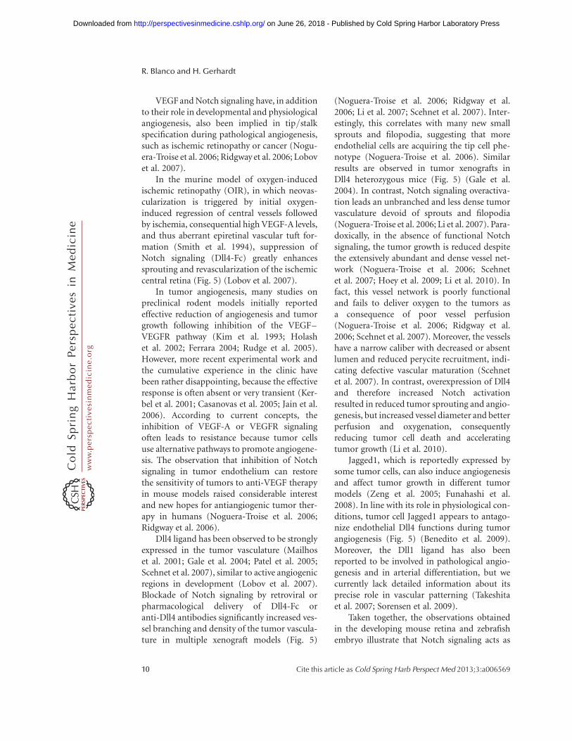
VEGF and Notch signaling have, in additionto their role in developmental and physiologicalangiogenesis, also been implied in tip/stalkspecification during pathological angiogenesis,such as ischemic retinopathy or cancer (Nogu-era-Troise et al. 2006; Ridgway et al. 2006; Lobovet al. 2007).
In the murine model of oxygen-inducedischemic retinopathy (OIR), in which neovas-cularization is triggered by initial oxygen-induced regression of central vessels followedby ischemia, consequential high VEGF-A levels,and thus aberrant epiretinal vascular tuft for-mation (Smith et al. 1994), suppression ofNotch signaling (Dll4-Fc) greatly enhancessprouting and revascularization of the ischemiccentral retina (Fig. 5) (Lobov et al. 2007).
In tumor angiogenesis, many studies onpreclinical rodent models initially reportedeffective reduction of angiogenesis and tumorgrowth following inhibition of the VEGF–VEGFR pathway (Kim et al. 1993; Holashet al. 2002; Ferrara 2004; Rudge et al. 2005).However, more recent experimental work andthe cumulative experience in the clinic havebeen rather disappointing, because the effectiveresponse is often absent or very transient (Ker-bel et al. 2001; Casanovas et al. 2005; Jain et al.2006). According to current concepts, theinhibition of VEGF-A or VEGFR signalingoften leads to resistance because tumor cellsuse alternative pathways to promote angiogene-sis. The observation that inhibition of Notchsignaling in tumor endothelium can restorethe sensitivity of tumors to anti-VEGF therapyin mouse models raised considerable interestand new hopes for antiangiogenic tumor ther-apy in humans (Noguera-Troise et al. 2006;Ridgway et al. 2006).
Dll4 ligand has been observed to be stronglyexpressed in the tumor vasculature (Mailhoset al. 2001; Gale et al. 2004; Patel et al. 2005;Scehnet et al. 2007), similar to active angiogenicregions in development (Lobov et al. 2007).Blockade of Notch signaling by retroviral orpharmacological delivery of Dll4-Fc oranti-Dll4 antibodies significantly increased ves-sel branching and density of the tumor vascula-ture in multiple xenograft models (Fig. 5)
(Noguera-Troise et al. 2006; Ridgway et al.2006; Li et al. 2007; Scehnet et al. 2007). Inter-estingly, this correlates with many new smallsprouts and filopodia, suggesting that moreendothelial cells are acquiring the tip cell phe-notype (Noguera-Troise et al. 2006). Similarresults are observed in tumor xenografts inDll4 heterozygous mice (Fig. 5) (Gale et al.2004). In contrast, Notch signaling overactiva-tion leads an unbranched and less dense tumorvasculature devoid of sprouts and filopodia(Noguera-Troise et al. 2006; Li et al. 2007). Para-doxically, in the absence of functional Notchsignaling, the tumor growth is reduced despitethe extensively abundant and dense vessel net-work (Noguera-Troise et al. 2006; Scehnetet al. 2007; Hoey et al. 2009; Li et al. 2010). Infact, this vessel network is poorly functionaland fails to deliver oxygen to the tumors asa consequence of poor vessel perfusion(Noguera-Troise et al. 2006; Ridgway et al.2006; Scehnet et al. 2007). Moreover, the vesselshave a narrow caliber with decreased or absentlumen and reduced perycite recruitment, indi-cating defective vascular maturation (Scehnetet al. 2007). In contrast, overexpression of Dll4and therefore increased Notch activationresulted in reduced tumor sprouting and angio-genesis, but increased vessel diameter and betterperfusion and oxygenation, consequentlyreducing tumor cell death and acceleratingtumor growth (Li et al. 2010).
Jagged1, which is reportedly expressed bysome tumor cells, can also induce angiogenesisand affect tumor growth in different tumormodels (Zeng et al. 2005; Funahashi et al.2008). In line with its role in physiological con-ditions, tumor cell Jagged1 appears to antago-nize endothelial Dll4 functions during tumorangiogenesis (Fig. 5) (Benedito et al. 2009).Moreover, the Dll1 ligand has also beenreported to be involved in pathological angio-genesis and in arterial differentiation, but wecurrently lack detailed information about itsprecise role in vascular patterning (Takeshitaet al. 2007; Sorensen et al. 2009).
Taken together, the observations obtainedin the developing mouse retina and zebrafishembryo illustrate that Notch signaling acts as
R. Blanco and H. Gerhardt
10 Cite this article as Cold Spring Harb Perspect Med 2013;3:a006569
ww
w.p
ersp
ecti
vesi
nm
edic
ine.
org
on June 26, 2018 - Published by Cold Spring Harbor Laboratory Press http://perspectivesinmedicine.cshlp.org/Downloaded from
a negative regulator of VEGF-induced angio-genesis and is essential for proper vascularmorphogenesis. Moreover, these data indicatethat the tip/stalk specification principle, whichworks during physiological angiogenesis, alsoapplies to pathological sprouting angiogenesis(Figs. 4,5).
CROSS TALK BETWEEN THE VEGFAND NOTCH SIGNALING PATHWAYS
The VEGF and Notch signaling pathways areboth key regulators of the angiogenic process,being both involved in the specification of thetip and stalk cell phenotype (Fig. 4). It is there-fore imperative that we ask how these twopathways work together to achieve functionalpatterning during sprouting angiogenesis.
Several lines of evidence indicate that VEGFsignaling acts upstream of the Notch pathwayduring physiological and pathological angio-genesis (Stone et al. 1995; Lawson et al. 2002;Patel et al. 2005; Ridgway et al. 2006; Lobovet al. 2007), controlling the expression of differ-ent Notch components.
In in vitro studies, VEGF-A stimulationconsistently increases the expression of Dll4protein on the surface of human umbilicalvein endothelial cells (HUVECs) (Ridgwayet al. 2006). Also in human tumor samples,the expression of Dll4 mRNA increased up toninefold in tumor blood vessels comparedwith vessels in the surrounding healthy tissueand correlated with increased local VEGF-Aexpression (Patel et al. 2005). Conversely, arapid decrease of Dll4 expression has beenreported in tumor blood vessels of differentxenograft models after VEGF-A blockade(Noguera-Troise et al. 2006; Thurston and Kita-jewski 2008).
Additionally to the observations reportedbefore, hypoxic conditions, which prominentlyinduce VEGF-A expression, appear to directlyup-regulate Dll4 mRNA expression in differentendothelial cell lines in an Hif1a-dependentmanner (Fig. 4) (Patel et al. 2005; Williamset al. 2006). Consistent with this notion, inthe OIR model, high expression of Dll4 hasbeen observed in veins and capillaries or sprouts
in close proximity to the avascular-hypoxicregions (Lobov et al. 2007).
The same principles apply to normal devel-opment angiogenesis in the mouse retina,where high Dll4 expression is found at the vas-cular sprouting front, immediately adjacent tothe avascular and hypoxic retinal peripherythat expresses high levels of VEGF-A (Fig. 4)(Stone et al. 1995; Gerhardt et al. 2003; Liuet al. 2003; Lobov et al. 2007; Suchting et al.2007). Retinal VEGF blockade using the VEGF-Trap inhibited Dll4 mRNA expression in thegrowing retinal vascular plexus. Conversely,intravitreal injection of VEGF increases Dll4expression, collectively showing that VEGF-Asignaling in endothelial cells in vitro and invivo quantitatively regulates Dll4 expression(Lobov et al. 2007; Suchting et al. 2007).
This is not only true for the sprouting front,because VEGF-A also acts upstream of Dll4 dur-ing the determination of arterial–endothelialcell fate in the zebrafish model (Lawson et al.2002).
Exactly how VEGF-A signaling controlsDll4 is less well understood. In arteries,VEGF-A-mediated induction of Dll4 has beenreported to involve both PI3K activation andthe activity of the Forkhead family transcrip-tions factors FoxC1 and FoxC2 (Hayashi andKume 2008). A recent study further identifieda conserved transcriptional repressor complexcomprising Tel and CtBP, which occupy theDll4 promoter, and upon VEGF-A stimulationtransiently disassemble, leading to temporalderepression of Dll4 transcription (Roukenset al. 2010). The timing of this event correlateswith Erk1/2 phosphorylation, but the detailedactivation pathway remains unclear.
In addition to VEGF-A acting upstream ofDll4, it has become clear that Notch, in turn,controls VEGF signaling. In general, all obser-vations support the concept that Notch signal-ing via Dll4 principally acts as a negativefeedback regulator to control VEGF-inducedangiogenesis. Accordingly, endothelial Notchactivation regulates the expression of the differ-ent VEGFRs (VEGFR1, 2, and 3) as well as thecoreceptor Nrp1 (Fig. 4). Notch activation inHUVECS leads to VEGFR1 mRNA induction
VEGF and Notch in Tip and Stalk Cell Selection
Cite this article as Cold Spring Harb Perspect Med 2013;3:a006569 11
ww
w.p
ersp
ecti
vesi
nm
edic
ine.
org
on June 26, 2018 - Published by Cold Spring Harbor Laboratory Press http://perspectivesinmedicine.cshlp.org/Downloaded from
(Harrington et al. 2008; Funahashi et al. 2010).In contrast, VEGFR2 and Nrp1 mRNA is mark-edly reduced by Notch activation in HUVECs(Soker et al. 1998; Ridgway et al. 2006; Williamset al. 2006), indicating that Notch signalingis able to regulate how the endothelial cellsrespond to VEGF. In fact, it has been suggestedthat the reduction of VEGFR2 and Nrp1 expres-sion may be responsible for the low prolifera-tive and migratory response observed in Dll4-overexpressing endothelial cells (Williamset al. 2006).
Similar interactions between VEGF andNotch pathways have been described in vivo.Down-regulation of VEGFR2 has been reportedin blood vessels in Dll4-overexpressing tumors(Li et al. 2007). In contrast, genetic inactivationof one allele of Dll4 in mice significantlyincreases the expression of VEGFR2 mRNAwhile decreasing the levels of VEGFR1 mRNA(Suchting et al. 2007). Moreover, independentreports show that endothelial cells in Dll4heterozygous retinas or in wild-type micetreated with g-secretase inhibitors up-regulateVEGFR3 (Tammela et al. 2008). Equivalentresults were obtained in zebrafish (Siekmannand Lawson 2007), where VEGFR3 is involvedon driving sprouting angiogenesis in interseg-mental arteries (Covassin et al. 2006; Tammelaet al. 2008). Thus, Notch activity appears tomodulate the sensitivity of endothelial cells tothe inductive activity of VEGF-A, but up-regu-lating the decoy receptor VEGFR1 and down-regulating the signaling receptors VEGFR2and VEGFR3 (Fig. 4).
TIP/STALK SELECTION MODEL
In accordance with the experimental observa-tions, the current concepts propose that theVEGF and Notch signaling pathways cooperatein tight coordination to specify and balance thetip and stalk cell phenotype between the endo-thelial cells that constitute the sprouts duringthe physiological and pathological angiogenicprocess. To this end, Notch signaling acts in anegative feedback loop with VEGF signalingduring sprouting angiogenesis (Fig. 4). Themodel stipulates that the initial angiogenic
response is induced by VEGF gradients estab-lished in the hypoxic tissues. Under VEGF stim-ulation, the endothelial cells of the pre-existingvessel become activated and compete for the tipcell position by Dll4/Notch signaling (Fig. 4).
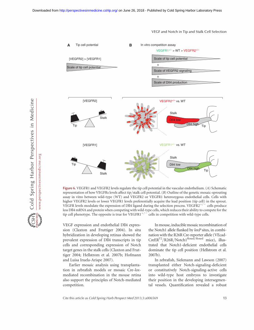
Small stochastic differences in VEGFR2expression or activity between endothelial cells,or differences in the local VEGF environmentof the individual cells could initially give tosome endothelial cells a competitive advantageagainst their neighbor to acquire the tip pheno-type (Fig. 6). Indeed, the ability of an individualcell to become a tip cell appears to be strictlydependent on its VEGFR expression profilein comparison with that of the neighboringcells (Fig. 6). Specifically, in in vitro sproutingcompetition assays, in developmental analysisof chimeric mouse retinal vasculature, and ina new computational model of tip cell selec-tion, endothelial cells with genetically encodedhigher VEGFR2 or lower VEGFR1 levels showeda remarkable competitive advantage to developand maintain the tip cell behavior compared totheir wild-type neighbors (Fig. 6) (Jakobssonet al. 2010). Surprisingly, the VEGFR levelshave no effect on migration speed or cell prolif-eration. Also, this competition is remarkablyrobust to changes in relative cell contribution,together illustrating that the mechanism ofcompetition for the tip cell is not a migrationrace to the tip or a relative overgrowth of onepopulation. Instead, it turns out that theVEGFR levels directly translate into Dll4 expres-sion and that VEGFR levels only affect tip cellpotential in the presence of active Notch signal-ing. In this “battle for the lead,” the endothelialcell that expresses more Dll4 ligand than itsneighbors acquires the tip cell phenotype be-cause it can more effectively inhibit its neigh-bors in the growing sprout (Figs. 4 and 6).Recently, the role of VEGFR1 as negative regula-tor of tip cell formation has been confirmed alsoin zebrafish, indicating that Notch-mediatedregulation of VEGFR1 represents an evolutio-narily conserved mechanism to regulate compe-tition for the tip position (Krueger et al. 2011).
The idea of competition via VEGFR andDll4 levels are consistent with previouslyobserved correlation between locally high
R. Blanco and H. Gerhardt
12 Cite this article as Cold Spring Harb Perspect Med 2013;3:a006569
ww
w.p
ersp
ecti
vesi
nm
edic
ine.
org
on June 26, 2018 - Published by Cold Spring Harbor Laboratory Press http://perspectivesinmedicine.cshlp.org/Downloaded from
VEGF expression and endothelial Dll4 expres-sion (Claxton and Fruttiger 2004). In situhybridization in developing retinas showed theprevalent expression of Dll4 transcripts in tipcells and corresponding expression of Notchtarget genes in the stalk cells (Claxton and Frut-tiger 2004; Hellstrom et al. 2007b; Hofmannand Luisa Iruela-Arispe 2007).
Earlier mosaic analysis using transplanta-tion in zebrafish models or mosaic Cre-lox-mediated recombination in the mouse retinaalso support the principles of Notch-mediatedcompetition.
In mouse, inducible mosaic recombination ofthe Notch1 allele flanked by loxP sites, in combi-nation with the R26R Cre-reporter allele (VEcad-CreERT2/R26R/Notch1floxed/floxed mice), illus-trated that Notch1-deficient endothelial cellsdominate the tip cell position (Hellstrom et al.2007b).
In zebrafish, Siekmann and Lawson (2007)transplanted either Notch-signaling-deficientor constitutively Notch-signaling-active cellsinto wild-type host embryos to investigatetheir position in the developing intersegmen-tal vessels. Quantification revealed a robust
Tip cell potentialA B In vitro competition assay
[VEGFR2] VEGFR2+/– vs. WT
VEGFR1+/– vs. WT[VEGFR1]
Tip Tip
Tip
StalkDII4 low
Dll4 high
Dll4 highDll4 low
Stalk
Stalk
Stalk
Tip
VEGFR1+/– > WT > VEGFR2+/–
Scale of tip cell potential
Scale of tip cell potential
[VEGFR2] > [VEGFR1]
Scale of VEGFR2 signaling
Scale of DII4 production
=̃
=̃
Figure 6. VEGFR1 and VEGFR2 levels regulate the tip cell potential in the vascular endothelium. (A) Schematicrepresentation of how VEGFRs levels affect tip/stalk cell potential. (B) Outline of the genetic mosaic-sproutingassay in vitro between wild-type (WT) and VEGFR2 or VEGFR1 heterozygous endothelial cells. Cells withhigher VEGFR2 levels or lower VEGFR1 levels preferentially acquire the lead position (tip cell) in the sprout.VEGFR levels modulate the expression of Dll4 ligand during the selection process. VEGFR2þ/2 cells produceless Dll4 mRNA and protein when competing with wild-type cells, which reduces their ability to compete for thetip cell phenotype. The opposite is true for VEGFR1þ/2 cells in competition with wild-type cells.
VEGF and Notch in Tip and Stalk Cell Selection
Cite this article as Cold Spring Harb Perspect Med 2013;3:a006569 13
ww
w.p
ersp
ecti
vesi
nm
edic
ine.
org
on June 26, 2018 - Published by Cold Spring Harbor Laboratory Press http://perspectivesinmedicine.cshlp.org/Downloaded from
overrepresentation of Notch-signaling-deficientcells in the tip cell position, whereas Notch-active cells were excluded from the tip positionand instead were confined to the base of thesprouts (Siekmann and Lawson 2007).
More recently, mouse ES cells lacking oneallele of Nrarp, a negative regulator of Notch,showed reduced ability to form tip cells whencompeting with wild-type embryonic stem(ES) cells in the three-dimensional (3D) sprout-ing assay. These results illustrate that even asmall increase in Notch signaling robustly inter-feres with the ability of cells to compete withtheir neighbors for the tip (Jakobsson et al.2010). Also, Dll4-heterozygous ES cells cannotcompete with wild-type cells for the tip. In con-trast, in a mosaic context, endothelial cells defi-cient for Jagged1 ligand preferentially acquirethe tip cell position (Benedito et al. 2009), con-sistent with the idea that Jagged1-deficient cellswill more efficiently activate Notch in neighbor-ing cells. Altogether, the mosaic analyses in var-ious model systems corroborate a model inwhich VEGFR signaling quantitatively regulatesDll4 levels, which, in turn, determine the tip cellpotential of the individual cell in a competitivemanner. A cell that in this process receives moreNotch input will invariably adopt the stalk cellphenotype because it is effectively inhibitedfrom becoming a tip cell (Fig. 4).
The spatial requirements of contact-mediated “lateral inhibition” via Dll4/Notchsignaling theoretically demand that, in a rowof endothelial cells stimulated by VEGF-A,maximally two consecutive stalk cells canform between tip cells. A subsequent divisionof stalk cells should in theory lead to three ormore stalk cells, which, however, cannot inhibiteach other from becoming a tip cell. Any endo-thelial cell that is activated by VEGF-A and notdirectly inhibited by a neighbor expressing highlevels of Dll4 will default to the tip cell pheno-type and thus initiate a new sprout (Fig. 4).This fundamental principle is now believed torepresent the key mechanism of spacing vascu-lar sprouts during normal development.
An agent-based computational model ofthis VEGF–VEGFR–Dll4–Notch–VEGFR regu-latory feedback loop between endothelial cells
(Bentley et al. 2008) strongly supports thetheoretical model and illustrates that underappropriate VEGF stimulation, this feedbackmechanism is sufficient to mediate tip–stalkselection (Bentley et al. 2008). However, thevery same feedback regulation shows very dif-ferent behavior when simulating pathologicallyhigh VEGF concentrations. Instead of establish-ing alternating tip and stalk cells, the endothe-lial cells synchronously oscillate between thetwo phenotypes and thus fail to coordinate asprouting and branching process. Intriguingly,this emergent behavior of the computationalmodel raises the possibility that the aberrantvascular patterning observed in pathologicalsituations could be a consequence of localNotch oscillations in endothelial cell popula-tions exposed to high VEGF-A concentrations.Such synchronous oscillations of Notch signal-ing occur in other developmental systems, mostprominently during somitogenesis (Kageyamaet al. 2007). Although experimental evidencefor the existence of Notch oscillations in theendothelium is currently lacking, the implica-tions warrant efforts to understand the dynamiccontrol of Dll4/Notch signaling in more detail.
TIP/STALK SELECTION IS DYNAMIC
During the continued sprouting, branching,and anastomosis process as the vascular net-work expands, new tip cells will have to beformed, whereas others may convert to stalkcells. Indeed, emergent behavior in the compu-tational model of tip/stalk selection illustratedthat meeting of two tip cells during anastomosiswill trigger renewed competition (Bentley et al.2009). As a consequence of changing endothe-lial neighbors, previously inhibited stalk cellscan be relieved from their inhibition andbecome new tip cells. Thus, endothelial tipand stalk cell specification does not representpermanent cell fate decisions but, rather,dynamic phenotypical specification in flux.
Time-lapse confocal microscopy of clonallylabeled cell populations in zebrafish embryos invivo and in 3D sprouting assays in vitro deliv-ered new insight into the surprisingly dynamicbehavior of tip and stalk cells within individual
R. Blanco and H. Gerhardt
14 Cite this article as Cold Spring Harb Perspect Med 2013;3:a006569
ww
w.p
ersp
ecti
vesi
nm
edic
ine.
org
on June 26, 2018 - Published by Cold Spring Harbor Laboratory Press http://perspectivesinmedicine.cshlp.org/Downloaded from
sprouts. Endothelial cells in the extendingsprouts appear to engage in iterative or con-tinued battles for the tip position, leading tofrequent exchange of the tip cells. The precisereason for this behavior is not fully understood,but Notch signaling again appears to play a partin its regulation (Jakobsson et al. 2010).
The dynamic selection of phenotypes andcontinued competition must arguably serve animportant function, because they come at con-siderable cost; stalk cells actively form lumenand must engage in a repolarization effort, toestablish apical (luminal) and basal domainsduring tubulogenesis. However, if they fre-quently switch between the migratory, leadingtip cell phenotype, and the lumen-forming stalkcell, the entire architecture of the developingvessel is in constant flux, defying the notion ofstable cell identities and functions. Futureefforts will therefore be focused on both under-standing the molecular regulation of the dy-namic behavior and unraveling its importantbiological function.
FUTURE DIRECTIONS
In this article, we have summarized key compo-nents and current concepts of the VEFG andNotch signaling pathways and how they crit-ically regulate vascular patterning by control-ling endothelial tip and stalk cell specificationin sprouting angiogenesis. Despite the vastamount of literature on both pathways and theplethora of recent studies on their interactionsin angiogenesis, many questions remain unan-swered. Are tip and stalk cells universal conceptsapplicable to all areas of angiogenesis? What isthe role of extracellular matrix in their behavior?How is the polarity of tip and stalk cells regu-lated? How is Dll4 expression dynamically con-trolled, and what is the ultimate effector ofNotch determining the stalk cell phenotype?And in disease processes, does the predictionof synchronous Notch signaling oscillationshold true? Will we be able to identify novelcritical parameters and regulators in theVEGF-Dll4/Notch feedback loop that will allowus to devise effective strategies for vascular nor-malization or antiangiogenesis therapy? The
pace of recent development in this area, thegrowing number of researchers focusing onsimilar powerful developmental systems, andthe integration of insights from computationalmodels, developmental biology, and clinicalstudies raise hopes that we may soon be ableto find answers to these and further questions.
ACKNOWLEDGMENTS
H.G. and R.B. are supported by CancerResearch UK, the Lister Institute of PreventiveMedicine, the EMBO Young Investigator Pro-gramme, the Leducq Transatlantic NetworkARTEMIS, and a HFSP Long-term Fellowship.
REFERENCES
Autiero M, Waltenberger J, Communi D, Kranz A, Moons L,Lambrechts D, Kroll J, Plaisance S, De Mol M, Bono F,et al. 2003. Role of PlGF in the intra- and intermolecularcross talk between the VEGF receptors Flt1 and Flk1. NatMed 9: 936–943.
Barleon B, Sozzani S, Zhou D, Weich HA, Mantovani A,Marme D. 1996. Migration of human monocytes inresponse to vascular endothelial growth factor (VEGF)is mediated via the VEGF receptor flt-1. Blood 87:3336–3343.
Bellon A, Luchino J, Haigh K, Rougon G, Haigh J, ChauvetS, Mann F. 2010. VEGFR2 (KDR/Flk1) signalling medi-ates axon growth in response to semaphorin 3E in thedeveloping brain. Neuron 66: 205–219.
Benedito R, Roca C, Sorensen I, Adams S, Gossler A, Frut-tiger M, Adams RH. 2009. The notch ligands Dll4 andJagged1 have opposing effects on angiogenesis. Cell137: 1124–1135.
Bentley K, Gerhardt H, Bates PA. 2008. Agent-based simu-lation of notch-mediated tip cell selection in angiogenicsprout initialisation. J Theor Biol 250: 25–36.
Bentley K, Mariggi G, Gerhardt H, Bates PA. 2009. Tippingthe balance: Robustness of tip cell selection, migrationand fusion in angiogenesis. PLoS Comput Biol 5:e1000549. doi: 10.1371/journal.pcbi.1000549.
Bettenhausen B, Gossler A. 1995. Efficient isolation of novelmouse genes differentially expressed in early postimplan-tation embryos. Genomics 28: 436–441.
Bray SJ. 2006. Notch signalling: A simple pathway becomescomplex. Nat Rev 7: 678–689.
Carmeliet P, Ferreira V, Breier G, Pollefeyt S, Kieckens L,Gertsenstein M, Fahrig M, Vandenhoeck A, Harpal K,Eberhardt C, et al. 1996. Abnormal blood vessel develop-ment and lethality in embryos lacking a single VEGFallele. Nature 380: 435–439.
Casanovas O, Hicklin DJ, Bergers G, Hanahan D. 2005.Drug resistance by evasion of antiangiogenic targetingof VEGF signalling in late-stage pancreatic islet tumors.Cancer Cell 8: 299–309.
VEGF and Notch in Tip and Stalk Cell Selection
Cite this article as Cold Spring Harb Perspect Med 2013;3:a006569 15
ww
w.p
ersp
ecti
vesi
nm
edic
ine.
org
on June 26, 2018 - Published by Cold Spring Harbor Laboratory Press http://perspectivesinmedicine.cshlp.org/Downloaded from
Claxton S, Fruttiger M. 2004. Periodic Delta-like 4 expres-sion in developing retinal arteries. Gene Expr Patterns 5:123–127.
Covassin LD, Villefranc JA, Kacergis MC, Weinstein BM,Lawson ND. 2006. Distinct genetic interactions betweenmultiple Vegf receptors are required for development ofdifferent blood vessel types in zebrafish. Proc Natl AcadSci 103: 6554–6559.
Davis RL, Turner DL. 2001. Vertebrate hairy and Enhancerof split related proteins: Transcriptional repressors regu-lating cellular differentiation and embryonic patterning.Oncogene 20: 8342–8357.
Dejana E, Tournier-Lasserve E, Weinstein BM. 2009. Thecontrol of vascular integrity by endothelial cell junctions:Molecular basis and pathological implications. Dev Cell16: 209–221.
Duarte A, Hirashima M, Benedito R, Trindade A, Diniz P,Bekman E, Costa L, Henrique D, Rossant J. 2004. Dosage-sensitive requirement for mouse Dll4 in artery develop-ment. Genes Dev 18: 2474–2478.
Dumont DJ, Jussila L, Taipale J, Lymboussaki A, MustonenT, Pajusola K, Breitman M, Alitalo K. 1998. Cardiovascu-lar failure in mouse embryos deficient in VEGFreceptor-3. Science 282: 946–949.
Dunwoodie SL, Henrique D, Harrison SM, Beddington RS.1997. Mouse Dll3: A novel divergent Delta gene whichmay complement the function of other Delta homo-logues during early pattern formation in the mouseembryo. Development 124: 3065–3076.
Dvorak HF. 2005. Angiogenesis: Update 2005. J ThrombHaemost 3: 1835–1842.
Ellisen LW, Bird J, West DC, Soreng AL, Reynolds TC, SmithSD, Sklar J. 1991. TAN-1, the human homolog of the Dro-sophila notch gene, is broken by chromosomal transloca-tions in T lymphoblastic neoplasms. Cell 66: 649–661.
Ferrara N. 2004. Vascular endothelial growth factor as a tar-get for anticancer therapy. Oncologist 9: 2–10.
Ferrara N, Carver-Moore K, Chen H, Dowd M, Lu L, O’SheaKS, Powell-Braxton L, Hillan KJ, Moore MW. 1996. Het-erozygous embryonic lethality induced by targeted inac-tivation of the VEGF gene. Nature 380: 439–442.
Ferrara N, Gerber HP, LeCouter J. 2003. The biology ofVEGF and its receptors. Nat Med 9: 669–676.
Fisher ND, Hughes M, Gerhard-Herman M, HollenbergNK. 2003. Flavanol-rich cocoa induces nitric-oxide-dependent vasodilation in healthy humans. J Hypertens21: 2281–2286.
Fong GH, Rossant J, Gertsenstein M, Breitman ML. 1995.Role of the Flt-1 receptor tyrosine kinase in regulatingthe assembly of vascular endothelium. Nature 376:66–70.
Fruttiger M. 2002. Development of the mouse retinal vascu-lature: Angiogenesis versus vasculogenesis. Invest Oph-thalmol Vis Sci 43: 522–527.
Fryer CJ, Lamar E, Turbachova I, Kintner C, Jones KA. 2002.Mastermind mediates chromatin-specific transcriptionand turnover of the Notch enhancer complex. GenesDev 16: 1397–1411.
Funahashi Y, Hernandez SL, Das I, Ahn A, Huang J, Vor-ontchikhina M, Sharma A, Kanamaru E, Borisenko V,Desilva DM, et al. 2008. A notch1 ectodomain construct
inhibits endothelial notch signalling, tumor growth, andangiogenesis. Cancer Res 68: 4727–4735.
Funahashi Y, Shawber CJ, Vorontchikhina M, Sharma A,Outtz HH, Kitajewski J. 2010. Notch regulates the angio-genic response via induction of VEGFR-1. J AngiogenesRes 2: 3. doi: 10.1186/2040-2384-2-3.
Gale NW, Dominguez MG, Noguera I, Pan L, Hughes V,Valenzuela DM, Murphy AJ, Adams NC, Lin HC, HolashJ, et al. 2004. Haploinsufficiency of delta-like 4 ligandresults in embryonic lethality due to major defects inarterial and vascular development. Proc Natl Acad Sci101: 15949–15954.
Gerhardt H, Golding M, Fruttiger M, Ruhrberg C, Lundkv-ist A, Abramsson A, Jeltsch M, Mitchell C, Alitalo K,Shima D, et al. 2003. VEGF guides angiogenic sproutingutilizing endothelial tip cell filopodia. J Cell Biol 161:1163–1177.
Harrington LS, Sainson RC, Williams CK, Taylor JM, Shi W,Li JL, Harris AL. 2008. Regulation of multiple angiogenicpathways by Dll4 and Notch in human umbilical veinendothelial cells. Microvasc Res 75: 144–154.
Hayashi H, Kume T. 2008. Foxc transcription factors directlyregulate Dll4 and Hey2 expression by interacting with theVEGF–Notch signalling pathways in endothelial cells.PLoS ONE 3: e2401. doi: 10.1371/journal.pone.0002401.
Hellstrom M, Phng LK, Gerhardt H. 2007a. VEGF andNotch signalling: The yin and yang of angiogenic sprout-ing. Cell Adh Migr 1: 133–136.
Hellstrom M, Phng LK, Hofmann JJ, Wallgard E, Coultas L,Lindblom P, Alva J, Nilsson AK, Karlsson L, Gaiano N,et al. 2007b. Dll4 signalling through Notch1 regulates for-mation of tip cells during angiogenesis. Nature 445:776–780.
Hoey T, Yen WC, Axelrod F, Basi J, Donigian L, Dylla S,Fitch-Bruhns M, Lazetic S, Park IK, Sato A, et al. 2009.DLL4 blockade inhibits tumor growth and reducestumor-initiating cell frequency. Cell Stem Cell 5:168–177.
Hofmann JJ, Luisa Iruela-Arispe M. 2007. Notch expressionpatterns in the retina: An eye on receptor–ligand distri-bution during angiogenesis. Gene Expr Patterns 7:461–470.
Holash J, Davis S, Papadopoulos N, Croll SD, Ho L, RussellM, Boland P, Leidich R, Hylton D, Burova E, et al. 2002.VEGF-Trap: A VEGF blocker with potent antitumoreffects. Proc Natl Acad Sci 99: 11393–11398.
Iruela-Arispe ML, Davis GE. 2009. Cellular and molecularmechanisms of vascular lumen formation. Dev Cell 16:222–231.
Iso T, Kedes L, Hamamori Y. 2003. HES and HERP families:Multiple effectors of the Notch signalling pathway. J CellPhysiol 194: 237–255.
Isogai S, Lawson ND, Torrealday S, Horiguchi M, WeinsteinBM. 2003. Angiogenic network formation in the devel-oping vertebrate trunk. Development 130: 5281–5290.
Jain RK, Duda DG, Clark JW, Loeffler JS. 2006. Lessons fromphase III clinical trials on anti-VEGF therapy for cancer.Nat Clin Pract Oncol 3: 24–40.
Jakobsson L, Franco CA, Bentley K, Collins RT, Ponsioen B,Aspalter IM, Rosewell I, Busse M, Thurston G, Medvin-sky A, et al. 2010. Endothelial cells dynamically compete
R. Blanco and H. Gerhardt
16 Cite this article as Cold Spring Harb Perspect Med 2013;3:a006569
ww
w.p
ersp
ecti
vesi
nm
edic
ine.
org
on June 26, 2018 - Published by Cold Spring Harbor Laboratory Press http://perspectivesinmedicine.cshlp.org/Downloaded from
for the tip cell position during angiogenic sprouting. NatCell Biol 12: 943–953.
Jeffries S, Robbins DJ, Capobianco AJ. 2002. Characteriza-tion of a high-molecular-weight Notch complex in thenucleus of Notch(ic)-transformed RKE cells and in ahuman T-cell leukemia cell line. Mol Cell Biol 22:3927–3941.
Kageyama R, Masamizu Y, Niwa Y. 2007. Oscillator mecha-nism of Notch pathway in the segmentation clock. DevDyn 236: 1403–1409.
Kao HY, Ordentlich P, Koyano-Nakagawa N, Tang Z,Downes M, Kintner CR, Evans RM, Kadesch T. 1998. Ahistone deacetylase corepressor complex regulates theNotch signal transduction pathway. Genes Dev 12:2269–2277.
Karsan A. 2005. The role of notch in modeling and main-taining the vasculature. Can J Physiol Pharmacol 83:14–23.
Kerbel RS, Yu J, Tran J, Man S, Viloria-Petit A, Klement G,Coomber BL, Rak J. 2001. Possible mechanisms ofacquired resistance to anti-angiogenic drugs: Implica-tions for the use of combination therapy approaches.Cancer Metastasis Rev 20: 79–86.
Kim KJ, Li B, Winer J, Armanini M, Gillett N, Phillips HS,Ferrara N. 1993. Inhibition of vascular endothelialgrowth factor-induced angiogenesis suppresses tumourgrowth in vivo. Nature 362: 841–844.
Krebs LT, Xue Y, Norton CR, Shutter JR, Maguire M, Sund-berg JP, Gallahan D, Closson V, Kitajewski J, Callahan R,et al. 2000. Notch signalling is essential for vascular mor-phogenesis in mice. Genes Dev 14: 1343–1352.
Krebs LT, Shutter JR, Tanigaki K, Honjo T, Stark KL, GridleyT. 2004. Haploinsufficient lethality and formation ofarteriovenous malformations in Notch pathway mutants.Genes Dev 18: 2469–2473.
Krueger J, Liu D, Scholz K, Zimmer A, Shi Y, Klein C, Siek-mann A, Schulte-Merker S, Cudmore M, Ahmed A, et al.2011. Flt1 acts as a negative regulator of tip cell formationand branching morphogenesis in the zebrafish embryo.Development 138: 2111–2120.
Lardelli M, Dahlstrand J, Lendahl U. 1994. The novel Notchhomologue mouse Notch 3 lacks specific epidermalgrowth factor-repeats and is expressed in proliferatingneuroepithelium. Mech Dev 46: 123–136.
Lawson ND, Vogel AM, Weinstein BM. 2002. sonic hedgehogand vascular endothelial growth factor act upstream of theNotch pathway during arterial endothelial differentia-tion. Dev Cell 3: 127–136.
Lecourtois M, Schweisguth F. 1995. The neurogenic sup-pressor of hairless DNA-binding protein mediates thetranscriptional activation of the enhancer of split com-plex genes triggered by Notch signalling. Genes Dev 9:2598–2608.
Leslie JD, Ariza-McNaughton L, Bermange AL, McAdow R,Johnson SL, Lewis J. 2007. Endothelial signalling by theNotch ligand Delta-like 4 restricts angiogenesis. Develop-ment 134: 839–844.
Li JL, Sainson RC, Shi W, Leek R, Harrington LS, PreusserM, Biswas S, Turley H, Heikamp E, Hainfellner JA,et al. 2007. Delta-like 4 Notch ligand regulates tumorangiogenesis, improves tumor vascular function, and
promotes tumor growth in vivo. Cancer Res 67:11244–11253.
Li JL, Jubb AM, Harris AL. 2010. Targeting DLL4 in tumorsshows preclinical activity but potentially significant tox-icity. Future Oncol 6: 1099–1103.
Limbourg FP, Takeshita K, Radtke F, Bronson RT, Chin MT,Liao JK. 2005. Essential role of endothelial Notch1 inangiogenesis. Circulation 111: 1826–1832.
Lindsell CE, Shawber CJ, Boulter J, Weinmaster G. 1995.Jagged: A mammalian ligand that activates Notch1. Cell80: 909–917.
Liu ZJ, Shirakawa T, Li Y, Soma A, Oka M, Dotto GP,Fairman RM, Velazquez OC, Herlyn M. 2003. Regulationof Notch1 and Dll4 by vascular endothelial growthfactor in arterial endothelial cells: Implications for mod-ulating arteriogenesis and angiogenesis. Mol Cell Biol 23:14–25.
Lobov IB, Renard RA, Papadopoulos N, Gale NW, ThurstonG, Yancopoulos GD, Wiegand SJ. 2007. Delta-like ligand4 (Dll4) is induced by VEGF as a negative regulator ofangiogenic sprouting. Proc Natl Acad Sci 104: 3219–3224.
Louvi A, Artavanis-Tsakonas S. 2006. Notch signalling invertebrate neural development. Nat Rev Neurosci 7:93–102.
Lu X, Le Noble F, Yuan L, Jiang Q, De Lafarge B, Sugiyama D,Breant C, Claes F, De Smet F, Thomas JL, et al. 2004. Thenetrin receptor UNC5B mediates guidance events con-trolling morphogenesis of the vascular system. Nature432: 179–186.
Mailhos C, Modlich U, Lewis J, Harris A, Bicknell R, Ish-Horowicz D. 2001. Delta4, an endothelial specific notchligand expressed at sites of physiological and tumorangiogenesis. Differentiation 69: 135–144.
Mumm JS, Kopan R. 2000. Notch signalling: From the out-side in. Dev Biol 228: 151–165.
Muramatsu M, Yamamoto S, Osawa T, Shibuya M. 2010.Vascular endothelial growth factor receptor-1 signallingpromotes mobilization of macrophage lineage cellsfrom bone marrow and stimulates solid tumor growth.Cancer Res 70: 8211–8221.
Nakagawa O, McFadden DG, Nakagawa M, Yanagisawa H,Hu T, Srivastava D, Olson EN. 2000. Members of theHRT family of basic helix–loop–helix proteins act astranscriptional repressors downstream of Notch signal-ling. Proc Natl Acad Sci 97: 13655–13660.
Noguera-Troise I, Daly C, Papadopoulos NJ, Coetzee S,Boland P, Gale NW, Lin HC, Yancopoulos GD, ThurstonG. 2006. Blockade of Dll4 inhibits tumour growth by pro-moting non-productive angiogenesis. Nature 444:1032–1037.
Olsson AK, Dimberg A, Kreuger J, Claesson-Welsh L. 2006.VEGF receptor signalling—In control of vascular func-tion. Nat Rev 7: 359–371.
Park JE, Keller GA, Ferrara N. 1993. The vascular endothelialgrowth factor (VEGF) isoforms: Differential depositioninto the subepithelial extracellular matrix and bioactivityof extracellular matrix-bound VEGF. Mol Biol Cell 4:1317–1326.
Park JE, Chen HH, Winer J, Houck KA, Ferrara N. 1994. Pla-centa growth factor. Potentiation of vascular endothelial
VEGF and Notch in Tip and Stalk Cell Selection
Cite this article as Cold Spring Harb Perspect Med 2013;3:a006569 17
ww
w.p
ersp
ecti
vesi
nm
edic
ine.
org
on June 26, 2018 - Published by Cold Spring Harbor Laboratory Press http://perspectivesinmedicine.cshlp.org/Downloaded from

growth factor bioactivity, in vitro and in vivo, and highaffinity binding to Flt-1 but not to Flk-1/KDR. J BiolChem 269: 25646–25654.
Patel NS, Li JL, Generali D, Poulsom R, Cranston DW, HarrisAL. 2005. Up-regulation of delta-like 4 ligand in humantumor vasculature and the role of basal expression inendothelial cell function. Cancer Res 65: 8690–8697.
Phng LK, Gerhardt H. 2009. Angiogenesis: A team effortcoordinated by notch. Dev Cell 16: 196–208.
Phng LK, Potente M, Leslie JD, Babbage J, Nyqvist D, LobovI, Ondr JK, Rao S, Lang RA, Thurston G, et al. 2009.Nrarp coordinates endothelial Notch and Wnt signallingto control vessel density in angiogenesis. Dev Cell 16:70–82.
Radtke F, Clevers H, Riccio O. 2006. From gut homeostasisto cancer. Curr Mol Med 6: 275–289.
Rao PK, Dorsch M, Chickering T, Zheng G, Jiang C, Good-earl A, Kadesch T, McCarthy S. 2000. Isolation and char-acterization of the notch ligand delta4. Exp Cell Res 260:379–386.
Reichelt M, Shi S, Hayes M, Kay G, Batch J, Gole GA, Brown-ing J. 2003. Vascular endothelial growth factor-B and ret-inal vascular development in the mouse. Clin ExperimentOphthalmol 31: 61–65.
Ridgway J, Zhang G, Wu Y, Stawicki S, Liang WC, ChantheryY, Kowalski J, Watts RJ, Callahan C, Kasman I, et al. 2006.Inhibition of Dll4 signalling inhibits tumour growth byderegulating angiogenesis. Nature 444: 1083–1087.
Risau W. 1997. Mechanisms of angiogenesis. Nature 386:671–674.
Roukens MG, Alloul-Ramdhani M, Baan B, Kobayashi K,Peterson-Maduro J, van Dam H, Schulte-Merker S, BakerDA. 2010. Control of endothelial sprouting by a Tel-CtBPcomplex. Nat Cell Biol 12: 933–942.
Rudge JS, Thurston G, Davis S, Papadopoulos N, Gale N,Wiegand SJ, Yancopoulos GD. 2005. VEGF trap as a novelantiangiogenic treatment currently in clinical trials forcancer and eye diseases, and VelociGene-based discoveryof the next generation of angiogenesis targets. Cold SpringHarb Symp Quant Biol 70: 411–418.
Ruhrberg C, Gerhardt H, Golding M, Watson R, IoannidouS, Fujisawa H, Betsholtz C, Shima DT. 2002. Spatiallyrestricted patterning cues provided by heparin-bindingVEGF-A control blood vessel branching morphogenesis.Genes Dev 16: 2684–2698.
Sainson RC, Aoto J, Nakatsu MN, Holderfield M, Conn E,Koller E, Hughes CC. 2005. Cell-autonomous notch sig-nalling regulates endothelial cell branching and prolifer-ation during vascular tubulogenesis. FASEB J 19: 1027–1029.
Scehnet JS, Jiang W, Kumar SR, Krasnoperov V, Trindade A,Benedito R, Djokovic D, Borges C, Ley EJ, Duarte A, et al.2007. Inhibition of Dll4-mediated signalling inducesproliferation of immature vessels and results in poor tis-sue perfusion. Blood 109: 4753–4760.
Shalaby F, Rossant J, Yamaguchi TP, Gertsenstein M, Wu XF,Breitman ML, Schuh AC. 1995. Failure of blood-islandformation and vasculogenesis in Flk-1-deficient mice.Nature 376: 62–66.
Shawber C, Boulter J, Lindsell CE, Weinmaster G. 1996.Jagged2: A serrate-like gene expressed during rat embryo-genesis. Dev Biol 180: 370–376.
Shutter JR, Scully S, Fan W, Richards WG, Kitajewski J,Deblandre GA, Kintner CR, Stark KL. 2000. Dll4, a novelNotch ligand expressed in arterial endothelium. GenesDev 14: 1313–1318.
Siekmann AF, Lawson ND. 2007. Notch signalling limitsangiogenic cell behaviour in developing zebrafisharteries. Nature 445: 781–784.
Smith LE, Wesolowski E, McLellan A, Kostyk SK, D’AmatoR, Sullivan R, D’Amore PA. 1994. Oxygen-inducedretinopathy in the mouse. Invest Ophthalmol Vis Sci 35:101–111.
Soker S, Takashima S, Miao HQ, Neufeld G, Klagsbrun M.1998. Neuropilin-1 is expressed by endothelial and tumorcells as an isoform-specific receptor for vascular endothe-lial growth factor. Cell 92: 735–745.
Sorensen I, Adams RH, Gossler A. 2009. DLL1-mediatedNotch activation regulates endothelial identity in mousefetal arteries. Blood 113: 5680–5688.
Stone J, Itin A, Alon T, Pe’er J, Gnessin H, Chan-Ling T,Keshet E. 1995. Development of retinal vasculature ismediated by hypoxia-induced vascular endothelialgrowth factor (VEGF) expression by neuroglia. J Neurosci15: 4738–4747.
Suchting S, Freitas C, le Noble F, Benedito R, Breant C,Duarte A, Eichmann A. 2007. The Notch ligand Delta-like 4 negatively regulates endothelial tip cell formationand vessel branching. Proc Natl Acad Sci 104: 3225–3230.
Takahashi H, Shibuya M. 2005. The vascular endothelialgrowth factor (VEGF)/VEGF receptor system and itsrole under physiological and pathological conditions.Clin Sci (Lond) 109: 227–241.
Takeshita K, Satoh M, Ii M, Silver M, Limbourg FP, Mukai Y,Rikitake Y, Radtke F, Gridley T, Losordo DW, et al. 2007.Critical role of endothelial Notch1 signalling in postnatalangiogenesis. Circ Res 100: 70–78.
Tammela T, Zarkada G, Wallgard E, Murtomaki A, SuchtingS, Wirzenius M, Waltari M, Hellstrom M, Schomber T,Peltonen R, et al. 2008. Blocking VEGFR-3 suppressesangiogenic sprouting and vascular network formation.Nature 454: 656–660.
Tanaka K, Yamaguchi S, Sawano A, Shibuya M. 1997. Char-acterization of the extracellular domain in vascular endo-thelial growth factor receptor-1 (Flt-1 tyrosine kinase).Jpn J Cancer Res 88: 867–876.
Thurston G, Kitajewski J. 2008. VEGF and Delta-Notch:Interacting signalling pathways in tumour angiogenesis.Br J Cancer 99: 1204–1209.
Tischer E, Mitchell R, Hartman T, Silva M, GospodarowiczD, Fiddes JC, Abraham JA. 1991. The human gene forvascular endothelial growth factor. Multiple proteinforms are encoded through alternative exon splicing.J Biol Chem 266: 11947–11954.
Uyttendaele H, Marazzi G, Wu G, Yan Q, Sassoon D, Kita-jewski J. 1996. Notch4/int-3, a mammary proto-oncogene, is an endothelial cell-specific mammalianNotch gene. Development 122: 2251–2259.
Uyttendaele H, Ho J, Rossant J, Kitajewski J. 2001. Vascularpatterning defects associated with expression of activated
R. Blanco and H. Gerhardt
18 Cite this article as Cold Spring Harb Perspect Med 2013;3:a006569
ww
w.p
ersp
ecti
vesi
nm
edic
ine.
org
on June 26, 2018 - Published by Cold Spring Harbor Laboratory Press http://perspectivesinmedicine.cshlp.org/Downloaded from

Notch4 in embryonic endothelium. Proc Natl Acad Sci98: 5643–5648.
Weinmaster G. 1998. Notch signalling: Direct or what? CurrOpin Genet Dev 8: 436–442.
Weinmaster G, Roberts VJ, Lemke G. 1992. Notch2: A secondmammalian Notch gene. Development 116: 931–941.
Williams CK, Li JL, Murga M, Harris AL, Tosato G. 2006.Up-regulation of the Notch ligand Delta-like 4 inhibitsVEGF-induced endothelial cell function. Blood 107:931–939.
Xue Y, Gao X, Lindsell CE, Norton CR, Chang B, Hicks C,Gendron-Maguire M, Rand EB, Weinmaster G, GridleyT. 1999. Embryonic lethality and vascular defects inmice lacking the Notch ligand Jagged1. Hum Mol Genet8: 723–730.
Zeng Q, Li S, Chepeha DB, Giordano TJ, Li J, Zhang H, Pol-verini PJ, Nor J, Kitajewski J, Wang CY. 2005. Crosstalkbetween tumor and endothelial cells promotes tumorangiogenesis by MAPK activation of Notch signalling.Cancer Cell 8: 13–23.
Cite this article as Cold Spring Harb Perspect Med 2013;3:a006569 19
VEGF and Notch in Tip and Stalk Cell Selection
ww
w.p
ersp
ecti
vesi
nm
edic
ine.
org
on June 26, 2018 - Published by Cold Spring Harbor Laboratory Press http://perspectivesinmedicine.cshlp.org/Downloaded from

October 19, 20122013; doi: 10.1101/cshperspect.a006569 originally published onlineCold Spring Harb Perspect Med
Raquel Blanco and Holger Gerhardt VEGF and Notch in Tip and Stalk Cell Selection
Subject Collection Angiogenesis
miRNAs as Modulators of Angiogenesis
William C. SessaShira Landskroner-Eiger, Isabelle Moneke and Malformation Syndromes
Arteriovenous Malformations and Other Vascular
Y. LiKevin J. Whitehead, Matthew C.P. Smith and Dean
VEGF and Notch in Tip and Stalk Cell SelectionRaquel Blanco and Holger Gerhardt Development
Molecular Parallels between Neural and Vascular
Anne Eichmann and Jean-Léon Thomas
Regulating AngiogenesisThe Role of the Tumor Microenvironment in
Randolph S. WatnickResponses, Resistance, and the Path ForwardThe VEGF Pathway in Cancer and Disease:
Mark W. Kieran, Raghu Kalluri and Yoon-Jae Cho
DisordersAngiogenic Factors in Preeclampsia and Related
Ana Sofia Cerdeira and S. Ananth Karumanchi
Common Polymorphisms in AngiogenesisMichael S. Rogers and Robert J. D'Amato
Anti-VEGF Therapies in the ClinicKellen L. Meadows and Herbert I. Hurwitz Signaling in Physiology and Pathology
Endothelial Cell-to-Cell Junctions: Adhesion and
Maria Grazia Lampugnani
Tie Signaling Pathway−Angiopoietin The Complex Role of Angiopoietin-2 in the
Gavin Thurston and Christopher DalyCells to OrganismVEGF-Directed Blood Vessel Patterning: From
Victoria L. Bautch
Disease-Restricted ActivityPlGF: A Multitasking Cytokine with
Mieke Dewerchin and Peter CarmelietModels for Therapeutic TrialsVascular Anomalies: From Genetics toward
VikkulaMelanie Uebelhoer, Laurence M. Boon and Miikka
Human Endothelial Progenitor CellsMervin C. Yoder Growth Factor Receptors
Signal Transduction by Vascular Endothelial
Sina Koch and Lena Claesson-Welsh
http://perspectivesinmedicine.cshlp.org/cgi/collection/ For additional articles in this collection, see
Copyright © 2013 Cold Spring Harbor Laboratory Press; all rights reserved
on June 26, 2018 - Published by Cold Spring Harbor Laboratory Press http://perspectivesinmedicine.cshlp.org/Downloaded from