volume reproductive center oocyte retrieval skills
TRANSCRIPT

Page 1/18
De�ning the Learning Curve Is Necessary to ImproveOocyte Retrieval Skills: Lessons From a Single High-volume Reproductive CenterMingzhu Cao
The Third A�liated Hospital of Guangzhou Medical UniversityZhi Liu
Southern Medical UniversitySichen Li
The Third A�liated Hospital of Guangzhou Medical UniversityYixuan Wu
The Third A�liated Hospital of Guangzhou Medical UniversityHaiying Liu
The Third A�liated Hospital of Guangzhou Medical UniversityQing Huang
The Third A�liated Hospital of Guangzhou Medical UniversityJianqiao Liu ( [email protected] )
The Third A�liated Hospital of Guangzhou Medical University
Research Article
Keywords: Learning curve, oocyte retrieval, training, assisted reproductive
Posted Date: August 9th, 2021
DOI: https://doi.org/10.21203/rs.3.rs-774823/v1
License: This work is licensed under a Creative Commons Attribution 4.0 International License. Read FullLicense
Page 2/18
AbstractBackground: A lack of formal and standard training program of assisted reproductive techniques, includingoocyte retrieval procedure, is one common problem in China. It is obscure that how a novice trainee was trained tobe quali�ed to perform oocyte retrieval procedure. The objective of this study was to determine the novice trainee’slearning curve for oocytes retrieval procedure through assessment of oocytes retrieval e�ciency, operative time,and other operative characteristics.
Methods: This retrospective cohort study included 200 consecutive patients undergoing transvaginal ultrasoundguided oocytes retrieval procedure. Those patients underwent oocyte retrieval procedure by a single operator andone experienced supervisor. Their clinical data, including demographic data, ovarian stimulation cycleinformation, surgical procedure, and laboratory data were collected over 3 months. CUSUM analyses based on theoperative time were performed to determine the learning curve.
Results: The mean operative time was 10.10 min. Based on the CUSUM plot of operative time, the learning curvecan be divided into three separated phases, phase 1 (case 1 to case 49) was learning phase, phase 2 (case 50 tocase 130) was acquisition phase, and phase 3 (case 131 to case 200) was pro�ciency phase. The operative timewas signi�cantly shortened from phase 1 to phase 3 (phase 1, 13.37 ± 4.83min; phase 2, 10.21 ± 3.30 min; phase3, 7.67 ± 3.24 min, P < 0.001). The oocyte retrieval e�ciency was also notably improved from phase 1 to phase 3(78.2% to 100% based on method 1 to determine oocyte retrieval e�ciency, and 104.2% to 121.1% based onmethod 2 to determine oocyte retrieval e�ciency). The retrieved oocytes number, the fertilization rate, clinicalpregnancy rate among the three phases showed no signi�cant differences. No patients had severe adverseevents. As determined by multiple linear regression, learning phase is the only independent predictor of oocyteretrieval e�ciency.
Conclusion: Trainees practice transvaginal ultrasound guided oocytes retrieval are expected to achieve astabilized procedure over consecutive training cases, with acquisition of the skills at 49 cases, and pro�ciency at130 cases. Cumulative operative experience can improve the operative time and oocytes retrieval e�ciency, butshowed minimal in�uence on retrieved oocytes number and reproductive outcomes.
IntroductionIn 2016, over 500,000 oocytes retrieval cycles were performed in China for infertile couples every year [1]. And thenumber of oocyte retrieval cycles is increasing year by year [1]. Oocyte retrieval is one critical procedure ofassisted reproductive techniques (ART). It is commonly considered as an easily-learned and minimally-invasiveoperation. However, the procedure remains to be technically challenging, which requires skills in sonography,reproductive medicine, and minimal invasive surgeries. With such large number of ART cycles performed in China,a lack of formal and standard training program of ART procedures is one common problem.
De�ning the learning curve for the individual trainee is one necessary step of training procedure and qualitycontrol. For novice trainee, performing the procedure under the supervision of an experienced operator is acommon practice. However, a tailored learning curve of the individual trainee has not been widely used. Thanks tocumulative summation test (CUSUM) approach, a learning curve with distinct learning phases can be preciselyevaluated. CUSUM plot has already been demonstrated to be effective to assess the pro�ciency level of a traineeby plotting the incremental changes in the measured outcomes during the learning process [2].

Page 3/18
To the best of our knowledge, no required practice number of oocytes retrieval is established for Chinese novicetrainees in ART �eld. And, there is no such study systematically examined the learning curve of oocytes retrievalprocedures in Chinese reproductive institutions. The hypothesis of this study was that the cumulative experiencefrom high-volume cases can help to improve the oocyte retrieval operative skills. The purpose of this study was tode�ne the learning curve of transvaginal ultrasound guided oocytes retrieval of a single trainee, and providehelpful references for novice operators during their training process.
Materials And Methods
Hospital settings and patients inclusionThis is a retrospective cohort study conducted at the locally largest Reproductive Center. Consecutive patientsrequiring transvaginal ultrasound guided oocytes retrieval by a single operator were recruited from October toDecember 2019. The operator had 3-year experience in sonography and reproductive medicine, but no previousexperience in oocytes retrieval procedure. Every patient signed informed consent to the collection of their clinicaldata for research use. The study protocol was approved by the hospital ethics committee (No. 2020-037). Allprocedures performed in the study were in accordance with institutional standard operation procedure (SOP) andART regulation.
Data from patients met the following criteria were excluded. 1) Poor responders were excluded based on Bolognacriteria (Women who followed at least 2 of the following 3 criteria. Women aged ≥ 40 years old, women withprevious poor response with no more than 3 oocytes retrieved using a conventional stimulation protocol, andwomen with abnormal ovarian reserve test with antral follicle counting number (AFC) < 7 follicles and anti-Mullerian hormone (AMH) < 1.1 ng/ml [3]. 2) Hyper-responders, determined as women with ≥ 15 oocytes to beretrieved [4].
Procedure techniqueThe demographic data of all patients including female age, male age, infertile duration, infertile type and factors,body mass index (BMI), AFC, and AMH were collected.
Standard ovarian stimulation protocols were provided based on women’s age, ovarian reservation function,ovarian response in the previous cycles, patients’ and physicians’ preferences. Majority of women receivedovarian stimulation using gonadotropin releasing hormone (GnRH) agonist and antagonist protocols. Otherwise,micro�are or luteal phase stimulation were provided. Gonadotropin (Gn) were administrated using follicle-stimulating hormone (FSH, either Gonal-F, Merck Serono, Modugno, Italy, or Puregon, MSD Organon, Oss,Netherlands), human menopausal gonadotropin (HMG, Livzon, Zhuhai, China), or luteinizing hormone (LH,Luveris, Merck Serono, Modugno, Italy). Follicle growth was monitored regularly using transvaginal ultrasound.When at least two follicles reached an average diameter at 18 mm, or at least three follicles reached an averagediameter at 17 mm, human chorionic gonadotropin (HCG, Merck Serono, Geneva, Switzerland, or Livzon, Zhuhai,China) were used for triggering of ovulation.
The single trainee performed oocyte retrieval under the monitoring of a 7-year experienced tutor. The tutor wasresponsible for the supervision of the trainee during the entire learning period, until the trainee had the �nal

Page 4/18
assessment of technique pro�ciency examination after performing about 300 cases of oocyte retrieval.
Oocytes were aspirated about 36- to 37-hour following HCG triggering. The whole procedures were conductedunder general anesthesia (Propofol, Libang Pharmaceutical, Xi’an, China) or with the help of pain relief drug(Ibuprofen, SmithKline & French Laboratories Ltd., Tianjin, China). The aspirations were conducted using an 18-gauge needle under vacuum and guided by transvaginal ultrasound monitoring. The vacuum suction pressurewas set to about 150–160 mmHg, and kept constant pressure during the procedures. Follicles with estimatedsize ≥ 10 mm were aspirated. The aspirants were transferred to the embryologist immediately, and the recoveredoocytes were counted and further inseminated with sperms through either in vitro fertilization (IVF) or intra-cytoplasmic sperm injection (ICSI) based on the partners’ sperm quality.
At the end of oocytes retrieval, a vaginal checking with gauze was performed to prevent or stop the oozing atpuncture sites. If the vaginal bleeding cannot be stop within around one minute. One or two gauze were packedinside vaginal for vaginal compression to stop the bleeding. Patients with vaginal compression to stop prolongedvaginal bleeding at puncture sites were also recorded and marked as “vaginal bleeding” in this study.
Up to 2 embryos were transferred at either cleavage stage or blastocyst stage based on the number and quality ofembryos and the age of female patients. The remaining usable embryos were cryopreserved by vitri�cation.
Study outcomesThe primary outcome measured of the study was operative time and oocytes retrieval e�ciency. The operativetime was used for determining the learning curve, and de�ned as the time duration between the operator startedthe �rst puncture and operator �nish the vaginal checking. The oocytes retrived e�ciencies were measured bydividing the number of oocytes retrived by the number of oocytes expected as determined by transvaginalultrasound on trigger day, and were calculated based on the following methods. Method 1: Oocytes retrivede�ciency (%, ≥ 10 mm) = number of oocytes retrived/number of follicles sized ≥ 10 mm on trigger day * 100%.Method 2: Oocytes retrived e�ciency (%, ≥ 14 mm) = number of oocytes retrived/number of follicles sized ≥ 14mm on trigger day * 100%.
The secondary outcomes assessed here including number of oocytes retrieved, fertilization rate, number of usableembryos on day 3, and number of embryos with top quality. The fertilization rate was evaluated as the proportionof 2 pronucleus oocytes out of number of oocytes inseminated or MII injected based on either IVF or ICSIperformed. Embryo quality was evaluated on day 3 based on the assessment of blastomere number, extent offragmentation and the symmetry of blastomere. The total score of each embryo were calculated by the sum ofabove parameters’ evaluation as reported [5]. Usable embryos on day 3 were those with > 5 blastomere, < 50%fragmentation and an overall score > 5 [5]. Quality of blastocysts was evaluated based on the Gardner grade [6].Embryos with top quality were evaluated on day 3, and those with 8 to 10 regularly formed blastomere, no morethan 20% fragmentation were marked as top quality.
Reproductive outcomes including number and reasons of cancelled embryo transfer cycles, stage and number ofembryos transferred, clinical pregnancy rate, biochemical pregnancy rate, miscarriage rate, ectopic pregnancy rate,ongoing pregnancy rate were also measured. Clinical pregnancy rate was calculated as the proportion of patientswith clinical pregnancy, who showed gestational sac on gestation 5 to 6 weeks, out of patients with embryos

Page 5/18
transferred. Miscarriage rate was calculated as the proportion of patients with spontaneous miscarriage out ofpatients with clinical pregnancy. Biochemical pregnancy rate was de�ned as the proportion of patients withbiochemical pregnancy, who showed positive serum HCG levels but no gestational sac under sonographyexamination, out of the patients with embryo transfer. Ectopic pregnancy rate was de�ned as the proportion ofpatients with ectopic pregnancy whose gestational sac reside out of intrauterine cavity, out of those with embryotransfer. Ongoing pregnancy rate was de�ned as the proportion of patients with the presence of viable intrauterinefetus at gestational 14 weeks as con�rmed by ultrasound out of those with embryo transfer.
The safety outcome was the number of vaginal bleeding at puncture site. No other complications, for instance,intraperitoneal bleeding, infection, organ injuries, occurred during the study period.
Statistical analysisAll statistical analyses were processed using SPSS 19.0 (Chicago, Illinois, US). Continuous data are demonstratedas mean ± standard deviation (SD) if they followed a normal distribution, and median and interquartile range(IQR) if not. Categorical data are demonstrated as numbers and proportions. Statistical analyses were performedusing one way ANOVA for normally distributed continuous data. Nonparametric Kruskal-Wallis tests were used fordata not following normal distribution. Chi-square tests and Fisher’s exact test were used to compare thecategorical data. A P value < 0.05 was considered as statistically signi�cant.
A CUSUM method was performed to determine the changing trend of operative time across the case series. Themean operative time was selected as a reference value. The sequential differences between the mean operativetime and each individual operative time (DOT) were calculated. Then the CUSUM for each case was calculated assummation of DOT of this case and the previous sum of operative time differences. These summations of caseseries yield a curve showing the trend of change in operative time, and help to determine the critical points when achange of learning phase occurs. Therefore, the learning curve based on the operative time was demonstrated byplotting the CUSUM outcomes. Multiple linear regression analyses were performed to determine the predictor ofoocytes retrieval e�ciencies. A stepwise regression approach was used to eliminate the inter-relationship amongthe independent variables. Possible confounders (phases of learning curve, female age, AFC, AMH, total Gn daysfor ovarian stimulation, total Gn doses for ovarian stimulation, endometrial thickness on trigger day, operativetime, etc) which might affect the oocytes retrieval e�ciencies were introduced into the regression analyses.
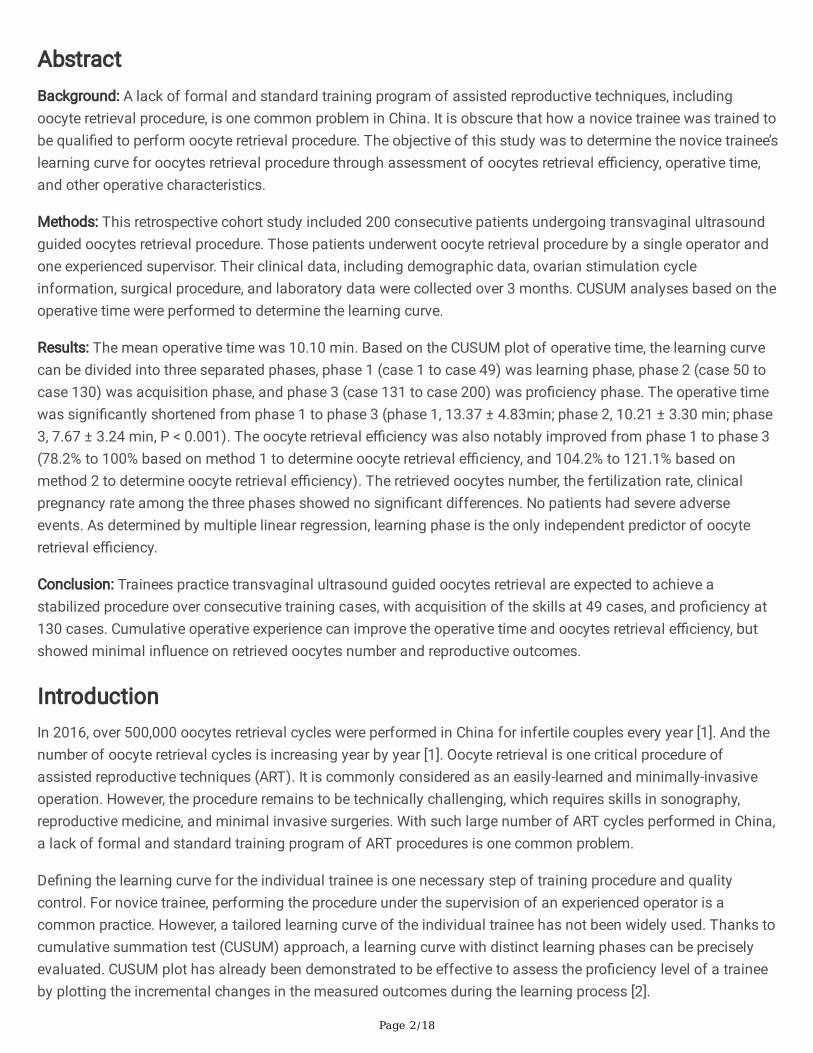
ResultsA total of 200 consecutive patients operated by a single operator were included. The mean operative time was10.1 ± 4.29 min. DOT were calculated by deduction of each individual operative time and the mean operative time.The sum of those DOT based on the case sequence were plotted to form a CUSUM (Fig. 1). CUSUM analysis ofoperative time shows 3 distinguished phases (Fig. 1A). Within phase 1 (case 1 to case 49, Fig. 1B), the learningphase, an increase trend in the summation was observed until the sum reaches a peak at case 49. During phase 2(case 50 to case 130, Fig. 1C), the acquisition phase, the summation plateaued with minor �uctuation. Phase 3(case 131 to case 200, Fig. 1D), the pro�ciency phase began with an in�exion at case 131, and then sumgradually reduced to the baseline. The �rst vertical line in Fig. 1A represents the transition from Phase 1 (learningphase) to Phase 2 (acquisition phase) of learning curve, and the second vertical line in Fig. 1A represents the
Page 6/18
transition from Phase 2 (acquisition phase) to Phase 3 (pro�ciency phase) of the learning curve for oocytesretrieval.
Table 1 summarizes the demographic information of patients from all 3 phases of learning curve who underwentoocytes retrieval. The baseline clinical characteristics among the three consecutive phases showed no signi�cantdifferences, except for AMH, which was obviously lower in phase 3 (median 2.11 ng/ml) than those from phase 1(median 2.73 ng/ml) and 2 (median 2.92 ng/ml) (P = 0.036).
Table 1Comparisons of demographic characteristics
All
N = 200
Phase 1
N = 49
Phase 2
N = 81
Phase 3
N = 70
Pvalue
Female age (yearsold)
32.40 ± 4.70 32.71 ± 4.56 32.42 ± 4.81 32.14 ± 4.72 0.808
Male age (years old) 34.58 ± 5.23 35.41 ± 5.75 34.69 ± 4.86 33.87 ± 5.25 0.281
Infertile duration(years)
3 (2, 5) 4 (3, 6.5) 3 (2, 4) 4 (2, 6) 0.076
Infertile type 0.384
Primary infertility 91/45.50% 19/38.78% 36/44.44% 36/51.43%
Secondary infertility 109/54.50% 30/61.22% 45/55.56% 34/48.57%
Infertile factors 0.526
Tubal 85/42.50% 18/36.73% 39/48.15% 28/40.00%
Ovulation disorder 9/4.50% 2/4.08% 4/4.94% 3/4.29%
Male 35/17.50% 9/18.37% 11/13.58% 15/21.43%
Multiple 43/21.50% 13/26.53% 16/19.75% 14/20.00%
Unexplained 23/11.50% 7/14.29% 9/11.11% 7/10.00%
PGT 5/2.50% 0/0% 2/2.47% 3/4.29%
BMI (kg/m2) 22.29 ± 3.29 21.61 ± 3.00 22.70 ± 3.75 22.28 ± 2.86 0.188
AFC 16 (12, 22) 17 (12, 26) 17 (13, 22) 14 (11, 19) 0.059
AMH (ng/ml) 2.64 (1.71,4.27)
2.73 (1.62,4.21)
2.92 (1.89,4.73)*
2.11 (1.57,3.40)
0.036
Abbreviations: PGT, pre-implantation genetic test; BMI, body mass index; AFC, antral follicle count; AMH, anti-Mullerian hormone.
* denotes a signi�cant difference when comparing patients between phase 2 and phase 3.
Table 2 shows the comparisons of ovarian stimulation cycle related characteristics of patients from all 3 phasesof learning curve. The current ovarian stimulation cycle, ovarian stimulation protocols, total Gn doses for ovarian

Page 7/18
stimulation, Gn days for ovarian stimulation, and endometrial thickness on trigger day showed no differences ofpatients among the 3 phases. However, the numbers of follicles ≥ 10 mm and ≥ 14 mm on trigger day in phase 3were less than those numbers from either phase 1 or phase 2 (P = 0.001 for number of follicles ≥ 10 mm, and P < 0.001 for number of follicles ≥ 14 mm).
Table 2Comparisons of ovarian stimulation cycle related characteristics
All
N = 200
Phase 1
N = 49
Phase 2
N = 81
Phase 3
N = 70
Pvalue
Current cycle number 0.934
1st cycle 144/72.00% 34/69.39% 59/72.84% 51/72.86%
2nd cycle 45/22.50% 13/26.53% 17/20.99% 15/21.43%
≥ 3 cycles 11/5.50% 2/4.08% 5/6.17% 4/5.71%
Stimulation protocol 0.760
GnRH agonist 121/60.50% 28/57.14% 51/62.96% 42/60.00%
GnRH antagonist 70/35.00% 20/40.82% 28/34.57% 22/31.43%
Others 9/4.50% 1/2.04% 2/2.47% 6/8.57%
Gn dose (IU) 2162.5 (1500,2775)
1875 (1388,2550)
2225 (1650,2850)
2288 (1575,2888)
0.117
Gn duration (days) 11 (9, 12.5) 10 (8, 12) 11 (9, 13) 11 (8.75, 13) 0.407
Number of follicles ≥ 10 mm ontrigger day
11.18 ± 4.79 12.00 ± 4.27# 12.28 ± 5.14* 9.33 ± 4.17 < 0.001
Number of follicles ≥ 14 mm ontrigger day
8.21 ± 3.31 8.76 ± 2.97# 8.88 ± 3.46# 7.04 ± 3.09 0.001
Endometrial thickness ontrigger day (mm)
10.14 ± 2.18 10.45 ± 2.17 10.26 ± 2.20 10.05 ± 2.69 0.660
Fertilization method 0.058
IVF 149/74.50% 41/83.67% 63/77.78% 45/64.29%
ICSI 43/21.50% 7/14.29% 14/17.28% 22/31.43%
Half-ICSI 6/3.00% 1/2.04% 3/3.70% 2/2.86%
Abbreviations: Gn, gonadotropin; IVF, in vitro fertilization; ICSI, intra-cytoplasmic sperm injection.
# denotes a signi�cant difference when comparing patients between phase 1 and phase 3.
* denotes a signi�cant difference when comparing patients between phase 2 and phase 3.
Oocyte retrieval procedure related characteristics were summarized in Table 3. The operative durations werenotably shortened from phase 1 to phase 2 and 3 (P < 0.001, Table 3 and Fig. 2A). Apparently, a signi�cant
Page 8/18
improvement of oocyte retrieval e�ciencies at phase 3 was observed as compared with phase 1 and phase 2(Table 3, Fig. 2C and 2D), regardless of the methods to determine the oocyte retrieval e�ciencies. The number ofretrieved oocytes (Table 3 and Fig. 2B), fertilization rate, number of day 3 usable embryos (Table 3 and Fig. 2E),number of embryos with top quality, and the vaginal bleeding rate at the puncture site showed no differences.
Table 3Comparisons of oocytes retrieval related features
All
N = 200
Phase 1
N = 49
Phase 2
N = 81
Phase 3
N = 70
Pvalue
Operative time (min) 10.10 ± 4.29 13.37 ± 4.83# 10.21 ± 3.30* 7.67 ± 3.24 < 0.001
Vaginal bleeding atpuncture site (n/%)
24/11.00% 6/12.24% 11/13.58% 5/7.14% 0.429
Retrieved oocytes number 8.76 ± 3.21 8.51 ± 2.95 9.07 ± 3.27 8.56 ± 3.32 0.511
Oocytes retrieval e�ciency
(%, ≥ 10 mm)
78.2 (61.5,107.7)
76.9 (61.5,92.1) #
70.60 (60.0,100.0)*
100.0 (66.7,115.7)
0.002
Oocytes retrieval e�ciency
(%, ≥ 14 mm)
104.2 (86.4,137.5)
100.0 (85.70,130.3) #
93.30 (82.3,133.3)*
121.1 (100.0,150.0)
0.011
Fertilization rate (%) 76.49 ± 25.59 73.24 ± 27.22 73.97 ± 25.33 83.54 ± 20.04 0.051
Number of day 3 usableembryos
3 (2, 5) 3 (2, 4) 3.5 (2, 5.75) 3 (2, 5) 0.385
Number of embryos withtop quality
1 (0, 2) 0 (0, 1.5) 1 (0, 2) 1 (0, 2) 0.070
# denotes a signi�cant difference when comparing patients between phase 1 and phase 3.
* denotes a signi�cant difference when comparing patients between phase 2 and phase 3.
The reproductive outcomes were demonstrated in Table 4. Part of the patients had cancelled embryo transfer. Thereasons for embryo transfer cancelling have been listed in Table 4, and no differences of the cancellation ratewere observed in patients among the three phases. For patients had embryos transferred, either the number or thestage of embryos showed no signi�cant differences in patients among the three phases. The clinical pregnancyrates were similar in patients all through different phases (45.16%, 62.50%, and 54.00% for each phase, P = 0.316,Table 4 and Fig. 2F). The ongoing pregnancy rate also did not differ notably among the three phases (41.95%,52.08%, and 48.00% for phase 1, 2 and 3, individually, P = 0.680, Table 4 and Fig. 2F)

Page 9/18
Table 4Comparisons of reproductive outcomes
All
N = 200
Phase 1
N = 49
Phase 2
N = 81
Phase 3
N = 70
P value
Cycles withcancelledembryotransferred
71/35.50% 18/36.73% 33/40.74% 20/28..57% 0.110
Risk ofOHSS
7/9.86% 1/5.56% 5/15.15% 1/5.00%
Elevatedprogesterone
7/9.86% 2/11.11% 3/9.09% 2/10.00%
Endometrialdisorders
12/16.90% 2/11.11% 5/15.15% 5/25.00%
Hydrosalpinx 1115.49% 2/11.11% 6/18.18% 3/15.00%
No usableembryos
24/33.80% 9/50.00% 9/27.27% 6/30.00%
PGT 4/5.63% 1/5.56% 26.06% 1/5.00%
Personalreasons
6/8.45% 1/5.56% 3/9.09% 2/10.00%
Cycles withembryotransferred
129/64.50% 31/63.27% 48/59.26% 50/71.43%
One embryo 84/65.12% 2270.97% 34/70.83% 28/56.00% 0.990
Twoembryos
45/34.88% 9/29.03% 14/29.17% 22/44.00%
Cleavege 103/79.84% 26/83.87% 38/79.17% 39/78.00% 0.605
Blastocyst 26/20.16% 5/16.13% 10/20.83% 11/22.00%
Reproductiveoutcomes
Biochemicalpregnancy(n/%)
2/1.55% 0/0% 0/0% 2/4% 0.203
Clinicalpregnancy(n/%)
71/55.04% 14/45.16% 30/62.50% 27/54.00% 0.316
Miscarriage(n/%)
8/11.27% 0/0% 5/16.67% 3/11.11% 0.270
Ectopicpregnancy(n/%)
1/0.78% 1/3.23% 0/0% 0/0% 0.313
Abbreviations: OHSS, ovarian hyper-stimulation syndrome. PGT, pre-implantation genetic test.

Page 10/18
All
N = 200
Phase 1
N = 49
Phase 2
N = 81
Phase 3
N = 70
P value
Ongoingpregnancy(n/%)
62/48.06% 13/41.95% 25/52.08% 24/48.00% 0.680
Abbreviations: OHSS, ovarian hyper-stimulation syndrome. PGT, pre-implantation genetic test.
The multilinear regressions (Table 5) were performed to identify the predictor(s) of oocytes retrieval e�ciencies.As demonstrated in Table 5, after adjusting several confounding factors, the phases of learning curve are the onlyindependent predictor of oocytes retrieval rate.

Page 11/18
Table 5Multiple linear regression coe�cients for oocytes retrieval e�ciencies
Outcomes Univariate analysis Multivariate analysis
Coe�ciency 95% CI P Coe�ciency 95% CI P
Oocytes retrieval e�ciency
(%, out of follicles sized ≥ 10mm)
Learning phases
Phase 1 -32.255 -53.365,-11.145
0.003 -22.602 -46.991,1.787
0.069
Phase 2 -26.006 -44.501,-7.510
0.006 -21.704 -40.938,-2.470
0.027
Phase 3 Ref Ref
Age (years old) 0.498 -1.255, 2.251 0.576
AFC -0.822 -1.933, 0.288 0.146
AMH (ng/ml) -1.679 -4.901, 1.543 0.305
Gn days (days) -0.176 -3.536, 3.184 0.918
Gn doses (IU) 0.002 -0.006, 0.010 0.638
Endometrium (mm) -0.634 -4.112, 2.844 0.720
Operative time (min) -2.741 -4.626,-0.857
0.005 -1.695 -3.861,0.472
0.124
Oocytes retrieval e�ciency
(%, out of follicles sized ≥ 14mm)
Learning phases
Phase 1 -22.599 -34.985,-10.212
< 0.001
-16.773 -31.078,-2.468
0.022
Phase 2 -20.174 -31.027,-9.322
< 0.001
-17.578 -28.860,-6.296
0.002
Phase 3 Ref Ref
Age (years old) 0.100 -0.946, 1.145 0.851
AFC -0.607 -1.267, 0.052 0.071
AMH (ng/ml) -0.585 -2.508, 1.339 0.550
Gn days (days) -0.352 -2.354, 1.649 0.729
Abbreviations: AMH, anti-Mullerian hormone; AFC, antral follicle count; Gn, gonadotropin; 95% CI, 95%con�dential index.

Page 12/18
Outcomes Univariate analysis Multivariate analysis
Coe�ciency 95% CI P Coe�ciency 95% CI P
Gn doses (IU) 0.002 -0.003, 0.006 0.507
Endometrium (mm) -0.179 -2.252, 1.894 0.865
Operative time (min) -1.803 -2.921,-0.686
0.002 -1.023 -2.293,0.248
0.114
Abbreviations: AMH, anti-Mullerian hormone; AFC, antral follicle count; Gn, gonadotropin; 95% CI, 95%con�dential index.
DiscussionAt present, no standard training curriculum has been established for novice trainees in China. The trainingprograms varied from institutions to institutions. Results from this study can shed a light on the minimal pre-required number of cases for practice before a trainee get an accreditation of performing oocytes retrievalprocedure independently. A minimal number of 49 consecutive cases of oocytes retrieval were required toovercome the initial learning phase, followed with 81 cases for practice before the trainee reach competency ofoocytes retrieval skills. Not only the operative time, but also the oocytes retrieval e�ciency was notably improvedfrom the initial learning phase to pro�ciency phase.
It is not easy to decide whether a trainee has reached pro�ciency of a new procedure. Learning curve based onCUSUM plot enables a quantitative analysis for the tutor about the minimal cases and training period required forthe trainee to achieve stabilized skills. Operative time is the most commonly used marker of pro�ciency of certainskills and procedures [2, 7, 8]. It is generally acknowledged that the learning phases involved three distinctivephases [9]. During the �rst phase, trainees focus on the basic skills of performing the procedure and how todiminish the mistakes or complications. During the second phase, trainees can perform the procedures withacceptable skills and more con�dence. And during the �nale phase, trainees perform the procedures with adeptskills and less cognitive pressure [9]. The learning curve based on the CUSUM of operative time in this studyshows a similar trend of the learning phase. The trainee can become more pro�cient in the oocytes retrievalprocedures as the practice case number increased. Speci�cally, the improvement in technique skills wasdemonstrated not only by operative time, but also the oocytes retrieval e�ciency. In the �nal phase of learningcurve, the trainee could safely and e�ciently collect the oocytes.
There are only a few publications showing the trainee’ learning performance in oocytes retrieval. Goldman et al[10] have proposed that a minimal number of 20 cases of oocyte retrievals was suitable for the trainees toachieve a pro�ciency level. However, the practice number is quite limited without adequate methods to detect thechange of learning curve phases. Besides, the trainees perform oocytes retrieval on those with easier procedurewhile the experienced tutor performed the more challenging cases, which made the results of pro�ciency lesscomparable between the trainees and the tutors. Dessolle et al [11] reported that 43 cases are required before theperformance of one trainee reached pro�ciency, which is similar to the case required to the acquisition phase inthis study. Dessolle et al [7] reported a learning curve from CUSUM data of oocyte retrieval. However, in their study,follicles from one side of the ovary were aspirated by the trainees and the other side by the tutor using arandomized manner. Even though this method seems to be more reasonable to compare the operative

Page 13/18
performance between the trainee and the tutor, it is not quite applicable and e�cient in clinical practice. Besides,their CUSUM were plotted based on the performance score (either success or failure) rather than operative time.Operative time, or the speed of operation is a useful parameter to determine CUSUM [8, 12, 13], including oocyteretrieval procedures [14]. In the current study, the CUSUM was determined based on the change trend of operativetime, a more appropriate parameter to de�ne the learning phases. After separation of learning phases, theoperative performance can be compared among each learning phase with more details.
There is no uniform de�nition of oocyte retrieval e�ciency or oocyte retrieval rate. In most reproductiveinstitutions, follicles with diameter ≥ 10 mm were aspirated during oocyte collection procedures. Thus the oocyteretrieval rate was usually determined as the ratio of the number of oocytes collected to the number of follicles ≥ 10 mm [15, 16]. However, several studies debated that reduced maturity and fertilization ability for oocytescollected from small follicles and aspiration of those follicles were not recommended [17, 18]. One studysuggested that oocyte retrieval e�ciency should be based on expected number of oocytes collected from folliclessized ≥ 14 mm [11]. Due to the discordant de�nition, both methods to determine the oocyte retrieval e�ciencieswere evaluated in the present study. As expected, using either method to determine oocyte retrieval e�ciency, asimilar trend of �uctuation in oocyte retrieval e�ciency was observed.
One of the key points during oocytes retrieval is to stabilized probe and correct orientation in the vaginal [19]. Webelieve that with further practice, especially after the operative time reaches a plateau, the trainee got morecon�dence, less anxiety, and better knowledge on maintaining a stable probe. Thus, unsurprisingly, after thefurther training during the acquisition phase, the operative time shortened further with improvement in oocytesretrieval rate during pro�ciency phase. The number of retrieved oocytes, fertilization rate, and reproductiveoutcomes were stable across the three consecutive phases, which suggest that the trainee gradually acquire theoocytes retrieval skills without compromising patients’ reproductive outcomes. After eliminating the confoundingvariables which might affect the oocyte retrieval rate, phases of learning curve seem to be the only variable thatin�uence the oocytes retrieval. Data here further implies that an oocyte retrieval procedure is operator-dependentand the techniques can be improved with cumulative practice. And it is also support the necessity ofestablishment of technique training schedule.
The operative time and di�culties of techniques varies signi�cantly among patients with different ovarianstimulation responses, and also locations of the ovaries. During the training program, almost only those withnormal ovarian stimulation response and good prognosis were assigned for the novice trainee. Interestingly, wedid observe that patients from phase 3 showed reduced ovarian reserve as depicted by lower AMH, AFC andnumbers of follicles ≥ 10 and 14mm on trigger day. This phenomenon was probably because trainees wereassigned with patients with good prognosis in the early phases of learning program, whereas more complex orsub-optimal prognosis patients in the later phases of learning. However, due to the improvement in oocytesretrieval rate, the retrieved number of oocytes did not show a reduced trend compared to those in phase 1 andphase 2.
There were no severe complications occurred in this study. The most common and minor complication in oocytesretrieval was the vaginal bleeding at puncture site, which could be easily controlled by local compression. Thevaginal bleeding at puncture site happened evenly throughout the 3 phases of learning period, which indicate thatthe safety issues might not be related with trainees’ experience. Given the stable number of oocytes retrieved,stable reproductive outcomes, and no severe complications during the learning period, a trainee might be

Page 14/18
underperformed in the beginning of learning curve, the trainee was still a responsible operator, without sacri�cingthe reproductive outcome and safety of the patients.
To the best of our knowledge, there are very limited reports on the learning curve of transvaginal ultrasoundguided oocytes retrieval, and this study provided a largest sample size and quantitative parameters for analysis oflearning curve. However, there are several limitations that we should acknowledged. First, even though the studywas conducted at the locally largest and high-volume center with more than 9000 oocyte retrieval cycles per year,we cannot ignore the drawback that the study was conduct at one single center retrospectively. Another limitationof the study is that all procedures were conducted by a single operator. The length of learning process couldvaried largely among trainees [20]. Hence, the results of this study are not applicable to every individual withvarious backgrounds and training experiences. An individual tailored learning program can be established basedon the individual CUSUM curve. Lastly, the stress levels of the trainee across the phases of learning curve were notrecorded and evaluated, which might potentially in�uence their technical skills.
ConclusionIn conclusion, trainees’ experience in transvaginal ultrasound guided oocytes retrieval can be expected to acquirea stabilized procedure skill after 49 consecutive cases, and reaching about 10-minture operative time. Continuedimprovements in operative time and oocytes retrieval rate were noted with additional cases and experience. Herefrom our results of learning curve, we provide some implications on how to optimize the training curricula ofoocytes retrieval procedure. With the help of these tips and caveats from learning curve, the establishment oftraining program should be more feasible and effective.
AbbreviationsART: assisted reproductive technique; CUSUM; cumulative summation test; AFC: antral follicle counting number;AMH: anti-Mullerian hormone; BMI: body mass index; GnRH: gonadotropin releasing hormone; Gn: gonadotropin;FSH: follicle-stimulating hormone; HMG: human menopausal gonadotropin; LH: luteinizing hormone; HCG: humanchorionic gonadotropin; IVF: in vitro fertilization; ICSI: intra-cytoplasmic sperm injection; SD: standard deviation;IQR: interquartile range; DOT: difference between the mean operative time and each individual operative time.
DeclarationsAcknowledgement
Not applicable.
Funding
This work was supported by the National Natural Science Foundation of China (Grant No. 81701400), NaturalScience Foundation of Guangdong Province (Grant No. 2017A030310447)
Availability of data and materials
The datasets used and/or analyzed during the current study are available from the corresponding author onreasonable request.

Page 15/18
Authors’ contributions
M.C. analyzed and interpreted the patients’ data, and was a major contributor in writing the manuscript. Z.L.analyzed the data and contributed in manuscript drafting. S.L. collected and interpreted the patients’ data. Y.W.collected and analyzed the patients’ data. H.L. collected the data. Q.H. collected the data. J.L. designed the studyand collected the data. All authors read and approved the �nal manuscript.
Ethical approval and consent to participate
The study protocol was approved by the local ethics committee (Approval No. 2020-037) of The Third A�liatedHospital of Guangzhou Medical University. Each participant in the study has signed informed consent statement.
Statement
All surgical procedures were performed in accordance with the relevant guidelines and regulations.
Consent for publication
Each participant had signed the consent form for the use of her personal data for research use and publication.
Competing interests
The authors declare no competing interests.
References1. Bai F, Wang DY, Fan YJ, Qiu J, Wang L, Dai Y et al. Assisted reproductive technology service availability,
e�cacy and safety in mainland China: 2016. Hum Reprod. 2020;35:446-52.http://dx.doi.org/10.1093/humrep/dez245.
2. Holzhey DM, Seeburger J, Misfeld M, Borger MA, Mohr FW. Learning minimally invasive mitral valve surgery:a cumulative sum sequential probability analysis of 3895 operations from a single high-volume center.Circulation. 2013;128:483-91. http://dx.doi.org/10.1161/circulationaha.112.001402.
3. Ferraretti AP, La Marca A, Fauser BC, Tarlatzis B, Nargund G, Gianaroli L. ESHRE consensus on the de�nitionof 'poor response' to ovarian stimulation for in vitro fertilization: the Bologna criteria. Hum Reprod.2011;26:1616-24. http://dx.doi.org/10.1093/humrep/der092.
4. Roque M, Haahr T, Geber S, Esteves SC, Humaidan P. Fresh versus elective frozen embryo transfer in IVF/ICSIcycles: a systematic review and meta-analysis of reproductive outcomes. Hum Reprod Update. 2019;25:2-14.http://dx.doi.org/10.1093/humupd/dmy033.
5. Jahromi BN, Mosallanezhad Z, Matloob N, Davari M, Ghobadifar MA. The potential role of granulosa cells inthe maturation rate of immature human oocytes and embryo development: A co-culture study. Clin ExpReprod Med. 2015;42:111-7. http://dx.doi.org/10.5653/cerm.2015.42.3.111.
�. Van den Abbeel E, Balaban B, Ziebe S, Lundin K, Cuesta MJ, Klein BM et al. Association between blastocystmorphology and outcome of single-blastocyst transfer. Reprod Bio-Med Online. 2013;27:353-61.http://dx.doi.org/10.1016/j.rbmo.2013.07.006.

Page 16/18
7. Dessolle L, Biau DJ, de Larouziere V, Ravel C, Antoine JM, Darai E et al. Learning curve of vitri�cationassessed by cumulative summation test for learning curve (LC-CUSUM). Fertil Steril. 2009;92:943-5.http://dx.doi.org/10.1016/j.fertnstert.2009.01.133.
�. Pernar LIM, Robertson FC, Tavakkoli A, Sheu EG, Brooks DC, Smink DS. An appraisal of the learning curve inrobotic general surgery. Surg Endosc. 2017;31:4583-96. http://dx.doi.org/10.1007/s00464-017-5520-2.
9. Forbes N, Mohamed R, Raman M. Learning curve for endoscopy training: Is it all about numbers? Best PractRes Clin Gastroenterol. 2016;30:349-56. http://dx.doi.org/10.1016/j.bpg.2016.04.003.
10. Goldman KN, Moon KS, Yauger BJ, Payson MD, Segars JH, Stegmann BJ. Pro�ciency in oocyte retrieval: howmany procedures are necessary for training? Fertil Steril. 2011;95:2279-82.http://dx.doi.org/10.1016/j.fertnstert.2011.02.055.
11. Dessolle L, Leperlier F, Biau DJ, Freour T, Barriere P. Pro�ciency in oocyte retrieval assessed by the learningcurve cumulative summation test. Reprod Bio-Med Online. 2014;29:187-92.http://dx.doi.org/10.1016/j.rbmo.2014.03.016.
12. Vieira A, Bourdages-Pageau E, Kennedy K, Ugalde PA. The learning curve on uniportal video-assisted thoracicsurgery: An analysis of pro�ciency. J Thorac Cardiovasc Surg. 2019.http://dx.doi.org/10.1016/j.jtcvs.2019.11.006.
13. Kayani B, Konan S, Huq SS, Tahmassebi J, Haddad FS. Robotic-arm assisted total knee arthroplasty has alearning curve of seven cases for integration into the surgical work�ow but no learning curve effect foraccuracy of implant positioning. Knee Surg Sports Traumatol Arthrosc. 2019;27:1132-41.http://dx.doi.org/10.1007/s00167-018-5138-5.
14. Soave I, D'Angelo A, Piva I, Marci R. A Pilot Study on Oocyte Retrieval Simulator: A New Tool for Training? JMed Syst. 2019;43:202. http://dx.doi.org/10.1007/s10916-019-1340-3.
15. Lu X, Hong Q, Sun L, Chen Q, Fu Y, Ai A et al. Dual trigger for �nal oocyte maturation improves the oocyteretrieval rate of suboptimal responders to gonadotropin-releasing hormone agonist. Fertil Steril.2016;106:1356-62. http://dx.doi.org/10.1016/j.fertnstert.2016.07.1068.
1�. Benaglia L, Busnelli A, Biancardi R, Vegetti W, Reschini M, Vercellini P et al. Oocyte retrieval di�culties inwomen with ovarian endometriomas. Reprod Bio-Med Online. 2018;37:77-84.http://dx.doi.org/10.1016/j.rbmo.2018.03.020.
17. Bergh C, Broden H, Lundin K, Hamberger L. Comparison of fertilization, cleavage and pregnancy rates ofoocytes from large and small follicles. Hum Reprod. 1998;13:1912-5.http://dx.doi.org/10.1093/humrep/13.7.1912.
1�. Triwitayakorn A, Suwajanakorn S, Pruksananonda K, Sereepapong W, Ahnonkitpanit V. Correlation betweenhuman follicular diameter and oocyte outcomes in an ICSI program. J Assist Reprod Genet. 2003;20:143-7.http://dx.doi.org/10.1023/a:1022977002954.
19. D'Angelo A, Panayotidis C, Amso N, Marci R, Matorras R, Onofriescu M et al. Recommendations for goodpractice in ultrasound: oocyte pick up(dagger). Hum Reprod Open. 2019;2019:hoz025.http://dx.doi.org/10.1093/hropen/hoz025.
20. Dessolle L, Freour T, Barriere P, Jean M, Ravel C, Darai E et al. How soon can I be pro�cient in embryotransfer? Lessons from the cumulative summation test for learning curve (LC-CUSUM). Hum Reprod.2010;25:380-6. http://dx.doi.org/10.1093/humrep/dep391.

Page 17/18
Figures
Figure 1
Learning curve (3-phase) based on CUSUM plot. (A) Three distinct learning phases were identi�ed with visualinspection. The �rst vertical line represents the transition from Phase 1 (learning phase) to Phase 2 (acquisitionphase) of learning curve, and the second vertical line represents the transition from Phase 2 (acquisition phase) toPhase 3 (pro�ciency phase) of the learning curve for oocytes retrieval. (B-D) shows a close look of phase 1 to 3individually.

Page 18/18
Figure 2
Comparisons of critical variables of oocytes retrieval procedure. A) Operative time (min), B) number of oocytesretrieved, C) oocytes retrieval e�ciency (%, ≥ 10 mm), D) oocytes retrieval e�ciency (%, ≥ 14 mm), E) number ofusable embryos on day 3, and F) clinical pregnancy rate and ongoing pregnancy rate (%) were compared amongdifferent learning phases. * denotes P < 0.05. ** denotes P < 0.01. *** denotes P < 0.001.