why do drugs look the way they do_conference paper.pdf
TRANSCRIPT

157
Why Do Drugs Look the Way they Do?
By Wolfgang K.-D. Brill
Discovery Research Oncology, Pharmacia Corp.,
Viale Pasteur 10, I-20014 Nerviano, Italy; e-mail: [email protected]
I. Introduction Heterocycles are very common among drugs. According to the CMC2001.1 database, 56.8% of
the current drugs contain heterocyclic entities.1 Why are heterocycles so frequent among drug-like
molecules? Cyclic molecules provide the highest density of atoms per surface, heterocycles the highest
density of chemical functionalities with well-defined orientation in space per surface. In this paper I
will address why certain features, such as being a heterocycle, are determining whether a molecule is
drug-like. According to the FDA drugs are2….(B) articles intended for use in the diagnosis, cure,
mitigation, treatment, or prevention of disease in man or other animals; and (C) articles (other than
food) intended to affect the structure or any function of the body of man or other animals; and….
animals; and…. Thus in order to alter metabolic pathways in a favorable way, a drug has to interact
with adequate targets. The interaction of a drug to its target, whatever it may be, must be sustained by
specific interactions, which can only be provided between chemical functionalities of a drug and those
of its target. If cyclic structures provide the highest clustering of atoms and, in organic molecules,
heteroatoms provide most functional groups, then the greatest density of functionality can only be a
heterocycle.
II. Biologically Relevant Targets Among the biopolymers involved in all crucial cellular processes proteins and nucleic acids
clearly stand out as potential targets for chemotherapeutic agents. Paul Ehrlich has already proposed
receptor proteins as drug targets in the late 19th century. The concept in which the receptor serves as a
"switch" that receives and generates specific signals and can be either blocked by antagonists or turned
on by agonists was recognized by J. N. Langley in 1905.3 The pharmacological characterization of
receptors in almost all organs, including the brain, provided the basis for a large number of very diverse
drugs: β-blockers4; β-agonists5; benzodiazepines, which enhance the effects of γ-aminobutyric acid and
chloride flux by way of the benzodiazepine receptor6; and monoclonal antibodies, which block

158
receptors of growth or differentiation factors on tumor cells.7 A comprehensive analysis of the drug
targets underlying current drug therapy undertaken in 1996 showed that present-day therapy addresses
only about 500 molecular targets. According to this analysis, cell membrane receptors, largely
heterotrimeric GTP-binding protein (G protein)-coupled receptors, constitute the largest subgroup
with 45% of all targets, and enzymes account for 28% of all current drug targets.8 The number of
potential targets has been exploding as a result of the sequencing of the human genome. However, the
disease processes have to be considered at the molecular (genetic) level to determine the optimal
molecular targets for drug intervention. Not every product of "disease gene" may in itself be a suitable
target. However, its function will likely be linked to that of other proteins in physiological or
pathophysiological circuits. Based on the assumption that the number of such "linked" proteins that
constitute suitable targets for drug intervention is between 5 and 10 per disease gene, J. Drews
estimated the number of potential drug targets to lie between 5,000 and 10,000, with 10 times as many
still to be exploited for future drug therapy.9c
The other part of the story is to find a chemical entity, which is able to penetrate various organs
such as the digestive system, body fluids, such as the blood and in many cases cellular membranes to
reach its target. Among ADME (absorption, distribution, metabolism and excretion), the oral
absorption is highly desired in pharmaceutical industry and poor absorption characteristics constitute a
bottleneck in drug development.9 Statistical analysis of properties of drugs has lead to the “Rule of
Five“10 and other physicochemical constraints characterizing molecules, that are most probably orally
absorbed.11 These possibility schemes of properties are invariant, as they are determined by the
physiology of the patient. Computational methods have recently been designed to estimate these
properties in silco prior to synthesis of a drug with good accuracy.12
Table 1. Some features of compounds, which have a high probability of absorption. Entries 1-4 are the original “Rules of 5”10
No Properties Value 1 Number of hydrogen bond
donors:(NHs and OHs) 0-5
2 Number of hydrogen bond acceptors: (Ns and Os)
0-10
3 LogP -2 -+5 4 Molecular weight 200-500 5 Number of rotatable bonds 0-8 6 Formal charge -2 +2 7 Number of heavy atoms 20-50 8 Polar surface area (TPSA)11b <90 A2

159
Of course, not all biologically active compounds have to comply with those constraints. Whenever it is
possible for a drug to use special uptake or distribution mechanisms or vehicles, dramatic variation
from the above property constraints can be tolerated in active compounds.10
III: The “Drug-likeness” of a Small Molecule Determines which Target is
Drugable. Molecules that are orally bioavailable are found to be restricted to specific property ranges (Table
1)10-12 Generally these drug-like molecules are small compared to their targets. Yet, to bind to an
appropriate target, they must bind with as many of their surface features to as many of those of the
target protein epitopes. However the protein surfaces are generally covered with water, which has to be
displaced by a drug as described in equation (1)13 Can we, looking at the thermodynamics of drug-
target interactions, identify epitopes which are more likely to be addressed by drug-like compounds
than others?
Daq + Raq DRaq + mH2O
(1)
In this equation Daq is the drug solvated by water, Raq is the hydrated receptor, DRaq is the
receptor complex and mH2O is the amount of water released during the binding process.
Mechanistically such a binding event may be viewed as in Figure 1. according to Andrews et al.13a
Here, the binding interactions of a trifunctional drug with an optimal receptor are shown. The cyan
colored circles represent water molecules, the enthalpies of hydration of the drug and the receptor
being ∆HDW and ∆HRW, respectively. The free drug has an overall rotational and translational enthropy
of ∆Srt and an internal enthropy of ∆Sint. If a drug is dissolved in water, not all of its surface
functionalities can form hydrogen bonds with the solvent. Around the hydrophobic portions of the drug
molecule, the water molecules cluster to adopt an “iceberg-like” structure reminiscent of ice chlathrates
and lose enthropy.14 On binding, the drug is fixed on its receptor and both terms, ∆Srt and ∆Sint are
lost. This unfavorable contribution may be compensated for by an increase in enthropy (∆Sw ) due to
the loss of structured water which was formerly clustered around the drug and the receptor. Another
increase in enthropy (∆Svib) is caused by new low frequency vibrational modes associated with non-
covalent with drug-receptor interactions (Figure 1)

160
Figure 1: The Drug receptor binding event.
The thermodynamics of drug-receptor interactions may be expressed by the Gibbs free energy ∆G
which is directly correlated with the association constant Ka (equation 2)
aKRTGG ln+°∆=∆ (2)
[ ] [ ][ ] [ ]aqaq
maq
RDOHDRRTGG
⋅⋅+°∆=∆ 2ln (3)
and consists of the binding enthalpy and enthropy as indicated in Figure 1.
For equilibrium conditions
0=∆G (4) and the enthalpic and enthropic contributions may be shown as:
STHG ∆−∆=°∆ (5)
It is difficult to measure the amount of water, which is displaced when a drug binds to a receptor
according to Figure 1. In turn, the effect of the solvent was shown in the case of a number of different
drugs, binding to various (G protein)-coupled receptors and ligand–gated ion channels. The ∆S° versus
∆H° scatter plot of all those measured binding events produced a straight regression line. This means
that any decease of binding enthalpy was compensated by a parallel decrease of binding enthropy and
vice versa. Tight binding to receptor can be achieved either enthalpy or enthropy-driven, depending on
which interactions the drug establishes with the receptor. For example, the entropy driven binding of
agonists to adenosine A1 receptor was attributed to the displacement of water from a pocket of the
receptor by a ribose residue of the agonists. The binding of antagonists of the same receptor, which do
not have a ribose residue to fill this pocket, and replace the water is enthalpy driven. The scatter plot
also reveals affinity constant values (Ka) cannot be greater than 0.01 nM-1.13b It is intriguing, that this
value is completely independent of the chemical entities that are involved in the reversible drug–
protein interactions, or whether the drug binds in a more enthalpy or more enthropy driven mode.13b

161
IV. Which Forces Make Drugs Bind to their Targets? Hydrogen bonding, though very significant for molecular recognition, cannot be the key player
for drug receptor interactions unless hydrogen bonds with water are significantly weaker than those in
a drug-receptor complex. However hydrogen bonds do not have a well-defined length, strength and
orientation. They are generally 20 to 30 times weaker than covalent bonds and extremely susceptible to
stretching and bending. Exchange of hydrogen bonds with water or other polar residues is isoenthalpic,
if there are no geometric constraints.15
Unpolar residues of molecules will appear to attract and combine via hydrophobic interactions
due to a favorable increase in entropy due to release of solvent from the highly ordered cluster around
the unpolar surface.16 Van der Waals forces17 are caused by induction of the polarization of a molecule
in an electric field. Thus at any given instant, the electronic distribution within atomic groups is
asymmetric due to electron fluctuations. Therefore, dipoles in one group of atoms polarize the
electronic system of neighboring atoms or molecules, thus inducing dipoles which attract each other.18
The binding energy of hydrophobic interactions falls off approximately by the sixth power of the
molecular separation. Thus, tight contact between hydrophobic residues of protein and drug as in an
organic liquid18j or organic solid18e are prerequisite for binding. The π-interactions between aromatic
residues are common within proteins and aromatic residues19 resembling contact patterns in benzene
crystals and the rearrangement of aromatic rings in aromatic host guest complexes.20 In hydrophobic
interactions with amide bonds, the N-H bond dipole is oriented along the normal of the plane through
the phenyl ring, however perpendicular to the amide plane.21 Interactions with hydroxyl functions22,
cations and aromatic residues are of importance as shown in various biological systems.23
Drug binding can be approximated as the sum of all the above-mentioned interactions13a, which
can be attributed to the functional groups of the drug, which is making the interaction (equation 6).
Thus the free energy of drug-receptor binding may be ∆G may be written in the following way (if one
neglects coupling terms between functional groups: compare also with calculations of TPSA11b):
ΧΧ∑++∆=∆ EnEnTG dofdofrtS (6)
Herein ∆Srt is the loss of overall rotational and translational enthropy of the bound drug-molecule
(Scheme 1). ndof are the number of internal degrees of conformational freedom in the drug molecule
and Edof is the change in energy associated with the loss of each such degree of conformational
freedom. EΧ is the intrinsic binding energy of a functional group Χ.
EΧ consists of the enthalpy of interaction between functional groups with the receptor and enthropy
associated with the displacement of water by the functional group and subsequent integration into the

162
solvent. The examination of various compound data sets leads to the following average intrinsic
binding energies. These may vary upon the alignment of functional groups. (Table 2)
Table 2: Intrinsic binding energies:
No. group Energy kcalmol-1
rangea
1 DOFb -0.7 -0.7- -1.0 2 C(sp2) 0.7 0.6- 0.8 3 C(sp3) 0.8 0.1- 1.0 4 N+ 11.5 11.4-15.0 5 N 1.2 0.8- 1.8 6 CO2
- 8.2 7.3- 10.3 7 OPO3
- 10.0 7.7- 10.6 8 OH 2.5 2.5- 4.0 9 C=O 3.4 3.2- 4.0
10 O,S 1.1 0.7- 2.0 11 halogen 1.3 0.2- 2.0
a)Range of binding energies for six random 100-compound data sets. b)Degrees of internal conformational freedom.
For small drug-like molecules the value of ∆Srt is mainly determined by physical constants and
calculated to be –14 kcalmol-1. The potential of a drug molecule to bind a receptor, the expected
binding free energy ∆G, may be calculated. It appears that, in order to have sufficient binding to a
target, a drug has to have enough surface area to provide a sufficient number of functionalities to
interact with its target in an optimal way. Considering the limits of molecular weight (Table 1) implied
by the bioavailability only structures can be drugs, which provide the maximal surface per molecular
weight.
V. How Must a Protein Surface Look like to Allow Tight Binding with Small
Hydrophobic Molecules? Hypothetical polar, shallow protein domains, which provide many hydrogen bonds and few
hydrophobic contact-surfaces, can only bind polar drugs, which complement the hydrogen bonds.
However the isoenergetic trans-hydration of a hypothetical very polar drug with a protein epitope does
render this type of interaction unlikely to provide tight drug-target binding, especially considering the
physicochemical constraints implied by the bioavailability.
Low molecular weight compounds may only bind to predominantly hydrophobic pockets on
proteins. Though hydrogen bonds or charge-charge interaction within a hydrophobic environment
enhance binding dramatically, if being complemented by the drug, the consequence of the Lennard

163
Jones potential is that the hydrophobic contacts between drug and receptor have to be maximized. This
can only be the case, if the drug molecule has a shape complementing that of its binding site on a
protein.23b,c Thus a tight bound drug molecule is likely to be buried deeply in a hole or a fold of its
receptor. Only proteins, having deep hydrophobic folds or grooves, are likely to be drugable
targets for small molecules. This principle has been recognized in computer-assisted identification of
binding sites for drug-like molecules on proteins with known structure.24 Of course shallow epitopes,
such as minor groves in DNA, are also known to bind drugs. However these drugs will likely have a
much larger molecular weight, in many cases a greater chemical complexity and must plug into certain
active uptake mechanisms. The shape of the binding site must be unique to allow selectivity. Often
more than one adjacent hydrophobic fold may have to be used to allow differentiation between
different target protein subtypes.25 It is very welcome, if some adjacent hydrophobic pockets have
derived from different evolutionary predecessors.
VI. Protein Kinases as Example for a Drug Target
OOPO
OO P
O
OP OO O
OHOH
N
NN
NO
NH2OH
Protein
OOPO
OP OO O
OHOH
N
NN
NO
NH2O
OPO
O
Protein
+
kinaseMg2+
+
Figure 2. The kinase reaction.

164
Figure 3. Human receptor protein-tyrosine kinases
Figure 3. The symbols α− and β denote distinct RPTK subunits. RPTK members in bold and italic type
are implicated in human malignancies The horizontal double line represents the cell membrane. The
receptor binding sites are in the extracellular domains of the receptor (below the double line). The
kinase activity is associated with the intracellular domains (above the double line, red rectangles) of
most receptors and represents the drugable target. Some receptors are associated with kinases, which
are not covalently bound to the receptor. An asterisk indicates that the member is devoid of intrinsic
kinase activity.26
Protein kinase activities are often associated with receptors, which can bind to specific effectors,
often other proteins. Upon binding to the effector, they become activated and can catalyze the transfer of
a phosphate group from an ATP molecule onto a tyrosine, serine or threonine of another protein or onto
another domain of themselves.26 (Figure 2)
The resulting phosphorylated proteins are enabled to interact with other proteins differently than in their
unphosphorylated form. Thus phosphorylation of certain enzymes (among them other receptor kinases)
alters their catalytic functions, which leads to build up or depletion of their substrates or products. The
types and concentrations of phosphorylated proteins, which are empowered by external or internal
signals, have a profound impact on every aspect of the cell life. These signal transuduction pathways
regulate a number of cellular functions, such as cell growth, differentiation, and cell death. Figure 3
shows a schematic representation of some membrane bond receptor kinases27

165
A variety of tumor types have dysfunctional growth factor receptor tyrosine kinases, resulting in
inappropriate mitogenic signaling. Protein tyrosine kinases (PTKs) are therefore attractive targets for
therapeutic agents, not only against cancer, but also against many other diseases.18d Protein kinases also
posses with their ATP-binding site a structural feature which renders them drugable targets. The
possibility of competitive displacement of the ATP-cofactor by filling up hydrophobic pockets
associated with the adenine portion of the cofactor (and neither phosphate nor substrate binding sites)
was first recognized by Pascal Furet.27
The ATP-binding site is composed of deep hydrophobic folds or grooves. The natural cofactor
ATP is not very tightly bound to the catalytic site28, since, after phosphorylation of the substrate, its
reaction product ADP and the phosphorylated substrate have to leave their binding site to make room
for new cofactor and substrate. Despite the fact, that the catalytic domains of kinases share significant
amino acid homology and conserved core structures29 the structural diversity between ATP-binding sites
is sufficient to allow the development of selective inhibitors.30 The development of many potent
inhibitors in recent years supports the significance for this binding site.31,32
The binding pocket consists of various regions (Figure 4), which are favorable for a drug target.8, 33
1) Adenine region
2) Sugar rocket
3) Hydrophobic region I
4) Hydrophobic region II
5) Phosphate binding region
Figure 4. The ATP binding site*
Figure 4: The enumeration of amino acid residues is based on c-AMP dependent protein kinase.

166
VII. How Can Drugs Fill Hydrophobic Pockets?
Binding to one or a few adjacent hydrophobic pockets requires the following: 1) The shape of the drug has to complement that of its binding site on a protein.23c,d 2) The geometries of optimal hydrogen bonding between polar residues have to be fulfilled. 3) The various functionalities that interact upon binding have to be pre-oriented so that
binding results in minimal conformational strain on drug and target. 4) Electric fields within the binding pocket should be compensated. 5) The conformational flexibility should be as low as possible. (see DOF Table 2) The conformation of the drug molecule bound to its target relative to that in solution has to be considered, especially if rotational barriers are high. For example, many amide bonds do not rotate at physiological temperature. In turn, the Gibbs free energy associated with the rotational barrier of some carbamates was found to be between 15-20 kcal/mol34 that of some anilides and toluamides 12-14 kcal/mol35 which accounts for up to 5-10 uncharged average hydrophobic contact interactions according to Table 2! The preference for more rigid structures with in drugs is desired in order to minimize enthropic loss due to fixation on its target (Figure 1). Thus small aliphatic rings with 3 and 4 members are often preferred over linear alkyl chains. 6-membered aliphatic rings are mainly used, if ring inversion barriers are high, which can be implemented by appropriate ring substituents. (This may explain why N, N disubstituted piperazines are very frequent within drugs.)
The alignment of the functional groups in their optimal binding positions is essential as seen in rate acceleration of enzymic and intramolecular reactions.35 In narrow protein folds the optimal orientation of clustered functional groups may only be achieved upon fixation onto or integration into cyclic structures.36 Aromatic heterocycles provide great specifically functionalized surface provided by a minimum of atoms. Figure 5. Different H-bondpatterns of kinase inhibitors with the “hinge region” of the ATP-binding site.a
A
HN N
H
NN
NNO
rib
B
HN N
N
NN
NHO
R1
R2
R3

167
C
N
N
NOH
NH
Cl
HN NN
O N
NH
H
Traxler et al ref. 31(EGFR Kinase, 3nM)
R1
R2
R3
Eb
N
O H
NN
H
R1
R2
R3
NNH
CH2 NHN
CH2
D
HN O
O NH
A
NH
O
NH
SU 5416(Sugen VEGFR Kinase)
NH
O
NNH
Br
SNH2
OO
Bramson et al. ref.CDK2 IC50 60nM
a The animated structures are taken from Noble et al.18j The ATP-binding site in Figure 4 is an excellent example how a hydrophobic pocket in an enzyme can
be filled by a drug. The adenine binding region itself is very narrow and of mostly hydrophobic
character. For example, in the c-AMP dependent protein kinase the N1 and N6 of the adenine ring are
engaged in hydrogen bonds with the carbonyl of Glu121 and the NH of the amide of Val 123. The
hydrogen bonds surrounded by a hydrophobic environment are extremely attractive to promote tight
drug-target interaction and are therefore also used by other inhibitors. Scheme 4 indicates in how many
different ways various heterocyclic inhibitors align to the hinge region to make the contacts. While ATP
displays a hydrogen-bonding pattern similar to that in DNA, roscovitine interacts with CDK2 in a
Hoogsteen type hydrogen-bond pattern. In turn, pyrrolopyrimidines (7-deazapurines)31 have been shown
to bind via N3 and H9 to the hinge region. The natural product staurosporine, Sugen 5416 and also an
isatine hydrazone38 bind via alignment of an oxopyrrole moiety. In case of the pyrazole binding the X-
ray structure of the phenyl methyl pyrazole is reported, however only the IC50 of the methylene-bridged

168
compound is reported. It is interesting to note that the tautomerism of the pyrazoles allows displaying
two inverse hydrogen bond donor-acceptor patterns.
It is likely that the IC50 of the unbridged compound (Figure 5, E, CH2 in gray) is much lower, due
to unfavorable entropic contributions caused by fixation of the aryl rings on the same plane. In one
model for the binding of a quinazoline with EGFR kinase, the inhibitor is interacting with the backbone
NH of Met-769 and with Tyr 766. In this model the aminoaryl substituent fills up the hydrophobic
region 1 (Figure 4), while the ribose pocket is not being used. (Figure 6, A) If, further hydrogen bonds
with the hinge region are established, either by alkoxy substituents in position 7 of the quinazoline or by
condensation with a pyrrole or pyrazole the IC50 is still dramatically lowered.39 Alternatively the
quinazolines, which probably lack the central hydrogen-bonding interaction, may however be
Figure 6. Superimposititon of a quinazoline and ATP binding to EGFR.
The gray rods represent the peptide backbone.

169
O O
O
O
OOH
HN
HN
H
H
H
N
N
NH
O
OBr
N
N
NH Cl
F
O
ONO
ZD1839 (Astra-Zeneca, EGFR kinase)
HN NN
H
OHN
NR
O
R2R1
O
OOH
OH
OH
N
N
NH
O
OOO
genistein
CP 358774 (Pfizer, EGFR Kinase)
HN O
HN
NN
H
NR
HN
O
R2
R1
SU 5271/PD 133035 (Sugen, EGFR/Psoriasis)
A B
The animated pictures are taken from Palmer et al.39 engaged in another hydrogen bond in the “hydrophobic region 1 (Figure 4). A similar hydrogen-bonding pattern is also proposed for genistein.18d (Figure 6, B) The sugar pocket is of hydrophilic character in most kinases. However within the EGFR family a
cysteine residue is present in this region. Aromatic residues, such as chlorophenyl, have been found to
be effective replacements for the much more polar ribosyl moiety in that case.31 It is assumed that
halogen substituted aromatics are engaged in an interaction with the cysteine residue also present in that
pocket in some kinases. In the EGF-receptor kinases, R-methylbenzyl has been demonstrated to be very
effective. Some more hydrophilic groups have been found very effective32a,33, however they cause the
drug to be more solvated by water, which might overcompensate the gain of making a new hydrogen
bond. Figure 7 shows how aryl groups of various inhibitors may act as bioisostere for the ribosyl
moiety. In a special case acetylene acts as a lipophilic spacer, bypasses most of the sugar pocket and
allows polar residues to bind directly to residues in the phosphate region.40 (Figure 8)

170
Figure 7. Superposition of dianilinophthalimide (gray), 4-(phenylamino)-7H-pyrrolo[2,3-d]pyrimidine (EGF IC50: 1.9µµµµM) (cyan), and ATP (blue).
Source: Traxler et al. 31
Figure 8. An acetylene spacer allows the binding of an OH-group (red) with polar residues in the phosphate region.
Source: Ducrot et al. 40
The hydrophobic region I (Figure 4) is not occupied by adenine residues, but can be used by
inhibitors such to gain activity and selectivity. Various purine derivatives such as olomoucine32c,41,
purvalanol B33 (Figure 5) but also the flavonoid L 86827642 and roscovitine33 (Figure 5) fill this pocket
and establish π-interactions with the peptide side chains and Van der Waals interactions with alkyl

171
residues. Thus purvalanol B bearing a 3-chloro-4-carboxyanilino group has an IC50 against
CDK2/cyclin A, which is 1000 fold lower than that of olomoucine.33 Among different kinases there is
variability of amino acids involved in generating this pocket, which is advantageous for the
development of selective inhibitors.43 The hydrophobic region II (Figure 4) is a hydrophobic slot open to the solvent. This region is also
not used by ATP and may be used by inhibitors. The phosphate binding pocket is very solvent exposed
and very polar. Inhibitors addressing this region consequently also have
Figure 9. Two inhibitors filling hydrophobic pocket of CDK2.
A
B
IC50= 60nM
Compound A (5-aryl-1H-pyrazole) is reported by Furet et al.37, compound B by Bramson et al.38
to bear polar, hydrated groups. As a consequence, considering the physicochemical constraints implied
by bioavailability and the competition with hydration this binding pocket is not of primary importance.
172
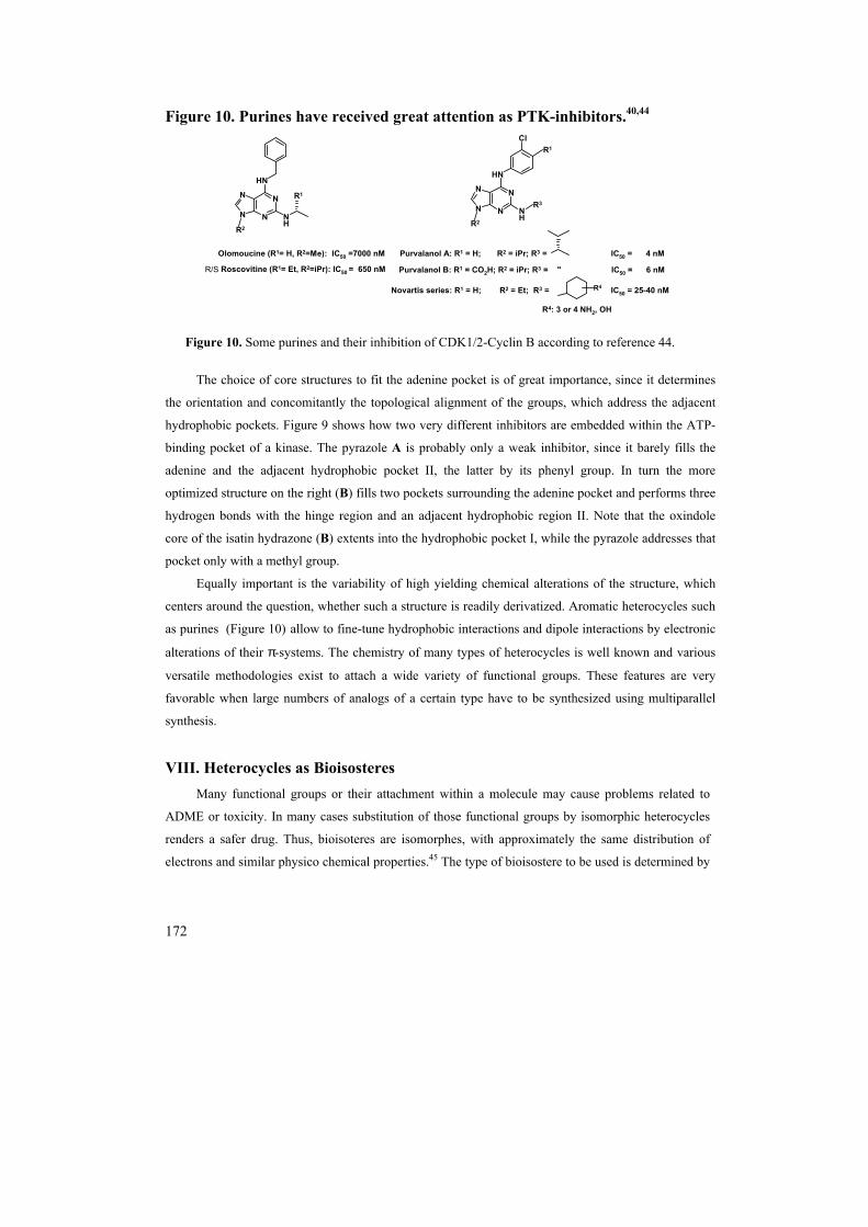
Figure 10. Purines have received great attention as PTK-inhibitors.40,44
N
NN
N
NH
NH
N
NN
N
NH
NH
Cl
R1
R2
Olomoucine (R1= H, R2=Me): IC50 =7000 nM
R/S Roscovitine (R1= Et, R2=iPr): IC50 = 650 nM
R1
Novartis series: R1 = H; R2 = Et; R3 = IC50 = 25-40 nM
Purvalanol A: R1 = H; R2 = iPr; R3 = IC50 = 4 nM
Purvalanol B: R1 = CO2H; R2 = iPr; R3 = " IC50 = 6 nM
R2
R4: 3 or 4 NH2, OH
R3
R4
Figure 10. Some purines and their inhibition of CDK1/2-Cyclin B according to reference 44.
The choice of core structures to fit the adenine pocket is of great importance, since it determines
the orientation and concomitantly the topological alignment of the groups, which address the adjacent
hydrophobic pockets. Figure 9 shows how two very different inhibitors are embedded within the ATP-
binding pocket of a kinase. The pyrazole A is probably only a weak inhibitor, since it barely fills the
adenine and the adjacent hydrophobic pocket II, the latter by its phenyl group. In turn the more
optimized structure on the right (B) fills two pockets surrounding the adenine pocket and performs three
hydrogen bonds with the hinge region and an adjacent hydrophobic region II. Note that the oxindole
core of the isatin hydrazone (B) extents into the hydrophobic pocket I, while the pyrazole addresses that
pocket only with a methyl group.
Equally important is the variability of high yielding chemical alterations of the structure, which
centers around the question, whether such a structure is readily derivatized. Aromatic heterocycles such
as purines (Figure 10) allow to fine-tune hydrophobic interactions and dipole interactions by electronic
alterations of their π-systems. The chemistry of many types of heterocycles is well known and various
versatile methodologies exist to attach a wide variety of functional groups. These features are very
favorable when large numbers of analogs of a certain type have to be synthesized using multiparallel
synthesis.
VIII. Heterocycles as Bioisosteres Many functional groups or their attachment within a molecule may cause problems related to
ADME or toxicity. In many cases substitution of those functional groups by isomorphic heterocycles
renders a safer drug. Thus, bioisoteres are isomorphes, with approximately the same distribution of
electrons and similar physico chemical properties.45 The type of bioisostere to be used is determined by

173
its ability to reconstitute the desirable interactions with the target, its effect on ADME and toxicity of a
drug. Isoelectronic properties and the (later so called) “isollobality”46 were recognized early by
Langmuir47, Grimm (Grimm´s Hydride Displacement Law)48 and Erlenmeyer.49 Some features of
bioisosterism implied by heterocycles involve the orientation of hydrogen bonds within a hydrophobic
environment within a hydrophobic pocket are often considered sticky points24, which have to be bound
by a drug. Heterocycles are an excellent way to arrange hydrogen donors or acceptors appropriately in
the binding pocket. The tautomerization37,50 found in several heteroromatic compounds allows them to
adapt to their environment. Thus a C-OH moiety of a heterocycle will tautomerize to C=O, and a
C=NH to C-NH- functions.51 Heterocycles with similar topological and electronic features may be
bioisosters of each other. Ring equivalents are another frequent type of bioisosterism. In many cases
pyridyl (2) and phenyl (1) have been demonstrated to be bioisosteres however, the metabolism and
solubility of pyridyl functions are very different from that of phenyl moieties.
Certain nitrogen heterocycles can serve as bioisosteres for phenols (3), which may cause ADME and
toxicological problems (Figure 11). Heterocycles such as indoles (4) and benzimidazoles (6) have been
shown to be particularly effective.52 The replacement of a phenol-residue by a pyrrolo ring has been
ascribed to the ability of both groups to hydrogen bond.53 The, very polar catecholes (5) could be
replaced by benzimidazoles (6)54, 3-hydroxypyrid-4-ones (7a), 3-hydroxypyranones (7b) 1-
hydroxypyrid-2-ones (8) and 3-hydroxypyridines (9).55
Figure 11. Bioisosteres of phenol and catechol.
N
OH NH
OHOH N
NH
Y
OOH N
OOH
N
OH
1 2
3 4
5 6 7a Y: O7b Y: NH
8 9
A common bioisostere for the carboxylic acid function (10), is the tetrazole moiety (11).
Comparison between carboxylic acids and tetrazoles at physiological pH reveals that the tetrazole
group is almost 10 times more lipophilic, while having similar acidity. (pKa tetrazole= 4.9, pKaRCOOH 4.2-

174
4.4).56 In the case of model complexes with an amidine the tetrazolate anion with its four N-atoms was
found to be a flexible hydrogen bond acceptor, which adapts quite easily to different binding
(coordination) modes. However, the tetrazole is smaller in size than the carboxylate anion and
therefore cannot bind an amidine as tightly.57 Other monocyclic bioisosteres for carboxylates are
isoxazoles (12)58 oxadiazolones (13).59
Several biosteres have been used for esters (15) to increase hydrolytic stability and
bioavailability.60 Some heterocycles employed are piperazine diones (16), oxazolidinones (17, 18)60,
1,2,4-oxadiazoles (19a), 1,3,4-thiadiazoles (19b)61 isoxazoles and isothiazoles (20a, 21a), furans
(22)61, 1,2,5–oxadiazoles, (23a) 1,2,5-thiadiazoles (23b)62, 1,2,3-triazol-4-yl (24a), tetrazol-5-yl groups
(24b)63. In some bicyclic isosteres for carboxylic acids or esters are isoxazolidines (14a, 25a) and
isothiazole (14b, 25b) fused with saturated rings64 which may be favorable for functional group
orientation.
For amides (27) several heterocycles such as 1,2,4 oxadiazoles (28)65, 1,3,4-oxadiazoles (29)66,
1,2,4-triazoles (30)67, isoxazolines (31)68 and imidazoline (32)69 have been used. (Figure 12)
Figure 12. Bioisosteres for carboxylic acids, esters and amides
YN
R
YN
OR
YNN
O R NN
NY
RO
R
NH
NYOR
N
NOOR
23a Y: O23b Y: S
20a Y: O20b Y: S
25a Y: O25b Y: S
21a Y: O21b Y: S
22 24a Y: CH24b Y: N
26
OH
O
NN
NNH
NO
NHO
NHN
O
O
R
YN R
O
OO
N
O
R
OR' Y
NN
R
N
NYOH
NO
O
OR
10 11 12 13 14a Y: O14b Y: S
1617a Y: O17b Y: CH217c Y: NH17d Y: NR'
18 19a Y: O19b Y: S
15

175
NH2
O
ON
N
NNH
N
NN
O
N
NH
NO
27 28 29 30
31 32
IX. Toxic structures Many chemically feasible heterocycles, which are tight binders to a target, fail to become drugs
due to toxicity. In most cases transition metals, and functionalities, which could modify biopolymers
are discarded such as alkylating and acylating agents etc.70
Expert systems such as TOPDCAT, DEREK, StAR, OncoLocic and MULTICASE have been
developed to predict toxic substructures, toxicophores, mainly based on training sets of existing
structures.71 Though, these methods have become more frequently used to predict toxicity, however the
relevance of the prediction is difficult to assess. The improvement of expert systems remains an
ongoing effort.72
Some toxic substructures are72b:
1-alkylamino benzene
4-heteroiminomethyl
N-heteroarylimine
Phenylhydrazone
Aryldimethyl amine
1,2 dihydroxybenzene
Some toxic heterocycles are often tricyclic aromatics such as dibenzodioxines 73, acridines74,
dibenzofuranes75 and the food toxin 8MeQX76 (Figure 13), the latter can both intercalate and
aminoarylate DNA.

176
Figure 13. Formation of a 8MeGX adduct with DNAa.
OOPOO
O
N
NHN
NO
O
NH2
N
N NN
NH2
N
N NN
NH
OSO3-
OOPOO
O
N
NHN
NO
O
NH2
NH
NN
NN
8-MeIQx
Metabolism
35 (DNA)
mutagenic adduct
3334
36
Source: Gooderham et al76a and Gauvin et al.76b
1. The synthesis in solution phase: Here the desired reaction product is separated from side
products upon precipitation, liquid-liquid extraction, solid phase-extraction or derivatization of the side
product or the desired one to an easily extractable form. During the synthesis often polymer-supported
reagents81 are used, which can readily be removed by filtration. 2. The solid phase synthesis: Here the product is bound to the solid phase via a covalent linker.
The linker must allow selective removal of the final product from the support, but must be stable under
the reaction conditions throughout the synthesis. The advantage of a solid phase approach is that
reagents can be used in large excess to drive reactions to completion and most side products are just
washed off from the solid phase. However, the solid phase implies steric constraints onto the reactions
performed. Many reactions, which proceed well in solution, proceed on a solid support with a lower
rate or not at all. The choice of synthesis method depends on the synthetic problem, is often not
obvious and the result of a reaction optimization.

177
Often neither solution nor solid phase approaches lead to sufficient compound purity so that high
throughput MS or UV-triggerd HPLC purification has to be performed.80
In the following section substituted purines may serve as representative examples for the
derivatization of aromatic heterocycles, which are very common in medicinal chemistry. The
characteristic reactivities are nucleophilic aromatic substitution (A), functional group interchanges (B)
cyclo condensation reactions (C) and oxidations (D). The fact that many heteroatoms are present
within a ring provides a wide variety of opportunities for synthetic disconnections.(Figure 14)
The problem with application of toxicophor predictions is that the definition of toxicity is very much
dependant on the cost/benefit ratio for any specific compound in any particular pharmacological or
clinical area or endpoint. Oncology, for instance provides many illustrations of the need for good
judgment following post-efficacy testing in deciding what constitutes a toxicophore. Nucleoside and
nucleotide analogs, nitrogen mustards, folic acid antagonists, etc. are definitely toxic classes of
chemicals, but they still provide drugs for use in treating disease such as cancer.
X. Combinatorial Synthesis of Heterocycles The suitability of a certain molecule type, to become a drug is very much determined by the
complexity of its synthesis. In fact the art of medicinal chemistry is to find the structure, which, being
assessable with the most facile synthesis, obtains the best biological activity. The variability of high
yielding chemical alterations of a drug is highly desirable.77 Whether a lead-structure is readily
derivatizable by high throughput multiparallel or combinatorial synthesis schemes is a key factor in the
competitive industrial environment. Many heterocycles meet these criteria. In recent years many
combinatorial methods have been developed to provide either large numbers of compounds for lead
finding or smaller compound collections focused on a specific target for lead optimization. Originally,
combinatorial chemistry set out to provide mostly peptide like molecules, often in mixtures78 out of
which the active ingredient, the lead, would be obtained after an often lengthy deconvolution process.79
However, many times an intrinsic activity in a screen of a compound mixture could not be attributed to
a single compound. Such “false positives” caused an enormous work, since often all compounds of a
large mixture would have to be re-synthesized. The current trend is therefore to synthesize single
compounds and, if necessary, purify them.80 In this way compounds from combinatorial schemes are
indistinguishable from traditionally synthesized compounds and the amount of false positives is
reduced. Various combinatorial schemes exist, many of which are frequently applied in the
derivatization of heterocycles.

178
Figure 14. Some characteristic reactions used for derivatizations of aromatic heterocycles
N
(N,C)
OH N
(N,C)
X
N
(N,C)
NH2 N
(N,C)
N+
N N
(N,C)
Y
POX3
diazotationNuc: Y-
or radical Y.
N
(N,C)
X N
(N,C)
Y
NH2
NH2
(RO,O R'(RO,O)
(O,OR)
NH
NR'
NH
NH
R'NH
NR'
NH
N
NH
NX
oxidation:
oxidation:
A: B:
C:
Y- Nuclephile
Y-
condensation+ H2O or ROH+
D:
The ability of purines to allow nucleophilic aromatic substitution under relatively mild conditions,
which are compatible with many functional groups was exploited in many syntheses of combinatorial
libraries.33,82 To increase the hit-rate of these libraries certain filters for bioavailability prediction12,
toxicity71,72 and solubility are often applied.83
Figure 15. Solution phase synthesis of 2, 6, 9 triamino purines.
N
NN
NH
X
Y N
NN
NH
NH
Y N
NN
NH
NH
NH
N
NN
N
NH
NH
R1
R2
R3
R1 R1
R2
i ii iii1
23
4
56
7
8
937 38 39 40
Figure 15. X, Y: Cl; i) amine R1NH2 (1.05eq.), DMF, 80°C, 2-24 h; ii) amine R2NH2 (5eq.) DMF, Et3N (1.1eq.), 150°C, 30h, then addition of formylpolystyrene beads; iii) R3-Cl, K2CO3, DMF, rt, 24 h or R3OH (1.05eq), DEAD (3eq.), PPh3 (3eq.), dioxane, rt, 6h, chromatography.

179
Thus 2,6,9-trisubstituted purines could be synthesized upon selective displacement of the chloro
functions of 2,6-dichloro-purine, followed by N7-alkylation82a,82d However the aminations are restricted
to nucleophilic amines R2NH2 in step ii (Figure 10) only. The alkylation of N9, is often incomplete.
When excess of alkylating agent is used the desired product is contaminated with N6- and N7alkylated
product. In order to widen the scope 2-fluoro 6-chloropurine was being used as starting material with a
Y=F being more readily displaced. The syntheses of some mixed halopurines is also performed via
Schiemann reaction on 42.84(Figure 16).
N
NHN
NH
O
NH2 N
NN
NH
Cl
NH2
N
NN
NH
Cl
N+
N
N
NN
NH
Cl
F
i
ii
41 42
43 44
Figure 16. i) POCl3; ii) a) aq. NaNO3, b) 48% aq. HF
In a solution phase approach, “overalkylation” of N6 was avoided by performing the alkylation
step on N9 first, prior to substituting the halides on the heterocycle. Though the fluoro-atom on C2 is
more readily displaced than the chloro-atom, the substitution on C2 is still limited to very nucleophilic
primary amines. However, if N6 was acylated the substition on C2 is dramatically facilitated.
Subsequent N6-protection also enhances the nucleophilicity of N2 to allow N-alkylation under
Mitsunobu conditions.85 The choice of Boc as “N6 protecting/N2 activating group” was governed by its
ability to give volatile cleavage products.82f (Figure 17)

180
N
NN
N
Cl
F N
NN
N
NH
F N
NN
N
N
F
O
O
N
NN
N
N
NH
O
O
N
NN
N
N
N
O
O
N
NN
N
NH
N
R1
i ii iii44
45 46
R2
R1
iv
47
48
R3
R1
R2
R1
49R2'
R3 R3
R3R3
vvi
R2
R1
50R2'R3
Figure 17. i)R3OH (1.2 eq.), DEAD (1.3 eq.), PPh3 (1.3 eq.), THF –10°C-rt.; ii) R1NH2 (R1=aryl) (1.0
eq) DIPEA, nBuOH, 140°C; iii) Boc2O, DIEA, DMAP, THF, rt.; iv) R3NH2 (1.2 eq.) DIEA,
DMSO, 70-80°C; v) R2’Br, Bu4NI, NaH, DMF; vi) pTSA, MeOH/DCM, rt.
In order to avoid purification steps in between the reactions several solid phase approach were
performed. These begin with the immobilization of an amine R1NH2 onto an appropriate linker via
reductive amination. Subsequently the polymer bound amine is allowed to react with C6 of 44.
Subsequent steps are N9-alkylation, C2-substitution and cleavage from the resin. Drawback of this
approach is the low reactivity of a polymer bound amine. This problem could be addressed even upon
activation of the heterocycle with ammonium salts86 and by the choice of linker. Mainly acid labile
indole87 53b and PAL-linkers82a,88 53a had been employed. The activation of C2 by acylation of N6 was
not exploited.82f,89(Figure.18)

181
O
OMe
OMe
O
NH
NO O
P O P NH
N
NN
NH
N
F N
NN
N
N
F
N
NN
N NH
N
N
NN
N NH
NH
P P
P
NoR1i
AMS resin
R1 R1
ii iii
iv
54 55
R2R2
v
56 57
R3
R3
R1 R1
R3
51
52
44
53a
53b
Crowns
Figure 18. i) R1NH2, [Me4N]+[HB(OAc)3]-, then NaBH3CN; ii) 53b, DIEA, THF, 60°C, 16h; iii)
R2OH (10 eq.) P(Ph)3 (10 eq.), DEAD (10 eq.), THF; iv) R3NH2, n-BuOH/DMSO, 120°C; v) 5%
TFA/DCM.
Another approach to overcome the lack of reactivity at the C2-position is the use of Pd-catalyzed
C-N and also C-C formations.90 Some combinatorial approaches lead to other substitution patterns of
the purine. The 2,6,8- trisubstituted purines are synthesized from dichloropurine bound to a polymeric
support via N9. After synthesis of 2,6-diaminopurines via selective substitution reactions, the C8-
position was oxidized with a bromine lutidine complex to give compound 72.91 The latter could
undergo Stille couplings to afford the trisubstituted purine.90a(Figure 19)

182
N
NN
N
Cl
Cl
OH
N
NN
N N
N
N
NN
N N
N
BrN
NN
N N
N
N
NN
NH N
N
NBr
Br
i iii
69
iv
R2
70
R1 R1'
R2
R2
72
R3
R1 R1'
R2
R2
73
R1 R1'
R2
R3R2
R1 R1'
R2
v
ii
68
74
71
Figure 19. i) TFAA, 2,6-lutidine, then 37, NMP, 2,6-lutidine; ii) aminations; iii) 71(5x) NMP, 3h, r.t.;
iv) R3-Sn(Bu)3, Cu2O, Pd(OAc)2 (0.2 eq.), dppp (=1,3-bis(diphenylphosphino)propane) (0.2eq.).
In many cases, high yields (>75% based on polymer loading) of the final, pure product were
obtained. Drawback of this approach is some dehalogenation of 72 to 70 which is accompanying the
Stille coupling and Pd impurities in some products and structural constraints imposed by the
bromination reaction.
The assembly of a heterocyclic skeleton on polymeric supports often has low yield, due to the
steric constraints imposed by the support. The fact, that some heterocyclic synthesis intermediates have
very poor solubility in about every solvent may however be exploited to facilitate purification in
solution phase approaches. Unfortunately poor solubility often results in low reactivity. For example, a
solution phase synthesis approach based on the de novo synthesis of the purine ring from 4,5,6-
triamino pyrimidine and carboxylic acids: The resulting 4-acylated 4,5,6-triamino pyrimidines could be
readily purified by precipitation into ethyl acetate. Cyclyzation with NaOMe resulted in highly
insoluble purines, which could be further alkylated under Mitsunobu conditions85 using a parallel
synthesizer, giving mixtures of 1,8-dialkyl adenines and 8,9-dialkyl adenines.92(Figure 20)

183
N
NNH2
NH2
NH2
N
NN
NH
NH2
OH
O
N
NNH2
NH
NH2
O
N
NN
N
NH2
N
NN
N
NH2
R1
ii
75
iii
+
R1
R1 R1 R1
R2 R2
+
76 77
78 79 80
i
Figure 20. i) 77, cyanuric fluoride, pyridine, DCM, 1.5 h, then 78, DIEA, DMAP, r. t. 1h (60-80%); ii)
NaOMe/MeOH, reflux, 3.5h (80-90%); iii) R2OH, P(Ph)3, DEAD, toluene/DCM 6.5/1 (v/v); r. t., 0.2-
1h 79: (65-80%), 80: (14-33%).
2,6,9 tri-substituted purines were also synthesized using a cyclization route with orthoester.93 The
amino substituent at C2 was converted to an iodo substituent via a diazonium salt.94 The iodo
substituent at C2 allows further aminations and Sonogashira couplings.95 (Figure 21)
N
NNH2
Cl
Cl
NH2 N
NN
N
Cl
NH2N
NNH2
NH
Cl
NH2
N
NN
N
Cl
I N
NN
N
NH
NH
ii
iii
R1
R1 R1
R2
81 82 83
84 85
i
R1
R3
iv
Figure 21. i) R1NH2, nBuOH, 100°C; ii) CH(OEt)3, H+, DMA, r. t.; iii) a) isoamyle nitrite (3 eq.) 45
min.; b) I2 (1eq.) CH2I2 (10 eq.), THF, reflux; iv) thermal aminations or Sonogashira couplings at C2.
Another approach to assemble the purine skeleton begins with mono amination of the 5-amino-
4,6-dichloropyrimidine.33,96 The resulting heterocycle (88) is not electron deficient enough to allow
further amination. Oxidative cyclization had to be performed under anhydrous conditions using silica
supported FeCl3. In turn aqueous conditions would lead to hypoxanthines97, whereas the use of “free”
FeCl3 would lead to filtration problems during workup. (Figure 22).

184
N
NNH2
Cl
Cl
N
NN
NH
Cl
N
NNH2
NH
Cl
ii
R1
R1
87 88
i
89 Figure 22: i) R1NH2, nBuOH, 100°C; ii) R2CHO, 15% FeCl3-SiO2, dioxane, 100°C, 44h;
A solid phase approach to obtain 6,8,9-triaminopurines using a “cyclocondensation route“ begins
by reacting 4,6-dichloro 5-nitro pyrimidine less hindered amine-bound to an acid labile support.98
Subsequently the remaining, less reactive chloro function of the resulting polymer bound pyrimidine
was substituted by an amine. After reduction of the nitro group employing aq. sodium dithionate and
dioctyl viologen99 as electron phase transfer catalyst a polymer bound triamino pyrimidine was
obtained. The latter was cyclized with various isothiocyanates to give the trisubstituted purine of high
purity. Due to the fact that often only primary amines are sufficiently reactive in the pyrimidine
substitutions, some times mixtures of regioisomers are obtained. (Figure 23)
N
N
Cl
ClO2N
ONH2
O
N N
NH
ClNO2 O
N N
NH
NHNO2
O
N N
NH
NHNH2
O O
N N
NH N
NNH
N
N N
N
R2
90
91
i
R1
R1 R1ii
R2
R1
R2
R1
R2
R1R3 R3
92 93
94
95a 95b
iii
iv iv
Figure 23. i) DIEA, DMF; ii) R2NH2, DIPEA, NMP; iii) 1,1’-dioctyl viologen, DCM, K2CO3,
Na2S2O4, H2O, 20h, r. t.; iv)R3SCN, DMF, DIC, 20-25min., 80°C
In an extension of the combinatorial approaches a heterocyclic drug may be conjugated with long
hydrophilic chains. The conjugates may be attached to hydrophilic matrix for affinity chromatography,
which then may be used for profiling of the selectivity of a drug. In turn, an affinity column bearing a
2,6,9-trisubstituted purine was found to bind various kinases. The set of bound proteins was dependent

185
upon the cell lines or tissue the protein extract was obtained from. This approach, was suggested to be
a general method for identifying intracellular targets of a given ligand.100
XI. Conclusion Heterocycles provide the highest possible density of directed chemical functionality per
molecular surface. This high density of functional groups is the prerequisite for a drug to complement
shape and functionality of hydrophobic binding pockets in targeted proteins. In turn, hydrophobic
binding pockets are the only target structures, which allow small molecules (MW<700 daltons) to bind
with sufficient affinity (IC50 < 50nM). Only molecules, such as heterocycles, which, while being
mostly hydrophobic, allow precise positioning of hydrogen bonds within hydrophobic protein pockets
allow high affinity binding with great selectivity. Only small molecules bearing hydrophobic
functionalities can reach their molecular target via a passive uptake mechanism, which includes
passage through hydrophobic membranes. Heterocycles can be synthesized in many ways using
multiparallel synthesis schemes. This allows the synthesis of a large number of derivatives around a
core structure, which is necessary to establish a SAR. Their electronic properties, as well as the
topology of heterocycles can be influenced independently upon introduction of a wide variety of
substituents. An electronic and structural variation within similar structures can be used to obtain
bioisosteres for various functional groups to improve the ADME properties of drugs.
Obviously only few of the aspects associated with syntheses and applications of low molecular
weight drug-like heterocycles can be mentioned here. Many other types of heterocycles with biological
relevance, such as cyclopeptides and oligosaccharides, as well as saturated heterocycles have not been
covered here.
Future developments in the synthetic preparation may involve the development of more mild,
robust and oxygen insensitive functionalization of aromatics and heteroaromatics. More efficient and
environmentally friendly high throughput purification methods are of need. The development of better
predictive tools for their properties, such as receptor binding, solubility and toxicity will dramatically
increase the hit rate of compound libraries, which will lead to a reduction of library sizes. The
identification of rules for active uptake mechanisms together with will expand the compound classes
considered to be drug-like and certainly enlarge the pharmacopia in the future. However heterocycles
are and will continue to determine how most drugs look like.

186
XII. Bibliography 1) The CMC 2001.1 database is a product of MDL Information Systems Inc. The search was
performed on Jan 9th 2001. 2) http://www.fda.gov/opacom/laws/fdcact /fdctoc.htm 3) Langley, J. N. J. Physiol. (London) 1905, 33, 374. 4) Black, J. W. in Drug Responses in Man (CIBA Foundation Symposium, Basel, Switzerland)
1967, pp. 111-117. 5) Bacq. Z. in Discoveries in Pharmacology, M. J. Parham, J. Braunfels, Eds. Elsevier, Amsterdam,
1983, vol.1, pp. 49-101. 6) Hunkeler, W.; Moehler, H.; Pieri, L.; Polc, P.; Bonetti, E. P.; Cumin, R.; Schaffner, R.; Haefely,
W. Nature (London) 1981, 290, 514-516. 7) Buske, C.; Feuring-Buske, M.; Unterhalt, M.; Hiddemann, W. Eur. J. Cancer 1999, 35, 549-557. 8) a) Drews, J. in Human Disease--from Genetic Causes to Biochemical Effects, J. Drews, J., Ryser,
St. Eds. (Blackwell, Berlin 1997), pp. 5-9; b) J. Drews, J.; Ryser, St. Classic Drug Targets. Special Pullout look again Nature Biotechnol.1997, 15 ; c) Drews, J. Science 2000, 287, 1960-1963.
9) Oprea, T. I; Gottfries, J. J. Mol. Graph. Modelling 1999, 17, 261-264. 10) Lipinski, C. A. Lombardo, F.; Dominy, B. W.; Feeney, P. J. Adva. Drug Delivery Reviews 1997,
23 3-25. 11) a) Lipinski, C. A. J. Pharm. Tox. Method. 2000, 44, 235-249; b) Ertl, P.; Rohde, B.; Selzer, P. J.
Med. Chem. 2000, 43, 3714-3717; c) Clark, D. E. J. Pharm. Sci. 1999, 88, 807-814; d) Lewis, D. F. V. Biochem. Pharm. 2000, 60,
293-396. 12) A filter implied by the REOS-program: a) Walters, W. P.; Stahl, M. T.; Murcko, M. A. Drug
Discovery Today 1998, 3, 160-178; b) Walters, P. W.; Murcko, Mark A. Methods Princ. Med. Chem. 2000, 10, 15-32. 13) a) Andrews, P. R.; Craik, D. J.; Martin, J. L. J. Med. Chem. 1984, 27, 1648-1657; b) Borea, P. A;
Dalpiaz, A.; Varani, K.; Gilli, P.; Gilli, G. Biochem. Pharmacol. 2000, 60, 1549-1556. 14) Frank, H. S., Evans, M. W. J. Chem. Phys. 1945, 13, 507-532. 15) a) Fersht, A. R. Trends Biochem. Sci 1984, 9, 145-147; b) Fersht, A. R.; Shi, J. P.; Knill-Jones, J.;
Lowe, D. M.; Wilkinson, A. J.; Blow, D. M.; Brick, P.; Carter, P.; Waye, M. M. Y.; Winter, G. Nature (London) 1985, 314, 235-238.
16) a) Pearlman, D. A.; Connelly, P. R. J. Mol. Biol. 1995, 248, 696-717; b) Poornima, C. S., Dean, P. M. J. Comput.-Aided Mol. Design 1995, 9, 513-20; c) Ladbury, J. E. Chem. Biol. 1996, 3, 973-980; d) Mikol, V.; Papageorgiou; Borer, X. J. Med. Chem. 1995, 38, 3361-3367; e) Honig, B. Nicholls, A. Science 1995, 268, 1144-1149; f) Warshel, A.; Papazyan, A. Curr. Opin. Struct. Biol. 1998, 8, 211-217.
17) a)Tame, J. R. H. J. Computer-Aided Mol. Design 1999, 13, 99-108; b) London, Z. Phys. Chem. Abt. B !930, 11, 222-251.
18) a) Privalov, P. L.; Gill, S. J. Adv. Protein Chem. 1988, 40, 191-234; b) Green, N. M. Biochem. J. 1963, 89, 599-609; c) Green, N. M. Adv. Protein Chem. 1975, 29, 85-133; d) Traxler, P.; Furet, P. Pharmacol. Ther. 1999, 82, 195-206; e) Holler, E.; Rainey, P.; Orme, A.; Bennett, E. L.; Calvin, M. Biochemistry 1973, 12, 1150-1159; f) Dreyer, M. K.; Borcherding, D. R.; Dumont, J. A.; Peet, N. P.; Tsay, J. T.; Wright, P. S.; Bitonti, A. J.; Shen, J.; Kim, S.-H. J. Med. Chem. 2001, 44, 524-530; g) Owens, S. L; Bell, F. E.; J. Biol. Chem. 1970, 24, 5515-5523; h) Hetnarski, B.; O´Brien, R. D. J. Agric. Food Chem. 1975, 23, 709-713; i) Metcalf, R. L.; Fukuto, T. E. J. Agric. Food Chem. 1965, 13, 220-231; j) Noble, E. M.; Endicott, J. A. Pharmacol. Ther. 1999, 82, 269-278; k) Klebe, 17. G. Ernst Schering Res. Found. Workshop 1998, 26 (Recent Trends in Molecular Recognition), 97-118.

187
19) Horvath, M. P.; Schweiker, V. L.; Bevilacqua, J. M.; Ruggles, J. A.; Schultz, S. C. Cell 1998, 95, 963-974.
20) a) Boehm, H.-J.; Klebe, G. Angew. Chem. Int. Ed. Engl. 1996, 35, 2588-2614; b) Klebe, G.; Diederich, F. Philos. Trans. R. Soc. London A 1993, 345, 37-48.
21) Klebe, G.; Mietner, T. J. Computer aided Design 1994, 8, 583-606; 22) Pavia, M. R.; Cohen, M. P.; Dilley, G. J.; Dubuc, G. R.; Durgin, T. L.; Forman, F. W.; Hediger,
M. E.; Milot, G.; Powers, T. S.; Sucholeiki, I.; Zhou, S.; Hangauer, D. G. Bioorg. Med. Chem. 1996, 4, 659-666.
23) a) Dougherty , D. A. Science 1996, 271, 163-168; b) Dougherty, D. A.; Stauffer, D. A. Science 1990, 250, 1558-1560; c) Kumpf, R. A.; Dougherty, D. A. Science 1993, 261, 1708-1710; d) Jencks, W. P. Proc. Natl. Acad. Sci USA 1981, 78, 4045-4050; e) Conte, L. L. ; Chothia, C.; Janin, J. J. Mol. Biol. 1999, 285, 2177-2198.
24) a) Ruppert, J.; Welch, W.; Jain, A. N. Protein Sci. 1997, 6, 524-533; b) Kuntz, I. D.; Blaney, J. M. ; Oatley, S. J.; Langridge. R.; Ferrin, T. E. J. Moll. Biol. 1982, 161, 269-288; c) Laskowski, R. A. J. Mol. Graph. 1995, 13, 2735-2748; d) Levitt, G. L.; Banaszak, L. J. J. Mol. Graph. 1992, 10, 229-234; e) Liang, J.; Edelsbrunner, H.; Woodward, C. Protein Sci. 1997, 7, 1884-1897; f) Peters, K. P.; Fauck, J.; Frommel, C. J. Mol. Biol. 1996, 256, 201-213; g) Schmitt, S.; Hendlich, M.; Klebe, G. Angew. Chem. Int. Ed. 2001, 40, 3141-3144; g) Otyepka, M.; Krystof, V.; Havlicek, L.; Siglerova, V.; Strnad, M.; Koca, J. J. Med. Chem. 2000, 43, 2506-2513.
25) a) Jacoby, E.; Fauchere, J.-L.; Raimbaud, E.; Ollivier, S.; Michel, A.; Spedding, M. Quant. Struct.-Act. Relat. 1999,18, 561-572;
b) Denessiouk, K. A.; Johnson, M. S. Proteins: Structure Function Genetics, 2000, 38, 310-326. 26) Blume-Jensen, P.; Hunter, T. Nature (London) 2001, 411, 355-365. 27) Furet, P.; Caravatti, G.; Lydon, N.; Priestle, J.; Sowadski, J.; Trinks, U.; Traxler, P. J. Comp. Aided. Mol. Des. 1995, 9, 465-571. 28) Toledo. L.M.; Lydon, N. B. Structure 1997, 5, 1551-1556. 29) a) Smith C. I.; Islam T. C.; Mattsson P. T.; Mohamed A. J.; Nore, B. F.; Vihinen, M. Bioessays
2001, 23, 436-446; b) Sierra, D. A.; Gilbert, D. J.; Householder, D.; Grishin, N. V.; Yu, K.; Ukidwe, P.; Barker, S. A.; He, W.; Wensel, T. G.; Otero, G.; Brown, G.; Copeland, N. G.; Jenkins, N. A.; Wilkie, T. M. Genomics 2002, 79, 177-185; c) Wajant, H.; Henkler, F.; Scheurich, P. Cell. Signalling 2001, 13, 389-400; d) Muller, W. E.G.; Skorokhod, A.; Muller, I. M. Acta Biol. Hung. 1999, 50, 395-411; Lieschke G. J. Int. J. Hematology 2001, 73, 23-31.
30) Frey, S. V. Chem. Biol. 1999, 6, R3-R7. 31) Traxler, P.; Furet, P.; Mett, H.; Buchdunger, E; Meyer, T.; Lydon, N. J. Med. Chem.1996, 38, 2285-2292. 32) a) Lawie, A. M. Noble, M. E. M., Tunnah, P.; Brown, N. R.; Johnson, L. N.; Endicott, J. A.
Nature Struct. Biol. 1997, 4, 796-801; b) Traxler, P.; Bold, G.; Frei, J.; Lang, M.; Lydon, N.; Mett, H.; Buchdunger, E.; Meyer, T.; Mueller, M.; Furet, P. J. Med. Chem. 1997, 40, 3601-3616: c) Schulze-Gahmen, U.; Brandsen, J.; Jones, H. D.; Morgan, D. O.; Meijer, L.; Vesely, J. Kim, S. H. Proteins 1995, 22, 378-391.
33) Gray, N. S.; Wodicka, L.; Thunnissen, A. M. W. H.; Norman, T., C.; Kwon, S.; Espinoza, F. H.; Morgan, D. O.; Barnes, G.; Leclerc, S.; Meijer, L.; Kim, S.-H.; Lockhart, D. J.; Schultz, P. G. Science 1998, 281, 533-538.
34) Cox, C.; Lectka, T. J. Org, Chem. 1998, 63, 2426-2427. 35) Bennet, A. J.; Somayaji, V.; Brown, R. S.; Santarsiero, B. D. J. Am. Chem. Soc. 1991, 113, 7563-
7571. 36) a) Page, M. I.; Jencks, W. P. Proc. Natl. Acad. Sci. USA 1971, 68, 1678-1683; b) Page M. I.
Angew. Chem. Int. Ed. Engl. 1977, 16, 449-459. 37) Furet, P.; Meyer, T.; Strauss, A.; Raccuglia, S.; Rondeau, J.-M. Bioorg. Med. Chem. Lett. 2002,
12, 221-224.

188
38) Bramson, H. N.; Corona, J.; Davis, S. T.; Dickerson, S. H.; Edelstein, M.; Freze, S. V.; Gampe, R. T. Jr. ; Harris, P. A.; Hassel, A.; Holmes, W. D.; Hunter, R. N.; Lackey, K. E.; Lovejoy, B.; Luzzio Val Montana, M. J.; Rocque, W. J.; Rusnak, D.; Shewckuk, L.; Veal, J. M.; Walker, D. H.; Kuyper, L. F. J. Med. Chem. 2001, 44, 4339-4358.
39) a) Palmer, B. D.; Trumpp-Kallmeyer, Fray, D. W.; Nelson, J. M.; Showalter, H. D. H.; Denny, W. A. J. Med. Chem. 1997, 40, 1519-1529; b) Rewcastle, G. W.; Palmer, B. D.; Bridges, A. J.; Showalter, H. D. H.; Sun, L.; Nelsen, J.; McMichael, A.; Kraker, A. J.; Fry, D. W.; Denny, W. A. J. Med. Chem. 1996, 39, 918-928.
40) Ducrot, P.; Legraverend, M.; Grierson, D. S. J. Med. Chem. 2000, 43, 4098-4108. 41) De Azevedo, W. F.; Leclerc, S; Meijer, L.; Havlicek, L.; Strnad, M; Kim, S.-H. Eur. J. Biochem.
1997, 243, 518-526. 42) De Azevedo, W. F.; Mueller-Dieckmann, H.-J.; Schulze-Gahmen, U.; Wordland, P. J.; Sausville,
E.; Kim, S.-H. Proc. Natl. Acad. Sci. USA 1996, 93, 2735-2740. 43) Hanks, S. K.; Hunter, T. FASEB J. 1995, 9, 576-595. 44) Legraverend, M.; Tunnah, P.; Noble, M. Ducrot, P.; Ludwig, O.; Grierson, D. S.; Leost, M.;
Meijer, L.; Endicott, J. J. Med. Chem. 2000, 43, 1282-1292. 45) a) Burger, A. C. Progr. Drug. Res. 1991, 37, 287-371; b) Friedman, H. L. NASNRS 1951, 205,
295-358. 46) Williams, C. I.; Whitehead, M. A. THEOCHEM 1997, 393 9-24. 47) Langmuir, I. J. Am. Chem. Soc. 1919, 41, 1543-1559. 48) a) Grimm, H. G. Z. Electrochem. 1925, 31, 474-480; b) Grimm, H. G. Naturwissenschaften 1929,
17, 557-564. 49) Erlenmeyer, H.; Leo, M. Helv. Chim . Acta 1932, 15, 1171-1186. 50) a) Elguero, J.; Marzin, C. Katritzky, A. R.; Linda, P. Advances in Heterocyclic Chemistry
Katritzky, A. R.; Boulton, A. J. , Eds. Academic Press Inc.: New York, 1976; Suppl. 1; b) Fusco, T.; Chiavarelli, S.; Palazzo, G.; Bovet, D. Gazz. Chim. Ital. 1948, 78, 951-964.
51) Fersht, A. R. Trends Biochem. Sci. 1987, 12, 301-304. 52) a) Seiler, M. P.; Markstein, R. Mol. Pharmacol. 1982, 22, 281-289; b) Mc Dermed, J. D.;
McKenzie, G. M.; Freeman, H. S. J. Med. Chem. 1976, 19, 547-549. 53) Asselin, A.; Humber, L. Voith, K.; Metcalf, G.; J. Med. Chem. 1986, 29, 648-654. 54) Thornber, C. W.; Chem. Soc. Rev. 1979, 8, 563-580. 55) a) Hashiguchi, H.; Takahashi, H. Mol. Parmacol. 1977, 13, 362-367; b) Inoue, S.; Shamura, T.;
Tsurvoka, T.; Ogawa, Y.; Wantanabe, H.; Yoshidea, J.; Nuda, T. Chem. Pharm. Bull. 1975, 23, 2669-2677; c) Norton, S. J.; Sanders, E.; J. Med. Chem. 1976, 10, 961-963; d) Harris, R. N. L.; Tetel, T. Austr. J. Chem. 1977, 36, 649-655.
56) Hansch, C.; Leo, L. Exploring QSAR. Fundamentals and Applications in Chemistry and Biolog, Heller, S. R.; Cons, S. R. Eds. American Chemical Society, Washington 1995 Chapter 13.
57) Peters, L.; Fröhlich, R.; Boyd, A. S. F.; Kraft, A. J. Org. Chem. 2001, 66, 3291-3298. 58) Pevarello, P.; Amici, R.; Brasca, M. G.; Villa, M.; Varasi, M. Targets Heterocycl. Syst. 1999, 3
301-339. 59) Tedesco, J. T.; Seeman, P.; Mc Dermewd, J. D. Mol. Pharmacol. 1976, 16, 369-381. 60) Lewis, R. T.; Mcleod, A. M. Merchant, K. J. Kelleher, F.; Sanderson, I.; Herbert, R. H.; Cascieri,
M. A.; Sadowski, S.; Ball, R. G.; Hoogsteen, K. J. J. Med. Chem. 1995, 38, 923-933. 61) a) Freeman, S. B.; Harley, E. A.; Patel, S.; Newberry, N. R.; Gilbert, M. J.; McKnight, A. T.;
Tang, J. K.; Maguire, J. J.; Mundunkotuwa, N. T.; Baker, R.; Street, L. J.; MacLeod, A. M.; Saunders, J.; Iversen, L. L. Br. J. Pharmacol. 1990, 101, 575-580; b) Eglen, R. M.; Harris, G. C.; Ford, A. P. D. W. ; Wong, , E. H. F.; Pfister, J. R.; Whiting, R. L. Naunyn-Schmiedeberg´s Arch. Pharmacology 1992, 345, 375-381; c) Sauerberg, P.; Kindtler, J. W.; Nielsen, L.; Sheardown, M. J.; Honore, T. J. Med. Chem. 1991, 34, 687-692.d) Saunders, J.; Cassidy, M.; Fredman, S. B.; Harley, E. A.; Iversen, L. L.; Kneen, C.; MacLeod, A. M.; Merchant, K.; Snow, R. J.; Baker, R. J. Med. Chem. 1990, 33, 1128-1138; e) Street, L. J.; Baker, R.; Book, T.; Kneen, C. O. MacLeod, A.

189
M.; Merchant, K. J.; Showell, G. A.; Saunders, J.; Herbert, R. H. Freedman, S. B.; Harley, E. A. J. Med. Chem. 1990, 33, 2690-2697; f) Freedman, S. B.; Harley, E. A.; Marwood, R. S.; Patel, S. Eur. J. Pharm. 1990, 187, 193-199.
62) Sauerberg, P.; Olsen, P. H.; Nielsen, S.; Treppendahl, S.; Sheardown, M. J.; Honore, T.; Mitch, C. H.; Ward, J. S.; Pike, A. J.; Bymaster, F. P.; Sawyer, B. D.; Shannon, H. E.; J. Med. Chem. 1992, 35, 2274-2283.
63) Moltzen, E. K.; Pedersen, H.; Bogese, K. P.; Meier, E. Frederiksen, K.; Sanchez, C. Lembol, L. H. J. Med. Chem. 1994, 37, 4085-4099.
64) a) Krogsgaard-Larsen, P. ; Hjeds, H.; Curtis, D. R.; Lodge, D.; Johnston, G. A. R. J. Neurochem. 1979, 32, 1717-1724; b) Krogsgaard-Larsen, P.; Johnston, G. A. R.; Lodge, D.; Curtis, D. R. A. ; Nature, (London) 1977, 268, 53-55; c) Arnt, J.; Krogsgaard-Larsen, P. Brain. Res. 1978 177, 395-400.
65) a) Boyd, S. A.;. Fung, A. K. L.; Baker, W. R.; Mantei, R. A; Stein, H. H.; Cohen, J.; Barlow, J. L.; Klinghofer, V.; Wessale, J. L.; Verburg, K. M.; Polakowski, J. S.; Adler, A. L.; Calzadilla, S., V.; Kovarr, P.; Yao, Z.; Hutchins, C. W.; Denissen, J. F.; Grabowski, B. A.; Cepa, S.; Hoffman, D. J.; Garren, K. W.; Kleinert, H. D. J. Med. Chem. 1994, 37, 2991-3001; b)Andersen, K. E.; Joergensen, A. S.; Braestrup, C. Eur. J. Med. Chem.1994, 29, 393-399; c) Carroll, F. I.; Gray, J. L.; Abraham, P.; Kuzemko, M. A.; Lewin, A. H.; Boja, J. W.; Kuhar, M. J. Med. Chem. 1994, 29, 393-399; d) Street, L. J.; Baker, R.; Castro, J. L.; Chambers, M. S.; Guiblin, A. R.; Hobbs, S. C.; Matassa, V. G.; Reeve, A. J.; Beer, M. S.; Middlemiss, D. N.; Noble, A. J.; Stanton, J. A.; Scholey, K.; Hargreaves, R. J. J. Med. Chem. 1993, 36, 1529-1538; e) Dunbar, P. G.; Durant, G. J.; Fang, Z.; Abuh, Y. F.; El-Assadi, A. A.; Ngur, D. O.; Periyasamy, S.; Hoss, W. P.; Messer, W. P. Jr; J. Med. Chem. 1993, 36, 842-847.
66) a) Adelstein, G. W.; Yen, C. H.; Dajani, E. Z.; Bianchi, R. G. J. Med. Chem. 1976, 19, 1221-1225; b) Tully, W. R. Gardner, C. R.; Gillespie, R. J.; Westwood, R. J. Med. Chem. 1991, 34, 2060-2067; c) Orlek, B. S.; Blaney, F. E.; Brown, F.; Clark, M. S. G.; Hadley, M. S.; Hatcher, J.; Riley, G. J.; Rosenberg, H. E.; Wadsworth, H. J.; Wyman, P. J. Med. Chem. 1991, 34, 2726-2735.
67) Thompson, S. K. Eppley, A. M.; Frazee, J. S.; Darcy, M. C.; Lum, R. T.; Tomaszek, T. A., Jr.; Ivanhoff, L. A.; Morris, J. F.; Sternberg, E. J.; Lambert, D. M.; Fernandez, A. V.; Petteway, S. R. Jr; Meek, T. D. ; Metcalf, B. W.; Gleasen, J. G. Bioorg. Med. Chem. Lett. 1994, 4, 2441-2446.
68) Kim, B. H.; Chung, Y. J., Keum, G.; Kim, J.; Kim, K. A. Tetrahedron Lett. 1992, 33, 6811-6814. 69) Jones, C. F. R.; Ward, G. J.; Tetrahedron Lett. 1988, 29, 3853-3856. 70) a) Riston, G. M.; Drug Discovery Today 1997, 2, 382-384; b) Sadowski, J. Virtual Screening for
Bioactive Molecules, Boehm, H.-J.; Schneider, G. Eds. Wiley-VCH: Weinheim, New, York, 2000: pp117-129.
71) a) Durham, S. K.; Pearl, G. M. Curr. Opin. Drug Discovery Dev. 2001, 4, 110-115. Cronin, Mark T. D. Curr. Opin. Drug Discovery Dev. 2000, 3, 292-297; c) Greene, N.; Judson, P. N.; Langowski, J. J.; Marchant, C. A. SAR QSAR Environ. Res. 1999, 10, 299-314, 2 plates; d) Richard, A. M. Mutat. Res.1998, 400, 493-507.
72) a) Barratt, M. D.; Castell, J. V.; Miranda, M. A.; Langowski, J. J. J. Photochemistry Photobiology, B: Biology 2000, 58, 54-61;
b) Roche, O.; Schneider, P.; Zuegge, J.; Guba, W.; Kansy, M.; Alanine, A.; Bleicher, K.; Danel, F.; Gutknecht, E.-M.; Roger-Evans, M.; Niedhart, W.; Stadler, H.; Dillon, M.; Sjogren, E.; Fotouhi, N.; Gillespie, P.; Goodnow, R.; Harris, W.; Jones, P.; Tanigucchi, M.; Tsujii, S.; von der Saal, W.; Zimmermann, G.; Schneider, G. J. Med. Chem. 2002, 45, 137-142; c) Keseru, G. M. J. Computer-Aided Mol. Des. 2001, 15, 649-657.
73) Heid, S. E.; Walker, M. K.; Swanson, H. I. Toxicol. Sci. 2001, 61, 187-196. 74) Siim, B. G.; Hicks, K. O.; Pullen, S. M.; van Zijl, P. L.; Denny, W. A.; Wilson, W. R. Biochem.
Pharmacol. 2000, 60, 969-978. 75) Heid, Scott E.; Walker, Mary K.; Swanson, Hollie I. Toxicol. Sci. 2001, 61, 187-196.

190
76) a) Gooderham, N. J.; Murray, S.; Lynch, A. M.; Yadollahi-Farsani, M.; Zhao, K.; Boobis, A. R.; Davies, D. S. Drug Metab. Dispos. 2001, 29 529-534; b) Gauvin, J.; Broyde, S.; Shapiro, R. Chem. Res. Toxicol. 2001, 14, 476-482.
77) a) Baker, D.; Mocek, U.; Garr, C. Spec. Publ. - R. Soc. Chem. 2000, 257(Biodiversity), 66-72; b) Bellamy, F. Actual. Chim. 2000, 4-6; c) Fauchere, J-L.; Boutin, J. A.; Henlin, J.-M.; Kucharczyk, N.; Ortuno, J.-C. Chemom. Intell. Lab. Syst. 1998, 43, 43-68; d) Wess, G.; Urmann, M.; Sickenberger, B. Angew. Chemie, Int. Ed. Engl. 2001, 40, 3341-3350. e) Adang, A. E. P.; Hermkens, P. H. H. Curr. Med. Chem. 2001, 8, 985-998. f) Duke, S. O.; Rimando, A. M.; Dayan, F. E.; Canel, C.; Wedge, D. E.; Tellez, M. R.; Schrader, K. K.; Weston, L. A.; Smillie, T. J.; Paul, R. N.; Duke, M. V. Phytochem. Bioact. Agents 2000, 1-20.
78) a) Houghten, R. A.; Wilson, D. B.; Pinilla, C. Drug Discovery Today 2000, 5, 276-285; b) Dooley, C. T.; Houghten, R. A. Biopolymers 1999, 51, 379-390.
79) a) Maclean, D.; Schullek, J. R.; Murphy, M. M.; Ni, Z-J.; Gordon, E. M.; Gallop, M. A. Proc. Natl. Acad. Sci. U. S. A. 1997, 94, 2805-2810; b) Konings, D. A. M.; Wyatt, J. R.; Ecker, D. J.; Freier, S. M. J. Med. Chem. 1997, 40, 4386-4395; c) Wilson-Lingardo, L.; Davis, P. W.; Ecker, D. J.; Hebert, N.; Acevedo, O.; Sprankle, K.; Brennan, T.; Schwarcz, L.; Freier, S. M.; Wyatt, J. R. J. Med. Chem. 1996, 39, 2720-2726.
80) a) Weller, H. N. Mol. Diversity 1998-99, 4, 47-52. b) Peng, S. X.; Henson, C.; Strojnowski, M. J.; Golebiowski, A.; Klopfenstein, S. R. Anal. Chem. 2000, 72, 261-266; c) Schultz, L.; Garr, C. D.; Cameron, L. M.; Bukowski, J. Bioorg. Med. Chem. Lett. 1998, 8, 2409-2414. d) Zeng, L.; Kassel, D. B. Anal. Chem. 1998, 70, 4380-4388. e) Ripka, W. C.; Barker, G.; Krakover, J. Drug Discovery Today 2001, 6, 471-477.
81) a) Ley, S. V.; Baxendale, I. R. Spec. Publ.-R. Soc. Chem. 2001, 266 (Supported Catalysts and Their Applications), 9-18; b) Gelbard, G.; Vielfaure-Joly, F. React. Funct. Polym. 2001, 48, 65-74; c) Sherrington, D. C. J. Polym. Sci., Part A: Polym. Chem. 2001, 39, 2364-2377; d) Eames, J.; Watkinson, M. Eur. J. Org. Chem. 2001, 1213-1224; e) Ley, S. V.; Baxendale, I. R.; Bream, R. N.; Jackson, P. S.; Leach, A. G.; Longbottom, D. A.; Nesi, Marcella; Scott, J. S.; Storer, R. Ian; Taylor, S. J. J. Chem. Soc. Perkin 1, 2000, 3815-4195. f) Brummer, O.; Clapham, B.; Janda, K. D. Curr. Opin. Drug Discovery Dev. 2000, 3, 462-473.
82) a) Fiorini, M. T.; Abell, C. Tetrahedron Lett. 1998, 39, 1827-1830; b) Norman, T. C.; Gray, N. S.; Koh, J. T.; Schultz, P. G.; J. Am. Chem. Soc. 1996, 118, 7430-7431; c) Gray, N. S.; Kwon, S.; Schultz, P. G. Tetrahedron Lett.. 1997, 38, 1161-1164; d) Show, S. R.; Mackman, R. L.; Blum, C. L.; Brooks, E.; Horsma, A. G.; Joly, A.; Kerwar, S.; Lee, G.; Shiffman, D.; Nelson, M.; Marek, G.; Wang, X.; Wick, M. M.; Zhang, X.; Lum, R. T. Bioorg. Med. Chem. Lett.1997, 7, 2697-2702.e) Cook, P. D.; An, H.; Guinosso, C.; J.; Fraser, A. S.; Kawasaki, A. M. PCT Int. Appl. 1999, PIXXD2 WO 9900669 A1 19990107; f) Chang, Y. -T.; Gray, N. S.; Rosania, G. R.; Sutherlin, D. P.; Kwon, S.; Norman, T. C.; Sarohia, R.; Leost, M.; Meijer, L.; Schultz, P. G. Chem. Biol. 1999, 6, 361-375; g) Fraser, A. S.; Kawasaki, A. M.; Cook, P. D. Nucleosides, Nucleotides 1999, 18, 1087-1089; h) Gray, N. S.; Schultz, P.; Wodicka, L. Meijer, L.; Lockhart, D. J. PTC Int. Appl.1999 PIXXD2 WO 9934018 A1 19990708; i) Kim, K.; McComas, W. Book of Abstracts 218th ACS National Meeting, New Orleans, Aug. 22-26, 1999, ORG-359. Publisher: American Chemical Society, Washington, D. C., Chem. Abstr. 1999, 54271; j) Kim, K.; McComas, W. Book of Abstracts 218th ACS National Meeting, New Orleans, Aug. 22-26 1999, ORG-597. Publisher: American Chemical Society, Washington, D. C., Chem. Abstr. 1999, 54271; k) Ding, S.; Gray, N. S.; Ding, Q.; Schultz, P. G. J. Org. Chem. 2001, 66, 8273-8276; l) Ding, S.; Gray, N. S.; Ding, Q.; Schultz, P. G. Tetrahedron Lett. 2000, 42, 8751-8755.
83) a) Hansch, C.; Quinlan, J. E.; Lawrence, G. L. J. Org. Chem. 1968,33, 347-350; b) Lien, E. J.; Lien, L. L.; Gao, H. Structure-System-Activity-Relationship Analysis of Drug Desposition in QSAR and Molecular Modelling: Concepts, Computational Tools and Biological Applications, Sans, F. G.; Manut, F eds., Porous Science Publishers, Barcelona, Spain 1995, 94-100; c) Yalkowsky, S. H.; Valvani, S. V. J. Pharm. Sci. 1980, 69, 912-922; d) Yalkowsky, S. H.; Ran, Y.

191
J. Chem. Info. Comp. Sci. 2001, 41, 354-357; e) Yalkowsky, S. H.; J. Pharm. Sci, 2001, 90, 234-25; f) Klopman, S.; Wang, S.; Balthasar, D. M. J. Chem.. Inf. Comput. Sci. 1992, 32, 474-482; g) Huuskonen, J. J. Chem. Inf. Comput. Sci. 2000, 40, 773-777; h) Bodor, N.; Huang, M. J. J. Pharm. Sci. 1992, 81, 954-959.
84) a) Montgomery, J. A.; Hewson, K.J. Am. Chem. Soc. 1957, 82, 463-464. b) Daves; Jr. et al.; J. Amer. Chem. Soc. 1960, 82, 2633,2637; c) Robins, M. J.; Uznanski, B. Can. J. Chem. 1981, 59, 2600-2611.
85) Mitsunobu, O. Synthesis 1981, 1-28; 86) Brill, W. K.-D.; Riva-Toniolo, Mueller, S. Synlett 2001, 7, 1097-1100. 87) a) Estep, K. G.; Neipp, C. E. Stramiello, L. M. S.; Adam, M. D.; Allen, M. P.; Robinson, S.;
Roskamp, E. J. J. Org. Chem. 1998, 63, 5300-5301; b) Indole resin was prepared using AMS resin (2 mmol/gm 450°) beads.
88) 5-(formyl-2,5-formyl-dimethoxyphenyloxy)valeric acid (PAL) derivatized crowns: a) Albericio, F.; Kneib-Cordonier N.; Biancolana, S.; Gera, L.; Masada, R. I.; Hudson, D.; Barany, G. J. Org. Chem. 1990, 55, 3730-3734; b) Rodda, S.; Tribbick, G.; Geysen, M. Comb. Pept. Nonpept. Libr. 1996, 303-326.
89) Dorff, P. H.; Garigipati, R. S. Tetrahedron 2001, 42, 2771-2773. 90) a) Brill, W. K.-D. Riva-Toniolo, C. Tetrahedron Lett. 2001, 42, 6515-5618; b) Brun, V.;
Legraverend, M.; Grierson, D. S. Tetrahedron Lett. 2001, 42, 8169-8171; c) Brun, V.; Legraverend, M.; Grierson, D. S. Tetrahedron Lett. 2001, 42, 8161-8164; d) Chmela, Z.; Vesely, J.; Lemr, K.; Rypka, M.; Hanus, J.; Havlicek, L.; Krystof, V.; Michnova, L.; Fuksova, K.; Lukes, J. Drug Metab. Dispos. 2001, 29, 329-334.
91) a) Brill, W. K.-D.; Riva-Toniolo, C. Terahedron Lett. 2001, 41, 6279-6282; b) Bruns, G. Ber. Dtsch. Chem. Ges. 1890, 23, 225-229.
92) Lucas, B.; Rosen, N.; Chiosis, G; J. Comb. Chem. 2001, 3, 518-520. 93) Legraverend, M.; Ludwig, O.; Bisagni, E.; Leclerc, S.; Meijer, L.; Giocanti, N.; Sadri, R.;
Favaudon, V. Bioorg. Med. Chem. 1999, 7, 1281-1293. 94) a) Nair, V.; Richardson, S. G. Synthesis 1982, 670-672; b) Matsuda, A.; Shinozaki, M.;
Yamaguchi, T.; Homma, H.; Nomoto, R.; Miyasaka, T.; Wantanabe, Y.; Abiru, T. J. J. Med. Chem. 1992, 35, 241-252.
95) Sonogashira, K.; Tohda, Y.; Hagihara, N. Tetrahedron Lett. 1975, 50, 4467-4470. 96) Dang Q.; Brown, B. S.; Erion, M. D. Tetrahedron Lett. 2000, 41, 6559-6562. 97) Traube, W.; Nithack, W. Ber. Dtsch. Chem. Ges. 1906, 39, 227-235. 98) Makara, G. M.; Ewing, W.; Ma, Y.; Wintner, E. J. Org. Chem. 2001, 66, 5783-5789. 99) a) Park, K. K.; Oh, C. H.; Joung, W. K. Tetrahedron Lett. 1993, 34, 7445-7446; b) Park, K. K.;
Oh, C. H.; Sim, W.-J. J. Org. Chem. 1995, 60, 6202-6204; Scheuerman, R. A.; Tumelty, D. Tetrahedron Lett. 2000, 41, 6531-6535.
100) Knockaert, Gray, N.; Damiens, E.; Chang, Y.-T.; Grellier, P.; Grant, K.; Fergusson, D.; Mottram, J.; Soete, M.; Dudremetz, J.-F.; Le Roch, K.; Doerig, C.; Schultz, P. G.; Meijer, L. Chem. Biol. 2000, 7, 411-422.
